Embed Size (px)
DESCRIPTION
Cytopathology Challenge! Weekly Cases. September 1 st , 2008. 34 year old female, FNA of parotid Benign salivary gland tissue Pleomorphic adenoma Warthin’s tumor Adenoid cystic carcinoma Acinic cell carcinoma. 1. 34 year old female, FNA of parotid Benign salivary gland tissue - PowerPoint PPT Presentation
Citation preview
September 1st, 2008

34 year old female, FNA of parotidA. Benign salivary gland tissueB. Pleomorphic adenomaC. Warthin’s tumorD. Adenoid cystic carcinomaE. Acinic cell carcinoma
1

1
34 year old female, FNA of parotidA. Benign salivary gland tissueB. Pleomorphic adenomaC. Warthin’s tumorD. Adenoid cystic carcinomaE. Acinic cell carcinomaSerous cells with granular cytoplasm
resembling normal acinic cell, but unlike the distinct “bunches of grapes” arrangement of benign acinic cells intermixed with ductal cells, this FNA shows only sheets of acinic cells.
2
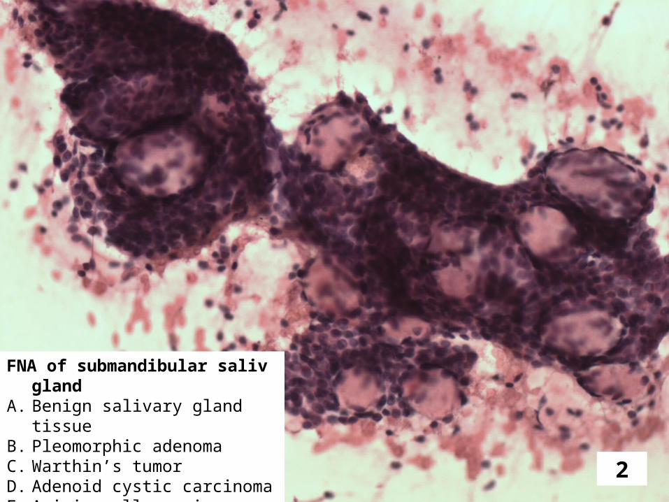
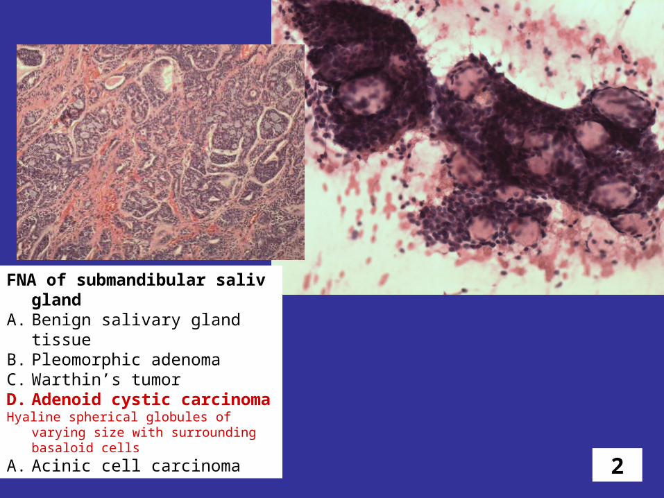
FNA of submandibular saliv glandA. Benign salivary gland tissueB. Pleomorphic adenomaC. Warthin’s tumorD. Adenoid cystic carcinomaE. Acinic cell carcinoma
2
FNA of submandibular saliv glandA. Benign salivary gland tissueB. Pleomorphic adenomaC. Warthin’s tumorD. Adenoid cystic carcinomaHyaline spherical globules of varying size with
surrounding basaloid cells
A. Acinic cell carcinoma

3
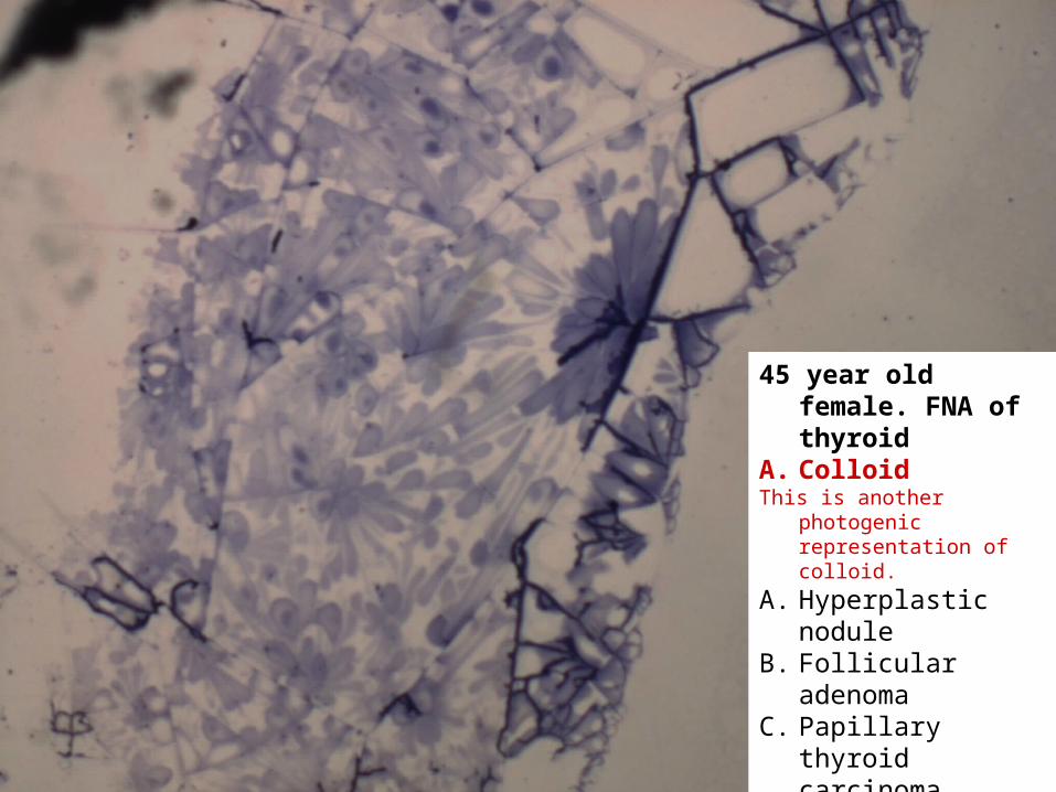
45 year old female. FNA of thyroid
A. ColloidB. Hyperplastic noduleC. Follicular adenomaD. Papillary thyroid
carcinomaE. Hashimoto’s
thyroiditis
3
45 year old female. FNA of thyroid
A. ColloidThis is another photogenic
representation of colloid.
A. Hyperplastic noduleB. Follicular adenomaC. Papillary thyroid
carcinomaD. Hashimoto’s
thyroiditis

4
49 year old female. FNA of parotid
A. Benign salivary glandB. Warthin’s tumorC. Pleomorphic adenomaD. Mucoepidermoid
carcinomaE. Adenoid cystic
carcinoma

4
49 year old female. FNA of parotid
A. Benign salivary glandB. Warthin’s tumorC. Pleomorphic adenomaD. Mucoepidermoid
carcinomaSquamous cells (black arrow),
intermediate cells (green), and mucin secreting cells (yellow).
A. Adenoid cystic carcinoma

5
Lung FNAA. GranulomasB. HamartomaC. AdenoCaD. SCCE. Small cell carcinoma

5
Lung FNAA. GranulomasB. HamartomaC. AdenoCaD. SCCE. Small cell carcinoma
6
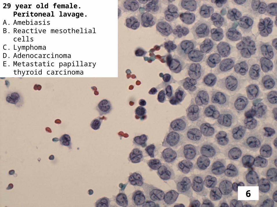
29 year old female. Peritoneal lavage.
A. AmebiasisB. Reactive mesothelial cellsC. LymphomaD. AdenocarcinomaE. Metastatic papillary thyroid
carcinoma

6
29 year old female. Peritoneal lavage.
A. AmebiasisB. Reactive mesothelial cells“Daisy cells” are normal mesothelial cells with
multiple deep nuclear indentations. Apparently, this type of cell is only seen in peritoneal mesothelial cells.
A. LymphomaB. AdenocarcinomaC. Metastatic papillary thyroid
carcinoma

7
35 year old female, FNA of parotid
A. Benign salivary gland tissue
B. Pleomorphic adenoma
C. Warthin’s tumorD. Adenoid cystic
carcinomaE. Acinic cell
carcinoma

7
35 year old female, FNA of parotid
A. Benign salivary gland tissue
B. Pleomorphic adenoma
Poorly cohesive epithelial-like cells associated with fibromyxoid stroma.
A. Warthin’s tumorB. Adenoid cystic
carcinomaC. Acinic cell carcinoma

8
69 year old male. FNA of neck mass
A. Negative for malignancyB. Reactive lymphoid hyperplasiaC. Metastatic squamous cell caD. Metastatic melanomaE. Amebic abscess

8
69 year old male. FNA of neck mass
A. Negative for malignancyB. Reactive lymphoid hyperplasiaC. Metastatic squamous cell caD. Metastatic melanomaE. Amebic abscess

9
BALA. CandidaB. AspergillusC. PCPD. AmebiasisE. Scedosporium

9
BALA. CandidaB. AspergillusC. PCPD. AmebiasisE. ScedosporiumIf you picked this one all I can say is WOW!

10
63 year old male. FNA of parotidA. Non diagnostic FNAB. Reactive lymph node in parotid
glandC. Warthin’s tumorD. Acinic cell carcinomaE. Mucoepidermoid carcinoma

10
63 year old male. FNA of parotid
A. Non diagnostic FNAB. Reactive lymph node
in parotid glandC. Warthin’s tumorAlso called “papillary
cystadenoma lymphomatosum papilliferum.” Lymphocytes and oncocytic cells.
A. Acinic cell carcinomaB. Mucoepidermoid
carcinoma