Embed Size (px)
Citation preview
The
Jou
rnal
of
Exp
erim
enta
l Bio
logy
– A
CC
EPT
ED
AU
TH
OR
MA
NU
SCR
IPT
1
TITLE: 1
2
An Automated Training Paradigm Reveals Long-term Memory in Planaria 3
and Its Persistence Through Head Regeneration 4
5
6
AUTHORS: 7
Tal Shomrat, and Michael Levin 8
9
Biology Department and Tufts Center for Regenerative and Developmental Biology, 10
Tufts University, 11
200 Boston Ave., Suite 4600, 12
Medford, MA 02155, USA. 13
14
CORRESPONDING AUTHOR 15
Dr. Michael Levin 16
Biology Department and 17
Tufts Center for Regenerative and Developmental Biology 18
Tufts University 19
200 Boston Ave., Suite 4600, 20
Medford, MA 02155-4243 21
Tel. (617) 627-6161 22
Fax: (617) 627-6121 23
email: [email protected] 24
25
RUNING TITLE: Memory in Regenerating Planaria 26
27
KEYWORDS: flatworms, training, conditioning, learning, planaria, regeneration, behavior 28
29
LIST OF ABBREVIATIONS: Automated Training Apparatus (ATA). 30
31
32
33
http://jeb.biologists.org/lookup/doi/10.1242/jeb.087809Access the most recent version at J Exp Biol Advance Online Articles. First posted online on 2 July 2013 as doi:10.1242/jeb.087809
Copyright (C) 2013. Published by The Company of Biologists Ltd
http://jeb.biologists.org/lookup/doi/10.1242/jeb.087809Access the most recent version at First posted online on 2 July 2013 as 10.1242/jeb.087809
The
Jou
rnal
of
Exp
erim
enta
l Bio
logy
– A
CC
EPT
ED
AU
TH
OR
MA
NU
SCR
IPT
2
SUMMARY 34
Planarian flatworms are a popular system for research into the molecular mechanisms 35
that enable these complex organisms to regenerate their entire body, including the brain. 36
Classical data suggest that they may also be capable of long-term memory. Thus, the planarian 37
system may offer the unique opportunity to study brain regeneration and memory in the same 38
animal. To establish a system for the investigation of the dynamics of memory in a regenerating 39
brain, we developed a computerized training and testing paradigm that avoided the many issues 40
that confounded previous, manual attempts to train planaria. We then used this new system to 41
train flatworms in an environmental familiarization protocol. We show that worms exhibit 42
environmental familiarization, and that this memory persists for at least 14 days – long enough 43
for the brain to regenerate. We further show that trained, decapitated planaria exhibit evidence 44
of memory retrieval in a savings paradigm after regenerating a new head. Our work establishes 45
a foundation for objective, high-throughput assays in this molecularly-tractable model system 46
that will shed light on the fundamental interface between body patterning and stored memories. 47
We propose planaria as a key emerging model species for mechanistic investigations of the 48
encoding of specific memories in biological tissues. Moreover, this system is likely to have 49
important implications for the biomedicine of stem cell-derived treatments of degenerative brain 50
disorders in human adults. 51
52
INTRODUCTION 53
One of the most interesting capabilities of living systems is processing information. 54
Biological information in multicellular organisms comes in at least 2 flavors: spatial information, 55
needed to create and maintain specific anatomical structures during embryogenesis and 56
regeneration, and temporal information abstracted and stored from environmental stimuli over 57
time by the central nervous system. The intersection of these two fundamental processes has 58
implications for basic neurobiology and engineering of the brain:body interface (Pfeifer and 59
Gomez, 2009; Sampaio et al., 2001), for the synthetic bioengineering of cybernetic systems 60
(Macia et al., 2012; Sole et al., 2007), and for the biomedicine of degenerative brain disease 61
(Murre et al., 2001; Perry and Hodges, 1996). For example, what happens to the personality 62
and mental content of an adult patient with decades of stored memories when his brain is 63
repopulated by the descendants of implanted stem cells (Martino et al., 2011; van Velthoven et 64
al., 2009)? Answering questions about the storage of information in dynamically-remodeling 65
biological tissues, and specifically about the dynamics of memory during brain regeneration, 66

The
Jou
rnal
of
Exp
erim
enta
l Bio
logy
– A
CC
EPT
ED
AU
TH
OR
MA
NU
SCR
IPT
3
requires a tractable model system with both – a robust CNS repair mechanism and the ability to 67
learn and remember. 68
Free-living, planarian flatworms represent the “first” class of organism to have a 69
centralized brain with true synaptic transmission (Sarnat and Netsky, 1985), and shares the 70
majority of neurotransmitters that occur in vertebrate brains (Buttarelli et al., 2008). Planaria 71
have primitive eyes and other sensory capabilities including sensitivity to chemical gradients 72
(Mason, 1975; Miyamoto and Shimozawa, 1985), vibration (Fulgheri and Messeri, 1973), 73
electric fields (Brown and Ogden, 1968), and magnetic fields (Brown and Chow, 1975; Brown, 74
1966). Their sensory reception mechanisms are integrated by the worm’s nervous system into a 75
rich and complex set of behaviors as they navigate their environment. 76
Adult stem cell populations (neoblasts) underlie their remarkable regenerative abilities 77
(Reddien and Sanchez Alvarado, 2004; Wagner et al., 2011), and whole worms can regenerate 78
from only a small proportion of the adult worm: a cut off (or damaged) head is rebuilt perfectly 79
within few days (Inoue et al., 2004; Umesono et al., 2011). Recently, planaria have become a 80
popular molecular-genetic system for the investigation of the pathways that allow complex 81
structures such as the head to be regenerated after damage (Aboobaker, 2011; Gentile et al., 82
2011; Lobo et al., 2012; Newmark and Sánchez Alvarado, 2002; Salo et al., 2009; Sánchez 83
Alvarado, 2006). Thus, planaria are an ideal system in which to probe the dynamics of 84
information stored in the CNS during massive remodeling and repair. While studies have 85
identified several insect organisms in which memories survive the drastic reorganization of 86
metamorphosis (Alloway, 1972; Blackiston et al., 2008; Hepper and Waldman, 1992; Ray, 1999; 87
Sheiman and Tiras, 1996; Tully et al., 1994), planaria are a uniquely tractable system for 88
molecular-biological analyses of large-scale regeneration of adult brains. But can they learn? 89
Nearly 55 years ago it was demonstrated that planarians could be trained to learn a task, 90
and following amputation of the head, the animals regenerating from the original tail sections 91
remembered the original training (Best, 1963; Corning and John, 1961; McConnell, 1965; 92
McConnell et al., 1959). This stunning finding, suggesting that some memory may be stored 93
outside of the head and imprinted on the new brain during regeneration, led to a myriad of 94
subsequent associative learning studies (Cherkashin et al., 1966; Corning, 1966; Corning, 1967; 95
McConnell, 1965; Morange, 2006; Sheiman and Tiras, 1996). The most common procedure was 96
a classical conditioning protocol based on planarians’ well-known photosensitivity (Dasheiff and 97
Dasheiff, 2002; Inoue et al., 2004; Prados et al., 2013; Stephen, 1963). Acquired memories that 98
could survive the process of head regeneration were demonstrated by measuring a direct 99

The
Jou
rnal
of
Exp
erim
enta
l Bio
logy
– A
CC
EPT
ED
AU
TH
OR
MA
NU
SCR
IPT
4
display of a conditioned response or a faster learning rate (“savings”) among worm fragments 100
generated from head and tail pieces of previously trained planarians (McConnell et al., 1959). 101
While learning induced by classical conditioning could be attributed to sensory 102
adaptation rather than consolidation and retrieval of “real, encoded” memory (Halas et al., 1962; 103
Halas et al., 1961), other studies showed that memories formed in more complex discrimination 104
tasks, e.g., eliciting movement in a specific direction in a two-choice maze (Best, 1963; Corning 105
and John, 1961; Corning, 1966; Corning, 1967; Corning et al., 1967; Humphries, 1961; 106
McConnell, 1965; Roe, 1963) or learning to associate odorant cues (Wisenden and Millard, 107
2001), likewise survived regeneration of the head (Corning, 1966; Ernhart and Sherrick, 1959). 108
The reports of persistent memory in an animal that had to regenerate its entire head (Corning, 109
1967) suggests approaches for investigating how information can be stored outside of the brain 110
and imprinted on a newly-regenerating brain – a truly fascinating possibility. 111
These remarkable discoveries have not had sufficient impact on the field and were 112
largely abandoned due to practical difficulties inherent in manual experiments. While the basic 113
findings were validated in some cases, they failed to be reproduced in others (Corning and 114
Riccio, 1970; McConnell, 1966), and the whole line of research became abandoned (Rilling, 115
1996). While modern discoveries such as epigenetic modification (Arshavsky, 2006; Day and 116
Sweatt, 2010; Ginsburg and Jablonka, 2009; Levenson and Sweatt, 2005) and RNAi 117
(Smalheiser et al., 2001) now offer mechanistic explanations of some of the original results, the 118
primary barrier to molecular-level investigations into the dynamics of memory during CNS 119
regeneration has remained: the difficulty of developing a robust learning assay in planaria. 120
Manual behavior experiments involve limited sample sizes, difficulties in precise reproduction of 121
protocols, and lack of quantitative analysis (Corning and Riccio, 1970; Hartry et al., 1964; 122
Morange, 2006; Travis, 1981). As a result of these difficulties, even, planarians’ capacity for 123
long term memory has been questioned (Abbott and Wong, 2008; Takeda et al., 2009; Travis, 124
1981). 125
As part of our investigations into information processing by dynamically-organizing 126
tissues, we have begun to develop automated approaches for eliciting learning and recall in 127
planaria to overcome the problems inherent in manual methods (Nicolas et al., 2008; Oviedo et 128
al., 2008b). We thus developed two platforms that allow automated, parallelized, quantitative, 129
and fully objective training and testing of planaria in a wide range of feedback paradigms 130
(Blackiston et al., 2010; Hicks et al., 2006). The aim of this study was to find a learning 131
paradigm that overcomes a number of problems in previous attempts and establishes a modern 132
platform for the use of regenerative planaria for the study of learning and memory. 133
The
Jou
rnal
of
Exp
erim
enta
l Bio
logy
– A
CC
EPT
ED
AU
TH
OR
MA
NU
SCR
IPT
5
Best and Rubinstein (Best, 1963; Best and Rubinstein, 1962a; Koopowitz, 1970) showed 134
that planarians which had been fed in a familiar environment will start to eat more quickly than 135
naïve worms which never been exposed to the feeding arena before. As in prior studies, their 136
manually-performed experiments contained small sample sizes and limited controls (Davenport 137
and Best, 1962; Dufort, 1962), and it appears that there have been no later attempts to use or 138
improve this non-punishing paradigm. Here, we modified this environmental familiarization 139
approach, adapting it to the use with a textured substrate (to provide clear haptic cues to the 140
animals) and an automated behavior analysis system (Blackiston et al., 2010). Our protocol 141
minimizes bias caused by manual procedures, allows an unprecedented level of quantitative 142
rigor in behavioral analysis, and applies the procedure to a large sample size in a relatively 143
short time frame. Additionally, in contrast to Best and Rubinstein’s protocol, our procedure 144
checks for long-term memory, several days after the training ended. Our results support the 145
findings of Best and Rubenstein, and show a statistically-significant shorter feeding delay for the 146
familiarized worms compare to unfamiliarized worms. Most importantly, the memory survives 147
long enough to allow for regeneration after amputation, and indeed we show that memory traces 148
survive entire brain regeneration in a “saving” paradigm. This simple and promising approach 149
opens great opportunities for the use of planaria as a model organism to understand how 150
specific memories survive large-scale regeneration of neural tissues. 151
152
MATERIALS AND METHODS 153
Experiment apparatus 154
For training and testing we used a custom-made fully automated training apparatus 155
(ATA) (Blackiston et al., 2010; Blackiston and Levin, 2012) (Figs.1A,2L,M), which minimized 156
bias caused by manual procedures and facilitated the training and testing of large numbers of 157
control and experimental worms, simultaneously within the same conditions including time of the 158
day, temperature, and type of arena. However, we settled on a paradigm that requires path 159
tracking of the animals (Fig. 1B) but no complex training algorithm with instantaneous feedback 160
(light or shock) to each animal. Therefore, this protocol could be done with any of the off-the-161
shelf system capable of multiple video tracking (Marechal et al., 2004; Noldus et al., 2001). 162
The ATA “familiarized” chamber environment contained a Petri dish with rough-textured 163
floor surrounded by the ATA electrode walls (Fig. 2). Rough-textured petri dishes have been 164
made from commercially available polystyrene 15x60mm petri dishes (Fisher Scientific, 165
0875713A), altered by laser etching (universal laser systems versaLASER VL-300). The laser 166
cuts the circles to a depth of 0.2mm below the level of the dish's floor, but the displaced melted 167

The
Jou
rnal
of
Exp
erim
enta
l Bio
logy
– A
CC
EPT
ED
AU
TH
OR
MA
NU
SCR
IPT
6
polystyrene also builds up around each circle to a height of about 0.05mm above the floor of the 168
dish. The pattern (Fig. 2N) is made up of circles drawn at 1.4mm in diameter and spaced 169
2.15mm at their centers. As cut, the outer diameter of each circle ends up being closer to 170
1.5mm and 1.2mm inner diameter (the trough that the laser cuts for each circle is about 0.3mm 171
wide). 172
173
Worm colonies’ maintenance 174
All planaria used in the current study were Dugesia japonica. After examining three 175
planarian species: Dugesia japonica, Dugesia dorotocephala, and Schmidtea mediterranea, we 176
found Dugesia japonica to be the most suitable for this project. It has remarkable regenerating 177
capabilities, high tolerance for training and dissection procedures, and is very active. Before 178
experiments, planarian colonies were stored in rectangular plastic containers, filled with Poland 179
Springs natural spring water (Oviedo et al., 2008a). Dugesia japonica has a high tendency to 180
spontaneously fission. In order to prevent spontaneous fission and allow worms to reach a 181
suitable size for the experiment (1-1.5 cm), containers were stored in an incubator at 10°C in 182
continuous darkness (Morita and Best, 1984) and fed once or twice a week with organic beef 183
liver. 184
185
Handling and maintenance during the experiment 186
In addition to suppressing fissioning, keeping the worms in darkness has been reported 187
to enhanced negative phototaxis (McConnell, 1965)(an important feature for the testing 188
procedure). Worms were kept in continuous darkness during the entire experimental period 189
except for brief periods during water changes and transfers between the experimental 190
environment and their resting petri dish/wells plate. Planarians are more active and display 191
longer exploration phase when kept in 18°C (as compared to 10°C). The experiment room 192
temperature was also kept at 18°C. Therefore, during the experimental period the worms were 193
held in incubator at 18°C. The tails’ regeneration rate is also higher in 18°C compared to 10°C, 194
allowing testing the headless fragments worms after only 10 days from decapitation (Fig. 4). 195
Culturing the worms at high density was also found to be effective in suppressing spontaneous 196
fission (Best et al., 1969). Thus, the worms were held in groups, in high density (~12 worms / 197
2ml water). This high density required water to be changed every day. 198
Every morning, during the training phase, the experimental apparatus was cleaned and 199
the water was changed. The worms were taken out of the ATA and placed in petri dishes with 200
fresh water in the dark for the cleaning period. The familiarized groups were placed in a dish 201

The
Jou
rnal
of
Exp
erim
enta
l Bio
logy
– A
CC
EPT
ED
AU
TH
OR
MA
NU
SCR
IPT
7
with a rough textured floor and the unfamiliarized groups were placed into standard Petri dishes. 202
Rough-textured and standard Petri dishes were reused during the training after being thoroughly 203
cleaned with Kimwipes soaked with ethanol 70% and positionally randomized between trials. 204
The ATA electrodes, used as walls for the “familiar” environment, were also cleaned with 205
Kimwipes soaked with ethanol 70%. At the end of the cleaning procedure the worms were 206
placed back into their experimental environments. In order to suppress fission, the experimental 207
environment was filled with low water levels (~12 worms / 2 ml water) to maintain high density of 208
animals. During the testing sessions the experimental apparatus (ATA-electrodes and dishes) 209
were cleaned between every testing trial. For all worms’ handling, we used a plastic transfer 210
pipette with the tip cut off to make a slightly larger opening. During the training, separate 211
pipettes were used for the familiarized and unfamiliarized groups. 212
213
Training procedure 214
Groups of 20-40 experimental worms were placed in an individual ATA chamber (while 215
testing was done on individual animals, familiarization proceeded in groups). The ATA chamber 216
environment contained a Petri dish with rough-textured floor surrounded by the ATA electrode 217
walls (Fig. 2A). The training period last 10-11 consecutive days. The chambers were filled with 218
water (~12 worms / 2ml water) and the lids were closed for darkness. Unfamiliarized (control) 219
worms went through the same procedure, simultaneously with the familiarized (experimental) 220
group but were placed in the ATA in non-textured standard Petri dish (Fig. 2B). Every morning 221
during the training phase, the worms were taken out of the ATA for water change and cleaning. 222
Before being inserted back into the chambers, the worms were inspected and tail fragments 223
caused by spontaneous fissions were extracted. After a 10 day familiarization period, the worms 224
were taken out from the ATA and divided into smaller groups and were kept in 12 multiwell 225
plates (Greiner Bio-One: part number 665102, hydrophobic surface (no treatment)) till the 226
testing (12 worms in a well filled with 2 ml water Fig. 2E). The water in the wells was changed 227
every day. Worms for regeneration experiments were kept in a Petri-dish for a 24-hour rest 228
phase before dissection and division into smaller groups in small wells (to allow all eaten food to 229
be digested before dissection). 230
231
Feeding during the training period 232
Worms were fed throughout the training period, in order to suppress fissioning, and 233
eliminate the possibility of differential starvation levels among worms. The worms were fed in 234
the ATA for one hour, with 1-2 small drops of liver (less than what they are capable of 235
The
Jou
rnal
of
Exp
erim
enta
l Bio
logy
– A
CC
EPT
ED
AU
TH
OR
MA
NU
SCR
IPT
8
consuming, Fig. 2C,D). Feeding took place in the morning after every third days of 236
familiarization training (days 1, 4, 7, 10). Just before feeding, chambers were filled with 237
additional ~10ml of water. On the last morning of familiarization training (day 10), the worms 238
were fed intensively with 1-2 drops of liver every 20 minutes, until satiety (revealed by the last 239
drop of liver remaining intact). This procedure “synchronizes” the hunger level of the worms 240
which were tested 4 days later, and suppresses fissioning of the worms during a longer resting 241
phase before testing. In addition, this feeding protocol is designed to create a positive 242
association with the experimental environment. Worms that were tested 11-15 days after the 243
end of training were fed again 1-2 times before the test. 244
245
Testing procedure 246
The ATA contains 12 identical chambers (Fig. 1A). During each testing trial, 6 247
familiarized and 6 unfamiliarized worms were tested simultaneously, each worm in its own 248
individual chamber. All chambers contained a rough textured floor (a separate set of dishes 249
from those used for the training), surrounded by the ATA electrode walls (Fig. 2J,K). A very 250
small amount of liver was spread with a fine paintbrush on small area of the roughened dishes 251
(Fig. 2J,K,O), and allowed to dry for about 5 minutes before being placed in the ATA and filled 252
with 11 ml of water. In the absence of food, worms prefer to stay on the edge of the dish. 253
Therefore, the liver was applied away from the arena wall (Fig. 2J) so that familiarized worms 254
would be more willing to leave the edge and move toward the center of the dish (Fig. 2P). The 255
worms were inserted to the ATA chambers with a plastic transfer pipette, in alternating order, 256
starting with the unfamiliarized. The worms were placed in the chambers, opposite the liver 257
spot. Worm transfer for all chambers averaged <1min. After all the 12 worms were inside the 258
chamber, the lids were closed and the tracking was initiated. 259
To identify feeding, we capitalized upon the planarians’ strong negative phototaxis 260
(Inoue et al., 2004). Since the worms generally avoid illuminated areas, the quadrant with the 261
spot of liver was illuminated with a strong blue LED light (Azuma et al., 1994; Brown et al., 262
1968) (Fig. 2L) thus, no worm would stay in this quadrant unless its desire for the liver, 263
overcame their natural light aversion (Fig. 2P). As an indication of feeding, we measured how 264
long it took the worms to reach the criterion of 3 consecutive minutes in the illuminated 265
quadrant, containing the liver spot. Any worms that didn’t reach criterion within 60 minutes (e.g., 266
never attempted to eat the liver), as well as worms that showed evidence of any health issue 267
like injuries caused by the transfer pipette, or worms that were in the process of fissioning, were 268
not included in the results. 269

The
Jou
rnal
of
Exp
erim
enta
l Bio
logy
– A
CC
EPT
ED
AU
TH
OR
MA
NU
SCR
IPT
9
At the end of each testing trial, the worms were inspected individually, under a dissection 270
microscope, for general health, injuries caused by the transfer pipette, fission, lesions, or 271
incomplete head regeneration in the case of the headless fragment worms. In order to avoid 272
possible interference from moving worms for testing in sequential groups, in the evening before, 273
the testing worms were divided into two groups of 6 familiarized and 6 unfamiliarized worms and 274
each group was placed in a separate well of 12-well plates, filled with 1ml of water (Fig. 2I). As 275
in the experimental period, plates were placed in dark at 18°C till the beginning of the test at the 276
next day. 277
278
Producing Headless Fragments 279
Worms were decapitated 24 hours after the final feeding which occurred at the end of 280
the familiarization session. So that no brain remained, the worms were decapitated at the point 281
between the auricles and the anterior side of the pharynx (Figs. 2F,4). Headless fragments were 282
kept in groups of 12 worms in one well of 12 multiwell plates, in 2ml of water (Fig. 2E), in a dark 283
incubator at 18°C. As with the intact worms, water was changed every day. After 7 days of 284
regenerating at 18°C, the headless fragments were capable of eating (Fig. 4). Seven to nine 285
days after decapitation, the regenerated worms were fed to satiety. Three to four days after 286
feeding the worms were tested for recall. The worms were fed a second time, in cases when the 287
duration between the first feeding to the recall test was longer than 3-4 days. For example, 288
worms that tested at days 13 after decapitation were fed at days 7 and then again at day 9 or 10 289
from decapitation. 290
291
Savings Paradigm 292
In contrast to the headless fragments’ regular protocol, where the feeding took place in 293
the worms’ home wells, in the saving protocol, the worms were fed in the familiarization arena. 294
Seven to nine days after decapitation, groups, of both, familiarized and unfamiliarized 295
regenerated worms were inserted in to the ATA’s chambers with the surrounding electrode 296
surfaces and the rough floor (the familiarization arena, Fig. 2H). After 30 minutes of exploration 297
phase, drops of liver were placed in the chamber and the worms were allowed to eat until 298
satiety. At the end of the session, the worms were placed back in the multiwall plate (~12 worms 299
in well/2 ml water; Fig. 2E). At the evening, 3 days after the savings session, the worms were 300
divided into groups of 6 familiarized and 6 unfamiliarized (Fig. 2I) and placed back in the dark at 301
18°C until the beginning of the test at the next day, 4 days after saving session. 302
303

The
Jou
rnal
of
Exp
erim
enta
l Bio
logy
– A
CC
EPT
ED
AU
TH
OR
MA
NU
SCR
IPT
10
Data analysis 304
The ATA’s tracking log files were converted to excel file for data analysis. Because the 305
delay values were not normally distributed (Kolmogorov-Smirnov test), we used the 306
nonparametric Mann-Whitney U test to evaluate statistical significance (Bevins et al., 2001). 307
Fisher's exact test was applied to determine statistical significance of the total number of worms 308
that reach criterion in less than 8 minutes. Tests were one tailed since the direction was 309
predicted in advance based on the previous work of Best & Rubinstein (1962a). To check for 310
any mobility-impairment that might be responsible for behavior differences between the 311
familiarized and unfamiliarized worms, the average movement rate (Pixels/Second) was 312
calculated for the first minute, when the majority of worms were still engaged in exploration 313
behavior. 314
315
RESULTS 316
Worms remember a familiar environment 317
Worms were familiarized to the automated behavior analysis platform (ATA) chambers 318
as described in Methods, and then tracked by the ATA (Fig. 1). The retrieval test for familiar 319
environment took place 4 - 15 days after the ending of the 10 days familiarization period, during 320
which the familiarized worms were kept and fed in ATA chambers in Petri dishes with a rough 321
bottom surface (Fig. 2C). The “unfamiliarized” group were also kept and fed in the ATA but in a 322
standard, smooth-bottom Petri dish (Fig. 2D). During each test session, 6 familiarized worms 323
and 6 “unfamiliarized” control worms were placed individually in the ATA chambers with a rough 324
floor (the familiar environment). A small area of the dish was covered with liver (Fig. 2J,O) and 325
a strong blue light illuminated the quadrant with the liver stain (Fig. 2L). As indication of feeding, 326
we measured how long it took for the worms to reach the criterion of 3 consecutive minutes 327
spent in the illuminated quadrant near the liver. The testing trials lasted 60 minutes. To rule out 328
general physical condition differences between the worms, we checked their movement rate 329
during the first minute, a time period while most of the worms were still during their exploration 330
phase before settled down on the liver area. No significant differences were found between the 331
two groups’ motility (Table 1). 332
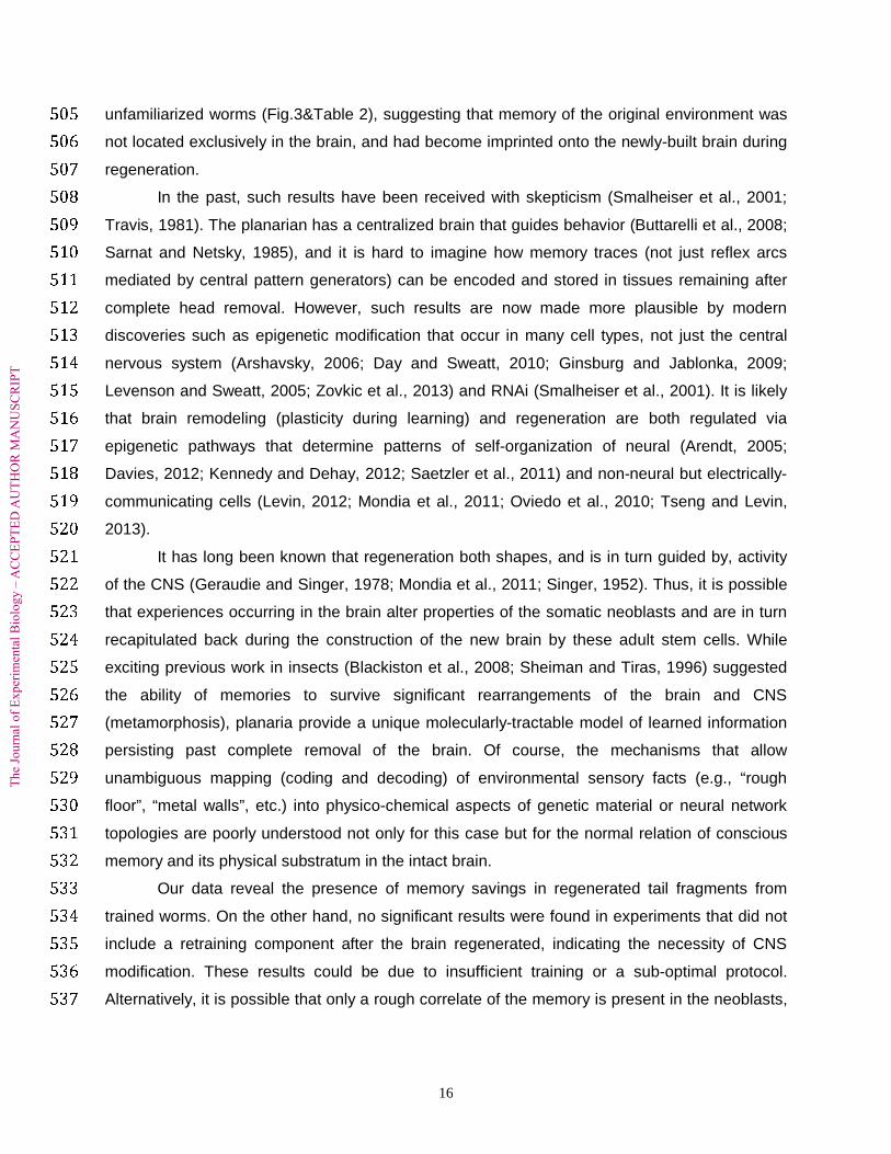
We tested for recall of a familiar environment 4 days after the familiarization period. 333
Familiarized worms displayed significantly shorter time to reach criterion compared to the 334
“unfamiliarized” worms (one tailed U-test, P < 0.001, Fig. 3B&Table 2). Similarly, testing for the 335
number of worms to reach criteria in less than 8 minutes revealed significant differences 336

The
Jou
rnal
of
Exp
erim
enta
l Bio
logy
– A
CC
EPT
ED
AU
TH
OR
MA
NU
SCR
IPT
11
between the trained and control worms (Fisher's exact test, P=0.005, one tailed, Fig. 3Aa&Table 337
2). 338
Different groups of worms were tested 12-15 days following training. The familiarized 339
worms displayed significantly shorter time to reach criterion compared to the unfamiliarized 340
control worms (one tailed U-test, P < 0.001, Fig. 3Aa and Table 2). Testing for the number of 341
worms to reach criterion in less than 8 minutes revealed significant difference between the 342
trained and control worms (P=0.014; one tailed, Fisher's exact test, Fig. 3Ab and Table 2). We 343
conclude that worms can remember a familiar environment for at least 14 days. 344
345
Worms with regenerated heads also retain some memory in a savings paradigm 346
The finding that this memory persists for at least 14 days – long enough for the brain to 347
regenerate (Fig. 4), allowed us to check the possibility that this memory can survive brain 348
regeneration. Headless fragments regenerated from familiarized worms displayed slightly 349
shorter feeding latency compared to headless fragments from unfamiliarized worms when 350
tested 10-14 days after decapitation (Fig. 3B&Table 2). However, the effect was not statistically 351
significant. We then checked for the phenomenon of savings (See methods for detailed 352
protocol), as McConnell found in his classical conditioning procedures (McConnell, 1965), 353
where memory was revealed by a significantly faster training in a specific task in groups that 354
had been trained on this task prior to decapitation. Worms that regenerated from headless 355
fragments from original familiarized worms (Fig. 4) displayed significantly shorter feeding 356
latency in the testing assay compared to regenerated worms that had not been familiarized to 357
the environment prior to decapitation (One tailed U-test, P = 0.027; Table 2&Fig. 3B). Testing 358
for the number of worms to reach criterion in less than 8 minutes revealed significant difference 359
between the original familiarized worms and control worms (P=0.013; one tailed, Fisher's exact 360
test, Fig. 3Ac &Table 2). We conclude that some memory of the place familiarization survives 361
decapitation and brain regeneration. 362
363
DISCUSSION 364
During the last decade, planaria have become an important model organism in the field 365
of developmental and regenerative biology; because of their extensive regenerative capacity 366
(driven by an adult stem cell population) and complex CNS, significant efforts are underway to 367
understand the molecular mechanisms behind neural repair and patterning (Aoki et al., 2009; 368
Gentile et al., 2011; Newmark and Sánchez Alvarado, 2002; Nishimura et al., 2011; Salo et al., 369
2009; Sánchez Alvarado, 2006; Tanaka and Reddien, 2011; Umesono and Agata, 2009). 370
The
Jou
rnal
of
Exp
erim
enta
l Bio
logy
– A
CC
EPT
ED
AU
TH
OR
MA
NU
SCR
IPT
12
However, due to their rich behavioral repertoire and ability to learn (Corning, 1967; Oviedo and 371
Levin, 2008), this model system also has the potential to offer unique opportunities for 372
understanding the dynamics of memory during brain regeneration. This question has not only 373
obvious clinical implications for stem cell therapies of adult neurological disorders but also bears 374
on the fundamental issues of mechanisms of memory encoding and storage in the physical 375
processes of the brain. 376
While planaria are now being used for studies of drug addiction and withdrawal (Pagan 377
et al., 2012; Raffa et al., 2008; Raffa and Valdez, 2001; Ramoz et al., 2012; Rawls et al., 2011; 378
Rawls et al., 2010; Sacavage et al., 2008), the usage of planaria as a model for learning and 379
memory is still very limited (Nicolas et al., 2008; Nishimura et al., 2010; Oviedo and Levin, 380
2008). Although extensive work on planarians’ learning and memory have long suggested that 381
memories can survive brain regeneration (McConnell, 1966), the limitations of previous manual 382
experiments have lead to these important questions being largely neglected by recent workers; 383
these limitations included small sample sizes, difficulties in precise reproduction of protocols, 384
and lack of quantitative analysis (Corning and Riccio, 1970; Travis, 1981). The aim of this work 385
was to find a reliable, state-of-the-art approach that moves beyond past controversies to identify 386
quantitative, objective, high-throughput protocols for eliciting and characterizing planarian long 387
term memory capabilities. By demonstrating evidence for the acquisition of relatively complex, 388
explicit-like memories, the planarian system becomes even more central in modern research 389
into learning and memory. 390
Environmental familiarity is a well-accepted paradigm for the study of explicit memory 391
mechanism in vertebrates (Heyser and Chemero, 2012; Heyser and Ferris, 2013; Teyke, 1989). 392
Although some invertebrates such as bees and ants are capable of spatial memory and 393
environmental recognition (Collett et al., 2003; Horridge, 2005), environmental familiarity has not 394
been frequently used in learning and memory research with invertebrates. Best & Rubinstein 395
(Best and Rubinstein, 1962a) showed that worms display a shorter feeding delay, when being 396
fed in familiar environment 90 minutes after single, 25 minutes, familiarization session. Here we 397
modified their environmental familiarization protocol and adapted it to the use with an automated 398
behavior analysis system (Blackiston et al., 2010). This system minimizes bias caused by 399
manual procedures, allows an unprecedented level of quantitative, objective rigor in behavioral 400
analysis and data reporting, and applies the procedure to a large sample size in a relatively 401
short time frame. In addition to more rigorous controls (Davenport and Best, 1962; Dufort, 402
1962), our protocol allows retrieval after at least 14 days from the end of the training. 403

The
Jou
rnal
of
Exp
erim
enta
l Bio
logy
– A
CC
EPT
ED
AU
TH
OR
MA
NU
SCR
IPT
13
Since this protocol measures feeding behavior, the worms’ performance in the retrieval 404
test is dependent on their baseline appetite level. We examined different starvation periods 405
between 1-30 days (unpublished data) and found differences in the results’ significance and 406
variance as a function of the worms’ starvation period, as did Best and Rubinstein (Best, 1963; 407
Best and Rubinstein, 1962a). We observed that the best results, in our procedure, were 408
obtained when the worms were fed 3-4 days before being tested. Future users of this procedure 409
must establish the correct hunger level in the worms to observe the best results in this assay. 410
Because hunger level is a pivotal parameter in this approach and could be affected by many 411
variables as manipulation intensity, maintenance temperature, size of the worms, the species of 412
worms, and type of food, we offer an additional heuristic to other workers reproducing this 413
protocol. As a heuristic, the proper hunger level seems to be achieved when not more than a 414
third of the worms initiate feeding in less than 1 minute from the start of the testing trial and stay 415
there until criterion is reached. Also, as seen from the results (fig.3B), although the general 416
protocol was similar between the different groups, there were still differences in the general 417
latency of feeding, between the different categories. Even so, in any of the experiments, both 418
control and experimental groups from each category were from the same colony, trained and 419
tested in the same time and went through identical conditions of feeding and maintenance 420
temperature. As a result, the changes in latency of feeding in each of the categories are both in 421
the experimental and control groups, indicate the importance of rigor with respect to identical 422
parameters and conditions for the experimental and control worms. 423
Importantly, in contrast to the most commonly-used procedures (classical conditioning 424
protocols), this environmental familiarity protocol cannot be attributed to pseudoconditioning or 425
sensitization effects (Halas et al., 1962; Halas et al., 1961) rather than consolidation and 426
retrieval of “real, encoded” memory and behavior controlled by the brain. Planarians’ feeding is 427
a true complex behavior. Although composed of a series of stereotypic actions, it is coordinated 428
and initiated by the central nervous system (Pearl, 1903; Sheiman et al., 2002). The feeding 429
behavior is dependent on sensory integration (Pearl, 1903) , as in our paradigm, of tactile/ 430
mechanical stimulation (Best and Rubinstein, 1962b) , chemotactic (Ash et al., 1973; Pearl, 431
1903) and optical sensations (Inoue et al., 2004). 432
Previous studies have shown that when food is placed in direct contact with the opening 433
of the folded pharynx, it can activate the reflexes of extending the pharynx and swallowing, even 434
in decapitated worms (Pearl, 1903; Wulzen, 1917). However, activation of these reflexes in 435
decapitated worm is exceptional (Bardeen, 1901; Pearl, 1903) and the worms need to be 436

The
Jou
rnal
of
Exp
erim
enta
l Bio
logy
– A
CC
EPT
ED
AU
TH
OR
MA
NU
SCR
IPT
14
starved (Bardeen, 1901; Wulzen, 1917) and tested directly after decapitation (Bardeen, 1901; 437
Sheiman et al., 2002; Wulzen, 1917). 438
We never observed such behavior in our worms (Dugesia japonica, which fasted for less 439
than a week) and consistent with others’ observations (Pearl, 1903; Sheiman et al., 2002), our 440
headless fragments with an intact pharynx did not demonstrate any interest in food until head 441
regeneration (5-7 days after decapitation), even when the tail fragment passed immediately 442
adjacent to the food. Moreover, we observed that extrusion of the pharynx happened just after 443
the head made a first contact with the food, sometime with a kind of stereotypic, drilling-like, 444
movements into the liver. We cannot completely rule out the possibility that the modifications in 445
the peripheral nervous system contribute to change in feeding latency. However, it is well-446
accepted that the recognition of food and moving directly to it, as in our case, with decision 447
making and a cautious approach, against their natural preference (under the strong light above 448
and away from the edge of the dish, Fig. 2P, Movie S1), are behaviors that are controlled by the 449
CNS (Bardeen, 1901; Pearl, 1903; Sheiman et al., 2002). Finally, our results that show that in 450
contrast to intact worms tested two weeks after training, regenerated worms, with an intact 451
pharynx required “retraining” to demonstrate retrieval (Fig.3, Table 2), suggest that the 452
difference found in latency of feeding is due to modification in the CNS and not/or not just a 453
reflex or peripheral nerve system modification. Thus, our data show the survival of a true 454
complex, brain-regulated behavior program through the process of head regeneration. 455
The procedure is ideally suited for automated apparatus with minimal handling and does 456
not required manual analysis, as was required for example in studies of conditioned response 457
intensity in classical conditioning procedures (Corning, 1967; Prados et al., 2013; Wells, 1967). 458
Our paradigm requires path tracking of the animals but no complex training algorithm with 459
instantaneous feedback (light or shock) to each animal. Therefore, this protocol could also be 460
done with any of the off-the-shelf systems capable of multiple video tracking (Marechal et al., 461
2004; Noldus et al., 2001). The protocol avoids operator fatigue and ensures that no scoring 462
biases are introduced into the data by subjective analysis of animal behavior. 463
While seeking the best complex learning protocol we observed the phenomenon 464
previously called planarian's lethargy (Best, 1963; Best and Rubinstein, 1962b; Corning, 1964; 465
McConnell, 1966; McConnell, 1965). Worms’ learning curves during the training phase can 466
suddenly reverse after a steady improvement, while healthy and active worms can begin to 467
refuse to behave at all when inserted into the training apparatuses (Best and Rubinstein, 1962b; 468
McConnell, 1965). Evidence suggests that this phenomenon could be related to familiarization 469
to a dangerous environment, i.e. one in which the animal previously received noxious stimulus 470

The
Jou
rnal
of
Exp
erim
enta
l Bio
logy
– A
CC
EPT
ED
AU
TH
OR
MA
NU
SCR
IPT
15
(Shomrat, unpublished data and Best, 1963). The protocol reported here involves natural 471
behavior with minimal handling and without negative reinforcement. This overcomes planarians’ 472
lethargy and thus also allows the application to much more sensitive species such as Schmidtea 473
mediterranea (Sanchez Alvarado et al., 2002). 474
No differences were found in general motility between familiarized and unfamiliarized 475
worms (Table 1). Thus, any behavioral differences are not due to simple changes of overall 476
activity level due to the familiarization protocol. The training occurred in complete darkness and 477
the type and amount of water, food, handling and maintenance were identical between the 478
familiarized (experimental) and the unfamiliarized (control) groups. Therefore, the learned 479
difference between the two environments was mainly tactile. In the majority of their exploration 480
phase, the worms were crawling around the edge on the bottom of the chamber. Hence, the 481
experimental worms could feel the roughness of the floor and the dodecagon shape of the 482
chamber walls, which alternated between delrin-plastic and iridium oxide-coated titanium 483
electrode (Fig. 2). Although no shock was delivered and the electrode material does not give off 484
electrolysis products such as metal ions (Blackiston et al., 2010), there is a possibility that 485
additional chemical cues from the electrode metal also facilitated place recognition. 486
Our results show that planarians can remember previously-encountered habitats for at 487
least 14 days (Fig.3&Table 2). Dugesia japonica regenerates a functional head and CNS after 7 488
days, and in 14 days the worms are fully regenerated (Agata and Umesono, 2008; Inoue et al., 489
2004), (Fig. 4). Encouraged by the long-term retrieval, we investigated whether trained worms 490
can display retrieval after decapitation and regeneration of a new head (Corning, 1966; Corning, 491
1967; McConnell et al., 1959). Worms regenerating from decapitated familiarized worms 492
displayed a slightly shorter average, feeding latency compared to regenerated fragments from 493
unfamiliarized worms (Fig. 3 & Table 2), but this effect was not statistically significant. Future 494
work will explore longer training phases and further optimize different starvation periods to 495
determine whether the strength of this effect can be increased. 496
McConnell’s original results revealed a pattern of “savings”, where the learning curve of 497
retrained animals is better (faster) relative to that of to naïve animals (McConnell, 1965; 498
McConnell et al., 1959). Therefore, we checked for the presence of savings in the regenerated 499
worms. In our savings protocol, regenerated worms were fed in the testing arena (familiarization 500
environment) in a single 3 hour session, 4 days before the retrieval test. Therefore the feeding 501
session was a previously-encountered environment for the familiarized worms and a first 502
introduction for the unfamiliarized. Worms that had regenerated from headless fragments from 503
original familiarized worms, displayed significant shorter feeding latency compare to 504
The
Jou
rnal
of
Exp
erim
enta
l Bio
logy
– A
CC
EPT
ED
AU
TH
OR
MA
NU
SCR
IPT
16
unfamiliarized worms (Fig.3&Table 2), suggesting that memory of the original environment was 505
not located exclusively in the brain, and had become imprinted onto the newly-built brain during 506
regeneration. 507
In the past, such results have been received with skepticism (Smalheiser et al., 2001; 508
Travis, 1981). The planarian has a centralized brain that guides behavior (Buttarelli et al., 2008; 509
Sarnat and Netsky, 1985), and it is hard to imagine how memory traces (not just reflex arcs 510
mediated by central pattern generators) can be encoded and stored in tissues remaining after 511
complete head removal. However, such results are now made more plausible by modern 512
discoveries such as epigenetic modification that occur in many cell types, not just the central 513
nervous system (Arshavsky, 2006; Day and Sweatt, 2010; Ginsburg and Jablonka, 2009; 514
Levenson and Sweatt, 2005; Zovkic et al., 2013) and RNAi (Smalheiser et al., 2001). It is likely 515
that brain remodeling (plasticity during learning) and regeneration are both regulated via 516
epigenetic pathways that determine patterns of self-organization of neural (Arendt, 2005; 517
Davies, 2012; Kennedy and Dehay, 2012; Saetzler et al., 2011) and non-neural but electrically-518
communicating cells (Levin, 2012; Mondia et al., 2011; Oviedo et al., 2010; Tseng and Levin, 519
2013). 520
It has long been known that regeneration both shapes, and is in turn guided by, activity 521
of the CNS (Geraudie and Singer, 1978; Mondia et al., 2011; Singer, 1952). Thus, it is possible 522
that experiences occurring in the brain alter properties of the somatic neoblasts and are in turn 523
recapitulated back during the construction of the new brain by these adult stem cells. While 524
exciting previous work in insects (Blackiston et al., 2008; Sheiman and Tiras, 1996) suggested 525
the ability of memories to survive significant rearrangements of the brain and CNS 526
(metamorphosis), planaria provide a unique molecularly-tractable model of learned information 527
persisting past complete removal of the brain. Of course, the mechanisms that allow 528
unambiguous mapping (coding and decoding) of environmental sensory facts (e.g., “rough 529
floor”, “metal walls”, etc.) into physico-chemical aspects of genetic material or neural network 530
topologies are poorly understood not only for this case but for the normal relation of conscious 531
memory and its physical substratum in the intact brain. 532
Our data reveal the presence of memory savings in regenerated tail fragments from 533
trained worms. On the other hand, no significant results were found in experiments that did not 534
include a retraining component after the brain regenerated, indicating the necessity of CNS 535
modification. These results could be due to insufficient training or a sub-optimal protocol. 536
Alternatively, it is possible that only a rough correlate of the memory is present in the neoblasts, 537

The
Jou
rnal
of
Exp
erim
enta
l Bio
logy
– A
CC
EPT
ED
AU
TH
OR
MA
NU
SCR
IPT
17
requiring a brief re-exposure to the trigger in order to consolidate into measurable effects on 538
animal behavior (as occurs in the savings paradigm). 539
We suggest that some trace of memory is stored in locations distributed beyond the 540
brain (since the place conditioning association survives decapitation). A straightforward model 541
implies that information acquired during training must be imprinted on the regenerating (naïve) 542
brain in order to result in the observed subsequent recall behavior. Future work must investigate 543
the properties and mechanisms of such instructive interactions between remaining somatic 544
organs and the regenerating CNS. However, two additional possibilities must be considered. 545
First is the possibility that the memory is executed entirely by the peripheral nervous 546
system, not involving the brain in learning or recall. Given the similarities between the planarian 547
brain and that of higher animals (in terms of structure, biochemistry, and complex ethology 548
(Nicolas et al., 2008; Oviedo and Levin, 2008; Rawls et al., 2011; Sarnat and Netsky, 1985)), 549
and the fact that worms exhibit no behavior prior to the regrowth of the brain, it is most likely that 550
the planarian brain indeed drives behavior. A pivotal role for the brain is also supported by the 551
need for the Savings portion of the paradigm, and the complexity of the behavior that is very 552
unlikely to be implemented by receptor sensitivity and reflex modifications only (e.g., Fig. 2P 553
and Movie S1). However, if true, this would suggest a remarkable capacity for integration of 554
complex information in the peripheral nervous system of an animal that normally has access to 555
an efficient brain, and thus would suggest a research program into the untapped information-556
processing abilities of the PNS in other advanced organisms. 557
Second is the possibility that the new brain is regenerated as a Tabula Rasa and is not 558
imprinted by any traces of the previous memory. Instead, on this model the familiarized worms’ 559
PNS (which would have been modified and tuned, e.g., increased/decreased receptor sensitivity 560
to a given stimuli during the training phase) is retraining the new brain: “burning” the association 561
into the new CNS, during the short “Saving” session (which suffices because it is more efficient 562
than in the unfamiliarized worms, due to the modified PNS sensitivity). We believe this scenario 563
is less likely, because of the behavioral complexity of the learned task (Fig. 2P & Movie S1). 564
Experimental and control worms were fed with liver during the entire procedure, and the liver 565
odor would be everywhere in the dish – this means the worms did not have to rely on the rough 566
texture to know that food was somewhere in the vicinity, and both the trained and control groups 567
could have developed positive associations to the smell of the liver. As can be seen in Movie 568
S1, the behavior does not resemble a simple reflex modification but rather the whole 569
environment that makes trained worms initiate feeding sooner. We cannot completely rule out 570
the possibility that the modifications in the peripheral nervous system contribute to change in 571

The
Jou
rnal
of
Exp
erim
enta
l Bio
logy
– A
CC
EPT
ED
AU
TH
OR
MA
NU
SCR
IPT
18
feeding latency. However, it should be noted that in order for receptor sensitivity to a particular 572
stimulus to change after training, a kind of learning had to take place - the system as a whole 573
(including learning, appropriate modification of PNS, and facilitation of re-training phases) 574
implements an association between the presence of liver and the salient predictor of its 575
presence, the rough surface, out of many other possible sensory modes that could have 576
become more or less sensitized. Thus, this system would provide a novel model in which to 577
examine the interactions between a mature PNS modified by specific experiences and learning 578
in a newly-developed brain (Inoue et al., 2004; Koopowitz and Holman, 1988). 579
580
Conclusions: 581
Our results, obtained using a highly-sensitive, objective, quantitative analysis system, 582
support previous findings of Best and Rubenstein (Best and Rubinstein, 1962a) , that planarians 583
are capable of acquiring a relatively complex, explicit-like memories of environmental familiarity. 584
Moreover, this memory survives long enough to allow full regeneration, after amputation. 585
Remarkably, headless fragments, regenerated from original environment-familiarized worms, 586
display significant environmental familiarity in a saving paradigm. This simple and promising 587
approach opens great opportunities for the use of planaria as a model organism for modern 588
research of learning and memory. Importantly, planarians are the only molecularly-tractable 589
system in which memory and brain regeneration can be studied in the same animal. This is a 590
crucial advantage allows the Investigation of innovative hypothesis as the role of epigenetic and, 591
self-organization mechanisms in memory encoding, brain development, and brain regeneration. 592
593
ACKNOWLEDGEMENTS 594
We thank Punita Koustubhan for general laboratory assistance, Junji Morokuma and 595
Wendy Beane for advice and help with the planarian model system, Douglas Blackiston and 596
Robert Cook for many helpful discussions about behavioral paradigms, Durwood Marshall, 597
Dany S. Adams, and Laura Vandenberg for assistance with statistics, Douglas Blackiston, 598
Michael Romero, and Philip Starks for comments on early versions of the manuscript, and 599
Ethan Golden for fabrication of the rough-textured, petri dishes. This work is dedicated to Paul 600
Van Oye and James V. McConnell, two pioneers of learning and memory in planaria. 601
602
FUNDING 603
This research was funded by the G. Harold and Leila Y. Mathers Charitable Foundation. 604
605
606

The
Jou
rnal
of
Exp
erim
enta
l Bio
logy
– A
CC
EPT
ED
AU
TH
OR
MA
NU
SCR
IPT
19
REFERENCES 607
Abbott, S. M. and Wong, G. K. (2008). The conditioning and memory retention of 608
planaria (Dugesia tigrina) for directional preferences. Bios 79, 160-170. 609
Aboobaker, A. A. (2011). Planarian stem cells: a simple paradigm for regeneration. 610
Trends in Cell Biology 21, 304-11. 611
Agata, K. and Umesono, Y. (2008). Brain regeneration from pluripotent stem cells in 612
planarian. Philos Trans R Soc Lond B Biol Sci 363, 2071-8. 613
Alloway, T. M. (1972). Retention of Learning through Metamorphosis in Grain Beetle 614
(Tenebrio-Molitor). American Zoologist 12, 471-472. 615
Aoki, R., Wake, H., Sasaki, H. and Agata, K. (2009). Recording and spectrum analysis 616
of the planarian electroencephalogram. Neuroscience 159, 908-14. 617
Arendt, T. (2005). Alzheimer's disease as a disorder of dynamic brain self-organization. 618
Progress in brain research 147, 355-78. 619
Arshavsky, Y. I. (2006). "The seven sins" of the Hebbian synapse: can the hypothesis 620
of synaptic plasticity explain long-term memory consolidation? Progress In Neurobiology 80, 99-621
113. 622
Ash, J. F., McClure, W. O. and Hirsch, J. (1973). Chemical studies of a factor which 623
elicits feeding behaviour in Dugesia dorotocephala. Animal Behaviour 21, 796-800. 624
Azuma, K., Okazaki, Y., Asai, K. and Iwasaki, N. (1994). Electrical responses and K+ 625
activity changes to light in the ocellus of the planarian Dugesia japonica. Comparative 626
biochemistry and physiology. Part A, Physiology 109, 593-9. 627
Bardeen, C. R. (1901). The Function of the Brain in Planaria Maculata. American 628
Journal of Physiology 4, 175-179. 629
Best, J. B. (1963). Protopsychology. Sci Am 208, 54-62. 630
Best, J. B., Goodman, A. B. and Pigon, A. (1969). Fissioning in planarians: control by 631
the brain. Science 164, 565-6. 632
Best, J. B. and Rubinstein, I. (1962a). Environmental familiarity and feeding in a 633
planarian. Science 135, 916-8. 634
Best, J. B. and Rubinstein, I. (1962b). Maze learning and associated behavior in 635
planaria. J Comp Physiol Psychol 55, 560-6. 636
Bevins, R. A., Koznarova, J. and Armiger, T. J. (2001). Environmental familiarization 637
in rats: differential effects of acute and chronic nicotine. Neurobiology of Learning and Memory 638
75, 63-76. 639
Blackiston, D., Shomrat, T., Nicolas, C. L., Granata, C. and Levin, M. (2010). A 640
second-generation device for automated training and quantitative behavior analyses of 641
molecularly-tractable model organisms. PLoS ONE 5, e14370. 642
Blackiston, D. J. and Levin, M. (2012). Aversive training methods in Xenopus laevis: 643
general principles. Cold Spring Harbor Protocols 2012. 644
Blackiston, D. J., Silva Casey, E. and Weiss, M. R. (2008). Retention of memory 645
through metamorphosis: can a moth remember what it learned as a caterpillar? PLoS ONE 3, 646
e1736. 647
Brown, F. and Chow, C. (1975). Differentiation between clockwise and 648
counterclockwise magnetic rotation by the planarian dugesia-dorotocephala. Physiological 649
Zoology 48, 168-176. 650
Brown, F. A., Jr. (1966). Effects and after-effects on planarians of reversals of the 651
horizontal magnetic vector. Nature 209, 533-5. 652
Brown, H. M., Ito, H. and Ogden, T. E. (1968). Spectral sensitivity of the planarian 653
ocellus. The Journal of general physiology 51, 255-60. 654
Brown, H. M. and Ogden, T. E. (1968). The electrical response of the planarian ocellus. 655
Journal of General Physiology 51, 237-53. 656

The
Jou
rnal
of
Exp
erim
enta
l Bio
logy
– A
CC
EPT
ED
AU
TH
OR
MA
NU
SCR
IPT
20
Buttarelli, F. R., Pellicano, C. and Pontieri, F. E. (2008). Neuropharmacology and 657
behavior in planarians: translations to mammals. Comparative biochemistry and physiology. 658
Toxicology & pharmacology : CBP 147, 399-408. 659
Cherkashin, A. N., Sheiman, I. M. and Bogorovskaya, G. I. (1966). Uslovniye reflexi y 660
planarii i opiti s regenerazei. Zhurnal Vysshei Nervnoi Deiatelnosti Imeni I. P. Pavlova XVI, 661
1110-1115. 662
Collett, T. S., Graham, P. and Durier, V. (2003). Route learning by insects. Current 663
Opinion in Neurobiology 13, 718-25. 664
Corning, W. and John, E. (1961). Effect of ribonuclease on retention of conditioned 665
response in regenerated planarians. Science 134, 1363-1365. 666
Corning, W. C. (1964). Evidence of right-left discrimination in planarians. The Journal of 667
Psychology 58, 131-139. 668
Corning, W. C. (1966). Retention of a position discrimination after regeneration in 669
planarians. Psychanomic Science 5, 17-18. 670
Corning, W. C. (1967). Regeneration and retention of acquired information: NASA. 671
Corning, W. C., Ratner, S. C. and American Institute of Biological Sciences. (1967). 672
Chemistry of learning; invertebrate research. New York,: Plenum Press. 673
Corning, W. C. and Riccio, D. (1970). The planarian controversy. In Molecular 674
approaches to learning and memory, (ed. W. Byrne), pp. 107-150. New York: Academic Press. 675
Dasheiff, B. D. and Dasheiff, R. M. (2002). Photonegative response in brown planaria 676
(Dugesia tigrina) following regeneration. Ecotoxicology & Environmental Safety 53, 196-9. 677
Davenport, D. and Best, J. B. (1962). On Planarian Behavior. Science 137, 452-6. 678
Davies, P. C. W. (2012). The epigenome and top-down causation. Interface Focus 2, 679
42-48. 680
Day, J. J. and Sweatt, J. D. (2010). DNA methylation and memory formation. Nature 681
neuroscience 13, 1319-23. 682
Dufort, R. H. (1962). On Planarian Behavior. Science 138, 400-2. 683
Ernhart, E. N. and Sherrick, C. (1959). Retention of a maze habit following 684
regeneration in planaria (D. aculatd). In Paper presented at Midwestern Psychology 685
Association. St. Louis, Mo. 686
Fulgheri, D. and Messeri, P. (1973). The use of 2 different reinforcements in light 687
darkness discrimination in planaria. Bollettino - Societa Italiana Biologia Sperimentale 49, 1141-688
1145. 689
Gentile, L., Cebria, F. and Bartscherer, K. (2011). The planarian flatworm: an in vivo 690
model for stem cell biology and nervous system regeneration. Dis Model Mech 4, 12-9. 691
Geraudie, J. and Singer, M. (1978). Nerve dependent macromolecular synthesis in the 692
epidermis and blastema of the adult newt regenerate. J Exp Zool 203, 455-60. 693
Ginsburg, S. and Jablonka, E. (2009). Epigenetic learning in non-neural organisms. J 694
Biosci 34, 633-46. 695
Halas, E. S., James, R. L. and Knutson, C. S. (1962). An attempt at classical 696
conditioning in the planarian. Journal of comparative and physiological psychology 55, 969-71. 697
Halas, E. S., James, R. L. and Stone, L. A. (1961). Types of responses elicited in 698
planaria by light. J Comp Physiol Psychol 54, 302-5. 699
Hartry, A. L., Morton, W. D. and Keithlee, P. (1964). Planaria - Memory Transfer 700
through Cannibalism Reexamined. Science 146, 274-275. 701
Hepper, P. G. and Waldman, B. (1992). Embryonic olfactory learning in frogs. Quarterly 702
Journal of Experimental Psychology. B, Comparative and Physiological Psychology 44, 179-97. 703
Heyser, C. J. and Chemero, A. (2012). Novel object exploration in mice: not all objects 704
are created equal. Behav Processes 89, 232-8. 705
Heyser, C. J. and Ferris, J. S. (2013). Object exploration in the developing rat: 706
Methodological considerations. Developmental Psychobiology 55, 373-81. 707

The
Jou
rnal
of
Exp
erim
enta
l Bio
logy
– A
CC
EPT
ED
AU
TH
OR
MA
NU
SCR
IPT
21
Hicks, C., Sorocco, D. and Levin, M. (2006). Automated analysis of behavior: A 708
computer-controlled system for drug screening and the investigation of learning. J Neurobiol 66, 709
977-90. 710
Horridge, G. A. (2005). Recognition of a familiar place by the honeybee (Apis mellifera). 711
Journal of comparative physiology. A, Neuroethology, sensory, neural, and behavioral 712
physiology 191, 301-16. 713
Humphries, B. (1961). Maze learning in planaria. Worm Runner’s Digest 3, 114–115. 714
Inoue, T., Kumamoto, H., Okamoto, K., Umesono, Y., Sakai, M., Sanchez Alvarado, 715
A. and Agata, K. (2004). Morphological and functional recovery of the planarian photosensing 716
system during head regeneration. Zoolog Sci 21, 275-83. 717
Kennedy, H. and Dehay, C. (2012). Self-organization and interareal networks in the 718
primate cortex. Progress in brain research 195, 341-60. 719
Koopowitz, H. (1970). Feeding Behaviour and Role of Brain in Polyclad Flatworm, 720
Planocera-Gilchristi. Animal Behaviour 18, 31-&. 721
Koopowitz, H. and Holman, M. (1988). Neuronal Repair and Recovery of Function in 722
the Polyclad Flatworm, Notoplana-Acticola. American Zoologist 28, 1065-1075. 723
Levenson, J. M. and Sweatt, J. D. (2005). Epigenetic mechanisms in memory 724
formation. Nat Rev Neurosci 6, 108-18. 725
Levin, M. (2012). Molecular bioelectricity in developmental biology: new tools and recent 726
discoveries: control of cell behavior and pattern formation by transmembrane potential 727
gradients. BioEssays : news and reviews in molecular, cellular and developmental biology 34, 728
205-17. 729
Lobo, D., Beane, W. S. and Levin, M. (2012). Modeling planarian regeneration: a 730
primer for reverse-engineering the worm. PLoS Comput Biol 8, e1002481. 731
Macia, J., Posas, F. and Sole, R. V. (2012). Distributed computation: the new wave of 732
synthetic biology devices. Trends Biotechnol 30, 342-9. 733
Marechal, J. P., Hellio, C., Sebire, M. and Clare, A. S. (2004). Settlement behaviour of 734
marine invertebrate larvae measured by EthoVision 3.0. Biofouling 20, 211-7. 735
Martino, G., Pluchino, S., Bonfanti, L. and Schwartz, M. (2011). Brain regeneration in 736
physiology and pathology: the immune signature driving therapeutic plasticity of neural stem 737
cells. Physiol Rev 91, 1281-304. 738
Mason, P. R. (1975). Chemo-klino-kinesis in planarian food location. Animal Behaviour 739
23, 460-9. 740
McConnell, J. (1966). Comparative physiology: learning in invertebrates. Annu Rev 741
Physiol 28, 107-36. 742
McConnell, J. V. (1965). A Manual of psychological experimentation on planarians. Ann 743
Arbor, Mich. 744
McConnell, J. V., Jacobson, A. L. and Kimble, D. P. (1959). The effects of 745
regeneration upon retention of a conditioned response in the planarian. Journal of Comparative 746
Physiology and Psychology 52, 1-5. 747
Miyamoto, S. and Shimozawa, A. (1985). Chemotaxis in the freshwater planarian 748
Dugesia-japonica-japonica. Zoological Science (Tokyo) 2, 389-396. 749
Mondia, J. P., Levin, M., Omenetto, F. G., Orendorff, R. D., Branch, M. R. and 750
Adams, D. S. (2011). Long-distance signals are required for morphogenesis of the regenerating 751
Xenopus tadpole tail, as shown by femtosecond-laser ablation. PLoS ONE 6, e24953. 752
Morange, M. (2006). What history tells us VI. The transfer of behaviours by 753
macromolecules. Journal of biosciences 31, 323-7. 754
Morita, M. and Best, J. B. (1984). Effects of Photoperiods and Melatonin on Planarian 755
Asexual Reproduction. Journal of Experimental Zoology 231, 273-282. 756
Murre, J. M., Graham, K. S. and Hodges, J. R. (2001). Semantic dementia: relevance 757
to connectionist models of long-term memory. Brain 124, 647-75. 758

The
Jou
rnal
of
Exp
erim
enta
l Bio
logy
– A
CC
EPT
ED
AU
TH
OR
MA
NU
SCR
IPT
22
Newmark, P. and Sánchez Alvarado, A. (2002). Not your father's planarian: a classic 759
model enters the era of functional genomics. Nat Rev Genet 3, 210-9. 760
Nicolas, C., Abramson, C. and Levin, M. (2008). Analysis of behavior in the planarian 761
model. In Planaria: A Model for Drug Action and Abuse, eds. R. Raffa and S. Rawls), pp. 83-94. 762
Austin: RG Landes Co. 763
Nishimura, K., Inoue, T., Yoshimoto, K., Taniguchi, T., Kitamura, Y. and Agata, K. 764
(2011). Regeneration of dopaminergic neurons after 6-hydroxydopamine-induced lesion in 765
planarian brain. Journal of Neurochemistry 119, 1217-31. 766
Nishimura, K., Kitamura, Y., Taniguchi, T. and Agata, K. (2010). Analysis of motor 767
function modulated by cholinergic neurons in planarian Dugesia japonica. Neuroscience 168, 768
18-30. 769
Noldus, L. P., Spink, A. J. and Tegelenbosch, R. A. (2001). EthoVision: a versatile 770
video tracking system for automation of behavioral experiments. Behav Res Methods Instrum 771
Comput 33, 398-414. 772
Oviedo, N. and Levin, M. (2008). The planarian regeneration model as a context for the 773
study of drug effects and mechanisms. In Planaria: A Model for Drug Action and Abuse, eds. R. 774
Raffa and S. Rawls). Austin: RG Landes Co. 775
Oviedo, N., Morokuma, J., Walentek, P., Kema, I., Gu, M., Ahn, J., Hwang, J., 776
Gojobori, T. and Levin, M. (2010). Long-range neural and gap junction protein-mediated cues 777
control polarity during planarian regeneration. Dev Biol 339, 188-99. 778
Oviedo, N. J., Nicolas, C. L., Adams, D. S. and Levin, M. (2008a). Establishing and 779
maintaining a colony of planarians. CSH Protoc 2008, pdb prot5053. 780
Oviedo, N. J., Nicolas, C. L., Adams, D. S. and Levin, M. (2008b). Planarians: a 781
versatile and powerful model system for molecular studies of regeneration, adult stem cell 782
regulation, aging, and behavior. Cold Spring Harb Protoc 2008, pdb.emo101-. 783
Pagan, O. R., Baker, D., Deats, S., Montgomery, E., Tenaglia, M., Randolph, C., 784
Kotturu, D., Tallarida, C., Bach, D., Wilk, G. et al. (2012). Planarians in pharmacology: 785
parthenolide is a specific behavioral antagonist of cocaine in the planarian Girardia tigrina. The 786
International journal of developmental biology 56, 193-6. 787
Pearl, R. (1903). The movements and reactions of fresh-water planarians : a study in 788
animal behaviour: London :J. & A. Churchill. 789
Perry, R. J. and Hodges, J. R. (1996). Spectrum of memory dysfunction in 790
degenerative disease. Current Opinion in Neurology 9, 281-5. 791
Pfeifer, R. and Gomez, G. (2009). Morphological Computation - Connecting Brain, 792
Body, and Environment. Creating Brain-Like Intelligence: From Basic Principles to Complex 793
Intelligent Systems 5436, 66-83. 794
Prados, J., Alvarez, B., Howarth, J., Stewart, K., Gibson, C. L., Hutchinson, C. V., 795
Young, A. M. and Davidson, C. (2013). Cue competition effects in the planarian. Animal 796
cognition 16, 177-86. 797
Raffa, R. B., Stagliano, G. W., Ross, G., Powell, J. A., Phillips, A. G., Ding, Z. and 798
Rawls, S. M. (2008). The kappa-opioid receptor antagonist nor-BNI inhibits cocaine and 799
amphetamine, but not cannabinoid (WIN 52212-2), abstinence-induced withdrawal in 800
planarians: an instance of 'pharmacologic congruence'. Brain Res 1193, 51-6. 801
Raffa, R. B. and Valdez, J. M. (2001). Cocaine withdrawal in Planaria. Eur J Pharmacol 802
430, 143-5. 803
Ramoz, L., Lodi, S., Bhatt, P., Reitz, A. B., Tallarida, C., Tallarida, R. J., Raffa, R. B. 804
and Rawls, S. M. (2012). Mephedrone ("bath salt") pharmacology: insights from invertebrates. 805
Neuroscience 208, 79-84. 806
Rawls, S. M., Patil, T., Tallarida, C. S., Baron, S., Kim, M., Song, K., Ward, S. and 807
Raffa, R. B. (2011). Nicotine behavioral pharmacology: clues from planarians. Drug and Alcohol 808
Dependence 118, 274-9. 809

The
Jou
rnal
of
Exp
erim
enta
l Bio
logy
– A
CC
EPT
ED
AU
TH
OR
MA
NU
SCR
IPT
23
Rawls, S. M., Patil, T., Yuvasheva, E. and Raffa, R. B. (2010). First evidence that 810
drugs of abuse produce behavioral sensitization and cross sensitization in planarians. 811
Behavioural Pharmacology 21, 301-13. 812
Ray, S. (1999). Survival of olfactory memory through metamorphosis in the fly Musca 813
domestica. Neuroscience Letters 259, 37-40. 814
Reddien, P. W. and Sanchez Alvarado, A. (2004). Fundamentals of planarian 815
regeneration. Annu Rev Cell Dev Biol 20, 725-57. 816
Rilling, M. (1996). The mystery of the vanished citations: James McConnell's forgotten 817
1960s quest for planarian learning, a biochemical engram, and celebrity (vol 51, pg 589, 1996). 818
American Psychologist 51, 1039-1039. 819
Roe, K. (1963). In search of the locus of learning in planarians. Worm Runner's Digest 5, 820
16-24. 821
Sacavage, S., Patel, H., Zielinski, M., Acker, J., Phillips, A. G., Raffa, R. B. and 822
Rawls, S. M. (2008). Withdrawal-like behavior in planarians is dependent on drug exposure 823
duration. Neuroscience Letters 439, 84-8. 824
Saetzler, K., Sonnenschein, C. and Soto, A. M. (2011). Systems biology beyond 825
networks: generating order from disorder through self-organization. Seminars in cancer biology 826
21, 165-74. 827
Salo, E., Abril, J. F., Adell, T., Cebria, F., Eckelt, K., Fernandez-Taboada, E., 828
Handberg-Thorsager, M., Iglesias, M., Molina, M. D. and Rodriguez-Esteban, G. (2009). 829
Planarian regeneration: achievements and future directions after 20 years of research. Int J Dev 830
Biol 53, 1317-27. 831
Sampaio, E., Maris, S. and Bach-y-Rita, P. (2001). Brain plasticity: 'visual' acuity of 832
blind persons via the tongue. Brain Res 908, 204-7. 833
Sánchez Alvarado, A. (2006). Planarian regeneration: its end is its beginning. Cell 124, 834
241-5. 835
Sanchez Alvarado, A., Newmark, P. A., Robb, S. M. and Juste, R. (2002). The 836
Schmidtea mediterranea database as a molecular resource for studying platyhelminthes, stem 837
cells and regeneration. Development 129, 5659-65. 838
Sarnat, H. B. and Netsky, M. G. (1985). The brain of the planarian as the ancestor of 839
the human brain. Canadian Journal of Neurological Sciences 12, 296-302. 840
Sheiman, I. M. and Tiras, K. L. (1996). Memory and morphogenesis in planaria and 841
beetle. In Russian contributions to invertebrate behavior, eds. C. I. Abramson Z. P. Shuranova 842
and Y. M. Burmistrov), pp. 43-76. Westport, CT: Praeger. 843
Sheiman, I. M., Zubina, E. V. and Kreshchenko, N. D. (2002). Regulation of the 844
feeding behavior of the planarian Dugesia (Girardia) tigrina. Journal of Evolutionary 845
Biochemistry and Physiology 38, 414-418. 846
Singer, M. (1952). The influence of the nerve in regeneration of the amphibian 847
extremity. Q Rev Biol 27, 169-200. 848
Smalheiser, N. R., Manev, H. and Costa, E. (2001). RNAi and brain function: was 849
McConnell on the right track? Trends Neurosci 24, 216-8. 850
Sole, R. V., Munteanu, A., Rodriguez-Caso, C. and Macia, J. (2007). Synthetic 851
protocell biology: from reproduction to computation. Philos Trans R Soc Lond B Biol Sci 362, 852
1727-39. 853
Stephen, W. S. (1963). The influence of varying light intensities on speed of movement 854
in Planaria Lugubris. Worm Runner's Digest 5, 40-45. 855
Takeda, H., Nishimura, K. and Agata, K. (2009). Planarians maintain a constant ratio 856
of different cell types during changes in body size by using the stem cell system. Zoological 857
science 26, 805-13. 858
Tanaka, E. M. and Reddien, P. W. (2011). The cellular basis for animal regeneration. 859
Developmental Cell 21, 172-85. 860

The
Jou
rnal
of
Exp
erim
enta
l Bio
logy
– A
CC
EPT
ED
AU
TH
OR
MA
NU
SCR
IPT
24
Teyke, T. (1989). Learning and Remembering the Environment in the Blind Cave Fish 861
Anoptichthys-Jordani. Journal of Comparative Physiology a-Sensory Neural and Behavioral 862
Physiology 164, 655-662. 863
Travis, G. D. L. (1981). Replicating Replication - Aspects of the Social Construction of 864
Learning in Planarian Worms. Social Studies of Science 11, 11-32. 865
Tseng, A. and Levin, M. (2013). Cracking the bioelectric code: Probing endogenous 866
ionic controls of pattern formation. Communicative & Integrative Biology 6, 1-8. 867
Tully, T., Cambiazo, V. and Kruse, L. (1994). Memory through metamorphosis in 868
normal and mutant Drosophila. Journal of Neuroscience 14, 68-74. 869
Umesono, Y. and Agata, K. (2009). Evolution and regeneration of the planarian central 870
nervous system. Dev Growth Differ 51, 185-95. 871
Umesono, Y., Tasaki, J., Nishimura, K., Inoue, T. and Agata, K. (2011). Regeneration 872
in an evolutionarily primitive brain--the planarian Dugesia japonica model. The European journal 873
of neuroscience 34, 863-9. 874
van Velthoven, C. T., Kavelaars, A., van Bel, F. and Heijnen, C. J. (2009). 875
Regeneration of the ischemic brain by engineered stem cells: fuelling endogenous repair 876
processes. Brain Res Rev 61, 1-13. 877
Wagner, D. E., Wang, I. E. and Reddien, P. W. (2011). Clonogenic neoblasts are 878
pluripotent adult stem cells that underlie planarian regeneration. Science 332, 811-6. 879
Wells, P. H. (1967). Training flatworms in a Van Oye maze. In Chemistry of Learning, 880
eds. W. C. Corning and S. C. Ratner), pp. 251-254. New York: Plenum. 881
Wisenden, B. D. and Millard, M. C. (2001). Aquatic flatworms use chemical cues from 882
injured conspecifics to assess predation risk and to associate risk with novel cues. Animal 883
Behaviour 62, 761-766. 884
Wulzen, R. (1917). Some chemotropic and feeding reactions of Planaria maculata. Biol 885
Bull 33, 67-69. 886
Zovkic, I. B., Guzman-Karlsson, M. C. and Sweatt, J. D. (2013). Epigenetic regulation 887
of memory formation and maintenance. Learn Mem 20, 61-74. 888
889
890
891

The
Jou
rnal
of
Exp
erim
enta
l Bio
logy
– A
CC
EPT
ED
AU
TH
OR
MA
NU
SCR
IPT
25
FIGURE LEGENDS 892
893
Fig. 1. The Automated Training Apparatus (ATA). 894
(A) A picture of the 12 channel fully automated device we used. The device contained 4 895
blocks of 3 isolated chambers. Each chamber contained 1 worm in a petri dish, allowing the 896
simultaneous tracking and training of 12 individual worms (Blackiston et al., 2010). All 897
coordinate data are processed, allowing an objective and quantitative analysis of each animal’s 898
behavior during testing trials. 899
(B) The basic workflow loop of the device. Continuously and independently, cameras in 900
each cell determine the position of each worm and record it. The device also has provisions for 901
providing changes of light or electric shock in response to specific worm positions. Such 902
negative reinforcement was not used in these experiments, but the ability to provide real-time 903
feedback to each individual animal allows very sophisticated training and testing paradigms to 904
be employed. 905
906
Fig. 2. Experimental protocol 907
Training phase: Groups of worms were placed in the ATA’s chambers for 10 consecutive 908
days. (A) The “familiarized” group was in Petri dishes with a rough textured bottom, while the 909
“unfamiliarized” (control) group was placed in standard Petri dishes with smooth bottoms (B). 910
(C&D) In the morning days 1, 4, 7, 10, the worms were fed in the ATA with 1-2 small drops of 911
liver (white arrows). On the morning of the last day the worms were fed extensively by being 912
given more liver than they could consume. Every day, the experiment arenas (dishes + 913
electrodes) were cleaned and water was changed. During the cleaning procedure the 914
familiarized worms were placed in a dish with a rough textured floor and the unfamiliarized 915
worms were placed into standard Petri dishes, in the dark. 916
Resting phase: (E) After 10 familiarization days, the worms were kept in 12 multiwell 917
plates in the dark until testing. The wells’ water was changed every day. (F) Illustration of a 918
worm before and after decapitation. To ensure that no brain tissue remained, the worms were 919
decapitated at the point between the auricles and the anterior side of the pharynx (White arrow). 920
Worms were fed in the 12 multiwell plates 4 days before retrieval test (G). Saving session: (H) 921
regenerated worm were fed in the ATA chambers with a rough floor (the familiar environment), 4 922
days before retrieval test. (I) In the evening before the testing day, the worms were divided into 923
two groups of 6 familiarized and 6 unfamiliarized worms and placed in separate wells of a 12-924
well plate. 925

The
Jou
rnal
of
Exp
erim
enta
l Bio
logy
– A
CC
EPT
ED
AU
TH
OR
MA
NU
SCR
IPT
26
Testing phase: After the resting period, the retrieval test took place. To test recall, 6 926
familiarized worms and 6 unfamiliarized worms were placed individually in the ATA chambers 927
with a rough floor (the familiar environment). (J&K) A small area of the dish was covered with 928
liver (red arrow point on the liver stain) and (L) a strong blue light was illuminating, from above 929
the quadrant with the liver stain (opened lid of the ATA with the light setting during the test). The 930
device measured how long it took each animal to begin feeding. Panel (M) shows the worm as 931
seen from below by the tracking camera, Red arrow indicates the worm’s pharynx. (N) 932
Enlargement of the rough textured bottom of the experimental environment with worm for 933
comparison. (O) Enlargement of the testing dish floor with the small stain of liver (inside the 934
dashed red circle). The black stain in the middle is made on the outer side of the dish by a black 935
marker to label the area where liver is. This enabled to place the dish in the right position with 936
the liver under the illuminated quadrant. (P) Typical exploration/foraging trail during the test. At 937
the start (red arrow) the worms are mainly moving around the edge of the chamber, avoiding the 938
illuminated quadrant (Blue area) containing the liver stain (dashed red circle). In some cases, as 939
in this example, the worm will make more than one, short, enters to the illuminated quadrant 940
with the liver, before making a sharp turn toward the liver stain and initiating feeding. 941
942
Fig. 3. Worms in a familiar environment display significantly shorter exploration phase before 943
initiating feeding: 944
A. Percentage of worms to reach criterion in less than 8 minutes. (a) Intact-4-days: 60.4% of 945
familiarized worms (n=225, red column) and 48% of the unfamiliarized worms (n=229, black 946
column), which have been tested 4 days after training, reach criterion in less than 8 minutes (<8 947
minutes, p=0.005; one-tailed, Fisher's exact test). (b) Intact-14-days: 84.2% of familiarized 948
worms (n=70, red column) and 67.1% of the unfamiliarized worms (n=70, black column), which 949
have been tested 12-15 days after training, reach criterion in less than 8 minutes (<8 minutes, 950
p=0.014; one-tailed, Fisher's exact test). (c) Saving paradigm: 79.5% of familiarized worms 951
(n=106, red column) and 64.5% of the unfamiliarized worms (n=104, black column), which have 952
been tested, 11-13 days after decapitating, with the Saving paradigm, reach criterion in less 953
than 8 minutes (<8 minutes, p=0.013; one-tailed, Fisher's exact test). B. Median delay of 954
feeding (time in minutes). The same groups as in A, including the category of Headless 955
Fragments, Regular Protocol which are worms regenerated from tail fragments and have been 956
tested, 10-14 days after decapitating, (Familiarized n=164, Unfamiliarized n=171).The right 957
points are form the familiarized groups, (Trained), and the left points are from the 958
Unfamiliarized, (Control) groups. Red line: Intact-4-days (Familiarized 6.641±0.47; 959

The
Jou
rnal
of
Exp
erim
enta
l Bio
logy
– A
CC
EPT
ED
AU
TH
OR
MA
NU
SCR
IPT
27
Unfamiliarized 8.341±0.48, P<0.001; one-tailed, U-test). Black line: Intact-14-days (Familiarized 960
5.012±0.49; Unfamiliarized 6.991±0.41, P<0.001; one-tailed, U- test). Green line: Headless 961
fragments, Regular Protocol (Familiarized 10.15±0.7; Unfamiliarized 10.325±0.69, No statistical 962
significance). Blue-line, Saving paradigm (Familiarized 7.166±0.58; Unfamiliarized 8.304±0.55, 963
P=0.027; one-tailed, U-test). 964
Error bars show SEM. 965
# Criterion was 3 consecutive minutes in the illuminated quadrant, containing the liver spot. 966
967
968
Fig. 4. Decapitation and regeneration 969
Illustration of worm regeneration sequence in our protocol conditions of 12 worms / 2ml 970
water in 18°C and constant darkness (not the same worm in each of the panels). Worms were 971
decapitated at the point between the auricles and the anterior side of the pharynx (red arrows). 972
973

The
Jou
rnal
of
Exp
erim
enta
l Bio
logy
– A
CC
EPT
ED
AU
TH
OR
MA
NU
SCR
IPT

The
Jou
rnal
of
Exp
erim
enta
l Bio
logy
– A
CC
EPT
ED
AU
TH
OR
MA
NU
SCR
IPT
The
Jou
rnal
of
Exp
erim
enta
l Bio
logy
– A
CC
EPT
ED
AU
TH
OR
MA
NU
SCR
IPT

The
Jou
rnal
of
Exp
erim
enta
l Bio
logy
– A
CC
EPT
ED
AU
TH
OR
MA
NU
SCR
IPT

The
Jou
rnal
of
Exp
erim
enta
l Bio
logy
– A
CC
EPT
ED
AU
TH
OR
MA
NU
SCR
IPT
Table 1: Motility During the Testing Session
Movement Rate
Average Pixels/Second (± s.e.m)
Protocol Familiarized: Unfamiliarized:
Intact: tested 4 days after end of training 8.775±0.2 8.818±0.2
Intact: tested 12-15 days after end of training 8.102±0.33 8.859±0.27
Headless fragments (saving paradigm): tested 11-13 days after decapitating 7.34±0.24 7.858±0.25
Table 2: Latency of Feeding During the Testing Session
N
Average Latency
Minutes to reach criteria
(± s.e.m)
Median Latency
Minutes to reach criteria
(± s.e.m)
Statistical Significance
Protocol
Reach Criteria
/ Tested
F
C
F
C
U-test
(One tailed)
Fisher's exact test (n<8min) (One tailed)
Intact: tested 4 days after end of training
Familiarized : 225/233 Unfamiliarized: 229/238
8.817±0.47
10.339±0.48
6.641±0.47
8.341 ±0.48
P < 0.001
P=0.005
Intact: tested 12-15 days after end of training
Familiarized: 70/72 Unfamiliarized: 70/72
5.932±0.49
7.326 ±0.41
5.012±0.49
6.991 ±0.41
P < 0.001
P=0.014
* Regular Protocol Headless fragments tested 10-14 days after decapitating
Familiarized: 171/201 Unfamiliarized: 164/199
12.934±0.7
12.603±0.69
10.15±0.7
10.325±0.69
No statistical significance
No statistical significance
**Savings Protocol Headless fragments tested 11-13 days after decapitating
Familiarized: 106/117 Unfamiliarized:104/115
8.532±0.58
9.545 ±0.55
7.166±0.58
8.304±
0.55
P = 0.027
P=0.013
Legend: F = familiarized; C = controls (unfamiliarized)
*Regular Protocol: The feeding session before the test was taken place in the worm multi plate wells (Fig. 2G)
**Saving Protocol: The feeding session before the test was taken place in the in the familiarization arena (ATA
chamber with the electrode insert and the rough floor (fig. 2H).


![CarloA.Furia,MatteoPradella,andMatteoRossi April2008 arXiv ... · arXiv:0804.4383v2 [cs.LO] 4 Jul 2008 Practical Automated Partial Verification of Multi-Paradigm Real-Time Models](https://img.dokumen.tips/doc/110x75/5fd8819eff02d46f244a4e71/matteopradellaandmatteorossi-april2008-arxiv-arxiv08044383v2-cslo-4-jul.jpg)