Embed Size (px)
Citation preview

THE FETAL BULLETIN
Issue 2 | June 2020
AMOGS e-newsletter I Page 1
The Association of MaharashtraObstetric & Gynaecological Societies
For any queries or feedback, please write to Dr. Pooja Lodha at [email protected]
fetopanishad|| ||
MEDICAL DISORDERS IN PREGNANCY
Dr. Pooja LodhaAMOGS’ Chairperson, Fetal Medicine Committee
Dr. Nandita PalshetkarAMOGS’ President
Dr. Arun NayakAMOGS’ Secretary
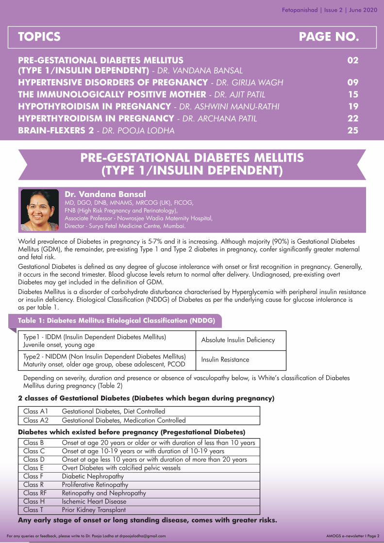
World prevalence of Diabetes in pregnancy is 5-7% and it is increasing. Although majority (90%) is Gestational Diabetes Mellitus (GDM), the remainder, pre-existing Type 1 and Type 2 diabetes in pregnancy, confer significantly greater maternal and fetal risk.Gestational Diabetes is defined as any degree of glucose intolerance with onset or first recognition in pregnancy. Generally, it occurs in the second trimester. Blood glucose levels return to normal after delivery. Undiagnosed, pre-existing overt Diabetes may get included in the definition of GDM.Diabetes Mellitus is a disorder of carbohydrate disturbance characterised by Hyperglycemia with peripheral insulin resistance or insulin deficiency. Etiological Classification (NDDG) of Diabetes as per the underlying cause for glucose intolerance is as per table 1.
2 classes of Gestational Diabetes (Diabetes which began during pregnancy)
Diabetes which existed before pregnancy (Pregestational Diabetes)
Any early stage of onset or long standing disease, comes with greater risks.
Type1 - IDDM (Insulin Dependent Diabetes Mellitus)Juvenile onset, young age
Depending on severity, duration and presence or absence of vasculopathy below, is White’s classification of Diabetes Mellitus during pregnancy (Table 2)
Absolute Insulin Deficiency
PRE-GESTATIONAL DIABETES MELLITUS 02(TYPE 1/INSULIN DEPENDENT) - DR. VANDANA BANSALHYPERTENSIVE DISORDERS OF PREGNANCY - DR. GIRIJA WAGH 09THE IMMUNOLOGICALLY POSITIVE MOTHER - DR. AJIT PATIL 15HYPOTHYROIDISM IN PREGNANCY - DR. ASHWINI MANU-RATHI 19HYPERTHYROIDISM IN PREGNANCY - DR. ARCHANA PATIL 22BRAIN-FLEXERS 2 - DR. POOJA LODHA 25
AMOGS e-newsletter I Page 2For any queries or feedback, please write to Dr. Pooja Lodha at [email protected]
Fetopanishad | Issue 2 | June 2020
TOPICS PAGE NO.
PRE-GESTATIONAL DIABETES MELLITIS(TYPE 1/INSULIN DEPENDENT)
Type2 - NIDDM (Non Insulin Dependent Diabetes Mellitus)Maturity onset, older age group, obese adolescent, PCOD
Class A1 Gestational Diabetes, Diet Controlled Class A2 Gestational Diabetes, Medication Controlled
Insulin Resistance
Class B Onset at age 20 years or older or with duration of less than 10 yearsClass C Onset at age 10-19 years or with duration of 10-19 yearsClass D Onset at age less 10 years or with duration of more than 20 yearsClass E Overt Diabetes with calcified pelvic vesselsClass F Diabetic NephropathyClass R Proliferative RetinopathyClass RF Retinopathy and NephropathyClass H Ischemic Heart DiseaseClass T Prior Kidney Transplant
Table 1: Diabetes Mellitus Etiological Classification (NDDG)
Dr. Vandana BansalMD, DGO, DNB, MNAMS, MRCOG (UK), FICOG,FNB (High Risk Pregnancy and Perinatology),Associate Professor - Nowrosjee Wadia Maternity Hospital,Director - Surya Fetal Medicine Centre, Mumbai.

AMOGS e-newsletter I Page 3For any queries or feedback, please write to Dr. Pooja Lodha at [email protected]
Fetopanishad | Issue 2 | June 2020
Table 2: White’s classification of DiabetesMellitus during pregnancy
As per the Gestational Diabetes ACOG 2011, IADPSG 2010, Diabetes first recognised in pregnancy can still be classified as Gestational or overt (Pre-diabetes), as per the criteria given in Table 3.
Risks related to Pre-gestational Diabetes
Complications of Diabetes is common in those with uncontrolled and fluctuating blood sugar levels.
Miscarriage Risk
Pre-gestational Diabetes is directly related to the degree of maternal Hyperglycemia with upto 44% with HbA1C >12%.
Other Fetal Risks
• Fetal growth restriction• Stillbirth/sudden fetal death (overall Perinatal mortality 2-3 times higher)• Macrosomia• Birth trauma • Shoulder dystocia
Neonatal Risks
• Hypoglycemia• Hypocalcemia, Hypomagnesemia, Hyperbilirubinemia • Respiratory Distress Syndrome • Birth Asphyxia• Transient tachypnea of newborn
Long-term Offspring Risks
• Obesity in adulthood• Type 2 Diabetes
Maternal Risks
• Pre-eclampsia in 10-25% of all pregnant diabetics• Polyhydramnios• Preterm• Recurrent infections, Chorioamnionitis• Operative delivery - instrumental and LSCS• Postpartum Hemorrhage• Postpartum Endometritis• Worsening end organ damage (Nephropathy, Retinopathy, Cardiovascular disease)• Hypoglycemic episodes and Ketoacidosis
Long-term
• Overt Diabetes in 50% in 20 years• Recurrent GDM 50%
Fetal age at glycemic exposure determinesthe fetal damage
• Before the 20th day after fertilisation. - All or none effect - Increased miscarriage• During organogenesis (between 20 and 56 days after fertilisation - 3-8 weeks post conception/ 5-10 weeks Gestational age) - Teratogenic effect• After organogenesis (2nd and 3rd trimester) - Teratogenic effect unlikely but Hyperglycemia may effect growth and function.
Congenital Malformation Risk
Maternal Diabetes is one of the strongest teratogen. There is a two to three fold increase in malformation in infants of Pre-gestational Diabetes with a baseline risk in general population of 1-2%, increasing to 5-9% in overt uncontrolled Diabetes. This increase may not be seen in well-controlled Gestational Diabetes.Hyperglycemia can effect any organ system, but more than 50% of anomalies effect CNS and CV system. Wide spectrum of malformations related to maternal DM are:• Cardiac anomalies• Craniofacial abnormalities• Gastrointestinal • Musculoskeletal• Urogenital• Caudal Regression Syndrome• Strongest association• 200 times more frequently in Diabetic mothers
Fasting plasma glucose >92 mg/dl GDMFasting plasma glucose >126 mg/dl Overt DiabetesHbA1c >6.5%Random plasma glucose >200 mg/dl
First Antenatal Visit
Table 3: International Association of Diabetes and Pregnancy Study Group (IADPSG) Diabetes in pregnancy

AMOGS e-newsletter I Page 4For any queries or feedback, please write to Dr. Pooja Lodha at [email protected]
The rate of birth defects increase linearly with the degree of maternal Hyperglycemia HbA1c <6.5%: Associated with lowest risk of congenital anomaliesHbA1C >7-8.5%: 5% risk of anomaliesHbA1C >10% risk of anomalies rises to >22%: Strongly advised to avoid pregnancy
Glycemic threshold for birth defects(HbA1c values and the risk of malformations)
Two-step approach:Glucose Challenge Test (O Sullivans Test) at 24-28 weeksPlasma glucose measured 1 hour after 50 gm anhydrous glucose load irrespective of time of day or last meal(Table 4)
<140 mg/dl Normal
>200 mg/dl Consider GDM 140-200 mg/dl Perform 100 gm 3 hour
OGTT as per old carpenter couston or NDDG criteria
Plasma glucose value Interpretation
Table 4:Screening for Gestational Diabetes (ACOG)
American Diabetic Association recommends targeted screening of high-risk women for Gestational Diabetics but the disadvantage is that a lot of clinically asymptomatic diabetic pregnant women, will miss the opportunity for adequate care and early correction of high glycemic blood values which is essential for both the mother and the fetus. In addition, for a country like ours, we anyway fall in the high-risk category due to our ethinicity and hence universal screening is recommended for Indian population.
Screening for Gestational Diabetes:(Universal Vs Targeted)
• Age >30 yrs• High-risk ethnic group- Asian, Native American, African• Obese (BMI >15% of normal ideal weight, >90 kg,
>30 kg/m2)• Family history of Diabetes• Personal history of abnormal glucose metabolism,
previous GDM• Bad obstetric history: recurrent abortions, stillbirth,
macrosomia, malformed fetus• Present pregnancy: recurrent candidiasis, UTI,
polyhydramnios, glycosuria, macrosomia
High-risk pregnant women for GDM(American Diabetic Association)
One-step approach:
Table 5: IADPSG, ADA screening for Diabetes in pregnancy
Table 5: Screening for Gestational Diabetes (IADPSG, ADA)
Follows selective screening for high-risk pregnant women at 24-28 weeks by single step, 2 hour 75 gm OGTT (WHO criteria). Women with previous GDM are offered OGTT at 6-8 weeks and repeat at 24-28 weeks.
Screening for Gestational Diabetes(NICE guideline)
Fasting plasma glucose >92 mg/dl
GDM
Fasting plasma glucose >126 mg/dlHbA1c >6.5%Random plasma glucose >200 mg/dl
Overt Diabetes
Fasting plasma glucose<92 mg/dl
Perform 75 gm OGTT at 24-28 wks as per ADA OGTT criteria
First antenatal visit
Fetopanishad | Issue 2 | June 2020
AMOGS e-newsletter I Page 5For any queries or feedback, please write to Dr. Pooja Lodha at [email protected]
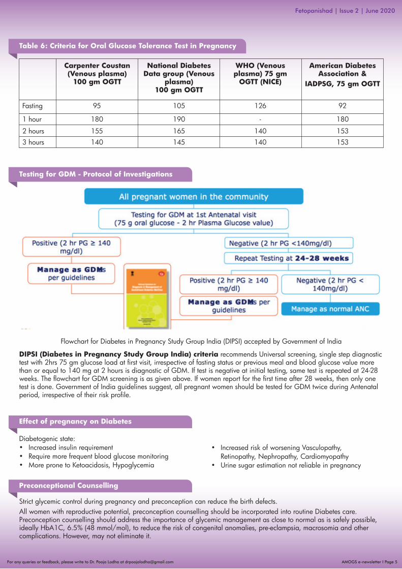
Table 6: Criteria for Oral Glucose Tolerance Test in Pregnancy
Testing for GDM - Protocol of Investigations
Fasting 95 105 126 92
1 hour 180
Flowchart for Diabetes in Pregnancy Study Group India (DIPSI) accepted by Government of India
DIPSI (Diabetes in Pregnancy Study Group India) criteria recommends Universal screening, single step diagnostic test with 2hrs 75 gm glucose load at first visit, irrespective of fasting status or previous meal and blood glucose value more than or equal to 140 mg at 2 hours is diagnostic of GDM. If test is negative at initial testing, same test is repeated at 24-28 weeks. The flowchart for GDM screening is as given above. If women report for the first time after 28 weeks, then only one test is done. Government of India guidelines suggest, all pregnant women should be tested for GDM twice during Antenatal period, irrespective of their risk profile.
Strict glycemic control during pregnancy and preconception can reduce the birth defects. All women with reproductive potential, preconception counselling should be incorporated into routine Diabetes care. Preconception counselling should address the importance of glycemic management as close to normal as is safely possible, ideally HbA1C, 6.5% (48 mmol/mol), to reduce the risk of congenital anomalies, pre-eclampsia, macrosomia and other complications. However, may not eliminate it.
190 - 180
2 hours 155 165 140 153
3 hours 140 145 140 153
Carpenter Coustan(Venous plasma)
100 gm OGTT
National Diabetes Data group (Venous
plasma) 100 gm OGTT
WHO (Venous plasma) 75 gm
OGTT (NICE)
American Diabetes Association &
IADPSG, 75 gm OGTT
Diabetogenic state:• Increased insulin requirement• Require more frequent blood glucose monitoring• More prone to Ketoacidosis, Hypoglycemia
Effect of pregnancy on Diabetes
Preconceptional Counselling
• Increased risk of worsening Vasculopathy, Retinopathy, Nephropathy, Cardiomyopathy• Urine sugar estimation not reliable in pregnancy
Fetopanishad | Issue 2 | June 2020

AMOGS e-newsletter I Page 6For any queries or feedback, please write to Dr. Pooja Lodha at [email protected]
• Avoid unplanned pregnancies• Importance of good glycemic control periconception and in pregnancy• Education about tight control of sugar in pregnancy• Frequent sugar estimation in pregnancy• Screen for vasculopathy, HbA1c monthly• Periconceptional folic acid• Patients on all oral hypoglycemics except Metformin – change to Insulin• Patients on Metformin for PCOD, continue same in pregnancy• Educate on the symptoms of hypoglycemiaWomen with pre-existing Type 1 or Type 2 Diabetes who are planning pregnancy or who have become pregnant, should be counselled on the risk of development and/or progression of Diabetic Retinopathy. Dilated eye examinations should occur ideally before pregnancy or in the first trimester and then patients should be monitored every trimester and for 1-year Postpartum as indicated by the degree of retinopathy and as recommended by the eye care provider.Women with pre-existing Diabetes should ideally be managed in a multidisciplinary clinic, including an Endocrinologist, Maternal-fetal Medicine Specialist, Dietitian, and Diabetes Educator, when available.
• Early dating and viability scan 7-9 weeks• Combined first trimester aneuploidy screening using maternal age, NT, Nasal bone, Ductus venosus, tricuspid regurgitation and biochemistry BHCG and PAPP-A at 11-13+6 weeks or if registers later, Quadruple test at 16-20 weeks• Malformation scan at 18-20 weeks • Fetal ECHO at 18-20 weeks• Color Doppler at 24-28 weeks • USG Growth assessment and Amniotic fluid volume every 4-6 weekly from 28-36 weeks • Daily fetal kick count • NST/Biophysical profile/Modified Biophysical profile weekly after 38 weeks, as per clinical condition
Fetal Surveillance
Medical nutrition therapy includes nutritional counselling, a personalized nutrition plan and a moderate exercise program, with the goal of achieving normoglycemia, preventing ketosis, provide adequate calorie intake to facilitate adequate weight gain and contribute to fetal well-being.
If target glucose levels (2 hrs postprandial blood sugar <120 mg/dl) cannot be met with nutrition therapy alone in two weeks, medical therapy should be initiated.
Calories required:
Obese (BMI >30 kg/m2) 1200-1800 Kcal/dayNon-obese 2000-2500 Kcal/day
40-50% of calories from complex carbohydrate25-30% fat (unsaturated)30% proteinsDiet-rich in fibre and low glycemic index
Divided meals: 3 small meals and 3 snacks
Management Medical Nutrition Therapy (First-line Therapy)
Dietary Restriction
As per the ADA, Insulin is the first-line agent recommended for treatment of GDM. While individual RCTs support limited efficacy of Metformin (biguanides) and glyburide (sulphonyl ureas) in reducing glucose levels for the treatment of GDM, these agents are not recommended as first-line treatment for GDM because they are known to cross the placenta and data on safety for offspring is lacking. However as per ACOG, if medications are needed, insulin and oral medications are equally effective and appropriate for first-line therapy. As per 2018 Indian guidelines, Metformin or Insulin therapy is accepted in management of GDM. Use of Metformin is popular in clinical practice, especially in patients who have started preconceptionally for obesity and PCOD.
However, if blood sugars are uncontrolled (2-h PPBS ≥120 mg/dL) with maximum dose of Metformin (2 g/day), insulin therapy is added. Dose of Insulin/metformin is titrated as per blood sugar level and follow-up schedule.
Metformin in Gestational Diabetes trial (MIG) showed that use of Metformin reduced the required dose of Insulin and also reduced the total weight gain and allowed better acceptability.
Pharmacologic Therapy
Mild to moderate exercise (Brisk walking for 30 minutes daily especially after a meal)Aerobic exercises
Exercise
Fetopanishad | Issue 2 | June 2020

1 hr postprandial <140 mg/dl (7.8 mmol/L)
2 hrs postprandial <120 mg/dl (6.7 mmol/L)
< 95 mg/dl(5.3 mmol/L)
AMOGS e-newsletter I Page 7For any queries or feedback, please write to Dr. Pooja Lodha at [email protected]
Insulin is the preferred agent for management of both Type 1 Diabetes and Type 2 Diabetes in pregnancy because it does not cross the placenta and because oral agents are generally insufficient to overcome the insulin resistance in Type 2 Diabetes and are ineffective in Type 1 Diabetes. The physiology of pregnancy necessitates frequent titration of insulin to match changing requirements and underscores the importance of daily and frequent self-monitoring of blood glucose.
Management of Pre-existing Type 1 and Type 2 Diabetes in Pregnancy
If fasting plasma glucose >105 mg/dlor2 hours postprandial glucose >140 mg/dl after diet control
Insulin therapy has been gold standard in treatment of GDM and pre-gestational Diabetes. Most associations recommend short acting regular and intermediate acting NPH Insulin. Newer ultra rapid acting lispro and aspart (action begins in 15 mins) are also acceptable.
Insulin Therapy
Early pregnancy is a time of enhanced insulin sensitivity, lower glucose levels, and lower Insulin requirements in women with Type 1 diabetes. The situation rapidly reverses as insulin resistance increases exponentially during the second and early third trimesters and levels off toward the end of the third trimester.
Insulin Physiology
Fasting and postprandial self-monitoring of blood glucose are recommended in both, Gestational Diabetes Mellitus and pre-existing Diabetes in pregnancy, to achieve glycemic control. Initially, SMBG may be required six or seven times a day, but it may be reduced to three times, once stabilised. Usual practice is to do fasting and 2 hour after lunch and dinner. A 3am blood glucose level may also be required to document nocturnal Hypoglycemia due to excess bedtime Insulin resulting in fasting Hyperglycemia (Somogyi phenomenon).
Due to increased red blood cell turnover, A1C is slightly lower in normal pregnancy than in normal non-pregnant women. Ideally, the A1C target in pregnancy is 6% (42 mmol/mol) if this can be achieved without significant Hypoglycemia, but the target may be relaxed to 7% (53 mmol/mol) if necessary, to prevent Hypoglycemia. In the second and third trimesters, A1C of 6% (42 mmol/mol) has the lowest risk of large-for-gestational-age infants, preterm delivery and Preeclampsia.
Glycemic Targets in Pregnancy
Similar to the targets recommended by the American College of Obstetricians and Gynecologists for GDM, the ADA-recom-mended targets for women with Type 1 or Type 2 Diabetes are as follows (Table 7).
Escalate insulin requirement when 2 or more measurements exceed the threshold in 2 weeks’ period.
Table 7:Therapeutic Target for Plasma Glucose (ADA)
Table 8:Insulin and their mechanism of action
Therapeutic Target for Plasma Glucose (ADA)
Insulin dose divided into: 2/3rd Intermediate acting and 1/3rd Short acting2/3rd dose in the morning, 1/3rd at night
Morning dose: 2/3rd NPH+1/3rd Short actingPre-dinner dose: ½NPH+½ Short acting
Monitor with daily fasting and 2 hour postprandial blood sugar levels using dextrostix. Patient should be aware of symptoms of Hypoglycemia with Insulin.
FastingPlasma Glucose
RegularSemi Lente
Intermediate (NPH)Lente
30 mins1-2 hrs3 hrs3 hrs
2-4 hrs4-6 hrs8-12 hrs8-12 hrs
6-8 hrs12-16 hrs18-24 hrs18-28 hrs
Type ofInsulin
Start ofAction
Peak Duration
Fetopanishad | Issue 2 | June 2020

AMOGS e-newsletter I Page 8For any queries or feedback, please write to Dr. Pooja Lodha at [email protected]
Timing of DeliveryDeliver at term (40 weeks), if GDM well-controlledDeliver at 38 weeks, if uncontrolled GDM or Overt DiabetesMacrosomia >4Kg deliver by LSCS
Intrapartum management• Morning dose of Insulin omitted• Neutralising Insulin drip in 5% dextrose started• Regular 1 hourly blood glucose estimation (Maintain between 4 and 7 mmol/l or 72-126 mg%)• 4 hourly urine ketones, intake/output• Antibiotics• Regular monitoring of fetus by electronic fetal heart monitor• Progress of labour by partogram• Deliver by senior with all preparation for Shouder Dystocia, PPH• Neonatologist standby• First 24 hours NICU monitoring for possible neonatal Hypoglycemia (Blood sugar <45 mg/dl) within first hour of birth and subsequently 4 hours interval• Newborns are also monitored for Respiratory Distress, Convulsions, and Hyperbilirubinemia
Precautions during LSCS• Anaesthetic fitness prior to LSCS• Consent and blood crossmatch• Preferably early morning first section• Omit morning dose of Insulin• Fasting blood glucose levels done and sliding scale of
Insulin can be given intraoperative and immediate postoperative
• Early mobilisation and check for need for thrombopro-phylaxis
• Adequate incision, anticipate need for forceps/vaccum for floating head
• Adequate antibiotics
Postnatal Management• GDM women can discontinue their drug if started in
pregnancy• Overt diabetics will need lowering of medication to
pre-pregnant levels• 3rd day once patient is on full diet fasting and
postprandial sugar levels done, to decide further treatment
• Early and exclusive breastfeeding• Contraceptive advise• Lifestyle modifications• Regular blood sugar monitoring• ACOG recommends testing all cases of GDM 6-12
weeks postpartum, to identify women with glucose intolerance or Diabetes and then 3 yearly screening
Obstetric Management
<90 mg/dl
90-126 mg/dl
126-180 mg/dl
180-240 mg/dl
>240 mg/dl
0.5 U/hr
1 U/hr
2 U/hr
3 U/hr
4 U/hr
Table 9: Neutralising drip in labour
Neutralizing drip in labour (50 U of regularInsulin in 50 ml of normal saline)
Fetopanishad | Issue 2 | June 2020

AMOGS e-newsletter I Page 9For any queries or feedback, please write to Dr. Pooja Lodha at [email protected]
Fetopanishad | Issue 2 | June 2020
Hypertension in pregnancy is on the rise and contributes significantly to Perinatal morbidity and mortality. Importantly, it leads to a lifetime cardiovascular abnormalities in the mother and intrauterine epigenetic programming of the fetus to future possibilities of non-communicable diseases. Thus, Hypertension in pregnancy needs careful attention and approach due to its immediate, devastating and long-term consequences.
Classification of Hypertension and Proteinuria in Pregnancy Proteinuria
Pre-existing/chronic <20 weeks
Gestational Hypertension/Pregnancy–induced >20 weeks
Pre-eclampsia: new onset Hypertension >20 weeks + new onset Proteinuria
Super-imposed Pre-eclampsia over Chronic Hypertension
• Essential• Secondary
More than 0.3 g/d in a complete 24-h urine collection or when urinary dipstick Proteinuria is more than or equal to 2+ and even if tests 1+ it needs to be evaluated thoroughly
Protein: Creatinine ratio≥30mg/mmol: singleton≥40mg/mmol: multiple pregnancy
Proteinuria
* FGR: Fetal Growth Restriction
Uteroplacental Dysfunction FGR*Maternal Systemic DysfunctionsRenal InsufficiencyLiver InvolvementNeurological ComplicationsHematological Complications
Pre-eclampsia is Hypertension after 20 weeks of Gestation along with the following associated new onset conditions
A concept of early onset and late onset Pre-eclampsia is accepted recently at 34 weeks cut-off as early onset Pre-eclampsia is identified to be associated with severe consequences.
Introduction
Abnormal Blood Pressure (Hypertension) is considered as a reading of 140/90 mm of Hg in women before, during, or after a pregnancy. Hypertension before pregnancy identifies a pre-existing essential Hypertension or a need for identifying other pre-existing disorders before planning a pregnancy. It increases the possibility of women developing Preclampsia (new onset Hypertension with new onset Proteinuria) during pregnancy and its related complications such as HELLP and Eclampsia. Postpartum Hypertension is a newly understood entity, either a reflection of a previously existing disorder, or a result of the vasculo inflammatory programming due to Preclampsia.
Many definitions and classifications exist for better clinical approach. The ISSHP classifies the entire Hypertensive Disorders in pregnancy, based on Gestational age cut-off of 20 weeks and Proteinuria and has further defined pre-eclampsia recently.
Definitions
Table 1: Classification of Hypertension and Proteinuria in Pregnancy
Table 2: ISSHP revised definition of Pre-eclampsia (2014)
HYPERTENSIVE DISORDERS OF PREGNANCY
Dr. Girija WaghM.D. FICOG FICS Dip Endoscopy. Consultant OBGYN specialised in Maternal-fetal Medicine and Infertility.Practicing for 29 years and Postgraduate teacher over 20 years. www.drgirijawagh.com | [email protected] | 9422000584, Pune, Maharashtra
AMOGS e-newsletter I Page 10For any queries or feedback, please write to Dr. Pooja Lodha at [email protected]
Fetopanishad | Issue 2 | June 2020
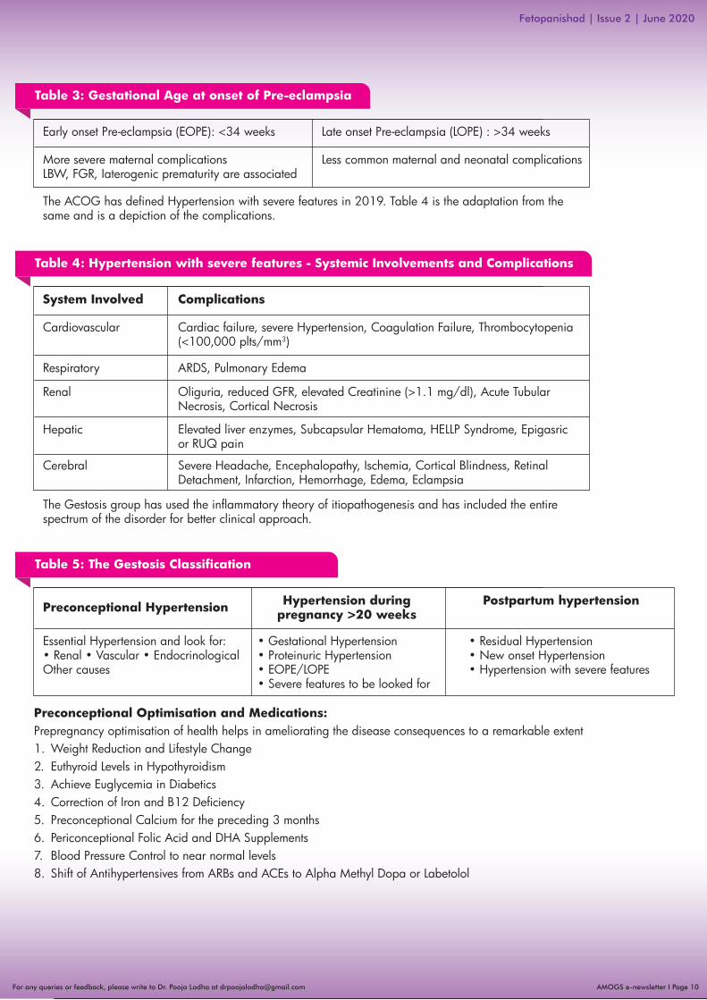
Early onset Pre-eclampsia (EOPE): <34 weeks
The ACOG has defined Hypertension with severe features in 2019. Table 4 is the adaptation from the same and is a depiction of the complications.
Late onset Pre-eclampsia (LOPE) : >34 weeks
More severe maternal complicationsLBW, FGR, Iaterogenic prematurity are associated
Less common maternal and neonatal complications
Table 3: Gestational Age at onset of Pre-eclampsia
System Involved
The Gestosis group has used the inflammatory theory of itiopathogenesis and has included the entire spectrum of the disorder for better clinical approach.
Complications
Cardiovascular Cardiac failure, severe Hypertension, Coagulation Failure, Thrombocytopenia(<100,000 plts/mm3)
Respiratory ARDS, Pulmonary Edema
Renal Oliguria, reduced GFR, elevated Creatinine (>1.1 mg/dl), Acute Tubular Necrosis, Cortical Necrosis
Hepatic Elevated liver enzymes, Subcapsular Hematoma, HELLP Syndrome, Epigasric or RUQ pain
Cerebral Severe Headache, Encephalopathy, Ischemia, Cortical Blindness, Retinal Detachment, Infarction, Hemorrhage, Edema, Eclampsia
Table 4: Hypertension with severe features - Systemic Involvements and Complications
Preconceptional Hypertension
Preconceptional Optimisation and Medications:Prepregnancy optimisation of health helps in ameliorating the disease consequences to a remarkable extent 1. Weight Reduction and Lifestyle Change2. Euthyroid Levels in Hypothyroidism 3. Achieve Euglycemia in Diabetics4. Correction of Iron and B12 Deficiency5. Preconceptional Calcium for the preceding 3 months6. Periconceptional Folic Acid and DHA Supplements7. Blood Pressure Control to near normal levels8. Shift of Antihypertensives from ARBs and ACEs to Alpha Methyl Dopa or Labetolol
Hypertension during pregnancy >20 weeks
Postpartum hypertension
Essential Hypertension and look for: • Renal • Vascular • EndocrinologicalOther causes
• Gestational Hypertension • Proteinuric Hypertension • EOPE/LOPE • Severe features to be looked for
• Residual Hypertension • New onset Hypertension • Hypertension with severe features
Table 5: The Gestosis Classification

AMOGS e-newsletter I Page 11For any queries or feedback, please write to Dr. Pooja Lodha at [email protected]
Fetopanishad | Issue 2 | June 2020
Laboratory tests for comorbidities and CKD
Maternal Surveillance and Treament in Pregnancy:Gestosis HDP needs meticulous surveillance for early pick up for effective corrective measures. The HDP Gestosis score depicts both existing as well as emerging risk factors to classify mothers needing close surveillance.
The antenatal care has to be focused on early detection of Gestosis and every visit specific assessments are necessary(Table 8). Also the family of the mother should be educated with the set of signs (Table 8), which need to be paid attention to and medical advice sought.
Additional tests
• Urinalysis and Culture• Creatinine and Uric Acid • Glucose Level and HBA1C • Albumin Creatinine Ratio• Thyroid profile
• Electrocardiogram• Echocardiography• Fundoscopy• Cardiovascular Evaluation, if necessary
Table 6: Pre-existing Hypertension and other disorders
Table 7: HDP Gestosis Score
Table 8: Focused Questions and Warning Signs HDP
Specific symptoms to be asked ideally to all ANCs after 20 weeks
Additional checks
• Visual Disturbances• Persistent Headaches• Epigastric or Right Upper Quadrant Pain• Increased oedema
• Upper Abdominal Pain/Acute Pain in Lower Abdomen• Headaches Feeling Unwell and Nauseous or like Throwing Up• Blurring of Vision or Seeing Flashing Lights• Swelling on Hands and/or Face• Reduced Fetal Movements• Loss of Consciousness/Seizures

AMOGS e-newsletter I Page 12For any queries or feedback, please write to Dr. Pooja Lodha at [email protected]
Fetopanishad | Issue 2 | June 2020
Lab Tests Levels
HemoglobinPCVTLCPlateletsSGOT & SGPTSAPCreatinineLDHSUA
<11 g/dL & >12 g/dL<30 & >40Leukocytosis<1.5 lacs>45 U/L>250 U/L0.9 mg/dL> 600 U/L>7.5 U/L
Inference
Anemia/HemoconcentrationHemodilution/HemoconcentrationInfection/SIRSThrombocytopeniaElevated liver enzymesElevatedRenal Involvement HemolysisAdverse Neonatal Outcome & Abruption Possible
PCV: Packed Cell Volume; TLC: Total Leucocyte Count; SGOT: Serum Glutamic Oxaloacetic Transferase; SGPT: Serum Glutamic Pyridoxal Phosphate Dependent Transaminase; SAP: Serum Alkaline Phosphatase, LDH: Lactate Dehydrogenase; SUA: Serum Uric Acid
Table 9: Laboratory Tests for HDP
Antihypertensive Mechanism of Action
Labetalol Alpha 1 and selective beta 2 Adrenergic Receptor Blocker
Dose
• 100 mg bid initially; increased by 100 mg 12 hourly every 2-3 days
• Usual dosage range: 200-400 mg bid not to exceed 2400 mg/day
Nifedipine Calcium Channel Blocker • 10 mg bid may be increased to 120 mg/day• Slow release preparations preferred
Alpha Methyldopa Centrally acting α2 Adrenergic Agonist
• 500-2000 mg per day orally in 3-4 divided doses
Management of Severe Hypertension needs immediate Blood Pressure Control and Seizure Prophylaxis with magnesium sulphate and Table 11 depicts preparedness for the same.
Table 10: Treatment of Hypertension - To be started at 140 / 90 mm Hg as per the CHIP study
Table 11: Hypertensive Crisis Management
Drugs for Urgent Control
Lower BP promptly but slowlyThe DBP not >30 mm Hg and MAP not >25 mm HgDBP: 90-100 mm Hg

AMOGS e-newsletter I Page 13For any queries or feedback, please write to Dr. Pooja Lodha at [email protected]
Fetopanishad | Issue 2 | June 2020
Table 12: HELLP and D/D of HDP
Table 13: Fetus as the Patient in HDP
Table 14: HDP: Maternal and fetal follow up surveillance strategy
Features of HELLP Differential Diagnosis
• Hemolysis (abnormal smear)• Elevated Liver Enzymes (serum SGOT >70 U/L serum
LDH >600 U/L • Low Platelets (<100000) • Occurrence- 20% of Pre-eclampsia Patients• Cause - It is thought to arise because of endothelial
and microvascular injury, increased vascular tone and platelet aggregation
Fetal surveillance and treatment in pregnancy: Table 13
Abbreviations: AFI: Amniotic Fluid Index; UAD: Uterine Artery Doppler; UmAD: Umbilical Artery Doppler; FGR: Fetal Growth Restriction; AEDF: Absent End Diastolic flow; REDF: Reduced End Diastolic Flow; CS: Cesarean Section; CPR: Cerebral Placental Ratio
Initial Assessment - Biometry, AFI & UAD, UmAD, MCA
If FGR: Doppler and AFI estimation: 1/15 days, till delivery
UmAD REDF week Droppler: AEDF
If AEDF: Doppler: 2/week: REDF
With REDF: >70% of the intervillous circulation is compromised and it is time to deliver by CS.
Ductus Venosus/Umbilical Vein: Fetal Acidemia: Poor Prognosis
Magnesium Sulphate: to be given for Fetal Neuroprotection <32 weeks
Arterial Doppler significant for Fetal Pognosis before 32 weeks
Venous Droppler Later
A CPR of >1 gives us time to postpone delivery
• Renal Disease• Acute Fatty Liver• Cholestasis of Pregnancy• Hemolytic Uremic Syndrome• Thrombotic• Thrombocytopenic Purpura • Pheochromocytoma• Cardiovascular Diseases: Coarctation, Subclavian
Stenosis, Aortic Dissection and Vasculitis

AMOGS e-newsletter I Page 14For any queries or feedback, please write to Dr. Pooja Lodha at [email protected]
Fetopanishad | Issue 2 | June 2020
Delivery Decision
Table 15: Postpartum Care
When to deliver ? How to Deliver ?
Place
No advantage of Cesarean Delivery over Vaginal Route. Individualise the route of delivery.
Postpartum disease needs vigilance and treatment
Conclusion: Hypertension in pregnancy is an important component of Antenatal and Postnatal Care, and preconceptional optimisation, useful. Careful vigilance throughout pregnancy and after delivery is important. Patient should be educated about associated future health risks.
Well-equipped wth infrastructure, expertise such as Neonatal, Critical, Anaesthesiology and Obstetrician Care with Blood Bank-support.
Transfer
Antihypertensives, Antenatal Steroids if less than37 weeks and Magnesium Sulphate for seizure prophylaxis.

THE IMMUNOLOGICALLY-POSITIVE MOTHER
AMOGS e-newsletter I Page 15For any queries or feedback, please write to Dr. Pooja Lodha at [email protected]
Fetopanishad | Issue 2 | June 2020
Pregnancy is sort of Allograft Implantation in the mother’s womb. There is bound to be Graft Rejection and Nature has to device strategies to overcome these Antigen-antibody Reactions, so that the fetus can survive further. Unfortunately, in some women, these rejection mechanisms play dominant role over the protective mechanism and lead to rejection of the pregnancy. This mechanism is likely to manifest in different forms, like Thrombosis, Miscarriages, Pre-eclampsia, Fetal Growth Restrictions or Postpartum Thrombosis.In order to cure, we need to understand these mechanisms happening at the molecular level.
Introduction
Placenta plays an important role by acting as an immunological barrier between the mother and the fetus, this is achieved by interplay of various mechanisms, few of which include, secretion of Neurokinin B to dodge the detection by immune system, suppression of cytotoxic T cells by inhibiting the response to Interleukin 2 and formation of atypical MHC class I isotypes like HLA-E and HLA - creating an immunologically-privileged site. One model for the induction of tolerance during the very early stages of pregnancy is the Eutherian Fetoembryonic Defence System (eu-FEDS) Hypothesis, which states that both soluble and cell surface associated Glycoproteins, present in the reproductive system and expressed on Gametes, suppress any potential immune responses and inhibit rejection of the fetus. The eu-FEDS model further suggests that specific carbohydrate sequences (Oligosaccharides) are covalently linked to these immunosuppressive Glycoproteins and act as “functional groups” that suppress the immune response. The major Uterine and Fetal Glycoproteins that are associated with the eu-FEDS model in the human include Alpha-fetoprotein, CA125, and Glycodelin-A (also known as placental protein 14 (PP14)).Regulatory T cells also likely play a role.Also, a shift from cell-mediated immunity toward humoral immunity, is believed to occur.
How is the fetus protected?
In certain conditions, such as Ischemia, Cell Injury or Autoimmunity, the negatively charged Phospholipids get exposed and stimulate the production of anti-phospholipid Anti-phospholid Antibodies, or permit the passage of Pro-coagulant Proteins like b2 Glycoproteins, Prothrombin, Protein-C, Protein S and Annexin-V, leading to formation of aPL. The aPL needs another co-factor - b2GP1 (Apolipoprotein H) to exert its effect. This initiates a cascade of events such as Trophoblasticinhibition, Inflammation, Defective Angiogenesis and Thrombosis leading to Impaired Placentation and, thus, Pregnancy Rejection.1
Some women appear to be genetically predisposed.2
Significant correlation has been shown in Polymorphisms involved with Blood Coagulation and pro-inflammatory states with Thrombotic APS.3
The Hypercoagulable state is APS also appears to be a result of defects in Haemostasis viz. Platelet Function, Fibrinolysis and Coagulation Cascade.4
Why does then pregnancy fail in some women?
aPL also alters the balance of PGI2/TXA2 leading to Vaso-constriction and Platelet Activation causing decreased blood supply to the fetus.5
aPL in some form, also exaggerates the proinflammatory response leading to Complement Activation, Release of TNE and Chemokines, causing Thrombosis and Fetal Growth Restriction. The protective role of Heparin appears more due to its anti-compliment effect rather than as an anticoagulant.6
Defective Endometrial Angiogenesis is also a result of modulation of VEGF and MMP activity by aPL leading to Defective Angiogenesis, reduction in length and number of capillaries and blocking the Secretion and Trophoblastic Invastion and, thus, Disruption of Spiral Arterial Remodelling. Addition of LMWH restores VEGF and MMP activity.This decreased uteroplacental blood supply due to defective spiral arterioles, combined with initiation of decidual inflammation, formation of syncytial knots and decrease in vasculo- syncytial membranes lead to
defective placentation.Understanding these mechanisms is important because from clinical point of view, Uterine Doppler provides a non-invasive indirect method of studying the utero-placental circulation before any clinical signs of either Pre-eclampsia or Growth Restriction are seen, normal Doppler being a good predictor of good pregnancy outcome while abnormal Doppler indicating high risk of Maternal Fetal complications.
Dr. Ajit PatilConsultantPristine Fetal Medicine UnitKolhapur

In certain conditions, such as Ischemia, Cell Injury or Autoimmunity, the negatively charged Phospholipids get exposed and stimulate the production of anti-phospholipid Anti-phospholid Antibodies, or permit the passage of Pro-coagulant Proteins like b2 Glycoproteins, Prothrombin, Protein-C, Protein S and Annexin-V, leading to formation of aPL. The aPL needs another co-factor - b2GP1 (Apolipoprotein H) to exert its effect. This initiates a cascade of events such as Trophoblasticinhibition, Inflammation, Defective Angiogenesis and Thrombosis leading to Impaired Placentation and, thus, Pregnancy Rejection.1
Some women appear to be genetically predisposed.2
Significant correlation has been shown in Polymorphisms involved with Blood Coagulation and pro-inflammatory states with Thrombotic APS.3
The Hypercoagulable state is APS also appears to be a result of defects in Haemostasis viz. Platelet Function, Fibrinolysis and Coagulation Cascade.4
aPL also alters the balance of PGI2/TXA2 leading to Vaso-constriction and Platelet Activation causing decreased blood supply to the fetus.5
aPL in some form, also exaggerates the proinflammatory response leading to Complement Activation, Release of TNE and Chemokines, causing Thrombosis and Fetal Growth Restriction. The protective role of Heparin appears more due to its anti-compliment effect rather than as an anticoagulant.6
Defective Endometrial Angiogenesis is also a result of modulation of VEGF and MMP activity by aPL leading to Defective Angiogenesis, reduction in length and number of capillaries and blocking the Secretion and Trophoblastic Invastion and, thus, Disruption of Spiral Arterial Remodelling. Addition of LMWH restores VEGF and MMP activity.This decreased uteroplacental blood supply due to defective spiral arterioles, combined with initiation of decidual inflammation, formation of syncytial knots and decrease in vasculo- syncytial membranes lead to
AMOGS e-newsletter I Page 16For any queries or feedback, please write to Dr. Pooja Lodha at [email protected]
Fetopanishad | Issue 2 | June 2020
Retrospective suspicion is easy in those women who had:1. More than 3 recurrent pregnancy losses <10 wks2. One or more, otherwise unexplained fetal deaths >10 wks3. Delivery prior to 34 wks for PE or Placental Insufficiency2-6% of women with RPL have positive aPL antibody results. But fetal death and early delivery for severe PE are considered to be more specific features of APS. Placental Insufficiency in absence of PE however, less well studied.But all these criteria are after the loss of a pregnancy and thus all efforts should be directed to identify prospectively, a woman at risk of APS. Lab evaluation is out of question in the first pregnancy but Uterine Artery Doppler is a better non-invasive tool to understand the defective Uteroplacental circulation and gives an opportunity for early and effective intervention.
Identifying Women with High Risk of APS7
The incidence of PE, across nations, religions, socio-economic groups is 5-7%. PE is bad for both. For every maternal death, there are another 10 women who suffer from PE-related sequel. And, thus, it is the duty of all Obstetricians to make conscious efforts to identify such high-risk women and intervene.Setting up of a “PRE-ECLAMPSIA CLINIC” is simple. Important components of such a clinic are:1. A station for proper history, taking blood pressure measurement in sitting position - a well-trained nurse can easily do
this. BP machines need to be automated machines and should be calibrated as per the interval suggested by the manufacturer.
2. Training in obtaining uterine Doppler measurements. This is easy and every OB can easily do this. It is part of NT certification. Learning curve for proper Uterine Artery Doppler measurements, is small and if the OB has access to FMF algorithm/online calculators, risk prediction becomes easy.
3. Serum testing for PAPP-A and PlGF, both are anyway done as part of Aneuploidy evaluation.Such a combined screening detects 76.6% of cases of preterm PE and 38.3% of term PE at a FPR of 10%.
The Need for "Pre-eclampsia Clinic"
FMF recommends use of Low-dose Aspirin, 150 mg at bedtime for all those women who are at high risk (>1:100). In the ASPRE study, eligible women with an estimated risk for preterm PE of >1 in 100 were invited to participate in a double‐blind trial of Aspirin (150 mg per day) vs placebo from 11–14 weeks until 36 weeks' Gestation. In the Aspirin group, the incidence of preterm PE was reduced by 62%.8
Intervention
Managing these high risk pregnant women
These women on Aspirin should be followed up regularly with repeat Uterine Artery Doppler at 18-20 weeks and around 24-26 weeks to see what has happened to Defective Trophoblast Invasion. However, Aspirin should not be stopped even if Doppler changes revert back to normal. Aspirin is continued till 36 completed weeks of pregnancy.
Follow-up
The aim of managing pregnancy with APS, is to minimise risk of adverse maternal and fetal events. Thus, counselling plays an important role. The triple-positive women, especially, are at increased risk of adverse events and should be counselled about adherence to the drug regimens and regular clinical follow-up.Current treatment of choice is LMWH and/or Low Dose Aspirin (LDA). This regime provides adequate Thromboprophylaxis and improves pregnancy outcome.11
LMWH is used in a standard dose of 4000IU/Enoxaparin 40 mg/0.4 ml as a Subcutaneous Injection. It may cause some local irritation and superficial staining of the skin (Ecchymosis) which disappears in some days. Women receiving LMWH can be taught to self-inject subcutaneously in the anterolateral abdominal wall on the right and left sides. Alternatively, they can use cold pack locally and keep on changing injection site, daily.
LDA (Aspirin) is available as 150 mg tablet, to be taken at bedtime, daily.Aspirin is stopped after 36 completed weeks. LMWH is continued till delivery. Before planned induction of Labour or Elective Caesarean Delivery, LMWH treatment should be ceased to prevent Intraoperative Haemorrhage.Preconceptional LDA provides beneficial effects on implantation and improves live birth rates and hence recommended.Two trials were conducted where LMWH was compared to unfractionated Heparin along with LDA, failed to find any differences in pregnancy outcomes. This is important, especially in low economic groups as UFH is affordable and thus compliance is better.12,13
This dose of Heparin, esp. LMWH is very unlikely to cause side effects such as Osteopaenia/Clinically-significant Bleeding or Heparin-induced Thrombocytopaenia (HIT). But the very nature of heterogeneous design of various trials, differences in laboratory criteria, lack of confirmatory repeat tests tempts the clinician to use these regimes empirically. Empirical use of Heparin in absence of positive laboratory criteria is not approved by majority of organizations. In fact, an open multi-centre randomised controlled trial, registered at EudraCT (protocol number: 2010-022715-19) was conducted in Sweden found immunological differences not in favour of using LMWH as an anti-inflammatory drug during the second and third trimesters of pregnancy, in women with otherwise unexplained recurrent pregnancy losses.Further studies are needed to explore the hypothesis of using LMWH-associated pro-inflammatory effects to ameliorate the defective implantation process in RPL in the earliest stages of pregnancy.
defective placentation.Understanding these mechanisms is important because from clinical point of view, Uterine Doppler provides a non-invasive indirect method of studying the utero-placental circulation before any clinical signs of either Pre-eclampsia or Growth Restriction are seen, normal Doppler being a good predictor of good pregnancy outcome while abnormal Doppler indicating high risk of Maternal Fetal complications.

AMOGS e-newsletter I Page 17For any queries or feedback, please write to Dr. Pooja Lodha at [email protected]
Fetopanishad | Issue 2 | June 2020
These women are at risk of increased adverse events in spite of use of Heparin and LDA. The prospective PROMISSE study showed that in women with APS, risk of adverse events was 20%. This is in spite of use of Heparin and Aspirin and women need to be counselled before, to avoid any unpleasant events later on.14
And, thus, it is recommended that these women be followed up frequently with Serial OB Sonography, Maternal BP Check-ups and Fetal Surveillance - beginning from 32 weeks or earlier depending on past history.
Relevant maternal labs should include platelet counts at least 4 weeks interval.
Antenatal Care in APS PregnancyThe aim of managing pregnancy with APS, is to minimise risk of adverse maternal and fetal events. Thus, counselling plays an important role. The triple-positive women, especially, are at increased risk of adverse events and should be counselled about adherence to the drug regimens and regular clinical follow-up.Current treatment of choice is LMWH and/or Low Dose Aspirin (LDA). This regime provides adequate Thromboprophylaxis and improves pregnancy outcome.11
LMWH is used in a standard dose of 4000IU/Enoxaparin 40 mg/0.4 ml as a Subcutaneous Injection. It may cause some local irritation and superficial staining of the skin (Ecchymosis) which disappears in some days. Women receiving LMWH can be taught to self-inject subcutaneously in the anterolateral abdominal wall on the right and left sides. Alternatively, they can use cold pack locally and keep on changing injection site, daily.
LDA (Aspirin) is available as 150 mg tablet, to be taken at bedtime, daily.Aspirin is stopped after 36 completed weeks. LMWH is continued till delivery. Before planned induction of Labour or Elective Caesarean Delivery, LMWH treatment should be ceased to prevent Intraoperative Haemorrhage.Preconceptional LDA provides beneficial effects on implantation and improves live birth rates and hence recommended.Two trials were conducted where LMWH was compared to unfractionated Heparin along with LDA, failed to find any differences in pregnancy outcomes. This is important, especially in low economic groups as UFH is affordable and thus compliance is better.12,13
This dose of Heparin, esp. LMWH is very unlikely to cause side effects such as Osteopaenia/Clinically-significant Bleeding or Heparin-induced Thrombocytopaenia (HIT). But the very nature of heterogeneous design of various trials, differences in laboratory criteria, lack of confirmatory repeat tests tempts the clinician to use these regimes empirically. Empirical use of Heparin in absence of positive laboratory criteria is not approved by majority of organizations. In fact, an open multi-centre randomised controlled trial, registered at EudraCT (protocol number: 2010-022715-19) was conducted in Sweden found immunological differences not in favour of using LMWH as an anti-inflammatory drug during the second and third trimesters of pregnancy, in women with otherwise unexplained recurrent pregnancy losses.Further studies are needed to explore the hypothesis of using LMWH-associated pro-inflammatory effects to ameliorate the defective implantation process in RPL in the earliest stages of pregnancy.
Clinical Manifestation of APS
Treatment of APS during pregnancy
APS with history of Thrombosis Patient on Long-term Anticoagulant - Full Anti-coagulant Dose of LMWH and Low-dose Aspirin
Patient not on Long-term Anticoagulation - Intermediate Dose or Full Anticoagulant Dose LMWH and Low-dose Aspirin
Patient with Thrombotic APS are at risk for Recurrent Thrombosis and are often managed using Long-term Anticoagulation e.g., with Warfarin. Warfarin should be discontinued prior to 6 weeks Gestation to avoid risk of Warfarin Embryopathy.
APS without a history and Thrombosis and Recurrent Early Miscarriages
Low-dose Aspirin or Prophylactic Dose LMWH and Low-dose Aspirin
Recurrent Early Miscarriage, some studies show high rate of successful pregnancy on Low-dose Aspirin alone and often show no benefit of addition of Heparin
History of Fetal Death or history of Early Delivery for Severe Pre-eclampsia or Placental Insufficiency
Prophylactic Dose LMWH and Low-dose Aspirin
The evidence that a Heparin agent improves pregnancy outcome in women with history of Fetal Death or Early Delivery for Pre eclampsia or Placental Insufficiency, is low quality.
Women with repeatedly positive LAC results or repeatedly positive for LAC and moderate to high titers of aCL or b2-GP1 antibodies are likely at increased risk of Thrombosis during pregnancy, Prophylactic LMWH should be considered.
Treatment Options Comments

AMOGS e-newsletter I Page 18For any queries or feedback, please write to Dr. Pooja Lodha at [email protected]
Fetopanishad | Issue 2 | June 2020
These women are at increased risk of Postpartum Thrombosis and thus, need drugs to be continued after delivery. Clinical judgements about the choice of drugs is necessary.
Postpartum Care
As mentioned in PROMISSE trial, almost 20% of women develop complications in spite of LMWH/LDA. These women will need higher dose of LMWH next time.
In view of failure of standard regimes, clinicians have sought combinations of “alternative treatments” along with Heparin. These include low dose prednisolone - 10 mg per day till 14 weeks, or HCQ-hydroxychloroquine in addition. IVIG Infusion or Plasmapheresis has also been tried with varied success rates. Pravastatin is also shown to be effective in one series. However, most of these type of evidences come from infertile women who have recurrent implantation failure, and, thus, interpolation of these results to RPL with APS women is difficult.15,16
Further studies are needed to explore the hypothesis of using these various combinations to ameliorate the defective implantation process in RPL, in the earliest stages of pregnancy.
But such a properly designed, powered, controlled, randomised trial is difficult in High-risk Refractory APS women who are in agony of losing pregnancies repeatedly, and clinicians will often face ethical dilemma of refusing such a drug, even if there is no adequate scientific proof, and will thus tempt to use them.
Word of caution is therefore necessary before a doctor takes decision based on results of such trials.
Reference: 1. Lockwood CJ, Rand JH. The immunobiology and obstetrical consequences of antiphospholipid antibodies. Obstet Gynecol Surv. 1994;49(6):432–41.2. Sebastiani GD, Iuliano A, Cantarini L et al. Genetic aspects of the antiphospholipid syndrome: An update. Autoimmun Rev. 2016;15(5):433–9. 3. Xie H, Kong X, Zhou H et al. TLR4 is involved in the pathogenic effects observed in a murine model of antiphospholipid syndrome. Clin Immunol. 2015;160(2):198–210.4. De Groot PG, Horbach DA, Derksen RH. Protein C and other cofactors involved in the binding of antiphospholipid antibodies: Relation to the pathogenesis of thrombosis.
Lupus. 1996;5(5):488–93.5. Carreras LO, Vermylen JG. “Lupus” anticoagulant and thrombosis--possible role of inhibition of prostacyclin formation. Thromb Haemost. 1982;48(1):38–40.6. Tedesco F, Borghi MO, Gerosa M et al. Pathogenic role of complement in antiphospholipid syndrome and therapeutic implications. Front Immunol. 2018;19(9):1388.7. Miyakis S, Lockshin MD, Atsumi T et al. International consensus statement on an update of the classification criteria for definite antiphospholipid syndrome (APS). J Thromb
Haemost. 2006;4(2):295–306.8. D. L. Rolnik et alASPRE trial: performance of screening for preterm pre‐eclampsia,Ultraound in OB & Gynecol, Volume 50, Issue 4 October 2017, Pages 492-4959. Miyakis S, Lockshin MD, Atsumi T et al. International consensus statement on an update of the classification criteria for definite antiphospholipid syndrome (APS). J Thromb
Haemost. 2006;4(2):295–306.10. Pengo V, Banzato A, Bison E et al. Laboratory testing for antiphospholipid syndrome. Int J Lab Hematol. 2016;38 (Suppl 1):27–31.11. Lockshin MD, Kim M, Laskin CA et al. Prediction of adverse pregnancy outcome by the presence of lupus anticoagulant, but not anticardiolipin antibody, in patients with
antiphospholipid antibodies. Arthritis Rheum. 2012;64(7):2311–812. Stephenson MD, Ballem PJ, Tsang P et al. Treatment of antiphospholipid antibody syndrome (APS) in pregnancy: A randomized pilot trial comparing low molecular weight
heparin to unfractionated heparin. J Obstet Gynaecol Can. 2004;26(8):729–34. 13. Noble LS, Kutteh WH, Lashey N, Franklin RD, Herrada J. Antiphospholipid antibodies associated with recurrent pregnancy loss: Prospective, multicenter, controlled pilot
study comparing treatment with low-molecular-weight heparin versus unfractionated heparin. Fertil Steril. 2005;83(3):684–90.14. JE Salmon PROMISSE: progress in understanding pregnancy complications in patients with SLE Arthritis Res Ther. 2012; 14(Suppl 3): A39.15. Bramham K, Thomas M, Nelson-Piercy C, Khamashta M, Hunt BJ. First-trimester low-dose prednisolone in refractory antiphospholipid antibody-related pregnancy loss.
Blood. 2011;117(25):6948–5116. Mekinian A, Lazzaroni MG, Kuzenko A et al. The efficacy of hydroxychloroquine for obstetrical outcome in anti-phospholipid syndrome: Data from a European multicenter
retrospective study. Autoimmune Rev. 2015;14(6):498–502.1
Refractory Cases

Pregnancy is associated with significant but reversible changes in Maternal Thyroid Physiology that can lead to confusion in the diagnosis of Thyroid abnormalities. There is moderate Thyroid enlargement as a result of pregnancy hormone–induced Glandular Hyperplasia and increased vascularity.
As shown in the figure, there are well described changes in Thyroid function tests during pregnancy that are related to 1) an Estrogen-mediated increase in circulating levels of Thyroid-binding Globulin, which is the major transport protein for Thyroid hormone, 2) thyroid stimulation due to a “spillover” effect, especially in the first trimester, by hCG, which shares some structural homology with TSH, and 3) a relative decline in availability of Iodine related to increased renal clearance and overall losses to the fetus and placenta.
Maternal T4 is transferred to the fetus throughout the entire pregnancy and is important for normal fetal brain development. It is especially important before the fetal Thyroid gland begins concentrating Iodine and synthesizing Thyroid hormone at approximately 12 weeks of Gestation.
Thyroid Function and the Fetus
Universal screening for Thyroid Disease in pregnancy is not recommended because identification and treatment of Maternal Subclinical Hypothyroidism has not been shown to result in improved Neurocognitive function in offspring. Indicated testing of Thyroid function should be performed in women with a personal history of Thyroid Disease or symptoms of Thyroid Disease.
Which Pregnant Patients should be screened for Thyroid Disease?
HYPOTHYROIDISM IN PREGNANCY
AMOGS e-newsletter I Page 19For any queries or feedback, please write to Dr. Pooja Lodha at [email protected]
Fetopanishad | Issue 2 | June 2020
Levels of TSH and free T4 should be measured to diagnose Thyroid Disease in pregnancy. The first-line screening test is measurement of the TSH level. Assuming Normal Hypo-thalamic-pituitary Function, an inverse log-linear relationship exists between serum TSH and serum Thyroid hormone, such that small alterations in circulating hormone levels will produce much larger changes in TSH. Therefore, TSH is the most reliable indicator of Thyroid status because it indirectly reflects Thyroid hormone levels as sensed by the Pituitary Gland. When the TSH level is abnormally high or low, a follow-up study to measure the free T4 level should be performed. Trimester-specific reference ranges have been given for Indian population that can be followed.
Laboratory Tests are used to diagnose Thyroid Disease during Pregnancy?
Dr. Ashwini Manu-RathiFetal Medicine Consultant, MD (OBGY), MRCOG, Fellowship in Fetal Medicine.Mumbai Fetal Medicine Centre, Mumbai.Visiting Consultant at Surya Mother and Child Hospital, Mumbai.918669227227 / 919920199029 | [email protected] | www.mumbaifetalmedicine.com

AMOGS e-newsletter I Page 20For any queries or feedback, please write to Dr. Pooja Lodha at [email protected]
Fetopanishad | Issue 2 | June 2020
Overt Hypothyroidism complicates between 1 of 1,000 and 3 of 1,000 pregnancies. The most common cause of primary hypothyroidism in pregnancy is chronic autoimmune thyroiditis (Hashimoto’s thyroiditis). Other important causes include endemic iodine deficiency and a history of either ablative radioiodine therapy or thyroidectomy.
Vague, nonspecific signs or symptoms that are often insidious in onset and easily confused with complaints of pregnancy itself characterize it. Initial symptoms include fatigue, constipation, cold intolerance and muscle cramps. These may progress to insomnia, weight gain, carpal tunnel syndrome, hair loss, voice changes and intellectual slowness. Other signs of hypothyroidism include periorbital edema, dry skin and prolonged relaxation phase of deep tendon reflexes.
Overt Hypothyroidism
Women with Overt Hypothyroidism are at an increased risk for pregnancy complications such as early pregnancy failure, preeclampsia, placental abruption, low birth weight, and stillbirth. Treatment of women with Overt Hypothyroidism has been associated with improved pregnancy outcomes.
Maternal risks
Overt, untreated maternal hypothyroidism has been associated with an increased risk of low birth weight and impaired neuropsychologic development of the offspring. However, it is rare for maternal thyroid inhibitory antibodies to cross the placenta and cause fetal hypothyroidism.
Fetal and Neonatal Effects
Very few studies demonstrate benefits from the identification and treatment of Euthyroid pregnant women who have Thyroid Antibodies. Thus, Universal Screening for Thyroid Autoantibodies in pregnancy currently is not recommended.
Screening or Testing for Thyroid Autoantibodies in Pregnancy?
Overt vs Subclinical Thyroid Dysfunction
AMOGS e-newsletter I Page 21For any queries or feedback, please write to Dr. Pooja Lodha at [email protected]
Fetopanishad | Issue 2 | June 2020
References:1. ACOG Practice Bulletin VOL. 125, NO. 4, APRIL 2015 2. Marwaha R, Chopra S, Gopalakrishnan S, Sharma B, Kanwar R, Sastry A, Singh S. Establishment of reference range for thyroid hormones in normal pregnant Indian women.
BJOG 2008;115:602–606. 3. Thyroid disease in pregnancy. Obstet Gynecol 2006;108:1283–92
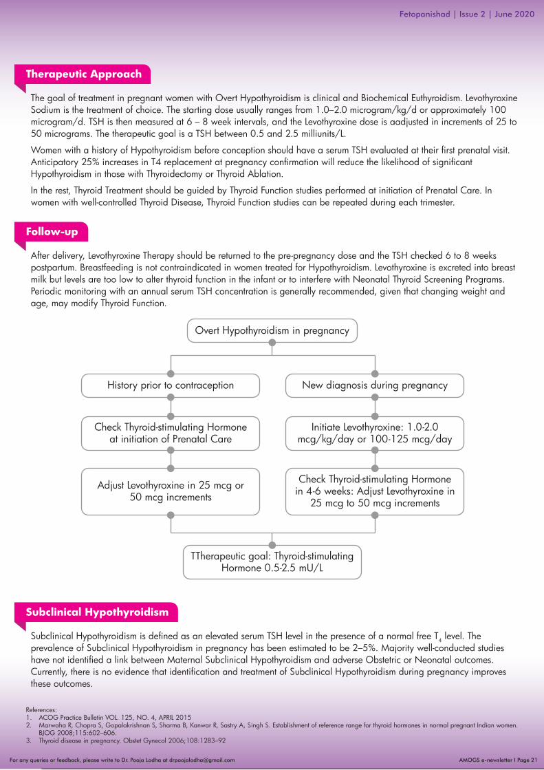
After delivery, Levothyroxine Therapy should be returned to the pre-pregnancy dose and the TSH checked 6 to 8 weeks postpartum. Breastfeeding is not contraindicated in women treated for Hypothyroidism. Levothyroxine is excreted into breast milk but levels are too low to alter thyroid function in the infant or to interfere with Neonatal Thyroid Screening Programs. Periodic monitoring with an annual serum TSH concentration is generally recommended, given that changing weight and age, may modify Thyroid Function.
Follow-up
Subclinical Hypothyroidism is defined as an elevated serum TSH level in the presence of a normal free T4 level. The prevalence of Subclinical Hypothyroidism in pregnancy has been estimated to be 2–5%. Majority well-conducted studies have not identified a link between Maternal Subclinical Hypothyroidism and adverse Obstetric or Neonatal outcomes. Currently, there is no evidence that identification and treatment of Subclinical Hypothyroidism during pregnancy improves these outcomes.
Subclinical Hypothyroidism
Overt Hypothyroidism in pregnancy
History prior to contraception New diagnosis during pregnancy
Check Thyroid-stimulating Hormoneat initiation of Prenatal Care
TTherapeutic goal: Thyroid-stimulating Hormone 0.5-2.5 mU/L
Initiate Levothyroxine: 1.0-2.0 mcg/kg/day or 100-125 mcg/day
Adjust Levothyroxine in 25 mcg or50 mcg increments
Check Thyroid-stimulating Hormonein 4-6 weeks: Adjust Levothyroxine in
25 mcg to 50 mcg increments
The goal of treatment in pregnant women with Overt Hypothyroidism is clinical and Biochemical Euthyroidism. Levothyroxine Sodium is the treatment of choice. The starting dose usually ranges from 1.0–2.0 microgram/kg/d or approximately 100 microgram/d. TSH is then measured at 6 – 8 week intervals, and the Levothyroxine dose is aadjusted in increments of 25 to 50 micrograms. The therapeutic goal is a TSH between 0.5 and 2.5 milliunits/L.
Women with a history of Hypothyroidism before conception should have a serum TSH evaluated at their first prenatal visit. Anticipatory 25% increases in T4 replacement at pregnancy confirmation will reduce the likelihood of significant Hypothyroidism in those with Thyroidectomy or Thyroid Ablation.
In the rest, Thyroid Treatment should be guided by Thyroid Function studies performed at initiation of Prenatal Care. In women with well-controlled Thyroid Disease, Thyroid Function studies can be repeated during each trimester.
Therapeutic Approach

AMOGS e-newsletter I Page 22For any queries or feedback, please write to Dr. Pooja Lodha at [email protected]
Fetopanishad | Issue 2 | June 2020
Refers to Biochemical & Physiological manifestation of Excessive Thyroid Hormones.
In Non-pregnant Women:• Menstrual Irregularity• Infertility
In Pregnant Women:• Pulse Rate > 100 bpm• Wide Pulse Pressure• Weight Loss• Nausea & Excessive Vomiting • Pre-eclampsia • Diarrhoea • Myopathy• Lymphadenopathy • Congestive Cardiac Failure
HYPERTHYROIDISM IN PREGNANCY
Future Pregnancy Planning: 2 stable Thyroid Function Tests at least once a month, apart in those women who are off Antithyroid Drugs and in remission.
TSH at/below 2.5th percentile for Gestational Age, Free T4:
1.75ng/dL or less
TSH decreases up to / below0.1mi U/L
Regardless of trimester T/t not required
Subclinical Hyperthyroidism
Occur in 1st trimester with 2.4% prevalence, transient.
lts due to hCG stimulation of thyroid – paralleled to hCG decline
May need B blocker for T/t
Gestational TransientThyrotoxicosis
Caused by a mutation in TSH receptor - hypersensitive to hCG
Recurrent GestationalHyperthyroidism
Definition Presentation & Association
• Grave’s Disease• Toxic Adenoma• Toxic Multinodular Goiter• Hyperemesis Gravidarum• Gestational Trophoblastic Disease• Metastatic Follicular Cell Carcinoma• Exogenous T3 & T4• De Quervin Thyroiditis • Painless Lymphotic Thyroiditis• Stuma Ovarii
• Free T3 & T4: Increased• TSH: Suppressed• Normal Free T4: T3 Toxicosis• Normochromic, Normocytic Anaemia, Mild Neutropenia• Liver Enzymes: Increased• Alkaline Phosphatase: Raised• Mild Hypercalemia (Sr hCG>40,625 ML u/L interferes with TSI Bioassay & gives False Negative Results, if found negative - should be repeated after 20 weeks, when hCG Concentration is Low)
Diagnosis of Hyperthyroidism
Future Pregnancy & Forms
Dr. Archana Patil(MBBS, DGO, CCGDM, Fellowship in Advanced Obstetrics Ultrasound,Certificate course in Advanced Fetal Echocardiography, Hyderabad)Consultant, Fetal Medicine, MGM MCRI, Aurangabad

• Always best to optimize prior to conception• Risk of Uncontrolled Hyperthyroidism far outweighs Risk of Therapy• Can consider no T/t ,if very minimal elevation of T4/T3• Goal of therapy is control without causing Fetal/ Neonatal Hyperthyroidism• Keep free T4 in high normal range or total T4 at 1.5x upper limit of normal
Fetal• Fetal Hyperthyroidism predicted when maternal TSIs
levels in excess of 300% of control values• Miscarriage• Preterm Delivery• Low Birth Weight• Fetal Thyrotoxicosis: Life-threatening, needs T/t features:
FHR >160 bpm, Growth Restriction, Advanced Bone Age, Cranyosynostenosis, Non-immune Fetal Hydrops, Fetal Death (detectable on Ultrasound)
Neonatal• Neonatal Hyperthyroidism – transplacental passage of
stimulating & blocking antibodies• Neonatal Thyrotoxicosis: all newborns should be
screened features: Hyperkineiss, Diarrhoea, Poor Weight Gain, Vomiting, Exopthalmos, Arrythmias, Cardiac Failure, Systemic and Pumonary Hypertension, Hepatospenomegaly, Thrombocytopenia Remission by 20 weeks common, but usually occurs by 48 weeks,if persistent, indicates strong Family History of Grave's Disease.
Medications:Current Thioamide Recommendations:• Used drugs: Propylthiouracil (PTU) & Methimazole (MMI)• Limit PTU use to first trimester only• Switch from PTU to MMI at 13 – 14 weeks• Monitor free T4 every 4 weeks• Other therapies: Beta Blockers, Iodides, Radioactive Ablation (Contraindicated), Surgery
Management Protocol
Complications: Occur in 1% - 5% due to Transplacental Passage of TSIs & Activation of Fetal Thyroid
Confirm diagnosis
Propylthioracil 100-150 mgx3/day
Low-dose ATD up and during labor
Monitor Thyroid Function(4 weekly) and adjust ATD
(if necessary)
Review Postpartum - check for Exacerbation
Discuss treatment: Maternal-fetal and
Breastfeeding effects
Check the infant for Thyroid Dysfunction
TST Receptor Antibodies at
36 weeks
Euthyroid status
AMOGS e-newsletter I Page 23For any queries or feedback, please write to Dr. Pooja Lodha at [email protected]
Fetopanishad | Issue 2 | June 2020
Hyperthyroidism in Pregnancy

• PTU in first trimester/MMI after first trimester• Ultrasound at 18 – 22 weeks for Fetal Anatomy• Growth Scan at 32 weeks• Weekly NSTs at 34 weeks (optional)• Mode of Delivery - Vaginal, until valid indication for CS• Notify Paediatrician• Postpartum Follow-up
Reference:• Belforte M, Saade G, Foley M, Phelan J, Didy G, Critical Care Obstetrics, Fifth Edition. Wiley- Blackwell, 2010.• Casey BM. Thyroid Disease in Pregnancy. ACOG Practice Bulletin.2015 number 148.• Creasy RK, Resnik R, Lams JD, LockwoodCJ, Moore TR, GreeneMF. Creasy & Resnik’s Maternal Fetal Medicine: Priciples & Practice 7th Edition.Elsvier Saunders,2014• De Groot L, Abalovich M, Alexander EK, Amino N, Barbour L, et al. Management of thyroid dysfunction during pregnancy & postpartum : An Endocrine Society Clinical
Practice Guideline.J Cin Endocrinol Metab 2012;97: 2543-2565
AMOGS e-newsletter I Page 24For any queries or feedback, please write to Dr. Pooja Lodha at [email protected]
Fetopanishad | Issue 2 | June 2020
Special Remark

AMOGS e-newsletter I Page 25For any queries or feedback, please write to Dr. Pooja Lodha at [email protected]
1) Gestation Diabetes is –
a. Detected 1st during 3rd trimester
b. Detected 1st postnatally
c. Very High Maternal BSL detected 1st time during 1st trimester
d. All of the above
2) Which of the following is not a risk for Neonates born to Hyperglycemic mothers?
a. Hyperbilirubinemia
b. Hypercalcemia
c. Hypoglycemia
d. Hypomagnesemia
7) Which of the following are the predictors of Pre-eclampsia in pregnancy?
a. Mean Uterine Artery PI >2.85 at 12 weeks
b. Mean Uterine Artery PI >1.85 at 24 weeks
c. Low PAPP-a at 12 weeks
d. All of the above
8) Low-dose Aspirin is...
a. 75 mg Aspirin
b. 150 mg Aspirin
c. 100 mg Aspirin
d. <300 mg Aspirin
3) Miscarriage Rate in Pre-gestational DM is directly proportional to...
a. Maternal Fasting BSL
b. Postprandial BSL
c. Maternal BMI and Co-existing Hypertension
d. Periconceptional HbA1c
4) Which of the following is considered as a limit for abnormal BP in pregnancy –
a. 150/100 mm of Hg
b. 140/90 mm of Hg
c. Depends on the trimester
d. Depends on Proteinuria
5) Which of the following drug is not indicated for use in pregnancy?
a. Calcium Chanel Blockers
b. Alpha Methyldopa
c. ACE Inhibitors
d. None of the above
6) Which of the following are drugs for urgent control of Blood Pressure in pregnancy?
a. Labetelol
b. Nidefepine
c. Alpha Methyldopa
d. ACE Inhibitors
Fetopanishad | Issue 2 | June 2020
Dr. Pooja LodhaAMOGS’ ChairpersonFetal Medicine Committee
BRAIN-FLEXERS 2

AMOGS e-newsletter I Page 26For any queries or feedback, please write to Dr. Pooja Lodha at [email protected]
9) Which is the single most reliable indicator of Maternal Thyroid Function in pregnancy?
a. TSH
b. T3
c. FreeT4
d. T4
10) Therapeutic goal of TSH in pregnancy is –
a. <2 mU/L
b. <5 mU/L
c. <2.5 mU/L
d. Depends on the trimester
11) Which of the following is a relative less-common Fetal Complication in a Hypothyroidism patient who is on Anti-thyroid Drugs?
a. Preterm Delivery
b. Low Birth Weight
c. Fetal Hypothyroidisim
d. Fetal Hyperthyroidism
12) Which of the following is true about Treatment of Maternal Hyperthyroidismin pregnancy?
a. PTU in 1st trimester
b. MMI after 1st trimester
c. Both of the above
d. PTU throughout pregnancy
Fetopanishad | Issue 2 | June 2020
BRAIN-FLEXERS WINNERS FOR ISSUE 1
1. Dr. Manoj Agalawe 2. Dr. Uzma Shaikh3. Dr. Sayali Jahagirdar
BRAIN-FLEXERS ANSWERS ISSUE 1 APRIL 2020 1-b, 2-c, 3-a, 4-b, 5-c, 6-c, 7-a, 8-c, 9-a, 10-c, 11-b, 12-a,
13-b, 14-c, 15-b, 16-b, 17-b, 18-c, 19-a, 20-a

AMOGS e-newsletter I Page 26For any queries or feedback, please write to Dr. Pooja Lodha at [email protected]
Fetopanishad | Issue 2 | June 2020
For your on-priority patients, where Genetic Counselling is essential and necessary & cannot wait due to current situation,
Lilac Insights announces
A service to provide seamless genetic counselling to your patients,without compromising the safety of you and your patients.
SAFE & CONVENIENT: No travel required &
person-to-person contactAccessible from anywhere &
any device
PRIVACY GUARANTEE: Through use of secure
platforms
NO WAITING PERIOD:Appointment as per
convenience
PREMARITAL COUNSELLING:
Eg. Planning a marriage in
consanguinity (i.e. in relation), Family history of a suspected genetic disorder or Late onset genetic disorders, etc
PRECONCEPTION COUNSELLING:
Eg. Bad obstetric history/ recurrent pregnancy loss,
Advanced maternal/ paternal age, Chromosomal
rearrangements in blood karyotyping
report, Family history of chromosomal/ genetic
disorder, etc
PRENATAL COUNSELLING:
Eg. Positive biochemical screening reports, Soft-markers/ anomalies on USG,
Plan prenatal diagnosis of genetic conditions
diagnosed/ yet undiagnosed in family,
Sudden fetal embryonic/demise, etc
POSTNATAL (PAEDIATRIC)
COUNSELLING: Eg. Child born with
suspected metabolic disorder/ other genetic
disorder, Intellectual disability, Suspected single gene disorder,
etc
Benefits of e-Genetic Counselling
e-Genetic Counselling can be helpful in
Email: [email protected] patients to book appointments: Call: 91366 29430 or