Embed Size (px)
Citation preview

―227―
Tokai J Exp Clin Med., Vol. 41, No. 4, pp. 227-229, 2016
Amoebiasis Presenting as Acute Appendicitis
Hitoshi ICHIKAWA*1, Jin IMAI*1, Hajime MIZUKAMI*2, Shuji UDA*3, Soichiro YAMAMOTO*3, Eiji NOMURA*3, Takuma TAJIRI*4, Norihito WATANABE*1 and Hiroyasu MAKUUCHI*3
*1Division of Gastroenterology and Hepatology, Department of Internal Medicine, Tokai University Hachioji Hospital *2Division of Gastroenterology and Hepatology, Department of Internal Medicine, Tokai University School of Medicine
*3Department of Gastrointestinal Surgery, Tokai University Hachioji Hospital *4Department of Pathology, Tokai University Hachioji Hospital
(Received July 6, 2016; Accepted October 3, 2016)
We report a case of amoebic appendicitis without colitis symptoms. Acute appendicitis is commonly encoun-tered by gastroenterologists in their daily practice. The number of cases of amoebiasis increases annually in Japan, and is thought to be associated with an increase in sexually transmitted disease or travel to endemic areas. However, acute amoebic appendicitis is rare and the prognosis is very poor compared to nonamoebic appendicitis. In our case, appendectomy was performed immediately after onset, and the patient was dis-charged without complications. It is difficult to differentiate between amoebic and nonamoebic appendicitis preoperatively, and the possibility of amoebic appendicitis should be kept in mind.
Key words: Entamoeba histolytica, Appendicitis
INTRODUCTION
In Japan, amoebiasis is typically found in men who have sex with men [1] and individuals with recent trav-el to endemic areas. Common symptoms are diarrhea, bloody mucoid stool, and abdominal pain, and the course may become chronic. Amoebiasis induces in-flammation of the colon [2], especially the rectum and cecum, but does not commonly cause acute appendici-tis. Here, we present the case of a 47-year-old man with no risk factors as noted above, who developed acute amoebic appendicitis.
CASE REPORT
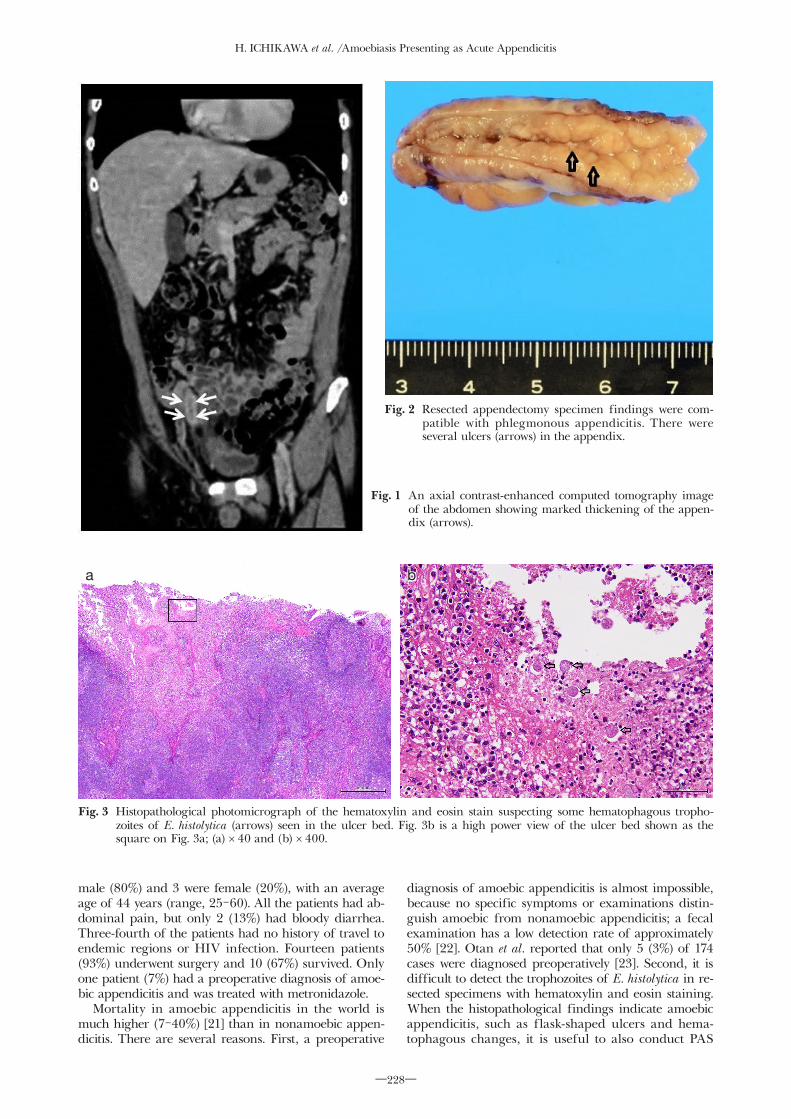
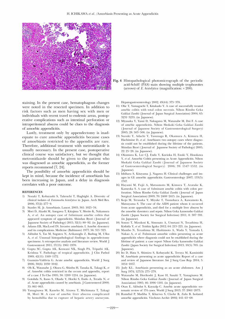
A 47-year-old man was referred to our hospital with acute right lower abdominal pain. He had no history of recent travel to endemic areas or acquired immuno-deficiency syndrome. On admission, his temperature was 36.5°C. Physical examination revealed localized tenderness in the right lower abdomen without muscle guarding or rebound tenderness. Laboratory tests revealed a white blood cell count of 12,800 cells/µL and a C-reactive protein (CRP) level of 1.421 mg/dL. Computed tomography revealed a dilated appendix with a maximum diameter of 15 mm and thickened cecal wall (Fig. 1). He was diagnosed with acute appen-dicitis and underwent surgery. His appendix showed acute inflammation from the cecum to the root of the appendix, without necrosis, perforation, or formation of localized abscess. There were several ulcers in the inflamed appendix (Fig. 2). Some hematophagous trophozoites of Entamoeba histolytica were suspected in the ulcer bed by pathological examination with hematoxylin and eosin stain (Fig. 3) and additional pathological examination with periodic acid-Schiff
stain revealed multiple trophozoites of E. histolytica (Fig. 4). Histopathological examination revealed amoe-biasis presenting as phlegmonous appendicitis. After appendectomy, he recovered uneventfully. As fecal examination was negative, he received no additional treatment with metronidazole. He was discharged on postoperative day 5.
DISCUSSION
Cases of amoebiasis are divided into intestinal amoebiasis and extraintestinal amoebiasis. Intestinal amoebiasis cases are further classified as asymptomatic cyst, acute colitis, and chronic colitis. The proportion of patients with symptoms is less than 10% after infec-tion with E. histolytica [3]. Most patients with symptoms have a clinical course similar to chronic colitis, but some present symptoms of acute colitis. Approximately 3% of acute colitis cases progress to fulminant colitis with a high mortality rate [4].
Acute appendicitis, which occurs when the appendix lumen is obstructed, is the most common cause of acute abdomen. Appendix obstruction can be caused by a coprolith, lymphoid hyperplasia, tumor, or parasitic infection. Akbulut et al. reviewed studies on the etiology of acute appendicitis published between January 2000 and November 2010 and reported that amoebiasis was detected in 118 (0.15%) of 80,698 cases [5].
Amoebic appendicitis is considered to be a rare disease, with a reported incidence of 0.5-2.3%, even in endemic regions [6, 7]. We searched for cases of amoe-bic appendicitis reported from 1996 to 2016 using Igakuchuo-Zasshi and reviewed the clinical features, treatment of choice, and outcomes of 15 patients [3, 8-20] including the present case. Twelve patients were
Hitoshi ICHIKAWA, Division of Gastroenterology and Hepatology, Department of Internal Medicine, Tokai University Hachioji Hospital, 1838 Ishikawa ma-chi, Hachioji, Tokyo 192-0032, Japan Tel: +81-42-639-1111 Fax: +81-42-639-1144 E-mail: [email protected]
H. ICHIKAWA et al. /Amoebiasis Presenting as Acute Appendicitis
―228―
male (80%) and 3 were female (20%), with an average age of 44 years (range, 25-60). All the patients had ab-dominal pain, but only 2 (13%) had bloody diarrhea. Three-fourth of the patients had no history of travel to endemic regions or HIV infection. Fourteen patients (93%) underwent surgery and 10 (67%) survived. Only one patient (7%) had a preoperative diagnosis of amoe-bic appendicitis and was treated with metronidazole.
Mortality in amoebic appendicitis in the world is much higher (7-40%) [21] than in nonamoebic appen-dicitis. There are several reasons. First, a preoperative
diagnosis of amoebic appendicitis is almost impossible, because no specific symptoms or examinations distin-guish amoebic from nonamoebic appendicitis; a fecal examination has a low detection rate of approximately 50% [22]. Otan et al. reported that only 5 (3%) of 174 cases were diagnosed preoperatively [23]. Second, it is difficult to detect the trophozoites of E. histolytica in re-sected specimens with hematoxylin and eosin staining. When the histopathological findings indicate amoebic appendicitis, such as flask-shaped ulcers and hema-tophagous changes, it is useful to also conduct PAS
Fig. 1 An axial contrast-enhanced computed tomography image of the abdomen showing marked thickening of the appen-dix (arrows).
Fig. 3 Histopathological photomicrograph of the hematoxylin and eosin stain suspecting some hematophagous tropho-zoites of E. histolytica (arrows) seen in the ulcer bed. Fig. 3b is a high power view of the ulcer bed shown as the square on Fig. 3a; (a) × 40 and (b) × 400.
Fig. 2 Resected appendectomy specimen findings were com-patible with phlegmonous appendicitis. There were several ulcers (arrows) in the appendix.
ba
H. ICHIKAWA et al. /Amoebiasis Presenting as Acute Appendicitis
―229―
staining. In the present case, hematophagous changes were noted in the resected specimen. In addition to risk factors such as men having sex with men or individuals with recent travel to endemic areas, postop-erative complications such as intestinal perforation or intraperitoneal abscess could be clues to the diagnosis of amoebic appendicitis.
Lastly, treatment only by appendectomy is inad-equate to cure amoebic appendicitis because cases of amoebiasis restricted to the appendix are rare. Therefore, additional treatment with metronidazole is usually necessary. In the present case, postoperative clinical course was satisfactory, but we thought that metronidazole should be given to the patient who was diagnosed as amoebic appendicitis, as the former reports recommend [7, 24].
The possibility of amoebic appendicitis should be kept in mind, because the incidence of amoebiasis has been increasing in Japan, and a delay in diagnosis correlates with a poor outcome.
REFERENCES1) Nozaki T, Kobayashi S, Takeuchi T, Haghighi A. Diversity of
clinical isolates of Entamoeba histolytica in Japan. Arch Med Res. 2006; 37(2): 277-9.
2) Stanley SL Jr. Amoebiasis. Lancet. 2003; 361: 1025-34.3) Mori D, Akashi M, Anegawa T, Kitajima Y, Morito K, Nakamura
A, et al. An autopsy case of fulminant amebic colitis that appeared symptom of appendicitis. Shindan Byori (Journal of Japanese Society of Pathology) 2015; 32(1): 88-92. (in Japanese).
4) Adams EB, MacLeod IN. Invasive amebiasis. I. Amebic dysentery and its complications. Medicine (Baltimore) 1977; 56: 315-323.
5) Akbulut S, Tas M, Sogutcu N, Arikanoglu Z, Basbug M, Ulku A, et al. Unusual histopathological findings in appendectomy specimens: A retrospective analysis and literature review. World J Gastroenterol. 2011; 17(15): 1961-1970.
6) Gupta SC, Gupta AK, Keswani NK, Singh PA, Tripathi AK, Krishna V. Pathology of tropical appendicitis. J Clin Pathol 1989; 42(11): 1169-1172.
7) Guzmán-Valdivia G. Acute amebic appendicitis. World J Surg 2006; 30(6): 1038-1042.
8) Oh K, Watanabe J, Okura G, Shinbu H, Tanaka K, Takagi K, et al. Amoebic colitis restricted in the cecum and appendix, report of a case. I To Cho 1995; 30: 1209-1214. (in Japanese).
9) Gotohda N, Itano S, Okada Y, Horiki S, Endo A, Terada N, et al. Acute appendicitis caused by amebiasis. J Gastroenterol 2000; 35: 861-863.
10) Yanagisawa M, Kaneko M, Aizawa T, Michimata T, Takagi H, Mori M. A case of amebic liver abscess complicated by hemobillia due to rupture of hepatic artery aneurysm.
Hepatogastroenterology 2002; 49(44): 375-378.11) Ohe T, Yamaguchi T, Kitakado Y. A case of successfully treated
amebic colitis with total colon necrosis. Nihon Rinsho Geka Gakkai Zasshi (Journal of Japan Surgical Association) 2004; 65: 3231-3235. (in Japanese).
12) Miyasaka Y, Yasui D, Nakagawa M, Watanabe M, Doi F. A case of amebic appendicitis. Nihon Shokaki Geka Gakkai Zasshi (Journal of Japanese Society of Gastroenterological Surgery) 2005; 28: 503-506. (in Japanese).
13) Suzuki Y, Adachi Y, Yasunaga R, Okamura A, Kimura H, Hachimine D, et al. Amebiases: two autopsy cases where diagno-sis could not be established during the lifetime of the patients. Shindan Byori (Journal of Japanese Society of Pathology) 2005; 22: 25-28. (in Japanese).
14) Okumura K, Lee CJ, Endo Y, Kurioka H, Enoki Y, Hosokawa Y, et al. Amoebic Colitis presenting as Acute Appendicitis. Nihon Shokaki Geka Gakkai Zasshi (Journal of Japanese Society of Gastroenterological Surgery) 2006; 39: 1547-1552. (in Japanese).
15) Ishihara S, Kitayama J, Nagawa H. Clinical challenges and im-ages in GI: amoebic appendicitis. Gastroenterology 2007; 133(5): 1747.
16) Hayami M, Fujii A, Matsumoto M, Kimura T, Aratake K, Kameoka S. A case of fulminant amebic colitis with colon per-foration. Nihon Rinsho Geka Gakkai Zasshi (Journal of Japan Surgical Association) 2009; 70: 2408-2415. (in Japanese).
17) Kojo M, Terasaka Y, Miyake T, Tsunohara A, Kawamoto K, Matsuyama S. The case of the AIDS patient whom it occurred from acute appendicitis, and died for a multiple liver abscess by the amoebic dysentery and sepsis. Nihon Geka kansensho Gakkai Zasshi (Japan Society for Surgical Infection) 2011; 8: 387-391. (in Japanese).
18) Sonoo T, Mizukosi K, Simosato A, Umetani N, Terashima H, Kishida Y, et al. Teishin Igaku 2012; 64: 213-221. (in Japanese).
19) Mambo N, Terashima M, Hashimoto A, Wada Y, Yamada I, Nakao A, et al. Fulminant amoebic colitis presenting as acute appendicitis where diagnosis could not be established during the lifetime of patient: a case report Nihon Geka kansensho Gakkai Zasshi (Japan Society for Surgical Infection) 2013; 10(5): 705. (in Japanese).
20) Ito D, Hata S, Shimizu S, Kobayashi K, Teruya M, Kaminishi M. Amebiasis presenting as acute appendicitis: Report of a case and review of Japanese literature. Int J Surg Case Rep. 2014; 5: 1054-1057.
21) Judy KL. Amebiasis presenting as an acute abdomen. Am J Surg 1974; 127(3): 275-279.
22) Watanabe M, Horikoshi J, Kase H, Sasaki T, Yanagisawa M. Nihon Rinsho Geka Gakkai Zasshi (Journal of Japan Surgical Association) 1985; 46: 1096-1101. (in Japanese).
23) Otan E, Akbulut S, Kayaalp C. Amebic acute appendicitis: sys-tematic review of 174 cases. World J Surg 2013; 37: 2061-2073.
24) Ramdial P, Madiba T, Kharwa S, Clarke B, Zulu B. Isolated amoebic appendicitis. Virchows Archiv 2002; 441: 63-68:
Fig. 4 Histopathological photomicrograph of the periodic acid-Schiff (PAS) stain showing multiple trophozoites (arrows) of E. histolytica (magnification: × 200).