Embed Size (px)
Citation preview
Amiodarone Ocular Toxicity, Emphasizing Optic Neuropathy: a Teaching Case Report | 1
| 1
Abstract
Amiodarone is one of the most effective antiarrhythmic drugs, but its use is limited by toxicity affecting many organsincluding the eyes. Corneal verticillata is the most common ocular finding associated with amiodarone therapy. A lesscommon but potentially sight-threatening complication is optic neuropathy. Amiodarone-induced optic neuropathy sharesmany clinical features with non-arteritic ischemic optic neuropathy making clinical diagnosis difficult. However, severalimportant distinctions exist including an insidious onset, bilateral involvement, male predilection and delayed resolution ofdisc edema. This case report highlights the unique features of amiodarone-associated optic neuropathy and presentsappropriate diagnostic and management strategies.
Key Words: amiodarone, ocular toxicity, optic neuropathy, optic disc edema, whorl keratopathy, corneal verticillata, non-arteritic ischemic optic neuropathy
Background
Amiodarone is the most common antiarrhythmic drug prescribed for treatment of atrial fibrillation. Its efficacy has beenchallenged by its ubiquitous organ toxicity resulting in nearly 50% of long-term users discontinuing the drug.1 Oculartoxicity most commonly presents as corneal deposits referred to as whorl keratopathy or corneal verticillata, which occurin the majority of amiodarone users.1,2 Whorl keratopathy is typically asymptomatic with reported glare and halos occurringin less than 5% of patients.1 This report is aimed toward third- and fourth-year optometric students and optometrists.
The rare occurrence of optic neuropathy can be debilitating to vision. Various studies have reported the incidence ofamiodarone optic neuropathy to be as high as 2.0%; however, the exact incidence is unknown.3,4 Most cases of amiodarone-associated optic neuropathy occur within a year of treatment initiation.1,5 Characteristics include gradual onset, bilateralinvolvement and slow resolution. Amiodarone-induced optic neuropathy remains a controversial diagnosis because itshares many clinical features with non-arteritic ischemic optic neuropathy (NAION), the most common optic nerve disordercausing sudden onset vision loss in elderly patients.4 Patients on amiodarone therapy often have the same risk factors aspatients who experience NAION, making it difficult to separate the two diagnoses.1,6 However, several distinguishingclinical features highlighted in this case suggest amiodarone-associated optic neuropathy is a distinct clinical entity.
Case Description
A 70-year-old Caucasian male complained of intermittent left upper visual field disturbances for two days. He described itas a window shade moving up and down that he initially noticed upon awakening. He also experienced soreness duringextreme eye movements.
The patient’s medical history was remarkable for hypertension, which was controlled with three medications: lisinoprilQPM, terazosin QPM and metoprolol BID. In-office blood pressure was 135 mmHg/81 mmHg. He was also takingatorvastatin daily to control hypercholesterolemia. Due to history of a myocardial infarction, he was prescribed aspirin,clopidogrel and warfarin. His medical history was also remarkable for atrial fibrillation, treated with amiodarone for thepast nine months. His dosage of amiodarone was 200 mg BID with an initial higher loading dose. Drug allergies includedsulfa drugs and tamsulosin. He had a 40-year history of heavy alcohol and tobacco use, both of which he stopped threeyears prior.
Best-corrected Snellen visual acuity was 20/20 in each eye with no change in manifest refraction. There were no grossdefects or constriction identified on confrontation visual fields or extent of fields. Extraocular motilities were full withoutpain or diplopia. Pupils were equal, round and reactive with no afferent pupillary defect (APD).
Anterior segment findings were remarkable for gray lines extending into whorl-like patterns on the inferior cornealepithelium in both eyes, which were not noted at his last eye examination six months ago. The patient also had mildnuclear sclerotic cataracts. Goldmann applanation tonometry intraocular pressures were 10 mmHg OD and 12 mmHg OS.
Both optic nerve heads appeared crowded with heaping rim tissue (Figure 1). The left optic nerve appeared to haveblurred margins inferiorly with an overlying disc hemorrhage superior-nasally. Cup-to-disc ratios were 0.05 OD and 0.1 OS,which was consistent with the previous eye examination. Other retinal findings, including macula, vasculature andperiphery, were normal in both eyes.

Amiodarone Ocular Toxicity, Emphasizing Optic Neuropathy: a Teaching Case Report | 2
| 2
Figure 1. Fundus photography taken at the patient’s first visit. (A) Right optic nerve headexhibits an indistinct inferior disc margin. (B) Left optic nerve head has a small cup-to-disc ratio,blurring of the inferior disc margin and a Drance hemorrhage (arrow). No apparent hyperemia orpallor is observed in either eye. Click to enlarge
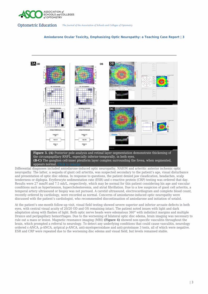
Visual field testing was ordered prior to dilation and revealed mild to moderately reduced superior-temporal defectsextending from the blind spot in the left visual field. Optical coherence tomography (OCT) (Figure 2) confirmed elevatedrim tissue in both eyes, especially inferiorly in the left eye. Posterior pole analysis showed retinal nerve fiber layer (RNFL)thickening into the arcuate bundles but not extending into the fovea. Ganglion cell layer and inner plexiform layer analysisof the fovea and perifovea revealed normal thickness values (Figure 3).
Figure 2. (A-C) A dense vertical OCT of the left optic nerve head shows elevated rim tissueinferiorly. In the infrared image, the hyporeflectivity of the edematous inferior peripapillaryRNFL can be seen. (D) OCT circumpapillary RNFL also shows thickening, especially inferiorlyOS, compared to the normative database. Click to enlarge
Amiodarone Ocular Toxicity, Emphasizing Optic Neuropathy: a Teaching Case Report | 3
| 3
Figure 3. (A) Posterior pole analysis and retinal layer segmentation demonstrate thickening ofthe circumpapillary RNFL, especially inferior-temporally, in both eyes.(B-C) The ganglion cell-inner plexiform layer complex surrounding the fovea, when segmented,appears normal. Click to enlarge
Differential diagnoses included amiodarone-induced optic neuropathy, NAION and arteritic anterior ischemic opticneuropathy. The latter, a sequela of giant cell arteritis, was suspected secondary to the patient’s age, visual disturbanceand presentation of optic disc edema. In response to questions, the patient denied jaw claudication, headaches, scalptenderness or diplopia. Erythrocyte sedimentation rate (ESR) and c-reactive protein (CRP) testing was ordered that day.Results were 27 mm/H and 7.5 mh/L, respectively, which may be normal for this patient considering his age and vascularconditions such as hypertension, hypercholesteremia, and atrial fibrillation. Due to a low suspicion of giant cell arteritis, atemporal artery ultrasound or biopsy was not pursued. A carotid ultrasound, electrocardiogram and complete blood count,recently ordered by cardiology, were recorded as normal. Concerns of amiodarone-induced optic neuropathy werediscussed with the patient’s cardiologist, who recommended discontinuation of amiodarone and initiation of sotalol.
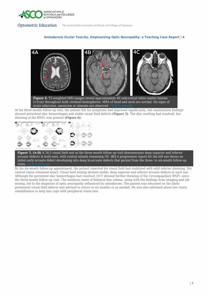
At the patient’s one-month follow-up visit, visual field testing showed severe superior and inferior arcuate defects in botheyes, with central visual acuity of 20/20 OD and OS remaining intact. The patient noted issues with light and darkadaptation along with flashes of light. Both optic nerve heads were edematous 360° with indistinct margins and multipleDrance and peripapillary hemorrhages. Due to the worsening of bilateral optic disc edema, brain imaging was necessary torule out a mass or lesion. Magnetic resonance imaging (MRI) (Figure 4) showed non-specific vasculitis throughout thebrain, which prompted a referral to neurology. To detect any underlying conditions that could cause vasculitis, neurologyordered c-ANCA, p-ANCA, aytpical p-ANCA, anti-myeloperoxidase and anti-proteinase 3 tests, all of which were negative.ESR and CRP were repeated due to the worsening disc edema and visual field, but levels remained stable.
Amiodarone Ocular Toxicity, Emphasizing Optic Neuropathy: a Teaching Case Report | 4
| 4
Figure 4. T2-weighted MRI images reveal approximately 40 subcortical white matter lesions(<1cm) throughout both cerebral hemispheres. MRA of head and neck are normal. No signs ofacute infarction, aneurysm or stenosis are observed. Click to enlarge
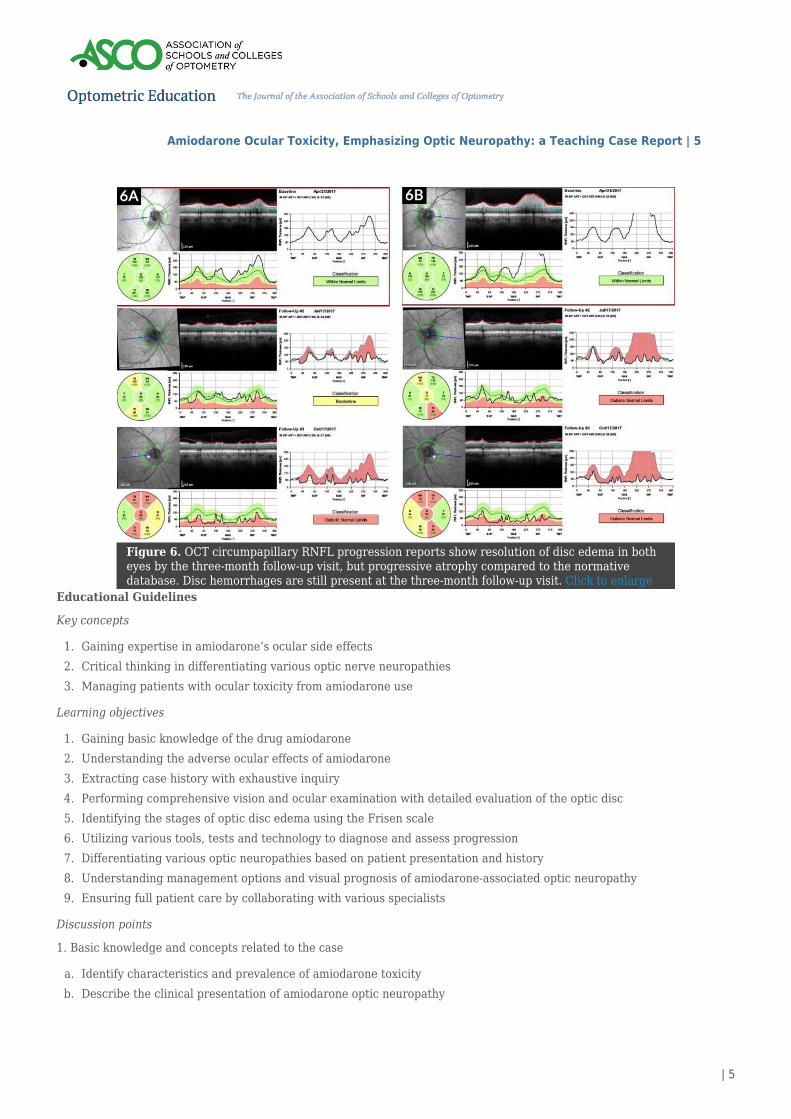
At his three-month follow-up visit, the patient felt his symptoms had improved significantly, but examination findingsshowed persistent disc hemorrhages and stable visual field defects (Figure 5). The disc swelling had resolved, butthinning of the RNFL was present (Figure 6).
Figure 5. (A-B) A 24-2 visual field test at the three-month follow-up visit demonstrates deep superior and inferiorarcuate defects in both eyes, with central islands remaining OU. (C) A progression report for the left eye shows aninitial early arcuate defect developing into deep bi-arcuate defects that persist from the three- to six-month follow-upvisits. Click to enlarge
By his six-month follow-up appointment, the patient reported his visual field had stabilized with mild inferior dimming. Hiscentral vision remained intact. Visual field testing showed stable, deep superior and inferior arcuate defects in each eye.Although the persistent disc hemorrhages had resolved, OCT showed further thinning of the circumpapillary RNFL sincethe three-month follow-up visit. The insidious onset of bilateral disc edema, along with the findings from imaging and labtesting, led to the diagnosis of optic neuropathy influenced by amiodarone. The patient was educated on the likelypermanent visual field defects and advised to return in six months or as needed. He was also informed about low visionrehabilitation to help him cope with peripheral vision loss.
Amiodarone Ocular Toxicity, Emphasizing Optic Neuropathy: a Teaching Case Report | 5
| 5
Figure 6. OCT circumpapillary RNFL progression reports show resolution of disc edema in botheyes by the three-month follow-up visit, but progressive atrophy compared to the normativedatabase. Disc hemorrhages are still present at the three-month follow-up visit. Click to enlarge
Educational Guidelines
Key concepts
Gaining expertise in amiodarone’s ocular side effects1.Critical thinking in differentiating various optic nerve neuropathies2.Managing patients with ocular toxicity from amiodarone use3.
Learning objectives
Gaining basic knowledge of the drug amiodarone1.Understanding the adverse ocular effects of amiodarone2.Extracting case history with exhaustive inquiry3.Performing comprehensive vision and ocular examination with detailed evaluation of the optic disc4.Identifying the stages of optic disc edema using the Frisen scale5.Utilizing various tools, tests and technology to diagnose and assess progression6.Differentiating various optic neuropathies based on patient presentation and history7.Understanding management options and visual prognosis of amiodarone-associated optic neuropathy8.Ensuring full patient care by collaborating with various specialists9.
Discussion points
1. Basic knowledge and concepts related to the case
Identify characteristics and prevalence of amiodarone toxicitya.Describe the clinical presentation of amiodarone optic neuropathyb.
Amiodarone Ocular Toxicity, Emphasizing Optic Neuropathy: a Teaching Case Report | 6
| 6
Identify clinical features of amiodarone optic neuropathy that distinguish it from ischemic optic neuropathyc.Describe diagnostic tools and appropriate ancillary testing that aid in diagnosisd.
2. Differential diagnosis
What are differential diagnoses based on the patient’s symptoms?a.What are differential diagnoses for unilateral vs. bilateral optic disc edema?b.What is the appropriate work-up to exclude differential diagnoses?c.
3. Critical thinking
Should amiodarone be discontinued when neuropathy is identified? How do you make that decision as a healthcarea.provider?What is the appropriate follow-up for patients currently taking amiodarone with no signs of neuropathy?b.What is the visual prognosis of amiodarone toxicity?c.Were brain imaging and blood work necessary in this patient’s case?d.Is amiodarone-induced optic neuropathy a diagnosis of exclusion?e.What are the differences between non-arteritic ischemic optic neuropathy and amiodarone-induced optic neuropathy?f.
Literature Review
Amiodarone
Amiodarone is approved by the Food and Drug Administration (FDA) for the treatment of refractory ventriculararrhythmias,7,8 but it is often prescribed for atrial arrhythmias such as atrial fibrillation or flutter. An initial higher dose(800 to 1600 mg daily) is needed to achieve therapeutic plasma levels because, with a bioavailability of approximately 30%,the drug is poorly absorbed. Maintenance dose (100 to 400 mg daily) varies depending on multiple factors, including heartrate.9 Amiodarone’s primary mechanism of action is to block potassium channels, but it also weakly blocks sodium andcalcium channels as well as beta and alpha adrenergic receptors. Direct effects on the myocardium cause a delayedrepolarization and an increased duration of action potential.10 The half-life of amiodarone is long yet variable. It has beenreported in the literature ranging from 13 days to 180 days.1,4,5,8–10 Elimination occurs mostly by hepatic metabolism.9
Geriatric patients have a slower drug clearance and may be more sensitive to adverse effects.10
Long-term oral therapy can lead to accumulation in tissue and adverse effects including pulmonary disease, thyroiddysfunction, cardiac toxicity, skin reactions, gastrointestinal and genitourinary problems, neurological dysfunction, oculartoxicity and drug interactions.7–9
Clinical manifestations
Whorl keratopathy, also known as corneal verticillata, is the most common ophthalmologic finding in amiodarone patients,with a prevalence of 70-100% with long-term therapy.9,11 These corneal microdeposits appear in a brown or gray swirl-likeor whisker-like pattern at the junction of the middle and lower thirds of the corneal epithelium. Lacrimal gland secretion ofamiodarone results in accumulation of these microdeposits on the corneal surface.11 The keratopathy can be classified intothree stages9 (Table 1), although stepwise classification serves minimal practical value.11 Clinically, this keratopathy isindistinguishable from that caused by chloroquine derivatives or Fabry disease.9 The microdeposits do not typically reducevisual acuity, but some patients may experience halos around lights, especially at night, and photophobia.9,11 Thesesymptoms can also be attributable to age-related lens changes and other sequelae.9 These ocular adverse effects aretypically dose- and duration-dependent, as shown by a meta-analysis of 1,465 patients that found an incidence of 1.5% withlow-dose intake compared to 0.1% incidence with placebo.7 The keratopathy disappears within 3-20 months ofdiscontinuation of the medication.9,11 Cataracts from amiodarone use have also been noted. These punctate, yellowish-whitelens opacities are located in the anterior subcapsule within the pupillary margin in a vertical pattern of about 2 mm indiameter.9 A study performed by Ingram et al. of more than 100 patients found lid irritation to be the most common ocularsymptom associated with amiodarone. This is most likely from photosensitivity of the eyelid skin,11 which occurs in 10-15%of patients.10 There are rare reports of dry eyes, macular degeneration,10 multiple chalazia9 and thyroid eye disease.12
Amiodarone Ocular Toxicity, Emphasizing Optic Neuropathy: a Teaching Case Report | 7
| 7
Table 1. Click to enlarge
Amiodarone has also been associated with visual disturbancessecondary to optic neuropathy. The exact annual incidence ofamiodarone-associated optic neuropathy is unknown. Variousreports estimate an incidence of 0.36% to 2.0% in amiodaroneusers, but specific parameters are often not specified.3,4 A fewreports detected a male predilection for amiodarone-associatedoptic neuropathy.1,5,6 Gender differences in body mass, fatdistribution enzymatic activity and hormonal stimuli1 may accountfor a slightly faster drug clearance in females.5
A few key features can aid in diagnosing amiodarone-associatedoptic neuropathy as well as differentiating it from otherpathologies. The optic neuropathy associated with amiodarone isdescribed as having an insidious onset, slow progression andprotracted disc swelling.4 Most cases that present with theseclinical features are simultaneously bilateral, but can beasymmetric.4,6 A small percentage of cases that were reported topresent unilaterally were either eventually bilateral,6 notattributable to amiodarone,13 or symptomatically unilateral butbilaterally edematous.1 Visual acuity at presentation rangessignificantly from 20/15 to light perception.1,4 Visual field can benormal, but most patients present with altitudinal defects, arcuatedefects or a generalized depression.13 A case series review byJohnson et al. found that only 9% of cases of amiodarone opticneuropathy had normal visual fields at presentation, and almosthalf of patients presented with either altitudinal or arcuatedefects. Visual field loss is also typically permanent.6
A review of eighty cases of amiodarone-induced optic neuropathy by Passman et al. found that the average intervalbetween starting amiodarone and onset of visual disturbances was approximately nine months, with a median of six monthsand a range of 1-84 months. After discontinuing the medication, 58% of 61 patients experienced improvement in visualacuity, 21% had unchanged visual acuity, and 21% had worsening of visual acuity. Permanent <20/200 visual acuity in atleast one eye resulted in 20% of cases.1 Even with discontinuation of amiodarone intake, optic atrophy can develop afterresolution of disc swelling.9
Cheng et al. performed a retrospective population-based cohort study involving 6,175 amiodarone-treated patients and24,700 age- and gender-matched controls to determine whether amiodarone use was associated with an increased risk ofoptic neuropathy. Optic neuropathy developed in 17 amiodarone-treated patients (0.3%) and 30 control patients (0.1%).Analysis of this data, with adjustments for age, gender and comorbidities, showed a two-fold increased risk of opticneuropathy. Longer exposure to amiodarone also increased the risk of optic neuropathy, but average daily dose did notdemonstrate a correlation.5 However, this study did not evaluate the features of optic neuropathy (i.e., bilateral orunilateral, type of vision loss, duration of disc edema). Therefore, appropriate classification and cause is difficult toproperly assess. Another limitation of the study is that the patients treated with amiodarone had a higher proportion ofmedical comorbidities than the controls. A correspondence by Mindel et al. reports that these incidence rates may bebiased estimates due to these limitations.14
PathophysiologyThe pathophysiology of amiodarone neuropathy is not completely understood. It is believed that the drug’s lipophilicnature, high volume of distribution, and high tissue affinity result in ultrastructural changes in the optic nerve head.5,6 In-vitro studies indicate that low-dose amiodarone causes inhibition of lysosomal sphingomyelinase activity and high-doseamiodarone leads to intracellular lipid accumulation and probable phospholipidosis.5,9 Histopathology after amiodarone useshows lysosome-like intracytoplasmic membranous lamellar bodies.1,15 These lamellar inclusion bodies have been found innearly all ocular tissues, including retrobulbar optic nerve large-diameter axons, extraocular muscle fibers, cornea,conjunctiva, scleral, lens, iris, ciliary body, choroid, retinal pigment epithelium, ganglion cells and endothelium of ocularblood vessels.9 It is theorized that these inclusions mechanically or biochemically inhibit axoplasmic flow, resulting in opticdisc edema. The resulting optic nerve head edema can persist as long as axonal transport is inhibited, which results indelayed resolution of optic nerve swelling.1,5,9,15 These bodies are also seen in peripheral nerves that have been affected by
Amiodarone Ocular Toxicity, Emphasizing Optic Neuropathy: a Teaching Case Report | 8
| 8
amiodarone-associated peripheral neuropathy. The peripheral nerves have shown signs of demyelination and large axonloss, which is absent in the retrobulbar optic nerve axons, possibly due to different types of myelination or otheretiology.1,16 Amiodarone can also cause vasodilation, possibly leading to oxidative damage and optic neuropathy.5
Differential diagnosis
Table 2. Click to enlarge
The most obvious differential diagnosis for amiodarone-inducedoptic neuropathy is NAION. NAION is the most common cause ofvision loss from optic nerve disease in individuals older than 50years with an incidence of 0.3%.5 The incidence of NAION inpatients with cardiac dysrhythmia is higher than this becausemany patients taking amiodarone often have severe vasculardisease.5,17 Amiodarone-associated optic neuropathy isindistinguishable from NAION with regard to optic discappearance, but the two clinical entities differ in multiple aspects(Table 2). NAION initially presents unilaterally, whileamiodarone-associated optic neuropathy often presents bilaterallysimultaneously as expected with systemic toxicity.3,4,14 Vision loss issudden and complete at onset of NAION, unlike the insidious onsetwith protracted disc swelling in amiodarone-associated opticneuropathy. NAION also typically resolves in a few weeks18
compared to months. NAION generally occurs in patients with asmall optic nerve cup-to-disc ratio or crowded disc, but this hasnot been an identifiable risk factor in cases of amiodarone-associated optic neuropathy. Amiodarone-associated opticneuropathy has a predilection toward men, whereas NAION has nogender predilection.1,5,6
Along with NAION, there are other conditions that are important to consider given the clinical presentation of optic discedema. Although amiodarone-associated optic neuropathy typically presents bilaterally, it may present asymmetrically,which warrants thorough investigation to rule out other potential causes of unilateral disc edema. Differential diagnosesinclude neoplastic, inflammatory, infectious, metabolic, demyelinating, hereditary and vascular etiologies. Neoplasticcauses such as optic nerve glioma or optic nerve sheath meningioma can be ruled out based on neuroimaging.Inflammatory etiologies such as sarcoidosis or systemic lupus erythematosus will likely present with other signs ofintraocular or systemic inflammation. Infectious causes such as syphilis, Lyme disease, or cat-scratch disease are bestidentified through bloodwork.19 The most common demyelinating cause is optic neuritis secondary to multiple sclerosis.Optic neuritis can result in optic disc edema; however, onset is usually unilateral, acute and painful and typically occurs inyounger patients.20
Differential diagnoses for bilateral optic disc edema include an intracranial mass, malignant hypertension, systemicmedications, toxic/nutritional neuropathy, meningitis and pseudotumor cerebri. A space-occupying lesion must always beconsidered in cases of bilateral disc swelling;19 however, no lesions were identified in this case based on neuroimaging. Thepatient in this case report presented with a blood pressure of 135 mmHg/81 mmHg, which ruled out malignanthypertension. Aside from amiodarone, other systemic drugs have been found to be associated with optic disc edema,including tuberculostatic drugs, antimicrobial agents, antiepileptic drugs, disulfiram, halogenated hydroquinolones,antimetabolites, tamoxifen and phosphodiesterase type 5 inhibitors. Toxins, such as metals, organic solvents, methanol andcarbon dioxide, as well as nutritional deficits, such as vitamin B, folic acid and proteins with sulfur-containing amino acidscan also cause optic neuropathy.21
Pseudotumor cerebri, or idiopathic intracranial hypertension (IIH), presents with bilateral disc swelling. In contrast toamiodarone optic neuropathy, IIH often presents with headaches, nausea or transient vision loss, and patients are typicallyyoung, overweight females of childbearing age. IIH is a diagnosis of exclusion and must be ruled out with neuroimagingincluding MRI and magnetic resonance venography. Diagnosis is confirmed with lumbar puncture showing elevatedopening pressure. Finally, to avoid unnecessary neuroimaging, causes of pseudopapilledema should not be overlooked.Optic disc drusen can present with unilateral or bilateral disc edema; however, appropriate ancillary testing such as OCT,fundus autofluorescence or B-Scan ultrasonography can help differentiate pseudo from true optic disc edema.19
Diagnostic tools
Amiodarone Ocular Toxicity, Emphasizing Optic Neuropathy: a Teaching Case Report | 9
| 9
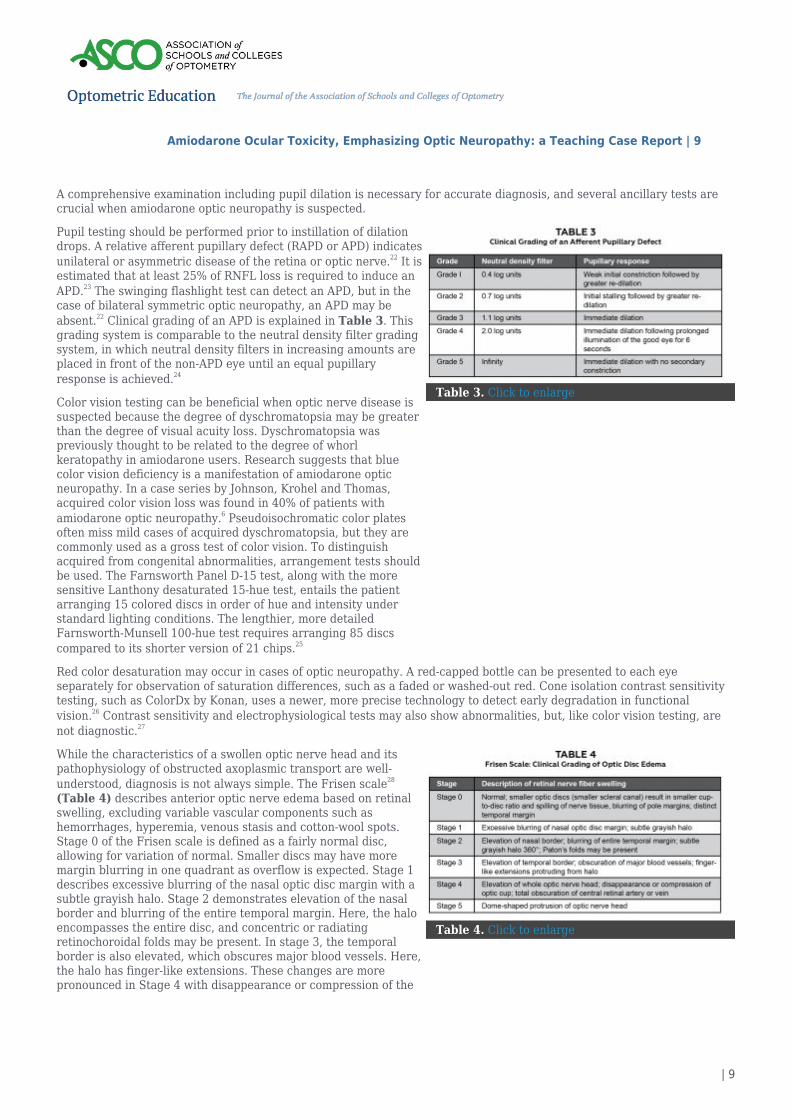
A comprehensive examination including pupil dilation is necessary for accurate diagnosis, and several ancillary tests arecrucial when amiodarone optic neuropathy is suspected.
Table 3. Click to enlarge
Pupil testing should be performed prior to instillation of dilationdrops. A relative afferent pupillary defect (RAPD or APD) indicatesunilateral or asymmetric disease of the retina or optic nerve.22 It isestimated that at least 25% of RNFL loss is required to induce anAPD.23 The swinging flashlight test can detect an APD, but in thecase of bilateral symmetric optic neuropathy, an APD may beabsent.22 Clinical grading of an APD is explained in Table 3. Thisgrading system is comparable to the neutral density filter gradingsystem, in which neutral density filters in increasing amounts areplaced in front of the non-APD eye until an equal pupillaryresponse is achieved.24
Color vision testing can be beneficial when optic nerve disease issuspected because the degree of dyschromatopsia may be greaterthan the degree of visual acuity loss. Dyschromatopsia waspreviously thought to be related to the degree of whorlkeratopathy in amiodarone users. Research suggests that bluecolor vision deficiency is a manifestation of amiodarone opticneuropathy. In a case series by Johnson, Krohel and Thomas,acquired color vision loss was found in 40% of patients withamiodarone optic neuropathy.6 Pseudoisochromatic color platesoften miss mild cases of acquired dyschromatopsia, but they arecommonly used as a gross test of color vision. To distinguishacquired from congenital abnormalities, arrangement tests shouldbe used. The Farnsworth Panel D-15 test, along with the moresensitive Lanthony desaturated 15-hue test, entails the patientarranging 15 colored discs in order of hue and intensity understandard lighting conditions. The lengthier, more detailedFarnsworth-Munsell 100-hue test requires arranging 85 discscompared to its shorter version of 21 chips.25
Red color desaturation may occur in cases of optic neuropathy. A red-capped bottle can be presented to each eyeseparately for observation of saturation differences, such as a faded or washed-out red. Cone isolation contrast sensitivitytesting, such as ColorDx by Konan, uses a newer, more precise technology to detect early degradation in functionalvision.26 Contrast sensitivity and electrophysiological tests may also show abnormalities, but, like color vision testing, arenot diagnostic.27
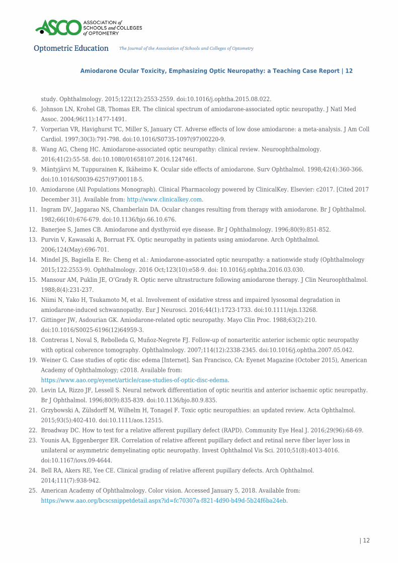
Table 4. Click to enlarge
While the characteristics of a swollen optic nerve head and itspathophysiology of obstructed axoplasmic transport are well-understood, diagnosis is not always simple. The Frisen scale28
(Table 4) describes anterior optic nerve edema based on retinalswelling, excluding variable vascular components such ashemorrhages, hyperemia, venous stasis and cotton-wool spots.Stage 0 of the Frisen scale is defined as a fairly normal disc,allowing for variation of normal. Smaller discs may have moremargin blurring in one quadrant as overflow is expected. Stage 1describes excessive blurring of the nasal optic disc margin with asubtle grayish halo. Stage 2 demonstrates elevation of the nasalborder and blurring of the entire temporal margin. Here, the haloencompasses the entire disc, and concentric or radiatingretinochoroidal folds may be present. In stage 3, the temporalborder is also elevated, which obscures major blood vessels. Here,the halo has finger-like extensions. These changes are morepronounced in Stage 4 with disappearance or compression of the
Amiodarone Ocular Toxicity, Emphasizing Optic Neuropathy: a Teaching Case Report | 10
| 10
optic cup or total obscuration of a central retinal artery or vein.Stage 5 is defined as a dome-shaped protrusion of the optic nervehead.28
Visual field testing is an important tool in both the diagnosis and management of amiodarone optic neuropathy. Manypatients present with visual field disturbances that gradually worsen over time and become permanent. Visual field defectsoften present as altitudinal or arcuate defects6 or a general depression.13 Thus, visual field testing is a crucial diagnosticand management tool for predicting visual prognosis and facilitating appropriate patient education.
The number of published studies in which OCT was used to monitor the behavior of disc edema in patients withamiodarone-associated optic neuropathy is limited. As stated earlier, the disc swelling is typically bilateral, insidious andprolonged compared to the swelling in NAION.4 A study by Akbari et al. evaluated peripapillary RNFL thickness andmacular thickness, specifically ganglion cell-inner plexiform layer (GCIPL) thickness, in NAION patients. At initialpresentation, peripapillary RNFL and outer macula thickness were elevated. This edema began to decrease by the one-month follow-up, but the GCIPL of the macula began to show thinning until about six months. This is likely due to neuronalloss,29 with areas of damage commonly correlating with visual field loss.30 This may be similar in amiodarone-associatedoptic neuropathy, although swelling may be present for a longer duration. OCT can be used to observe retinal edema,followed by axonal thinning and atrophy.31
Less threatening conditions should also be considered, especially in the absence of disc hemorrhages. Optic disc drusensometimes display a bumpy appearance on funduscopy, exhibit hyper-autofluorescence on fundus autofluorescence, andreveal a hyper-reflective border with posterior shadowing on cross-sectional OCT. A narrow scleral channel or hyperopicdisc can have a crowded appearance that is not considered edematous.32
Some, but not many, studies utilizing optical coherence tomography angiography (OCTA) in patients with amiodarone-associated optic neuropathy have been published. Evaluations of optic disc blood flow in NAION eyes portray significantnon-perfusion, which correlates with the degree of mean deviation of visual field loss. Fluorescein angiography may exhibitsimilar results but is a more invasive technique.30
Optic disc edema with unknown etiology may warrant brain imaging and additional laboratory or serological testing.Bilateral presentations may be secondary to increased intracranial pressure, malignant hypertension, brain mass or lesion,infection, inflammation or toxicity. Unilateral optic disc edema can result from optic neuritis, ischemia or compression, butthese can also occur bilaterally.33
Management
Patients using amiodarone who report visual disturbances should be promptly examined, and additional testing should beordered as indicated above. Amiodarone-associated optic neuropathy is a diagnosis of exclusion. Imaging to rule out acerebral mass or other etiologies is needed, especially in the case of visual disturbances and absence of disc edema.6,27
Patients taking amiodarone have serious cardiovascular illnesses, making it difficult to associate neuropathies withamiodarone rather than a manifestation of systemic disease. If optic neuropathy due to amiodarone usage is highlysuspected, consultation with the patient’s cardiologist is necessary to discuss discontinuation and substitution with anotherantiarrhythmic drug. Abrupt cessation of amiodarone without a therapeutic alternative can lead to a potentially fatalarrhythmia.7,8,10,13 Once diagnosed, patients should also be aware of the visual prognosis and lengthy timeline of resolution.
Many case reports highlight similar presentations, in which bilateral optic neuropathy occurred after a patient was placedon amiodarone and optic nerve swelling persisted for a few months. In such cases, after discussion with cardiology,amiodarone dosage was decreased or stopped, with resulting resolution of optic nerve swelling and hemorrhages.9,13,34
The presence of whorl keratopathy is not an indication to discontinue amiodarone because visual symptoms are rare andnot sight-threatening.9 Corneal refractive laser surgery is contraindicated in patients using amiodarone as it may affectlaser accuracy and postoperative healing.10,35
Patients with amiodarone-associated optic neuropathy may present to eyecare professionals for irritation of the eyelids,which can be caused by a variety of mechanisms. Consideration of skin photosensitivity should be on the list of differentialsin patients taking amiodarone.11 Ultraviolet radiation protection, cool compresses, and soothing creams may alleviate thisdrug-induced photosensitivity similar to a sunburn. A corticosteroid cream may be needed for significant inflammation, andan antibacterial cream may be necessary to prevent skin infection if the skin blisters and breaks.10,36
There has not been consensus on a follow-up protocol for patients taking amiodarone. Amiodarone manufacturers
Amiodarone Ocular Toxicity, Emphasizing Optic Neuropathy: a Teaching Case Report | 11
| 11
recommend routine screenings, but do not specify time intervals. The Heart Rhythm Society suggests a baseline evaluationfor patients with pre-existing visual impairment.1 Many physicians recommend an annual comprehensive examination, withan expedited appointment with onset of any visual symptoms.4,5,10 Johnson et al. recommend a few evaluations within thefirst year of starting amiodarone as ocular toxicity occurs at an average time point of nine months. Because onset isinsidious, periodic evaluation is needed rather than at longer intervals.6 Beneficial testing at baseline and follow-up mayinclude OCT of the optic nerve head, visual field testing and color vision testing. If ocular toxicity is suspected, physiciansshould report examination findings, drug dose, drug duration, concomitant drugs and follow-up findings to the FDA’sMedWatch program to increase publicity of a potentially serious drug reaction.1
Discussion
Due to a $22.8 million judgment against Wyeth-Ayerst Pharmaceuticals in 1997,2 the FDA added optic neuropathy as acaution in the amiodarone package insert, but not in a black box warning as a serious or life-threatening risk.1,2,37 Otherdrugs, including ethambutol, have long been accepted as a cause of optic neuropathy with an incidence of <1%. Similar toamiodarone, there are no evidence-based studies to demonstrate true causation.1
Many question the existence of amiodarone-associated optic neuropathy.1,3,5,17,38,39 A trial by Mindel et al. that involved 1,600patients and compared amiodarone to placebo did not find any cases of bilateral vision loss.3 However, results were basedsolely on patients who answered “yes” to the query of optic neuritis. A comprehensive eye examination was not performedon these patients, and analysis included only patients who had bilateral vision loss, not mildly reduced visual acuity orvisual field defects.1
Direct causation between amiodarone usage and optic neuropathy remains unproven, and many consider the clinicalfindings to be a variant of NAION.5,38,39 Patients share similar features and systemic risk factors, such as hypertension,diabetes mellitus and older age, which complicates the ability to distinguish the two conditions.1,6 Challenges exist toestablishing the relationship between amiodarone and optic neuropathy. Additional research is needed as numeroushistorical reports have not included pertinent information such as funduscopic features, visual field findings, time courseand clinical outcomes. This task is challenging because using a placebo medication on a patient with a life-threateningcondition is not ethical. A study comparing incidence of optic neuropathy in patients taking amiodarone vs. anotherantiarrhythmic medication may be warranted. Sotalal’s efficacy is comparable, but amiodarone has the least recurrence ofatrial fibrillation.10
Conclusion
Amiodarone, although an extremely effective antiarrhythmic drug, can have numerous adverse systemic and ocular effects,some of which are rare but serious. Controversy persists as to whether prolonged, bilateral optic neuropathy occurssecondary to amiodarone use or the plethora of vascular issues exacerbated by amiodarone. Presentation of vision or visualfield loss varies significantly and generally has a slow onset, progression and resolution. To distinguish amiodarone-induced optic neuropathy from NAION, differences in laterality and duration of disc edema should be considered. Earlyrecognition is important as the long half-life of amiodarone can lead to severe damage even after cessation of the drug.Active inquiry regarding visual manifestations, thorough clinical evaluation and deliberate ancillary testing can prompt anearly diagnosis and facilitate appropriate management. In collaboration with cardiology, attempts should be made todiscontinue amiodarone if amiodarone-associated optic neuropathy is highly suspected.
References
Passman RS, Bennett CL, Purpura JM, et al. Amiodarone-associated optic neuropathy: a critical review. Am J Med.1.2012;125(5):447-453. doi:10.1016/j.amjmed.2011.09.020.Chen D, Hedges TR. Amiodarone optic neuropathy – review. Semin Ophthalmol. 2003;18(4):169-173.2.doi:10.1080/08820530390895163.Mindel JS, Anderson J, Johnson G, et al. Absence of bilateral vision loss from amiodarone: a randomized trial.3.2008;153(5):837-842. doi:10.1016/j.ahj.2007.02.010.MacAluso DC, Shults WT, Fraunfelder FT. Features of amiodarone-induced optic neuropathy. Am J Ophthalmol.4.1999;127(5):610-612. doi:10.1016/S0002-9394(99)00016-1.Cheng HC, Yeh HJ, Huang N, Chou YJ, Yen MY, Wang AG. Amiodarone-associated optic neuropathy: a nationwide5.
Amiodarone Ocular Toxicity, Emphasizing Optic Neuropathy: a Teaching Case Report | 12
| 12
study. Ophthalmology. 2015;122(12):2553-2559. doi:10.1016/j.ophtha.2015.08.022.Johnson LN, Krohel GB, Thomas ER. The clinical spectrum of amiodarone-associated optic neuropathy. J Natl Med6.Assoc. 2004;96(11):1477-1491.Vorperian VR, Havighurst TC, Miller S, January CT. Adverse effects of low dose amiodarone: a meta-analysis. J Am Coll7.Cardiol. 1997;30(3):791-798. doi:10.1016/S0735-1097(97)00220-9.Wang AG, Cheng HC. Amiodarone-associated optic neuropathy: clinical review. Neuroophthalmology.8.2016;41(2):55-58. doi:10.1080/01658107.2016.1247461.Mäntyjärvi M, Tuppurainen K, Ikäheimo K. Ocular side effects of amiodarone. Surv Ophthalmol. 1998;42(4):360-366.9.doi:10.1016/S0039-6257(97)00118-5.Amiodarone (All Populations Monograph). Clinical Pharmacology powered by ClinicalKey. Elsevier: c2017. [Cited 201710.December 31]. Available from: http://www.clinicalkey.com.Ingram DV, Jaggarao NS, Chamberlain DA. Ocular changes resulting from therapy with amiodarone. Br J Ophthalmol.11.1982;66(10):676-679. doi:10.1136/bjo.66.10.676.Banerjee S, James CB. Amiodarone and dysthyroid eye disease. Br J Ophthalmology. 1996;80(9):851-852.12.Purvin V, Kawasaki A, Borruat FX. Optic neuropathy in patients using amiodarone. Arch Ophthalmol.13.2006;124(May):696-701.Mindel JS, Bagiella E. Re: Cheng et al.: Amiodarone-associated optic neuropathy: a nationwide study (Ophthalmology14.2015;122:2553-9). Ophthalmology. 2016 Oct;123(10):e58-9. doi: 10.1016/j.ophtha.2016.03.030.Mansour AM, Puklin JE, O’Grady R. Optic nerve ultrastructure following amiodarone therapy. J Clin Neuroophthalmol.15.1988;8(4):231-237.Niimi N, Yako H, Tsukamoto M, et al. Involvement of oxidative stress and impaired lysosomal degradation in16.amiodarone-induced schwannopathy. Eur J Neurosci. 2016;44(1):1723-1733. doi:10.1111/ejn.13268.Gittinger JW, Asdourian GK. Amiodarone-related optic neuropathy. Mayo Clin Proc. 1988;63(2):210.17.doi:10.1016/S0025-6196(12)64959-3.Contreras I, Noval S, Rebolleda G, Muñoz-Negrete FJ. Follow-up of nonarteritic anterior ischemic optic neuropathy18.with optical coherence tomography. Ophthalmology. 2007;114(12):2338-2345. doi:10.1016/j.ophtha.2007.05.042.Weiner G. Case studies of optic disc edema [Internet]. San Francisco, CA: Eyenet Magazine (October 2015), American19.Academy of Ophthalmology; c2018. Available from:https://www.aao.org/eyenet/article/case-studies-of-optic-disc-edema.Levin LA, Rizzo JF, Lessell S. Neural network differentiation of optic neuritis and anterior ischaemic optic neuropathy.20.Br J Ophthalmol. 1996;80(9):835-839. doi:10.1136/bjo.80.9.835.Grzybowski A, Zülsdorff M, Wilhelm H, Tonagel F. Toxic optic neuropathies: an updated review. Acta Ophthalmol.21.2015;93(5):402-410. doi:10.1111/aos.12515.Broadway DC. How to test for a relative afferent pupillary defect (RAPD). Community Eye Heal J. 2016;29(96):68-69.22.Younis AA, Eggenberger ER. Correlation of relative afferent pupillary defect and retinal nerve fiber layer loss in23.unilateral or asymmetric demyelinating optic neuropathy. Invest Ophthalmol Vis Sci. 2010;51(8):4013-4016.doi:10.1167/iovs.09-4644.Bell RA, Akers RE, Yee CE. Clinical grading of relative afferent pupillary defects. Arch Ophthalmol.24.2014;111(7):938-942.American Academy of Ophthalmology. Color vision. Accessed January 5, 2018. Available from:25.https://www.aao.org/bcscsnippetdetail.aspx?id=fc70307a-f821-4d90-b49d-5b24f6ba24eb.
Amiodarone Ocular Toxicity, Emphasizing Optic Neuropathy: a Teaching Case Report | 13
| 13
Konan Medical. ColorDx CCT-HD Cone-isolation contrast sensitivity. Accessed January 5, 2018. Available from:26.https://konanmedical.com/colordx/.Behbehani R. Clinical approach to optic neuropathies. Clin Ophthalmol. 2007;1(3):233-246.27.doi:10.1097/NRL.0b013e3181be6fad.Frisen L. Swelling of the optic nerve head: a staging scheme. J Neurol Neurosurg Psychiatry. 1982;45(1):13-18.28.doi:10.1136/jnnp.45.1.13.Akbari M, Abdi P, Fard MA, et al. Retinal ganglion cell loss precedes retinal nerve fiber thinning in nonarteritic29.anterior ischemic optic neuropathy. J Neuro-Ophthalmology. 2016;36(2):141-146.doi:10.1097/WNO.0000000000000345.Ling JW, Yin X, Lu QY, Chen YY, Lu PR. Optical coherence tomography angiography of optic disc perfusion in non-30.arteritic anterior ischemic optic neuropathy. Int J Ophthalmol. 2017;10(9):1402-1406. doi:10.18240/ijo.2017.09.12.Grzybowski, Andrzej, Barboni, Piero (Eds.). OCT in Central Nervous System Diseases. 1st ed. Springer International31.Publishing; 2016. doi:10.1007/978-3-319-24085-5.Feldman BH, Harak K. Optical coherence tomography in neuro-ophthalmology. American Academy of Ophthalmology.32.Published 2015. Accessed January 5, 2018. Available from:http://eyewiki.org/Optical_Coherence_Tomography_in_Neuro-ophthalmology.Sadaka A, Lee AG, Berry S, Smith S V. Bilateral optic disc edema. American Academy of Ophthalmology. Published33.2015. Accessed January 5, 2018. Available from: http://eyewiki.org/Bilateral_Optic_Disc_Edema.Gittinger JW, Asdourian GK. Papillopathy caused by amiodarone. Arch Ophthalmol. 1987;105(3):349-351.34.doi:10.1001/archopht.1987.01060030069028.Facts You Need to Know About CustomVue™ Laser Assisted In-Situ Keratomileusis (LASIK) Laser Treatment.35.Accessed January 5, 2018. Available from: https://www.accessdata.fda.gov/cdrh_docs/pdf/P930016S017d.pdf.Moore DE. Drug-induced cutaneous photosensitivity: incidence, mechanism, prevention and management. Drug36.Safety. 2002;25(5):345-372. doi:10.2165/00002018-200225050-00004.Label: Amiodarone HCL – amiodarone hydrochloride tablet. National Institute of Health: National Library of Medicine.37.Available from: https://dailymed.nlm.nih.gov/dailymed/drugInfo.cfm?setid=08641e51-abca-4c1c-ba19-d24435332018.Hayreh SS. Amiodarone, erectile dysfunction drugs , and non-arteritic ischemic optic neuropathy. J Neuro-Ophthalmol.38.2006;26(2):154-155.Younge BR. Amiodarone and ischemic optic neuropathy. J Neuro-Ophthalmology. 2007;27(1):85-86.39.doi:10.1097/WNO.0b013e3180334a06.
Dr. Ragha [[email protected]] is an Instructor at Southern College of Optometry in Memphis, Tenn. She completed herresidency at Memphis Veterans Affairs Medical Center.
Dr. Williams is a primary care resident at Memphis Veterans Affairs Medical Center. She graduated from Southern Collegeof Optometry in Memphis, Tenn.