Embed Size (px)
Citation preview

Alveolar bone loss associated to periodontaldisease in lead intoxicated rats underenvironmental hypoxia
Antonela R. Terrizzi a, Javier Fernandez-Solari a,c, Ching M. Lee a,Clarisa Bozzini a, Patricia M. Mandalunis b, Juan C. Elverdin a,Marıa Ines Conti a, Marıa Pilar Martınez a,*aDepartment of Physiology, Faculty of Dentistry, University of Buenos Aires, ArgentinabDepartment of Histology, Faculty of Dentistry, University of Buenos Aires, ArgentinacNational Council for Scientific and Technical Research (CONICET), Argentina
a r c h i v e s o f o r a l b i o l o g y 5 8 ( 2 0 1 3 ) 1 4 0 7 – 1 4 1 4
a r t i c l e i n f o
Article history:
Accepted 12 June 2013
Keywords:
Lead poisoning
Environmental hypoxia
Bone loss
Periodontitis
a b s t r a c t
Previously reported studies from this laboratory revealed that rats chronically intoxicated
with lead (Pb) under hypoxic conditions (HX) impaired growth parameters and induced
damages on femoral and mandibular bones predisposing to fractures. We also described
periodontal inflammatory processes under such experimental conditions. Periodontitis is
characterised by inflammation of supporting tissues of the teeth that result in alveolar bone
loss. The existence of populations living at high altitudes and exposed to lead contamination
aimed us to establish the macroscopic, biochemical and histological parameters consistent
with a periodontal disease in the same rat model with or without experimental periodontitis
(EP). Sixty female rats were divided into: Control; Pb (1000 ppm of lead acetate in drinking
water); HX (506 mbar) and PbHX (both treatments simultaneously). EP was induced by
placing ligatures around the molars of half of the rats during the 14 days previous to the
autopsy. Hemi-mandibles were extracted to evaluate bone loss by histomorphometrical
techniques. TNFa plasmatic concentration was greater ( p < 0.01) in Pb and HX animals.
TBA-RS content was significantly higher in gums of rats with or without EP only by means of
Pb. The SMG PGE2 content increased by Pb or HX was higher in PbHX rats ( p < 0.01). Pb and
HX increased EP induced alveolar bone loss, while Pb showed spontaneous bone loss also. In
conclusion, these results show that lead intoxication under hypoxic environment enhanced
not only alveolar bone loss but also systemic and oral tissues inflammatory parameters,
which could aggravate the physiopathological alterations produced by periodontal disease.
# 2013 Elsevier Ltd. All rights reserved.
Available online at www.sciencedirect.com
journal homepage: http://www.elsevier.com/locate/aob
1. Introduction
More than 200 million people live at altitudes above 2500 m
around the world frequently exposed to environmental
pollutants. Among these, lead (Pb) is of particular interest
* Corresponding author at: Marcelo T. de Alvear 2142 3A, 1122 BuenosE-mail addresses: [email protected], pilarmartinez@
0003–9969/$ – see front matter # 2013 Elsevier Ltd. All rights reservehttp://dx.doi.org/10.1016/j.archoralbio.2013.06.010
because of its wide distribution in the environment. Previously
reported studies from this laboratory suggested that chronic
intoxication with Pb in immature rats under hypoxic condi-
tions impaired growth parameters and induced negative
effects on femoral and mandibular structural properties
decreasing their maximal load supported at fracture and their
Aires, Argentina. Tel.: +54 11 4964 1275; fax: +54 11 4508 3958.fibertel.com.ar (M.P. Martınez).
d.
C Pb HX PbHX C Pb HX PbHX0
50
100
150
200
250 EP
a a aba
b
c
c
)torpg
m/gp(ahpla
FNT
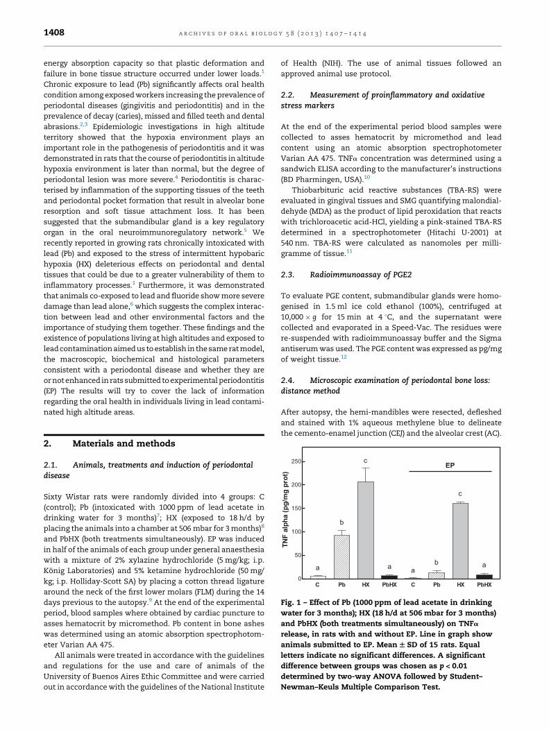
Fig. 1 – Effect of Pb (1000 ppm of lead acetate in drinking
water for 3 months); HX (18 h/d at 506 mbar for 3 months)
and PbHX (both treatments simultaneously) on TNFa
release, in rats with and without EP. Line in graph show
animals submitted to EP. Mean W SD of 15 rats. Equal
letters indicate no significant differences. A significant
difference between groups was chosen as p < 0.01
determined by two-way ANOVA followed by Student–
Newman–Keuls Multiple Comparison Test.
a r c h i v e s o f o r a l b i o l o g y 5 8 ( 2 0 1 3 ) 1 4 0 7 – 1 4 1 41408
energy absorption capacity so that plastic deformation and
failure in bone tissue structure occurred under lower loads.1
Chronic exposure to lead (Pb) significantly affects oral health
condition among exposed workers increasing the prevalence of
periodontal diseases (gingivitis and periodontitis) and in the
prevalence of decay (caries), missed and filled teeth and dental
abrasions.2,3 Epidemiologic investigations in high altitude
territory showed that the hypoxia environment plays an
important role in the pathogenesis of periodontitis and it was
demonstrated in rats that the course of periodontitis in altitude
hypoxia environment is later than normal, but the degree of
periodontal lesion was more severe.4 Periodontitis is charac-
terised by inflammation of the supporting tissues of the teeth
and periodontal pocket formation that result in alveolar bone
resorption and soft tissue attachment loss. It has been
suggested that the submandibular gland is a key regulatory
organ in the oral neuroimmunoregulatory network.5 We
recently reported in growing rats chronically intoxicated with
lead (Pb) and exposed to the stress of intermittent hypobaric
hypoxia (HX) deleterious effects on periodontal and dental
tissues that could be due to a greater vulnerability of them to
inflammatory processes.1 Furthermore, it was demonstrated
that animals co-exposed to lead and fluoride show more severe
damage than lead alone,6 which suggests the complex interac-
tion between lead and other environmental factors and the
importance of studying them together. These findings and the
existence of populations living at high altitudes and exposed to
lead contamination aimed us to establish in the same rat model,
the macroscopic, biochemical and histological parameters
consistent with a periodontal disease and whether they are
or not enhanced in rats submitted to experimental periodontitis
(EP) The results will try to cover the lack of information
regarding the oral health in individuals living in lead contami-
nated high altitude areas.
2. Materials and methods
2.1. Animals, treatments and induction of periodontaldisease
Sixty Wistar rats were randomly divided into 4 groups: C
(control); Pb (intoxicated with 1000 ppm of lead acetate in
drinking water for 3 months)7; HX (exposed to 18 h/d by
placing the animals into a chamber at 506 mbar for 3 months)8
and PbHX (both treatments simultaneously). EP was induced
in half of the animals of each group under general anaesthesia
with a mixture of 2% xylazine hydrochloride (5 mg/kg; i.p.
Konig Laboratories) and 5% ketamine hydrochloride (50 mg/
kg; i.p. Holliday-Scott SA) by placing a cotton thread ligature
around the neck of the first lower molars (FLM) during the 14
days previous to the autopsy.9 At the end of the experimental
period, blood samples where obtained by cardiac puncture to
asses hematocrit by micromethod. Pb content in bone ashes
was determined using an atomic absorption spectrophotom-
eter Varian AA 475.
All animals were treated in accordance with the guidelines
and regulations for the use and care of animals of the
University of Buenos Aires Ethic Committee and were carried
out in accordance with the guidelines of the National Institute
of Health (NIH). The use of animal tissues followed an
approved animal use protocol.
2.2. Measurement of proinflammatory and oxidativestress markers
At the end of the experimental period blood samples were
collected to asses hematocrit by micromethod and lead
content using an atomic absorption spectrophotometer
Varian AA 475. TNFa concentration was determined using a
sandwich ELISA according to the manufacturer’s instructions
(BD Pharmingen, USA).10
Thiobarbituric acid reactive substances (TBA-RS) were
evaluated in gingival tissues and SMG quantifying malondial-
dehyde (MDA) as the product of lipid peroxidation that reacts
with trichloroacetic acid-HCl, yielding a pink-stained TBA-RS
determined in a spectrophotometer (Hitachi U-2001) at
540 nm. TBA-RS were calculated as nanomoles per milli-
gramme of tissue.11
2.3. Radioimmunoassay of PGE2
To evaluate PGE content, submandibular glands were homo-
genised in 1.5 ml ice cold ethanol (100%), centrifuged at
10,000 � g for 15 min at 4 8C, and the supernatant were
collected and evaporated in a Speed-Vac. The residues were
re-suspended with radioimmunoassay buffer and the Sigma
antiserum was used. The PGE content was expressed as pg/mg
of weight tissue.12
2.4. Microscopic examination of periodontal bone loss:distance method
After autopsy, the hemi-mandibles were resected, defleshed
and stained with 1% aqueous methylene blue to delineate
the cemento-enamel junction (CEJ) and the alveolar crest (AC).

a r c h i v e s o f o r a l b i o l o g y 5 8 ( 2 0 1 3 ) 1 4 0 7 – 1 4 1 4 1409
A stereomicroscope (Stemi DV4 Stereomicroscope, Carl Zeiss
MicroImaging, Gottingen, Germany) and a digital calliper
(Digimess, Geneva, Switzerland) were used to measure three
lingual/palatal and three buccal distances (mesial, central and
distal) from the CEJ to the most apical area of the AC. The sum
of the three distances on each side of each molar was used as a
measure of the alveolar bone loss in millimeters10 (Fig. 3,
Upper).
2.5. Statistic
Statistical analyses were performed by two-way ANOVA
followed by multiple comparison Student–Newman–Keuls
tests (GraphPad Software Inc., San Diego, USA).
C Pb C Pb 0
10
20
30
40
50
60
70
80
a a
c
b
EP
)torpg
m/lomn (
mugni
SR-ABT
C HX0
10
20
30
40
50
60
70
80
a a
TBA
-RS
in g
um (n
mol
/mg
prot
)
C Pb C Pb 0
5
10
15
20
a
aaa
EP
)torpg
m /lom n(
GMS
niSR-
A BT
C HX0
5
10
15
20
a
b
TBA
-RS
in S
MG
(nm
ol/m
g pr
ot)
Fig. 2 – Effect of Pb (left), HX (middle) and the combination of bot
SMG (DOWN) from rats with or without bilateral EP. Data are rep
half of the animals of each group). Equal letters indicate no sig
groups was chosen as p < 0.01 determined by two-way ANOVA f
Test.
3. Results
3.1. Hematocrit % and lead content
Significantly high-level lead accumulation was observed
in ashes from bones in Pb (640.53 � 86.32 mg/g) and PbHX
(695 � 63.23 mg/g) groups against control animals (0.98 �0.35 mg/g) indicating that the administered Pb was deposit-
ed in the skeleton in significant amounts. Exposure of
rats to hypobaric hypoxia produced an expected
significant increase in hematocrit values (C: 43.51 � 3.07%;
Pb: 35.08 � 4.11%; HX: 74.05 � 7.04% and PbHX: 61.37 �5.91%).
C HX
a
EP
a
C PbHX C PbHX0
10
20
30
40
50
60
70
80
a a
c
b
EP
TBA
-RS
in g
um(n
mol
/mg
prot
)
C HX
a
EP
a
C PbHX C PbHX0
5
10
15
20
a
a
c
b
EP
TBA
-RS
in S
MG
(nm
ol/m
g pr
ot)
h treatments (right) on TBA-RS content in gum (UPPER) and
orted as means W SD (15 rats per group. EP was induced in
nificant differences. A significant difference between
ollowed by Student–Newman–Keuls Multiple Comparison

a r c h i v e s o f o r a l b i o l o g y 5 8 ( 2 0 1 3 ) 1 4 0 7 – 1 4 1 41410
3.2. Plasmatic TNF alpha content
We observed a significant increased concentration of TNFa in
the group intoxicated with lead ( p < 0.01) against the control
group being the enhancement even greater in the group
exposed to HX. When both treatments were applied simulta-
neously, no significant difference against the control group
was observed. The same response pattern was observed in the
EP groups (Fig. 1).
3.3. TBA-RS content in gingival tissue (gums) and SMG
In order to explore the effect of treatments on some oral target
tissues before or after periodontitis induction, we first
evaluated an oxidative stress marker that might be modified
in inflammatory processes such as TBA-RS in gingival tissue
and salivary glands. Rats chronically intoxicated with lead
showed significant higher values of TBA-RS content in gums,
but not in SMG. On the contrary, hypoxia significantly
enhanced TBA-RS content in SMG, but not in gums. These
results were independent of the existence or not of EP (Fig. 2).
3.4. PGE2 content in SMG
PGE2 content, measured by RIA, was increased in every
experimental group against its control in rats with and
without EP. The group with lead intoxication under hypoxia
environment showed even higher values (Fig. 3).
3.5. Microscopic examination of periodontal bone loss:distance method
Regarding alveolar bone loss, we measured the distance in
mm between the enamel-cement junction and the alveolar
crest, in lingual and buccal side of the mandible. In every
experimental group, no significant difference against the
control group was observed in the animals without EP (with
C Pb C Pb 0
200
400
600
800
1000
1200
1400
a cd
b
EP
PGE 2
wei
ght)
wet
SMG
(pg/
mg
SMG
in
C HX0
200
400
600
800
1000
1200
1400
a
b
PGE 2
in S
MG
(pg/
mg
SMG
wet
wei
ght)
Fig. 3 – Effect of Pb (left), HX (middle) and the combination of both
without bilateral EP. Data are reported as means W SD (15 rats p
group). Equal letters indicate no significant differences. A signi
determined by two-way ANOVA followed by Student–Newman
the exception of HX animals in the buccal side). We observed
bone loss in the control group with EP, which was increased
not only by lead intoxication (Fig. 4A) but also by exposition to
HX (Fig. 4B) or by means of both treatments applied
simultaneously (Fig. 4C).
3.6. Interradicular bone volume
Interradicular bone volume was measured using Image Pro
Plus software, expressed as % of total bone volume. Significant
bone loss was observed in the control group with EP, being
even higher in those rats with EP exposed to HX and with both
treatments simultaneously. Furthermore, the PbHX rat was
the only group that showed interradicular bone loss when the
experimental periodontitis was not applied (Fig. 5).
4. Discussion
In this paper, we demonstrated that chronic lead intoxication
under normoxic and hypoxic environment modify some
systemic and oral tissues inflammatory parameters, which
could lead to a periodontal disease in individuals living in lead
contaminated high altitude areas. Periodontitis is an infec-
tious disease characterised by inflammation of tooth-support-
ing tissues and by periodontal pocket formation, which results
in alveolar bone resorption and loss of periodontal attachment
tissue with evidence indicating the role of submandibular
glands in the regulation of immune/inflammatory reactions.9
It may be influenced by several environmental factors. Among
these, the effects of Pb or hypobaric hypoxia on oral health are
of particular relevance to understanding the pathogenesis of
periodontitis in populations living and working in areas lead
contaminated at high altitude. It was demonstrated, in a rat
model under hypoxic conditions, that the degree of periodon-
tal lesions and the microbial community in gingival crevicular
fluid were affected by the altitude hypoxia environment.4 Oral
C HX
c
EP
d
C PbHX C PbHX0
200
400
600
800
1000
1200
1400
aa
b
b
EP
PGE 2
in S
MG
(pg/
mg
SMG
wet
wei
ght)
treatments (right) on PGE2 content in SMG from rats with or
er group. EP was induced in half of the animals of each
ficant difference between groups was chosen as p < 0.01
–Keuls Multiple Comparison Test.

Fig. 4 – Effect of alveolar bone loss after Pb and HX treatments in rats with 14-days experimental periodontitis (EP). Upper:
Distance method: diagram of a section of the lower first molar. Three distances (arrows) were measured from the cement–
enamel-junction (CEJ) to the most apical area of the alveolar crest (AC). Middle: Measurements by distance method showing
lingual and buccal sections of mandible first molars with/without induction of experimental periodontitis (EP) in C, control
rats; Pb, lead-treated rats (A); HX, hypoxic rats (B); PbHX, lead-treated hypoxic rats (C). Lines in each graph show animals
submitted to EP. Data are reported as means W SD (15 rats per group. EP was induced in half of the animals of each group).
Equal letters indicate no significant differences. A significant difference between groups was chosen as p < 0.01 determined
by two-way ANOVA followed by Student–Newman–Keuls Multiple Comparison Test. Down: Photographs of one animal per
group selected randomly. Scale bar 1 mm.
a r c h i v e s o f o r a l b i o l o g y 5 8 ( 2 0 1 3 ) 1 4 0 7 – 1 4 1 4 1411
epidemiologic investigations in China western territory have
showed that the immigrants in the plateau have a higher
morbidity with periodontitis. Moreover, chronic exposure to
lead significantly affects oral health among exposed workers
and strongly correlates with increasing level of blood lead
among them. The most common adverse effects of lead on
dental health of exposed workers were the significant increase
in the prevalence of periodontal diseases, caries, missed and
filled teeth and dental abrasions.3 The deleterious effects on
periodontal and dental tissues, which we recently reported by
means of Pb and HX, aimed us to investigate in the same rat
model, local and systemic inflammatory parameters linked to
periodontal disease in rats with or without experimental
periodontitis (EP). We used the ligature-induced periodontitis
in rats as an experimental model where ligation acts as a
mechanical trauma on the gingival area, reducing tissue
integrity, allowing host–plaque interaction and bacterial
plaque-formation.13
To establish the effect of Pb and HX as risk factors for
periodontitis, we evaluated the production of plasmatic TNFa
because it was demonstrated to be an important mediator in
the pathogenesis of this disease allowing the entrance of

Fig. 5 – Upper: Interradicular bone volume after Pb and HX treatments in rats with 14-days experimental periodontitis (EP) in
C, control rats; Pb, lead-treated rats (A), HX, hypoxic rats (B), PbHX, lead-treated hypoxic rats (C). Lines in each graph show
animals submitted to EP. Data are reported as means W SD (15 rats per group). EP was induced in half of the animals of each
group. Equal letters indicate no significant differences. A significant difference between groups was chosen as p < 0.01
determined by two-way ANOVA followed by Student–Newman–Keuls Multiple Comparison Test. Down: Photographs of
transverse slides of the longitudinal sections of the mandibular interradicular bone of one animal per group selected
randomly. Resected hemimandibles stained with H&E were observed under a stereomicroscope (T2.5).
a r c h i v e s o f o r a l b i o l o g y 5 8 ( 2 0 1 3 ) 1 4 0 7 – 1 4 1 41412
inflammatory cells into sites of infection, promoting bone
resorption and stimulating eicosanoid release, especially
prostaglandin E2.14 Previously reported studies showed a
release of TNFa in another experimental model of periodonti-
tis induced by exposure to lipopolysaccharide (LPS) from
gingival tissues.10 Surprisingly, we found a lack of response
in rats exposed to Pb and HX, simultaneously. These
findings could be due to a greater activation of the
hypothalamus-pituitary-adrenal axis, with increased levels
of corticosterone which leads to lower values of TNFa.
Periodontitis causes oxidative stress, whose consequences
occur in the oral cavity and in most distant organs.15 Patients
with periodontitis have a significantly higher level of TBA-RS
than healthy people and this suggests that TBA-RS of gingival
tissue are closely associated with periodontal status.16 In this
study we found that rats chronically intoxicated with lead

a r c h i v e s o f o r a l b i o l o g y 5 8 ( 2 0 1 3 ) 1 4 0 7 – 1 4 1 4 1413
showed significant higher values of TBA-RS content in gums.
However, TBA-RS content was significantly higher in SMG only
by means of HX. We hypothesise that these differences
between gums and SMG values could be due to the local
deleterious effect induced by lead intoxication versus the
systemic effect of hypoxia, since gums are daily exposed to
lead acetate in drinking water while SMG are more distant
organs responding to general immunological status. Anyway,
other authors concluded that intermittent hypobaric hypoxia
not produces significant imbalance in redox status, since not
increase oxidative stress assessed by measurements of
plasma TBARS and catalase and superoxide dismutase in
eritrocytes.17
The increase of SMG PGE2 content observed in rats
subjected to EP is consistent with inflammatory states as
those seen in gingival tissues of patients suffering periodontal
disease.18 High levels of PGE2 in the SMG were previously
reported to be associated to periodontitis, and are in
concordance with salivary hipofunction observed in these
conditions which also produce additional deleterious effect
contributing to the progression of the disease.5 Moreover, the
highest levels of PGE2 content observed in the SMG of rats
exposed to lead intoxication and hypoxia environment,
simultaneously, could explain the spontaneous bone loss
observed in these rats, and shows clearly the existence of
crosstalk between oral pathologies and the SMG.
Bone loss was determined by the distance method in spite
of its limitations because it is recommended for short
observation periods (<15 days EP) in relation to the area
method.19 We demonstrated that the hypoxic environment
enhanced alveolar bone loss in the buccal sections in animals
not submitted to EP. As expected, the molars with EP exhibited
significant bone loss which was aggravated by treatments.
These results suggest that the damages found in bone tissue
due to a periodontal disease could be worsening by the effect
of lead intoxication or under hypoxic environment. Moreover,
from the analysis of the histological data it seems that
combined treatments induce interradicular bone loss even in
the absence of periodontitis. We hypothesise that this could be
due to the fact that lead intoxication affects osteoblasts,
osteoclasts and chondrocytes producing osteoporosis and
inhibition of endochondral ossification20,21 and that the metal
has been found inside hydroxyapatite crystals.22 Lead could
also increase the activity of matrix metalloproteinases (MMPs)
by direct or indirect mechanisms under means of ROS
production.23 MMPs are a group of enzymes released from
inflammatory cells recruited by bacterial infection which play
an important role in periodontal disease pathogenesis by
cleaving collagen and other matrix proteins. This higher
activity induced by Pb would explain the mayor tissue
destruction and increased bone loss in the experimental
groups.24 On the other hand, the presence of different kind or
number of bacteria in the altitude environment compared to a
normal one or the greater osteoclastic activity plus the
changes in the quantity and quality of saliva might play a
role in these alterations.4
In conclusion, these results show that lead intoxication
under hypoxic environment enhanced not only alveolar bone
loss but also some systemic and oral tissues inflammatory
parameters, which could aggravate the physiopathological
alterations produced by periodontal disease. Further studies
should be necessary to fully understand the underlying
mechanisms to evaluate the risk of Pb intoxication in our
experimental model.
Funding
This work was supported by research grants from University
of Buenos Aires (UBACyT 20020110100014).
Competing interests
Drs. Fernandez-Solari, Bozzini, Mandalunis, Elverdin, Conti &
Martinez and dentists Terrizzi and Lee report no conflicts of
interest related to this study.
Ethical approval
All animals were treated in accordance with the guidelines
and regulations for the use and care of animals of the
University of Buenos Aires Ethic Committee and were carried
out in accordance with the guidelines of the National Institute
of Health (NIH). The use of animal tissues followed an
approved animal use protocol.
Acknowledgments
The authors acknowledge the collaboration of physiology
laboratory technicians Graciela M. Champin and Elsa Lingua,
Department of Physiology, School of Dentistry, University of
Buenos Aires.
r e f e r e n c e s
1. Conti MI, Terrizzi AR, Lee CM, Mandalunis PM, Bozzini C,Pineiro AE, et al. Effects of lead exposure on growth andbone biology in growing rats exposed to simulated highaltitude. Bulletin of Environmental Contamination and Toxicology2012;88(6):1033–7.
2. Won YS, Kim JH, Kim YS, Bae KH. Association of internalexposure of cadmium and lead with periodontal disease: astudy of the Fourth Korean National Health and NutritionExamination Survey. Journal of Clinical Periodontology2013;40(2):118–24.
3. El-Said KF, El-Ghamry AM, Mahdy NH, El-Bestawy NA.Chronic occupational exposure to lead and its impact onoral health. Journal of the Egyptian Public Health Association2008;83(5–6):451–66.
4. Xiao X, Li Y, Zhang G, Gao Y, Kong Y, Liu M, et al. Detectionof bacterial diversity in rat’s periodontitis model underimitational altitude hypoxia environment. Archives of OralBiology 2012;57(1):23–9.
5. Amer M, Elverdin JC, Fernandez-Solari J, Medina VA,Chiarenza AP, Vacas MI. Reduced methacholine-inducedsubmandibular salivary secretion in rats withexperimental periodontitis. Archives of Oral Biology2011;56(5):421–7.

a r c h i v e s o f o r a l b i o l o g y 5 8 ( 2 0 1 3 ) 1 4 0 7 – 1 4 1 41414
6. Leite GAS, Sawan RMM, Teofilo JM, Porto IM, Sousa FB,Gerlach RF. Exposure to lead exacerbates dental fluorosis.Archives of Oral Biology 2011;56:695–702.
7. Hamilton JD, O’Flaherty EJ. Effects of lead exposure onskeletal development in rats. Fundamental and AppliedToxicology 1994;22(4):594–604.
8. Bozzini CE, Barcelo AC, Conti MI, Martinez MP, Lezon CE,Bozzini C, et al. Unexpected hypoxia-dependenterythropoietin secretion during experimental conditionsnot affecting tissue O2 supply/demand ratio. KidneyInternational 1997;51:413–5.
9. Vacas MI, Amer M, Chiarenza AP, Luchelli MA, MandalunisPM, Elverdin JC. Influence of submandibulectomy onalveolar bone loss in rats. Journal of Periodontology2008;79:1075–80.
10. Ossola CA, Surkin PN, Pugnaloni A, Mohn CE, Elverdin JC,Fernandez-Solari J. Long-term treatment withmethanandamide attenuates LPS-induced periodontitis inrats. Inflammation Research 2012;61(9):941–8.
11. Maia Mda S, Bicudo SD, Sicherle CC, Rodello L, Gallego IC.Lipid peroxidation and generation of hydrogen peroxide infrozen-thawed ram semen cryopreserved in extenders withantioxidants. Animal Reproduction Science 2010;122(1-2):118–23.
12. Mohn CE, Fernandez-Solari J, De Laurentiis A, Bornstein SR,Ehrhart-Bornstein M, Rettori V. Adrenal gland responses tolipopolysaccharide after stress and ethanol administrationin male rats. Stress 2011;14:216–26.
13. Carvalho RS, de Souza CM, Neves JC, Holanda-Pinto SA,Pinto LM, Brito GA, et al. Effect of venlafaxine on bone lossassociated with ligature-induced periodontitis in Wistarrats. Journal of Negative Results in BioMedicine 2010;9:13.
14. Stashenko P, Jandinski JJ, Fujiyoshi P, Rynar J, Socransky SS.Tissue levels of bone resorptive cytokines in periodontaldisease. Journal of Periodontology 1991;62(8):504–9.
15. Ekuni D, Endo Y, Irie K, Azuma T, Tamaki N, Tomofuji T,et al. Imbalance of oxidative/anti-oxidative status induced
by periodontitis is involved in apoptosis of ratsubmandibular glands. Archives of Oral Biology 2010;55:170–6.
16. Fowler EB, Breault LG, Cuenin MF. Periodontal disease andits association with systemic disease. Military Medicine2001;166:85–9.
17. Esteva S, Pedret R, Fort N, Torrella JR, Pages T, Viscor G.Oxidative stress status in rats after intermittent exposure tohypobaric hypoxia. Wilderness & Environmental Medicine2010;21(4):325–31.
18. Preshaw PM, Heasman PA. Prostaglandin E2 concentrationsin gingival crevicular fluid: observation in untreated chronicperiodontitis. Journal of Clinical Periodontology 2002;29:15–20.
19. Dantas AM, Mohn CE, Burdet B, Zorrilla Zubilete M,Mandalunis PM, Elverdin JC, et al. Ethanol consumptionenhances periodontal inflammatory markers in rats.Archives of Oral Biology 2012;57(9):1211–7.
20. Vahter M, Akkeson A, Liden C, Ceccatelli S, Berglund M.Gender differences in the disposition and toxicity of metals.Environmental Research 2007;104:85–95.
21. Carmouche JJ, Puzas JE, Zhang X, Tiyapatanaputi P, Cory-Slechta DA, Gelein R, et al. Lead exposure inhibits fracturehealing and is associated with increased chondrogenesis,delay in cartilage mineralization, and a decrease inosteoprogenitor frequency. Environmental Health Perspectives2005;113:749–55.
22. Hamilton JD, O’Flaherty EJ. Influence of lead onmineralization during bone growth. Fundamental and AppliedToxicology 1995;26:265–71.
23. Rizzi E, Castro MM, Fernandes K, Barbosa F, MaisonnaveArisi G, Garcia-Cairasco N, et al. Evidence of earlyinvolvement of matrix metalloproteinase-2 in lead-inducedhypertension. Archives of Toxicology 2009;83:439–49.
24. Kim K, Chung SB, Hawng EY, Noh SH, Song KH, Kim HH,et al. Correlation of expression and activity of matrixmetalloproteinase-9 and -2 in human gingival cells ofperiodontitis patients. Journal of Periodontal & Implant Science2013;43:24–9.


![Periodontal Disease: General Aspects from Biofilm to the ... · gingival inflammation and loss of alveolar bone, which can be detected by x-ray (Figure 2) [15] [16]. Acute periodontal](https://img.dokumen.tips/doc/110x75/5e725d4a1a91891c5f67e737/periodontal-disease-general-aspects-from-biofilm-to-the-gingival-inflammation.jpg)


![Cronicon · of periodontal ligament and alveolar bone [1] that lead to damage of the periodontal tissues, formation of intrabony defects (ID) and subsequently tooth loss [1,2]. Treatment](https://img.dokumen.tips/doc/110x75/5fa63e7602b4a8288f613a70/cronicon-of-periodontal-ligament-and-alveolar-bone-1-that-lead-to-damage-of-the.jpg)


