Embed Size (px)
Citation preview
1
ALUMINUM ADJUVANT LINKED TO GULF WAR SYNDROME INDUCES MOTOR NEURON DEATH IN MICE M.S. Petrik1,2, M.C. Wong1,2, R.C. Tabata1, R.F. Garry5 and C.A. Shaw1,3,4 1Departments of Ophthalmology, 3Physiology, and 4Experimental Medicine 2Program in Neuroscience, University of British Columbia, Vancouver, British Columbia, Canada. 5Department of Microbiology and Immunology, Louisiana State University Health Sciences Center, Tulane University Health Sciences Center, New Orleans, Louisiana, USA. Corresponding Author: MS Petrik Email: [email protected] VGH Research Pavilion 828 W10th Ave., Rm. 386 Vancouver, BC, Canada V5Z 1L8 Tel: 604-875-4111 x68375 Fax: 604-875-4376

2
ABSTRACT Gulf War Syndrome (GWS) affects a high percentage of veterans of the 1991 conflict,
but its origins remain unknown. One neurological complication of GWS is an increased
incidence of amyotrophic lateral sclerosis (ALS). While many environmental factors
have been linked to GWS, the role of the anthrax vaccine administered to deployed
troops has come under increasing scrutiny. Among the vaccine’s potentially toxic
components are the adjuvant aluminum hydroxide and squalene. To examine whether
these materials might contribute to neurologic toxicity, we injected young male colony
CD-1 mice with these adjuvants at doses equivalent to those given to service personnel.
Mice were subjected to a battery of motor and cognitive behavioral tests over a six month
period. Following sacrifice, CNS tissue was examined using immunohistochemistry for
evidence of neural death. Behavioral testing showed both motor and cognitive functions
were impacted by the tested adjuvants to differing degrees. Apoptotic neurons were
identified in lumbar spinal cord and motor cortex in the groups receiving the adjuvants.
Aluminum injected animals also showed a significant increase of astrocytes in the lumbar
spinal cord. Our findings suggest a possible role for either or both compounds in some
neurological features associated with GWS.
KEY WORDS: ALS, GULF WAR SYNDROME, ADJUVANT, ALUMINUM
HYDROXIDE, SQUALENE, ANTHRAX, VACCINE, NEUROTOXICITY.
3
INTRODUCTION Gulf War Syndrome (GWS) is a cluster of illnesses in veterans of the Persian Gulf War
(1990–91) characterized by a group of variable and nonspecific symptoms such as
fatigue, emotional disorders, muscle and joint pains, headaches, memory loss, and
posttraumatic stress reactions (Haley et al., 1997; Fukuda et al., 1998). Previous studies
conducted on Gulf War veterans by the U.S. Department of Defense, the U.S.
Department of Veteran Affairs and the U.K. Gulf War Research Illness Unit have
established a strong link between Gulf War service and the occurrence of Gulf War
Syndrome (GWS) (Hom et al., 1997; Unwin et al., 1999; Kang et al., 2002; Wolfe et al.,
2002; Dyer, 2004).
Recent studies have established a correlation between Gulf War service and a
neurological cluster of amyotrophic lateral sclerosis – Gulf War Syndrome (ALS-GWS)
(Charatan, 2002; Horner et al., 2003; Weisskopf et al., 2005). According to a nationwide
study by the U.S. Department of Veterans Affairs, deployed veterans of the Persian Gulf
War are more than twice as likely to develop ALS than non-deployed veterans and the
civilian population (Samson, 2002). The most unique feature of this new cluster is that
the victims are younger than usual ALS patients (Haley, 2003). Due to the overlapping
symptomatology seen in GWS and ALS, GWS can be partially classified as a
neurological illness that may carry an ALS component. One major cluster of ALS in the
Western Pacific, amyotrophic lateral sclerosis-parkinsonism dementia complex (ALS-
PDC) (Kurland, 1988; Murakami, 1999), suggests an environmental cause. Gulf War
4
Syndrome ALS may comprise another cluster of the disease and as such may provide
clues to the etiology of ALS.
Epidemiological studies have suggested several potential environmental factors in GWS
such as exposure to depleted uranium, nerve gas, organophosphates, vaccinations, heavy
metals, and bacteria infections (Abou-Donia et al., 1996; Taylor et al., 1997; Kurt, 1998;
Hodgson and Kipen, 1999; Sartin, 2000; Hotopf, 2000; Ferguson and Cassaday, 2001-
2002; Nicolson et al., 2002). Of these, vaccines are highly suspected because multiple
epidemiological studies have found positive correlations between vaccinations and GWS,
and because some nondeployed but vaccinated troops have developed illnesses identical
to GWS. In particular, the anthrax vaccine is a major suspect in GWS as it contains two
materials of particular interest from a neurological perspective: aluminum hydroxide and
the lipid polymer squalene (which has been found in the vaccine at low concentrations)
(Sasaki et al., 1992); (Sahin et al., 1994; Schumm et al., 2002) forms of both having been
implicated in neurological disease (Garruto et al., 1989; Wagner-Recio et al., 1991).
Aluminum and squalene have been linked to neurotoxicity in a number of other studies
(Bilkei-Gorzo, 1993; Gajkowska et al., 1999; Nass, 2002), and antibodies to squalene
have shown up in those with GWS (Asa et al., 2000). LD50 values (via subcutaneous
injection) for either compound have not been published to date (see J.T. Baker Material
Safety Data Sheets).
5
METHODS Experimental animals, diet and tissue collection
Adult CD-1 male mice were used in the study (3 months old at experiment onset,
weighing approximately 35g). Younger animals were deliberately chosen to mimic the
age of onset in young Gulf War veterans (Haley, 2003). The control group contained 10
animals and each adjuvant group contained either 10 or 11 animals. All animals were
housed solitarily at the Jack Bell Animal Care Facility, where an ambient temperature of
22oC and a 12/12hr light cycle were maintained. All mice were consistently fed Purina®
mouse chow ad libitum. Mice were subjected to several behavioral tests including wire
mesh hang (2x/week), open field (1x/week), and water maze (1x/week) over a period of
six months. The behavioral tests were conducted in the same room over a total period of
24 weeks. The order of animals tested was randomized for each trial. At sacrifice, the
CNS tissue was collected for histological examinations. Brains and spinal cords of mice
were obtained from both adjuvant and control groups upon sacrifice by perfusion with
4% paraformadehyde (PFA). Fixed brains and spinal cords from adjuvant and control
mice were transferred to a 30% sucrose/PBS solution overnight and kept frozen until
sectioning. The CNS sections were cryoprotected in 30% ethylene glycol-20% glycerol-
dibasic and monobasic sodium phosphate solution and kept frozen at –20oC. Brains were
sectioned into 30 µm slices and spinal cords were sectioned at 25 µm in the transverse
plane on a cryostat mounted in Tissue-Tek O.C.T compound (Sakura, Zoeterwoude,
Netherlands).

6
Adjuvants
Alhydrogel®, an aluminum hydroxide (Al(OH)3) gel suspension is manufactured by
Superfos Biosector a/s (Denmark) and supplied by SIGMA, Canada. MPL® + TDM +
CWS (Monophosphoryl Lipid A, syntheitic Trehalose Dicorynomycolate, and cell wall
skeleton of mycobacteria), a commercial squalene (C30H50) containing adjuvant was
manufactured by Corixa Corporation (Seattle, USA). Both were supplied by SIGMA,
Canada.
To calculate equivalent to human dosages of aluminum hydroxide and squalene for our
experiments, we used the following information: The AVA vaccine for human use is
made by Bioport Corporation, of Lansing, Michigan. According to product data sheets
from the Michigan Biological Products Institute anthrax vaccine insert (Bioport’s
predecessor) a single dose of vaccine contains 2.4 mg of aluminum hydroxide (equivalent
to 0.83mg of aluminum). Based on an assumed average human body weight of 80
kg, the amount per kg body weight is approximately 30µg/kg. Soldiers or civilians
receiving the vaccine would have received between 30µg/kg (1 injection) up to 180ug/kg
if 6 injections were received. Bioport Corporation denies the addition of squalene in the
formulation. The company producing the vaccine during the 1990/1991 periods,
Michigan Biological Products Institute, also denied use of squalene in the formulation of
the product. However, antibodies to squalene has been found in blood samples from
patients with GWS and in higher in titers than those produced naturally by the body ((Asa
et al., 2000)), suggesting that it was present in at least some anthrax vaccine lots in use at
7
the time. Current vaccines in use outside the United States employ a squalene containing
adjuvant oil emulsion. MF59, an adjuvant in experimental influenza vaccines (Chiron
Corporation) use a 5% squalene concentration. Based on the total volume of the MF59
injection (0.5 ml), this would be equivalent to 0.025ml of squalene. Again, based on an
average 70kg human, the amount per injection would be approximately 21.5µg
(0.3µg/kg) for one injection, as much as 86µg (1.2µg/kg) for a full series of 4 injections.
The adjuvant injections in our mice were calibrated based on average animal weight for
3-month-old male CD-1 mice (approximately 35g). We chose to do two injections as an
approximate average rather than the range of 1 to 4 injections in the human subjects.
Based on the human values above, mice receiving aluminum hydroxide received two
doses of 50µg/kg (suspension) in a total volume of 200µL sterile PBS (0.9%). Mice
receiving squalene got the equivalent dose of 2% squalene suspension (MPL® + TDM +
CWS) in PBS. Mice in the aluminum hydroxide + squalene group had both adjuvants
administered the same PBS volume. Controls were injected with 200µL PBS.
The injection site for human administration is typically subcutaneous over the deltoid
muscle. For injections in mice we used a subcutaneous injection into the loose skin
behind the neck (the "scruff") to minimize discomfort and for ease of injection.
8
Immunization
Animals received two injections (two weeks apart) of aluminum hydroxide, squalene,
aluminum hydroxide and squalene or PBS. The adjuvants were then administered by
subcutaneous injection at the back of the neck. This immunization protocol mimicked the
anthrax vaccine dose schedule set by the Anthrax Vaccine Immunization Program
(AVIP) except for the location of the injection.
Behavioral Testing Wire Mesh Hang
The wire mesh hang was used to test for muscular strength and endurance (Crawley,
2000). The wire mesh hang consisted of a 6-inch wire mesh that was suspended 40 cm
high. Mice placed onto the wire grid and inverted for a maximum period of 60 s. Latency
to fall was measured and recorded three times per week.
Open Field
The open field test was used to evaluate anxiety (DeFries et al., 1974). The open field
arena consisted of a brightly lit open field pool, 1.3 m in diameter, 30 cm high containing
mouse bedding 2 inches thick. An overhead video camera was used to record mouse
locomotion in the open field environment. The investigator counted the number of
squares crossed in a measured area (outside, inside and center perimeters) over 5 min.
Anxiety, or fear-related behavior, is seen when the mouse remains in the corners or near
9
the edges of the arena (thigmotaxis) rather than moving out in the center of the arena
(Crawley et al., 1997). Testing was conducted once a week for the duration of the
experiment.
Morris Water Maze
The water maze was used to evaluate spatial and reference memory (Morris, 1984). The
water maze set-up included a pool, 1.3 m in diameter (Everts and Koolhaas, 1999), 5
radial arms, 30 cm high and a rescue platform 5 mm above the water level. The mice
were trained for 4 d at 3 trials/d prior to the injection paradigm. Mice were placed into the
pool at the same start location for each trial and were allowed to explore the pool for a
maximum of 60 s, after which they were guided to the platform using a ruler. At 90s, the
handler placed mice on the platform if they had still not reached it on their own. Training
was terminated when mice consistently found the platform within 25s on 4 consecutive
trials. Testing was conducted once a week for the duration of the experiment. During
testing, errors were counted if the mouse fully entered the incorrect arm.
Immunohistochemistry Neuronal Nuclei (NeuN) and activated caspase-3 labeling
Mouse NeuN antibody (Chemicon International; Temecula, CA, 1:300) was used to
identify neurons containing NeuN, a DNA-binding and neuron-specific nuclear protein
(Mullen et al., 1992; Wolf et al., 1996). Free-floating sections were rinsed in 10% tris-
EDTA buffer and microwaved for 10 min. After heating, sections were allowed to cool
for 20 min. Sections were then incubated in working solution of MOM Mouse IG

10
Blocking Reagent (MOM kit, Vector Laboratories) for 1 h. Sections were immersed in
MOM Diluent solution for 5 min and incubated in primary NeuN antibody for 30 min at
room temperature. Sections were then incubated in MOM Biotinylated Anti-Mouse IgG
Reagent for 10 min and incubated with Fluorescein Avidin DCS for 5 min, then blocked
with 10% NGS (normal goat serum) for 1 h. Sections were incubated with rabbit anti-
activated caspase-3 antibody (Promega; Madison, WI, 1:250) overnight and AlexaFluor
546™ for 30 min at room temperature (Molecular Probes; Eugene, OR, 1:500) to detect
cells undergoing apoptosis (Duan et al., 2003). Sections were mounted with fluorescent
DAPI (4’,6 diamidino-2-phenylindole, Vector Laboratories). A serial approach was used
for double-fluorescence labeling due to having to use the Vector mouse on mouse
(MOM) kit for NeuN. All steps were performed at room temperature unless specified
otherwise.
Choline acetyltransferase (ChAT) labeling
ChAT antibody (AB144P, Chemicon International; Temecula, CA, 1:100) was used to
identify cholinergic neurons in brain and spinal cord and serves as a specific marker for
motor neurons (Wetts and Vaughn, 1996; Maatkamp et al., 2004). Fluorescent
immunolabeling was performed on free-floating sections and pretreated in 0.5% Triton
X-100 in buffer for 2 x 15 min. Sections were then blocked in 5% NGS (normal goat
serum) with 5% BSA (bovine serum albumin) for 3 hours, then incubated in goat anti-
ChAT IgG antibody (in PBS with 5% NGS + 1% BSA, 1:100) overnight at 4°C. The
sections were incubated for 2 h each in rabbit anti-goat IgG antibody (1:200; DuoLuX™,
11
Elite ABC Kit, Vector Laboratories) at room temperature and mounted with fluorescent
DAPI (4’,6 diamidino-2-phenylindole, Vector Laboratories).
Glial fibrillary acidic protein (GFAP) labeling
GFAP is a member of the class III intermediate filament protein family. It is heavily, and
specifically, expressed in astrocytes and certain other astroglia in the central nervous
system (Lee et al., 1984; Tohyama et al., 1991; Lee et al., 1984). Anti-Glial Fibrillary
Acidic Protein Rat monoclonal antibody (345860, Calbiochem, San Diego, CA, 1:100)
was used to identify astrocytes in lumbar segment of animal spinal cord. Fluorescent
immunolabeling was performed on free-floating sections and pretreated in 0.5% Triton
X-100 in buffer (PBST) for 2 x 5 min. Sections were then blocked in 10% NGS +
1%BSA in PBST for 2 hours, then incubated with primary antibody rat-anti-GFAP (in
PBST with 1%NGS + 1%BSA) at 10ug/ml (1:100) in a humidified chamber for overnight
at room temperature (23°C). Sections were then incubated for 1 hour in anti-rat
Fluorescein Isothiocyanate (FITC) antibody (1:200 dilution in PBS, Serotec Laboratories)
incubate for at room temperature and mounted with fluorescent DAPI (4’,6 diamidino-2-
phenylindole, Vector Laboratories).
Microscopy
Brain and spinal cord sections processed with fluorescent materials were viewed with a
Zeiss Axiovert microscope at 40x magnification and 100x magnification under oil. When
these fluorescent markers are excited they can be easily detected by fluorescent
microscopy. DAPI (blue) was viewed with a 359/461 nm absorption/emission filter,

12
Alexa Fluor 546™ (red), and rabbit IgG DuoLuX™ (red) was viewed with
556,557/572,573 nm filter and FITC was viewed with a 490,494/520,525 nm filter.
Images were captured using AxioVision 4.3 software. Brains and spinal cords used for
histology were chosen randomly from each group. Cerebral cortices and lumbar cord
slices were sampled per animal per histological experiment.
NeuN and active caspase-3 quantification
NeuN and active caspase-3 assays were performed to examine CNS tissue for evidence of
neurodegeneration. NeuN staining was used to label neuronal cells and activated caspase-
3 to measure apoptosis. Multiple brain and lumbar spinal cord sections (n=3,8) from each
mouse were captured as previously described. Fluorescent intensity levels of NeuN and
activated caspase-3 were used to identify specific antibody labeling. Double labeling of
NeuN and activated caspase-3 indicates neurons undergoing programmed cell death.
Stained sections included primary motor cortex, red nucleus, substantia nigra, dentate
gyrus layer of hippocampus and lumbar spinal cord. Regions of interest (ROI) were
defined using landmarks from stereotaxic mouse brain and spinal cord atlases (Sidman et
al., 1971; Paxinos and Franklin, 2001). All sections were randomized and counted in an
unbiased manor. Cell counts included total number of cells labeled either NeuN, activated
caspase-3, or both markers (double labeling) and were counted under a 40x objective
lens, then compared for quantification. Five mice from each group were used for lumbar
spinal cord histology and five from each group for brain histology.

13
Choline acetyltransferase (ChAT) quantification
ChAT staining was used to identify motor neurons by labeling choline acetyltransferase;
an enzyme used by motor neurons to synthesize the neurotransmitter acetylcholine
(ACh). Lumbar spinal cord sections (n=8) from each mouse were captured and ROIs
defined using the methods previously described. Ventral root motor neurons were
counted under a 40x objective lens and the experimenter was blind to the counts. All
motor neurons in the field of view were counted in the results for quantification. Eight
mice from each group were used.
Glial fibrillary acidic protein (GFAP) quantification
GFAP stains reactive rodent and human brain astrocytes induced by a variety of central
nervous system injuries. Lumbar spinal cord sections (n=8) from each mouse were
captured and ROIs defined using the methods previously described. Only positively
GFAP labeled astrocytes in the ventral horn of the grey matter where included in the
counting. Counts were conducted under a 40x objective lens and the experimenter was
blind to the counts. All astrocytic cells in the field of view were counted in the results for
quantification. Eight mice from each group were used.
Anti-squalene antibody assay (ASA)
Squalene was diluted 10-, 100-, 1000-, and 10,000-fold in distilled water, applied to
nitrocellulose membranes using a cotton-tipped applicator, and allowed to air-dry. The
nitrocellulose membranes were then cut into 4-mm-wide strips, placed in 20-well trays,
14
and rinsed in wash buffer (Tris-buffered saline containing 0.3% polyoxyethylene sorbitan
monolaurate and 0.005% thimerosal, pH 7.4). The strips were incubated in 2 ml blocking
buffer (Tris-buffered saline containing 5% powdered instant milk, 4% goat serum, and
0.008% thimerosal, pH 7.4) for 45 min prior to the addition of 5 µl of mouse sera (1:100
to 400 dilution) followed by a further 90-min incubation. All incubations and washes
were carried out at room temperature on a rocking platform. The blocking buffer was
then removed and the strips were washed with washing buffer (three times for 5 min
each). After the strips were washed, 2 ml of blocking buffer containing biotin conjugated
to goat anti-mouse IgG (Sigma, St Louis, Mo), diluted 1:1000, was added. After a 60-min
incubation, the strips were again washed as above, and 2 ml of blocking buffer containing
avidin-conjugated horseradish peroxidase (Jackson ImmunoResearch, West Grove, PA),
diluted 1:500, was added. Following another 60-min incubation, the strips were washed,
as above, and 2 ml of buffered saline containing 30% methanol and the substrate 0.6
mg/ml 4-chloro-1-napthol, 0.03% hydrogen peroxide; pH 7.4) was added. The reaction
was allowed to proceed for 15 min and was stopped by rinsing the strips in distilled
water. The strips were allowed to air-dry for visual scoring on a scale of 0 to 4.
Statistics Values for each mouse on the individual tasks and cell counts were used to calculate
mean ± S.E.M. for each group. Behavioral scores and cell counts were normalized to the
mean value of controls. The means were compared using one-way ANOVA (Statistica,
Statsoft Inc., Tulsa, OK; GraphPad Prism, San Diego, CA).
15
RESULTS We used a combination of motor and cognitive behavioral tests over a six-month period,
including tests for muscular strength and coordination (wire mesh hang), spontaneous
locomotor activity and anxiety (open field) and reference memory (radial arm water
maze).
The greatest overall effects were seen in mice injected with aluminum hydroxide. These
mice showed a significant decrease in muscular strength and endurance (-50%) compared
to controls (Fig. 1A). Squalene (in the form of an adjuvant system, MPL® + TDM +
CWS; see Adjuvants in Methods) injected mice showed a minor decrease in muscular
strength that did not achieve significance. The aluminum hydroxide and squalene
(combined) group did not show any statistically significant differences in muscle strength
and endurance. Aluminum injected mice showed a significant increase in anxiety levels
(+38%), measured by open field testing, compared to controls (Fig. 1B). The squalene
group also showed a small increase in anxiety after week 20 but these results did not
achieve significance. There was no difference in anxiety levels between the combined
group and controls.
Assessment of cognitive performance on a Morris water maze showed that mice injected
with both adjuvants had significant late stage memory deficits with an increase in the
number of errors after week 20 (4.3 errors) compared to controls (0.2 errors) (Fig. 1C).
16
Mice injected with squalene (0.9 errors) or aluminum hydroxide (1.2 errors) also showed
an increase in the number of errors after week 20.
In addition to behavioral changes, physiological changes were observed. Four of the ten
mice injected with both adjuvants developed an allergic skin reaction (i.e. dermatitis)
approximately 0.5 cm diameter region around the injection site. Hair loss at the injection
site (0.5 cm to 1.0cm diameter region around the injections site) was also visible in all
groups; 3/10 mice from the combined group, 2/10 from the aluminum hydroxide group,
4/10 from the squalene group, and no control animals (injected with PBS) developed hair
loss in this area.
Previous studies of ALS pathology have shown increased numbers of dying cells
undergoing apoptosis, a normal form of programmed cell death (Troost et al., 1995;
Martin, 1999). To measure apoptosis, we used colabeling of NeuN, a neuronal marker;
and activated caspase-3, a key mediator of the apoptotic cell death pathway (Mullen et
al., 1992; Wolf et al., 1996; Duan et al., 2003). Both NeuN and activated caspase-3
primary antibodies were tagged with fluorescent secondary antibodies (Fluorescein
Avidin DCS and AlexaFluor 546™ ) in order to provide fluorescent labeling.
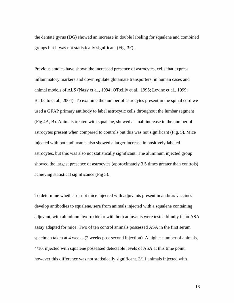
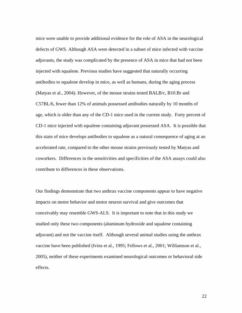
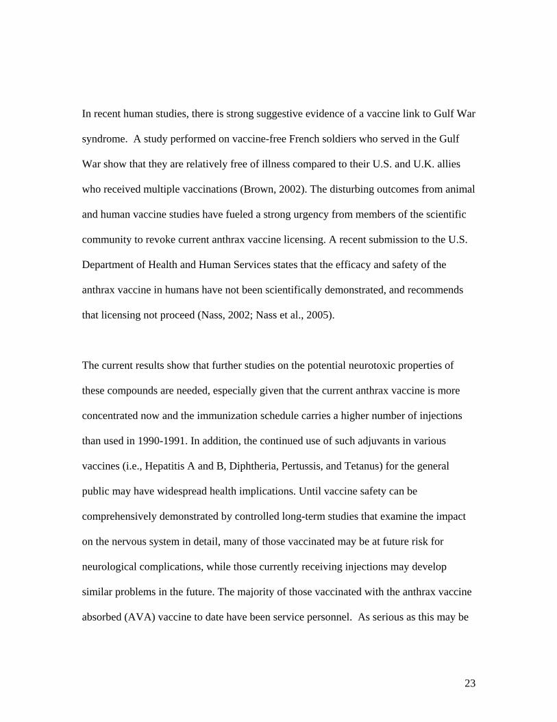
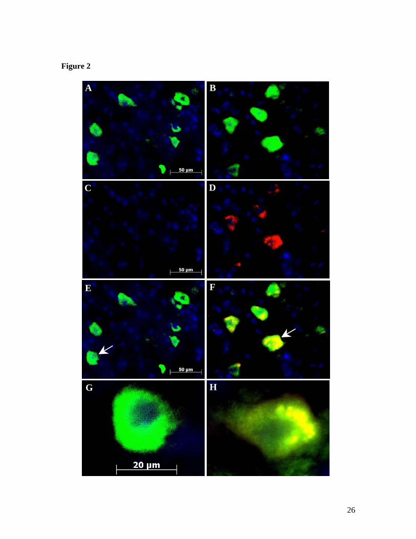
Mice injected with PBS showed little or no activated caspase-3 labeling in ventral lumbar
spinal cord (Fig 2C, E, G; 3A). In contrast, mice injected with aluminum hydroxide
showed a significant 155% increase in activated caspase-3 labeling alone and a
significant 133% increase in double labeling (Fig. 2D, F, H; 3A). Activated caspase-3
17
was also increased in the squalene group as well as the combined aluminum and squalene
group, but quantified cell counts were not significant. The difference between activated
caspase-3 and NeuN labeling between groups suggests that other cell types are
undergoing apoptosis. Aluminum injected mice also showed a significant reduction in
motor neurons (-35%) compared to controls (Fig. 3B). The squalene and combined group
also showed a reduction in motor neuron number that did not achieve significance (Fig.
3B).
In addition to the spinal cord, we also examined brain structures involved in motor
function and movement. NeuN and activated caspase-3 immunohistology was performed
on the primary motor cortex and brainstem (substantia nigra and red nucleus for evidence
of apoptotic neuropathy), since these areas are affected in ALS (Sasaki et al., 1992; Eisen
and Weber, 2001; Tsuchiya et al., 2002). Quantitative analysis of NeuN labeling showed
insignificant differences between groups indicating comparable numbers of labeled
neurons in all treatment groups (Fig 3A, C-F). Mice injected with aluminum hydroxide
showed a significant increase in activated caspase-3 (+92%) and double labeling (+85%)
in primary motor cortex compared to controls (Fig. 3C). The squalene and combined
group showed a small increase in both labels but it was not significant. Cell counts
performed in the red nucleus show increased activated caspase-3 and double labeling in
both aluminum groups, but this did not achieve statistical significance (Fig. 3D). Analysis
of the substantia nigra region did not reveal any differences in labeling between all
groups (Fig 3E). In the hippocampus, cell counts conducted on the polymorph layer of
18
the dentate gyrus (DG) showed an increase in double labeling for squalene and combined
groups but it was not statistically significant (Fig. 3F).
Previous studies have shown the increased presence of astrocytes, cells that express
inflammatory markers and downregulate glutamate transporters, in human cases and
animal models of ALS (Nagy et al., 1994; O'Reilly et al., 1995; Levine et al., 1999;
Barbeito et al., 2004). To examine the number of astrocytes present in the spinal cord we
used a GFAP primary antibody to label astrocytic cells throughout the lumbar segment
(Fig.4A, B). Animals treated with squalene, showed a small increase in the number of
astrocytes present when compared to controls but this was not significant (Fig. 5). Mice
injected with both adjuvants also showed a larger increase in positively labeled
astrocytes, but this was also not statistically significant. The aluminum injected group
showed the largest presence of astrocytes (approximately 3.5 times greater than controls)
achieving statistical significance (Fig 5).
To determine whether or not mice injected with adjuvants present in anthrax vaccines
develop antibodies to squalene, sera from animals injected with a squalene containing
adjuvant, with aluminum hydroxide or with both adjuvants were tested blindly in an ASA
assay adapted for mice. Two of ten control animals possessed ASA in the first serum
specimen taken at 4 weeks (2 weeks post second injection). A higher number of animals,
4/10, injected with squalene possessed detectable levels of ASA at this time point,
however this difference was not statistically significant. 3/11 animals injected with

19
aluminum hydroxide and 1/10 injected with both adjuvants possessed ASA. The
presence of ASA was generally stable over time in individual animals tested. However,
one animal that had been injected with both adjuvants developed ASA at a later time
point (24 weeks post last injection).
DISCUSSION Using the anthrax vaccine adjuvant aluminum hydroxide and squalene (not a licensed
component in North America) under minimal conditions (1-4 injection range), we
investigated the behavioral and neuropathological effects of these adjuvants in mice. Our
data suggest that the aluminum hydroxide adjuvant induces both behavioral and motor
deficits and the loss of motor neurons and increased presence of astrocytes in spinal cord
and neuronal apoptosis in the primary motor cortex while also affecting the red nucleus
region of the brain. The squalene adjuvant alone produced a small change in locomotion
and anxiety testing, but the histological results were not significant in the current
experiments. The combination of both adjuvants showed a significant memory deficit
with some indications of neuronal apoptosis in the red nucleus and DG region of the
hippocampus.
Several possibilities may explain the neurotoxic effects of these adjuvants demonstrated
in the current results. First, the adjuvant compounds may exert direct toxicity on some
neural cell populations in the CNS (Bilkei-Gorzo, 1993; Gajkowska et al., 1999).
Aluminum in particular has long been associated with neuronal degeneration and

20
neurodegenerative diseases (Rao et al., 1998; Savory and Garruto, 1998), and aluminum
adjuvanted vaccines have been shown to increase aluminum levels in the murine brain
(Redhead et al., 1992; Sahin et al., 1994). Aluminum treated animals have also shown
severe anterograde degeneration of cholinergic terminals in cortex and hippocampus
(Platt et al., 2001). Squalene has been shown to cause swelling in astrocytic processes
(Gajkowska et al., 1999).
Alternatively, the adjuvants may induce an indirect type of toxicity by stimulating an
immune response. Rook and Zumla (1997) hypothesize that multiple Th2 (T helper cell
type 2)-inducing vaccinations, stressful circumstances, and the method of vaccine
administration (oral vs. subcutaneous vs. intramuscularly) could lead to a shift the Th2
predominance, versus Th1 (T helper cell type 1), and maximize Th2 immunogenicity
(Rook and Zumla, 1997; Rook and Zumla, 1998). Both aluminum hydroxide and
squalene have previously been shown stimulate a Th2-cytokine response (Valensi et al.,
1994; Brewer et al., 1999). A latest study using inbred and outbred mice injected with
recombinant protective antigen (rPA) vaccine and challenged with Bacillus anthracis,
found that both mouse strains displayed a predominantly Th2 biased immune response
(Flick-Smith et al., 2005). Such a Th1 to Th2 shift could stimulate autoimmune
processes. A recent study of blood samples from Gulf War veterans, however, showed
evidence for Th1 immune activation (Skowera et al., 2004). Alternatively, the observed
effects of aluminum hydroxide and squalene (or other components in the squalene

21
containing adjuvant) in our study could result from direct action on neuronal cells in the
CNS.
In the present study, the combination of aluminum hydroxide and squalene seemed to
have less effect on motor behavior and anxiety testing than either aluminum hydroxide or
squalene alone. The possibility of competing effects on immune response cannot be
discounted and deserve further investigation. It is notable that while both compounds are
present in the anthrax vaccines administered to deployed service personnel, the company
making these vaccines has not confirmed the addition of squalene. Although squalene has
adjuvant properties, we are not claiming it was deliberately added to adjuvant the
vaccine, and it is not a licensed ingredient of the vaccine. However, blood samples from
patients with GWS have been reported to contain anti-squalene antibodies in much higher
in titers than those produced naturally (Asa et al., 2000), suggesting that it was present in
at least some anthrax vaccine lots in use at the time. Conversely, although anti-squalene
antibodies have been found in Gulf War vets, squalene is a component of human bodies
and such antibodies may be unrelated to the deliberate injection or ingestion of squalene
(which can be found in cosmetics and various foods).
Previous studies suggested that anti-squalene antibodies are present in the serum of
veterans with GWS at a greater frequency than the general population (Asa et al., 2000).
The presence of low levels of squalene in certain lots of anthrax vaccine correlated
significantly with the presence of ASA (Asa et al., 2002). Our current studies using CD-1
22
mice were unable to provide additional evidence for the role of ASA in the neurological
defects of GWS. Although ASA were detected in a subset of mice infected with vaccine
adjuvants, the study was complicated by the presence of ASA in mice that had not been
injected with squalene. Previous studies have suggested that naturally occurring
antibodies to squalene develop in mice, as well as humans, during the aging process
(Matyas et al., 2004). However, of the mouse strains tested BALB/c, B10.Br and
C57BL/6, fewer than 12% of animals possessed antibodies naturally by 10 months of
age, which is older than any of the CD-1 mice used in the current study. Forty percent of
CD-1 mice injected with squalene containing adjuvant possessed ASA. It is possible that
this stain of mice develops antibodies to squalene as a natural consequence of aging at an
accelerated rate, compared to the other mouse strains previously tested by Matyas and
coworkers. Differences in the sensitivities and specificities of the ASA assays could also
contribute to differences in these observations.
Our findings demonstrate that two anthrax vaccine components appear to have negative
impacts on motor behavior and motor neuron survival and give outcomes that
conceivably may resemble GWS-ALS. It is important to note that in this study we
studied only these two components (aluminum hydroxide and squalene containing
adjuvant) and not the vaccine itself. Although several animal studies using the anthrax
vaccine have been published (Ivins et al., 1995; Fellows et al., 2001; Williamson et al.,
2005), neither of these experiments examined neurological outcomes or behavioral side
effects.
23
In recent human studies, there is strong suggestive evidence of a vaccine link to Gulf War
syndrome. A study performed on vaccine-free French soldiers who served in the Gulf
War show that they are relatively free of illness compared to their U.S. and U.K. allies
who received multiple vaccinations (Brown, 2002). The disturbing outcomes from animal
and human vaccine studies have fueled a strong urgency from members of the scientific
community to revoke current anthrax vaccine licensing. A recent submission to the U.S.
Department of Health and Human Services states that the efficacy and safety of the
anthrax vaccine in humans have not been scientifically demonstrated, and recommends
that licensing not proceed (Nass, 2002; Nass et al., 2005).
The current results show that further studies on the potential neurotoxic properties of
these compounds are needed, especially given that the current anthrax vaccine is more
concentrated now and the immunization schedule carries a higher number of injections
than used in 1990-1991. In addition, the continued use of such adjuvants in various
vaccines (i.e., Hepatitis A and B, Diphtheria, Pertussis, and Tetanus) for the general
public may have widespread health implications. Until vaccine safety can be
comprehensively demonstrated by controlled long-term studies that examine the impact
on the nervous system in detail, many of those vaccinated may be at future risk for
neurological complications, while those currently receiving injections may develop
similar problems in the future. The majority of those vaccinated with the anthrax vaccine
absorbed (AVA) vaccine to date have been service personnel. As serious as this may be

24
for the potential for adjuvant-associated complications in this population, legislation now
before U.S. Congress may mandate similar vaccination regimes for the civilian
population as well (Biodefense and Pandemic Vaccine and Drug Development Act of
2005). If a significant fraction of the military and civilians vaccinated develop
neurological complications, the impact on U.S. society would be profound. Whether the
risk of protection from a dreaded disease outweighs the risk of toxicity is a question that
demands our urgent attention.

25
Figure 1
A B
Wire Hang
0 5 10 15 20 25A B0
25
50
75
* ** *** ***
**
ControlSqualeneAluminumAluminum+Squalene
Week
Late
ncy
to fa
ll (s
)
A
Open Field
10 15 20 250
100
200
300
*ControlSqualeneAluminumAluminum+Squalene
Week
Tim
e (s
) spe
nt in
out
erpe
rim
eter
in a
5 m
inse
ssio
n
B
A B
Water Maze
0 5 10 15 20 25A B0.0
2.5
5.0
7.5
10.0ControlSqualeneAluminumAluminum+Squalene
*
Week
Erro
rs/3
tria
ls
C
26
Figure 2
E
G H
A B
C D
F

27
Figure 3
NeuN and Caspase-3 LabelingLumbar SC
NeuN Caspase-3 Double0.0
0.5
1.0
1.5
2.0
2.5
3.0
3.5ControlSqualeneAluminumAluminum+Squalene
*#
*
**#
Marker
Nor
mal
ized
num
ber
ofpo
sitiv
e la
bele
d ce
lls p
ersa
mpl
e ar
ea
A
NeuN and Caspase-3 Labelingin Red Nucleus
NeuN Caspase-3 Double0.0
0.5
1.0
1.5
2.0ControlSqualeneAluminumAluminum+Squalene
Marker
Nor
mal
ized
num
ber
ofpo
sitiv
e la
bele
d ce
lls in
sam
ple
area
C
NeuN and Caspase-3 Labelingin Substantia Nigra
NeuN Caspase-3 Double0.0
0.5
1.0
1.5ControlSqualeneAluminumAluminum+Squalene
Marker
Nor
mal
ized
num
ber
ofpo
sitiv
e la
bele
d ce
lls p
ersa
mpl
e ar
ea
E
NeuN and Caspase-3 Labelingin Primary Motor Cortex
NeuN Caspase-3 Double0.0
0.5
1.0
1.5
2.0
2.5ControlSqualeneAluminumAluminum+Squalene
Marker
Nor
mal
ized
num
ber
ofpo
sitiv
e la
bele
d ce
lls p
ersa
mpl
e ar
ea
*#
#*
*
*
B
NeuN and Caspase-3 Labelingin DG of Hippocampus
NeuN Caspase-3 Double0
1
2ControlSqualeneAluminumAluminum+Squalene
**
Marker
Nor
mal
ized
num
ber
ofpo
sitiv
e la
bele
d ce
lls p
ersa
mpl
e ar
ea
D

28
Figure 4
A B

29
Figure 5
Motor Neuron Countin Lumbar SC
PBS SQE ALUM S+A0.0
0.2
0.4
0.6
0.8
1.0
1.2ControlSqualeneAluminumAluminum+Squalene
Group
Nor
mal
ized
num
ber o
fpo
sitiv
e la
bele
d ce
ll pe
rsa
mpl
e ar
ea
*
A
30
Figure 6
A B

31
Figure 7 GFAP Labeling in Lumbar SC
CON SQE ALUM A+S0
1
2
3
4
5ControlSqualeneAluminumAluminum+Squalene
Group
Nor
mal
ized
num
ber
ofpo
sitiv
e la
bele
d ce
ll pe
rsa
mpl
e ar
ea
***

32
Table 1 Comparison of human ALS and GWS symptomology
with GWS mouse model. Symptoms ALS* GWS† GWS
Mouse Model
Muscular motor loss Enhanced anxiety Memory impairment Dermatitis *(Bromberg, 2002). †(Haley et al., 1997).

33
Figure Captions Fig. 1. Motor and cognitive effects of adjuvants. A: The wire mesh hang test measures
the latency to fall when suspended from a wire mesh, as a test of muscle strength and
endurance. Mice injected with aluminum hydroxide showed a significant decrease in
muscular strength and endurance (-50%) compared to controls. Mice injected with
squalene or both adjuvants did not show a significant decrease in muscular strength. B:
Open field tests (during weeks 7-24) records the time the animal spends in the outer
perimeter, as a measure of anxiety. Animals with increased anxiety will increasingly
circle the perimeter. Mice injected with aluminum hydroxide show a significant increase
in anxiety (+38%) compared to controls. Mice injected with squalene or both adjuvants
did not show any significant effect. C: The radial arm water maze (5 arms) was used to
test spatial and reference memory; animals were required to memorize and locate a
rescue platform and their errors were recorded over 3 trials. Mice injected with both
adjuvants showed a significant increase in errors after week 20 (4.3 errors) while controls
achieved 0.2 errors. Mice injected with squalene (0.9 errors) or aluminum hydroxide (1.2
errors) did show increased errors after week 20 but these values were not significant.
A=1st injection, B=2nd injection. *, p<0.05, **, p<0.01, ***, p<0.001, one-way
ANOVA.
Fig. 2. NeuN and activated caspase-3 fluorescent labeling in ventral horn of lumbar
spinal cord. Green = Neuron specific nuclear protein (NEUronal Nuclei: NeuN); Red =
Activated caspase-3 antibody; specific marker for staining apoptotic cells. Yellow = Co-
localization of NeuN and activated caspase-3 indicating apoptotic neuronal cell death.

34
Blue = Nuclear DAPI (4’, 6 diamidino-2-phenylindole) counterstaining. A: Control
shows NeuN labeling (Magnification: 40x). B: NeuN labeling in aluminum injected
mouse. C: Control animals show no labeling of activated caspase-3 antibody. D: Animals
injected with aluminum hydroxide show clear labeling of activated caspase-3 antibody.
E: Tissue from control animal shows NeuN labeling but no activated caspase-3 labeling
(Magnification: 40x; white arrow indicates neuron enlarged in figure 1G). G:
Enlargement of neuron from figure 1E shows positive NeuN labeling with no activated
caspase-3 labeling (Magnification: 100x); F: Mice injected with aluminum hydroxide
show increased positive labeling of NeuN and anti-active caspase-3 compared to controls
indicating apoptosis (Magnification: 40x; white arrow indicates neuron enlarged in figure
1H). H: Enlargement of neuron from 1F shows clear double labeling of NeuN and anti-
active caspase-3. (Magnification: 100x). A-F: Scale bar = 50 µm. G, H: Scale bar =
20µm.
Fig. 3. A: Cell counts for NeuN and activated caspase-3 labeling in ventral horn of
lumbar spinal cord. NeuN counts between groups (n=32, 8 per group) show no significant
differences indicating similar numbers of neuronal cells labeled in all groups. Activated
caspase-3 marker shows significantly increased positive capsase-3 labeling (+155%) in
mice injected with aluminum hydroxide compared to controls. NeuN and activated
caspase-3 double labeling show significantly increased apoptotic neuronal cells (+133%)
in mice injected with aluminum hydroxide compared to control and squalene injected
groups. B: NeuN counts (n=20, 5 per group) show no significant difference between
groups. Animals injected with aluminum hydroxide show a significant increase in
35
activated caspase-3 (+92%) and double labeling (+85%) in primary motor cortex
compared to controls. Compared to squalene-injected mice, aluminum mice showed a
significant increase (+65%) in double labeling. C: Cell counts (n=20, 5 per group)
performed in the red nucleus show an increase in activated caspase-3 and double labeling
in both aluminum groups compared to controls, but this was not significant. D:
Hippocampal cell counts (n=20, 5 per group) performed on the polymorph layer of the
dentate gyrus (DG) show increased activated caspase-3 and double labeling in the
squalene group, while the combined group showed the greatest activated caspase-3 and
double labeling, but these results were not statistically significant. E: There was no
significant difference in cell counts (n=20, 5 per group) of NeuN and activated caspase-3
labeling between groups in the substantia nigra region. Data are means ± S.E.M *, #
p<0.05 versus control and squalene mice, **, p<0.01 versus control mice using one-way
ANOVA analysis.
Fig. 4. Cholinacetyltransferase (ChAT) fluorescent labeling in ventral horn of lumbar
spinal cord. A: Control animal shows clear ChAT labeling and health motor neuron shape
(20x magnification). B: Aluminum injected animal shows decreased ChAT labeling and
abnormal morphology of motor neurons compared to controls (20x magnification). Scale
bar = 50 µm.
Fig. 5. Motor neuron cell counts after ChAT fluorescent labeling in ventral horn of
lumbar spinal cord. Only cells positively labeled with ChAT were counted as motor

36
neurons (n=32, 8 per group). Mice injected with aluminum hydroxide showed a
statistically significant decrease in motor neuron number (-35%) compared to controls.
There was no significant difference in motor neuron counts between all other groups
compared to controls. Data are means ± S.E.M *** p<0.001 versus control mice using
one-way ANOVA analysis.
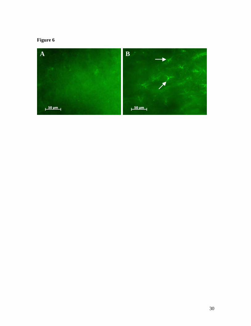
Fig. 6. Glial fibrillary acidic protein (GFAP) fluorescent labeling in ventral horn of
lumbar spinal cord. A: Control animal shows little GFAP labeling indicating rare
presence of astrocytes (40x magnification). B: Aluminum injected animal shows
increased GFAP labeling and greater number of astrocytes (white arrows) compared to
controls (40x magnification). Scale bar = 50 µm.
Fig. 7. Normalized cell counts for GFAP labeling of astrocytes in ventral horn of lumbar
spinal cord (n=32, 8 per group). Squalene treated animals show a small increase in GFAP
labeled astrocytes when compared to controls. Animals treated with both aluminum
hydroxide and squalene showed a larger increase in astrocyte cell number when
compared to controls, while mice injected with aluminum showed the greatest number of
astrocytes present (approximately 3.5 times greater than controls). Data are means ±
S.E.M *** p<0.001 versus control mice using one-way ANOVA analysis.
Table 1. Table summary of human ALS and GWS symptomology compared with GWS
mouse model. This table outlines the similarities between human ALS and Gulf War
37
syndrome. From this table, we can see that overlapping symptoms present in ALS and
some GWS patients are represented in our mouse model of GWS.
ANIMAL ETHICS COMMITTEE APPROVAL
Protocols governing the use of animals were approved by review committees of the
University of British Columbia and were in compliance with guidelines published by the
Canadian Council on Animal Care and are in accordance with the international guidelines
including the NIH Guide for the Care and Use of Laboratory Animals, as well as the EEC
Council Directive.
CONFLICT OF INTEREST STATEMENT Petrik has not received any grants or funding from Bioport, Chiron, Corixa, nor any other
pharmaceutical companies. All the other authors have viewed this article and declare that
they have no conflict of interest.
ACKNOWLEDGEMENTS This work was supported by grants from the US Army Medical Research and Materiel
Command (#DAMD17-02-1-0678), Scottish Rite Charitable Foundation of Canada, and
the Natural Science and Engineering Research Council of Canada (to CAS). We would
like to thank Dr. Jason Wilson (University of British Columbia, B.C., Canada), Dr. Meryl
Nass (Mount Desert Island Hospital, Maine, U.S.A.), and Dr. Reyniel Cruz-Aguado
(University of British Columbia, B.C., Canada), for their invaluable comments and
advisory contributions to this project and manuscript.

38
Reference List
Abou-Donia, M. B., Wilmarth, K. R., Jensen, K. F., Oehme, F. W., and Kurt, T. L. (1996 ) Neurotoxicity resulting from coexposure to pyridostigmine bromide, deet, and permethrin: implications of Gulf War chemical exposures. J Toxicol Environ Health. 48, 35-56.
Asa, P. B., Cao, Y., and Garry, R. F. (2000) Antibodies to squalene in Gulf War syndrome. Exp Mol Pathol. 68, 55-64.
Asa, P. B., Wilson, R. B., and Garry, R. F. (2002) Antibodies to squalene in recipients of anthrax vaccine. Exp Mol Pathol. 73 , 19-27.
Barbeito, L. H., Pehar, M., Cassina, P., Vargas, M. R., Peluffo, H., Viera, L., Estevez, A. G., and Beckman, J. S. (2004) A role for astrocytes in motor neuron loss in amyotrophic lateral sclerosis. Brain Res Brain Res Rev. 47, 263-74.
Bilkei-Gorzo, A. (1993) Neurotoxic effect of enteral aluminium. Food Chem Toxicol. 31, 357-61.
Brewer, J. M., Conacher, M., Hunter, C. A., Mohrs, M., Brombacher, F., and Alexander, J. (1999) Aluminium hydroxide adjuvant initiates strong antigen-specific Th2 responses in the absence of IL-4- or IL-13-mediated signaling. J Immunol. 163, 6448-54.
Bromberg, M. B. (2002) Diagnostic criteria and outcome measurement of amyotrophic lateral sclerosis. Adv Neurol. 88, 53-62.
Brown, P. (2002) French soldiers free of Gulf war illness. The Guardian.
Charatan, F. (2002) US links motor neurone disease with Gulf war service. BMJ. 324, 65.
Crawley, J. N. (2000) What' s Wrong With My Mouse? : Behavioral Phenotyping of Trangenic and Knockout Mice. 65--69.
Crawley, J. N., Belknap, J. K., Collins, A., Crabbe, J. C., Frankel, W., Henderson, N., Hitzemann, R. J., Maxson, S. C., Miner, L. L., Silva, A. J., Wehner, J. M., Wynshaw-Boris, A., and Paylor, R. (1997) Behavioral phenotypes of inbred mouse strains: implications and recommendations for molecular studies. Psychopharmacology (Berl). 132, 107-24.
DeFries, J. C., Hegmann, J. P., and Halcomb, R. A. (1974) Response to 20 generations of selection for open-field activity in mice. Behav Biol. 11, 481-95.
Duan, W. R., Garner, D. S., Williams, S. D., Funckes-Shippy, C. L., Spath, I. S., and Blomme, E. A. (2003) Comparison of immunohistochemistry for activated caspase-3 and cleaved cytokeratin 18 with the TUNEL method for quantification of apoptosis in histological sections of PC-3 subcutaneous xenografts. J Pathol. 199, 221-8.
Dyer, O. (2004) Inquiry finds that Gulf war veterans face extra burden of disease. BMJ. 329, 1257.
Eisen, A. and Weber, M. (2001) The motor cortex and amyotrophic lateral sclerosis. Muscle Nerve. 24, 564-73.
Everts, H. G. and Koolhaas, J. M. (1999) Differential modulation of lateral septal vasopressin receptor blockade in spatial learning, social recognition, and anxiety-related behaviors in rats. Behav Brain

39
Res. 99, 7-16.
Fellows, P. F., Linscott, M. K., Ivins, B. E., Pitt, M. L., Rossi, C. A., Gibbs, P. H., and Friedlander, A. M. (2001) Efficacy of a human anthrax vaccine in guinea pigs, rabbits, and rhesus macaques against challenge by Bacillus anthracis isolates of diverse geographical origin. Vaccine. 19, 3241-7.
Ferguson, E. and Cassaday, H. J. (2001-2002) Theoretical accounts of Gulf War Syndrome: from environmental toxins to psychoneuroimmunology and neurodegeneration. Behav Neurol. 13, 133-47.
Flick-Smith, H. C., Waters, E. L., Walker, N. J., Miller, J., Stagg, A. J., Green, M., and Williamson, E. D. (2005) Mouse model characterisation for anthrax vaccine development: comparison of one inbred and one outbred mouse strain. Microb Pathog. 38, 33-40.
Fukuda, K., Nisenbaum, R., Stewart, G., Thompson, W. W., Robin, L., Washko, R. M., Noah, D. L., Barrett, D. H., Randall, B., Herwaldt, B. L., Mawle, A. C., and Reeves, W. C. (1998) Chronic multisymptom illness affecting Air Force veterans of the Gulf War. JAMA. 280, 981-8.
Gajkowska, B., Smialek, M., Ostrowski, R. P., Piotrowski, P., and Frontczak-Baniewicz, M. (1999) The experimental squalene encephaloneuropathy in the rat. Exp Toxicol Pathol. 51, 75-80.
Garruto, R. M., Shankar, S. K., Yanagihara, R., Salazar, A. M., Amyx, H. L., and Gajdusek, D. C. (1989) Low-calcium, high-aluminum diet-induced motor neuron pathology in cynomolgus monkeys. Acta Neuropathol (Berl). 78, 210-9.
Haley, R. W. (2003) Excess incidence of ALS in young Gulf War veterans. Neurology. 61, 750-6.
Haley, R. W., Kurt, T. L., and Hom, J. (1997) Is there a Gulf War Syndrome? Searching for syndromes by factor analysis of symptoms. JAMA. 277, 215-22.
Hodgson, M. J. and Kipen, H. M. (1999) Gulf War illnesses: causation and treatment. J Occup Environ Med. 41, 443-52.
Hom, J., Haley, R. W., and Kurt, T. L. (1997) Neuropsychological correlates of Gulf War syndrome. Arch Clin Neuropsychol. 12, 531-44.
Horner, R. D., Kamins, K. G., Feussner, J. R., Grambow, S. C., Hoff-Lindquist, J., Harati, Y., Mitsumoto, H., Pascuzzi, R., Spencer, P. S., Tim, R., Howard, D., Smith, T. C., Ryan, M. A., Coffman, C. J., and Kasarskis, E. J. (2003) Occurrence of amyotrophic lateral sclerosis among Gulf War veterans. Neurology. 61, 742-9 .
Hotopf, M., David, A., Hull, L., Ismail, K., Unwin, C., and Wessely, S. (2000) Role of vaccinations as risk factors for ill health in veterans of the Gulf war: cross sectional study. BMJ. 320, 1363-7.
Ivins, B., Fellows, P., Pitt, L., Estep, J., Farchaus, J., Friedlander, A., and Gibbs, P. (1995) Experimental anthrax vaccines: efficacy of adjuvants combined with protective antigen against an aerosol Bacillus anthracis spore challenge in guinea pigs. Vaccine. 13, 1779-84.
Kang, H. K., Mahan, C. M., Lee, K. Y., Murphy, F. M., Simmens, S. J., Young, H. A., and Levine, P. H. (2002) Evidence for a deployment-related Gulf War syndrome by factor analysis. Arch Environ Health. 57, 61-8.
40
Kurland, L. T. (1988) Amyotrophic lateral sclerosis and Parkinson's disease complex on Guam linked to an environmental neurotoxin. Trends Neurosci. 11, 51-4.
Kurt, T. L. (1998) Epidemiological association in US veterans between Gulf War illness and exposures to anticholinesterases. Toxicol Lett. 102-103, 523-6.
Lee, V. M., Page, C. D., Wu, H. L., and Schlaepfer, W. W. (1984) Monoclonal antibodies to gel-excised glial filament protein and their reactivities with other intermediate filament proteins. J Neurochem. 42, 25-32.
Levine, J. B., Kong, J., Nadler, M., and Xu, Z. (1999) Astrocytes interact intimately with degenerating motor neurons in mouse amyotrophic lateral sclerosis (ALS). Glia. 28, 215-24.
Maatkamp, A., Vlug, A., Haasdijk, E., Troost, D., French, P. J., and Jaarsma, D. (2004 ) Decrease of Hsp25 protein expression precedes degeneration of motoneurons in ALS-SOD1 mice. Eur J Neurosci. 20, 14-28.
Martin, L. J. (1999) Neuronal death in amyotrophic lateral sclerosis is apoptosis: possible contribution of a programmed cell death mechanism. J Neuropathol Exp Neurol. 58, 459-71.
Matyas, G. R., Rao, M., Pittman, P. R., Burge, R., Robbins, I. E., Wassef, N. M., Thivierge, B., and Alving, C. R. (2004) Detection of antibodies to squalene: III. Naturally occurring antibodies to squalene in humans and mice. J Immunol Methods. 286, 47-67.
Morris, R. (1984) Developments of a water-maze procedure for studying spatial learning in the rat. J Neurosci Methods. 11, 47-60.
Mullen, R. J., Buck, C. R., and Smith, A. M. (1992) NeuN, a neuronal specific nuclear protein in vertebrates. Development. 116 , 201-11.
Murakami, N. (1999) Parkinsonism-dementia complex on Guam - overview of clinical aspects. J Neurol. 246 Suppl 2, II16-8.
Nagy, D., Kato, T., and Kushner, P. D. (1994) Reactive astrocytes are widespread in the cortical gray matter of amyotrophic lateral sclerosis. J Neurosci Res. 38, 336-47.
Nass, M. (2002) The Anthrax Vaccine Program: an analysis of the CDC's recommendations for vaccine use. Am J Public Health. 92, 715-21.
Nass, M., Fisher, B. L., and Robinson, S. (2005) Comments and Questions regarding FDA's proposed rule and order to license Anthrax Vaccine Absorbed. FDA Anthrax vaccine docket submission. Proposed rule and proposed order, 29 Fed. Reg. 78281-78293.
Nicolson, G. L., Nasralla, M. Y., Haier, J., and Pomfret, J. (2002) High frequency of systemic mycoplasmal infections in Gulf War veterans and civilians with Amyotrophic Lateral Sclerosis (ALS). J Clin Neurosci. 9, 525-9.
O'Reilly, S. A., Roedica, J., Nagy, D., Hallewell, R. A., Alderson, K., Marklund, S. L., Kuby, J., and Kushner, P. D. (1995) Motor neuron-astrocyte interactions and levels of Cu,Zn superoxide dismutase in sporadic amyotrophic lateral sclerosis. Exp Neurol. 131, 203-10.
Paxinos, G. and Franklin, K.B.J. (2001) The Mouse Brain in Stereotaxic Coordinates. Academic Press.

41
Sydney.
Platt, B., Fiddler, G., Riedel, G., and Henderson, Z. (2001) Aluminium toxicity in the rat brain: histochemical and immunocytochemical evidence. Brain Res Bull. 55, 257-67.
Rao, J. K., Katsetos, C. D., Herman, M. M., and Savory, J. (1998) Experimental aluminum encephalomyelopathy. Relationship to human neurodegenerative disease. Clin Lab Med. 18, 687-98, viii.
Redhead, K., Quinlan, G. J., Das, R. G., and Gutteridge, J. M. (1992) Aluminium-adjuvanted vaccines transiently increase aluminium levels in murine brain tissue. Pharmacol Toxicol. 70, 278-80.
Rook, G. A. and Zumla, A. (1997) Gulf War syndrome: is it due to a systemic shift in cytokine balance towards a Th2 profile? Lancet. 349, 1831-3.
Rook, G. A. and Zumla, A. (1998) Is the Gulf War syndrome an immunologically mediated phenomenon? Hosp Med. 59, 10-1.
Sahin, G., Varol, I., Temizer, A., Benli, K., Demirdamar, R., and Duru, S. (1994) Determination of aluminum levels in the kidney, liver, and brain of mice treated with aluminum hydroxide. Biol Trace Elem Res. 41, 129-35.
Samson, K. (2002) VA study finds ALS spike in Gulf War vets. Neurology Today. 2, 1, 13-14.
Sartin, J. S. (2000) Gulf War illnesses: causes and controversies. Mayo Clin Proc. 75, 811-9.
Sasaki, S., Tsutsumi, Y., Yamane, K., Sakuma, H., and Maruyama, S. (1992) Sporadic amyotrophic lateral sclerosis with extensive neurological involvement. Acta Neuropathol (Berl). 84, 211-5.
Savory, J. and Garruto, R. M. (1998) Aluminum, tau protein, and Alzheimer's disease: an important link? Nutrition. 14, 313-4.
Schumm, W. R., Reppert, E. J., Jurich, A. P., Bollman, S. R., Webb, F. J., Castelo, C. S., Stever, J. C., Sanders, D., Bonjour, G. N., Crow, J. R., Fink, C. J., Lash, J. F., Brown, B. F., Hall, C. A., Owens, B. L., Krehbiel, M., Deng, L. Y., and Kaufman, M. (2002) Self-reported changes in subjective health and anthrax vaccination as reported by over 900 Persian Gulf War era veterans. Psychol Rep. 90, 639-53.
Sidman, R. L., Angevine Jr., J. B., and Pierce, E. T. (1971) Atlas of the Mouse Brain and Spinal Cord.
Skowera, A., Hotopf, M., Sawicka, E., Varela-Calvino, R., Unwin, C., Nikolaou, V., Hull, L., Ismail, K., David, A. S., Wessely, S. C., and Peakman, M. (2004) Cellular immune activation in Gulf War veterans. J Clin Immunol. 24, 66-73.
Taylor, D. N., Sanchez, J. L., Smoak, B. L., and DeFraites, R. (1997) Helicobacter pylori infection in Desert Storm troops. Clin Infect Dis. 25, 979-82.
Tohyama, T., Lee, V. M., Rorke, L. B., and Trojanowski, J. Q. (1991) Molecular milestones that signal axonal maturation and the commitment of human spinal cord precursor cells to the neuronal or glial phenotype in development. J Comp Neurol. 310, 285-99.
Troost, D., Aten, J., Morsink, F., and de Jong, J. M. (1995) Apoptosis in amyotrophic lateral sclerosis is not

42
restricted to motor neurons. Bcl-2 expression is increased in unaffected post-central gyrus. Neuropathol Appl Neurobiol. 21, 498-504.
Tsuchiya, K., Takahashi, M., Shiotsu, H., Akiyama, H., Haga, C., Watabiki, S., Taki, K., Nakano, I., and Ikeda, K. (2002) Sporadic amyotrophic lateral sclerosis with circumscribed temporal atrophy: a report of an autopsy case without dementia and with ubiquitinated intraneuronal inclusions. Neuropathology. 22, 308-16.
Unwin, C., Blatchley, N., Coker, W., Ferry, S., Hotopf, M., Hull, L., Ismail, K., Palmer, I., David, A., and Wessely, S. (1999) Health of UK servicemen who served in Persian Gulf War. Lancet. 353, 169-78.
Valensi, J. P., Carlson, J. R., and Van Nest, G. A. (1994) Systemic cytokine profiles in BALB/c mice immunized with trivalent influenza vaccine containing MF59 oil emulsion and other advanced adjuvants. J Immunol. 153, 4029-39.
Wagner-Recio, M., Toews, A. D., and Morell, P. (1991) Tellurium blocks cholesterol synthesis by inhibiting squalene metabolism: preferential vulnerability to this metabolic block leads to peripheral nervous system demyelination. J Neurochem. 57, 1891-901.
Weisskopf, M. G., O'Reilly, E. J., McCullough, M. L., Calle, E. E., Thun, M. J., Cudkowicz, M., and Ascherio, A. (2005) Prospective study of military service and mortality from ALS. Neurology. 64, 32-7.
Wetts, R. and Vaughn, J. E. (1996) Differential vulnerability of two subsets of spinal motor neurons in amyotrophic lateral sclerosis. Exp Neurol. 141, 248-55.
Williamson, E. D., Hodgson, I., Walker, N. J., Topping, A. W., Duchars, M. G., Mott, J. M., Estep, J., Lebutt, C., Flick-Smith, H. C., Jones, H. E., Li, H., and Quinn, C. P. (2005) Immunogenicity of recombinant protective antigen and efficacy against aerosol challenge with anthrax. Infect Immun. 73, 5978-87.
Wolf, H. K., Buslei, R., Schmidt-Kastner, R., Schmidt-Kastner, P. K., Pietsch, T., Wiestler, O. D., and Blumcke, I. (1996) NeuN: a useful neuronal marker for diagnostic histopathology. J Histochem Cytochem. 44, 1167-71.
Wolfe, J., Proctor, S. P., Erickson, D. J., and Hu, H. (2002) Risk factors for multisymptom illness in US Army veterans of the Gulf War. J Occup Environ Med. 44, 271-81.