Embed Size (px)
Citation preview

RESEARCH ARTICLE
Alternative exon usage creates novel transcript variants of tumorsuppressor SHREW-1 gene with differential tissue expressionprofilePetra A. B. Klemmt1,‡, Eduard Resch1,*,‡, Isabell Smyrek2, Knut Engels3, Ernst H. K. Stelzer2 andAnna Starzinski-Powitz1,§
ABSTRACTShrew-1, also called AJAP1, is a transmembrane protein associatedwith E-cadherin-mediated adherence junctions and a putative tumorsuppressor. Apart from its interaction with β-catenin and involvementin E-cadherin internalization, little structure or function informationexists. Here we explored shrew-1 expression during postnataldifferentiation of mammary gland as a model system.Immunohistological analyses with antibodies against either theextracellular or the cytoplasmic domains of shrew-1 consistentlyrevealed the expression of full-length shrew-1 in myoepithelial cells,but only part of it in luminal cells. While shrew-1 localization remainedunaltered in myoepithelial cells, nuclear localization occurred inluminal cells during lactation. Based on these observations, weidentified two unknown shrew-1 transcript variants encodingN-terminally truncated proteins. The smallest shrew-1 protein lacksthe extracellular domain and is most likely the only variant present inluminal cells. RNA analyses of human tissues confirmed that thenovel transcript variants of shrew-1 exist in vivo and exhibit adifferential tissue expression profile. We conclude that our findingsare essential for the understanding and interpretation of futurefunctional and interactome analyses of shrew-1 variants.
KEY WORDS: AJAP1, Mammary gland, Lactation, Splice variants,Adherence junctions, Tumor suppressor protein
INTRODUCTIONShrew-1, also known as adherence junction-associated protein-1(AJAP1) is a vertebrate-specific transmembrane protein characterizedby an unusually long N-terminal signal peptide and one hydrophobictransmembrane segment (Bharti et al., 2004; Resch et al., 2008). Inpolarized cells, targeting to the basolateral plasma membranedepends on targeting motifs in the cytoplasmic tail (Jakob et al.,
2006). Once delivered to the plasma membrane, shrew-1 localizesbasolaterally in polarized epithelial cells and is associated withcomponents of the adherence junction complex and a directinteraction partner of β-catenin (Bharti et al., 2004). It is implicatedin modulating the internalization of E-cadherin upon growth factorstimulation in breast cancer cells (Gross et al., 2009). The function ofshrew-1 in epithelial tissues remains elusive, however, it is knownfrom progressive promoter silencing or gene deletion in brain tumorsthat shrew-1 can act as a tumor suppressor protein (Cogdell et al.,2011; Ernst et al., 2009; McDonald et al., 2006; Milde et al., 2009).More recent evidence indicates that the SHREW-1/AJAP1 gene is alsosilenced in other tumor types such as gastric (Matsusaka et al., 2011),cervical (Chen et al., 2014) and endometrial cancer (Lai et al., 2014),or hepatocellular carcinoma (Ezaka et al., 2015).
The mammary gland is a highly regenerative organ displayingmainly postnatal development under the control of coordinatedsignaling events (Hennighausen and Robinson, 2001). At birth, it is arudimentary ductal tree consisting of a bilayered epitheliumcomposed of luminal and myoepithelial cells surrounded bystromal cells and embedded in a mammary fat pad. Ductaloutgrowth and branching morphogenesis is initiated under thecontrol of pubertal ovarian hormones to fill the entire mammary fatpad (Hennighausen and Robinson, 2005). Further differentiationoccurs during pregnancy when luminal cells differentiate to milksecreting alveolar cells (alveologenesis) under the influence ofgrowth factors and hormones such as epidermal growth factor,progesterone and prolactin (Hennighausen et al., 1997). After thelactation period, and upon cessation of suckling, the mammary glandundergoes apoptotic removal of terminally differentiated cells duringinvolution and returns to a pre-pregnancy state. Thus, mammarygland development and function is instrumental to unravel proteinexpression, regulation and function in general.
EST libraries from different species and organs contain shrew-1sequences covering different parts of the annotated shrew-1transcript variants. This raises the possibility that shrew-1 exists indifferent transcript variants affecting its protein structure and/orregulation. This hypothesis is systematically addressed at the RNAand protein level both in vitro and in vivo. The mammary glandmodel system was chosen to characterize the expression pattern ofshrew-1 and its putative protein variants in the context of epithelialdifferentiation.
RESULTSShrew-1 is differentially expressed in human breast tissueandduringmousemammary gland outgrowth, differentiationand involutionWe reported that shrew-1 is located at cell-cell contacts in humanbreast tissue (Gross et al., 2009). A different intracellular localizationReceived 18 May 2016; Accepted 14 September 2016
1Institute of Cell Biology and Neuroscience, Department of Molecular Cell Biologyand Human Genetics, Goethe Universitat Frankfurt am Main, Max-von-Laue-Straße13, Frankfurt am Main D-60438, Germany. 2Physical Biology Group, BuchmannInstitute for Molecular Life Sciences (BMLS), Goethe Universitat Frankfurt am Main,Max-von-Laue-Straße 15, Frankfurt am Main D-60438, Germany. 3Center forPathology, Cytology and Molecular Pathology, Neuss D-41462, Germany.*Present address: Fraunhofer Institute for Molecular Biology and Applied EcologyIME, Project Group Translational Medicine and Pharmacology TMP, Theodor-Stern-Kai 7, Frankfurt am Main D-60596, Germany‡These authors contributed equally to this work.
§Author for correspondence ([email protected])
A.S., 0000-0003-4552-4193
This is an Open Access article distributed under the terms of the Creative Commons AttributionLicense (http://creativecommons.org/licenses/by/3.0), which permits unrestricted use,distribution and reproduction in any medium provided that the original work is properly attributed.
1607
© 2016. Published by The Company of Biologists Ltd | Biology Open (2016) 5, 1607-1619 doi:10.1242/bio.019463
BiologyOpen
by guest on September 9, 2018http://bio.biologists.org/Downloaded from

of shrew-1 was observed in healthy appearing mammary tissue ofpoorly differentiated invasive ductal carcinoma. Here shrew-1 was notlocated at cell-cell contacts but appeared within the cytoplasm ofmammary epithelial cells (Fig. 1A). In addition, we detected shrew-1in some nuclei of luminal epithelial cells (Fig. 1A insets). Based onthese findings and the availability of several novel antibodies raisedagainst different protein domains of shrew-1 (Table 1 and Fig. S1), weinvestigated its localization and distribution pattern in a well-definedmodel system for epithelial differentiation, i.e. the mouse mammarygland. The developmental stages of mouse mammary gland compriseof postnatal outgrowth during puberty, and repetitive pregnancy cyclesmarked by alveologenesis, lactogenesis and involution upon cessationof suckling (Hennighausen and Robinson, 2001; Inman et al., 2015).
We performed immunohistological analyses of mouse mammarygland tissue sections collected at different developmental stages(virgin, pregnancy and lactation) initially with a polyclonal antibodyagainst the cytoplasmic domain of shrew-1. Comparable to humanmammary gland (Fig. 1A), we detected the expression of shrew-1 inthe bilayered mammary epithelium, which is positive for E-cadherinexpression (Fig. 1B). However, the localization of shrew-1 in murinemammary gland varied in the myoepithelium and luminal cellsbetween the different developmental stages. Shrew-1 localized withinthe cytoplasm of virgin and mid-pregnant mice in both epithelial celllayers; however, its localization changed in luminal cells of late-pregnant mice [P16.5 days post coitum (dpc)]. Here shrew-1 isdetectable in the cytoplasm and in the nuclei of luminal cells, and this
Fig. 1. Localization of shrew-1 in breast tissue depends on differentiation stage. (A) Histological analysis of human breast tissue stained with a rat polyclonalanti-shrew-1 antibody (Genovac, green) revealed that shrew-1 is expressed in mammary epithelial cells in the cytoplasm and in nuclei of some luminal cells(insets). Insets represent higher magnification (zoom 4-fold). Single planes; scale bar: 15 μm; s, stroma; lu, lumen. (B) Histological analysis of mouse mammarygland tissue obtained at different developmental stages with antibodies against the cytoplasmic domain of shrew-1 (HPA012157, red) and the epithelial cellmarker E-cadherin (green). Shrew-1 expression appeared cytosolic in virgin mice and displayed nuclear localization during alveologenesis and lactation. Max Zprojections, scale bar: 15 μm; s, stroma; lu, lumen. Blue in A and B: DNA stained with DAPI.
1608
RESEARCH ARTICLE Biology Open (2016) 5, 1607-1619 doi:10.1242/bio.019463
BiologyOpen
by guest on September 9, 2018http://bio.biologists.org/Downloaded from

nuclear localization became more pronounced with the onset andduration of lactation. The nuclear appearance of shrew-1 in mousemammary glands coincided with phosphorylation and nucleartranslocation of a physiological marker for lactogenesis, signaltransducer and activator of transcription 5 (STAT5, Fig. S2A).In a second immunohistological analyses we used polyclonal
antibodies either against the extracellular (ED) or the cytoplasmicdomain (CD) of shrew-1 in combination with the myoepithelialmarker smooth muscle actin (SMA, Fig. 2A). We detectedco-immunostaining with both antibodies in the myoepitheliumin all mammary gland differentiation stages suggesting thepresence of the full-length shrew-1 protein. Unexpectedly,immunostaining for shrew-1 in luminal cells was present onlywith the antibody against the CD. The lactation-dependentemerging nuclear localization of shrew-1 in luminal cellspersisted during early involution (day 1 and 4) (Fig. 2A andFig. S2B) alongside the nuclear localization of a physiologicalmarker for involution, i.e. phosphorylated STAT3 (Fig. S2B,C).During the later stages of involution (day 9), the nuclear staining ofSTAT3 was lost (Fig. S2D) and shrew-1 staining in luminal cellswas again cytoplasmic (Fig. 2A), comparable to its localizationobserved in virgin mice.In order to assess whether the differential antibody recognition
also occurs in humans, we performed immunohistochemicalanalysis of healthy human breast tissue. Similar to the mousemammary gland, co-immunostaining of shrew-1 was only seen inmyoepithelial cells, but not in luminal cells (Fig. 2B). Thedifferential staining pattern of shrew-1 supports the hypothesisthat a modified shrew-1 protein species exists in luminal mammaryepithelium, which either lacks the extracellular part or underliespost-translational modification, thus masking the epitope bindingsites in luminal cells.The specificity of the available shrew-1 antibodies was tested
with relevant methods, for example selective detection of shrew-1protein isoforms in immunoblot and for immunohistology withantibody preabsorption prior to commencing with the stainingprocedure (Fig. S1).
Identification of novel shrew-1 transcript variants and theirexpression pattern in different organsShrew-1 expression has been confirmed in several organs, e.g.pancreas, uterus, brain and mammary gland (Bharti et al., 2004;Gross et al., 2009; McDonald et al., 2006). To date, two humanshrew-1 mRNA transcripts (NM_018836.3, NM_001042478.1) areannotated in the NCBI RefSeq database (Pruitt et al., 2012) based onthe entries in the GenBank (www.ncbi.nlm.nih.gov/genbank/).These two transcripts are composed of seven shrew-1 exons (E1, E2,E3, E4, E5, E6, and E6a) (Fig. 3A, gray boxes). As the open readingframe (ORF) of shrew-1 reaches from E1 to E5, the alternative usageof E6 and E6a does not affect the protein coding sequence. Thus,transcript variants 1 and 2 encode the 411 amino acid (aa) residues
long shrew-1 protein, which we refer to as shrew-1 protein isoform1. We identified a further exon of shrew-1, namely E1a, which islocated in the SHREW-1/AJAP1 gene between E1 and E2 (Fig. 3A,black box; Fig. S3A). Alternative splicing of this E1a to E2 (insteadof E1) results in a novel transcript, transcript variant 3. E1a lacks atranslation initiation codon so that the next possible translationinitiation codon is located on E2. The protein synthesized from thistranslation initiation codon lacks the first 11 aa residues of shrew-1protein, whereas the remaining 400 aa residues are identical to it(shrew-1 protein isoform 2).
We used a selection of human cDNA libraries generated from 16different organs in order to analyze the mRNA expression profile ofthe identified shrew-1 transcript variants so far (Fig. 3B). Twoprimer pairs were designed to amplify specifically either the cDNAencoding shrew-1 protein isoform 1 (E1 to E5; 1308 bp) or thecDNA encoding shrew-1 protein isoform 2 (E1a to E5; 1299 bp).Expression of shrew-1 encoding isoform 1 was detectable in mostanalyzed organs (Fig. 3B). In addition, for the first time, theexpression of isoform 2-encoding shrew-1 transcript was verifiedin vivo, encompassing the complete protein coding sequence(Fig. 3B). This transcript was detectable in prostate, testis,leukocyte, heart, placenta and kidney (verified by sequencing ofobtained RT-PCR products). Placenta was the only sampleexclusively expressing the transcript of isoform 2. In severalorgans (ovary, heart, kidney, pancreas and brain), an unexpectedPCR product of only 508 bp was noted with the primer pair to detectshrew-1 transcripts of isoform 1. Subsequent sequencing of thisPCR product revealed a novel alternative transcript splice variant ofshrew-1 precisely lacking the sequence of the 800 nucleotide (nt)long E2 (Fig. 3C and Fig. S3B). This finding suggests that a so farunknown additional shrew-1 mRNA transcript exists, which is atruncated variant of the shrew-1 protein isoforms 1 and 2 coveringthe rear stretch of the transmembrane segment (TMS) and thefollowing CD (isoform 3; Fig. 3C; transcript variant 4).
Based on these data, we refer to the long version of shrew-1(Fig. 3D) as shrew-1 isoform 1 (411 aa, encoded by transcript variants1 and 2), the N-terminally truncated shrew-1 protein as isoform 2(400 aa, transcript variant 3) and the even shorter transcript variant 4as isoform 3 with 120 aa.
Shrew-1 isoform 2 retains plasma membrane delivery andcell surface presentation, whereas isoform 3 is anintracellular membrane-associated proteinIt was shown previously that the targeting of shrew-1 isoform 1 tothe cell surface depends on its signal peptide (SP) and the TMS(Resch et al., 2008). The discovery of the novel exon 1a, and thenatural occurrence of the N-terminally truncated shrew-1 proteinisoform 2 with a shorter signal peptide, raised the question whetherthis protein isoform is delivered to the cell surface as efficiently asisoform 1. Immunofluorescence staining of HEK293T cellstransfected with either iso1-shrew-1-myc or iso2-shrew-1-myc
Table 1. Anti-shrew-1 antibody specificity in tested application
Anti-shrew-1/AJAP1 antibody Epitope Clonality Immunoblot IHCP
HPA012157 human shrew-1 aa 310-411 (CD) Rabbit polyclonal + +ab121361 human shrew-1 aa 310-411 (CD) Rabbit polyclonal + +AF7970 mouse shrew-1 aa 157-283 (ED) Sheep polyclonal + +AAS47449C human shrew-1 aa 130–190 (ED) Rabbit polyclonal Not tested +Nanotools human shrew-1 aa 239–250 (ED) Mouse monoclonal + −Genovac clone F human shrew-1 aa 384–403 (CD) Rat monoclonal + −
ED, extracellular domain; CD, cytoplasmic domain; IHCP, immunohistochemistry paraffin.
1609
RESEARCH ARTICLE Biology Open (2016) 5, 1607-1619 doi:10.1242/bio.019463
BiologyOpen
by guest on September 9, 2018http://bio.biologists.org/Downloaded from

showed prominent plasma membrane localization with a similardistribution pattern (Fig. 4A), which was confirmed by cell surfacebiotinylation for both isoforms (Fig. 4B). In contrast, the novel
shrew-1 isoform 3 lacking the SP, the ED, and parts of the TMS isconsiderably smaller in size (13.4 kDa) than the other shrew-1isoforms (precursor size approx. 44.5 kDa). Moreover, as isoform 3
Fig. 2. Differential localization of shrew-1 inmammary epithelial cell layers indicates novel shrew-1 protein isoforms. (A) Histological analyses of shrew-1localization in murine mammary gland tissue with polyclonal anti-shrew-1 antibodies against the cytoplasmic (CD, ab121361, red) or extracellular domain (ED,AF7970, green) in combination with the marker for myoepithelial cells smooth muscle actin (SMA, gray). Colocalized staining with both anti-shrew-1 antibodiescould only be observed in myoepithelial cells, whereas luminal cells were only stained with the anti-shrew-1 CD antibody. The nuclear localization of shrew-1 inluminal cells observed during alveologenesis and lactation persisted during early stages of involution. By later stages of involution (Inv9) shrew-1 was present inthe cytoplasm. Insets represent higher magnification (zoom 4.8-fold). Max Z projections; S, stroma; lu, lumen; white arrowhead, myoepithelial cell; scale bar:15 μm. (B) Histological analyses of human breast adenoma tissuewith antibodies against the cytoplasmic domain of shrew-1 (CD, ab121361) in combination withE-cadherin (green) or the extracellular domain of shrew-1 (ED, AF790) in combination with SMA (red). Shrew-1 localization in luminal cells is only detected withthe anti-shrew-1 CD antibody whereas colocalized staining with both anti-shrew-1 antibodies is observed in myoepithelial cells. Max Z projections; lu, lumen;white arrowhead, myoepithelial cell; scale bar: 15 μm. Blue in A and B, DNA stained with DAPI.
1610
RESEARCH ARTICLE Biology Open (2016) 5, 1607-1619 doi:10.1242/bio.019463
BiologyOpen
by guest on September 9, 2018http://bio.biologists.org/Downloaded from

lacks the SP and the truncated TMS is shunted to its N-terminus(Fig. 3D), we next addressed where this shrew-1 protein isoformwould localize within the cell. In silico analysis using SignalP(Petersen et al., 2011) predicted a SP of 17 aa residues in length forisoform 3 with a SP cleavage site in the former CD of shrew-1isoforms 1 and 2 (data not shown). Thus, the truncated TMS,together with the following five aa residues, were thought to targetand translocate the former CD across the ER membrane as the TMSis able to target shrew-1 to the secretory pathway on its own (Jakobet al., 2006). In order to test this prediction experimentally, thecDNA of shrew-1 isoform 3 was cloned into an expression vectorand transfected into MCF-7 cells. Immunofluorescence stainingusing the anti-shrew-1 antibody (Genovac F) raised against the CDof shrew-1 isoform 1 revealed that shrew-1 isoform 3 wasdistributed in the cytoplasm, thereby accumulating in dotted
structures (Fig. 4C). Co-expressed GFP fused to the N-terminalsignal-anchor of the human β-1,4-galactosyl-transferase was used asa marker for the trans cisternae of the Golgi apparatus showing thatshrew-1 isoform 3 colocalized with these structures. Immunoblotanalysis of exogenously expressed shrew-1 isoform 3 in HEK293Tcells revealed that two distinct protein bands were detectable for theprotein (Fig. 4D). The lower migrating band emerged at about theexpected size of 13.4 kDa, or somewhat higher (asterisk), whereasthe higher migrating band emerged at a considerably larger size thanexpected, about 17 kDa (arrowhead). Analogously, the same doubleband was observed for shrew-1 isoform 3 fused to a myc-tag at theC-terminus. As expected, iso3-shrew-1-myc exhibited a shiftcaused by the 1.2 kDa tag, thus supporting the estimated size ofthe upper and the lower bands. Next we assessed whetherexogenously expressed iso3-shrew-1 in HEK293T cells could befound associated with the secretory pathway and performed cellfractionation experiments (Fig. 4E). In immunoblot analysis ofcytosolic and microsomal fractions using the anti-shrew-1 antibody(Genovac F), iso3-shrew-1 emerged as two distinct protein bandsexclusively in the microsomal fraction comparable to non-fractionated cells (Fig. 4E). To validate the luminal localization ofiso3-shrew-1, the isolated microsomes were subjected to aproteinase K protection assay (Fig. 4F). Unexpectedly, theproteinase K protection assay revealed that, in contrast to thein silico prediction, iso3-shrew-1 was not luminal, but wasassociated with the membrane from the cytosolic side as iso3-shrew-1 was degraded alongside β-catenin but not Grp94, whichserved as control. However, the addition of Triton X-100 incombination with proteinase K degraded Grp94, indicating that themicrosomes were intact and protected luminal proteins underproteinase K digestion. These data suggest that shrew-1 isoform 3 isnot a secreted protein as predicted, but instead is targeted to themembranes of the secretory pathway and remains attached to them.These findings imply that shrew-1 isoform 3 retains the sameorientation with regard to the membrane as the CD of shrew-1isoforms 1 and 2.
Fig. 3. Alternative exon usage creates shrew-1 transcript variants codingfor three different protein isoforms with organ specific expressionpatterns. (A) The human SHREW1/AJAP1 gene, encompassing about129,000 bp, is encoded on chromosome 1p36.32 in seven annotated exons(E1 to E6, gray boxes) and a novel one (E1a, black box). The lengths of theintrons are given above and exons below. The schematic drawing is based onthe alignment of the human shrew-1 genomic sequence (NC_000001.10,position 4715104-4843850) with the two shrew-1 transcripts (NM_018836.3,NM_001042478.1) in line with the novel E1a. (B) cDNAs of several humanorgans were used to analyze the shrew-1 transcript variant expression. Usingprimers that bind to exons E1 and E5, the transcript variant encoding shrew-1isoform 1 was detectable by PCR (1308 bp). The band emerging at 508 bp is anovel transcript variant lacking E2. The transcript encoding shrew-1 isoform 2(1299 bp) was detected with an E1a-specific forward primer together with thereverse primer binding to E5. GAPDH, the transcript of a housekeeping genewas amplified as a positive control; m, DNA ladders. (C) (1) NM_018836.3 and(2) NM_001042478.1 are the two known shrew-1 transcript variants, whichdiffer in their 3′-untranslated region by alternative exon usage (E6 and E6a,respectively). The open reading frame (ORF) of the encoded shrew-1 protein(E1 to E5, red arrow) is not affected and gives rise to a 411 aa residue longpeptide (isoform 1). (3) Alternative usage of E1a instead of E1 leads to an N-terminal truncation of the first 11 aa residues (isoform 2). (4) Alternative splicingof E1 to E3 thus skipping E2, results in an N-terminally truncated proteinisoform lacking the entire ED and part of the TMS (isoform 3). (D) Primarystructure of the protein isoforms 1-3. Isoform 1 is the published shrew-1 proteinencompassing 411 aa residues. Isoform 2 is N-terminally truncated resulting ina shortened SP (orange) and a protein of 400 aa residues in length. Isoform 3 isa putative 120 aa residues long protein starting in the TMS (blue, underlined) ofisoform 1 and 2, respectively.
1611
RESEARCH ARTICLE Biology Open (2016) 5, 1607-1619 doi:10.1242/bio.019463
BiologyOpen
by guest on September 9, 2018http://bio.biologists.org/Downloaded from

Detection of Shrew-1 isoforms 1 and 3 in several murineorgansInterestingly, an expressed sequence tag (EST) clone exactlymatching the novel transcript variant encoding shrew-1 protein
isoform 3 exists for Mus musculus (BI990953.1). This indicatesthat the observed differences in antibody recognition betweenmyoepithelial and luminal cells might be explained by the presenceof different shrew-1 protein variants in murine and human mammary
Fig. 4. Shrew-1 protein isoforms 2 and 3 are targeted to the secretory pathway. (A) Constructs encoding iso1-shrew-1-myc and iso2-shrew-1-myc wereexpressed in HEK293T cells and detected with an anti-shrew-1 antibody (Nanotools, green). Nuclei stained with DAPI (blue). Both shrew-1 protein isoformspredominantly localized at the plasma membrane. Scale bar: 10 μm. (B) HEK293T cells expressing iso1-shrew-1-myc or iso2-shrew-1-myc were probed withbiotin, pulled down with NeutrAvidin beads and analyzed by SDS-PAGE and immunoblot (beads) alongside 13% each of the pull-down input (input) andsupernatant (sup) and visualized with an anti-shrew-1 antibody (Nanotools). Comparable pull-down efficiencies and purity of the cell surface samples wasconfirmed with Pan-cadherin and GAPDH detection. Both shrew-1 protein isoforms are O-glycosylated (arrowhead; Fig. S4 and main text), but also exhibit afraction of premature protein (bracket). (C) Co-transfection of MCF-7 cells with iso3-shrew-1-myc (Genovac F, red) and Golgi-GFP showed a co-localization ofiso3-shrew-1-myc with the trans cisternae of the Golgi apparatus (yellow). The nuclei were stained with DAPI (blue). Scale bar: 10 μm. (B) Ectopic Shrew-1isoform 3 expression in HEK293T cells revealed the presence of double bands at the estimated size of 13.4 kDa (asterisk) and at about 17 kDa (arrowhead)compared to the empty vector control (bracket). Shrew-1 isoform 3 fused with a myc-tag (1.2 kDa) exhibited for both bands a delayed migration. (D) Cytosol-microsomal fractionation of transfected HEK293T cells was performed followed by immunoblot analysis of both fractions (cytosol, microsomes) as well as wholecell lysate samples (total). As markers for the distinct fractions GAPDH (cytosol) and Grp94 (microsomes) were visualized. Shrew-1 isoform 3 was detected in themicrosomal fraction as a double band. (E) The isolated microsomal membranes of iso3-shrew-1 or empty vector transfected HEK293T cells were subjected to aproteinase K-protection assay. Whereas Grp94, the ER-lumenal protein, was protected by microsomal membranes from the proteolytic digestion, iso3-shrew-1and β-catenin, associated with the membrane at the cytosolic side, were not. (F) By addition of Triton X-100 the microsomal membranes were disrupted, resultingin the degradation of Grp94. These results suggest that shrew-1 isoform 3 is associated with the membrane at the cytosolic side.
1612
RESEARCH ARTICLE Biology Open (2016) 5, 1607-1619 doi:10.1242/bio.019463
BiologyOpen
by guest on September 9, 2018http://bio.biologists.org/Downloaded from
glands (Fig. 2). To address the question of whether shrew-1 proteinisoform 3 could be detected in vivo, several murine organs were lysedand subjected to immunoblot analysis with two different antibodiesagainst the CD recognizing different epitopes (Table 1 and Fig. S1)of shrew-1 on separate membranes (Fig. 5A). HEK293T cellsexogenously expressing shrew-1 isoform 3 served as a positivecontrol. Interestingly, both antibodies recognized proteins of the sizeof isoform 3 in the organs, with the strongest signal found in thespleen, the kidney, and the embryo head (Fig. 5A). In contrast to theexogenously expressed isoform 3, the proteins appearing in the organsamples displayed a double or triple band. As they exhibited the sizeof the upper isoform 3 band of about 17 kDa, it suggests that theymight also be post-translationally modified. The data indicate that ashrew-1 protein with the size, and presumably with themodifications,of isoform 3 exists in vivo. In order to align the immunostaining datawith the putative shrew-1 protein expression during mammary glanddifferentiation and remodeling (Fig. 5B), we analyzed the presence ofshrew-1 transcript variants in whole mammary gland protein extracts(Fig. 5C). Interestingly, we observed several protein bands rangingfrom approximately 13 kDa to 72 kDa with the polyclonal antibodyagainst the CD of shrew-1 (abcam). Longer exposure times indicatedthat protein bands for isoform 3 were present. The higher rangingbands correlated with the expression of shrew-1 isoform 1. Incontrast, only a signal at approximately 26 kDawas obtained with themonoclonal antibody Genovac F raised against the CD of shrew-1.The expected molecular weight of shrew-1 protein isoforms 1 and 3are 44.5 kDa and 13.4 kDa, respectively. The obtained band patternsuggests expression of both shrew-1 protein variants that might haveundergone post-translational modification which might affect therecognition by the two antibodies.
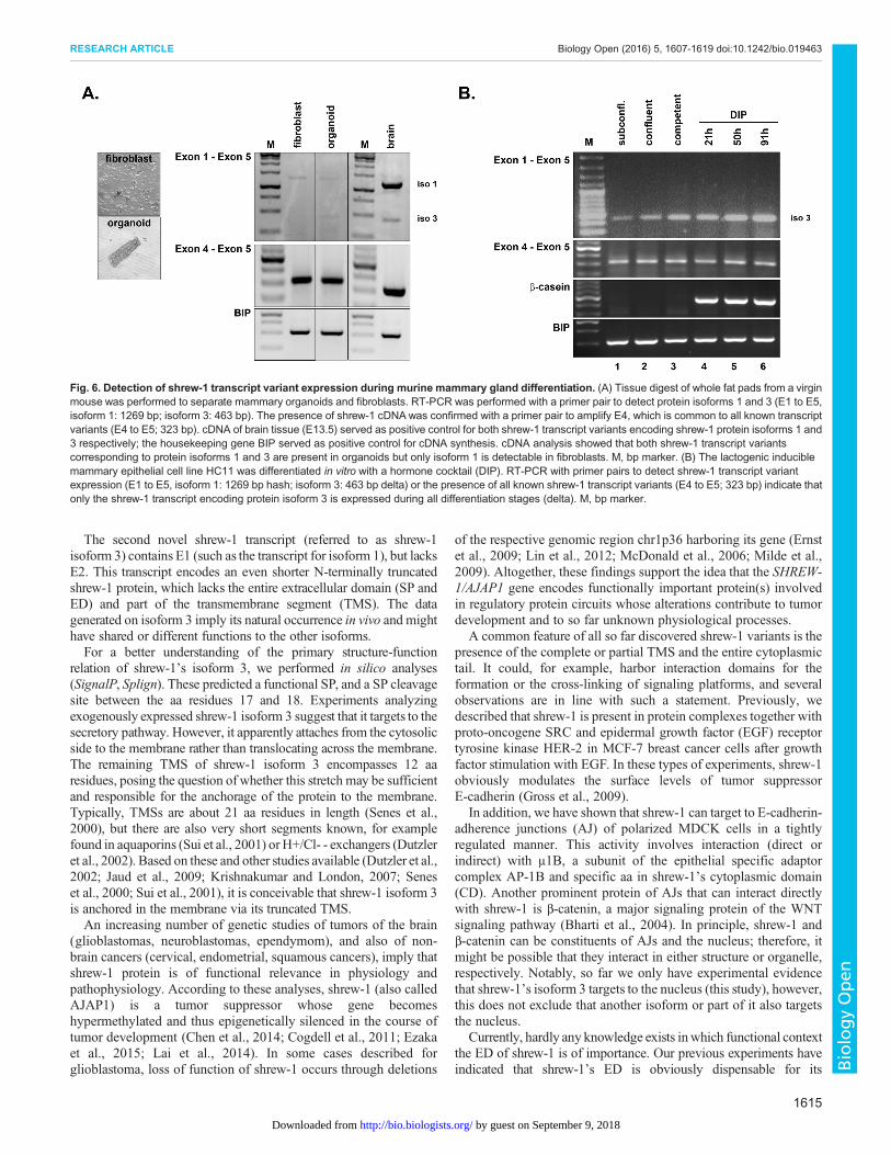
Predominant expression of shrew-1 protein isoform 3 is inmammary epitheliumThe whole fat pad protein lysates of mammary tissue collected atdifferent developmental stages confirmed the presence of shrew-1protein but revealed a discrepancy between deduced protein size ofthe identified transcript variants. As mammary fat pads consist of abilayered mammary epithelial tree surrounded by fibroblasts andadipocytes, we digested mammary gland tissue to analyze shrew-1transcript expression in purified mammary organoids (consisting ofmyoepithelial and luminal cells) and fibroblasts (Fig. 6A). RT-PCRwas performed with a primer pair to detect specifically the cDNAtranscript variants for protein isoform 1 (1269 bp) and 3 (463 bp)expression in one reaction (primer pair from E1 to E5). As a controlfor shrew-1 cDNA expression in general, exon 4 was amplified witha primer pair that is common to all identified shrew-1 transcriptvariants so far inMusmusculus (primer pair from E4 to E5; 323 bp).Brain tissue served as a positive control for shrew-1 expression,and the housekeeping gene BIP as a positive control for cDNAsynthesis. Both shrew-1 transcript variants encoding proteinisoform 1 and 3 respectively could be detected in mammaryorganoids, whilst in fibroblasts only the long shrew-1 transcriptvariant encoding protein isoform 1 could be detected. Next, weaddressed the question of whether luminal cells indeed only expressthe novel shrew-1 protein isoform 3 as suggested by the obtainedhistological analysis (Figs 1B and 2A). For this, we used HC11cells, a murine mammary luminal epithelial cell line capable oflactogenic differentiation (Ball et al., 1988). HC11 cells weredifferentiated with a hormone cocktail containing dexamethasone,insulin and prolactin (DIP) for several days to induce alveologenesisas previously described (Vafaizadeh et al., 2010). The mRNAexpression at different stages of lactogenic differentiation confirmed
that only the transcript encoding the shrew-1 protein isoform 3 ispresent in luminal cells (Fig. 6B).
Post-translational modification of shrew-1 affects proteinsizeIn previous reports (Bharti et al., 2004; Gross et al., 2009; Jakobet al., 2006; Schreiner et al., 2007), as well as in this study, shrew-1protein emerged in immunoblot analysis about 15 to 20 kDa abovethe expected size, raising the question about the nature of thisapparently increased molecular weight. This could be due to severalpossibilities such as transcript variant expression or post-translational modifications. Glycosylation of the protein wassuggested, as it is known that glycans reduce the SDS binding ofproteins and subsequently the reduced charge-to-mass ratio slowsdown the migration of the protein in the SDS-PAGE (Poduslo,1981).
Using the prediction software NetOGlyc v3.1 (Julenius et al.,2005), 34 putative O-glycosylation sites were identified in the ED ofshrew-1 (Fig. S4A). Human shrew-1 protein exhibits three clusters inthe ED with serines and threonines predicted to be O-glycosylated,encompassing the peptide stretches from the positions 126 to 137,211 to 237, and 251 to 266 in the shrew-1 sequence (Fig. S4A).Moreover, the clusters are largely conserved in shrew-1 orthologuesandwere also predicted to beO-glycosylated (data not shown). To testthe possible O-glycosylation of shrew-1, an O-glycosylation mutantwas generated substituting putative O-glycosylation sites (threoninesat the positions 230, 233 and 237 as well as serines at the positions239 and 251) by alanines. The O-glycosylation mutant, as well aswild-type shrew-1 (isoform 1), were expressed in MDCK cells.The immunoblot analysis of both shrew-1 proteins showed that thesubstitution of these five aa residues affected the apparent size of theshrew-1 protein (Fig. S4B). The shrew-1 O-glycosylation mutantappearedwith a distinct smaller size (delta) compared to thewild-typeprotein (arrowhead). Apparently, by targeting these amino acidresidues, relevant O-glycosylation sites were hit, resulting in areduced O-glycosylation and, therefore, in an apparent size closer tothe theoretical size (44.5 kDa) of the premature precursor protein.
Next, we treated lysates ofMDCK cells stably expressing shrew-1(isoform 1) with acetylneuraminyl hydrolase (neuraminidase)alone, or additionally with endo-α-N-acetylgalactosaminidase(O-glycosidase). A slight downshift of shrew-1 protein wasobserved after neuraminidase treatment (Fig. S4C, lane 2) andsubsequent additional digestion with O-glycosidase resulted in adramatic shift of the shrew-1 protein down to the calculated size ofthe precursor (brace; Fig. S4C, lane 3).
DISCUSSIONWe describe the discovery of two so far unknown transcriptvariants of the tumor suppressor shrew-1. Both novel transcriptsarise by alternative exon usage. The originally identified shrew-1transcript encodes a protein of 411 aa (isoform 1) with anunusually long signal peptide (SP, aa 1-43) that containsfunctional distinct targeting subdomains (Bharti et al., 2004;Hiss et al., 2008). The first newly identified shrew-1 transcriptvariant (referred to as isoform 2) starts with a novel exon (E1a),leading to an 11 aa shorter SP, that might bring so far unknownsubcellular targeting features to shrew-1. Notably, the NCBIRefSeq database (Pruitt et al., 2012) predicts a shrew-1 proteinisoform X2 (XP_011540089), which is identical to our newlydiscovered isoform 2. However, the putative transcript encodingisoform X2 (XM_011541787) contains additional informationnext to our identified E1a sequence.
1613
RESEARCH ARTICLE Biology Open (2016) 5, 1607-1619 doi:10.1242/bio.019463
BiologyOpen
by guest on September 9, 2018http://bio.biologists.org/Downloaded from

Fig. 5. Detection of shrew-1 isoforms in mouse organs.(A) Protein lysates of several murine organs were subjected toSDS-PAGE and analyzed by immunoblot using antibodiesrecognizing the CD of shrew-1, Sigma and Genovac F,respectively. Shrew-1 isoform 3 exogenously expressed inHEK293T was used as positive control and GAPDH as loadingcontrol. Both the Genovac F antibody as well as the Sigmaantibody recognized the double bands of iso3-shrew-1(13.4 kDa, asterisks; 17 kDa, arrowhead). Interestingly, also inthe organs (spleen, kidney, embryo head, and to a lesseramount in the liver and the brain) protein species at about17 kDa (arrowhead) were detected by both antibodies,suggesting the existence of a shrew-1 protein with the size ofisoform 3 in vivo. (B) Schematic overview of mammary glanddifferentiation cycle and intracellular localization of shrew-1isoforms by histological analysis. (C) Whole fat pad lysates ofmurine mammary gland at different developmental stages weresubjected to SDS-PAGE and analyzed by immunoblot usingantibodies recognizing the CD of shrew-1 to visualize all knownshrew-1 isoforms (abcam andGenovac F). The antibody againstβ-casein was used as differentiation marker and probing againstGAPDH as loading control. A double protein band atapprox.13 kDa representing isoform 3 could be detected withthe shrew-1 abcam antibody (long exposure) and additionalbands ranging from 34 to 72 kDa possibly representing isoform 1expression. The Genovac F antibody recognized a double bandat approx. 26 kDa. d, days; V, virgin; Inv, involution.
1614
RESEARCH ARTICLE Biology Open (2016) 5, 1607-1619 doi:10.1242/bio.019463
BiologyOpen
by guest on September 9, 2018http://bio.biologists.org/Downloaded from
The second novel shrew-1 transcript (referred to as shrew-1isoform 3) contains E1 (such as the transcript for isoform 1), but lacksE2. This transcript encodes an even shorter N-terminally truncatedshrew-1 protein, which lacks the entire extracellular domain (SP andED) and part of the transmembrane segment (TMS). The datagenerated on isoform 3 imply its natural occurrence in vivo and mighthave shared or different functions to the other isoforms.For a better understanding of the primary structure-function
relation of shrew-1’s isoform 3, we performed in silico analyses(SignalP, Splign). These predicted a functional SP, and a SP cleavagesite between the aa residues 17 and 18. Experiments analyzingexogenously expressed shrew-1 isoform 3 suggest that it targets to thesecretory pathway. However, it apparently attaches from the cytosolicside to the membrane rather than translocating across the membrane.The remaining TMS of shrew-1 isoform 3 encompasses 12 aaresidues, posing the question of whether this stretch may be sufficientand responsible for the anchorage of the protein to the membrane.Typically, TMSs are about 21 aa residues in length (Senes et al.,2000), but there are also very short segments known, for examplefound in aquaporins (Sui et al., 2001) or H+/Cl- - exchangers (Dutzleret al., 2002). Based on these and other studies available (Dutzler et al.,2002; Jaud et al., 2009; Krishnakumar and London, 2007; Seneset al., 2000; Sui et al., 2001), it is conceivable that shrew-1 isoform 3is anchored in the membrane via its truncated TMS.An increasing number of genetic studies of tumors of the brain
(glioblastomas, neuroblastomas, ependymom), and also of non-brain cancers (cervical, endometrial, squamous cancers), imply thatshrew-1 protein is of functional relevance in physiology andpathophysiology. According to these analyses, shrew-1 (also calledAJAP1) is a tumor suppressor whose gene becomeshypermethylated and thus epigenetically silenced in the course oftumor development (Chen et al., 2014; Cogdell et al., 2011; Ezakaet al., 2015; Lai et al., 2014). In some cases described forglioblastoma, loss of function of shrew-1 occurs through deletions
of the respective genomic region chr1p36 harboring its gene (Ernstet al., 2009; Lin et al., 2012; McDonald et al., 2006; Milde et al.,2009). Altogether, these findings support the idea that the SHREW-1/AJAP1 gene encodes functionally important protein(s) involvedin regulatory protein circuits whose alterations contribute to tumordevelopment and to so far unknown physiological processes.
A common feature of all so far discovered shrew-1 variants is thepresence of the complete or partial TMS and the entire cytoplasmictail. It could, for example, harbor interaction domains for theformation or the cross-linking of signaling platforms, and severalobservations are in line with such a statement. Previously, wedescribed that shrew-1 is present in protein complexes together withproto-oncogene SRC and epidermal growth factor (EGF) receptortyrosine kinase HER-2 in MCF-7 breast cancer cells after growthfactor stimulation with EGF. In these types of experiments, shrew-1obviously modulates the surface levels of tumor suppressorE-cadherin (Gross et al., 2009).
In addition, we have shown that shrew-1 can target to E-cadherin-adherence junctions (AJ) of polarized MDCK cells in a tightlyregulated manner. This activity involves interaction (direct orindirect) with μ1B, a subunit of the epithelial specific adaptorcomplex AP-1B and specific aa in shrew-1’s cytoplasmic domain(CD). Another prominent protein of AJs that can interact directlywith shrew-1 is β-catenin, a major signaling protein of the WNTsignaling pathway (Bharti et al., 2004). In principle, shrew-1 andβ-catenin can be constituents of AJs and the nucleus; therefore, itmight be possible that they interact in either structure or organelle,respectively. Notably, so far we only have experimental evidencethat shrew-1’s isoform 3 targets to the nucleus (this study), however,this does not exclude that another isoform or part of it also targetsthe nucleus.
Currently, hardly any knowledge exists inwhich functional contextthe ED of shrew-1 is of importance. Our previous experiments haveindicated that shrew-1’s ED is obviously dispensable for its
Fig. 6. Detection of shrew-1 transcript variant expression during murine mammary gland differentiation. (A) Tissue digest of whole fat pads from a virginmouse was performed to separate mammary organoids and fibroblasts. RT-PCR was performed with a primer pair to detect protein isoforms 1 and 3 (E1 to E5,isoform 1: 1269 bp; isoform 3: 463 bp). The presence of shrew-1 cDNAwas confirmed with a primer pair to amplify E4, which is common to all known transcriptvariants (E4 to E5; 323 bp). cDNA of brain tissue (E13.5) served as positive control for both shrew-1 transcript variants encoding shrew-1 protein isoforms 1 and3 respectively; the housekeeping gene BIP served as positive control for cDNA synthesis. cDNA analysis showed that both shrew-1 transcript variantscorresponding to protein isoforms 1 and 3 are present in organoids but only isoform 1 is detectable in fibroblasts. M, bp marker. (B) The lactogenic induciblemammary epithelial cell line HC11 was differentiated in vitro with a hormone cocktail (DIP). RT-PCR with primer pairs to detect shrew-1 transcript variantexpression (E1 to E5, isoform 1: 1269 bp hash; isoform 3: 463 bp delta) or the presence of all known shrew-1 transcript variants (E4 to E5; 323 bp) indicate thatonly the shrew-1 transcript encoding protein isoform 3 is expressed during all differentiation stages (delta). M, bp marker.
1615
RESEARCH ARTICLE Biology Open (2016) 5, 1607-1619 doi:10.1242/bio.019463
BiologyOpen
by guest on September 9, 2018http://bio.biologists.org/Downloaded from
basolateral targeting since shrew-1 without its ED integrates into AJs,although gives rise to less defined localization (Jakob et al., 2006).The first insight into a putative function of the shrew-1 ED as abinding partner for receptor complex composition comes from arecent publication onmammalian brain extracts. Here, proteomic dataindicate that shrew-1 peptides from both the extracellular andcytoplasmic domain could interact with GABAB receptor subunits(Schwenk et al., 2015). Since we show that shrew-1’s ED is a targetfor O-glycosylation, it would be of high interest to investigate to whatextent changes in the composition of the attached glycans may affectshrew-1’s ED protein-protein interaction.The mouse mammary gland is an informative model system
allowing analysis of shrew-1 isoforms in vivo at differentdevelopmental stages. The fact that shrew-1 detection byantibodies differed within the epithelium and during functionaldifferentiation indicated that at least two protein isoforms of shrew-1are present. A colocalization signal of the antibody staining forshrew-1 against the ED and CD in myoepithelial cells suggests thatshrew-1 isoform 1 or 2 is present. The discrimination between thesetwo shrew-1 isoforms is not feasible with the currently availableantibodies.In contrast, the absence of antibody staining against the ED of
shrew-1 in luminal cells suggests that only isoform 3 is present. Inline with this, the murine mammary luminal epithelial cell lineHC11 expresses only shrew-1 isoform 3. The main function ofluminal cells is the differentiation into milk-secreting alveolar cellsduring pregnancy, a process driven by prolactin signaling via theJAK-STAT5 pathway (Cui et al., 2004; Gallego et al., 2001;Hennighausen et al., 1997). In parallel, localization of shrew-1 inluminal epithelial cells changes during lactation up to earlyinvolution. Here, the nuclear localization could suggest aparticipation in specific signaling and transcription events duringdifferentiation and remodeling.In contrast, the main functional feature of myoepithelial cells is to
elicit structural support to the luminal cells by, for example, secretingbasement membrane components, matrix metalloproteinases orparacrine signals and exerting contractile force mediated byoxytocin during lactation. They also mediate signals from thestroma to the luminal cells influencing their polarity (Adriance et al.,2005; Gudjonsson et al., 2005, 2002; Pandey et al., 2010). Shrew-1isoform 1 and/or 2, the full-length transmembrane protein variants,show an overlapping distribution pattern with SMA, the contractileactin (ACTA2) in myoepithelial cells required for milk ejection(Haaksma et al., 2011). This could imply a role of shrew-1 isoform1and/or 2 in mediation of signals to exert mechanical force or pressurefor milk ejection.In summary, we conclude from our data that shrew-1 exists in
several protein variants depending on cellular and functionalcontext. Although the transcripts for the three shrew-1 isoformsare detectable in human and murine organs, our analyses indicatecell type- and tissue-specific, as well as overlapping, expressionpatterns. These data are important observations for further analysisof shrew-1 gene products and their putative functional relevance. Itis required to assess which shrew-1 isoforms are present within ananalyzed cell type or tissue in order to unravel the individualsubcellular targeting, function and/or regulation.
MATERIALS AND METHODSAntibodiesAntibodies were purchased from BD Transduction Laboratories (Heidelberg,Germany; E-cadherin clone 36; IF 1:100, IB 1:1000), Sigma-Aldrich(Munich, Germany; AJAP1 HPA012157, IF 1:100, WB 1:1000; SMA clone
1A4, IF 1:100; pan-cadherin CH-19, IB 1:1000; myc C3956, IF 1:100, IB1:1000), Ambion (Darmstadt, Germany; GAPDH clone 6C5, IB 1:10,000),Thermo scientific (Darmstadt, Germany; Grp94 clone 9G10.F8.2, IB 1:500),Abcam (Cambridge, UK; AJAP1 ab121361, IF 1:100, IB 1:1000), R&DSystems (Wiesbaden, Germany; AJAP1 AF7970), AntibodyVerify (LasVegas, NV, USA; AJAP1 AAS47449C), Santa Cruz (Heidelberg, Germany;β-catenin H102, IB 1:1000; β-casein M-14, IB 1:1000) and custom madeantibodies against shrew-1 (Nanotools, IB 1:500, polyclonal Genovac andmonoclonal Genovac clone F, IF 1:50 and IB 1:500). Secondary antibodies(Alexa Fluor 488-, 594- and 647-labeled antibodies, IF 1:400)were purchasedfrom Molecular Probes (Leiden, The Netherlands) or Abcam (Cambridge,UK) and Horseradish peroxidase (HRP)-conjugated secondary antibody fromJackson Immuno Research (Dianova, Hamburg, Germany) or Biosource(Camarillo, CA, USA).
Mice, cell lines, culture and lactogenic differentiationHEK293T cells (CRL-11268; ATCC, Manassas, VA, USA) and MCF-7cells (European Collection of Cell Cultures, Salisbury, UK) were cultured inDMEM high glucose supplemented with 10% fetal calf serum (FCS),100 U ml−1 penicillin and 100 µg ml−1 streptomycin (Sigma-Aldrich,Munich, Germany) at 37°C with 5% CO2. HC11 cells were cultured inRPMI supplemented with 10% heat-inactivated FCS, 5 µg ml−1 insulin,10 ng ml−1 EGF and 100 U ml−1 penicillin and 100 µg ml−1 streptomycin(growth medium; Sigma-Aldrich, Munich, Germany) at 37°Cwith 5%CO2.For lactogenic differentiation, HC11 cells were grown to confluence andkept for further 2 days in growth medium. To induce differentiation, insulinwas removed from the growing medium for 3 days before addition of thelactogenic hormone mix (DIP: 10−7 M dexamethasone, 5 µg ml−1 insulin,5 µg ml−1 prolactin; Sigma-Aldrich, Munich, Germany) to the growingmedium without EGF for 4 days as described (Taverna et al., 1991). All celllines were Mycoplasma-negative. Female BALB/c mice (10 weeks of age)were purchased from Harlan Laboratories (Roßdorf, Germany) and allmouse procedures were performed in compliance with the German animalwelfare law, guidelines and policies. Dissected Mammary gland tissue fromBalb/c mice was minced and homogenized with ice-cold RIPA buffer(approx. 50 mg of sample in 300 µl ice cold RIPA buffer). From the clearedextract, 40 µg of total protein was separated by SDS–PAGE andimmunoblotting.
Plasmids and cloningShrew-1-myc constructs (isoform 1 and isoform 2) were generated by PCRusing the vector pEGFP-shrew-1 (Bharti et al., 2004) as a template andcloned into pcDNA3.1(-) (Invitrogen, Karlsruhe, Germany) using NheI andAcc65I restriction sites. 5′-TTG GTACCT TACAGATCC TCT TCT GAGATG AGT TTT TGT TCG CAG GAG ATT TCA AAC CAT-3′ served asthe reverse primer, tagging shrew-1 with a myc-tag at the C-terminus. Thefollowing oligonucleotides were used as forward primers: Iso1-shrew-1-myc: 5′-AAT TGCTAGCATGTGGATTCAACAGCTT-3′, Iso2-shrew-1-myc: 5′- AAT TGC TAG CAT GTC CAT CCG CTG GC-3′. Shrew-1isoform 3 was PCR-amplified from MCF-7 cells-derived cDNA (forwardprimer 5′- TTTAAGCTTATGTGGATTCAACAG-3′; reverse primer 5′-TTG GTA CCT TAG CAG GAG ATT TCA AAC CAT TTC TC-3′) andcloned into pcDNA3.1(+) (Invitrogen, Karlsruhe, Germany) using HindIIIand Acc65I restriction sites. To generate pEGFP-Golgi the sequence of theN-terminal signal-anchor of the human beta-1,4-galactosyl-transferase(GenBank Acc. No. AK293418.1) was PCR-amplified from HEK293Tcells-derived cDNA (forward primer 5′-AAT TGC TAG CAT GAG GCTTCG GGA GCC GCT C-3′, reverse primer 5′-TTG GAT CCT TGG CCCCTC CGG TCC GGA GCT CCC C-3′) and fused N-terminally to the GFPin the pEGFP-N1 vector (Clontech, Saint-Germain-en-Laye, France) usingthe NheI and BamHI restriction sites. All constructs were checked bysequencing for integrity (SRD, Bad Homburg, Germany).
Cell transfectionCells were transfected with the transfection reagent jetPEI (PEQLABBiotechnologie GmbH, Erlangen, Germany) as described (Resch et al.,2011). Briefly, 6×10⁵ HEK293T cells and 7×10⁵ MCF-7 cells, respectively,
1616
RESEARCH ARTICLE Biology Open (2016) 5, 1607-1619 doi:10.1242/bio.019463
BiologyOpen
by guest on September 9, 2018http://bio.biologists.org/Downloaded from

were seeded in 10 cm2 culture dishes. After one day, plasmid DNA wasincubated with jetPEI and subjected to the cells according to themanufacturer’s instructions. Cells were cultivated for the desired time.
Cytosol-microsomal fractionationCytosol-microsomal fractionation was performed as described (Resch et al.,2008). Briefly, 24 h after transfection, cytosolic and microsomal fractions ofcells (three cell culture dishes of 10 cm2) were isolated using the QproteomeMitochondria Isolation Kit (Qiagen, Hilden, Germany). 20 µg of proteinof each fraction was separated by SDS-PAGE and analysed byimmunoblotting.
Proteinase K protection assayMicrosomal fractions which were prepared on the very same day asdescribed above were subjected to protein K protection assay. Therefore,250 µl of the microsomal fractions (approx. 100 µg of protein) weresupplemented with 2 µl of proteinase K solution (20 mg ml−1) (Fermentas,Darmstadt, Germany) and incubated on ice for 30 min. In a parallel reaction,13 µl of 20% Triton X-100 solution was added. Both reactions were stoppedby the addition of 5 µl 0.5 M EDTA solution (pH 7.6), 11 µl proteinaseinhibitor cocktail Complete (Roche Diagnostics GmbH, Mannheim,Germany) and 93 µl pre-heated (95°C) protein sample buffer (Roti-Load1; Carl Roth, Karlsruhe, Germany) followed by SDS-PAGE andimmunoblotting.
Biotinylation and precipitation of cell surface proteinsCell surface biotinylation experiments were performed as described (Reschet al., 2011). Briefly, 24 h after transfection, cells were washed with ice coldPBS pH 8.0 and incubated with 0.5 mg ml−1 EZ-Link Sulfo-NHS-LC-Biotin (Thermo Scientific, Darmstadt, Germany) diluted in PBS pH 8.0 onice for 1 h. Cells were washed twice with ice cold PBS pH 8.0 and once withice cold 25 mM Tris-HCl, pH 8.0, lysed with 200 µl RIPA buffer (150 mMNaCl, 50 mMTris-HCl, pH 7.5, 0.5% sodium deoxycholate, 1%Nonidet P-40, 0.1% SDS) containing proteinase inhibitor cocktail Complete (RocheDiagnostics GmbH, Mannheim, Germany). Cleared cell lysates with a totalprotein amount of 150 µg adjusted to a concentration of 1 µg µl−1 wereincubated under rotation with 100 µl of a 50:50 slurry of NeutrAvidinagarose resins (Thermo Scientific, Darmstadt, Germany; resuspended inRIPA buffer) overnight. The supernatants were collected and the resins werewashed three times with RIPA buffer, supplemented with protein samplebuffer (Roti-Load 1, Carl Roth, Karlsruhe, Germany) and subjected to SDS-PAGE and immunoblotting.
ImmunoblottingIn general, separated proteins were transferred onto nitrocellulose or PVDFmembranes in a semi-dry blotting chamber (PEQLAB BiotechnologieGmbH, Erlangen, Germany). A pre-stained protein standard was used as amolecular size marker (Fermentas, Darmstadt, Germany). Membranes wereblocked with 5% nonfat milk powder or 5% BSA in Tris-buffered salineTween-20 (TBST: 10 mM Tris–HCl, pH 7.4, 150 mM NaCl and 0.05%Tween-20) for 1 h. After a single wash step with TBST, the membranes wereincubated with primary antibody overnight at 4°C, and after intensivewashing, the bound primary antibody was detected with HRP-conjugatedsecondary antibodies (Dianova, Hamburg, Germany) and HRP substratesolution (2.5 mM luminol, 0.4 mM p-coumaric acid, 100 mM Tris–HCl,pH 8.5, and 0.009% H2O2).
Immunocytochemistry and immunohistochemistryCells were seeded at a density of 100,000 cells per well onto coverslips(18 mm diameter). Coverslips were washed once with pre-warmed PBS,fixed for 10 min in 4% PFA, permeabilized for 10 min (0.5% Triton X-100in PBS) and blocked for 30 min in 10% FCS in PBS (Sigma-Aldrich,Munich, Germany). Specific antigens were detected by incubation withprimary antibodies overnight at 4°C in 10% FCS in PBS followed byincubation with 10 μg ml−1 of Alexa Fluor-labeled secondary antibodiesfrom donkey for 45 min at room temperature in the dark, counterstained for5 min with 1.5 μg ml−1 DAPI in PBS and mounted in Mowiol (Carl Roth,
Karlsruhe, Germany). Immunohistochemical analysis was performed onformalin-fixed paraffin sections (5 μm), cleared in xylene, rehydratedthrough an alcohol series followed by Antigen retrieval (10 mM sodiumcitrate buffer pH 6) using a pressure cooker (3 min, setting 2). Sections wereblocked in 10% FCS in TBS (Sigma-Aldrich, Munich, Germany) for 1 h.Primary antibodies were incubated in 10% FCS/TBS overnight at 4°Cfollowed by incubation with 10 μg ml−1 Alexa Fluor-conjugated secondaryantibody from donkey (Invitrogen GmbH, Karlsruhe, Germany) for 2 h atroom temperature in the dark. Sections were counterstained with1.5 μg ml−1 DAPI in PBS for 10 min and mounted in Mowiol. Stainingwas assessed by using a Zeiss LSM780 confocal microscope and Zensoftware. Images were processed by Fiji software (Schindelin et al., 2012).
RNA isolation, cDNA synthesis and shrew-1 transcript variantanalysisRNA isolation was performed using the RNA Extract II kit (Macherey &Nagel, Düren, Germany), according to the manufacturer’s instructions.RNA was eluted with RNase-free ddH2O and stored at −80°C. cDNAsynthesis was carried out using the M-MLV reverse transcriptase (Promega,Mannheim, Germany) according to the manufacturer’s instructions. Shrew-1 transcript variants were detected by PCR in human cDNA libraries ofdifferent organs and tissues (Clontech, Saint-Germain-en-Laye, France).For the detection of human shrew-1 Exon1-specific oligonucleotide(F_exon1_27mer) 5′-TCT GAG GCC CCG CTC CCC GAA ACG TGA-3′ and Exon1a-specific oligonucleotide (F_exon1a_29mer) 5′-ATC CGGAGCAGATCTCAT TTC CCT GAG TA-3′were used as forward primers,Exon5-specific oligonucleotide (R_3UTR_28mer) 5′-GGC GTC TGCCCT GCC CCC AGG AGG TAA A-3′) was used as the reverse primer.GAPDH was detected as a positive control with following primers:F_GAPDH 5′- TGA AGG TCG GAG TCA ACG GAT TTG GT-3′,R_GAPDH 5′-CAT GTG GGC CAT GAG GTC CAC CAC-3′. For thedetection of murine shrew-1 Exon1-specific oligonucleotide(F_exon1_mur) 5′-GTG ACC ATG TGG ATC CAA CAG C-3′ andExon4-specific oligonucleotide (F_exon4_mur) 5′-ACA CTC GGA GGAATA GCC ACC A-3′ were used as forward primers, Exon5-specificoligonucleotide (R_3UTR_mur) 5′-AGG AGG TAA AAA GCC TTCGGC-3′) was used as the reverse primer. BIP was detected as a positivecontrol with following primers: F_BIP 5′- TAC ACT TGG TAT TGA AACTG-3′, R_BIP 5′-GGT GGC TTT CCA GCC ATT C-3′.
Neuraminidase and O-glycanase protein digestionTo remove O-linked glycans from proteins, whole cell lysates in RIPAbuffer were treated with Neuraminidase (New England Biolabs GmbH,Frankfurt am Main, Germany; Cat. No. P0720S) and Endo-α-N-acetylgalactosaminidase, also known as O-glycanase (New EnglandBiolabs; Cat. No. P0733S), according to the manufacturer’s instructions.Therefore, a total protein amount of 90 μg was adjusted with RIPA buffer toa final volume of 45 μl, followed by the addition of 5 μl of 10× GlycoproteinDenaturing Buffer (5% SDS, 0.4 M DTT). Proteins were denatured byheating at 95°C for 10 min. The chilled reaction was supplemented with 7 μlG7 Buffer (0.5 M sodium phosphate, pH 7.5) and 7 μl of 10% NP-40. Thereaction was divided in to three equal parts. One third was supplementedwith 2 μl ddH2O giving the negative control. The second third wassupplemented with 1 μl ddH2O and 1 μl Neuraminidase (50 U/μl). The lastthird was supplemented with 1 μl Neuraminidase and 1 μl O-glycanase(40,000 U/μl-1). All reactions were incubated at 37°C for 3.5 h. Thereactions were stopped by addition of protein sample buffer (Roti-Load 1;Carl Roth, Karlsruhe, Germany, K929.1).
IHCP with antibody preabsorptionPreabsorption of the antibody against the cytoplasmic domain of shrew-1(ab121361, abcam) was performed according to the manufacturer’sinstruction (PREST protocol, Atlas Antibodies, Stockholm, Sweden).Briefly, the primary antibody mixture (shrew-1 ab121361 and anti-SMA)together with shrew-1 peptides (5 μl each of in vitro translated human shrew-1 protein aa1-411 and aa1-282 together with 30 μl GST-Shrew-1 aa 304-411) was incubated rotating overnight at 4°C followed by a centrifugation
1617
RESEARCH ARTICLE Biology Open (2016) 5, 1607-1619 doi:10.1242/bio.019463
BiologyOpen
by guest on September 9, 2018http://bio.biologists.org/Downloaded from
step [(15 min at 16.8 g (13200 rpm)] to pellet immune complexes priorproceeding with the immunohistology protocol. As a control, the primaryantibody mixture without the addition of shrew-1 peptides was treated in thesameway. The addition of the anti-SMA antibody served as an internal controlfor successful staining procedure. Stainingwas assessed using a Zeiss LSM780confocalmicroscope and Zen software (http://www.zeiss.de/microscopy/de_de/produkte/mikroskopsoftware/zen.html#downloads). Images were processed byFiji software (Schindelin et al., 2012).
Computational analysisThe presence and location of signal peptide cleavage sites were analyzed withSignalP (http://www.cbs.dtu.dk/services/SignalP/; Petersen et al., 2011),putative splice sites were analyzed with Splign (http://www.ncbi.nlm.nih.gov/sutils/splign/splign.cgi; Kapustin et al., 2008) and O-glycosylation sites wereidentified with NetOGlyc (http://www.cbs.dtu.dk/services/NetOGlyc/;(Julenius et al., 2005).
AcknowledgementsWe thank Heinz Schewe from the FCAM facility (Goethe University) for technicalassistance.
Competing interestsThe authors declare no competing or financial interests.
Author contributionsE.H.K.S. analyzed the data and edited the paper. P.A.B.K., E.R. and A.S.-P.conceived and designed the experiments. P.A.B.K., E.R. and I.S. performed theexperiments and P.A.B.K., E.R., K.E. analyzed the data. P.A.B.K., E.R. and A.S.-P.wrote the paper, P.A.B.K. prepared the figures and all authors edited the paper priorsubmission.
FundingThis work was funded by the Deutsche Forschungsgemeinschaft (GermanResearch council) [grant no. DFG STA 187-17-1 and EXC 115].
Supplementary informationSupplementary information available online athttp://bio.biologists.org/lookup/doi/10.1242/bio.019463.supplemental
ReferencesAdriance, M. C., Inman, J. L., Petersen, O. W. and Bissell, M. J. (2005).Myoepithelial cells: good fences make good neighbors. Breast Cancer Res. 7,190-197.
Ball, R. K., Friis, R. R., Schoenenberger, C. A., Doppler, W. and Groner, B.(1988). Prolactin regulation of beta-casein gene expression and of a cytosolic 120-kd protein in a clonedmousemammary epithelial cell line. EMBO J. 7, 2089-2095.
Bharti, S., Handrow-Metzmacher, H., Zickenheiner, S., Zeitvogel, A., Baumann,R. and Starzinski-Powitz, A. (2004). Novel membrane protein shrew-1 targets tocadherin-mediated junctions in polarized epithelial cells. Mol. Biol. Cell 15,397-406.
Chen, Y.-C., Huang, R.-L., Huang, Y.-K., Liao, Y.-P., Su, P.-H., Wang, H.-C.,Chang, C.-C., Lin, Y.-W., Yu, M.-H., Chu, T.-Y. et al. (2014). Methylomicsanalysis identifies epigenetically silenced genes and implies an activation of beta-catenin signaling in cervical cancer. Int. J. Cancer 135, 117-127.
Cogdell, D., Chung, W., Liu, Y., McDonald, J. M., Aldape, K., Issa, J.-P. J., Fuller,G. N. and Zhang, W. (2011). Tumor-associated methylation of the putative tumorsuppressor AJAP1 gene and association between decreased AJAP1 expressionand shorter survival in patients with glioma. Chin. J. Cancer 30, 247-253.
Cui, Y., Riedlinger, G., Miyoshi, K., Tang,W., Li, C., Deng, C.-X., Robinson, G.W.and Hennighausen, L. (2004). Inactivation of Stat5 in mouse mammaryepithelium during pregnancy reveals distinct functions in cell proliferation,survival, and differentiation. Mol. Cell. Biol. 24, 8037-8047.
Dutzler, R., Campbell, E. B., Cadene, M., Chait, B. T. and MacKinnon, R. (2002).X-ray structure of a ClC chloride channel at 3.0 A reveals the molecular basis ofanion selectivity. Nature 415, 287-294.
Ernst, A., Hofmann, S., Ahmadi, R., Becker, N., Korshunov, A., Engel, F.,Hartmann, C., Felsberg, J., Sabel, M., Peterziel, H. et al. (2009). Genomic andexpression profiling of glioblastoma stem cell-like spheroid cultures identifies noveltumor-relevant genes associated with survival. Clin. Cancer Res. 15, 6541-6550.
Ezaka, K., Kanda, M., Sugimoto, H., Shimizu, D., Oya, H., Nomoto, S., Sueoka,S., Tanaka, Y., Takami, H., Hashimoto, R. et al. (2015). Reduced expression ofadherens junctions associated protein 1 predicts recurrence of hepatocellularcarcinoma after curative hepatectomy. Ann. Surg. Oncol. 22, 1499.
Gallego, M. I., Binart, N., Robinson, G. W., Okagaki, R., Coschigano, K. T.,Perry, J., Kopchick, J. J., Oka, T., Kelly, P. A. and Hennighausen, L. (2001).Prolactin, growth hormone, and epidermal growth factor activate Stat5 in differentcompartments of mammary tissue and exert different and overlappingdevelopmental effects. Dev. Biol. 229, 163-175.
Gross, J. C., Schreiner, A., Engels, K. and Starzinski-Powitz, A. (2009). E-cadherin surface levels in epithelial growth factor-stimulated cells depend onadherens junction protein shrew-1. Mol. Biol. Cell 20, 3598-3607.
Gudjonsson, T., Rønnov-Jessen, L., Villadsen, R., Rank, F., Bissell, M. J. andPetersen, O. W. (2002). Normal and tumor-derived myoepithelial cells differ intheir ability to interact with luminal breast epithelial cells for polarity and basementmembrane deposition. J. Cell Sci. 115, 39-50.
Gudjonsson, T., Adriance, M. C., Sternlicht, M. D., Petersen, O. W. and Bissell,M. J. (2005). Myoepithelial cells: their origin and function in breast morphogenesisand neoplasia. J. Mammary Gland Biol. Neoplasia 10, 261-272.
Haaksma, C. J., Schwartz, R. J. and Tomasek, J. J. (2011). Myoepithelial cellcontraction and milk ejection are impaired in mammary glands of mice lackingsmooth muscle alpha-actin. Biol. Reprod. 85, 13-21.
Hennighausen, L. and Robinson, G. W. (2001). Signaling pathways in mammarygland development. Dev. Cell 1, 467-475.
Hennighausen, L. and Robinson, G. W. (2005). Information networks in themammary gland. Nat. Rev. Mol. Cell Biol. 6, 715-725.
Hennighausen, L., Robinson, G. W., Wagner, K.-U. and Liu, W. (1997). Prolactinsignaling in mammary gland development. J. Biol. Chem. 272, 7567-7569.
Hiss, J. A., Resch, E., Schreiner, A., Meissner, M., Starzinski-Powitz, A. andSchneider, G. (2008). Domain organization of long signal peptides of single-passintegral membrane proteins reveals multiple functional capacity. PLoS ONE 3,e2767.
Inman, J. L., Robertson, C., Mott, J. D. and Bissell, M. J. (2015). Mammary glanddevelopment: cell fate specification, stem cells and the microenvironment.Development 142, 1028-1042.
Jakob, V., Schreiner, A., Tikkanen, R. and Starzinski-Powitz, A. (2006).Targeting of transmembrane protein shrew-1 to adherens junctions is controlledby cytoplasmic sorting motifs. Mol. Biol. Cell 17, 3397-3408.
Jaud, S., Fernandez-Vidal, M., Nilsson, I. M., Meindl-Beinker, N. M., Hubner,N. C., Tobias, D. J., von Heijne, G. and White, S. H. (2009). Insertion of shorttransmembrane helices by the Sec61 translocon. Proc. Natl. Acad. Sci. USA 106,11588-11593.
Julenius, K., Molgaard, A., Gupta, R. and Brunak, S. (2005). Prediction,conservation analysis, and structural characterization of mammalian mucin-typeO-glycosylation sites. Glycobiology 15, 153-164.
Kapustin, Y., Souvorov, A., Tatusova, T. and Lipman, D. (2008). Splign:algorithms for computing spliced alignments with identification of paralogs. Biol.Direct 3, 20.
Krishnakumar, S. S. and London, E. (2007). Effect of sequence hydrophobicityand bilayer width upon the minimum length required for the formation oftransmembrane helices in membranes. J. Mol. Biol. 374, 671-687.
Lai, H.-C., Wang, Y.-C., Yu, M.-H., Huang, R.-L., Yuan, C.-C., Chen, K.-J., Wu, C.-C., Chiang, K.-J. andChao, T.-K. (2014). DNAmethylation as a biomarker for thedetection of hidden carcinoma in endometrial atypical hyperplasia. Gynecol.Oncol. 135, 552-559.
Lin, N., Di, C., Bortoff, K., Fu, J., Truszkowski, P., Killela, P., Duncan, C.,McLendon, R., Bigner, D., Gregory, S. et al. (2012). Deletion or epigeneticsilencing of AJAP1 on 1p36 in glioblastoma. Mol. Cancer Res. 10, 208-217.
Matsusaka, K., Kaneda, A., Nagae, G., Ushiku, T., Kikuchi, Y., Hino, R., Uozaki,H., Seto, Y., Takada, K., Aburatani, H. et al. (2011). Classification of Epstein-Barr virus-positive gastric cancers by definition of DNAmethylation epigenotypes.Cancer Res. 71, 7187-7197.
McDonald, J. M., Dunlap, S., Cogdell, D., Dunmire, V., Wei, Q., Starzinski-Powitz, A., Sawaya, R., Bruner, J., Fuller, G. N., Aldape, K. et al. (2006). TheSHREW1 gene, frequently deleted in oligodendrogliomas, functions to inhibit celladhesion and migration. Cancer Biol. Ther. 5, 300-304.
Milde, T., Pfister, S., Korshunov, A., Deubzer, H. E., Oehme, I., Ernst, A.,Starzinski-Powitz, A., Seitz, A., Lichter, P., von Deimling, A. et al. (2009).Stepwise accumulation of distinct genomic aberrations in a patient withprogressively metastasizing ependymoma. Genes Chromosomes Cancer 48,229-238.
Pandey, P. R., Saidou, J. and Watabe, K. (2010). Role of myoepithelial cells inbreast tumor progression. Front. Biosci. 15, 226-236.
Petersen, T. N., Brunak, S., von Heijne, G. and Nielsen, H. (2011). SignalP 4.0:discriminating signal peptides from transmembrane regions. Nat Methods 8,785-786.
Poduslo, J. F. (1981). Glycoprotein molecular-weight estimation using sodiumdodecyl sulfate-pore gradient electrophoresis: comparison of tris-glycine and tris-borate-EDTA buffer systems. Anal. Biochem. 114, 131-139.
Pruitt, K. D., Tatusova, T., Brown, G. R. and Maglott, D. R. (2012). NCBIReference Sequences (RefSeq): current status, new features and genomeannotation policy. Nucleic Acids Res. 40, D130-D135.
Resch, E., Quaiser, S., Quaiser, T., Schneider, G., Starzinski-Powitz, A. andSchreiner, A. (2008). Synergism of shrew-1’s signal peptide and
1618
RESEARCH ARTICLE Biology Open (2016) 5, 1607-1619 doi:10.1242/bio.019463
BiologyOpen
by guest on September 9, 2018http://bio.biologists.org/Downloaded from

transmembrane segment required for plasma membrane localization. Traffic 9,1344-1353.
Resch, E., Hiss, J. A., Schreiner, A., Schneider, G. and Starzinski-Powitz, A.(2011). Long signal peptides of RGMa and DCBLD2 are dissectible intosubdomains according to the NtraC model. Mol. Biosyst. 7, 942-951.
Schindelin, J., Arganda-Carreras, I., Frise, E., Kaynig, V., Longair, M., Pietzsch,T., Preibisch, S., Rueden, C., Saalfeld, S., Schmid, B. et al. (2012). Fiji: anopen-source platform for biological-image analysis. Nat. Methods 9, 676-682.
Schreiner, A., Ruonala, M., Jakob, V., Suthaus, J., Boles, E., Wouters, F. andStarzinski-Powitz, A. (2007). Junction protein shrew-1 influences cell invasion andinteracts with invasion-promoting protein CD147. Mol. Biol. Cell 18, 1272-1281.
Schwenk, J., Perez-Garci, E., Schneider, A., Kollewe, A., Gauthier-Kemper, A.,Fritzius, T., Raveh, A., Dinamarca, M. C., Hanuschkin, A., Bildl, W. et al.(2015). Modular composition and dynamics of native GABAB receptors identifiedby high-resolution proteomics. Nat. Neurosci. 19, 233-242.
Senes, A., Gerstein, M. and Engelman, D. M. (2000). Statistical analysis of aminoacid patterns in transmembrane helices: the GxxxG motif occurs frequently and inassociation with beta-branched residues at neighboring positions. J. Mol. Biol.296, 921-936.
Sui, H., Han, B.-G., Lee, J. K., Walian, P. and Jap, B. K. (2001). Structural basis ofwater-specific transport through the AQP1 water channel. Nature 414, 872-878.
Taverna, D., Groner, B. and Hynes, N. E. (1991). Epidermal growth factor receptor,platelet-derived growth factor receptor, and c-erbB-2 receptor activation allpromote growth but have distinctive effects upon mouse mammary epithelial celldifferentiation. Cell Growth Differ. 2, 145-154.
Vafaizadeh, V., Klemmt, P., Brendel, C., Weber, K., Doebele, C., Britt, K., Grez,M., Fehse, B., Desrivieres, S. and Groner, B. (2010). Mammary epithelialreconstitution with gene-modified stem cells assigns roles to Stat5 in luminalalveolar cell fate decisions, differentiation, involution, and mammary tumorformation. Stem Cells 28, 928-938.
1619
RESEARCH ARTICLE Biology Open (2016) 5, 1607-1619 doi:10.1242/bio.019463
BiologyOpen
by guest on September 9, 2018http://bio.biologists.org/Downloaded from