Embed Size (px)
Citation preview

RESEARCH ARTICLE
Endothelial cells regulate neural crest and second heart fieldmorphogenesis
Michal Milgrom-Hoffman1, Inbal Michailovici1, Napoleone Ferrara2, Elazar Zelzer3 and Eldad Tzahor1,*
ABSTRACT
Cardiac and craniofacial developmental programs are intricately
linked during early embryogenesis, which is also reflected by a high
frequency of birth defects affecting both regions. The molecular
nature of the crosstalk between mesoderm and neural crest
progenitors and the involvement of endothelial cells within the
cardio–craniofacial field are largely unclear. Here we show in the
mouse that genetic ablation of vascular endothelial growth factor
receptor 2 (Flk1) in the mesoderm results in early embryonic
lethality, severe deformation of the cardio–craniofacial field, lack of
endothelial cells and a poorly formed vascular system. We provide
evidence that endothelial cells are required for migration and
survival of cranial neural crest cells and consequently for the
deployment of second heart field progenitors into the cardiac
outflow tract. Insights into the molecular mechanisms reveal marked
reduction in Transforming growth factor beta 1 (Tgfb1) along
with changes in the extracellular matrix (ECM) composition. Our
collective findings in both mouse and avian models suggest that
endothelial cells coordinate cardio–craniofacial morphogenesis, in
part via a conserved signaling circuit regulating ECM remodeling by
Tgfb1.
KEY WORDS: Endothelial cell, Neural crest, Second heart field,
ECM
INTRODUCTIONThe anterior region of the embryo gives rise to the head andheart through overlapping morphogenetic processes. Pharyngeal
mesoderm cells contribute to significant parts of the developingheart and the head musculature (Grifone and Kelly, 2007; Tzahor,
2009; Tzahor and Evans, 2011), as well as to endothelial cells
(EC). Pharyngeal mesoderm surrounds the pharynx and containsboth paraxial and splanchnic mesoderm regions that later form
the core of the pharyngeal arches (Nathan et al., 2008). Inaddition to pharyngeal muscles, pharyngeal mesoderm also
contributes to both poles of the heart, following the formationof the linear heart tube. The cardiogenic population of pharyngeal
mesoderm cells is known as the second heart field (SHF), or
anterior heart field (Buckingham et al., 2005; Tzahor and Evans,
2011). Perturbations in the recruitment of these cells to the heart
tube can lead to a wide range of congenital heart defects. Suchdefects occur in nearly 1% of live births, reflecting the complexcellular processes underlying heart development (Srivastava,
1999; Buckingham et al., 2005).
From an embryonic point of view, the development ofthe head–heart region should be considered as a singlemorphogenetic field, in which every tissue in it is influencedby neighboring tissues (Hutson and Kirby, 2003). Due to
the anatomical proximity during early embryogenesis andoverlapping progenitor populations, cardiac and craniofacialbirth defects are often linked (Grifone and Kelly, 2007; Tzahor,
2009; Tzahor and Evans, 2011). DiGeorge syndrome (DGS) isone of the most frequent chromosomal deletion syndrome inhumans (Yamagishi and Srivastava, 2003; Baldini, 2005). Its
clinical features broadly include cardiac defects, craniofacial andaortic arch anomalies. Previous studies addressing DGS etiologyimplicate a series signaling pathways such as FGF (Guo et al.,
2011), VEGF (Stalmans et al., 2003), retinoic acid (Roberts et al.,2006; Ryckebusch et al., 2010) and TGFbeta (Wurdaket al., 2005; Choudhary et al., 2006). These studies highlightthe linkage in signaling circuits in different cell types during
cardiac and craniofacial development processes, whose nature arelargely unknown.
Cranial neural crest (NC) cells migrate to the pharyngealarches (Noden, 1983; Trainor and Tam, 1995). Craniofacial
malformations are attributed to defects in the patterning,proliferation, migration or differentiation of this cell population(Noden and Trainor, 2005). Cardiac NC located caudal to the
cranial NC territory, were found to be critical for normal heartdevelopment (Kirby et al., 1983). Malfunction of the cardiacNC affects the caudal cardio–craniofacial field whereas the
involvement of cranial NC population in cardiac development isfar less studied.
Blocking agents against various ECM molecules perturb NCcell migration (Tucker, 2004). The ECM is known to regulate
numerous cellular processes as are ECM modulating factors,which affect the geometry and composition of the ECM.Individual ECM components are laid down, cross-linked, andorganized together via covalent and noncovalent modifications.
Deregulation of these control mechanisms can lead to varioushuman pathologies. TGFb signaling has been shown to be animportant modulator of the ECM by stimulating the synthesis of
ECM components (Wells and Discher, 2008). Conventionalknockout of either Tgfbr2 (Oshima et al., 1996) or Tgfb1(Dickson et al., 1995) in mice results in developmental
retardation and early mortality attributed to the loss of ECintegrity. Indeed, Tgfb1 signaling plays a dominant role indevelopment the vascular network (ten Dijke and Arthur, 2007).
During vertebrate embryogenesis the circulatory system is the
first functioning physiological system to emerge together with the
1Department of Biological Regulation, Weizmann Institute of Science, Rehovot76100, Israel. 2Department of Pathology, University of California at San Diego, LaJolla, CA 92093, USA. 3Department of Molecular Genetics, Weizmann Institute ofScience, Rehovot 76100, Israel.
*Author for correspondence ([email protected])
This is an Open Access article distributed under the terms of the Creative Commons AttributionLicense (http://creativecommons.org/licenses/by/3.0), which permits unrestricted use, distributionand reproduction in any medium provided that the original work is properly attributed.
Received 25 February 2014; Accepted 3 June 2014
� 2014. Published by The Company of Biologists Ltd | Biology Open (2014) 3, 679–688 doi:10.1242/bio.20148078
679
BiologyOpen
by guest on June 17, 2018http://bio.biologists.org/Downloaded from

blood system. Its main function is to deliver oxygen and nutrientsto the developing tissues, although accumulating evidence
supports a perfusion-independent signaling role(s) for EC.Development of the liver and pancreas has been shown to bedependent on signals from blood vessels (Lammert et al., 2001;Matsumoto et al., 2001). Thus, EC can provide instructive
regulatory signals to other cell types (Cleaver and Melton, 2003).In this study we addressed the roles of EC in the morphogenesis
of the cardio–craniofacial field. Embryos deficient of EC, via the
ablation of Flk1 in the cardio–craniofacial mesoderm, exhibitedarrested development of NC and SHF progenitors in additionto vascular defects. Several ECM genes were affected in these
conditional mutants, including EC-derived Tgfb1, suggesting thatEC are required for the integrity of the ECM. Conditional TgfbR2knockout in mesoderm progenitors resulted in cardiac SHF
phenotype supporting a critical role of Tgfb1 signaling in thecrosstalk within the cardio–craniofacial field. Finally, we were ableto phenocopy the mouse phenotype in the chick model using aVEGFR2 inhibitor. Chick embryos treated with the inhibitor had
similar phenotypic and molecular changes as in the mouse model.Taken together, our findings suggest that Tgfb1 secreted by ECregulates ECM remodeling that is crucial for proper cardio–
craniofacial morphogenetic development.
MATERIALS AND METHODSPreparation of chick/quail embryosFertilized chick/quail eggs were incubated at 38 C under 80% humidity;
embryos were staged according to Hamburger and Hamilton.
Inhibitors administrationThe VEGFR tyrosine kinase inhibitor II (EMD Millipore) at 5 mg/ml in
DMSO was diluted 1:150,000 in PBS and administered to stage 5 Quail/
chick embryos. As a control, the same dilution of DMSO was used.
Embryos were then further incubated until reaching the required
developmental stage.
RNA extraction and amplificationRNA was harvested using a microRneasy Kit (Quaigen). cDNA was
synthesized from DNase-treated total RNA, using a M-MLV reverse
transcriptase-mediated extension of random primers, followed by a RT-
PCR amplification using different sets of primers (available upon
request).
In situ hybridizationWhole-mount in situ hybridization was performed using digoxigenin-
labeled antisense riboprobes synthesized from the cDNAs. A full list of
the in situ hybridization probes and a detailed protocol are available upon
request. Images were obtained using a Leica MZ16FA stereomicroscope
attached to a digital camera (DC300F, Leica Microsystems).
Mouse linesConditional knockout embryos were generated using either MesP1Cre, or
Tie2Cre mice, crossed with the TgfbR2 conditional mice (Chytil et al.,
2002; Koni et al., 2001; Saga et al., 1996). For FACS isolation Tie2Cre
crossed with the Rosa26YFP reporter or Pax3Cre crossed with the
Rosa26Tomato reporter.
All animal experiments were performed in accordance with the
Weizmann Institute of Science regulations for animal care and handling.
ImmunofluorescenceCryo sections were blocked with 5% horse serum, and incubated with the
following antibodies: QH1, PECAM1, ISL1, PAX7 (DSHB), AP-2 alpha
(Novus Biologicals), P-smad2/3 and NKX2.5 (Santa-Cruz), Col1 and
Tgfb1 (Abcam) and FLK1 supplied by Philip Thorpe’s lab (UT
Southwestern Medical Center). Secondary antibodies used were Cy2-,
Cy3-, or Cy5-conjugated anti-mouse, anti-rat or anti-rabbit IgG, Cy3-
conjugated anti-mouse IgG1, and Cy5-conjugated anti-mouse IgG2b
(1:100, Jackson ImmunoResearch).
RESULTSFlk1 and endothelial cells regulate cardiac and craniofacialmorphogenesisThe expression of Vascular Endothelial Growth Factor Receptor 2
(Flk1) in early mesodermal cells marks progenitors with a broadlineage potential, although it is thought that this gene is primarilynecessary for the formation of endothelial and hematopoietic
lineages (Shalaby et al., 1995; Motoike et al., 2003). We havepreviously used the conditional allele of Flk1 with severalmesodermal Cre deletors, as a specific and effective method
to induce EC dysfunction (Milgrom-Hoffman et al., 2011).Conditional ablation of this gene in the MesP1 lineage,encompassing the entire anterior mesoderm of the embryo (Sagaet al., 1999), resulted in loss of EC in the anterior region of the
embryo and mutant embryos die at E9.5 (Fig. 1A,B,D,E). Thesemutant embryos were developmentally retarded; the pharyngeal
Fig. 1. Genetic ablation of Flk1 in mouse embryosleads to a loss of endothelial cells. (A) E9.5 controlembryo compared to matching stage (B)Mesp1Cre Flk1
and (C) Tie2Cre Flk1 mutants. Note the poorlydeveloped heart and pharyngeal arches in mutantembryos. Dotted lines indicate the level of sections.Transverse sections of E9.5 (D) control embryo,(E) Mesp1Cre Flk1 and (F) Tie2Cre Flk1 mutants.Sections are stained with the endothelial markerPECAM1 showing decrease in endothelial cells inmutant embryos. Scale bars: 100 mm.
RESEARCH ARTICLE Biology Open (2014) 3, 679–688 doi:10.1242/bio.20148078
680
BiologyOpen
by guest on June 17, 2018http://bio.biologists.org/Downloaded from

arches were smaller and malformed compared to control embryos,the heart tube was shorter and not properly looped (Fig. 1B,E).
We next generated EC specific Flk1 cKO mutants using the
Tie2Cre. As with the MesP1Cre, we observed a reduction inPECAM1+ EC (Fig. 1F). The phenotype of the Tie2Cre Flk1
mutant embryos was milder compared to the Mesp1Cre Flk1
mutants, yet preserved the trend of hypomorphic pharyngealarches and abnormal heart looping (Fig. 1C,F). Results forTie2Cre Flk1 cKO mutants are mostly shown in supplementary
material Figs S1 and S2, and Flk1 cKO mutants refer toMesP1Cre Flk1 mutants. Taken together, our Flk1 cKO mutantsreveal a possible link between the loss of EC and a cardio–craniofacial phenotype. These findings suggest a regulatory role
for EC in the morphogenesis of the cardio–craniofacial field.
Differential effects of endothelial cells on cranial neuralcrest and pharyngeal mesoderm progenitorsIn order to gain insights into the craniofacial phenotype observedin MesP1Cre and Tie2Cre Flk1 cKO mutants, we performed
in situ hybridization for different cranial NC and mesodermmarkers, the two resident cell populations in the pharyngealarches. Strikingly, NC markers were completely absent from the
second arch of the mutant embryos (Fig. 2; supplementarymaterial Fig. S1). Twist expression was decreased in the firstpharyngeal arch of Flk1 cKO mutants compared to the control
embryos, and undetected in the second arch (Fig. 2A,F). HoxA2,a specific marker for second arch NC cells (Kanzler et al., 1998),was undetected in the Flk1 cKO mutants (Fig. 2B,G). Likewise,
Dlx5 was downregulated in the first arch and undetected inthe second arch of mutant embryos compared to the control(Fig. 2C,H).
In comparison to this dramatic effect on NC gene expression,
the mesoderm markers Tbx1 and Tcf21 (Capsulin) were expressedat normal levels in the core of the two pharyngeal archesin MesP1Cre Flk1 mutants as well as in control embryos
(Fig. 2D,E,I,J,). In conclusion, these results indicate that ablationof EC in the Flk1 cKO mutants specifically affect NC but not thepharyngeal mesoderm gene expression.
Fig. 2. Cranial neural crest defects in Flk1 cKOmutants. In situ hybridization in E9.5 control and mutantembryos for the NC cell’s markers (A,F) Twist,(B,G) HoxA2 and (C,H) Dlx5. Note the down-regulationof these genes in the mutant embryo PAs. In situhybridization for the mesodermal markers (D,I) Tbx1and (E,J) Capsulin. (K–T) Immunostaining on transversesections of E9.5 embryos for (K,P) the endothelialmarker FLK1 and (L,Q) the NC progenitor marker PAX7.(M,R) Co-staining for the NC marker AP2 and cell deathmarker Casp-3. (N,S) Staining for pHis3 andcomparison of neuroepithelium proliferation in controland mutant embryos, indicated by arrowheads.(O,T) E8.5 embryo sections stained for the NCprogenitor marker PAX7. All fluorescent images exceptpanels L and Q are counterstained with DAPI (blue). pa,pharyngeal arch; nt, neural tube. Scale bars: 100 mm.
Fig. 3. Hypoxia-independent functions ofEC on NC development. (A,B) Transversesections of E9.5 Mesp1Cre Flk1 control andmutant embryos stained with hypoxiprobe. (C)Cranial neural tubes (blue) of stage 8 quailembryos dissected either with or without asmall portion of the underlying mesoderm(red). Explants were grown for 48 hours inculture. Staining for the endothelial (QH1) andNC (AP2)markers. (D) Cultures of neural tubeexplants with the underlying mesodermencompassing endothelial cells (n53/3) or (E)neural tube explant alone (n53/3). (F) Thehead region of E7.5 Mesp1Cre Flk1 controland mutant embryos dissected and culturedfor 48 hours. Co-staining for PECAM1 andAP2 in (G) control embryo explants (n55/5)and (H) mutant embryo explants (n52/2).Fluorescent images are counterstained withDAPI (blue). Scale bars: 10 mm.
RESEARCH ARTICLE Biology Open (2014) 3, 679–688 doi:10.1242/bio.20148078
681
BiologyOpen
by guest on June 17, 2018http://bio.biologists.org/Downloaded from

We next analyzed NC specific markers at a higher resolutionby staining E9.5 embryonic sections at the level of the second
arch. While FLK1 expressing cells were detected in controlembryos, underlining the ectoderm and the neuroepithelium, itsexpression in Flk1 cKO mutants was undetectable (Fig. 2K,P).PAX7 staining was comparable in the control and mutant
embryos (Fig. 2L,Q) indicating that the specification of NCwithin the neural tube was unaffected. Cranial NC cell death wasobserved by co-staining for AP2 and Caspase-3 (Fig. 2M,R).
Apart from the specific NC phenotype, sectioning the embryosenabled us to identify an additional neuroepithelium phenotype inthe Flk1 cKO mutants, which had a thin and deformed neural
tube, accompanied by a marked decrease in the proliferation ofthe neuroepithelium (Fig. 2N,S). Notably, the NC phenotype wasalso evident at E8.5 where PAX7+ NC cells in the mutant
migrated to a lesser extent compared to the control (Fig. 2O,T).Taken together, we suggest that EC loss affects specifically NCmigration and survival but not its specification.
In order to exclude the possibility that the cardio–craniofacial
defects were not a consequence of poor vasculature andimpending oxygen delivery we compared the hypoxic levelsbetween control E9.5 and Flk1 cKO embryos. Both control and
mutant embryos were positive for Hypoxiprobe staining withinthe neural tube and surrounding tissues in a comparable manner(Fig. 3A,B). Next, we employed explant culture assays to
determine the effect of EC on cranial NC migration andsurvival, by eliminating circulation and hypoxia as influencing
factors (Fig. 3C–H). Neural crest explants composed of the dorsalneural tube from stage 8 quail embryos were dissected with orwithout the adjacent mesoderm (Fig. 3C) and cultured for24 hours followed by staining for NC and endothelial markers
(Fig. 3D,E). AP2 staining was undetected in explants lacking theadjacent mesoderm, which includes QH1+ EC. Next we usedmouse embryo explants of the head region of E7.5 control
and Flk1 cKO mutant embryos (Fig. 3F). Explants were thensectioned and stained for NC and endothelial markers(Fig. 3G,H). As in the avian model, the lack of EC in the Flk1
mutants was associated with a failure of NC migration. Combinedwith the differential effects on pharyngeal mesoderm and NCgene expression patterns (Fig. 2), our data do not support the
classical explanation of a poorly formed vascular system to be themain cause for the loss of the cranial NC, but rather a signalingmechanism between EC and NC populations.
Endothelial cells affect the formation of second heart fieldderived structuresOur data revealed that NC development was compromised in
Flk1 cKO mutants (in both MesP1 and Tie2 lineages, Fig. 2). Wenext asked whether the effect on NC development can be linkedto the observed cardiac defects, which include hypotrophy and
Fig. 4. Second heart field phenotype inFlk1 cKO mutants. In situ hybridization atE9.5 for first and second heart field markersin control and mutant embryos. FHFmarkers (A,E) Tbx5, (B,F) Hand1 and (C,G)Mlc2a expression were comparablebetween control and mutant embryos. (D,H)A schematic representation of the spatiallocation of second (green) and first (yellow)heart fields in control and Mesp1Cre Flk1
mutant embryos. SHF markers (I,M) Bmp4,(J,N) Wnt11 and (K,O) Isl1 were affected inthe mutant embryos as indicated byarrowheads. (L,P) Immunostaining for ISL1on transverse sections of control andMesp1Cre Flk1 mutant embryos asindicated by arrowheads. Arrow indicatesNC cells entering the OFT. Counterstainingfor nuclei with DAPI (blue). pa, pharyngealarch; nt, neural tube; pnx, pharynx; oft,outflow tract. Scale bars: 100 mm.
RESEARCH ARTICLE Biology Open (2014) 3, 679–688 doi:10.1242/bio.20148078
682
BiologyOpen
by guest on June 17, 2018http://bio.biologists.org/Downloaded from

aberrant heart tube looping (Fig. 1). Expression of the first heartfield markers Tbx5, Hand1 and Mlc2a (Klaus et al., 2007) was
comparable between Flk1 cKO mutant and control embryos(Fig. 4A–C,E–G). In contrast, the expression of SHF markerswas changed. Wnt11, which is expressed in the outflow tract
(OFT), was completely missing in mutant embryos (Fig. 4J,N;supplementary material Fig. S2), indicating a shortening of theOFT in Flk1 cKO mutants. Interestingly, expression of Bmp4
was slightly decreased in the posterior SHF, but seem to be
upregulated in the anterior SHF (Fig. 4I,M) as was theexpression of Isl1 (Fig. 4K,O) in Flk1 cKO mutants. This wasfurther corroborated by staining for ISL1 protein, which
revealed accumulation of ISL1+ cells in the second pharyngealarch (Fig. 4L,P). In the Flk1 cKO mutants all the cells in thesecond arch and OFT were ISL1+, reflecting the loss of cranialNC cells, which are clearly seen in the control embryo (DAPI+
ISL12 in Fig. 4L). In summary, our findings suggest that theincorporation of first heart field progenitors into the linear hearttube was unaffected in Flk1 cKO mutants, while SHF
progenitors failed to migrate into the OFT and right ventricle,two structures that were mostly affected in Flk1 cKO mutants.We suggest that deployment of pharyngeal mesoderm/SHF into
the cardiac OFT is mediated by EC, directly or indirectly(Fig. 4D,H).
Endothelial cells are required for extracellular matrixremodelingThe migration of the cranial NC and SHF progenitors wasperturbed in both MesP1Cre and Tie2Cre Flk1 mutants, yielding
severe cardiac and craniofacial defects. Furthermore, a substantialdecrease in the proliferation of the neuroepithelium was detectedin these mutants (Figs 1 and 2; supplementary material Fig. S1).
This set of developmental processes have been previously foundto be dependent on extracellular cues, we therefore hypothesizedthat ECM is not properly formed in Flk1 cKO mutant embryos.
Scanning Electron Microscopy (SEM) analysis of E9 embryosrevealed structural differences between control and Flk1 cKOmutant embryos. The neural tube of the mutants was thinner anddeformed, validating our previous observations (Fig. 5A,D).
Cells delaminating from the neural tube could be identified inthe control embryo (Fig. 5B, arrowhead), but not in the mutantembryo (Fig. 5E), concurrent with the lack of NC in these
mutants. An abnormal ECM was detected in the vicinity of theneural tube of the mutant embryo (Fig. 5B,E). Highmagnification SEM images revealed relatively straight and
smooth ECM fibers in the control, compared to thinner, bentand occasionally split fibers seen in the mutants (Fig. 5C,F).
We next stained embryos’ sections for Collagen 1 (COL1), a
major component of the ECM (Lohler et al., 1984). COL1was expressed abundantly and spread throughout the entiremesenchyme in the Flk1 cKO mutants compared to the controls
Fig. 5. Extracellular matrix changes in Flk1 cKO mutants.(A,D) Scanning electron microscopy of Tie2Cre Flk1 control and mutantembryos, fractioned transversally. The neural tube is depicted in red.(B) Magnification of panel A. Arrowhead indicates a supposed NC cell.(E) Magnification of panel D. Arrowhead indicates excess ECM. (C,F) High-resolution magnification. ECM fibers morphology is different between controland mutant samples as indicated by arrowheads. (G,H) Col1 staining onsections of E9.5 Tie2Cre Flk1 control and mutant embryos. (I) Heads of E9.5Tie2Cre Flk1 control (n52 pools of 3) and mutant (n52 pools of 3) embryoswere isolated and analyzed for real time gene expression. (J–O) In situhybridization in E9.5 Mesp1Cre Flk1 control and mutant embryos for(J,N) Tgfb1, (K,O) Tgfb2, (P,L) TgfbR2 and (Q,M) Loxl2. Arrowheads indicatethe region of the SHF. (R–Y) Immunostaining on transverse sections of E9.5Mesp1Cre Flk1 control and mutant embryos. (R,V) Co-staining for TGFb1and PECAM1. (S,W) Immunostaining for p-Smad 2/3, (T,X) Collagen1 and(U,Y) Laminin. (Z) Heads of E9.5 Mesp1Cre Flk1 control (n53 pools of 3)and mutant (n53 pools of 3) embryos were isolated and analyzed for realtime gene expression. All fluorescent images are counterstained with DAPI(blue). Error bars indicate SD. nt, neural tube. Scale bars: 20 mm(A,D), 10 mm (B,E), 100 nm (C,F), 10 mm (G,H,R–Y), 100 mm (J–Q).
RESEARCH ARTICLE Biology Open (2014) 3, 679–688 doi:10.1242/bio.20148078
683
BiologyOpen
by guest on June 17, 2018http://bio.biologists.org/Downloaded from
(Fig. 5G,H). In order to gain more insight into the molecular basis
for the ECM mutant phenotype, we performed a qRT-PCRanalysis on the anterior region of Tie2Cre Flk1 mutant andcontrol embryos (Fig. 5I). As expected from our previous
findings (Fig. 1), the EC marker Pecam1 was downregulated.Transforming growth factor beta 1 (Tgfb1) (ten Dijke and Arthur,2007), implicated in both vascular and ECM development, wasalso decreased in the cKO mutants. Lysyl-oxidase-like 2 (Loxl2),
Lysyl-hydroxilase (Plod) 1 and 3, which are collagen cross linkersthat regulate ECM structure and rigidity (Kagan and Li, 2003;Myllyla et al., 2007) and Col1 were all upregulated in Tie2Cre
Flk1 mutants compared to control (Fig. 5I).These results were corroborated in the MesP1Cre Flk1
mutants. In situ hybridization revealed a marked decrease in
the expression of Tgfb1 in EC in the mutant compared to controlembryos (Fig. 5J,N). Tgfb2 has a classical SHF expressionpattern extending into the OFT (Fig. 5K). Its expression in
the Flk1 cKO mutants was consistent with a defect in thedeployment of SHF cells into the heart as these cells remainedwithin the distal part of the second arch (Fig. 5O). While TgfbR2
expression pattern and levels were comparable between control
and mutant embryos (Fig. 5L,P), Loxl2 was upregulated andshifted to the anterior SHF in the Flk1 cKO mutants comparedto control (Fig. 5M,Q). Tgfb1 protein expression was
downregulated in Flk1 cKO mutants (Fig. 5R,V), as well asthe phosphorylation of SMAD2/3, a readout for Tgfb signaling(Fig. 5S,W). Tgfb1 staining in the control embryo was notconfined to EC, suggesting other sources of Tgfb1 in the
adjacent tissues.COL1 and Laminin staining were upregulated in Flk1 cKO
mutants, in particular in the basement membrane of the neural
tube (Fig. 5T–Y). Real time gene expression analysis of bothTie2Cre and MesP1Cre mutant embryos revealed a decrease inPecam1 and Tgfb1 expression relative to the control (Fig. 5I,Z).
In contrast, Tgfb2, and the ECM modifiers Loxl2 and Plod1 andPlod3 were upregulated in the Flk1 cKO mutants (Fig. 5I,Z).Collectively we found that the loss of EC was accompanied with
a strong downregulation in both RNA and protein levels of Tgfb1,and broad changes in ECM composition and structure in Tie2Cre
and MesP1Cre Flk1 cKO mutants.
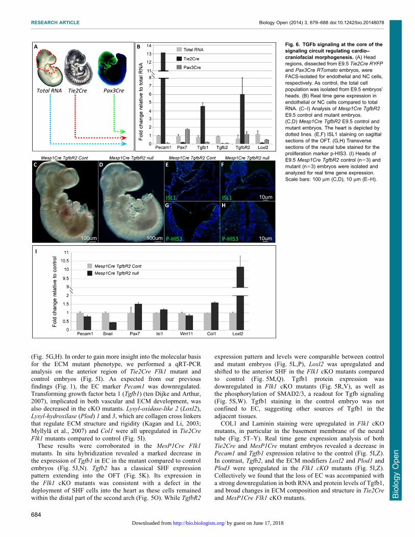
Fig. 6. TGFb signaling at the core of thesignaling circuit regulating cardio–craniofacial morphogenesis. (A) Headregions, dissected from E9.5 Tie2Cre RYFP
and Pax3Cre RTomato embryos, wereFACS-isolated for endothelial and NC cells,respectively. As control, the total cellpopulation was isolated from E9.5 embryos’heads. (B) Real time gene expression inendothelial or NC cells compared to totalRNA. (C–I) Analysis of Mesp1Cre TgfbR2
E9.5 control and mutant embryos.(C,D) Mesp1Cre TgfbR2 E9.5 control andmutant embryos. The heart is depicted bydotted lines. (E,F) ISL1 staining on sagittalsections of the OFT. (G,H) Transversesections of the neural tube stained for theproliferation marker p-HIS3. (I) Heads ofE9.5 Mesp1Cre TgfbR2 control (n53) andmutant (n53) embryos were isolated andanalyzed for real time gene expression.Scale bars: 100 mm (C,D), 10 mm (E–H).
RESEARCH ARTICLE Biology Open (2014) 3, 679–688 doi:10.1242/bio.20148078
684
BiologyOpen
by guest on June 17, 2018http://bio.biologists.org/Downloaded from

Loss of Tgfb signaling in mesoderm progenitors is partiallysimilar to second heart field phenotype observed in the Flk1mutant embryosThe finding of increased Loxl2 and decreased Tgfb1 expression inFlk1 cKO mutants led us to analyze the cell type specificexpression of these factors and other players in the TGF beta
signaling pathway. For this aim we isolated by FACS EC fromTie2Cre Rosa26YFP and NC progenitors from Pax3Cre
Rosa26YFP E9.5 embryos (Fig. 6A). The enriched expression
of Pecam1 in EC (Tie2Cre, Fig. 6B) and Pax7 in NC cells(Pax3Cre, Fig. 6B), compared to the total RNA, reflects thespecificity of our FACS assay (Fig. 6A). Tgfb1 ligand was highly
enriched in EC rather than in NC cells, whereas the receptorTgfbR2 was expressed by both cell populations.
Tissue specific ablation of TgfbR2 was previously performed
in both NC and mesoderm cells (Wurdak et al., 2005; Choudharyet al., 2006; Choudhary et al., 2009). We crossed the MesP1Cre
with the TgfbR2 floxed allele (Fig. 6C–I). Mutant embryos appearto be normal apart from a distinct cardiac phenotype of a
shortened OFT with abnormal looping (Fig. 6C,D). Sagittalsections of the embryos and ISL1 staining confirmed theshortening of the SHF and distal OFT in MesP1Cre TgfbR2
mutants (Fig. 6E,F). In addition, we observed a reduction in theproliferation of the neural epithelium of these mutants, similar tothe Flk1 cKO mutant embryos (Fig. 6G,H). Gene expression
analysis of these mutants revealed increased levels of Col1 andLoxl2 and a decrease in Snail1, a marker for migrating neuralcrest (Fig. 6I).
Inhibition of VEGFR2 signaling in chick embryosrecapitulates the Flk1 mutants’ phenotype in the mouseIn addition to the genetic ablation of Flk1 in the mouse, we used
the avian model to interfere with VEGFR2 signaling usingthe VEGFR2 tyrosine kinase inhibitor, BIO-676481. Embryosthat were treated with BIO-676481 exhibited abnormal heart
morphogenesis including shortened OFT (Fig. 7A,D). In situhybridization for Sox9 was consistent with a fewer number ofmigrating cranial NC cells in BIO-treated chick embryos
(Fig. 7B,E). The expression of the cardiac marker Nkx2.5
within the heart tube was not affected while its expressiondecreased in the SHF of the BIO-treated embryos (Fig. 7C,F).Next, BIO-treated quail embryos were sectioned and stained for
QH1, a quail specific EC marker. EC appear disorganized in BIO-treated embryos, with almost no staining in the dorsal aorta(Fig. 7G,J). The expression of the NC marker HNK1 was
markedly reduced and remains confined to the dorsal region ofthe treated embryo, compared to the control embryo (Fig. 7H,K).We also observed a decrease in the proliferation of the
neuroepithelia in BIO-treated chick embryos as shown by PH3staining (Fig. 7I,L).
Altogether these findings show that the BIO-676486 treatment
of avian embryos affected EC integrity as well as NC and SHFmorphogenesis, resembling the phenotype of the Flk1 cKO mousemutants. Our hypothesis was that excess ECM deposition(especially Collagen) and/or enhanced crosslinking of the
collagen perturb NC migration and thus lead to the cardio–craniofacial phenotype.
It is well established that cardiac NC are important for
migration of the SHF population during morphogenesis of theOFT, yet, the role of the cranial NC has not yet been addressed inthis context. In the mouse, cranial NC cells from the second arch
seem to enter the OFT together with ISL1+ cells and these cells
were absent in the Flk1 cKO mutants (Fig. 4L,P). In order toshow the importance of cranial NC in OFT morphogenesis wesurgically ablated the cranial neural tube at stage 8 chick
embryos. Cranial NC ablated chick embryos displayedmalformations of head structures, shortened OFT and abnormalcardiac looping (Fig. 7M–O) resembling the Flk1 cKO mousemutants.
Fig. 7. Inhibition of VEGFR2 signaling in chick embryos reveal cardiacand craniofacial defects and molecular changes resembling the mouseFlk1 mutants. (A–L) Analysis of HH stage 13 chick embryos treated withVEGFR2 inhibitor BIO-676481 or DMSO as control. (A,D) Control DMSO-treated (n516/18) and BIO-676481-treated (n514/18) chick embryos at HHstage 13. (B,E) ISH for the NC marker Sox9. Arrowheads indicate streams ofmigrating NC cells. (C,F) ISH for the cardiac marker Nkx2.5. Expression inthe SHF is depicted by dotted lines. (G,J) Staining for the quail endothelialspecific marker QH1 in sections of control and treated embryos HH stage 13quail embryos. (H,K) Staining for the migrating NC marker HNK-1 indicatedby arrowheads. (I,L) p-HIS3 staining in transverse sections of the neuraltube. Arrowheads point to differences in staining between control and treatedembryos. (M) Schematic illustration of cranial neural crest ablation of a HHstage 8 chick embryo. (N,O) Stage 15 control embryo (n55/6) comparedto a cranial NC ablated (n56/6) embryo. Scale bars: 200 mm (A–F), 100 mm(G–L).
RESEARCH ARTICLE Biology Open (2014) 3, 679–688 doi:10.1242/bio.20148078
685
BiologyOpen
by guest on June 17, 2018http://bio.biologists.org/Downloaded from

Taken together, our findings suggest that inhibition of VEGFR2signaling in chick embryos affects EC integrity, NC migration,
SHF looping that together result in aberrant morphogenesis of thecardio–craniofacial field. The involvement of VEGFR2 signalingand the role of EC in coordinating the cardio–craniofacialmorphogenetic field are therefore conserved in vertebrates.
DISCUSSIONThe developmental roles of EC in coordinating organogenesis
are far from being clear. Several studies gradually unravelthe significance of EC as organizers of early embryonicdevelopmental processes (Cleaver and Melton, 2003). In this
study, we shed light on a mechanism by which EC coordinatecardio–craniofacial morphogenesis, in part via a Tgfb1-mediatedECM remodeling program (Fig. 8). We have used conditional
knockout of Flk1 in either the anterior mesoderm (MesP1Cre) ormore specifically in EC (Tie2Cre) that led to a loss of EC andabnormal development of the cardio–craniofacial field. CranialNC migration and survival and SHF deployment into the cardiac
OFT and RV were abnormal in the Flk1 cKO mutants (Fig. 8A–C). Our data suggest that EC play a role in maintaining theintegrity of the extracellular environment (Fig. 8D). Further
experiments using conditional TgfbR2 knockout in mesodermprogenitors support a key role for Tgfb signaling in thisdevelopmental crosstalk. Finally, we were able to phenocopy
the mouse Flk1 cKO phenotype in the avian model using a
VEGFR2 inhibitor. We identified the collagen crosslinkingenzyme LoxL2 as a candidate gene within the ECM remodeling
program in the cardio–craniofacial field.As in many studies on EC signaling, a major issue is to
uncouple the signaling from the metabolic (systemic) roles of EC.It is likely that the loss of EC affects both functions. Consistent
with this, Hypoxia-inducible factor-1 alpha (HIF-1a) was shownto induce LoxL2 mRNA transcription in fibroblasts and renaltubular epithelial cells (Higgins et al., 2007), suggesting that the
absence of perfusion contributes to the overall phenotype.Nevertheless, our data also suggest an EC-mediated signalingcircuit. The EC-mediated cardio–craniofacial phenotype occurs
as early as E8.5 when circulation is just beginning and themajority of the embryo is hypoxic (Dunwoodie, 2009). At E9.5both control and cKO mutant embryos showed specific regions of
low oxygen levels, but without significant differences betweenthem. In addition, the molecular effects were specific for cranialNC but not pharyngeal mesoderm markers. Further, the hearts ofthe mutants were still beating and we were able to retrieve viable
mutant embryos as late as E10.5 suggesting that the embryos atE9.5 are not dying. We could show functional EC–NC crosstalkin explant culture assays, eliminating hypoxia or absence of
perfusion as major causes for the phenotype.Tgfb1 signaling has been shown to be important for EC
development as well as other cell types. Specifically, EC-derived
Tgfb1 was shown to promote smooth muscle differentiation of
Fig. 8. Endothelial cell signalingregulates the morphogenesis of thecardio–craniofacial field. (A) Endothelialcells (blue) are scattered throughout theentire dorso-ventral axis of the embryo,underlying the neural tube and entering theheart. Neural crest cells (green) follow thesame path and enter only the most arterialpole of the OFT together with SHFprogenitors (yellow). (B) The cranialprogenitor niche. ECs secrete Tgfb1 thatbalances secretion of Loxl2 and enablesproper remodeling of the ECM. (C) Loss ofECs results in reduction of Tgfb signalingand increased levels of Loxl2. Here, theECM loses its integrity causing migratorydefects of NC and SHF progenitors togetherwith reduction in proliferation of theneuroepithelium. (D) ECM remodeling ismediated by signaling from progenitors,which in turn rely on ECM integrity forproper migration and consequentmorphogenesis. nt, neural tube.
RESEARCH ARTICLE Biology Open (2014) 3, 679–688 doi:10.1242/bio.20148078
686
BiologyOpen
by guest on June 17, 2018http://bio.biologists.org/Downloaded from

trunk NC (Shah et al., 1996). Genetic ablation of Tgfb signalingin the NC, using Wnt1Cre mice, revealed a wide spectrum of
craniofacial and cardiovascular defects including specific featuresof DGS (Ito et al., 2003; Wurdak et al., 2005; Choudhary et al.,2006). Conditional knockout of the Tgfbr2 in the mesoderm waspreviously performed. MesP1Cre mutants die at E10.5 but
Mef2c-AHFCre mutants survive to E14 and display dilatedpulmonary trunk with ruptures. The ECM was highly disorganizedin the pulmonary trunk and ascending aorta (Choudhary et al., 2009).
We show that MesP1Cre TgfbR2 mutants display SHF phenotype ofshortened OFT, abnormal looping and reduced ISL1 expression.Collectively these studies suggest that Tgfb signaling is required in
both neural crest and pharyngeal mesoderm to control overlappingand distinct morphogenetic events within the cardio–craniofacialfield. Similarly, Semaphorins and VEGF signaling molecules, acting
through Npn1 (Gu et al., 2003) or PlexinD1 (Gitler et al., 2004) inEC, were shown to regulate cardiac outflow tract development.Interruption of these pathways in mice resulted in congenital heartdefects as well as vascular patterning defects. Together with our
findings, there is a strong evidence for a key role of EC inorchestrating critical aspects of cardiac and craniofacialmorphogenesis.
DGS patients are characterized with hemizygousmicrodeletions of chromosome 22q11.2. The T-box containingfamily of transcription factors TBX1 is located within this region
and haploinsufficiency of this gene promotes the manifestationsof DGS in humans. A common denominator of the organs that areaffected in DGS is their dependence on NC cells (Kochilas et al.,
2002). However, Tbx1 is not expressed and does not function inNC cells. We have recently revealed a genetic link betweenTcf21, Tbx1, and Lhx2 within the pharyngeal mesoderm. Geneticperturbation of these factors resulted in specific DGS-like
phenotypes (Harel et al., 2012). Furthermore, defective vascularorganization and EC dysfunction were recently shown to give riseto DGS-like phenotypes (Zhou et al., 2012).
Perturbation of EC development causes a distortion of ECMstructures, which in turn affects ECM-mediated neural crestmigration (Henderson and Copp, 1997; Coles et al., 2006).
Defects in NC cell migration often lead to cell death (Maynardet al., 2000), as we observed in Flk1 cKO mutant embryos. Ourdata suggest that one of the functions of EC is to modify thematrix in order to facilitate NC migration.
Cardiac NC cells have been shown to have a role in SHFdevelopment (Waldo et al., 2005). Furthermore, studies from ourlab indicated that cranial NC cells are intimately involved in a
crosstalk with pharyngeal mesoderm progenitors (including SHFcells) as they approach the distal heart tube (Tirosh-Finkel et al.,2006; Rinon et al., 2007; Tirosh-Finkel et al., 2010). Heart
looping defects were evident in cranial NC ablated chick embryos(Fig. 7). Therefore, the SHF phenotype observed in both mouseand chick embryos could be attributed to the perturbed crosstalk
between (cardiac and cranial) NC and SHF progenitors. Anotherpossible explanation for this is a direct role for EC on SHF cellmigration (Fig. 8A).
We propose that tissue crosstalk within the cardio–craniofacial
field is extensive and critical for proper embryonic development.Our findings provide insights into some of the molecular eventsthat are at the core of this tissues crosstalk. We suggest
that EC dysfunction (by loss of VEGF signaling) results indownregulation of Tgfb1, which affects both NC- and pharyngealmesoderm/SHF progenitors morphogenesis. ECM remodeling
genes appear to be directly affected by the loss of TGFb
signaling, in particularly Col1 and LoxL2. We suggest that anytissue or molecular perturbation within the cardio–craniofacial
field is likely to give rise to cardiac, pharyngeal, and craniofacialdefects as seen in DGS patients, due to the tight signaling circuitbetween all tissues.
LoxL2 is member of the Lysyl oxidase (LOX) protein family,
which is made up of copper-containing enzymes that catalyze theoxidative deamination of the e-amino groups in lysines (Smith-Mungo and Kagan, 1998). The striking upregulation of LoxL2 in
MesP1 and Tie2 Flk1 cKO mutants as well as in the MesP1
TgfbR2 cKO mutants (Figs 5 and 6) emerge as an importantfeature of the cardio–craniofacial phenotype. While we addressed
the involvement of LoxL2 as an ECM remodeling modifier it isimportant to note that this protein had been implicated in theregulation of EMT and metastasis formation via interaction with
Snail1 (Peinado et al., 2005). In addition, it was recentlydocumented that LoxL2 is a histone modifier enzyme thatcatalyzes H3K4me3 deamination (Herranz et al., 2012). Thus, theroles of LoxL2 as a key regulator of the cardio–craniofacial
morphogenetic field require further investigation.
AcknowledgementsWe thank Henry Sucov and Peleg Hasson for insightful discussions.
Competing interestsThe authors have no competing interests to declare.
FundingThis work was supported by grants to E.T. from the European Research Council;Israel Science Foundation; United States–Israel Binational Science Foundation;German Israeli Foundation; Association Francaise Contre les Myopathies and adonation from the Jack Gitlitz Estate.
ReferencesBaldini, A. (2005). Dissecting contiguous gene defects: TBX1. Curr. Opin. Genet.Dev. 15, 279-284.
Buckingham, M., Meilhac, S. and Zaffran, S. (2005). Building the mammalianheart from two sources of myocardial cells. Nat. Rev. Genet. 6, 826-837.
Choudhary, B., Ito, Y., Makita, T., Sasaki, T., Chai, Y. and Sucov, H. M. (2006).Cardiovascular malformations with normal smooth muscle differentiation inneural crest-specific type II TGFbeta receptor (Tgfbr2) mutant mice. Dev. Biol.289, 420-429.
Choudhary, B., Zhou, J., Li, P., Thomas, S., Kaartinen, V. and Sucov, H. M.(2009). Absence of TGFbeta signaling in embryonic vascular smooth muscleleads to reduced lysyl oxidase expression, impaired elastogenesis, andaneurysm. Genesis 47, 115-121.
Chytil, A., Magnuson, M. A., Wright, C.V. and Moses, H. L. (2002). Conditionalinactivation of the TGF-b type II receptor using Cre:Lox. Genesis 32, 73-75.
Cleaver, O. and Melton, D. A. (2003). Endothelial signaling during development.Nat. Med. 9, 661-668.
Coles, E. G., Gammill, L. S., Miner, J. H. and Bronner-Fraser, M. (2006).Abnormalities in neural crest cell migration in laminin alpha5 mutant mice. Dev.Biol. 289, 218-228.
Dickson, M. C., Martin, J. S., Cousins, F. M., Kulkarni, A. B., Karlsson, S.and Akhurst, R. J. (1995). Defective haematopoiesis and vasculogenesisin transforming growth factor-beta 1 knock out mice. Development 121, 1845-1854.
Dunwoodie, S. L. (2009). The role of hypoxia in development of the Mammalianembryo. Dev. Cell 17, 755-773.
Gitler, A. D., Lu, M. M. and Epstein, J. A. (2004). PlexinD1 and semaphorinsignaling are required in endothelial cells for cardiovascular development. Dev.Cell 7, 107-116.
Grifone, R. and Kelly, R. G. (2007). Heartening news for head muscledevelopment. Trends Genet. 23, 365-369.
Gu, C., Rodriguez, E. R., Reimert, D. V., Shu, T., Fritzsch, B., Richards, L. J.,Kolodkin, A. L. and Ginty, D. D. (2003). Neuropilin-1 conveys semaphorin andVEGF signaling during neural and cardiovascular development.Dev. Cell 5, 45-57.
Guo, C., Sun, Y., Zhou, B., Adam, R. M., Li, X., Pu, W. T., Morrow, B. E., Moon,A. and Li, X. (2011). A Tbx1-Six1/Eya1-Fgf8 genetic pathway controlsmammalian cardiovascular and craniofacial morphogenesis. J. Clin. Invest.121, 1585-1595.
Harel, I., Maezawa, Y., Avraham, R., Rinon, A., Ma, H. Y., Cross, J. W.,Leviatan, N., Hegesh, J., Roy, A., Jacob-Hirsch, J. et al. (2012). Pharyngealmesoderm regulatory network controls cardiac and head musclemorphogenesis. Proc. Natl. Acad. Sci. USA 109, 18839-18844.
RESEARCH ARTICLE Biology Open (2014) 3, 679–688 doi:10.1242/bio.20148078
687
BiologyOpen
by guest on June 17, 2018http://bio.biologists.org/Downloaded from

Henderson, D. J. and Copp, A. J. (1997). Role of the extracellular matrix inneural crest cell migration. J. Anat. 191, 507-515.
Herranz, N., Dave, N., Millanes-Romero, A., Morey, L., Dıaz, V. M., Lorenz-Fonfrıa, V., Gutierrez-Gallego, R., Jeronimo, C., Di Croce, L., Garcıa deHerreros, A. et al. (2012). Lysyl oxidase-like 2 deaminates lysine 4 in histoneH3. Mol. Cell 46, 369-376.
Higgins, D. F., Kimura, K., Bernhardt, W. M., Shrimanker, N., Akai, Y.,Hohenstein, B., Saito, Y., Johnson, R. S., Kretzler, M., Cohen, C. D. et al.(2007). Hypoxia promotes fibrogenesis in vivo via HIF-1 stimulation of epithelial-to-mesenchymal transition. J. Clin. Invest. 117, 3810-3820.
Hutson, M. R. and Kirby, M. L. (2003). Neural crest and cardiovasculardevelopment: a 20-year perspective.Birth Defects Res. C Embryo Today 69, 2-13.
Ito, Y., Yeo, J. Y., Chytil, A., Han, J., Bringas, P., Jr, Nakajima, A., Shuler, C. F.,Moses, H. L. and Chai, Y. (2003). Conditional inactivation of Tgfbr2 in cranialneural crest causes cleft palate and calvaria defects. Development 130, 5269-5280.
Kagan, H. M. and Li, W. (2003). Lysyl oxidase: properties, specificity, andbiological roles inside and outside of the cell. J. Cell. Biochem. 88, 660-672.
Kanzler, B., Kuschert, S. J., Liu, Y. H. and Mallo, M. (1998). Hoxa-2 restricts thechondrogenic domain and inhibits bone formation during development of thebranchial area. Development 125, 2587-2597.
Kirby, M. L., Gale, T. F. and Stewart, D. E. (1983). Neural crest cells contribute tonormal aorticopulmonary septation. Science 220, 1059-1061.
Klaus, A., Saga, Y., Taketo, M. M., Tzahor, E. and Birchmeier, W. (2007).Distinct roles of Wnt/beta-catenin and Bmp signaling during early cardiogenesis.Proc. Natl. Acad. Sci. USA 104, 18531-18536.
Kochilas, L., Merscher-Gomez, S., Lu, M. M., Potluri, V., Liao, J., Kucherlapati,R., Morrow, B. and Epstein, J. A. (2002). The role of neural crest duringcardiac development in a mouse model of DiGeorge syndrome. Dev. Biol. 251,157-166.
Koni, P. A., Joshi, S. K., Temann, U. A., Olson, D., Burkly, L. and Flavell, R. A.(2001). Conditional vascular cell adhesion molecule 1 deletion in mice: impairedlymphocyte migration to bone marrow. J. Exp. Med. 193, 741-754.
Lammert, E., Cleaver, O. and Melton, D. (2001). Induction of pancreaticdifferentiation by signals from blood vessels. Science 294, 564-567.
Lohler, J., Timpl, R. and Jaenisch, R. (1984). Embryonic lethal mutation inmouse collagen I gene causes rupture of blood vessels and is associated witherythropoietic and mesenchymal cell death. Cell 38, 597-607.
Matsumoto, K., Yoshitomi, H., Rossant, J. and Zaret, K. S. (2001). Liverorganogenesis promoted by endothelial cells prior to vascular function. Science294, 559-563.
Maynard, T. M., Wakamatsu, Y. and Weston, J. A. (2000). Cell interactions withinnascent neural crest cell populations transiently promote death of neurogenicprecursors. Development 127, 4561-4572.
Milgrom-Hoffman, M., Harrelson, Z., Ferrara, N., Zelzer, E., Evans, S. M. andTzahor, E. (2011). The heart endocardium is derived from vascular endothelialprogenitors. Development 138, 4777-4787.
Motoike, T., Markham, D. W., Rossant, J. and Sato, T. N. (2003). Evidence fornovel fate of Flk1+ progenitor: contribution to muscle lineage. Genesis 35, 153-159.
Myllyla, R., Wang, C., Heikkinen, J., Juffer, A., Lampela, O., Risteli, M.,Ruotsalainen, H., Salo, A. and Sipila, L. (2007). Expanding the lysylhydroxylase toolbox: new insights into the localization and activities of lysylhydroxylase 3 (LH3). J. Cell. Physiol. 212, 323-329.
Nathan, E., Monovich, A., Tirosh-Finkel, L., Harrelson, Z., Rousso, T., Rinon,A., Harel, I., Evans, S. M. and Tzahor, E. (2008). The contribution of Islet1-expressing splanchnic mesoderm cells to distinct branchiomeric musclesreveals significant heterogeneity in head muscle development. Development135, 647-657.
Noden, D. M. (1983). The embryonic origins of avian cephalic and cervicalmuscles and associated connective tissues. Am. J. Anat. 168, 257-276.
Noden, D. M. and Trainor, P. A. (2005). Relations and interactions betweencranial mesoderm and neural crest populations. J. Anat. 207, 575-601.
Oshima, M., Oshima, H. and Taketo, M. M. (1996). TGF-beta receptor type IIdeficiency results in defects of yolk sac hematopoiesis and vasculogenesis.Dev. Biol. 179, 297-302.
Peinado, H., Del Carmen Iglesias-de la Cruz, M., Olmeda, D., Csiszar, K.,Fong, K. S., Vega, S., Nieto, M. A., Cano, A. and Portillo, F. (2005). Amolecular role for lysyl oxidase-like 2 enzyme in snail regulation and tumorprogression. EMBO J. 24, 3446-3458.
Rinon, A., Lazar, S., Marshall, H., Buchmann-Møller, S., Neufeld, A.,Elhanany-Tamir, H., Taketo, M. M., Sommer, L., Krumlauf, R. and Tzahor,E. (2007). Cranial neural crest cells regulate head muscle patterning anddifferentiation during vertebrate embryogenesis. Development 134, 3065-3075.
Roberts, C., Ivins, S., Cook, A. C., Baldini, A. and Scambler, P. J. (2006).Cyp26 genes a1, b1 and c1 are down-regulated in Tbx1 null mice and inhibitionof Cyp26 enzyme function produces a phenocopy of DiGeorge Syndrome in thechick. Hum. Mol. Genet. 15, 3394-3410.
Ryckebusch, L., Bertrand, N., Mesbah, K., Bajolle, F., Niederreither, K., Kelly,R. G. and Zaffran, S. (2010). Decreased levels of embryonic retinoic acidsynthesis accelerate recovery from arterial growth delay in a mouse model ofDiGeorge syndrome. Circ. Res. 106, 686-694.
Saga, Y., Hata, N., Kobayashi, S., Magnuson, T., Seldin, M. F. and Taketo,M. M. (1996). MesP1: a novel basic helix–loop–helix protein expressed in thenascent mesodermal cells during mouse gastrulation. Development 122, 2769-2778.
Saga, Y., Miyagawa-Tomita, S., Takagi, A., Kitajima, S., Miyazaki, J. and Inoue,T. (1999). MesP1 is expressed in the heart precursor cells and required for theformation of a single heart tube. Development 126, 3437-3447.
Shah, N. M., Groves, A. K. and Anderson, D. J. (1996). Alternative neural crestcell fates are instructively promoted by TGFbeta superfamily members. Cell 85,331-343.
Shalaby, F., Rossant, J., Yamaguchi, T. P., Gertsenstein, M., Wu, X. F.,Breitman, M. L. and Schuh, A. C. (1995). Failure of blood-island formation andvasculogenesis in Flk-1-deficient mice. Nature 376, 62-66.
Smith-Mungo, L. I. and Kagan, H. M. (1998). Lysyl oxidase: properties, regulationand multiple functions in biology. Matrix Biol. 16, 387-398.
Srivastava, D. (1999). Developmental and genetic aspects of congenital heartdisease. Curr. Opin. Cardiol. 14, 263-268.
Stalmans, I., Lambrechts, D., De Smet, F., Jansen, S., Wang, J., Maity, S.,Kneer, P., von der Ohe, M., Swillen, A., Maes, C. et al. (2003). VEGF: amodifier of the del22q11 (DiGeorge) syndrome? Nat. Med. 9, 173-182.
ten Dijke, P. and Arthur, H. M. (2007). Extracellular control of TGFbeta signallingin vascular development and disease. Nat. Rev. Mol. Cell Biol. 8, 857-869.
Tirosh-Finkel, L., Elhanany, H., Rinon, A. and Tzahor, E. (2006). Mesodermprogenitor cells of common origin contribute to the head musculature and thecardiac outflow tract. Development 133, 1943-1953.
Tirosh-Finkel, L., Zeisel, A., Brodt-Ivenshitz, M., Shamai, A., Yao, Z., Seger,R., Domany, E. and Tzahor, E. (2010). BMP-mediated inhibition of FGFsignaling promotes cardiomyocyte differentiation of anterior heart fieldprogenitors. Development 137, 2989-3000.
Trainor, P. A. and Tam, P. P. (1995). Cranial paraxial mesoderm and neural crestcells of the mouse embryo: co-distribution in the craniofacial mesenchyme butdistinct segregation in branchial arches. Development 121, 2569-2582.
Tucker, R. P. (2004). Antisense knockdown of the beta1 integrin subunit in thechicken embryo results in abnormal neural crest cell development. Int. J.Biochem. Cell Biol. 36, 1135-1139.
Tzahor, E. (2009). Heart and craniofacial muscle development: a newdevelopmental theme of distinct myogenic fields. Dev. Biol. 327, 273-279.
Tzahor, E. and Evans, S. M. (2011). Pharyngeal mesoderm development duringembryogenesis: implications for both heart and head myogenesis. Cardiovasc.Res. 91, 196-202.
Waldo, K. L., Hutson, M. R., Stadt, H. A., Zdanowicz, M., Zdanowicz, J. andKirby, M. L. (2005). Cardiac neural crest is necessary for normal addition of themyocardium to the arterial pole from the secondary heart field. Dev. Biol. 281,66-77.
Wells, R. G. and Discher, D. E. (2008). Matrix elasticity, cytoskeletal tension, andTGF-beta: the insoluble and soluble meet. Sci. Signal. 1, pe13.
Wurdak, H., Ittner, L. M., Lang, K. S., Leveen, P., Suter, U., Fischer, J. A.,Karlsson, S., Born, W. and Sommer, L. (2005). Inactivation of TGFbetasignaling in neural crest stem cells leads to multiple defects reminiscent ofDiGeorge syndrome. Genes Dev. 19, 530-535.
Yamagishi, H. and Srivastava, D. (2003). Unraveling the genetic anddevelopmental mysteries of 22q11 deletion syndrome. Trends Mol. Med. 9,383-389.
Zhou, J., Pashmforoush, M. and Sucov, H. M. (2012). Endothelial neuropilindisruption in mice causes DiGeorge syndrome-like malformations viamechanisms distinct to those caused by loss of Tbx1. PLoS ONE 7, e32429.
RESEARCH ARTICLE Biology Open (2014) 3, 679–688 doi:10.1242/bio.20148078
688
BiologyOpen
by guest on June 17, 2018http://bio.biologists.org/Downloaded from