Embed Size (px)
Citation preview

= ~--~------~
Alignment and Occlusion of the Dentition
Occlusiofl is trw static of tile teeth and is basic to all aSt1fcts of
-JPO
The al ment and occlusion of the dentishytion are m function The basic activities of
swal and greatly not only on the position of teeth in the dental arches but
on the relationship of teeth as they are brought into occlusion Tooth are detershymined not chance but by numerous controlling factors such as arch width and tooth size are also such as those the This chapter is divided into sections The first
the factors and forces that detershyposition in the denta arches The second
section describes the normal of the teeth as they are wihin the arches [inraarch
The third section describes the normal of the arches each other as they are
FACTORS AND FORCES THAT DETERMINE TOOTH POSITION
The alignment of the dentition in the dental arches occurs as a result of complex multidirectional forces on ~he teeth d and after erupshytion As the teeth are directed into a
where
58
forces that inflJence tooth position nate from the surrOLnding musculashyture Labial to the teeth are the lips and which provide I ight but constant lingua Ily directed forces On the side of the denta I arches is the tongue which provides labially and
directed forces to the Ii surface of the teeth Both the forces labially the I and cheeks and I the tongue are but constant these are the types of forces that over time can move teeth wthin the dental arches
The labiolinl2ual and buccolin2ual forces are
In this
a tooth in
and cheeks if in facioversion) will force 1HlIlil ~
that tooth into the neutral position This normally nIJ_
OCCLl rs when there is space fo~ the tooth within the dental arch If space is not the surrounding muscular fo~ces are not sufficient to the tooth in proper arch ai ment The tooth then emains outside the LiHIHmiddot
normal arch form and is observed Th remai ns until additiclllal outside
correct the tooth and arch discrepancy lie orthodontia
Even after he magnitude direction or muscular will tend to move the tooth im
where the forces are againn equilib~1
jjjliiii~jjiii~ii - shy
59
~ gt bull - ---------__------shy
Fig 3middot1 NEUTRAL POSITION This is the position of the tooth when the lingual forces are in equilibrium with the labial forces (lips and cheeks) It exists for both anterior and posterior teeth
-his type of may result if the tongue is ~ther unusually active or This can result in -eater forces applied Ii Iy to the teeth than
by the I The neutra space is not lost to the labial This comshy
mly leads to a labial flaring of the anterior teeth ~ltl reach a position at which the labial and lgual forces are again in librium Clinically is asan anterior open bite Fig 3-2) Ifan
is merely
vidual with this condition is asked to swal
Aliglllllent and Occlusion of the DOIilioll
the tongue fills the anterior space (see Fig 3~2
Band 01 Originally it was assumed that the force by the tongue d this type of swallow
was responsible for the labial or flaring of the anterior teeth Recent evidence does not substantiate this concept In the greater likelihood is that the anterior teeth are displaced labially by the constant ion of the tongue and not the actual swallowing I The tongue thrusting forward during a swallow s more likely associated with the patients attempt to seal the mouth which is necessary for efficient swallowing
or posturing
The clinician should remember that these musshycular forces are constantly acting on and tooth function Forces not directly derived from the oral musculature but associated with oral habits can also influence tooth position biting on a for example can alter tooth position Musical instruments placed between the maxillary and mandibular teeth (eg a clarinet) may labial forces to the lingual surfaces of the maxillary anterior teeth resulting in a labial When abnormal tooth position is identified it is imporshytant to for these types of habits If the
of the position is not eliminated correcshytion of the tooth pOSition will surely faiL
The proximal surfaces of the teeth are also subjected to a of forces Proximal contact between teeth helps maintain the teeth in normal alignment A functional response of the a~veolar bone and the gingival fibers surrounding the teeth appears to result in a mesial drifting of the teeth toward the midline During mastication a
buccolingual as well as vertical movement of the teeth over time also results in wear of the proximal contact areas When these areas are worn the mesial drifting maintain contact between adjacent teeth and thus stabilizes the arch Mesial drift becomes most apparent when the surface of a posterior tooth is by caries or an entire tooth is extracted With the loss of proximal contact the tooth distal to the extracshytion site will drift mesially into the space which
Iy in the molar usually causes this tooth to tip into the space
Another factor that helps to stabilize tooth al is occlusal contact which prevents
Functiolwl Anatomy60
iii
Fig 3-2 A An anterior open bite in an adult associated with a large and active tongue B During a swallow the tongue is seen to fill the anterior space so that the mouth can be sealed for swallowing C A young individual who has developed an anterior open bite secondary to an active tongue 0 During a swallow the tongue is seen to fill the anterior space allowing the individual to swallow (Courtesy Dr Preston E Hicks University of Kentucky College of Dentistry Lexington)
BA
)jlillT~dlJi1IIII
IlijijMlilllr
i_dlJihillli- c o
IIgtOIUIII
liIIflshy
of teeth trus rra in- tooth can be dramatic In the loss of stability thshydental arches
closed occlusal contac pattern reern and rnaintains tootr If a
of the occlusal surface of INTRAARCH TOOTH ALIGNMENT the of the tures will allow sh Intmald tooth refers to the reiatlons 1
)
teeth are to supererupt untl occlusal contact the teeth each other within the denal is established Therefore when a tooth lost not section describes the norrnallntraarch ri
tics of the maxillary and mandibular teeth the The of OCciUS iOl1 the that wou ing established if a line were drawn throuQh c1
buccal cusp ad incisal of the mar-contacts are nt in rnaintain tGoth lar teeth 3-41 then broadened into a rnent and arch The effect (gtile miss include the Ii I cusp tps and conti
is tre distal tooth I
an occlusal con~act
apparert that the
co erupt see~-3- 31 Therefore
61
II
Fig 3middot3 The loss of a single tooth can have significant effects on the stability of both arches Note that with loss of the mandibular first molar the mandibular second and
J -~ird molars tip mesially the mandibular second premolar oves distally and the opposing maxillary first molar is 5 ~pererupted
arch to include the side buccal and cusp tips When the plane of occlusion is
middotmined it becomes apparent that it is not fat The temporomandibular which funcshy
with identical simultareous movements detershy~e much of the movement that is detectible
=ause most jaw movements are with centers of rotation a flat
II
A
Fig3-4 PLANE OF OCCLUSION A Curve of Spee B Curve of Wilson
Aligllmf11t illld Occlusion of the Dentition
occl ne wil not permit simultaneous funcshytionai contact in more than one area of the dental arch Therefore the occlusal of the dental arches are curved in a manner that maxishymum utilization of tooth contacts during function The curvature of the occlusal plane is primarily a result of the fact that the teeth are positioned in the arches at ng degrees of inclination
the arches from the lateral view the mesiodistal axial relationship can be seen If lines are extended through the axes of the roots occ usa Ily through the crOVJJlS (Fig 3-5 L the of the teeth with respect to the alveolar bone can be observed In the mandibular arch both the anterior and teeth are mesially incined The second and third molars are more incined than the In the maxillary arch a different pattern of inclination exists ( 3-6) The anterior teeth are generally mesially inclined vith the most molars being distally inclined If from the lateral view an line
drawn through the buccal cusp rior teeth (molars and premolars) a curved line folowing the of occlusion will be estabshylished isee 3-4 AI that is convex for the maxilshy
arch and concave for the mandibular arch These convex and concave lines match when the dental arches are into occlusion This curvature of the dental arches was first described
von and is therefore referred to as the WIpound of
When observing the dental arches from the fronta I view the buccal axial
B
bull 62 FuncliollOi Anatomy
Fig 3-5 ANGULATION OF THE MANDIBULAR TEETH Note that both the anterior and posterior teeth are inclined mesially (From Dempster WT Adams W) Duddles RA Arrangement in the jaws of the roots of the teeth JAm Dent Assoc 67 779-79 7 1963
Fig 3-6 ANGULATION OF THE MAXshyILLARY TEETH The anterior teeth are mesially inclined whereas the most posterior teeth become more distally inclined with refershyence to the alveolar bone (From Dempster WT Adams Wi Duddles RA Arrangement in the jaws of the roots of the teeth JAm Dent Assoc 67 77-7979 1963
SINGLE-ROOTED TEETH bull Incisors
Canines Premolars
FIRST PREMOLAR bull Buccal roots I Palatal roots
UPPER MOLARS
V Mesiobuccal roots + Distobuccal roots o Palatal roots
SINGLE-ROOTED TEETH bull Incisors
Canines Premolars
LOWER MOLARS Mesial roots bull Distal roots
i -e
---------------Aligflillent and O(clusion of [Ie Dentition 63
SINGLE-ROOTED TEETH bull Incisors
Canines Premolars
FIRST PREMOLAR bull Buccal roots I Palatal roots
UPPER MOLARS V Mesiobuccal roots + Distobuccal roots o Palatal roots
Fig3-7 ANGULATION OFTHE MAXILLARY TEETH Note that all the posterior teeth are slightly inclined buccally (From Dempster WT Adoms Wj Duddles RA Arrangement in the jows of the roots of the teeth JAm Dent Assoc 67 779-797 1963)
seen Generaly the teeth in the iary arch have a slightly buccal inclination
- 3-7) In the mandibular arch the ave a slightly lingual inclination i
- is drawn the buccal and I both the
of occlusion will be observed see 3-4 Bl The curvature is convex in the
- and concave in the mandibular arch if 1rches are into occlusion the tooth
will match This curvature in usal plane observed from the frontal view
lied the curve of Wilson in observers to develop
e standardized formulas that would describe 33rch Bonwill3 one of the first to -~ribe the dental arches noted that an equilatshy existed between the centers of the
and the mesial contact areas of the -dibular central incisors He this as iIlg 4-inch sides In other words the distance
_ Tl the mesial contact area of the mandibular
centcal incisor to the center of either was 4 inches and the distance between the centers of the was 4 inches in 1932 Monson 4 used
and a theory that a existed with a radius of 4 inches the center of which was an distance from the occl usa I surfaces of the teeth and froni the centers of the Although these concepts were roughly correct were oversimshyplifications and would not hold true in all instances Reaction to such si istic theories sti m ulated investigators to both oppose and defend these ideas From such controversies
the theories of occlusion that are used in
The occlusal surfaces of the teeth are made up of nJmerous cusps grooves and sulci During function these occlusal elements effective breaki up of the food and mixing with saliva to form aDous that is easily swallowed For discusshysion purposes the occl usa surfaces of the ctpr teeth can be divided into several areas
64
Ffmdiorwl Arwamy
SINGLE-ROOTED TEETH
bull Incisors
Canines
Premolars
LOWER MOLARS
A Mesial roots
bull Distal roots
Fig 3-8 ANGULATION OF THE MANDIBULAR TEETH Note that all the posterior teeth are slightly inclined lingually (From Dempster WT Adams Wi Duddles RA Arrangement in the jaws of the roots of the teeth JAm Dent Assoc 67 779-79 7 1963
Occlusional table (50 to 60)
Total buccolingual width
(100) Fig 3-9 Occlusal table of a maxillary premolar
II~t
1 bull
I
~
- shy~_-I~--
of the (tpnn
3-9) The of mast ca-The occlusal table
the tota I tooth ald
axis of the root struc-It is considered the i1l1e1 lspec of the tooth
it falls between the cusp
-e area of the tooth betweer he buccal and [inshy inclines are further detifed describng the ~~ I~cusp of which a For thE ~inner incline of the buccal cusp of the maxillar
first identifies a area in th ~
-~ dental arch Tootr inclines are also identilied witr respect to the surface toward which
~
shydireced lie rresial or distall Mesially incli miliill bull
surfaces are those hat face the mesial the tooth and distally inclined surfaces are thoshy
tilt
ccclusal area outside the cusp is called that face the distal PClrtiCln IFig III liter aspect The inner ard outer of the
ilL ~th are made up of inclines that extend from the
to either the central fossa ICFI areas or INTERARCH TOOTH ALIGNMENT of the contour on the or labial
of the teeth Thus these inclines are led IlitelllrCll tooti1 refers te the and outer imlines 3-101 Inner and cuter the teeth in c)fIe arch to these i1 the ether
bull
65
Outer incline
Inner incline
Fig 3-10 Inner and outer inclines of a maxillary premolar
t
he 3ry he th
3re ied
of
bull) of the
Aliglllllfili and occlusion 01 the Dentition
Fig 3-1 1 MESIAL INCLINES (MI) AND DISTAL INCLINES (DI) An incline adjacent to each posterior cusp tip is demonstrated
~~ches come irto contact as in mandibular the occlusal relationship of the teeth
~biished This section describes the normal rch characteristics of the maxi I and
~c bula r teeth in occlusion -0 maxillary and mandibular teeth occlude ii
bull ccise and exact ITanner The distance of a line at the distal surface of the third
-cc~ds meSially ail of the mal conshy reas around the entire arch and ends at the
i surface of the third mOlar represents Both arches rave the
rch
with the madibuiar arch being smaller Imaxii arch )28 mm mandibu
)26 mml7 This difference s a ~t of the narrower mesiodistal distance of the -dlbular incisors With t~1e maxli ors The arcl widtlJ is the dis(ance across the ~he width of the mandibular arch is
than that of the maxi arch thus when arches occlude each maxil tooth is more
thal~ the occl mandibular tootl
Because the maxi teeth are more facially lor at least have more facial inclinashy
tion) the normal occlusal relationship of the pos terior teeth is for the mandibular buccal cusps to occlude the CF areas of the teeth Ukewise the maxillary cusps occlude a the CF areas of the mandibUlar teeth ( 312) This occlusal protects the surround-
soft tissue The buccal cusps of the rnaxil teeth prevent the buccal mucosa of the cheek and lips from fal between the occlusal surface of the teeth the
frorn teeth
The role of the tongue and of function because
conti the food on the occlusal surshyfaces of the teeth for more breakdown
66
a ~ bull
e iiiISllgtirili~
Functional Anatomy
-l r
Fig312 NORMAL BUCCOLINGUALARCH RELAmiddot TIONSHIP The mandibular buccal cusps occlude in the central fossae of the maxillary teeth and the maxillary linshygual cusps occlude in the central fossae of the mandibular teeth
The normal buccolingual reationship heps maximize the efficiency of the musculature while minimizing any trauma to the soft tissue [from cheek or tongue biting) Occasionally because of discrepancies in skeletal arch size or eruption patshyterns the teeth occlude in such a manner that the maxillary buccal cusps contact in the CF area of the mandibular teeth This relationship is referred to as a crossbite (Fig 3-13)
The buccal cusps of the mandibular teeth and the lingual cusps of the maxijlary posshyterior teeth occl ude with the CF areas These cusps are called the supporting cusps or centric cusps and are pri responsible for maintaining the distance between the maxilla and mandible This distance supports the vertical facial height and is called the vertical aimerlsion of occlusion These cusps also a major role in mastication because contact occurs on both the inner and outer aspects
Fig 3middot13 POSTERIOR CROSSBITE When this condition exists the mandibular lingual cusps occlude in the central fossae of the maxillary teeth and the maxillary buccal cusps occlude in the central fossae of the mandibular teeth
Fig314 MANDIBULAR FIRST MOLAR The tion of the centric and noncentric cusp tips are de strated with respect to the entire buccolingual width c tooth
of the cusps The centric cusps are broad rounded When viewed from the occlusaL
are located approximately one third the tance into the totai buccol ingual width of the ~ I 3-141
The buccal cusps of the maxil post teeth and the lingual cusps of the mandibular terior teeth are called the guiding or noncentric l
These are sharp with definite one sixth the disc wioth of the tooth
Fig 3-141 A small area of the noncentric cuspshyhave functonal significance This area is 10C
on the inner incline of the noncentric cusps the CF of the tooth and either contacts with close to a small of the outer aspect ( opposing centric cusp The small area of centric cusp (about I mm) is the only area in an outer aspect has any functional This area has therefore been called the Qllter A small outer aspect on each ce cusp can function against the inner incline c noncentric CUSP 3-15 i Because th is assists in the shearing of food during mastica the noncentric cusps have also been calec sf1earing CLSpS
The ma role of the noncentric cus to minimize tissue impingement as al mentioned and to maintain the bolus of fomiddot~ the occlusal table for mastication The noneshycusps also give the mandible stabilitv so thai the teeth are in full occlusion a occusal relationshio results This relations
1
--- ---------------------------------------------------------shyAlignment and Oceusion of the Dellilioll 67
BO line
r FOA -I~~~~_-I- FOA
~sishyInshye
Fig 3-16 Buccoocclusal (BO) line of the left mandibular arch
2 Likewise if an ry line is extendedFig 3middot1 5 The functional outer aspect (FDA) of the centric _ r through the lingual cusps of the maxillary posshy_ is the only area of an outer incline with functional
terior teeth the I I LO) I ine isiicance observed This line reveals the arch form and represents the demarcation between the outer and inner of these centric
- ~ -2eth in their maximum intercuspation is called cusps IFig 3-171 _ lW1Him irlierwspal position (ICPL If the mandible 3 If a third imaginary line extended th
aterally from this position the noncentric central developmental of the lct will contact and guide it In the and mandibular
if the mouth is opened and then closed the established In the normal well-al arch cusps will help guide the mandible back this line is continuous and reveals the arch
ICE In addition during mastication these form IFig 3-18) ~=5 finish the contacts that feedshy Once the CF line is established it is worthwhile lt to the neuromuscular system which controls to note an important relationship of the proximal
- hewing stroke Therefore the noncentric cusps _ 31so appropriately referred to as gHiding (LSPS
BUCCOLINGUAL OCCLUSAL CONTACT RELATIONSHIP
-en the dental arches are viewed from the certain landmarks can be visual
to understand the interocclusal of the teeth
line is extended through all the buccal cusp tips of the mandibular teeth the buccoocclusal (BOI line is estabshylished In a normal arch this line flows smoothly and continuously revealing the general arch form It also the demarcation between the inner and outer aspects of the bucca cusps I Fig 3-16)
L LO line
Fig 3middot17 Unguoocclusal (LO) line of the right maxillary arch
____3 gt~gt ______
bull middotmiddotH~- 68 FW1Ctimlai Anatomy
Fig 3-18 Central fossa (eF) line of the left dental arches
contact areas These areas are Iy located slightly buccal to the CF lire 3-19) which allows for a greater lingual embrasure area and a smaller buccal embrasure area Durlng
the larger em brasure wil I act as aJ
maior spil for the food being masticated As the teeth are brought into contact the of the food will be shunted to the which is more efficient in returning food to the occlusal table than is the buccinator and perioral musculature
Buccal embrasure area
Lingual embrasure area
Fig 3-19 The proximal contact areas between posterior teeth are generally located buccal to the central fossa line
To visualize the relationsh ps the teeth in one ust ~Im match up the appropriate nary i
in 3-20 the 80 line of the mandibu shyteeth occludes with the CF line of the 5imuita the LO line of the m 1 teeth occludes with the CF line of the manc1lbu shyteeth
MESIODISTAL OCCLUSAL CONTACT RELATIONSHIP
iust occlusal contacts occur [len c
centric cusps contact the CF line leC from the facial these cusps typically contacl i of two areas~ lj CF areas and (2) margin and embrasure areas
The contacts between cusp tips and I
areas have been likened to the grinding of c pes in a mortar When two unlike curved surfacc~ me_
certain come into contact at ~
time leaving other areas free of contact t for the substance being crushed
mandible shifts durirg mastication differe t
contact creating differert This 11
increases the efficiercv of mastication
~ t
69
~ --------------shyAiiUllll1eflt and OeclliSioll of the Del1titiol1
A B
of
3f
~h
ry af
CF line
Fig 3middot20 NORMAL OCCLUDING RELATIONSHIP OF THE DENTAL ARCHES A The buccal cusps (centric) of the mandibular teeth occlude in the central fosshysae (CFj of the maxillary teeth B The lingual cusps (centric) of the maxillary teeth occlude in the central fossae of the mandibular teeth BO Buccoocclusal La linguoocclusal
LO line t
The seconci type of occlusal contact is between and raised convex areas at the mesial and
~tal borders of the occlusal surfaces that h the the teeth The
e
in ~ i directions As the mandible rroves lateraly the tual contact area shifts i of
is
not for occlusal contact A circushylar area around the true cusp with a radius of about 05 mm the contact area with the
tooth surface Vhen the normal ~nterarch tooth is
vieved from the lateral it can be seen that each tooth occludes with two ng teeth However there are two to this rule the mandibushylar central ncisors and tre maxil third molars In these cases the teeth occlude witl~ only one
tooth Therefore the arch tooth is found to occlude with its name-
arch plus an tooth
70 Functional Anatomy
-
Fig 3middot21 INTERARCH RELATIONSHIP OF THE MAXILLARY AND MANDIBULAR TEETH (The mandibular teeth are only outlined) Each mandibular posterior tooth is situated slightly lingual and mesial to its counterpart
This one-tooth-to-two-teeth relationship helps distribute occlusal forces to several teeth and ultimately over the entire arch It also mainshytain some arch even when a tooth is because stabilizing occlusal contacts are still mainshytained on all the remaining teeth
In the normal relationship the mandibular teeth are positioned lingual and mesial to their counterparts This is true of both the Dosterior and anterior teeth (Fig 3-21) In exami mon contact patterns of the denta I arches it is helpful to study the Dosterior teeth and anterior teeth separately
COMMON OCCLUSAL RELATIONSHIPS OF THE POSTERIOR TEETH
In examining the occlusal relationships of the posshyterior teeth much attention is centered around the first molar The mandibular first molar is normaJly situated slightly mesial to the maxillarv first molar
characteristics identify the most found in the natural
as a Class I
1 The mesiobuccal cusp of the mandibular first molar occludes in the embrasure area between the maxilary second premolar and first molar
2 The mesiobuccal cusp of the maxi first molar is over the buccal groove of the mandibular first molar
3 The mesiolingua l cusp of the maxillary first mOlar is situated in the CF area of the mandibular first molar In this relationship each mandibular tooth
occludes with its counterpart and the adjacent mesial tooth (For example the mandibular secshyond premolar contacts both the second
and the maxi first I The contacts between molars occur on both cusp and fossae and cusp tips and marginal
Two variations in the occlusal contact patterns can result with respect to the marginal areas In some instances a cusp contacts the embrasure area land often both adjacent marginal I
directly resulting in two contacts on the area of the cusp tip ( 3-22) In other instances the cusp is such that it contacts only one marshyginal resulting in one contact on the cusp The latter situation is used in the common molar relationships 3-23 the buccal view and typical occlusal contact patter~ of a Class I molar relationship
Class II In some the maxillary arch is large or advanced anteriorly or the mandibular arch is smal or posterioriy These conditions wl result in the mandibular lirst molar distal to the Class I molar relationship ( described as a Class II molar relationship It is of tel depicted the characteristics I The mesiobuccal cusp of the mandibular firs
molar occludes in the CF area of the maxi I first molar
2 The mesiobuccal cusp of the lTlandibular firs molar is aligned with the buccal groove of the maxillary first molar
shy
fIIshyir 111
I I I
~
wII iI ~Ii~_
--II~
bulla
AliqfWfllt alld Oa[lsioll of the Dentition 71
Fig 3middot22 Some centric cusps occlude in the embrasures erween opposing teeth This causes two contacts surshy-ounding the cusp tip (top) Others occlude in an embrasure 2rea and contact only one opposing marginal ridge (bottom)
A
B
Fig323 INTERARCH RELATIONSHIPS OF A CLASS I MOLAR OCCLUSION A Buccal B Occlusal showing typical contact areas
A
B
Fig 3middot24 INTERARCH RELATIONSHIPS OF A CLASS II MOLAR OCCLUSION A Buccal B Occlusal showing typical contact areas
3 The distolingual cusp of the maxillary first molar occludes in the CF area of the mandibular first molar When compared with the Class I relationship
each occlusal contact pair is situated distally approximately the mesiodistal width of a premolar
Class III A third type of molar relationship often found corresponding to a predominant growth of the mandible is termed Class Ill In this relationship growth positions the mandibular molars mesial to the maxillary molars as seen in Class I ( The Class III characteristics are as follows I The distobuccal cusp of the mandibular first
molar is situated in the embrasure between the maxillary second premolar and first molar
2 The mesiobuccal cusp of the maxillary first molar is situated over the embrasure between the mandibular first and second molar
3 The mesiolingual cusp of the maxillary first molar is situated in the mesial pit of the mandibular second molar
~ ~
~
72 FUllctionalnatol11lj
A
B
Fig 3-25 INTERARCH RELATIONSHIPS OF A CLASS III MOLAR OCCLUSION A Buccal B Occlusal showing typical contact areas
each occlusal contact is situated iust mesial to the contact in a Class I about the width of a premolar
The most com found molar relationship the Class I the conditions described for Class II and Class III are fairly uncommon Class II and Class III tCf1dencies are quite common A Class II or III describes a condition that is not Class I yet is not extreme to the
of Class II or [II The amerior teeth and their occlusal contacts can also be affected
patterns
COMMON OCCLUSAL RELATIONSHIPS OF THE ANTERIOR TEETH
Like the maxi I labia to the
mandibular anterior teeth Unlike the teeth however both and mandibular anteshyrior teeth are inclined to the labial 12 to 28 degrees from a vertical reference line- Although a
great amount of variation occurs the normal relashyViII find the incisal of the mandibular
incisors the I surfaces of the maxshyi 1 incisors These contacts commonly occur in the lingual fossae of the incisors aoproxshy
4 mm to the incisal In other words when viewed from the labial 3 to 5 mm of the mandibular anterior teeth are hidden the maxilshy
anterior teeth I 3-26) Because the crowns of the mandibJlar anterior teeth are 9 mm in a little more than half the crown is still visible from the labia view
The iabial inclination of the anterior teeth is indicative of a fmction different from that of the
teeth As oreviousiv mentioned the main function of the teeth is to aid in effecshy
food mastication while dimension of occlusion
teeth are al such that the vertical forces of closure can be on them with no adverse effect to either the teeth or their
structures The labial inclination of the anterior teeth and the manner in which
the mandibular teeth occlude with them do nOt favor resistance to occlusal forces If
forces occur on the anterior teeth mandibular closure the is to the teeth labially Therefore in a normal occlusion contacts on the anterior teeth in the fep are much lighter than on the teeth bull An absence of contacts on the anterior teeth in the
bullfCP is not uncommon The purpose of the anteshyrior teeth is not to maintain the vertcal dimension of occlusion but to wide the mandible
Fig 3-26 Normally the maxillary anterior teeth overla~ the mandibular anterior teeth almost half the length of ~hE mandibular crowns
bull
the various lateral movements The anteshyrior tooth contacts that gu idance of the mandible are called the antaior
The anterior an role in systen Its characshy
teristics are dictated by the exact and of the anterior teeth which can be
examined both and vertically The horishyzontal distance whJCh the anteriorteeth
the mandibular anterior teeth known as the ilrizol1tal (sometimes called overjet) 5 the distance betweet~ the labial incisal ~e maxil incisor and the labial surface of the ~landibular incisor in the ICP The anterior guidshycKe can also be examined in the vertical
l
I
lt~
e
e
~~I0~~~~~
~ J
I4f
- g 3middot27 Normal interarch relationsflips of the anterior ~~E -owing two types of overlap HO Horizontal va
Alignment and Occlusion of the Dentition 73
sometimes called overshybite The vertical is the distance between the incisal edges of the anterior teeth As nentioned the normal occlusion has 3 to 5 mm of vertical An characteristic of the anterior guidshyance is determined the intricate interrelationshyship of both these factors
inother fu nction of the anterior teeth is that of the initial acts of mastication The anterior teeth function to incise food as it is introduced into the oral Once it has been
carried to the teeth for a more breakdown The anterior teeth also playa role n speech lip support and aesthetics
In some persons this nornal anterior tooth relashytionship does not exist Variations car result from different and growth patterns Some of the relationships have been identified by
3-281 Wnen a person has mandible (Class II molar
the mandibular anterior teeth often
is If ir an anterior celtral and latshy
erals are a normal labial inclination it is considshyered to be a di1isiml I When the maxi I are the anterior termed a Class II ditisiof An extreme resu in contact with the to the incisors
In other persons in whom thee may be OfCshy
nounced mandibular the mandibular anteshyrior teeth are often forward and contact with the incisal of the maxil teeth molar Class IIi relationsh an euroild-to-rnd or in extreme cases the mandibular anterior teeth can be tioned so rar forward that no contact occurs in the ICP (ie Class Ill)
Another anterior tooth reiationship i one that has a vetical In other
words with the teeth in maxum inteshythe ng anteior teel do not
contact otner anterior relatjonship is termed an Cliltlnoropt11 ()ite person
-------------_bull
-------~~~~~~~~~=
74 Functional Anatomy
-
Class II division 1 Class I (normal) (deep bite) Class II division 2
A
I 1lt11
Class III (end to end) Class III Anterior open bite h Fig328 A Six variations of anterior tooth relationships
middotmiddotmiddotJJI~ I~II~lIm~~~I_
75 llJld Oalusioll 01 tIe Dellli1ioll
8 C
D E
F G
Fig 3-28 contd B Normal Class I C Class II division I deep bite 0 Class II division 2 E Class III end to end F Class III G Anterior open bite
with an anterior open no anterior conshytacts may occur during mandibular movement dynam The
nts and associated
however that tle masticatory
rnuscuiature mandible to move in all three I hoshyOCCLUSAL CONTACTS DURING izontal and frontal J Along wih movementsMANDIBULAR MOVEMENT corne tooth contacts An understanding
To this point only static relatio-sri of the of the andocation of tootr contacts that and anterior teeth have discussed occur the basic maldibular movements
76
-~
Functional Anatomy
GI
)
Fig 3middot29 The guiding inclines (GI) of the maxillary teeth are the surfaces responsible for the characteristics of anterior guidance
is important The term eccentric has been used to describe any movement of the mandible from the ICP that results in tooth contact Three basic eccentric movements are discussed (J) protrusive (2 laterotrusive and (3) retrusive
Protrusive Mandibular Movement A rVllmdiolJiar movement occurs when the mandible moves forward from the lCP Any area of a tooth that contacts an opposing tooth during protrusive movement is considered to be protrusive contact In a normal occlusal relationship the predominant protrusive contacts occur on the anterior teeth between the incisal and labial
of the mandibular incisors against the Ii fossa areas and incisal edges of the maxilshylary incisors These are considered the guiding inclines of the anterior teeth (Fig 3-29) On the posterior teeth the protrusive movement causes the mandibular centric cusps ) to pass ante-
across the occlusal surfaces of the maxillary teeth (Fig 3-30) Posterior protrusive contacts occur between the distal inclines of the maxillary lingual cusps and the mesial inclines of the
~
t_--
-1 --
-A
Fig 3middot30 Posterior protrusive contacts can occur between the distal inclines of maxillary teeth and the mesial inclines of mandibular teeth
77
used troo
Jasic
2n the rea of
= -~trusive -c-ip the
the labial
-5 the maxilshy
_ su )n the causes
anteshyaxillary
5 occur
Mediotrusive contacts
Laterotrusive contacts
-ssae and ridges Posterior _ntacts can also occur between the mesial inclines the mandibular buccal cusps and the distal -clines of the fossae and marginai
Laterotrusive Mandibular Movement a lateral mClI1divuar movtmtnt the and
mandibular posterior teeth move across their Jposing teeth In different directions
If for example the mandible moves laterally to --e left Fig 3-31) the left mandibular middoteth will move laterally across their 2eth However the mandibular 1 move medially across their e contact areas for these teeth are in
=fferent locations and are therefore different names more
sterior teeth on the left side during a left lateral --Jement reveals that contacts can occur on two - line areas One is between the inner inclines of -e maxillary buccal cusps and the outer inclines the mandibular buccal cusps The other is 2tween the outer inclines of the maxillary i sps and the inner inclines of the mandibular
cusps Both these contacts are termed 1tratrusive To differentiate those occurring between
cusps from those occurring tween bucca I cusps the term lingual-toshy
laterotrusive contact is used to describe the rmer The term working contact is also _sed for both these laterotrusive contacts Because ~-Jst function occurs on the side to which the
Aligl1mel1t poundlIld Oceusion of the Dentition
Fig 3-3 I LEFT LATEROTRUSIVE MOVEshyMENT Contacts can occur between the inner inclines of the maxillary buccal cusps and the outer inclines of the mandibular buccal cusps they can also occur between the outer inclines of the maxillary lingual cusps and the inner inclines of the mandibular lingual cusps Mediotrusive conshytacts can occur between the inner inclines of the
maxillary lingual cusps and the inner inclines of the mandibular buccal cusps When the mandible is moved to the right similar contacts can occur on the contralateral teeth
mandible is shifted the term contClct is
the same left lateral movement the right mandibular teeth are in a medial direcion across their opposing teeth The potenshytia sites for occlusal contacts are between the inner inclines of the maxillary cusps nd the inner inclines of the mandibular buccal cusps These are called mediotrusive contacts During a left ateral movement most function occurs on the left and therefore the side has been deSignated the nonworking side Thus these mediotrusive contacts a re a Iso ca lied nonworking contacts In earlier literature the term balClflcing clmtClct was used
If the mandible moves to the right the sites of contact will be identical with but
reversed from those occurring in left lateral movement right side now has laterotrusive contacts and the left side has mediotrusive contacts These contact areas are on the same inclines as in the left lateral movement but on the teeth in the side of the arch
As mentioned the anterior teeth an important gu role left and lateral mandibular movement In a normal
occlusal the maxil and mandibular canines contact right and left lateral moveshyments and therefore have laterotrusive contacts These occur between the labial surfaces and incisal
of the mandibular canines and the I fossae and incisal of the canines
78
I
Functional Anatomy
Fig 3middot32 Posterior retrusive contacts can occur between the mesial inclines of the maxillary teeth and the distal inclines of the mandibular teeth
are considered
~ ~urnrlary the laterotrusive contacts teeti-] occur on the inner inclines
naxi I buccal cusps the outer of the mandibular buccal cusps and the
ncll nes of the cusps opposshy~ e inner inclines of the mandibular lingual
lediotrusive (nonworking) contacts occur nner inclines of the maxil Ii cusps
the inner inclines of the mandibular cusps
Retrusile Malldihular Movemellt movemei1t occurs when tfJe mandible
From the ICP with movements a retrusive movement IS
~al i I or 2 mm) A retrusive movement is structu res menshy
I During a retrusive movement rclbular buccal cusps move distally across
usa surface of their ng maxiilary 332) Areas of potential contact occur
F
between the distal inclines of the mandibular buccal cusps (centric) and the mesial inclines of the opposing fossae and marginal In the maxillary arch retrusive contacts occur between the mesial inclines of the 00005in2 central fossae and marginal Retrusive contacts occur on the reverse inclines of the contacts because the movement is
Summary of Occlusal COlltacts When two normal manner lmaxillary lIngual cusps the central fossae and mandibular bucshy
the
contact area during any mandibular eccentric movement falis in a area of the occlusal surface of the tooth Each incline of ~ the centric cusp can eccentric ~~ contact with the tooth The inner incine ~ ~ of the noncentric cusp can also contact an oppos-
~ -shytooth during a eccentric movement 3- 33 the occlusal contacts that
occur on the maxillary and mandibular first molars
II Iii bull
79 Alignment and Occlusion of the Dentitiofl
M
lbular nes of
_-= In the ~tween
fossae cur on
mtacts
in a acting r bucshygtssae )
- ~~ libular - - uea of
- ine of centric - ncline
- gtpposshy2ment
-- olars
B L B L B LA
D Laterotrusive Mediotrusive
D
B M M
B L
Retrusive Protrusive M
Fig 3-33 A Potential sites of contact during eccentric movements (lateral and proximal view) B Potential sites of eccentric contacts surrounding the cusps of the maxillary and mandibular first molars (occlusal view)The contacts are demonstrated LT Laterotrusive MT mediotrusive p protrusive R retrusive
c 2member that these areas are only potential ntact areas because all posterior teeth do not ~ntact du all mandibular movements In ~ne instances a few teeth contact during a speshy ic mandibular movement which disarticulates ~e remaining teeth If however a tooth contacts 1 opposing tooth during a specific mandibular --lovement this diagram depicts the area of contact
Fig 3-34 Common sites for eccentric contacts on the maxillary anterior teeth LT Laterotrusive p protrusive
When the anterior teeth occlude in a usual manner the potential sites of contact during various mandibular movements are also preshydictable and are in 3-34
Ash 11 Nelson Sj Wileeers dental anatomy physiology and occulslOn ed 8 St Louis 2003 Saunders
Kraus BS Jordan RE Abrams L Dlntal anatomy and occlusion Baltimore 1973 Vaverly
Moyer RE Handbool of orthodontics for the student and ~eneml practitioner ed 3 Chicago 1973 Year Book MedicaL
1 Sarver OM Profilt WR Special considerations in diagnosis and treatment planning In Graber T Vanarsdall R Vig K editors Orthodontics current principles lind techniques ed 4 St Louis 2005 Mosby pp 3-70
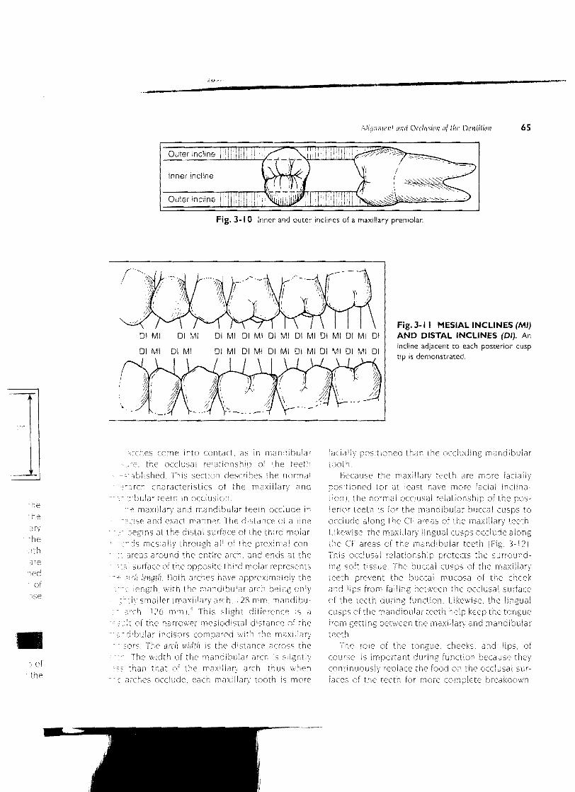
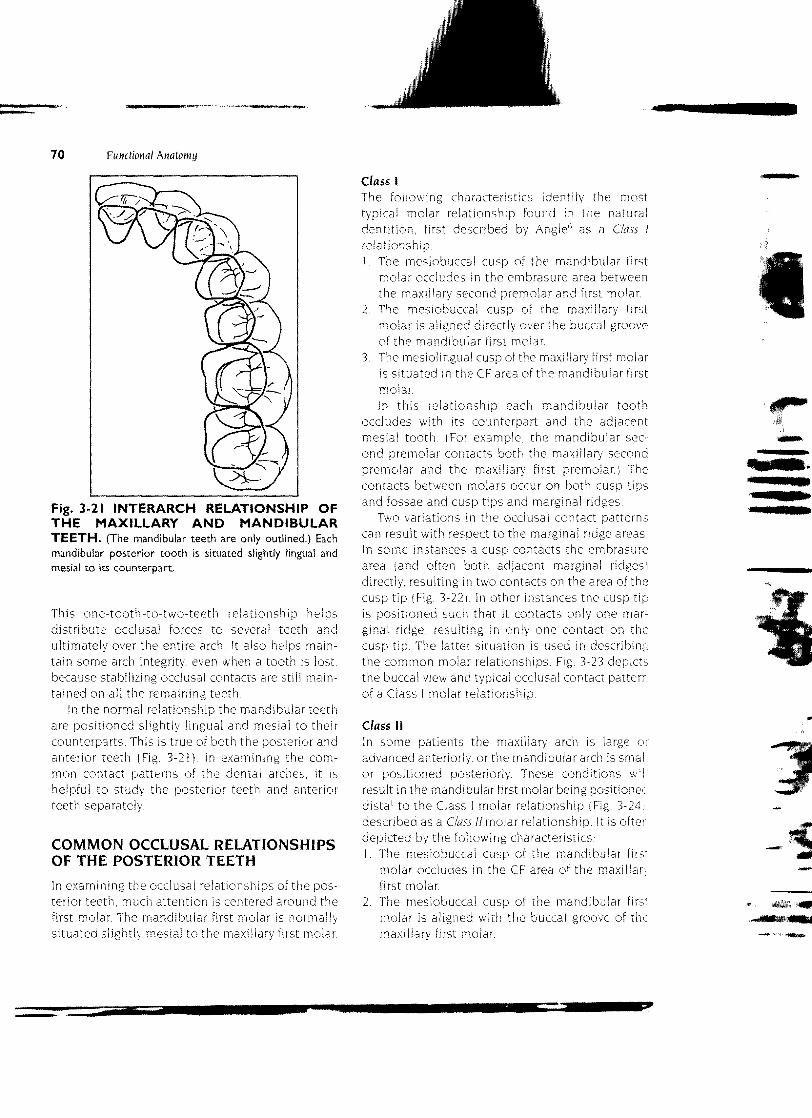
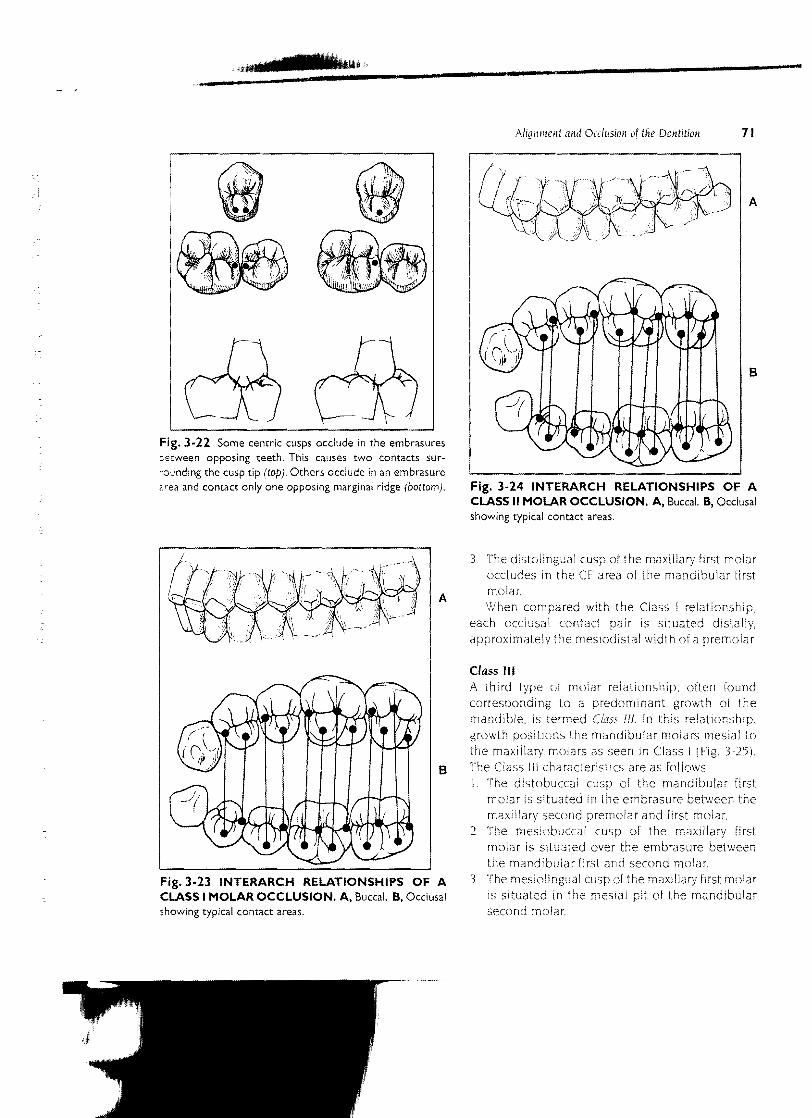
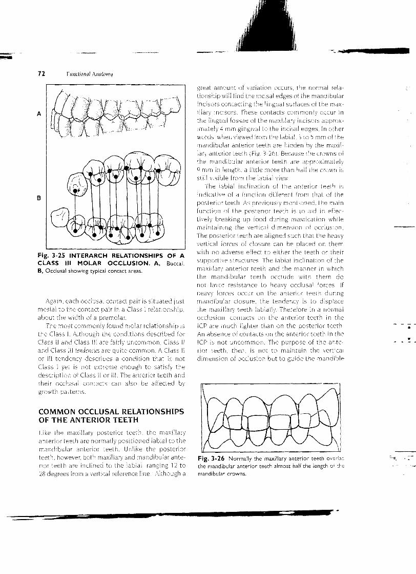
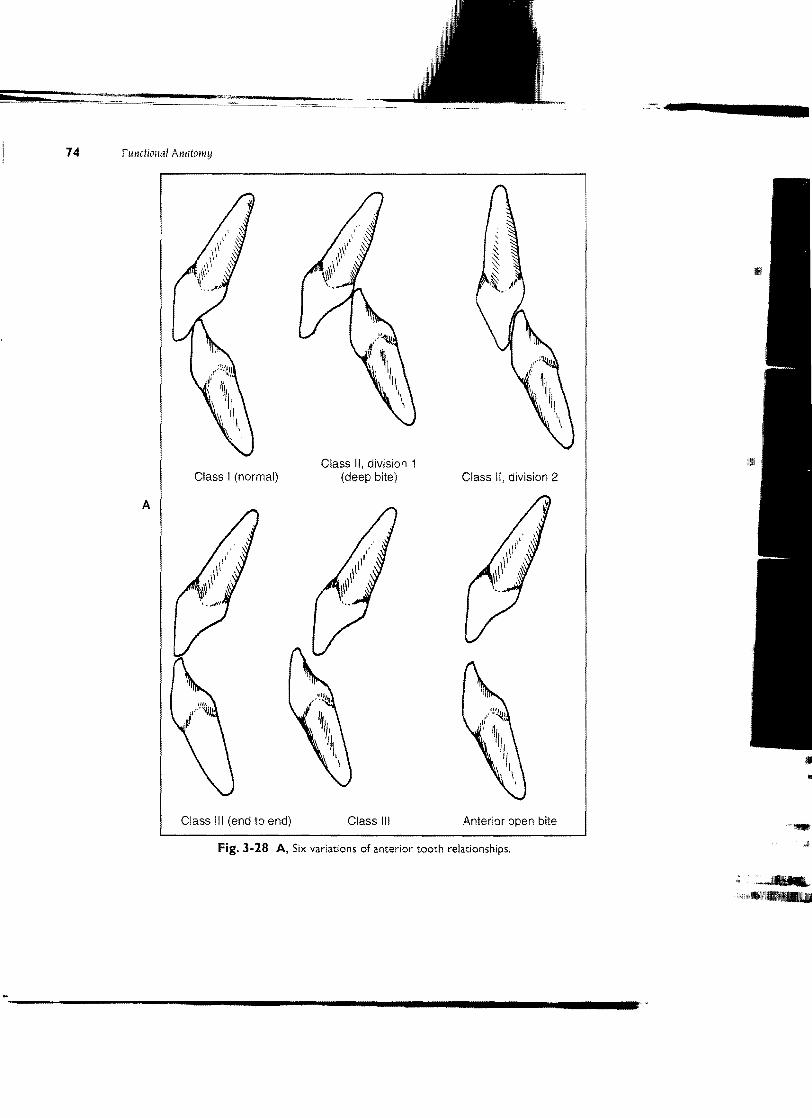
![Partial Face Recognition: An Alignment Free Approach · Alignment via landmarks 250 Cross-view [30,11,13,33] Limited FOV Skin texture [35] Frontal, partial face alignment 114 Occlusion,](https://img.dokumen.tips/doc/110x75/6001543a7033d50dfd266bbb/partial-face-recognition-an-alignment-free-approach-alignment-via-landmarks-250.jpg)


![Factored Occlusion: Single Spatial Light Modulator ... · tual content and occlusion masks, reducing form-factor and removing the need for careful SLM alignment. Cakmakci et al. [3]](https://img.dokumen.tips/doc/110x75/5e9c0e80d044482a7547703c/factored-occlusion-single-spatial-light-modulator-tual-content-and-occlusion.jpg)