Embed Size (px)
Citation preview
Novel biopesticides targeting the neuromuscular system of the peach
potato aphid Myzus persicae
Aishah Mohammed Alatawi
A thesis submitted for the Degree of Doctor of Philosophy
School of Biology, Faculty of science, Agriculture and Engineering
Newcastle University
June/2016
i
Table of contents:
List of figures: ............................................................................................................................. v
List of Tables: ............................................................................................................................ xi
List of Appendices: .................................................................................................................... xi
Acknowledgements .................................................................................................................. xii
Declaration: ............................................................................................................................. xiv
Abbreviations ........................................................................................................................... xv
Abstract: ................................................................................................................................ xviii
1 Chapter 1 Introduction ................................................................................................... 1
1.1 The global insect pest problem ........................................................................................ 1
1.1.1 Agricultural pests .................................................................................................. 1
1.2 Factors limiting the efficacy of conventional agrochemical pesticides ........................... 2
1.2.1 Insecticide resistance ............................................................................................ 3
Health consequences and environmental impacts ............................................................. 3
1.3 Development of bio- pesticides ....................................................................................... 4
1.3.1 Arthropod venoms as natural insect pest control agents ..................................... 5
Spider venoms: sources of novel bio-insecticides ............................................................ 6
Toxins purified from segestria florentina venom glands .................................. 7
Structure of spider venom peptides precursors and post-translational processing ............ 9
Structure and functional characteristics of Segestria. Florentina ...................... 9
Insecticidal targets of spider neurotoxins ........................................................ 11
Spider venom peptides targeting insect CaV Channels .................................. 12
Spider venom peptides targeting insect NaV channels ................................... 14
1.3.2 Marine cone snail from the Genus Conus: peptide nomenclature .................... 17
Structural motifs of cone snail venom peptides: ancestral variations ............................. 20
Potential insecticides targeting neurotransmitter receptors............................................. 21
Insecticides targeting nACh receptors ............................................................ 21
1.3.3 The venom peptide Acrorhagin-2a from the sea anemone Anthopleura maculate
23
Structural motifs of the sea anemone venom peptides .................................................... 23
Sodium ion channel as targets for neuropeptides as potential bioinsecticidal ................ 24
1.3.4 Marine cone snail from the Genus Conus: isolation ........................................... 24
Structure and function of the Genus Conus α -conotoxin E1 ......................................... 25
1.4 Neurotoxins and their use in fusion proteins ................................................................ 26
1.4.1 Spider venom toxins as insecticides.................................................................... 26
1.5 Expression of fusion proteins: Pichia pastoris as an expression host ............................ 27
1.5.1 pGAPZα as a plasmid vector for recombinant protein expression ..................... 28
1.6 Plant lectins .................................................................................................................... 28
ii
1.6.1 Roles in plant defence ......................................................................................... 29
1.6.2 Rols of snowdrop lectin as a carrier molecule .................................................... 30
1.7 Synthetic fusion proteins ............................................................................................... 30
1.8 Using the peach potato aphid Myzus persicae both as a target and for other
homopterans ........................................................................................................................... 31
1.8.1 Life cycle of Myzus persicae ................................................................................ 31
1.8.2 Control of Myzus persicae and need for alternative methods ........................... 32
1.9 Project aim ..................................................................................................................... 33
2 Chapter 2 Materials and Methods ................................................................................ 34
2.1 Microbial Expression systems and Insects ..................................................................... 34
2.2 Materials and Recombinant Techniques ....................................................................... 34
2.3 Standard molecular biology techniques ........................................................................ 35
2.3.1 Oligonuclueotide synthesis ................................................................................. 35
2.3.2 Polymerase Chain Reaction (PCR) ....................................................................... 35
2.3.3 Ligation and transformation of expression constructs into E. coli ..................... 37
2.3.4 Modification of plasmids for two copy insertion constructs .............................. 38
2.3.5 Transformation of expression constructs in Pichia Pastoris ............................... 38
2.3.6 Cloning and Expression of Cone Snail Toxins ...................................................... 39
2.3.7 Small-scale screening for fusion protein expression .......................................... 40
2.3.8 Protein Expression and Purification from P. pastoris ......................................... 40
Downstream processing of supernatant .......................................................................... 41
Ni-NTA Affinity Purification ......................................................................................... 41
2.4 Bioassays ........................................................................................................................ 42
2.4.1 Insects and Artificial Diet .................................................................................... 42
2.4.2 Insect Bioassays .................................................................................................. 43
Initial screening of recombinant fusion proteins from venom of the spider Segestria-
florentina ......................................................................................................................... 43
Dose response effect of recombinant fusion proteins 2 X SFI1/GNA, SFI5/GNA,
SFI8/GNA, and GNA alone ............................................................................................ 44
Initial screening of recombinant fusion protein from cone snails ................................... 44
Dose response effect of recombinant fusion proteins GNA/ alpha-Conotoxin EI .......... 44
2.5 Basic Analytical Techniques ........................................................................................... 45
2.5.1 Sodium Dodecyl Sulfate Polyacrylamide Gel Electrophoresis (SDS-PAGE) ......... 45
2.5.2 Immune assays by Western Blotting ................................................................... 45
2.5.3 Estimation of Protein concentration ................................................................... 46
2.6 DNA Sequencing ............................................................................................................ 47
2.7 Statistical Analysis .......................................................................................................... 47
iii
3 Chapter 3 Expression, purification and biological activity of fusion proteins
incorporating the toxins from the venom of the spider Segestria florentina (SFIx) and
snowdrop lectin (Galanthus nivalis agglutinin; GNA) ........................................................... 48
3.1 Introduction ................................................................................................................... 48
3.2 Results ............................................................................................................................ 49
3.2.1 Production and purification of fusion proteins incorporating SFI toxins and GNA
49
Assembly of the SFIx/GNA fusion protein construct ..................................................... 49
Assembly of the expression vector constructs containing two gene copies .................... 54
Expression of the recombinant protein SFIx/GNA ......................................................... 57
Yeast transformation and gene copy analysis ................................................................. 58
Purification of recombinant protein SFIx/GNA .............................................................. 61
3.2.2 Biological Activity of fusion proteins incorporating the toxins from the venom of
the spider segestria (SFIx) and snowdrop lectin (Galanthus nivalis agglutinin; GNA) ....... 72
Oral activity of fusion proteins incorporating SFI and GNA against survival trials set-up
over a period of 12 days .................................................................................................. 72
3.3 Discussion ...................................................................................................................... 81
3.3.1 Design of expression vectors for SFI peptides .................................................... 81
3.3.2 Expression of recombinant protein SFIs and GNA .............................................. 81
3.3.3 Expression and oral delivery of SF1/GNA, SFI2/GNA, SFI3/GNA, SFI5/GNA,
SFI6/GNA, and SFI8/GNA .................................................................................................... 83
3.3.4 Biological activity of fusion proteins SF1/GNA, SFI2/GNA, SFI3/GNA, SFI5/GNA,
SFI6/GNA, and SFI8/GNA .................................................................................................... 84
3.3.5 Increasing stability in gut environments ............................................................. 85
3.3.6 Functional characteristics of SFI toxins ............................................................... 85
4 Chapter 4 Expression, purification and biological activity of fusion proteins based on
snowdrop lectin (Galanthus nivalis agglutinin; GNA) and toxins from the cone snail (alpha-
Conotoxin EI, alpha-Conotoxin Sm1.1 and Acrorhagin-2a). ................................................... 87
4.1 Introduction ................................................................................................................... 87
4.2 Results: ........................................................................................................................... 89
4.2.1 Production and purification of fusion proteins ................................................... 89
Design of expression constructs GNA/Acrorhagin-2a, GNA/alpha-Conotoxin EI and
GNA/ alpha-Conotoxin sm1.1 ........................................................................................ 89
Expression of recombinant protein GNA/ alpha-Conotoxin EI, GNA/ alpha-Conotoxin
sm1.1 and GNA/ Acrorhagin-2a ..................................................................................... 93
Purification of GNA/ alpha-Conotoxin EI construct and Quantification ........................ 94
4.2.2 Biological Activity of of fusion proteins incorporating the toxins from the cone
snail alpha-Conotoxin EI and snowdrop lectin (Galanthus nivalis agglutinin; GNA)........... 98
Demonstration of insecticidal activity of GNA/ alpha-Conotoxin EI against Myzus
persicae ........................................................................................................................... 98
4.2.3 Transformed P.pastoris on Zeocin plates ......................................................... 100
iv
Expression of the fusion protein ................................................................................... 101
4.2.4 Oral activity of fusion proteins incorporating GNA and alpha-Conotoxin EI
against Myzus persicae ..................................................................................................... 105
4.3 Discussion .................................................................................................................... 109
4.3.1 Design of expression vectors for synthetic genes ............................................. 109
4.3.2 Expression of recombinant proteins: GNA/alpha-conotoxin EI, GNA/sm1.1,
GNA/Acrorhagin-2a ........................................................................................................... 109
4.3.3 Insecticidal activity of the recombinant fusion protein GNA/alpha-conotoxin EI
110
4.3.4 Insecticides targeting nicotinic acetylcholine receptors ................................... 111
5 Chapter 5 General Discussion ..................................................................................... 113
5.1 Mechanisms of insecticide resistance in Myzus persicae ............................................ 113
5.1.1 Overproduction of carboxylesterases ............................................................... 113
5.1.2 Mutation of the acetylcholinesterase enzyme ................................................. 113
5.1.3 Mutation of the voltage-gated sodium channel ............................................... 114
5.1.4 Enhanced expression of the cytochrome P450, CYP6CY3 and Mutation of the
nicotinic acetylcholine receptor ........................................................................................ 115
5.2 Mechanisms of peptide targeting to ion channels ...................................................... 116
5.3 Mechanisms for delivering venom derived neuropeptides ......................................... 117
5.4 Fusion with snowdrop lectin (GNA) ............................................................................. 118
5.5 Alternative delivery methods of toxins to insects ....................................................... 120
5.6 Integrated pest control approaches ............................................................................ 120
5.7 Conclusions .................................................................................................................. 121
References: ........................................................................................................................... 124
Appendix: .............................................................................................................................. 140
v
List of figures:
Figure 1-1: Size exclusion chromatography results of crude S. florentina (Lipkin et al., 2002) ......................................................................................................................... 8
Figure 1-2 A family of seven related protein structures (SFI2-SFI8) revealed after translation of cDNA sequences. Boxes show sequence similarity between SFI toxins and cysteine residues. Shading highlights non-identical residues (Lipkin et al., 2002). ................................................................................................................. 10
Figure 1-3: The amino acid sequence comparison between SFI1 (F5.6) S. florentina insecticidal toxin and other arthropod toxins. All sequences were arranged with respect to cysteine residue as shown in boxes. For better arrangement, gaps (-) were introduces. Shading show negatively charged amino acid residues and those highlighted in bold are positively charged amino acid residues. ............................ 11
Figure 1-4 Schematic representation for the membrane topology and molecular structure of CaV and NaV channels. The pore-forming - subunit (centre) in each case made up of 4 homologous domains I–IV. Cylinders represent the trans-membrane - helical segments (S1–S6) within each domain and the S4 voltage- sensor is shown in red. The walls of the ion- conducting pathway as shown in blue are formed by the pore- lining segments S5 - S6 and the intervening P loop. (A) The inactivation gate (magenta) of NaV channel is represented by the inactivation particle with magenta arrows which indicate the sites expected to form the inactivation gate receptor. (B) High voltage activated CaV channels usually comprises a single copy each of the α1, α2-δ, β, and γ subunits, however low voltage activated CaV channels contains only the pore- forming 1 subunit. The intracellular subunit comprises of a C- terminal guanylate kinase domain (cyan) and an N- terminal SH3 domain (blue). The guanylate kinase domain binds the interaction domain (AID; purple) which is located in the cytoplasmic loop and link domains I and II of 1, this interaction modulates the rate of channel inactivation. Calmodulin (CaM) interacts with CaV1 and CaV2 channels through a conserved sequence motif know as the ‘‘IQ motif’’ which is located in the cytoplasmic C- terminal region of 1. The C and N - terminal lobes of CaM (green) bind the IQ domain (red) at various sites and with varying affinities, making the interaction sensitive to both global Ca levels and the concentration of Ca in the area of the CaV channel pore. Ca ions bound to CaM in the CaM- IQ complex are shown by orange spheres (Adapted from King 2007; Nicholson 2007). ............................... 12
Figure 1-5 Voltage-gated Ca channels (CaV) topological structure(Catterall and Few, 2008; Pringos et al., 2011) ..................................................................................... 14
Figure 1-6 Schematic diagram representing the four possible disulfide bond isomers of α - conotoxins E1. .................................................................................................... 20
Figure 1-7 Alternative target sites of nAChRs. All panels (A to F) shows two muscle nAChRs; the top receptor is the “fetal” subtype having a γ subunit, and the bottom one is the “adult” subtype having an ε subunit. Receptors are closed in the absence of acetylcholine (blue triangles). A transition from a closed to an open state occurs (A to B) when 2 two molecules of acetylcholine bind (as shown in B). Effects of different conotoxins are shown in panels C, D, E, and F. One possible mechanism of non-competitive block is demonstrated in panel C. Orange globular geometry show the toxin occlude ion conductance through the channel pore. Competitive antagonists targeted to different pharmacological sites are shown in panels D, E, and F. A conotoxin highly specific for α1δ is shown in panel
vi
D (purple) which inhibits both fetal and adult subtypes. A conotoxin (red) which is specific for the α1γ interface, binds the fetal receptor but not to the adult as shown in E. On the contrary in F, the conotoxin (green) is specific for α1ε which does not inhibit the fetal subtype, however prevents acetylcholine from binding one of the two sites in the adult subtype (Olivera and Teichert, 2007). ............... 22
Figure 1-8 Life cycle of Myzus persicae (Anjum, 2014). ................................................. 32
Figure 3-1: Agarose gel electrophoresis of amplified SFI2, SFI3, SFI5, and SFI6 toxins using SFIX forward and SFIX reverse primers. Lane 1 represents the standard DNA ladder, 0.1Kb to 3Kb. Lanes 2, 3, 4, and 5 represent the successfully amplified SFI2, SFI3, SFI5, and SFI6 toxins of 200bp size. ....................................................... 49
Figure 3-2: Agarose gel electrophoresis of digested SFI2, SFI3, SFI5, SFI6, and SFI8 toxins using EcoRI gives two fragments. Lane 1 represents the standard DNA Ladder 0.1 Kb to 3 Kb. Each line represents two fragments, and the 3000 bp and 200 bp fragments correspond to vector pGAPZα and the amplified insert, respectively. ............................................................................................................ 50
Figure 3-3: Full SFI1/GNA fusion protein construct in the yeast pGAPZAP: full
determined nucleotide sequences, presumed amino acid sequences, and schematic
representation of fusion protein construct. The yeast alpha factor signal sequences
are shown with not highlighted, the SFI1 toxin sequences are highlighted in red,
and the GNA sequences are highlighted in green. The basic constructs of SFI1/GNA were designed by Fitches et al. (2004). .................................................................. 52
Figure 3-4: Sequence of predicted products from the expression constructs for the SFI2/GNA fusion protein (A), SFI3/GNA fusion protein (B), SFI5/GNA fusion protein (C), SFI6/GNA fusion protein (D), and SFI8/GNA fusion protein (E).Unshaded regions indicate the sequence of the yeast α-factor signal. Shaded regions in light grey and grey indicate the nucleotide sequences for SFIx and GNA, respectively. Underlined regions indicate the linker sequence contributed by the nucleotides, used to join the SFIx toxin and GNA coding sequences together. The highlighted region in red indicates sequences of the His tag to facilitate the purification of the expressed protein. .................................................................................................. 53
Figure 3-5: (A) Agarose gel electrophoresis of digested products using BamHI and BglII.Lane 1 represents the standard DNA ladder 0.1 Kb to 3 Kb. Lane 2 represents the control BamHI digest to linear plasmid. Lane 3 represents double digestion using BamHI and BglII, representing the vector and insert band. (B) Lane 2 represents the separation of the gene cassette from the 1x pGAPZα vector. Intact band in Lane 3 represents the pGAPZα vector. ...................................................... 55
Figure 3-6: Diagrammatic representation of the cloning strategy adopted to enable insertion of multiple copies of the fusion protein cassette into the Pichia yeast genome. ................................................................................................................... 56
Figure 3-7: The presence of GNA of the correct size in the positive standard lanes shows the blots have worked successfully. However, the fusion proteins SFI2/GNA, SFI3/GNA, SFI5/GNA, SFI6/GNA, and SFI8/GNA were subjected to high levels of expression, with a number of bands seen on the gel between 17 kDa and 14 kDa. ..................................................................................................................... 57
Figure 3-8: Western blot analysis of the protein expression of SFI1/GNA pGAPZα and 2 x SFI1/GNA pGAPZα using anti-GNA antibodies derived from the small-scale cultures’ supernatants. ........................................................................................... 58
Figure 3-9. A. shows the 72-hour fermentation run of SFI1/GNA, and the green curve in the graph shows the dissolved oxygen level and its consistency. B. shows the
vii
successful run of the fermenter for SFI2/GNA. C. represents the fermentation run of SFI3/GNA, the DO tend for the cell density. The 72-hour fermentation run of the SFI5/GNA fusion protein is represented in Figure 9.D, and the variation in the graph shows the dissolved oxygen level and its consistency. E. illustrates the successful run for fermenter for SFI6/GNA. ........................................................... 60
Figure 3-10: purification of His-tagged 2X SFI1/GNA proteins expressed in Pichia pastoris using a Liquid Chromatography SystemFiltered P. pastoris culture. The supernatant was diluted in a binding buffer (0.02 Sodium phosphate, 0.4 M NaCl, pH 7.4) and loaded onto a HisTrap™ (GE Healthcare) column. The protein was eluted with a binding buffer containing 0.2 M imidazole, and a NaCl gradient was held while the protein was eluted. (A) Typical purification trace. The absorbance trace is shown as a blue line. (B) 15 % SDS-PAGE analysis of the peak fractions collected from the recombinant protein purification. Fractions were not free from high molecular weight yeast proteins. Lane 7 of the eluted proteins have slightly visible bands around 17 kDa. ...................................................................... 62
Figure 3-11: Western blot (probed with anti-GNA ab) of recombinant 2 X SFI1/GNA expressed in Pichia pastoris.GNA was loaded in a known amount, 1mg/ml concentration. L1, L2, and L3 show the filtered supernatant, cultured supernatant diluted in binding buffer, and the supernatant after loading, respectively. Lane 4 of the wash stages shows multiple bands, indicating some protein was lost during was step. Lane 5 of the eluted protein shows an intact band at approximately the correct size for 17 kDa. GNA loaded in a known amount, 1 mg/ml, shows a single band at the correct size of 12.5 kDa. ...................................................................... 63
Figure 3-12: (A) Nickel column purification of His-tagged SFI3/GNA proteins expressed in Pichia pastoris using a Liquid Chromatography System Filtered P. pastoris culture. Binding to the column was carried out in 0.02 Sodium phosphate, 0.4 M NaCl, pH 7.4 and loaded onto a HisTrap™ (GE Healthcare) column. The protein was eluted with a binding buffer containing 0.2 M imidazole, and a NaCl gradient was held while the protein was eluted. (A) Typical purification trace. The absorbance trace is shown as a blue line. (B) 15% SDS-PAGE analysis of the peak fractions collected from the recombinant protein purification. Th lane 6 of the eluted protein shows the correct size for SFI3/GNA. ............................................. 64
Figure 3-13: Western blot (probed with anti-GNA ab) of recombinant SFI3/GNA expressed in Pichia pastoris. Recombinant GNA was used as a positive standard; GNA was loaded in a 1 mg/ml concentration. Fractions were free from high molecular weight yeast proteins. L1, L2, L3, and L4 show the unfiltered supernatant, filtered supernatant, cultured supernatant diluted in a binding buffer, and the supernatant after loading, respectively. Lane 5 of the wash stage shows few bands, indicating the loss of some protein during the wash step. The expected size of SFI3/GNA is 17 kDa. The blot shows a distinct band of approximately the correct size, as seen in lane 6. GNA loaded in a known amount, 1 mg/ml, shows a very light single band at the correct size of 12.5 kDa, as seen in Lane 7. ..................................................................................................................... 65
Figure 3-14: (A) Nickel column purification of His-tagged SFI5/GNA Proteins, expressed in Pichia pastoris using a Liquid Chromatography System Filtered P. pastoris culture. Binding to the column was carried out in 0.02 Sodium phosphate, 0.4 M NaCl, and pH 7.4 and was loaded onto a HisTrap™ (GE Healthcare) column. The protein was eluted with a binding buffer containing 0.2 M imidazole, and a NaCl
viii
gradient was held while the protein was eluted. (A) Typical purification trace. The absorbance trace is shown as a blue line. (B) 25 μl samples of different peak fractions collected from the recombinant protein purification were analysed on15% SDS-PAGE. L1, L2, L3, L4 show the unfiltered supernatant, the filtered supernatant, cultured supernatant diluted in binding buffer, the supernatant after loading. The single band in the eluted protein lanes is the correct size for SFI5/GNA. ................................................................................................................ 66
Figure 3-15: Purification of (His) 6 SFI6/GNA expressed in Pichia pastoris (strain X33) by a Liquid Chromatograph Nickel on a column. A filtered P. pastoris culture supernatant was diluted with a binding buffer and then loaded; the column was recharged with a Niso4 (2.0ml/l) HisTrap™ (GE Healthcare) column. It was then washed with a 1x binding buffer to elute any non-specific, unbound proteins. Washing of the nickel columns with 100ml binding buffer is required until the UV absorbance is measured at 280 nm and reaches a steady baseline. (A) A typical purification trace. (B) 25 μl samples of wash (W) and elution (E) fractions were then analysed on 15% SDS-PAGE gels. Lane 7 of eluted protein shows a single band at approximately the correct size for 17kDa. Elution fractions were dialyzed using 12 kDa MWCO tubing and lyophilized. .......................................................... 68
Figure 3-16: Western blots (probed with an anti-GNA antibody) of recombinant SFI6/GNA, expressed in Pichia pastoris (strain X33).Recombinant GNA of 1 mg/ml was used as a positive control, but no band was seen. Fractions were free from high molecular weight yeast proteins. L1, L2, L3, and L4 show the unfiltered supernatant, filtered supernatant, cultured supernatant diluted in a binding buffer, and the supernatant after loading, respectively. Line 5 of the wash stage shows few bands, indicating the loss of some protein during the wash step. The expected size of SFI6/GNA is 17 kDa. The blot did not show the required result, as there are no correct bands in lane 6 of the eluted protein. ................................... 69
Figure 3-17: (A) Nickel column purification of His-tagged SFI8/GNA proteins, expressed in Pichia pastoris using a Liquid Chromatography System Filtered P. pastoris culture. The protein was eluted with a binding buffer containing 0.2 M imidazole, and a NaCl gradient was held while the protein was eluted. (A) Typical purification trace. The absorbance trace is shown as a blue line. (A) Shows three peaks, and the eluted protein of SFI8/GNA was collected from the last fraction (green circle). (B) Shows 15% SDS-PAGE analysis of the peak fractions collected from the recombinant protein purification. The result of SDS-PAGE does not show the required result, but the presence of GNA bands at the correct size confirmed the gel was done correctly. ........................................................................................... 70
Figure 3-18: A Western blot (probed with anti-GNA ab) of recombinant SFI8/GNA expressed in Pichia pastoris. GNA was loaded in known amounts, with 1mg/ml and 2mg/ml concentrations. Lanes 1-5 show the expression of bands with immunoreactivity to anti-GNA antibodies. The lane of the eluted stages shows an intact band at approximately the correct size for 17 kDa. ..................................... 71
Figure 3-19: Survival of Myzus persicae on artificial diet bioassays. 2x SFI1/GNA is more toxic when fed at 0.1 mg/ml in artificial diet bioassays (n=30 aphids per treatment), as shown by Kaplan-Meier survival analysis ....................................... 73
Figure 3-20: Survival curve of surviving Myzus persicae fed recombinant GNA at 0.1, 0.05, and 0.001 mg g-1 and (His) 6-2 X SFI2-GNA, (His) SFI5/GNA, (His) SFI8/GNA 0.1, 0.05, and 0.001 mg g-1. (A) Kaplan–Meier survival curves at 0.1 mg g-1. (B)
ix
Kaplan–Meier survival curves at 0.055 mg g-1. (C) Kaplan–Meier survival curves at 0.001 mg g-1. Diet-only fed Myzus persicae are used as controls (n=30 aphids per treatment), as shown by Kaplan-Meier survival analysis. Treatments are depicted in the figure as follows: ........................................................................................... 76
Figure 3-21: Dose-response curves for 2 X SFI1/GNA in artificial diet bioassays show the relationship between percent of response and concentration. ............................. 78
Figure 3-22: Dose-response curves for SFI5/GNA in artificial diet bioassays show the relationship between percent of response and concentration .............................. 78
Figure 3-23: Dose-response curves for SFI8/GNA in artificial diet bioassays show the relationship between percent of response and concentration. ............................. 79
Figure 3-24: Dose-response curves for GNA in artificial diet bioassays show the relationship between percent of response and concentration. ............................. 79
Figure 3-25: the toxicity index (T.I); the 2xSFI1/GNA was the most effective construct, followed by SFI5/GNA and SFI8/GNA, respectively. ............................................... 80
Figure 4-1: Full GNA/Acrorhagin-2a fusion protein fusion protein construct in yeast pGAPZAP. Full determined nucleotide sequences, presumed amino acid sequences and schematic representation of fusion protein construct. Yeast alpha factor signal sequences are highlighted in blue, GNA sequences is highlighted in red and Acrorhagin toxin sequences are highlighted in green. .............................. 90
Figure 4-2: Full GNA/ alpha-Conotoxin EI fusion protein construct in yeast pGAPZAP. Full determined nucleotide sequences, presumed amino acid sequences, and schematic representation of fusion protein construct. The sequence for the yeast alpha-factor signal is highlighted with blue, GNA sequence is highlighted with red, and alpha-Conotoxin EI toxin sequence is highlighted with green. ....................... 91
Figure 4-3: Full GNA/sm1.1 fusion protein construct in yeast pGAPZAP. Full determined nucleotide sequences, presumed amino acid sequences and schematic representation of fusion protein construct. Yeast alpha-factor signal sequence is highlighted with blue, GNA sequence is highlighted with red, and a sm1.1 toxin sequence is highlighted with green. ....................................................................... 92
Figure 4-4: Western blot (probed with anti-GNA ab): of recombinant GNA/ alpha-Conotoxin EI expressed in Pichia pastoris. GNA was loaded in known amount, 1 mg/ml. The presence of GNA at 12.5-kDa in the positive standard lanes shows the blots have worked successfully. However, the fusion proteins GNA/ alpha-Conotoxin EI were subjected to express correctly with the single band seen on the gel at 15 kDa. ........................................................................................................... 94
Figure 4-5: Purification of His-tagged GNA/ alpha-Conotoxin EI proteins expressed in Pichia pastoris using a Liquid chromatography system. Filtered P. pastoris culture supernatant was diluted in binding buffer (0.02 Sodium phosphate, 0.4M NaCl, pH 7.4) and loaded onto HisTrap™ (GE Healthcare) column. Protein was eluted with 0.2M imidazole, whilst NaCl gradient was held. (A) Typical purification trace showing absorbance trace in blue line. (B) 15% SDS-PAGE analyses of the peak fractions collected from the recombinant protein purification. Fractions free from high molecular weight yeast proteins were pooled separately dialysed against distilled water at 4 OC using 12-15kDa MWCO tubing. The very light band represents the correct molecular weight of GNA/ alpha-Conotoxin EI. ................. 96
Figure 4-6: Western blot (probed with anti-GNA antibodies): of recombinant GNA/ alpha-Conotoxin EI expressed in Pichia pastoris. . 1mg/ml and 2mg/ml of GNA was loaded in L1 and L2 respectively. Line 3 and 4 shows expression of bands with

x
immunoreactivity to anti-GNA antibodies. Line 4 of the load stage shows intact band at approximately 15kDa, while line 3 from the eluted stage shows weak band at 15kDa. ........................................................................................................ 97
Figure 4-7: Biological activity of GNA/ alpha-Conotoxin EI agnaist Myzus persicae on artificial diet bioassay. GNA/ alpha-Conotoxin EI was most toxic compared to GNA alone in aritifical diet bioassay (n=30 aphids per treatment), as shown by Kaplan Meier survival analysis. ........................................................................................... 99
Figure 4-8: PCR results from a colony screening of GNA/Sm1.1 pGAPZα and GNA/α-conotoxins EI pGAPZα transformants. Lane M: 1 kb ladder. Lane 1-6: PCR reaction of the GNA/α-conotoxins EI transformed single band of about 500 bp. Lane 7 contains PCR reaction of GNA/Sm1.1 showing no expression.. GNA as seen in lane 8 produced intake band is about 500bp. Negative control was ran in the lane 9 and, has no band. ....................................................................................... 101
Figure 4-9: Graph of OD600 vs time for protein expression ........................................ 102
Figure 4-10: A) SDS- PAGE and Western blot (probed with anti-GNA ab): at 48h for expression screening of GNA/ alpha-Conotoxin EI fusion protein. 25-μl samples from different colony culture Supernatant was loaded onto SDS- PAGE gel, recombinant GNA was used as a positive standard. No bands were observed of all Colonies. The Presence of the GNA at approximately the correct size (12 kDa) was visualised by SDS-PAGE, followed by western blotting using anti-GNA antibodies (B). ......................................................................................................................... 103
Figure 4-11: SDS- PAGE and Western blot (probed with anti-GNA ab): at 96h, for expression screening of GNA/ alpha-Conotoxin EI fusion protein. 25-μl samples from different colony culture Supernatant was loaded onto SDS- PAGE gel, recombinant GNA was used as a positive standard. Colonies 1 and 6 show very low expression of fusion proteins, colony 8 shows intact band at approximately the correct size (12 kDa) for GNA, while line 9 represents the strain x33 as negative control with no band. ............................................................................. 104
Figure 4-12: Western blot (probed with anti-GNA antibodies) detecting the fusion protein GNA/ alpha-Conotoxin EI transformed yeast after cell lysis using the YeastBuster™ Protein Extraction. GNA were loaded as controls, 1 mg/ml, and 2 mg/ml concentration. The recombinant protein GNA/ alpha-Conotoxin EI was detected and estimated from the intensity of the GNA band. ............................. 105
Figure 4-13: A graph Myzus persicae survival feeding on artificial diet alone (control) and GNA incorporated arterial diet at 0.1mg/ml over 7 days. ............................. 106
Figure 4-14: Survival of Myzus persicae on artificial diet bioassays.GNA/ alpha-Conotoxin EI is more toxic when fed at 0.1 mg/ml in artificial diet bioassays (n=30 aphids per treatment), as shown by Kaplan-Meier survival analysis ................... 107
Figure 4-15: Dose-response curves for GNA/ alpha-Conotoxin EI in artificial diet bioassays shows the relationship between percent of response and concentration. ............................................................................................................................... 108
xi
List of Tables:
Table 1-1 Examples of Gene Superfamilies (Olivera and Teichert, 2007) ................... 18
Table 1-2 Families of Conopeptides and Families of Targets (Olivera and Teichert, 2007) ....................................................................................................................... 18
Table 1-3 Sequences of different cone snail toxins, the cysteine residues and mode of action ....................................................................................................................... 20
Table 2-1 Primers used for the amplification of the SFI2, SFI3/4/5, SFI6, SFI8, and GNA sequences. ............................................................................................................... 35
Table 2-2 Reaction component volumes used for PCR. The total reaction volume is 50 μl. ............................................................................................................................. 35
Table 2-3 PCR cycles for bacterial colony screening, including temperatures, time and cycles for each PCR step. ......................................................................................... 36
Table 2-4 PCR cycles for yeast colony screening, including temperatures, time and cycles for each PCR step. ......................................................................................... 39
List of Appendices:
Appendix A: Summary of the strategy to obtain single-copy expression vectors (Invitrogen manufacturer, Catalog nos. V200-20 and V205-20). ......................... 140
Appendix B: map for the strataclone pcr cloning vector psc-a-amp/kan (Instruction Manual, Catalog number: 240218) ....................................................................... 141
Appendix C: Recipe for separating and stacking gels for 16.5% Sodium Dodecyl Sulfate Polyacrylamide Gel ................................................................................................ 141
Appendix D: Summary of toxicological data obtained from the aphids’ bioassay after 7 days of exposure to 2 X SFI1/GNA, SFI5/GNA, SFI8/GNA, and GNA fusion proteins ............................................................................................................................... 142
Appendix E: Wild type P. pastoris X33 were grown on solid YPD medium at 30°C, for at least 72 h. Transformation of FP2.1/GNA pGAPZα, GNA/Sm1.1 pGAPZα and GNA/α-conotoxins EI pGAPZα into wild type P. pastoris X33 resulted in a few colonies after 3 days growth ................................................................................. 143

xii
Acknowledgements
Above all, I am thankful to God the Almighty who grants me this unique opportunity to
learn and to proceed successfully. This thesis is in its current form due to the assistance
and guidance of many people. Thanking others is part of gratitude to Allah. As The
Prophet -- peace and blessings be upon him -- said: “He who has not thanked people has
not thanked Allah.” Therefore, I would like to express my sincere thanks to everyone.
First and foremost, I am incredibly thankful to Prof. Angharad Gatehouse (School of
Biological, Newcastle University) for her helpful suggestions, feedback, her offering
valuable guidance throughout my PhD effort and especially for her warm
encouragement during the whole period of study. Prof. Gatehouse, I will never forget
your support and what you did for me. I would also like to thank my adviser, Dr.
Martin Edward (School of Biological, Newcastle University), for his excellent advice
and detailed review ever since I began my lab work.
I would also like to extend my thanks to Ms. Gillian Davison for providing such a good
atmosphere in the lab and for her guidance in using some of the equipment in the lab.
I also appreciate the financial support of the University of Tabuk during my PhD
studies -- thanks for providing me with this opportunity.
Erich Nakasu and Hesham Abd El Halim, I greatly appreciate your great help and
your spiritual support for me during my PhD studies. I will always remember the time
we spent together and shared useful discussions.
Janne Darell, Sreevijeth Ravi and Monica Athwani, thanks for excellent help in the
lab, particularly during the creation of some variants of constructs.
Thanks to all my friends and colleagues, Eman Salem, Dr. Shreya Wani, Rana
Alshegaihi, Uma Kupusamy, Thomas McDaniel, Alistair Polland, and Baida Alshukri.
Thank you for making my time here in Newcastle unforgettable; you always made me
feel as if I were at home. Special thanks to Dr. Dalal Albaijan for her listening, offering
me advice and supporting me through this entire process.
I cannot finish without thanking my family; Words cannot express how grateful I am to
my mother and father. I cannot thank you enough for all the support and love you have
given me. I love you both; I would not be where I am today without your prayers. It’s
because of your prayers that your daughter made the dream come true for both of you. I
know I cannot repay what you have done for me, but I promise I will always be by your
side whenever you need me. My sister Nora Alatawi, no words can truly express the

xiii
level of gratitude and appreciation I have for you. Thank you for being there whenever I
needed you; your encouragement and support made all the difference. I will always
remember the SMS, the phone calls, the guidance and so much more. At the end, I am
extremely thankful to my brother Mansor Alatawi, Thank you for your continued
support and love, especially during the worst moments. I will always love you.
Aishah

xiv
Declaration:
I declare that this thesis is my own work and that I have correctly acknowledged the
work of others. This submission is in accordance with University and School guidance
on good academic conduct. I certify that no part of the material offered has been
previously submitted by me for a degree or other qualification in this or any other
University. I confirm that the word length is within the prescribed range as advised by
my school and faculty
xv
Abbreviations
DDT: Dichlorodiphenyltrichloroethane
PCR: Polymerase chain reaction
FP: Fusion Protein
FP2.1: SFI1/GNA
FP2.3: SFI3/GNA
FP2.5: SFI5/GNA
FP2.6: SFI6/GNA
FP2.8: SFI8/GNA
SFI1: Segestria florentina toxin 1
SFI2: Segestria florentina toxin 2
SFI3: Segestria florentina toxin 3
SFI4: Segestria florentina toxin 4
SFI5: Segestria florentina toxin 5
SFI6: Segestria florentina toxin 6
SFI7: Segestria florentina toxin 7
SFI8: Segestria florentina toxin 8
GNA: Galanthus nivalis agglutinin
CaV: Calcium voltage channel
NaV: Sodium voltage channel
IFM: Amino acid sequence isoleucine, phenylalanine, and methionine
MACE: The modified a cetyle-cholinesterase aphids
α - conotoxins E1: cone snail venom peptides
sm1.1: cone snail venom peptides
Acrorhagin-2a: the sea anemone Anthopleura maculate.
nAChR: Nicotinic acetylcholine receptor
n-nAChR: Neuronal nicotinic acetylcholine receptor
SOC: Super Optimal Broth medium
X-gal: 5-Bromo-4-chloro-3-indolyl-β-D-galactopyranoside
SRDA: Single Residue Distribution Analysis
SDS-PAGE: Sodium dodecyl sulphate polyacrylamide gel electrophoresis
BSA: Bovine Serum Albumin
xvi
TEMED: Tetramethylethylenediamine
OD: Optical density
MWCO: Molecular weight cut off
bp: base pairs
LB: Luria-Bertani broth
YPG: Yeast peptone glucose medium
YPD: Yeast Extract-Peptone-Dextrose medium
LC50: Concentration of a toxin that causes the death of 50% of test insectsc
cDNA: Complementary DNA
NCBI-BLAST: National Centre for Biotechnology Information Basic Local
kDa: KiloDalton
FPLC: Fast Protein Liquid Chromatography
mg: milligram
w/v: Weight per volume
ml: Millilitres
mM: Millimolar
μl: Microlitre
nm: Nanometres
UV: Ultraviolet
V: Volts
M: Molar
rpm: Revolutions per minute
μg: Microgram ECL: Enhanced Chemi-luminescence reagents
PBS: Phosphate Buffered Saline
Nucleic acid abbreviations:
A: Adenine
T: Thymine
G: Guanine
C: Cytosine
Amino acid abbreviations:
Ala: Alanine
Arg: Arginine
Asn: Asparagine Asp: Aspartic Acid Cys: Cystiene Glu: Glutamic acid Gln: Glutamine
Gly: Glycine

xvii
His: Histidine
Val: Valine
xviii
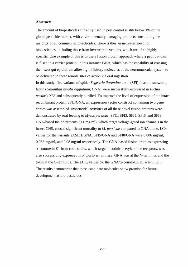
Abstract:
The amount of biopesticides currently used in pest control is still below 1% of the
global pesticide market, with environmentally damaging products constituting the
majority of all commercial insecticides. There is thus an increased need for
biopesticides, including those from invertebrate venoms, which are often highly
specific. One example of this is to use a fusion protein approach where a peptide-toxin
is fused to a carrier protein, in this instance GNA, which has the capability of crossing
the insect gut epithelium allowing inhibitory molecules of the neuromuscular system to
be delivered to these remote sites of action via oral ingestion.
In this study, five variants of spider Segestria florentina toxin (SFI) fused to snowdrop
lectin (Galanthus nivalis agglutinin; GNA) were successfully expressed in Pichia
pastoris X33 and subsequently purified. To improve the level of expression of the intact
recombinant protein SFI1/GNA, an expression vector construct containing two gene
copies was assembled. Insecticidal activities of all these novel fusion proteins were
demonstrated by oral feeding to Myzus persicae. SFI1, SFI3, SFI5, SFI6, and SFI8
GNA-based fusion proteins (0.1 mg/ml), which target voltage-gated ion channels in the
insect CNS, caused significant mortality to M. persicae compared to GNA alone. LC50
values for the variants 2XSFI1/GNA, SFI5/GNA and SFI8/GNA were 0.006 mg/ml,
0.038 mg/ml, and 0.08 mg/ml respectively. The GNA-based fusion proteins expressing
-conotoxin E1 from cone snails, which target nicotinic acetylcholine receptors, was
also successfully expressed in P. pastoris; in these, GNA was at the N-terminus and the
toxin at the C-terminus. The LC 50 values for the GNA/-conotoxin E1 was 8 μg/μl.
The results demonstrate that these candidate molecules show promise for future
development as bio-pesticides.
1
1 Chapter 1 Introduction
1.1 The global insect pest problem
1.1.1 Agricultural pests
Insects are the most diverse species in the animal kingdom, there are more than 900,000
species of insect worldwide. Most insects are directly important to both human life and
the environment. Several insect species are pollinators, decomposers of organic matter,
garbage collectors, soil conditioners and natural fertilizer producers. Interestingly, some
insect species are predators or parasitoids, which involves other harmful pests. In
general, insect pest contribute to the destruction of nearly 14 % of global crop product,
by either direct feeding on stored product or on the non-harvested crop in the field. It is
estimated that more than USD 100 billion is lost annually to the activity of insect pest
(Santos et al., 1990). Some of the most destructive insect pests of food products are
Coleoptera (beetles) and Lepidoptera (moths and butterflies). Indeed, 40% of chemical
insecticides are directed against larval forms of lepidopterans (Brooks, 1999). However,
it is worthy of note that other insect species like aphids are undoubtedly widespread,
with approximately 4400 species worldwide. Though, 250 species of aphids are
classified as serious crop pests, they still present major losses to production of important
crops (Remaudiére and Remaudiére, 1997; Blackman and Eastop, 2006).
Aphid species like the Myzus persicae (the peach potato aphid), is considered an
important agricultural pest worldwide. They cause direct damage through the extraction
of phloem sap that results in reduced crop yield. They also cause indirect damage by
transmitting plant viruses to economic crops. Nault (1997) demonstrated that over 50%
of insect borne plant viruses are transferred by aphids. Similarly, aphid’s excreta are
high in sucrose, which can cause indirect damage through increased insect population.
The excreta of aphids also help in growth of sooty mould that reduces the rate of
photosynthesis by covering plant leaves (Vickers, 2012). It has been shown that aphids
use both visual and chemical signals to select and locate a host plant (Vickers, 2012).
Over the past 5 decades, aphids and other phytophagous (plant- eating) insects have
been recognised as a major threat to food production for human consumption. In 2001,
the estimated combined cost of all protective approaches against crop damage in the US
alone is about USD 7.56 billion (Beckmann and Haack, 2003). Soybean aphids alone
are responsible for more than USD I billion in crop loss. A study conducted by (Pan-
UK, 2003), found that over 3 billion kilograms of pesticides is applied worldwide per
year at a cost of nearly USD 40 billion. Additionally, 500 million kilograms of more
2
than 600 different pesticide types are used in the United States only, with estimated cost
of USD 10 billion (Pimentel and Greiner, 1997).
European Union (EU) directives calling for legislation against the use of persistent
pesticides in crop protection and more recently the ban on neonicotinoids due to
potential negative effects on beneficial insects, in particular bees, have been the driving
factor for the renewed call for safe pest control methods. However, improvement in pest
control strategies should be based on the precautionary principle in existing EU
legislation. The EU regulation is targeted at reducing the negative impact of p9esticide
on both human and animal health, and the environment. Therefore, the need for safer
pest control should take precedent over the push to improve agricultural yield (EU
communication to the Council, 2006). To address the problem posed by toxic pesticides,
alternative approaches to chemical control are needed, which will help to develop high
quality and larger quantities of agricultural products.
1.2 Factors limiting the efficacy of conventional agrochemical pesticides
Since the introduction of the first DDT based product in 1940s, arthropod pests have
been successfully controlled using chemical agents. The successful application of DDT
in agriculture has been replicated in the fight against malaria (Attaran et al., 2000). Up
until the 1950s, it was thought that chemical pesticides could successfully lead
widespread control of insect pests. However, the development of resistance to such
chemical pesticides had occurred in many insect pest species, with over 400 arthropod
species gaining resistance to a wide range of chemical pesticides (Pospischil and Hanke,
1994). Additionally, part of the reason suggested for the development of alternatives to
chemical pest control is that chemical control has a limited number of nervous system
targets and lacks selectivity, which often targets both pests and non-target species
(Feyereisen, 1995). There is an increasing concern over the effects of chemical control
strategies on both humans and animals. Indeed, a study conducted by Metcalf (1994)
found that only 0.1 % of agrochemical control agents applied each year actually target
the intended pests, the study suggested that a large proportion of such chemical agents
remain in the environment to affect other organisms (Weisser and Siemann, 2004).
Moreover imbalance in the ecosystems gradually develops over time, which causes
more complications to the ecosystem (Weisser and Siemann, 2004).
3
1.2.1 Insecticide resistance
Insecticidal resistance is a recognised threat to human welfare because it negatively
affects crops and increases the cost of agricultural production. Over the years, many
mechanisms and non-exclusive mechanisms that enable a species to develop resistance
to insecticides have been identified, including changes in metabolic pathways and point
mutations at a site which renders an insect resistant to a particular insecticide. M.
persicae has been the most successful in exploiting the agricultural environment and it
is considered as one of the most important pests. Various insecticides have been used on
the peach aphid but the pressure from such control measures has facilitated the
development of resistance to most of them. In peach aphids, four mechanisms have been
described by various researchers. These mechanisms include target or metabolic site
mutations, this is important because these compounds modify the acetylcholine
receptors or cause mutations in Na+2 channels (Martinez-Torres et al., 1999). Anthony
et al. (1998) also described a mechanism based on a mutation in the GABA-Rdl
receptors. Also, Bass et al. (2011b) recently reported that they created a mutation in
nAChR b1 subunit that conferred resistance against insecticides. Another important
mechanism is the overproduction of E4 or EF4 esterases and cytochrome P450. Finally,
the plant allelochemical detoxification system, which is present in insects, has been
implicated in insecticidal resistance (Silva et al., 2012). Due to these molecular
mechanisms, most insect pests are immune to organophosphates, dimethyl carbamates
and pyrethroids. Currently, for the purpose of controlling insect pest populations,
neonicotinoids such as imidacloprid, thiamethoxam, clothianidin and acetamiprid have
been used. However, the use of neonicotinoids have been met with a few cases of
resistance in developed countries like USA. Moreover, biochemical and genomic
analysis of M. persicae samples collected from Greece was reported to have 40-fold
resistance to neonicotinoids. Resistant aphids have been shown to contain
approximately 18 copies of P450 genes compared to two copies in normal aphids, which
was linked to pesticide resistance (Bass et al., 2011b).
Health consequences and environmental impacts
In addition to issues associated with insecticidal resistance, a variety of chronic health
effects associated with insecticides in food production have been documented. For
instance, exposure to pesticides have been linked to the development of cancers.
Similarly, a study conducted by (Garabrant et al., 1992) found high mortalities among
4
pancreatic cancer patients often exposed to pesticides. Additionally, Beard (2006)
reported a link between exposure to pesticides and long-term neurological effects, and
McCarthy (1993) suggested that a number of potential hormone related diseases can be
induced by pesticides, including adverse reproductive outcomes in humans.
As a result of pesticides in both natural and agriculture ecosystems, many beneficial
species especially predators and parasites, are adversely affected (Pimentel et al.,
1993). On the one hand increased pesticide use reduces the population of beneficial
predators and parasites, on the other, pesticide can cause reduction in the number of
beneficial species. This can negatively impact on food security and agricultural
productivity. A number of pesticides are known to have lethal effect on bees, while
others have sub-lethal effect that decreases the ability of bees to thrive (Decourtye et al.,
2004). Subsequently, affecting pollination and crop yield, this is in line with the fact
that one third of all plants consumed by humans are pollinated by bees. About 90% of
flowering plants are pollinated by animals and insects. Increasing food productivity is
essential in meeting the requirements of the growing global population. Improvement
of pest management is the best approach to increasing crop protection, which can be
done by developing alternatives to chemical pesticides and new pest control strategies
that work through different modes of action.
1.3 Development of bio- pesticides
Several studies have highlighted the importance of exploiting a variety of natural
substances in the production of novel biopesticides. These natural substances can be
derived from several sources including animals (e.g. nematodes) (Fuxa , 1991; Beard et
al., 2001), microorganisms e.g. Bacillus thuringiensis, venoms of predator/ parasitoid
arthropods such as spiders (Tedford et al., 2004; Nicholson , 2006), and cone snails
(Olivera, 2002). Hence, biopesticides can provide a plethora of pest control methods
that are non- toxic to the environment and human health. Biological agents are highly
effective against pest species, often highly specific, and degrade rapidly in the
environment. Due to these unique features such compounds are considered as potential
stand-alone bioinsecticides. Moreover, transgenes of predator toxins can be expressed in
transgenic plants to enhance resistance to insect pests.
5
1.3.1 Arthropod venoms as natural insect pest control agents
Venom peptides isolated from predator/parasite arthropods such as spiders, scorpions,
snakes, wasps, predacious mites and aquatic cone snails provide a rich source of natural
insecticides. These peptides have evolved to target a wide range of receptors and ion
channels in the insect nervous system (Froy et al.,2000; Gould and Jeanne, 1984;
Tomalsk i et al., 1988; Olivera , 2002). These arthropod venoms are a heterogeneous
mixture of salts, low molecular weight, and polypeptides. Whilst some are specific to
invertebrates, others target vertebrates and yet others affect both (Escoubas et al., 2000;
Loret and Hammock, 1993). A subset of arthropod venom is often a mixture of
polypeptide toxins generally targeting specific subtypes of voltage ligand ion channels
(Lewis and Garcia, 2003; Sollod et al., 2005). Some of these peptides have unusual
targets such as the intracellular calcium – activated ryanodine channel (Fajloun et al.,
2000). The activity of such peptides make them useful therapeutic agents that can be
used to modify the activity of ion channels implicated in human disease. For instance,
one ω-conotoxin, has been shown to target different species of fish. Source of ω-
conotoxin include MVIIA from conus magus, which was the first ω-conotoxin to be
approved by the food and drug administration for the management of several human
chronic pains (Jain, 2000).
The venom of arachnids contains approximately 0.5 to 1.5 million insect active peptide
toxins, providing a rich source for novel biopesticides (Quistad and Skinner , 1989;
Wang et al., 2000; Tedford et al., 2004). For example, spiders can express 1000
different polypeptides (Escoubas et al., 2006). Interestingly, just four groups of spider
out of 10,000 are potentially fatal to humans (Isbister and White, 2004). Marine cone
snails of the Genus Conus use a potent cocktail of venomous peptides to catch their
prey. These venomous peptides are small in size (<50kDa) and are synthesised from a
few genes. As per an estimation by Becker and Terlau (2008) there are over 100,000
bioactive compounds present in the venomous cocktail synthesized by cone snails, each
with a distinct neurological target. Unfortunately, the use of these toxins as a successful
crop protection method is limited by an inability to reach target sites in the insect central
nervous system following oral consumption (Quistad et al., 1991a). However, many
purified toxins have been proven to be lethal to insect prey following injection of the
toxins directly to the nervous system, but lack insecticidal effect when delivered orally.
Fitches et al. (2002) fused peptides from spider venom to the carrier molecule snowdrop
6
lectin; (GNA), which directs transport across the insect gut epithelium following oral
delivery, where they have been shown to be effective and active insecticides.
Spider venoms: sources of novel bio-insecticides
Spiders are classified as the order Araneae. They have eight legs and chelicerae with
fangs that is used to inject venom. Spiders are found on every continent except
Antarctica. These are the largest order within the arachnids. Within all orders of
organisms, spiders rank seventh in total species diversity (Sebastian and Peter, 2009).
As per the world Spider Catalogue (Ver. 16.5), taxonomists have reported at least
45,752 spider species, and 114 families. Spiders differ anatomically from other
arthropods, where the usual body segments in spiders are fused into two tagmata (the
cephalothorax and abdomen), which are joined by a small cylindrical pedicel. Spiders
do not have antennae, unlike insects. Compared to all arthropods, spiders have the most
centralised nervous system except Mesothelae, the most primitive group of arachnids.
Also, exterior muscles are absent in limbs, where hydraulic pressure is used to extend
limbs, unlike arthropods. The spider abdomen bears appendages that are modified into
spinnerets and extrude silk.
Apart from Bagheera kiplingi, which is a herbivorous spider species as described in
2009 by Meehan et al, all other known spider species are predators. These mostly feed
on insects and on other spiders. Also, a few large species feed on birds and lizards. A
wide range of strategies are used by spiders to capture prey, including the trapping of
prey in sticky webs, mimicking the prey to avoid detection, or lassoing it with sticky
bolas. Most of the spiders detect prey mainly by sensing vibrations, but some of the
active hunters have acute vision. Spiders have very narrow guts and cannot take solids,
therefore they flood digestive enzymes on food to liquidise it and use the base of
pedipalps to grind it as true jaws are also absent. The lifespan of most spiders is
approximately two years, but tarantulas and other mygalomorph spiders can live up to
25 years.
Spiders have the ability to produce various complex venoms, which is one of the major
contributing factors to the evolutionary success of spiders. These venoms are used for
predation and predator deterrence (King, 2004). Unfortunately, the venom of spiders is
not well studied compared to other venomous taxa including scorpions (few species),
snakes, centipedes, and a few marine animals. Spider venoms contain a large number of
chemicals with a variety of biologically active components and toxins. These chemicals
7
fall into various chemical groups such as polyamine-like toxins that interfere with
glutamic acid receptors and block neuromuscular transmission (Grishin et al., 1986;
Kawai et al., 1991), low molecular weight proteins or peptides that affect neuronal or
membrane ion channels and receptors through pre- or post-synaptic actions, and high
molecular weight neurotoxins that interact with specific pre-synaptic receptors (Lipkin
et al., 2002). Insecticidal toxins synthesised by spiders typically cause paralysis due to
disruption of the activity of neuromuscular junctions. These toxins are usually cysteine
rich polypeptides with 55 to 60 amino acid residues or less. Some of the venomous
species that have been studied extensively include Segestria florentina, Agelenopsis
aperta, Hololena curta, Phoneutria nigriventer, Atrax robustus and Plectreurys tristis
(Pallaghy et al., 1997; Diniz et al., 1993; Reily et al., 1995; Quistad and Skinner ,
1994; Newcomb et al., 1995; Stapleton et al., 1990).
Toxins purified from segestria florentina venom glands
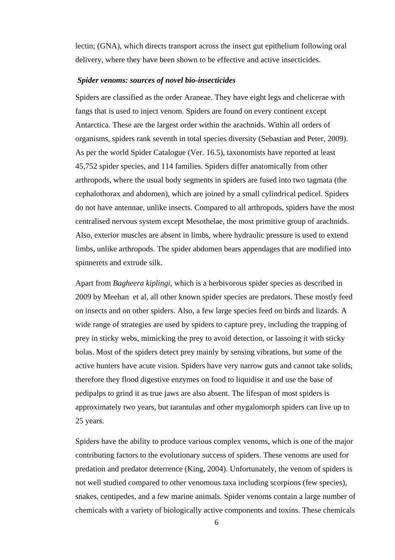
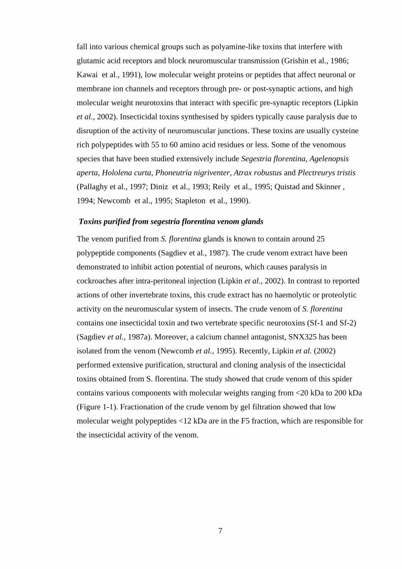
The venom purified from S. florentina glands is known to contain around 25
polypeptide components (Sagdiev et al., 1987). The crude venom extract have been
demonstrated to inhibit action potential of neurons, which causes paralysis in
cockroaches after intra-peritoneal injection (Lipkin et al., 2002). In contrast to reported
actions of other invertebrate toxins, this crude extract has no haemolytic or proteolytic
activity on the neuromuscular system of insects. The crude venom of S. florentina
contains one insecticidal toxin and two vertebrate specific neurotoxins (Sf-1 and Sf-2)
(Sagdiev et al., 1987a). Moreover, a calcium channel antagonist, SNX325 has been
isolated from the venom (Newcomb et al., 1995). Recently, Lipkin et al. (2002)
performed extensive purification, structural and cloning analysis of the insecticidal
toxins obtained from S. florentina. The study showed that crude venom of this spider
contains various components with molecular weights ranging from <20 kDa to 200 kDa
(Figure 1-1). Fractionation of the crude venom by gel filtration showed that low
molecular weight polypeptides <12 kDa are in the F5 fraction, which are responsible for
the insecticidal activity of the venom.
8
Figure 1-1: Size exclusion chromatography results of crude S. florentina (Lipkin et al., 2002)
Fraction F5 was further fractioned using RP-HPLC and obtained fractions were tested
on H. virescens larvae for toxic effects. Flaccid paralysis was caused by fractions F5.5,
F5.6 and F5.7. Also, reports of mass spectroscopic analysis of crude S. florentina
venom showed that many polypeptides are in the range of 4900 to 5100 Da (Lipkin et al.,
2002). Partial sequences were obtained when purified toxin was analysed by gas phase
N-terminal amino acid sequence:
F5.5 - AECMVDETVCYIHNXNNC
F5.6 - KECMTDGTVCYIHNXNDE
F5.7 - KECMADETVCYIHNXNNC
This data indicate that the purified toxins of S. florentina are related to each other,
although discrete from previously reported SIT S. florentina toxins (Sagdiev et al., 1987).
9
Structure of spider venom peptides precursors and post-translational processing
Spider venom peptide toxins are initially expressed as precursors (Sollod et al., 2005),
which are made up of an N-terminal signal peptide, a propeptide region of highly
variable length rich in the mature toxin sequence and acidic residues. The mature
peptide venom toxins are produced upon post-translational modification of the
precursors. The mature spider venom peptide sequence has evolved through time and
within different toxin super families, however the cysteine framework still remains
strictly unchanged. The majority of peptide toxins targeting ion channels, along with
those obtained from spiders (Kozlov and Grishin, 2005), have modified C- and N-
termini that promote in vivo stability. In addition to this, these peptide toxins also
possess several disulphide bonds that adopt a structural motif that is designated as
‘inhibitor cysteine-knot’ (ICK) motif. ICK provides a constrained globular
conformation to the molecule. The common configuration of this structural motif
consists of an ‘anti-parallel, triple-stranded β-sheet, which is stabilized by a cysteine
knot’(Pallaghy et al., 1994) . It has the following amino acid sequence: CX3-7CX3-6CX0-
5CX1-4CX4-13C, where X can be any amino acid (Norton and Pallaghy, 1998). Cysteine
knot is usually containing three disulphide bridges, but in some cases a fourth one can
also exist which stabilizes the fifth loop, as in the case of ω-agatoxin IVB.
Structure and functional characteristics of Segestria. Florentina
Lipkin et al. (2002) found that toxins F5.5, F5.6 and F5.7 are members of a family of
closely related toxins that has similar biological activities and N-terminal amino acid
sequences. F5.6 toxin was found to be representative of crude venom of S. florentina.
Studies that compared the primary structures of SFI toxins with other predator toxins,
suggested that they share some structural and functional characteristics with other spider
toxins (Lipkin et al., 2002). However, the eight variants including SFI1-SFI8 has been
demonstrated to possess 10 variable positions, whilst sharing the same number of amino
acid and cysteine residues (Lipkin et al., 2002). The amino acid sequences in some of
these variable positions are not conserved. For instance, the position 17 can contain
either Glu or Gly, while the position 17 can contain Asp or Asn (Lipkin et al., 2002).
Such non-conserved changes can modulate relative charge of the toxins, causing
differences in charges between the toxin variants, as well as potential differences in
charge distribution in the polypeptide (Lipkin et al., 2002). The occurrence of these
10
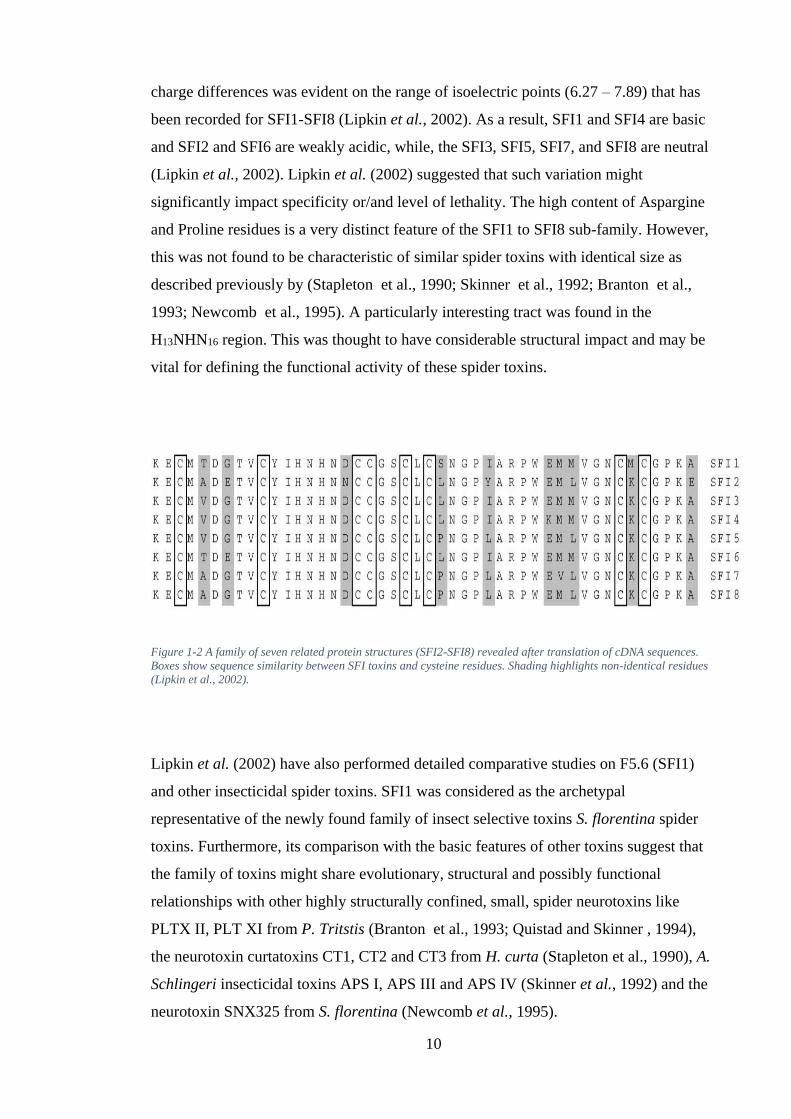
charge differences was evident on the range of isoelectric points (6.27 – 7.89) that has
been recorded for SFI1-SFI8 (Lipkin et al., 2002). As a result, SFI1 and SFI4 are basic
and SFI2 and SFI6 are weakly acidic, while, the SFI3, SFI5, SFI7, and SFI8 are neutral
(Lipkin et al., 2002). Lipkin et al. (2002) suggested that such variation might
significantly impact specificity or/and level of lethality. The high content of Aspargine
and Proline residues is a very distinct feature of the SFI1 to SFI8 sub-family. However,
this was not found to be characteristic of similar spider toxins with identical size as
described previously by (Stapleton et al., 1990; Skinner et al., 1992; Branton et al.,
1993; Newcomb et al., 1995). A particularly interesting tract was found in the
H13NHN16 region. This was thought to have considerable structural impact and may be
vital for defining the functional activity of these spider toxins.
Figure 1-2 A family of seven related protein structures (SFI2-SFI8) revealed after translation of cDNA sequences.
Boxes show sequence similarity between SFI toxins and cysteine residues. Shading highlights non-identical residues
(Lipkin et al., 2002).
Lipkin et al. (2002) have also performed detailed comparative studies on F5.6 (SFI1)
and other insecticidal spider toxins. SFI1 was considered as the archetypal
representative of the newly found family of insect selective toxins S. florentina spider
toxins. Furthermore, its comparison with the basic features of other toxins suggest that
the family of toxins might share evolutionary, structural and possibly functional
relationships with other highly structurally confined, small, spider neurotoxins like
PLTX II, PLT XI from P. Tritstis (Branton et al., 1993; Quistad and Skinner , 1994),
the neurotoxin curtatoxins CT1, CT2 and CT3 from H. curta (Stapleton et al., 1990), A.
Schlingeri insecticidal toxins APS I, APS III and APS IV (Skinner et al., 1992) and the
neurotoxin SNX325 from S. florentina (Newcomb et al., 1995).

11
Most of these spider toxins are known to be highly selective agonists or antagonists of
various voltage dependent Ca2+ channels (Stapleton et al., 1990; Branton et al., 1993;
Pallaghy et al., 1997), which could be potentially valuable reagents in the
neuromuscular function. Lipkin et al. (2002) suggest that if this SFI1 family possess a
similar mode of action, they have the potential to be used as selective reagents to target
Ca2+ channels. This possibility was studied by examining the alignment of sequences as
shown in Figure 1-3 that revealed that SFI1 bears a basic structure identical to other
neurotoxins, mainly associated to the distribution of cysteine residue. Also, more typical
characteristics of such structural motif may be the size of the amino acid tract between
successive cysteine residues. Compared to other toxins, SNX325 and SFI1 have a large
gap between the 6th and 7th cysteine residues. Lipkin et al. (2002) postulated that
variations observed in the folded configuration, due to various disulfide pairings, may
be important for the different receptor identification, specificity and biological modes of
activity among these spider neurotoxins.
Figure 1-3: The amino acid sequence comparison between SFI1 (F5.6) S. florentina insecticidal toxin and other
arthropod toxins. All sequences were arranged with respect to cysteine residue as shown in boxes. For better
arrangement, gaps (-) were introduces. Shading show negatively charged amino acid residues and those highlighted
in bold are positively charged amino acid residues.
Insecticidal targets of spider neurotoxins
The pore-forming α1 subunit of the calcium voltage (CaV) channels and α subunit of
the sodium voltage (NaV) channels together form a superfamily of structurally related
voltage-gated ion channels.
12
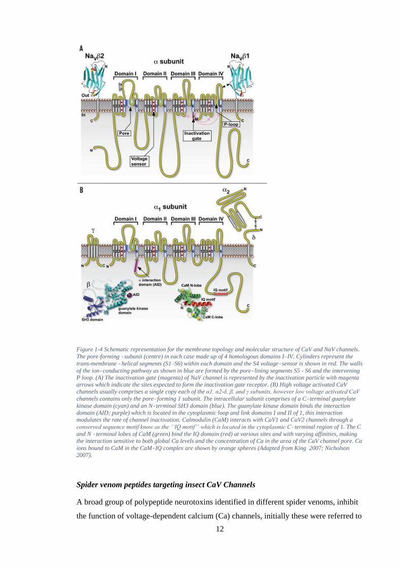
Figure 1-4 Schematic representation for the membrane topology and molecular structure of CaV and NaV channels.
The pore-forming - subunit (centre) in each case made up of 4 homologous domains I–IV. Cylinders represent the
trans-membrane - helical segments (S1–S6) within each domain and the S4 voltage- sensor is shown in red. The walls
of the ion- conducting pathway as shown in blue are formed by the pore- lining segments S5 - S6 and the intervening
P loop. (A) The inactivation gate (magenta) of NaV channel is represented by the inactivation particle with magenta
arrows which indicate the sites expected to form the inactivation gate receptor. (B) High voltage activated CaV
channels usually comprises a single copy each of the α1, α2-δ, β, and γ subunits, however low voltage activated CaV
channels contains only the pore- forming 1 subunit. The intracellular subunit comprises of a C- terminal guanylate
kinase domain (cyan) and an N- terminal SH3 domain (blue). The guanylate kinase domain binds the interaction
domain (AID; purple) which is located in the cytoplasmic loop and link domains I and II of 1, this interaction
modulates the rate of channel inactivation. Calmodulin (CaM) interacts with CaV1 and CaV2 channels through a
conserved sequence motif know as the ‘‘IQ motif’’ which is located in the cytoplasmic C- terminal region of 1. The C
and N - terminal lobes of CaM (green) bind the IQ domain (red) at various sites and with varying affinities, making
the interaction sensitive to both global Ca levels and the concentration of Ca in the area of the CaV channel pore. Ca
ions bound to CaM in the CaM- IQ complex are shown by orange spheres (Adapted from King 2007; Nicholson
2007).
Spider venom peptides targeting insect CaV Channels
A broad group of polypeptide neurotoxins identified in different spider venoms, inhibit
the function of voltage-dependent calcium (Ca) channels, initially these were referred to
13
as v-toxins (Grishin, 1999). Though pharmacological and physiological properties of
voltage-gated Ca channels can vary between different types of voltage-gated Ca
channels, they still share a common assembly of structural motifs. Different types of
CaV channels have an identical general structure. These Ca channels are heteromeric
proteins made up of a pore-forming α1 subunit, a δ transmembrane subunit with a
disulfide-link to the α2 protein, an intracellular subunit β, and also another
transmembrane subunit γ. These subunits form a fully functional calcium channel, as
shown in Figure 1-5 (Catterall and Few, 2008; Pringos et al., 2011).
The largest protein is the α1 subunit, which forms the channels, which also contains the
conduction pore, the voltage sensor and the gating mechanism. In addition, some of the
channel regulation sites (by second messengers, drugs or toxins) are also known to be
located on the α1 subunit. A trans-membrane protein is formed by the α1 subunit, as
predicted by amino acid sequencing, and organized into 4 repeated domains. Each of the
domains comprises 6 segments of α helical structures, as shown in figure1-5(b). The
green cylinders shown represent the pore-forming segments and yellow cylinders
represent the sensor segments and a loop (Catterall and Few, 2008). The intracellular
side of the membrane contains N- and C-termini of the protein.

14
Figure 1-5 Voltage-gated Ca channels (CaV) topological structure(Catterall and Few, 2008; Pringos et al., 2011)
SNX-325, a Ca channel antagonist peptide was purified from the venom of S. florentina.
SNX-325 toxin consists of 49 amino acid residues and contains 4 intramolecular disulfide
bridges. It was found to be a selective N-type channel blocker at nanomolar
concentrations, but not a blocker of other calcium channels such as class C, A, and E
(Newcomb et al., 1995).
Spider venom peptides targeting insect NaV channels
NaV channels are trans-membrane proteins that provide the pathway for the fast
depolarization of excitable cells, this a process that is necessary for the initiation of
action potentials (Hodgkin and Huxley, 1952). They are principally made up of a 220–
260 kDa single pore-forming glycoprotein, the α-subunit, as observed from neurotoxin
labelling, purification and successive functional expression studies (Catterall, 2001).
This α- subunit is made up of 4 internally homologous, non- identical domains (I–IV),
which are connected by intracellular linkers as shown in figure 1-4A (Yu and Catterall,
b
a
15
2003). Each domain is assumed to contain 6 trans-membrane segments (S1 – S6) that
are joined by intracellular or extracellular loops. Out of these, 4 domains fold jointly in
a clockwise orientation in an orientation where the outer pore vestibule is formed by
domains I and IV. Membrane re-entrant loops (P- loops) between trans-membrane
segments S5 and S6 from each domain dip into the trans-membrane region of the
protein and also form the narrow ion- selectivity filter at the extracellular end of the
pore. The most highly conserved S4 segments contain a positively charged Arg or Lys
amino acid at every third position. This section serves as a ‘voltage sensor’ which
initiates voltage- dependent activation through outward movement caused by changes in
electric field (Chanda and Bezanilla , 2002; Cestèle et al., 2006). Selective influx of
Na+ ions through the pore occurs as a response to membrane depolarization, causing the
channel to undergo a conformational change.
In mammals, one or two smaller auxiliary subunits of approximately 33–36 kDa are
also associated with the α- subunit (NaVβ1–NaVβ4). These subunits are required for
normal kinetics, as well as voltage- dependent gating of the channel, but not for ion
flux, ionic selectivity and ultimately pharmacological modulation (Yu et al., 2003).
Inactivation of the NaV channel is mediated through a short intracellular loop
connecting III and IV domains, containing inactivation gate IFM motif, which is the key
hydrophobic amino acid sequence isoleucine, phenylalanine, and methionine. The NaV
channel is the primary molecular target for numerous therapeutic drugs like local
anaesthetics, antiarrhythmics and anticonvulsants and also insecticides like pyrethroids,
dihydropyrazoles, DDT, N-alkylamides and oxadiazines. Most of the structural and
functional properties of NaV has been identified using guanidinium, small alkaloid
toxins and peptide of various plant and animal origins.
There are 4 binding sites for neurotoxin on the Nav channels, including the site 1
located on the extracellular surface of the pore. This site 1 is where the Tx1 peptide
toxins from the Phoneutria nigriventer spider and the µ - conotoxins from Conus
species, as well as the guanidinium alkaloids (saxitoxin (STX) and tetrodotoxin (TTX)
bind. The Type 1 and Type 2 sea anemone toxins, the scorpion a-toxins, and selected
spider toxins bind to the site S3 – S4 on the extracellular loop in domain IV and may
also bind to unidentified residues in the S5 – S6 linkers in domains I and IV. This site
has also been shown to possess complex allosteric interactions with site - 2, which binds
several alkaloid toxins like veratridine and batrachotoxin, and site - 5 which binds the
16
cyclic polyether toxins ciguatoxin and brevetoxin. Some site - 4 toxins include the
scorpion polypeptide b-toxins which bind to the S1 – S2 and S3 – S4 linkers in domain
II and help channel activation via trapping the S4 segment in its outward position and
shift the voltage-dependent activation to more hyperpolarized potentials. Site-3 and site-
4 toxins primarily increase the open status of NaV channels and hamper gating
transitions into the closed states. These are therefore classified as ‘gating-modifiers’.
Finally, site - 6 neurotoxins binds to the d-conotoxin TxVIA slowing channel
inactivation, this is contrary to the allosteric modulation observed in the binding of
neurotoxins targeting site-3 (Nicholson, 2007).
NaV channels have been preserved across evolution and therefore identical types of
neurotoxin receptor sites are found on both mammalian and insect neuronal NaV
channels. However, mammalian and insect NaV channels are pharmacologically
distinct. To date, 9 mammalian NaV channels (NaV1.1–1.9) have been cloned,
expressed functionally and characterised (Goldin et al., 2000). Evidence from these
studies suggests that the diversity of mammalian NaV channels is achieved mainly
through expression of distinct NaV channel genes. Certainly, a variety of non-
insecticidal spider toxins can be utilised in the study of the molecular differences in
NaV channel subtypes. Phrixotoxin 3 from Phrixotrichus auratus and ceratotoxins 1, 2,
and 3 from Ceratogyrus cornuatus have been shown to block ion conductance, which
results in a shift from the voltage-dependent activation to more positive values, not
affecting inactivation (Bosmans et al., 2006). Studies in certain toxins have shown that
slight differences in surface receptors can alter the specificity to NaV channel subtypes
(Nicholson, 2007). The structural differences between arthropod and mammalian NaV
channels is reflected on the allosteric modulation of neurotoxin receptor sites. The NaV
channels in insects are often more sensitive to the activity of neurotoxins. Indeed, m-
agatoxins (m-Aga-I to m-Aga-VI) that affect the functions of NaV channels has been
found in the venom of Agelenopsis aperta spider (Skinner et al., 1989). Currently, there
are 6 toxins of about 36 - 38 amino acids containing 8 cysteine residues which form 4
disulfide bridges. At least 4 toxins from this family possess an amidated C-terminal
residue. Furthermore, m-Agatoxins share similar activity characteristics with the
scorpion excitatory insectotoxins, including shift of the insect Na+ channel activation
curve to more negative potentials. Scorpion insectotoxins have be
17
en shown to exhibit similar action to the curtatoxins (Ct-I and Ct-II) isolated from the
venom of Hololena curta spider, which is attributed to structural similarities (Stapleton
et al., 1990b). They also resemble toxic polypeptides (Tx1 and Tx2-9) derived from the
venom of the Phoneutria nigriventer spider (Cordeiro Mdo et al., 1992) and also
several other polypeptide toxins from diverse spider venoms.
1.3.2 Marine cone snail from the Genus Conus: peptide nomenclature
The genus Conus is the most diverse genus of marine invertebrates (Kohn, 1998).The
speciation and rich endemism are evident from the morphology and toxicology of the
genus Conus (Rockel et al., 1995).This genus is broadly distributed throughout all
tropical oceans, which encompass 25% of the earth’s ocean area. Recent studies suggest
that the Indo-Pacific region accounts for around 60% of Conus habitation (Kumar et al.,
2015). It is thought that each Conus member has a range of 100–200 different venom
components, which are majorly disulfide-rich conopeptides. Moreover, the venom from
each species has its own distinct complement of peptides that lacks molecular overlap
with each other (Olivera, 1997). Therefore, the number of identified Conus species
(around 500–700) could potentially translate to a source for more than 140,000 diverse
conopeptides (Olivera, 2006). The diversity of conopeptides is interesting as it is
generated by a relatively small number of gene superfamilies expressed in the venom
duct of conus species (Terlau and Olivera, 2004). A single open reading frame is present
in conopeptide encoding mRNAs, which is translated into a canonical prepropeptide
(Beleboni et al., 2004). Generally, the mature peptides have a characteristic
arrangement of Cys residues in each gene superfamily (Table 1-1).
A strikingly conserved N-terminal signal sequence is shared by the peptide precursors
from a given gene superfamily. In contrast to this, the mature peptide sequence is hyper-
mutable, with the exception of the Cys residues. Thus, conotoxin precursors exhibit
striking juxtaposition of very conserved and hyper-variable sequences, similar to
antibodies. The hyper-mutable molecular diversity of Conus peptide superfamilies
constantly result to the generation of conotoxins with novel functions. For example, as
shown in Table 1-1, the C–C–CC–C–C Cys pattern is observed in all peptides in the O-
conotoxin superfamily and are translated with conserved signal sequences. While the ω-
conotoxins are calcium channel blockers, the δ-conotoxins inhibit the inactivation of
sodium channels as such enhancing the conductance of Na+ channel (Terlau and
Olivera, 2004). Though different members of a same Conus peptide family have
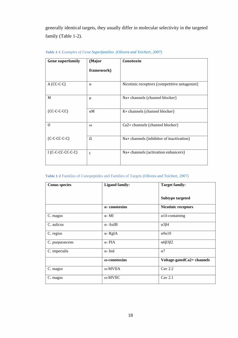
18
generally identical targets, they usually differ in molecular selectivity in the targeted
family (Table 1-2).
Table 1-1 Examples of Gene Superfamilies (Olivera and Teichert, 2007)
Gene superfamily (Major
framework)
Conotoxin
A (CC-C-C) α Nicotinic receptors (competitive antagonist)
M
(CC-C-C-CC)
µ Na+ channels (channel blocker)
κM K+ channels (channel blocker)
O
(C-C-CC-C-C)
ω Ca2+ channels (channel blocker)
Ω Na+ channels (inhibitor of inactivation)
I (C-C-CC-CC-C-C) ι Na+ channels (activation enhancers)
Table 1-2 Families of Conopeptides and Families of Targets (Olivera and Teichert, 2007)
Conus species Ligand family:
Target family:
Subtype targeted
α- conotoxins Nicotinic receptors
C. magus α- MI α1δ-containing
C. aulicus α- AuIB α3β4
C. regius α- RgIA α9α10
C. purpurascens α- PIA α6β3β2
C. imperialis α- ImI α7
ω-conotoxins Voltage-gatedCa2+ channels
C. magus ω-MVIIA Cav 2.2
C. magus ω-MVIIC Cav 2.1
19
Marine cone snails from the genus conus have received much interest in recent years as
rich sources of neurotoxins as they possess a potent cocktail of venomous peptides to
catch their prey. These venomous peptides are small in size (<50kDa) and a number of
genes are required for their synthesis. As per an estimation done by Terlau et al. (1996)
there are over 100,000 bioactive compounds present in t
he venomous cocktail synthesized by cone snails, each with a distinct neurological
target.
Venom peptides, called conopeptides, have evolved very recently and produced from
propeptides using specialized venom endoproteases. The toxicity of a venom peptide is
further increased by several forms of post-translational modification, which also renders
stability to the 3 D structure of the protein (Figure 1-6). Conotoxins are thought to be
good pharmacological probes due to the small size, stable structure and specificity for
their targets
. Conotoxins have been studied in detail by various researchers and have been
implicated as antagonists of nicotinic acetylcholine receptors (nAChR). These venoms
act as nAChR blockers at the neuromuscular synapse in the prey resulting in paralysis.
The action of conotoxins on nAChR is mediated through a competition between the
antagonist of acetylcholine receptors for the binding site on the receptor. The
antagonists of acetylcholine receptors are α-tubocurarine and α-bungarotoxins, which
bind in a reversible manner. A study conducted by Groebe et al in 1995 showed that
affinity for the acetylcholine binding site for the α- conotoxins MI, GI and SIA is 15000
fold higher compared to tubocurarine or bungarotoxins (Bass et al., 2011; Lewis et al.,
2012; Safavi - Hemami et al., 2011).
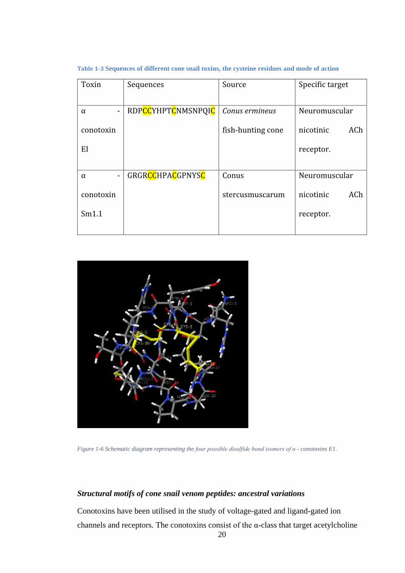
20
Table 1-3 Sequences of different cone snail toxins, the cysteine residues and mode of action
Toxin Sequences Source Specific target
α -
conotoxin
EI
RDPCCYHPTCNMSNPQIC Conus ermineus
fish-hunting cone
Neuromuscular
nicotinic ACh
receptor.
α -
conotoxin
Sm1.1
GRGRCCHPACGPNYSC Conus
stercusmuscarum
Neuromuscular
nicotinic ACh
receptor.
Figure 1-6 Schematic diagram representing the four possible disulfide bond isomers of α - conotoxins E1.
Structural motifs of cone snail venom peptides: ancestral variations
Conotoxins have been utilised in the study of voltage-gated and ligand-gated ion
channels and receptors. The conotoxins consist of the α-class that target acetylcholine
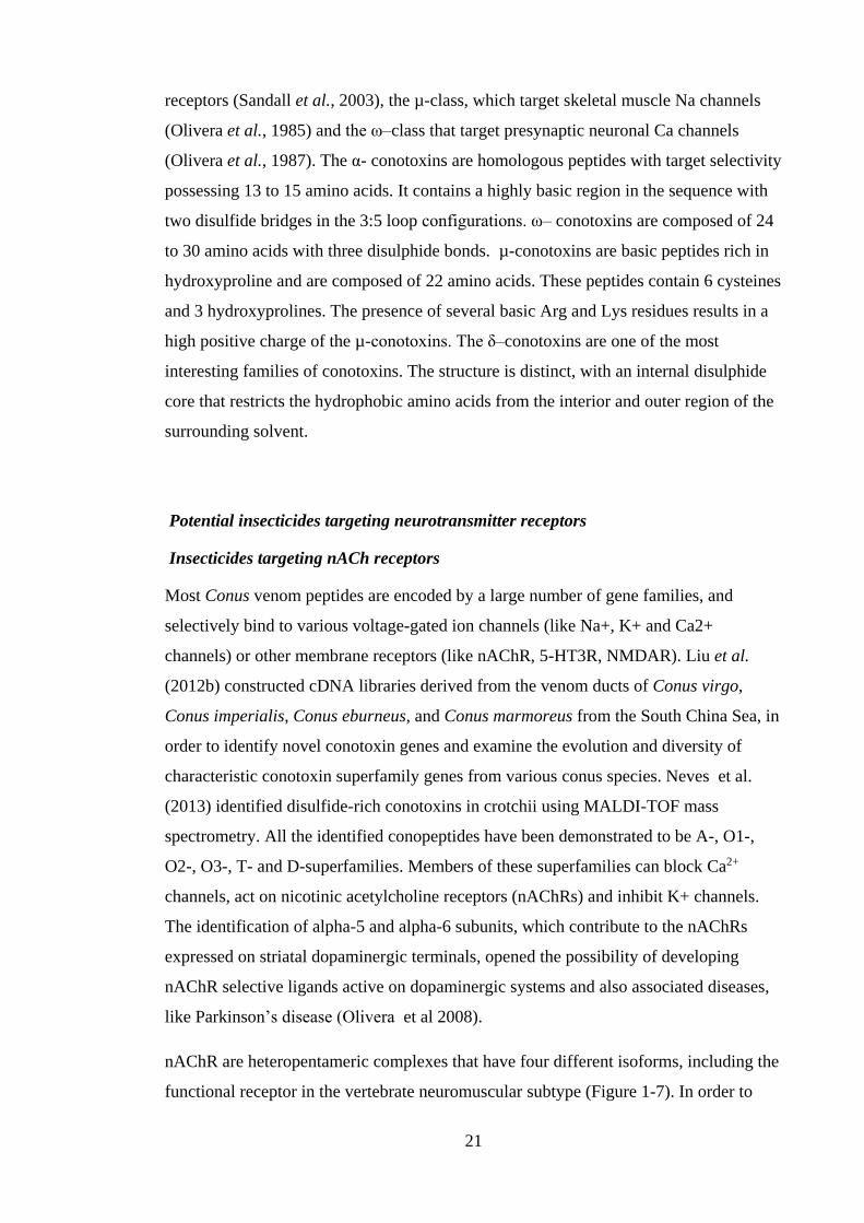
21
receptors (Sandall et al., 2003), the µ-class, which target skeletal muscle Na channels
(Olivera et al., 1985) and the ω–class that target presynaptic neuronal Ca channels
(Olivera et al., 1987). The α- conotoxins are homologous peptides with target selectivity
possessing 13 to 15 amino acids. It contains a highly basic region in the sequence with
two disulfide bridges in the 3:5 loop configurations. ω– conotoxins are composed of 24
to 30 amino acids with three disulphide bonds. µ-conotoxins are basic peptides rich in
hydroxyproline and are composed of 22 amino acids. These peptides contain 6 cysteines
and 3 hydroxyprolines. The presence of several basic Arg and Lys residues results in a
high positive charge of the µ-conotoxins. The δ–conotoxins are one of the most
interesting families of conotoxins. The structure is distinct, with an internal disulphide
core that restricts the hydrophobic amino acids from the interior and outer region of the
surrounding solvent.
Potential insecticides targeting neurotransmitter receptors
Insecticides targeting nACh receptors
Most Conus venom peptides are encoded by a large number of gene families, and
selectively bind to various voltage-gated ion channels (like Na+, K+ and Ca2+
channels) or other membrane receptors (like nAChR, 5-HT3R, NMDAR). Liu et al.
(2012b) constructed cDNA libraries derived from the venom ducts of Conus virgo,
Conus imperialis, Conus eburneus, and Conus marmoreus from the South China Sea, in
order to identify novel conotoxin genes and examine the evolution and diversity of
characteristic conotoxin superfamily genes from various conus species. Neves et al.
(2013) identified disulfide-rich conotoxins in crotchii using MALDI-TOF mass
spectrometry. All the identified conopeptides have been demonstrated to be A-, O1-,
O2-, O3-, T- and D-superfamilies. Members of these superfamilies can block Ca2+
channels, act on nicotinic acetylcholine receptors (nAChRs) and inhibit K+ channels.
The identification of alpha-5 and alpha-6 subunits, which contribute to the nAChRs
expressed on striatal dopaminergic terminals, opened the possibility of developing
nAChR selective ligands active on dopaminergic systems and also associated diseases,
like Parkinson’s disease (Olivera et al 2008).
nAChR are heteropentameric complexes that have four different isoforms, including the
functional receptor in the vertebrate neuromuscular subtype (Figure 1-7). In order to
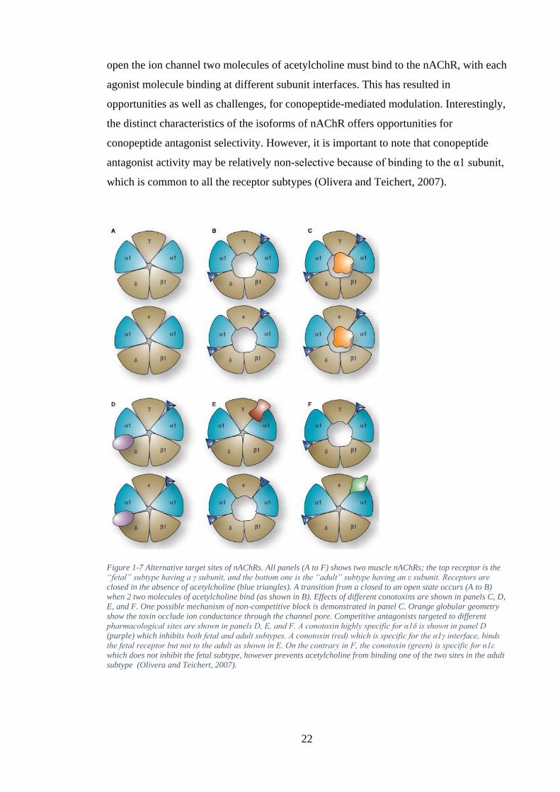
22
open the ion channel two molecules of acetylcholine must bind to the nAChR, with each
agonist molecule binding at different subunit interfaces. This has resulted in
opportunities as well as challenges, for conopeptide-mediated modulation. Interestingly,
the distinct characteristics of the isoforms of nAChR offers opportunities for
conopeptide antagonist selectivity. However, it is important to note that conopeptide
antagonist activity may be relatively non-selective because of binding to the α1 subunit,
which is common to all the receptor subtypes (Olivera and Teichert, 2007).
Figure 1-7 Alternative target sites of nAChRs. All panels (A to F) shows two muscle nAChRs; the top receptor is the
“fetal” subtype having a γ subunit, and the bottom one is the “adult” subtype having an ε subunit. Receptors are
closed in the absence of acetylcholine (blue triangles). A transition from a closed to an open state occurs (A to B)
when 2 two molecules of acetylcholine bind (as shown in B). Effects of different conotoxins are shown in panels C, D,
E, and F. One possible mechanism of non-competitive block is demonstrated in panel C. Orange globular geometry
show the toxin occlude ion conductance through the channel pore. Competitive antagonists targeted to different
pharmacological sites are shown in panels D, E, and F. A conotoxin highly specific for α1δ is shown in panel D
(purple) which inhibits both fetal and adult subtypes. A conotoxin (red) which is specific for the α1γ interface, binds
the fetal receptor but not to the adult as shown in E. On the contrary in F, the conotoxin (green) is specific for α1ε
which does not inhibit the fetal subtype, however prevents acetylcholine from binding one of the two sites in the adult
subtype (Olivera and Teichert, 2007).

23
1.3.3 The venom peptide Acrorhagin-2a from the sea anemone Anthopleura
maculate
The venom peptide Acrorhagin-2a from the sea anemone Anthopleura maculate targets
sodium channels by binding at site 3 of the sodium channels receptor in the excitable
membrane (Honma and Shiomi, 2006). Because of this unique biological effect, sea
anemone sodium channel toxins appear to be a valuable source of novel peptide toxins
for managing crop pests. However, efforts towards this direction have remained largely
unsuccessful due to non-target activity since the peptide has been shown to target
sodium channels in a wide range of animals following injection. For example,
preliminary studies have shown that injecting crabs with these toxins caused non-
specific lethal effects (Honma et al., 2005). Oral delivery of the toxin, on the other
hand, has been shown to have no toxic effects. It has thus been proposed that to
effectively use such molecules they should be fused to a carrier molecule to ensure that
the toxin reaches its site of action, in this case the sodium ion channels. However, such
a delivery strategy may also affect non-targets insects and vertebrates (Honma et al.,
2005). Therefore, understanding the structure of different sea anemone polypeptides is
necessary to enhance their development for control of insect pests that are both effective
and specific
Structural motifs of the sea anemone venom peptides
Sea anemone polypeptide toxins have different structures that are dependent on the
presence and arrangement of cysteine residues. Among these toxins, highly toxic, linear,
cysteine-free polypeptides make up a considerable part of known venom peptides. On
the basis of the specific arrangement of cysteine residues, cysteine-rich polypeptides
may be classified into several structural classes. The number of the cysteine residues
can be a crucial feature of polypeptide toxins because it determines the exact order of
the disulfide bonds and subsequent folding. Motif development was performed using
Single Residue Distribution Analysis (SRDA). In the mature venom toxin, the number
of cysteine residues varies from 2 to 10. Though toxins with 6 cysteines are most
common, there are differences in the cysteine motifs (Kozlov and Grishin, 2012).
24
Sodium ion channel as targets for neuropeptides as potential bioinsecticidal
NaV channel toxins are the most thoroughly studied of all the sea anemone toxins, as
these constitute a major fraction of the venom (Moran et al., 2009). Four types of sea
anemone toxic polypeptides (ranging from 3.5 to 6.5 kDa) bind to site three of the NaV
channel receptor during the depolarization process. The Type I and II toxin families
have 46–51 amino acids, anti-parallel β-sheet with 4 β-strands and a highly flexible
“Arg-14 loop” (lacking any –helix) (Smith and Blumenthal, 2007). Members of the
Type I and II toxin families have similar locations for the six half-cysteine residues
(forming three disulfide bonds). Similarly several other residues were thought to play a
role in biological activity and maintenance of the tertiary structure (Norton, 2009). They
also have a basic C-terminal sequences (Messerli and Greenberg, 2006). The type III
toxin family have 27–32 amino acids with rigid β and γ turns, like in ATX-III and
PaTX, which are cross-linked by three and four disulphide bonds respectively,
suggesting they do not share the structural scaffold (Honma and Shiomi, 2006). Reports
suggest that the bioactive surface of toxin ATX-III consist mainly of aromatic residues
that is distinct from other site-3 targeting toxins. However, ATX-III binds to NaV
channels at the site three of the receptor (Moran et al., 2007). Unlike Type I and Type
II, the Type III family toxins have only been reported to target NaV channels in a few
species, which are mainly common in various cnidarian venoms (Messerli and
Greenberg, 2006). In addition to these types of toxin, other group like Calitoxin I and
Calitoxin II (with 79 amino acid residues) resemble the Type I and II toxin family in the
number of disulfide bridges and the sequence length, but the amino acid sequence is
different. They also act on NaV channels in a similar manner to the Type I–III toxin
family (Honma and Shiomi, 2006).
1.3.4 Marine cone snail from the Genus Conus: isolation
Spence et al. (1977) attempted to purify the bioactive peptide components from cone
snail venom toxin and validated a peptide composition based on its amino acid
composition (µ-conotoxin). Chemical synthesis began with the α - conotoxin GI, (from
C. geographus venom), which confirmed its native amino acid sequence of 13-amino
acids with two disulfide bonds (Gray et al., 1981). Following the identification of the
venom peptide composition, the µ-conotoxins were isolated and characterised from C.
Geographus milk venom (Olivera et al., 1985). The presence of α - conotoxin and µ-
25
conotoxin functional peptides in C. geographus venom suggested that the cone snail
venom could be both a potent nicotinic receptor antagonist (α -conotoxin) and a sodium
channel blocker (µ-conotoxin) (Moczydlowski et al., 1986). These cone snail venom
molecules act synergistically to bind and inhibit different receptors of multiple
molecular pathways involved in the prey’s normal evasion behaviours. Ultimately, the
requirement for categorizing the paralytic peptides based on pharmacological targets
was expressed due to work that established the activity of various toxins on different
receptors (Janes, 2005).
Structure and function of the Genus Conus α -conotoxin E1
The α - conotoxins are a family of cone snail venom toxins, with 12 and 19 amino acid
residues and use key disulphide bonds to maintain the structure. These polypeptide
toxins are highly selective at blocking the nicotinic acetylcholine receptors (nAChRs),
ligand-gated K+, Na+ or Ca2+ ion channels across the synaptic membrane. nAChRs
exist in two classes including the neuronal and neuromuscular, with five subunits in
each homopentameric or heteropentameric membrane-bound channel structures. Two
molecules of the acetylcholine neurotransmitter are required to open the channel and
stabilise the receptor in the open active state for the conductance of ions. Most of the α -
conotoxins are competitive antagonists for the nAChRs, which bind at one or both of
these ACh binding sites with high affinity. Neuronal nAChRs (n-nAChRs) receptors are
highly diverse in structure with a variety of different α and β subunits. The α -
conotoxins are selective towards both specific subclasses of receptor and ACh binding
pockets between deferent subunit pairs. This makes them ideal tools to probe both the
central as well as peripheral nervous systems for receptor distribution. In addition to
this, a growing number of α - conotoxin structures create the potential for its use as
templates for deriving pharmaceutical agents.
The α - conotoxin EI was first identified in 1995 by (Martinez et al.)., from the Atlantic
fish hunting marine snail Conus ermineus. The α - conotoxin EI is a somewhat atypical
member of the neuromuscular conotoxins, which has an α4/7 loop motif that is more
associated with neuronal conotoxins (Martinez et al., 1995). Comparison of α -
conotoxin EI with α4/7 neuronal conotoxins shows that only Tyr6 and a Gln16 residues
in its sequence are unique and not found in any other neuronal conotoxins. There is
limited data on the binding of α - conotoxin EI to neuronal nAChRs. Similarly, the
26
determined structural conformation of α - conotoxin EI is nearly the same in its
backbone to the α4/7 neuronal α - conotoxins (Park et al., 2001). This suggests that
some of the main differences in the mechanisms of binding to specific receptor
subclasses are the surface charge, side-chain conformations and subtle differences in the
overall topologies of these α - conotoxin peptides.
1.4 Neurotoxins and their use in fusion proteins
Protein based biopesticides represent more environmentally friendly approaches to pest
control, as they are biodegradable and combine efficacy with specificity. In addition to
naturally occurring protein biopesticides like Bacillus thuringiensis toxins, recombinant
proteins with insecticidal activity can be produced using biotechnological methods. Use
of insecticidal fusion proteins containing a toxic peptide or protein fused to a “carrier” is
an example of such a fusion protein. The carrier molecule confers oral activity on the
toxin which would normally be toxic when injected into the insect since it directs the
transports of the fusion molecule across the insect gut, as so allow the toxin to reach its
site of action (Fitches et al., 2002, 2004). When fused to a carrier molecule the
neuropeptide remains active and is transported across the gut epithelium to the central
nerves system (Fitches et al., 2002). The δ-amaurobitoxin-PI1a spider venom peptide
that targets the NaV channels have been fused to the snowdrop lectin (GNA) “carrier”
to study its oral toxicity (Yang et al., 2014). The fusion was found to be approximately
6 times as effective as recombinant spider venom peptide on a molar basis. Originally,
the spider venom peptide lacked oral activity against cabbage moth larvae. However a
single 30 mg dose of the fusion protein presented 100% larval mortality in 6 days. Also,
fusion protein transports from the gut contents to the haemolymph of cabbage moth
larvae. Additionally, further binding to the nerve chord, was revealed by Western
blotting (Yang et al., 2014). Therefore, fusion protein found to be a promising candidate
for the development of biopesticide.
1.4.1 Spider venom toxins as insecticides
Some of the spider toxins have high phyletic specificity, and high potency mediated
through novel mode of action, which makes them an important compound for
bioinsecticide development. In fact, transgenes encoding insect-specific arachnid toxins
(including spider neurotoxins) have been successfully expressed in various crops and
27
entomopathogens. Another similar approach expressed a transgene encoding a spider
toxin in the entomopathogenic fungus Metarhizium anisopliae. The pathogenicity of
this fungus was found to be significantly increased against the tobacco hornworm
Manduca sexta and the dengue mosquito Aedes aegypti, without compromising host
specificity (Wang and St Leger, 2007). For the purposes of this research, only three
spider toxins have been evaluated to date.
1.5 Expression of fusion proteins: Pichia pastoris as an expression host
Pichia pastoris is amethylotrophic yeast, which is the eukaryotic expression system of
choice for large-scale production of active recombinant fusion proteins since
prokaryotes do not fold these proteins correctly. This system shows a major advantage
over Saccharomyces spp. as it contains an efficient secretion system, which helps to
direct larger amounts of recombinant protein secreted into the culture medium. This
makes purification of the desired product straightforward (Pyati et al., 2014). P.
pastoris is an ideal host for producing small proteins with high content of disulphide
bridges, since it is able to fold the protein into biological active forms (Daly and Hearn,
2005). This is essential for the production of Galanthus nivalis agglutinin
(GNA/snowdrop lectin), which is secreted as a fully active folded protein (Baumgartner
et al., 2003; Raemaekers et al., 1999). However, expression in E. coli produces an
insoluble inclusion body, where the process of denaturation and renaturation is required
to recover functionally active GNA (Luo et al., 2005).
Constructs driven by the alcohol oxidase (AOX1) promoter are usually used to
maximise expression of recombinant proteins in P. pastoris. Here, the expression can be
induced by addition of methanol to the growing culture. Fusion protein production
studies have shown that constitutively expressed GAP promoter gives better expression.
It is advantageous for industrial production as a methanol feed is not required. Studies
have shown that, during production by fermentation, insecticidal fusion proteins are
subjected to degradation due to yeast extracellular proteases (Trung et al., 2006;
Fitches et al., 2004; Fitches et al., 2012). The proteolysis occurs mainly at or near the
linker region between the carrier protein and the insecticidal peptide, resulting in a
decrease in yield of the intact fusion protein. This type of proteolysis is predominantly
evident when wild-type X33 P. pastoris strain is used for expression of fusion proteins.
However, using the P. pastoris strain SMD1168H has been found to reduce proteolysis,
28
as it is deficient in the extracellular vacuole peptidase A (pep4), which is responsible for
activating protease B1 and carboxypeptidase Y. The absence of extracellular vacuoles
increases the yield of intact fusion protein. However, for large-scale production, X33
strain is the preferred strain, as it is a protease deficient strain. It is important to note
that the X33 strain tends to be less robust compared to the wild-type strain, resulting in
poor survival and lower growth on storage (Gleeson et al., 1998).
1.5.1 pGAPZα as a plasmid vector for recombinant protein expression
Vectors pGAPZα A, B, and C (3.1 kb) (see Appendix A) use the GAP promoter for
constitutive expressing recombinant proteins in P. pastoris. pGAPZα produces a protein
that is fused to an N-terminal peptide and encoded with the Saccharomyces cerevisiae
α-factor secretion signal. Pyati et al. (2014) used pGAPZαB vector, a shuttle vector
propagated in E. coli, to facilitate the insertion of multiple fusion protein cassettes into
the yeast genome. The pGAPZαB vector was modified so as to contain a Hind III site in
the GAP promoter region using site-directed mutagenesis. Figure 1-8 shows a summary
of the strategy used to obtain a single-copy expression vector. The vector backbone was
modified by insertion of a Hind III site near the Bln I site in the GAP gene 5′ UTR to
obtain the multi-copy plasmid. This results in alteration in two adjacent bases, but does
not affect promoter function. The obtained expression vector (pGAPZαBH-
MODHv1a/GNA (FP)) was transformed into P. pastoris by Pyati et al. (2014) to check
for expression. The authors reported no differences in to the original pGAPZαB
expression construct, which gave the 1-copy baseline for subsequent manipulation.
1.6 Plant lectins
Many plant species have been shown to contain carbohydrate binding proteins (lectins).
Lectins are large class of proteins that are known to contain at least one noncatalytic
domain that binds reversibly to specific mono- or oligo-saccharides (Powell et al.,
1995). Since the first discovery of a plant lectin at the end of the nineteenth century by
Peter Hermann Stillmark, more than one hundred lectins have been purified and
characterized detailing their molecular structure, carbohydrate binding specificity, and
biochemical properties reported over the years. Plant lectins can be classified into four
main lectin families, based on protein structure and evolution (Legume, chitin binding,
type 2 RIPs and monocot mannose binding proteins) and 3 minor families. Currently,
29
researchers focused on the mannose-specific lectins such as Galanthus nivalis (GNA),
Narcissus pseudonarcissus (NPA) and Allium sativum (ASA) and found that they may
qualify as potential carrier molecule. These lectins have the ability to cross the midgut
epithelial through receptor binding, which suggests their roles in pest control.
1.6.1 Roles in plant defence
Since the initial discovery of plant lectins, important progress has been made in the
study of the physiological role of plant lectins. Many studies have investigated the
function of the different structures and sugar binding specificity between all families of
plant lectin. Sugar binding activity and specificity are responsible for the normal
function of lectins, which is mediated by sugar binding specificity to a glycoconjugate
receptor (Van Damme et al., 1998). Identification of their recptors is an essential step
for understanding their physiological role. However, as many storage tissues (e.g. seeds,
bulbs) are rich source in lectins they have been implicated to play a role in plant defence
against pathogens and pests (Van Damme et al., 1998; Powell et al., 1993).
There are a wide verity of plant lectins known to be involved in pest control such as
Canavalia ensiformis (con A). A standard bioassay conducted by Sauvion et al in 2004
to evaluate the toxicity of Con A against Pea aphids, Acyrthosiphon pisum showed a
significant negative effect toward Pea aphids. Furthermore it also showed a remarkable
change in the structure of the epithelial cells of this insect. This study confirmed the
important role of plants lectins such as Con A in plant defence against pests.
Biological activity of other plant lectins that are specific for mannose-binding sites such
as Galanthus nivalis (GNA), Narcissus pseudonarcissus (NPA) and Allium sativum
(ASA) have been assessed by bioassay against peach potato aphid, Myzus persicae. A
comparison of the differences in biological effect between GNA, NPA, and ASA
indicates a non-significant effect of NPA and ASA on survival, whereas GNA caused
45% mortality at 1500 µg/ml. However, GNA showed no significant effects on adult
aphids, their adverse effects being observed during aphid development (Sauvion et al.,
1996).
30
1.6.2 Rols of snowdrop lectin as a carrier molecule
Snowdrop lectin (Galanthus nivalis agglutinin, GNA) is a plant-derived insecticidal
protein, which has previously been shown to be toxic when included into artificial diets
of homopteran pest species, or when expressed in transgenic plants. However, the
effects observed are inconsistent between species (Down et al., 2006). The three-
dimensional structure GNA has been elucidated. It has 12 binding sites, but only the
subdomain I of each of the four subunits and subdomain III of one of its subunits have
been studied in detail (Hester et al., 1995a); these are specific for mannose.
Immunohistochemical studies carried out in the rice brown plant-hopper by Powell et
al. in 1998 showed that GNA binding was mainly concentrated on the luminal surface
of the midgut epithelial cells. The result suggested that GNA binds to the cell surface of
carbohydrate moieties in the gut. Furthermore, Fitches and Gatehouse (1998) showed
that GNA can also bind to glycoproteins (94 kDa) on the Lacanobia oleracea brush
border membrane. The presence of GNA was shown in the ovarioles, fat bodies, and
throughout the hemolymph using immunolocalisation, which suggest the ability of
GNA to cross the midgut epithelial barrier and further pass into the circulatory system
of the test insect (Fitches et al., 2001).
1.7 Synthetic fusion proteins
Synthetic fusion proteins are obtained by incorporating multiple proteins into one
complex and can be designed to accomplish improved properties or new functionality,
synergistically. The fusion of two or more protein domains improves bioactivities; it
may also be used to generate novel functional combinations that can show various
biotechnological and (bio) pharmaceutical applications (Yu et al., 2015). Various
studies on synthetic fusion proteins have been immunohistochemical studies carried out
in the rice brown plant-hopper, most of which caused improved insecticidal activity
(Down et al., 2006).
Down et al. (2006) studied the toxicity of the SFI1/GNA fusion protein against peach
potato aphids M. persicae. The effects of this fusion protein on survival, development,
feeding activity and nymph production of M. persicae were investigated when the
protein was added to artificial diets. The study revealed that SFI1/GNA fusion protein
was toxic with a faster mode of activity to this aphid species compared to GNA, when
31
developed via artificial diet, causing a significant delay in the onset of nymph
production.
1.8 Using the peach potato aphid Myzus persicae both as a target and for other
homopterans
Myzus persicae, found throughout the world (Peccoud et al., 2010), is an important pest
of vegetable crops (Heathcote, 1962), having originated in Asia (Zhang et al., 2008). It
is a primary pest of peach potato (Kuroli and Lantos, 2006), as secondary host plants.
However, it infests over 100 other crop and ornamental plants (Baker , 1994; Van
Emden and Harrington, 2007). The control of aphid is mainly dependent on chemical
insecticides (Robert, 1992), however, exposure to many insecticides has resulted in
aphids evolving resistance, making their control particularly challenging (Foster et al.,
2007). This has resulted in the search for new molecules with novel modes of action to
control this species and other homopteran pests.
1.8.1 Life cycle of Myzus persicae
The life cycle of M. persicae varies considerably, which is dependent on the presence of
cold winters (Van Emden et al., 1969). The development of this aphid can be rapid and
often takes 10 to 12 days for a complete generation. Over 20 annual generations have
been reported in mild climates. Aphids usually reproduce parthenogenetically, however,
if suitable host plants are not available, the aphid will produce eggs. When the plant
breaks dormancy in the spring and begins to grow, the eggs hatch and the nymphs start
feeding on young foliage, flowers, and stems. In cold climates, the adults M. persicae
return to Prunes spp. in the autumn and mating occurs, followed by disposition. All
generations culminating in egg production are parthenogenetic (non-sexual) except the
autumn generation (Capinera, 2001).

32
Figure 1-8 Life cycle of Myzus persicae (Anjum, 2014).
1.8.2 Control of Myzus persicae and need for alternative methods
M. persicae has developed resistance to insecticides and in the UK, females generally
reproduce parthenogenetically with rapid population build up. Currently, three different
resistance mechanisms have been observed in this aphid species. First, the esterase
resistant aphids produce large amounts of esterase enzymes, which help to break down
insecticides before they can reach their target sites. The esterase resistant activity is
highest for organophosphorus insecticides. Organophosphorus and carbamate
insecticides target acetylcholinesterase, however in the modified a cetyle-cholinesterase
(MACE) aphids, this enzyme becomes very insensitive to the blocking effect, making it
immune. A third mechanism is known as knockdown resistance, where aphids have a
modified sodium channel protein, insensitive to insecticides specifically to pyrethroids
(Insecticide Resistance Active Group, UK, 2008). This creates a need for development
of alternative past control methods such as the use of fusion proteins and development
of transgenic crops.
33
1.9 Project aim
The overall aim of this project is to explore the use of fusion proteins composed of
naturally occurring neurotoxic polypeptides fused to a carrier molecule, in this instance
the snowdrop lectin (GNA), as a biopesticide for the protection of plants against insect
pests: specifically Myzus persicae. The methylotrophic yeast Pichia pastoris was used
to produce these recombinant fusion proteins.
The objectives are:
1- Clone and express fusion proteins based on snowdrop lectin (GNA) and toxins
from the spider Segestria-florentina SFIx.
2- Assemble a construct containing two gene copies encoding SFI1/GNA
3- Determine a difference in protein expression levels between a one-copy and a
two-copy gene cassette insert using the pGAPZa plasmid.
4- Establish oral toxicity of the different variants of recombinant fusion proteins
SFIx/GNA against the aphid M. persica species using artificial diet
formulations.
5- Clone and express fusion proteins based on the snowdrop lectin (GNA) and
toxins from the cone snail (alpha-Conotoxin EI, Sm1.1 and Acrorhagin-2a).
6- Establish oral toxicity of recombinant fusion protein (GNA/ alpha-Conotoxin
EI) against the model aphid species M. persicae.
34
2 Chapter 2 Materials and Methods
2.1 Microbial Expression systems and Insects
Escherichia coli and Pichia pastoris were used for expression of recombinant protein.
Escherichia coli Dh5α was maintained on low salt LB broth, which was later replaced
by 2X YT broth to increase the cell density during growth. TOP10 Electrocomp™ E.
coli were provided for efficient transformation supplied by Invitrogen; catalogue
number C4040-52.
Pichia pastoris X33, was grown in 200 ml of starter cultures in a 1L baffled flask for 2-
3 days at 30 OC with shaking. We used YPD media was used which contains 1% yeast
extract, 2% peptone, 1% glucose supplemented with 100–1000 μg/ml Zeocin
(Invitrogen). For a negative control, non-selective YPD medium was also used for P.
pastoris. In many cases yeast was maintained in an orbital shaker at 30 °C and 200 rpm
for 96 h.
The aphid Myzus. persicae was maintained on Chinese cabbage plants (Brassica rapa)
under controlled environmental conditions of 25°C and a 16 hour light/ 8 hour dark
regime.
2.2 Materials and Recombinant Techniques
Basic protocols used in this work were as described by Fitches and colleagues (Fitches
et al., 2002) and Handbook of Molecular Biology (Sambrook and Russell., 2001). The
reagents and chemicals used were purchased from Sigma, VWR or Invitrogen. General
molecular cloning was performed using competent cells from Agilent Technologies
(StrataClone™). For yeast work, transformation of one strain of P. pastoris, the X33
wild type strains was attempted using the EasyComp kit (Invitrogen). GNA and anti-
GNA antibodies were bought from Vector Laboratories and Genosys Biotechnologies,
respectively. A TOPO Cloning kit from Invitrogen was used for subcloning.
Additionally, P. pastoris X33 wild-type strains and the expression vector pGAPZα
(originally purchased from Invitrogen), were constructed by Dr Martin Edwards with
the F1/GNA fusion protein gene cassette incorporated. A cDNA library was used for
amplifying S. florentina (SF) sequences (Lipkin et al., 2002) using PCR amplification.
PCR reactions were carried out using Pfu DNA polymerase Fermentas and plasmid
DNA was prepared using Fermentas miniprep kit.
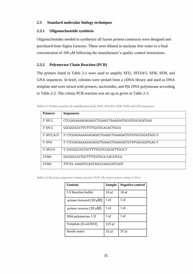
35
2.3 Standard molecular biology techniques
2.3.1 Oligonuclueotide synthesis
Oligonucleotides needed to synthesize all fusion protein constructs were designed and
purchased from Sigma Genosys. These were diluted in nuclease free water to a final
concentration of 100 μM following the manufacturer’s quality control instructions.
2.3.2 Polymerase Chain Reaction (PCR)
The primers listed in Table 2-1 were used to amplify SFI2, SFI3/4/5, SFI6, SFI8, and
GNA sequences. In brief, colonies were picked from a cDNA library and used as DNA
template and were mixed with primers, nucleotides, and Pfu DNA polymerase according
to Table 2-2. The colony PCR reaction was set up as given in Table 2-3.
Table 2-1 Primers used for the amplification of the SFI2, SFI3/4/5, SFI6, SFI8, and GNA sequences.
Primers Sequences
5’ SFI 2 CTCGAGAAAAGAGAGGCTGAAGCTAAAGAGTGCATGGCGGATGAG
3’ SFI 2 GGCGGCCGCTTCTTTTGGTCCACACTTGCA
5’ SFI3/4/5 5’ CTCGAGAAAAGAGAGGCTGAAGCTAAAGAGTGTATGGTGGATGGG 3’
5’ SFI6 5’ CTCGAGAAAAGAGAGGCTGAAGCTAAAGAGTGTATGACGGATGAG 3’
3’ SFI3-8 5’ GGCGGCCGCTGCTTTTGGTCCACACTTGCA 3’
5’GNA GGCGGCCGCTGCTTTTGGTCCA-CACATGCA
3’GNA TTCTA- GAAATCCAGTAGCCCAACGATCAGT
Table 2-2 Reaction component volumes used for PCR. The total reaction volume is 50 μl.
Content Sample Negative control
5 X Reaction buffer 10 μl 10 μl
primer forward (10 μM) 1 μl 1 μl
primer reverse (10 μM) 1 μl 1 μl
DNA polymerase 1 U 1 μl 1 μl
Template (E.coli/X33) 4/5 μl -
Sterile water 32 μl 37 μl
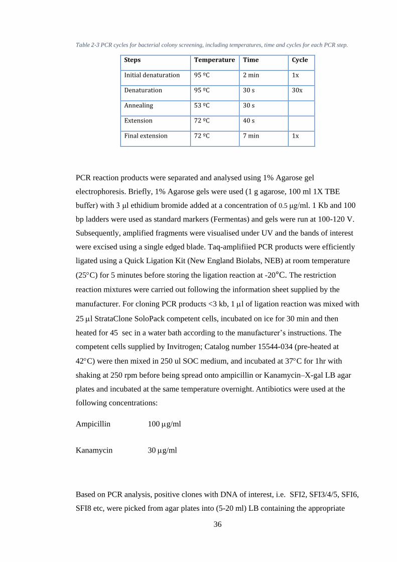
36
Table 2-3 PCR cycles for bacterial colony screening, including temperatures, time and cycles for each PCR step.
Steps Temperature Time Cycle
Initial denaturation 95 ºC 2 min 1x
Denaturation 95 ºC 30 s 30x
Annealing 53 ºC 30 s
Extension 72 ºC 40 s
Final extension 72 ºC 7 min 1x
PCR reaction products were separated and analysed using 1% Agarose gel
electrophoresis. Briefly, 1% Agarose gels were used (1 g agarose, 100 ml 1X TBE
buffer) with 3 μl ethidium bromide added at a concentration of 0.5 μg/ml. 1 Kb and 100
bp ladders were used as standard markers (Fermentas) and gels were run at 100-120 V.
Subsequently, amplified fragments were visualised under UV and the bands of interest
were excised using a single edged blade. Taq-amplifiied PCR products were efficiently
ligated using a Quick Ligation Kit (New England Biolabs, NEB) at room temperature
(25C) for 5 minutes before storing the ligation reaction at -20°C. The restriction
reaction mixtures were carried out following the information sheet supplied by the
manufacturer. For cloning PCR products <3 kb, 1 l of ligation reaction was mixed with
25 l StrataClone SoloPack competent cells, incubated on ice for 30 min and then
heated for 45 sec in a water bath according to the manufacturer’s instructions. The
competent cells supplied by Invitrogen; Catalog number 15544-034 (pre-heated at
42C) were then mixed in 250 ul SOC medium, and incubated at 37C for 1hr with
shaking at 250 rpm before being spread onto ampicillin or Kanamycin–X-gal LB agar
plates and incubated at the same temperature overnight. Antibiotics were used at the
following concentrations:
Ampicillin 100 g/ml
Kanamycin 30 g/ml
Based on PCR analysis, positive clones with DNA of interest, i.e. SFI2, SFI3/4/5, SFI6,
SFI8 etc, were picked from agar plates into (5-20 ml) LB containing the appropriate
37
antibiotic(s). Plasmids were extracted using a QIAprep Spin Miniprep Kit (QIAGEN;
catalogue number 27104). Briefly, overnight cultures (5-20 ml) were grown and
minipreps were prepared as described in the kit. The isolated DNA plasmids were
linearized with EcoRI (Fermentas catalogue number 15202-013). The restriction
enzyme digestion were set up for optimal reaction conditions following the information
sheet supplied by the manufacturer. The restriction reaction mixtures were: 20 μl in
volume using 15 μl of SFIx-Pbsk Vector, 1X enzyme buffer, 1 to 10 units of the
restriction enzyme with the addition of sterile distilled water up to the required volume.
Reaction mixtures were left for 3 hours at 37°C to ensure complete digestion. The
restriction products were separated using 1% agarose gel electrophoresis and then the
QIAquick gel extraction kit (QIAGEN; catalogue number 28704) was used for
purification of DNA excised from the gel. Purified DNA was eluted in 30µl of elution
buffer or distilled. water. Purity, quality and quantity of DNA were measured using a
Thermo Scientific NanoDropTM 1000 Spectrophotometer with UV/Vis analyses of 1 μl
samples. The plasmid concentration for positive clones was measured using a
biophotometer and sent for sequencing.
2.3.3 Ligation and transformation of expression constructs into E. coli
The amplified and purified sequences, SFI2, SFI3, SFI5, SFI6 and SFI8 were digested
with XhoI at NotI sites as the presence of these sites allows insertion of the amplified
toxins into the pGAPZα vector. The amplified coding sequences were then mixed with
cut plasmid pGAPZα containing GNA (in 3:1 ratio), 0.5μl T4 DNA ligase and 1 μl 10 X
ligase buffer in a 10 μl reaction in separate tubes. Reactions were left overnight at 4°C
to ensure complete ligation. Ligated fragments were further used to transform the TOP
10 electro-competent E. coli bacterial cells from Agilent Technologies (StrataClone™
SoloPack® Competent cells). Briefly, cells were removed from -80°C storage and
thawed on ice. Upon mixing with the required ligated plasmid, electroporation allowed
plasmid uptake. Electroporation was carried out using a Biorad Gene Pulser system
following recommendations by both Invitrogen (the cell provider) and Biorad (E. coli
electroporation protocol). Transformed E. coli cells were plated on LB agar plates
containing Zeocin™ (25 μg/ml) for selection of transformants containing the pGAPZα
plasmid. Plasmids from E. coli were extracted using the miniprep Kit and positive
transformants were confirmed by DNA sequencing and/or colony PCR. This will result
into the toxin sequences being inserted within the LacZ gene of the psc-A vector and
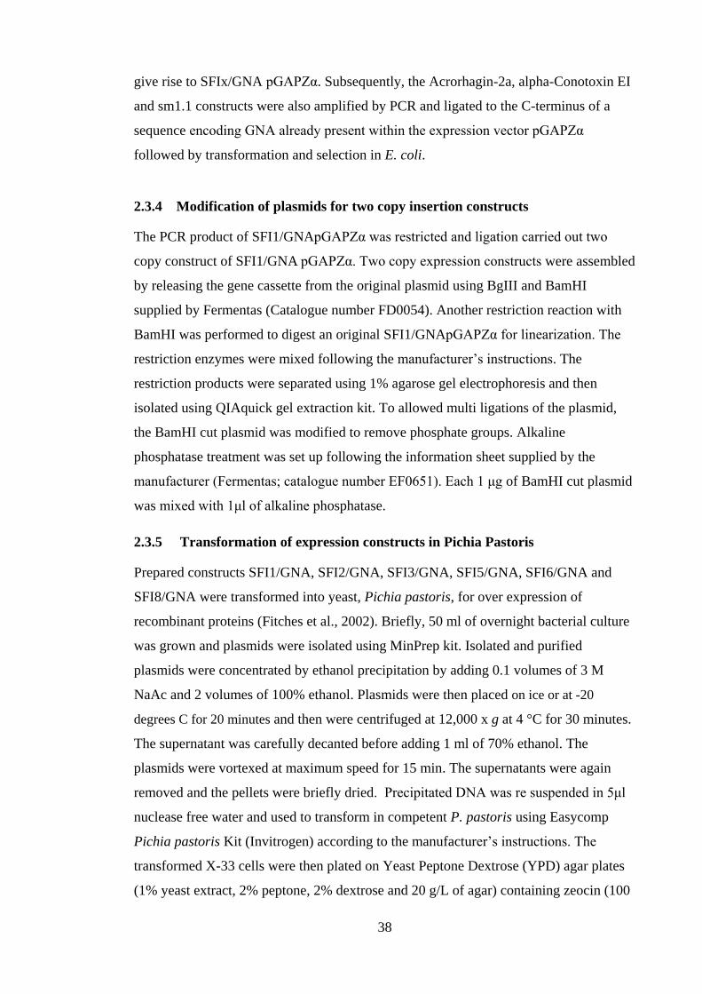
38
give rise to SFIx/GNA pGAPZα. Subsequently, the Acrorhagin-2a, alpha-Conotoxin EI
and sm1.1 constructs were also amplified by PCR and ligated to the C-terminus of a
sequence encoding GNA already present within the expression vector pGAPZα
followed by transformation and selection in E. coli.
2.3.4 Modification of plasmids for two copy insertion constructs
The PCR product of SFI1/GNApGAPZα was restricted and ligation carried out two
copy construct of SFI1/GNA pGAPZα. Two copy expression constructs were assembled
by releasing the gene cassette from the original plasmid using BgIII and BamHI
supplied by Fermentas (Catalogue number FD0054). Another restriction reaction with
BamHI was performed to digest an original SFI1/GNApGAPZα for linearization. The
restriction enzymes were mixed following the manufacturer’s instructions. The
restriction products were separated using 1% agarose gel electrophoresis and then
isolated using QIAquick gel extraction kit. To allowed multi ligations of the plasmid,
the BamHI cut plasmid was modified to remove phosphate groups. Alkaline
phosphatase treatment was set up following the information sheet supplied by the
manufacturer (Fermentas; catalogue number EF0651). Each 1 μg of BamHI cut plasmid
was mixed with 1μl of alkaline phosphatase.
2.3.5 Transformation of expression constructs in Pichia Pastoris
Prepared constructs SFI1/GNA, SFI2/GNA, SFI3/GNA, SFI5/GNA, SFI6/GNA and
SFI8/GNA were transformed into yeast, Pichia pastoris, for over expression of
recombinant proteins (Fitches et al., 2002). Briefly, 50 ml of overnight bacterial culture
was grown and plasmids were isolated using MinPrep kit. Isolated and purified
plasmids were concentrated by ethanol precipitation by adding 0.1 volumes of 3 M
NaAc and 2 volumes of 100% ethanol. Plasmids were then placed on ice or at -20
degrees C for 20 minutes and then were centrifuged at 12,000 x g at 4 °C for 30 minutes.
The supernatant was carefully decanted before adding 1 ml of 70% ethanol. The
plasmids were vortexed at maximum speed for 15 min. The supernatants were again
removed and the pellets were briefly dried. Precipitated DNA was re suspended in 5μl
nuclease free water and used to transform in competent P. pastoris using Easycomp
Pichia pastoris Kit (Invitrogen) according to the manufacturer’s instructions. The
transformed X-33 cells were then plated on Yeast Peptone Dextrose (YPD) agar plates
(1% yeast extract, 2% peptone, 2% dextrose and 20 g/L of agar) containing zeocin (100
39
μg/ml), and plates were incubated at 30°C for 3–4 days until colonies appeared on the
plate. About 6 conspicuous colonies were picked randomly from the YPD agar plate
and cultured in YPD media (containing 1% yeast extract, 2% peptone, 2% dextrose and
100 μg/mL zeocin) for 24–36 h. Glycerol stocks were made from these cultures and
stored at –80°C until later use for protein production. To ensure no errors had occurred,
colony screening of P. pastoris X33 wild-type was performed. Briefly, microcentrifuge
tubes containing 75 μl TE buffer were inoculated with the positive colonies.
Microcentrifuge tubes were heated using a microwave for 3.5, 2, 1.5, 1 and 0.5 minutes
and vortexed briefly in between. The tubes were then placed in a -80ºC freezer for 10
minutes, boiled at 95ºC for 2 minutes and then centrifuged at 1000 x g at room
temperature for 15 minutes. 5 μl of the supernatant was then analysed by PCR using
GNA primers (Table 2-1). PCR conditions are given in Table 2-3.
Table 2-4 PCR cycles for yeast colony screening, including temperatures, time and cycles for each PCR step.
Step Temperature Time Cycle
Initial denaturation 95 ºC 2 min 1x
Denaturation 95 ºC 1 min 30x
Annealing 54 ºC 1 min
Extension 72 ºC 1 min
Final extension 72 ºC 7 min 1x
2.3.6 Cloning and Expression of Cone Snail Toxins
Apart from spider toxins, another objective was to clone and express fusion proteins
based on snowdrop lectin (GNA) and toxins from the cone snail (alpha-Conotoxin EI,
Sm1.1 and Acrorhagin-2a). Obtained constructs, GNA/Acrorhagin-2a, GNA/ alpha-
Conotoxin EI and GNA/sm1.1 were amplified by PCR and ligated into the P. pastoris
expression vector pGAPZαB by restriction/ligation. The resulting plasmids with inserts
were sequenced and confirmed. Transformation of the x33 strain of P. pastoris was
performed by isolating the resulting transformed plasmids pGAPzaB, containing the
correct GNA/ alpha-Conotoxin EI and GNA/sm1.1 construct, from 50 ml of overnight
bacterial culture. Prior to transformation, the plasmids were then digested with either
0.1 μl BglII restriction, (supplied by Fermentas; catalogue number ER0081) or 1 μl
40
(FastDigest BglII enzyme purchased from Fermentas; catalogue number FD0083) per
μg plasmid DNA and the 10X Fast Digest™ Buffer made up 10% of the reaction.
Digests were incubated three hours or 30 min at 37°C, depending on which enzyme was
used (conventional or fast digest respectively). The fully digested DNA was then
inactivated by incubation at 80ºC for 5 minutes. Restriction fragments were isolated
from the agarose gel by QIAquick Gel extraction Kit (QIAGEN; catalogue number
28704) following manufacturer’s instructions.
For yeast expression of GNA/ alpha-Conotoxin EI and GNA/sm1.1 constructs, an
Easycomp Pichia pastoris Kit (Invitrogen) was used. The transformation was done
according to the manufacturer’s instructions. Transformants were analysed either by
colony PCR or western blot to confirm if the insert had been successfully integrated. To
ensure no errors had occurred when GNA/ alpha-Conotoxin EI , GNA/sm1.1 and the
GNA/Acrorhagin-2a constructs were inserted into the x33 strain of P. pastoris, a colony
screening of X33 was performed.
2.3.7 Small-scale screening for fusion protein expression
P. pastoris glycerol stocks were plated and single colonies from various strains were
inoculated in 50ml of YPD media 1% yeast extract, 2% peptone, 1% glucose)
supplemented with 100–1000 μg/ml Zeocin (Invitrogen) and incubated at 30°C for 1–2
days on shaker till the optical density at 600 nm (OD600) reached to 6 days. For negative
control, non-selective YPD medium was also inoculated with the x33 strain of P.
pastoris; glycerol was added to a final concentration of 0.5% every 24 h. Before the
overexpression of all recombinant fusion proteins, cell densities were measured. 1 ml of
the culture at time points 0, 48, 96 hours were withdrawn and measured by OD at
600nm. During this period, 1 mL of culture media was also collected every 48 h which
was centrifuged at 8000 xg for 5 min, and the supernatant was collected and protein
expression examined by SDS-PAGE and western blot.
2.3.8 Protein Expression and Purification from P. pastoris
Selected positive colonies were cultured in YPG growth media and grown in 200 ml of
starter cultures in a 1L baffled flask for 2-3 days at 30 OC with shaking, as discussed in
Fitches et al. (2004). The starter culture was used to inoculate 900 ml of basal media
(Higgins and Cregg, 1998) in a bench top fermenter (New Brunswick Scientific Bioflo
41
110). The bench fermenter with basal media was calibrated with a pH probe and
dissolved oxygen and sterilised in an autoclave at 121 OC, 15 lbs pressure for 20
minutes. The sterilised fermenter vessel was then set up for measuring pH and
temperature using a digital pH controller and digital temperature sensor. Cooling of the
water supply was used to maintain a 30 OC temperature. Two 500 ml bottles were
connected to the fermenter for the addition of acid and base and one 1000 ml bottle for
the addition of sterile 50% glycerol solution (v/v with distilled water) containing 9.6 ml
PTM1. After 24 hours of incubation, fermentation was initiated. Sterile media
supplemented with (3.92 ml/l) PTM1 salts was inoculated with 100ml starter cultures.
Cultivation was set up with input of the following parameters: 30% dissolved oxygen,
pH 4.5, 30 OC. A glycerol feed (4-9ml/h) was maintained during the fermentation
process. A decrease in glycerol feed and increase in the level of dissolved oxygen
makes the termination of the process. Selected proteins were separated from the culture
by centrifugation (30 min at 8000 g at 4 OC). A quick and alternative protocol was used
to extract maximum protein for quantitative and qualitative analysis. Briefly, 1.5 ml of
yeast culture (OD600) was harvested by centrifugation and re-suspended in 500 μl
YeastBuster™ Dill et al. (2004) reagent plus 5μl 100 X THP solution and the pelleted
cells were processed according to the YeastBuster™ Protein Extraction Reagent
manufacturer instructions.
Downstream processing of supernatant
The recombinant protein was separated from the medium by centrifugation (30 min at
8000 g at 4 OC). The supernatant was separated from the mycelium by filtering through
a 2.7 μm, 1.2 μm followed by 0.45 μm filter. 100 μl fractions were collected to use for
further analysis. The filtered supernatant was then diluted with 2 X binding buffer
(20Mm Sodium phosphate with 1 M NaCl, PH 7.2). Fast protein liquid chromatography
(FPLC) was used for the purification of the recombinant protein.
Ni-NTA Affinity Purification
In this project constructs were designed with a fused N-terminal His-6 tag motif. His-6
tag binds to nickel and can be used to purify tagged proteins using Ni-NTA (nickel-
nitrile-tri-acetic acid) affinity column. As a first step, the Ni-NTA column is charged by
washing NTA resin with 50 mM EDTA (containing 0.1% SDS, pH 8) followed by
distilled water to remove EDTA. This was followed by recharging the column with 100
42
mM nickel sulfate (5 column volume) turning the beads to green). Yeast supernatant
collected above diluted with 2X binding buffer (20Mm Sodium phosphate with 1 M
NaCl, 20-40 mM imidazole, PH 7.2) was equilibrated with 1x binding buffer (20 mM
sodium phosphate, 0.5 M Nacl, 20-40 mM imidazole, pH 7.4), and then loaded onto the
Ni-NTA column. The loaded column was washed with 50 ml of washing buffer (20 mM
sodium phosphate, 0.5 M Nacl, 50 mM imidazole, pH 8.00) and bound proteins were
eluted with 15 mL of elution buffer (20 mM Tris, 150 mM sodium chloride and 300
mM imidazole, pH 8.0). Eluted protein was dialysed overnight and the dialysed protein
was frozen in liquid nitrogen or lyophilised. About 2 mL of sample was collected during
all the purification steps for SDS-PAGE or western blot detection. Alternatively, the
supernatant along with 2x binding buffer was run through a nickel column on an
agarose support at 2 ml/min. Before loading the supernatant, the column was recharged
with NiSO4 (2 ml/l) and washed with distilled water. It was then equilibrated with 1x
binding buffer and the supernatant loaded on the nickel columns and washed overnight
at a flow rate of 2 ml/min. The nickel column was washed with 100 ml binding buffer
until the UV absorbance, measured at 280 nm, reached a steady baseline. After a
washing step, protein was eluted in 200 mM imidazole. Each single fraction generated
from the load, wash and elution stages was collected and analysed using SDS-PAGE.
After the termination of protein purification, the column was stripped and rinsed
following the manufacturer’s recommendations. The soluble fractions of the protein
were dialysed against distilled water at 4 OC using dialysis tubing with a molecular
weight cut off 12-14 kDa. Following dialysis, dialysed protein was transferred to a
round bottom flask and then it was frozen in liquid nitrogen and freeze dried.
2.4 Bioassays
2.4.1 Insects and Artificial Diet
Myzus persicae (peach potato aphid) was chosen as representing a major UK pest.
Insects were maintained on Chinese cabbage plants (Brassica rapa) under controlled
environmental conditions of 25°C and a 16 hour light/ 8 hour dark regime. Prior to
bioassays, the aphid artificial diet was prepared based that described by Febvay et al.
(1988). Adult aphids were collected from the base of a Chinese cabbage plant and
transferred to 90mm diameter Petri dishes and fed with artificial diet as described by
Down et al. (1996), and incubated overnight to reproduce nymph aphids (0-24 h old).
43
2.4.2 Insect Bioassays
Initial insect bioassays were conducted to check the toxicity of different versions of the
recombinant fusion proteins. 100 adult aphids were collected from the base of a Chinese
cabbage plant grown under controlled environmental condition and fed with artificial
diet and incubated overnight to produce nymph aphids (0-24 h old). Different droplet-
feeding assays were conducted to check the oral activity of the selected fusion proteins
and GNA towards the nymphs of M. persicae. The replicates were set for each single
treatment containing 10 neonate aphids in each petri-dish. Neonate aphids were fed on
artificial diet alone (control) for each assay. To ensure the oral toxicity of fusion
proteins was not due to the presence of GNA in the fusion proteins, Myzus persicae
nymphs (<24 h) were fed with GNA fusion protein only incorporated artificial diet.
Four representative assays are described herein.
Initial screening of recombinant fusion proteins from venom of the spider Segestria-
florentina
Bioassays were carried out with Myzus persicae nymphs (<24h). The following treatment
were:
1) Nymphs were fed with an artificial diet only (control)
2) Nymphs were fed with water only (control)
3) Nymphs were fed with diet containing 2 X SFI1/GNS (0.1 µg/µl)
4) Nymphs were fed with diet containing SFI2/GNA (0.1 µg/µl)
5) Nymphs were fed with diet containing SFI3/GNA (0.1 µg/µl)
6) Nymphs were fed with diet containing SFI5/GNA (0.1 µg/µl)
7) Nymphs were fed with diet containing SFI6/GNA (0.1 µg/µl)
8) Nymphs were fed with diet containing SFI8/GNA (0.1 µg/µl)
9) Nymphs were fed with diet containing GNA alone (0.1 µg/µl)
For each treatment, 3 replicates were carried out, each using 10 nymphs.
Diets (150 µl) were placed between the two layers of Para film stretched over the
top of the petri dishes. Bioassays were carried out for 12 days under optimal
conditions. Survival was monitored daily and diets were changed after every 72
hours avoiding contamination.
44
Dose response effect of recombinant fusion proteins 2 X SFI1/GNA, SFI5/GNA,
SFI8/GNA, and GNA alone
Feeding assays were set up to check the oral activity of the selected fusion proteins (2
X SFI1/GNA, SFI5/GNA and SFI8/GNA) and GNA towards the nymphs of M.
persicae. Three replicates of 10 nymphs per concentration (0.1 mg/ml, 0.05 mg/ml, and
0.001 mg/ml) for different proteins were assayed. Nymphs were fed with artificial diet
for the control. Nymphs were starved for 2 h prior to feeding in order to encourage the
consumption of the droplet. Survival was recorded daily over the bioassay period (7
days) and analysed by Sigmaplot 11(2008).
Initial screening of recombinant fusion protein from cone snails
Nymph aphids were collected and exposed to one of the four treatments in artificial diet:
i) Water alone (negative control)
ii) An artificial diet as positive control
iii) 0.1 mg/ml GNA
iv) 0.1 mg/ml GNA/ alpha-Conotoxin EI
Survival was monitored daily for seven days and diets were changed every 48 hours,
avoiding contamination. Thirty nymphs per treatment were used, 10 nymphs in each petri
dish. Survival analysis was carried out using Sigmaplot 11 (2008).
Dose response effect of recombinant fusion proteins GNA/ alpha-Conotoxin EI
To compare the effects of feeding recombinant GNA/ α-conotoxins EI against M.
persicae, 24 hour old M. persicae were raised on different treatments of GNA/ α-
conotoxins EI and GNA containing artificial diet. Concentrations of 0.1 mg/ml, 0.01
mg/ml and 0.001 mg/ml recombinant GNA/ α-conotoxins EI and 0.1 mg/ml GNA were
tested. Neonates were also fed on artificial diet alone (control). Survival was monitored
daily for seven days and diets were changed every 48 hours avoiding contamination.
Thirty nymphs per treatment were used, 10 nymphs in each petri dish. Survival analysis
was carried out using Sigmaplot 11.
45
2.5 Basic Analytical Techniques
2.5.1 Sodium Dodecyl Sulfate Polyacrylamide Gel Electrophoresis (SDS-PAGE)
Sodium Dodecyl Sulfate Polyacrylamide Gel Electrophoresis (SDS-PAGE) is routinely
used to separate macro-molecules based on ionic charge and molecular mass. The gels
were prepared using the recipe given in Appendix A.
Samples collected at various stages of purification (10 μL) were mixed with 10 μL
loading dye (2% SDS, 0.1% bromophenol blue, 10% glycine) and boiled at 95°C for 5
min. The denatured samples were loaded on SDS-PAGE gel and were electophoresed in
BIO-RAD Mini-PROTEAN® Tetra cell gel electrophoresis apparatus with
electrophoresis buffer (25 mM Tris, 192 mM glycine, 0.1% SDS, pH 8.3). Gels were
electrophoresed at 80 V for 15-20 min, and then the voltage was increased to 120 V for
another 45 min. Gel were then either stained with Coomassie brilliant blue staining
solution (40% methanol, 10% acetic acid, 0.1% coomassie brilliant blue) overnight at a
room temperature or used immediately for western blot analysis. Stained gels were then
immersed in de-staining solution (20% methanol, 10% acetic acid, 70% double distilled
water) for 2-3 days at room temperature with shaking until a clear background was
obtained. Gel images were recorded using a BIO-RAD Gel-Doc® system. Routinely,
for separation of protein in the range of 10-40 kDa, 15% acrylamide gels were used.
2.5.2 Immune assays by Western Blotting
Following SDS-PAGE, the SEMI DRY method was used to transfer protein from the
gel to nitrocellulose membranes. A 0.2 µm pore size membrane is suitable for use with a
low molecular weight of proteins. Blotting of the gel was done by soaking nitrocellulose
membranes and pieces of blotting paper in Towbin transfer buffer (Towbin et al. (1979)
and then assembling in the following order: Cathode Plate, 3 sheets of blotting paper,
Acrylamide Gel, Nitrocellulose membrane, 3 sheets of blotting paper, and Anode Plate;
these were then placed in an electro blotter. The process of electro-blotting was done at
0.1 A for 45 mins. The transfer process followed by blocking the membrane with 50 ml
of blocking solution (5% non-fat milk powder, 1X PBS, 0.1% Tween-20) for 1 hour
with gentle agitation at room temperature. After blocking it is necessary to rinse the
membrane with (50 ml) antisera buffer (5% Non-fat milk powder, 1X PBS, 0.1%
Tween-20) in order to remove residues of block solution.
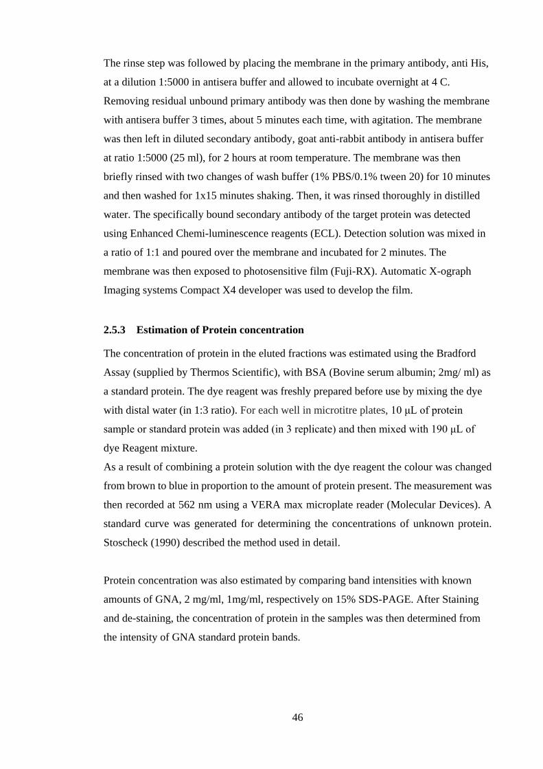
46
The rinse step was followed by placing the membrane in the primary antibody, anti His,
at a dilution 1:5000 in antisera buffer and allowed to incubate overnight at 4 C.
Removing residual unbound primary antibody was then done by washing the membrane
with antisera buffer 3 times, about 5 minutes each time, with agitation. The membrane
was then left in diluted secondary antibody, goat anti-rabbit antibody in antisera buffer
at ratio 1:5000 (25 ml), for 2 hours at room temperature. The membrane was then
briefly rinsed with two changes of wash buffer (1% PBS/0.1% tween 20) for 10 minutes
and then washed for 1x15 minutes shaking. Then, it was rinsed thoroughly in distilled
water. The specifically bound secondary antibody of the target protein was detected
using Enhanced Chemi-luminescence reagents (ECL). Detection solution was mixed in
a ratio of 1:1 and poured over the membrane and incubated for 2 minutes. The
membrane was then exposed to photosensitive film (Fuji-RX). Automatic X-ograph
Imaging systems Compact X4 developer was used to develop the film.
2.5.3 Estimation of Protein concentration
The concentration of protein in the eluted fractions was estimated using the Bradford
Assay (supplied by Thermos Scientific), with BSA (Bovine serum albumin; 2mg/ ml) as
a standard protein. The dye reagent was freshly prepared before use by mixing the dye
with distal water (in 1:3 ratio). For each well in microtitre plates, 10 μL of protein
sample or standard protein was added (in 3 replicate) and then mixed with 190 μL of
dye Reagent mixture.
As a result of combining a protein solution with the dye reagent the colour was changed
from brown to blue in proportion to the amount of protein present. The measurement was
then recorded at 562 nm using a VERA max microplate reader (Molecular Devices). A
standard curve was generated for determining the concentrations of unknown protein.
Stoscheck (1990) described the method used in detail.
Protein concentration was also estimated by comparing band intensities with known
amounts of GNA, 2 mg/ml, 1mg/ml, respectively on 15% SDS-PAGE. After Staining
and de-staining, the concentration of protein in the samples was then determined from
the intensity of GNA standard protein bands.

47
2.6 DNA Sequencing
The sequencing of samples was done by Geneius (Genevision) at Newcastle University.
GNA Primers and alpha factor were used to carry out the sequencing. Samples were
completed by use of 10 ng of DNA for every 100 bp of template DNA and 3.2 pmol of
the primer. Post-sequencing reactions were purified from unincorporated dye
terminators using ExoSAP IT, GE Healthcare. The ABI 3730 Sequencer was used for
the determination of the sequences using vector specific primers. BLAST was
performed on the sequences obtained by using the tool at the ENAserver. Translation of
the sequences was done using tools at ExPaSy.
2.7 Statistical Analysis
Kaplan-Meier survival curves were constructed using R software (Pinheiro et al., 2013)
or Sigmaplot 12.0 and further data were analysed using one-way ANOVA Excel to
estimate the lethal concentration needed to kill 50%. A P value less than 0.05 were
considered statistically significant. The toxicity index of the tested fusion proteins were
also calculated according to SUN (1950).

48
3 Chapter 3 Expression, purification and biological activity of fusion
proteins incorporating the toxins from the venom of the spider
Segestria florentina (SFIx) and snowdrop lectin (Galanthus nivalis
agglutinin; GNA)
3.1 Introduction
Neurotoxic polypeptides were isolated from the venom of the spider Segestria
florentina by Newcomb et al. (1995). These neurotoxic polypeptides are encoded by a
family of eight genes (SFI1-SFI8); all the deduced polypeptides contain 46 amino acid
residues and eight cysteine residues. These toxins are known to block neuromuscular
junctions via direct action on the voltage calcium channels (Lipkin et al., 2002). Fitches
et al. (2004) have shown that small venom peptides from spiders are degraded by the
gut epithelium enzymes when delivery orally, as the biological activity of the purified S.
florentina SFI1 was only observed when injected into a pest insect.
GNA is a plant lectin which was isolated from the bulbs of snowdrops. The ability of
GNA to cross the gut epithelium gives this protein the potential to be used as a carrier
protein in fusion protein technology. Turner (1996) reported that there is specificity
between GNA and mannose binding. This gives the technology an added advantage, as
mammals do not have mannose receptors in their gut so the fusion protein will not cross
their gut barrier. Fusing the coding sequence of a carrier protein such as snowdrop lectin
GNA with the spider toxin peptides helps the toxic protein to be transported to the
haemolymph through the insect’s gut epithelium. This would therefore allow delivery
of the toxin to its target site in the central nervous system.

49
3.2 Results
3.2.1 Production and purification of fusion proteins incorporating SFI toxins
and GNA
Assembly of the SFIx/GNA fusion protein construct
The sequence encoding the 46-amino acid insecticidal neurotoxins SFI2, SFI3, SFI5,
SFI6, and SFI8 was successfully assembled using a series of overlapping
oligonucleotides. Primers used are listed in Table 2-1 (see Chapter 2) and are known to
contain 5’ Xho I and 3’ Not I sites to facilitate ligation of the PCR fragment into vector
pGAPZα. PCR products were electrophoretically separated on a 1% agarose gel and
amplification was obtained, as seen in Figure 3-1. Bands of the correct size of about 200
bp were seen on the agarose gel for all amplified samples.
Figure 3-1: Agarose gel electrophoresis of amplified SFI2, SFI3, SFI5, and SFI6 toxins using SFIX forward and
SFIX reverse primers. Lane 1 represents the standard DNA ladder, 0.1Kb to 3Kb. Lanes 2, 3, 4, and 5 represent the
successfully amplified SFI2, SFI3, SFI5, and SFI6 toxins of 200bp size.
Following assembly, the coding sequences, all positive clones with DNA of interest,
were subjected to digestion using the Fermenter EcoRI enzyme as described in 2.3.2
(see Chapter 2). The digested products were run on a 1% agarose gel to analyse the
reaction. EcoRI digestion of SFI2, SFI3, SFI5, SFI6, and SFI8 toxin clones revealed
two fragments. The fragments were found to be of about 3000 bp and 200 bp,
corresponding to vector pGAPZα and the amplified insert respectively, as seen in Figure
3-2.
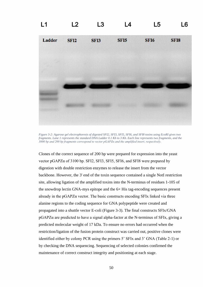
50
Figure 3-2: Agarose gel electrophoresis of digested SFI2, SFI3, SFI5, SFI6, and SFI8 toxins using EcoRI gives two
fragments. Lane 1 represents the standard DNA Ladder 0.1 Kb to 3 Kb. Each line represents two fragments, and the
3000 bp and 200 bp fragments correspond to vector pGAPZα and the amplified insert, respectively.
Clones of the correct sequence of 200 bp were prepared for expression into the yeast
vector pGAPZα of 3100 bp. SFI2, SFI3, SFI5, SFI6, and SFI8 were prepared by
digestion with double restriction enzymes to release the insert from the vector
backbone. However, the 3' end of the toxin sequence contained a single NotI restriction
site, allowing ligation of the amplified toxins into the N-terminus of residues 1-105 of
the snowdrop lectin GNA-mys epitope and the 6× His tag-encoding sequences present
already in the pGAPZα vector. The basic constructs encoding SFIx linked via three
alanine regions to the coding sequence for GNA polypeptide were created and
propagated into a shuttle vector E-coli (Figure 3-3). The final constructs SFIx/GNA
pGAPZα are predicted to have a signal alpha-factor at the N-terminus of SFIx, giving a
predicted molecular weight of 17 kDa. To ensure no errors had occurred when the
restriction/ligation of the fusion protein construct was carried out, positive clones were
identified either by colony PCR using the primers 5’ SFIx and 3’ GNA (Table 2-1) or
by checking the DNA sequencing. Sequencing of selected colonies confirmed the
maintenance of correct construct integrity and positioning at each stage.
L1 L2 L3 L4 L5 L6

51
The sequence was translated using the translate tool at open reading frame finder
(http://www.ncbi.nlm.nih.gov/gorf/gorf.html). A schematic diagram of the expression
constructs, the determined nucleotides, and the predicted protein sequences for the
SFI2/GNA, SFI3/GNA, SFI5/GNA, SFI6/GNA, and SFI8/GNA are shown in Figure 3-
4. DNA from a verified clone of the complete expression constructs SFIx/GNA
pGAPZα was linearized and transformed into competent cells of the Pichia pastoris X
33 strain, and selected on zeocin-containing plates. An attempt to transform the
SFIx/GNA pGAPZα constructs into the P. pastoris X 33 strain resulted in few clones
(result not shown). The selected clones were further analysed for expression to select for
the highest-yield expressing clone. The expression constructs were assessed by Western
blot using anti GNA-antibodies derived from the small-scale cultures’ supernatants.
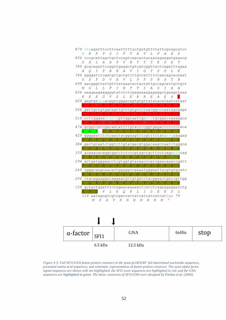
52
879 atgagatttccttcaatttttactgctgttttattcgcagcatcc
M R F P S I F T A V L F A A S
834 tccgcattagctgctccagtcaacactacaacagaagatgaaacg
S A L A A P V N T T T E D E T
789 gcacaaattccggctgaagctgtcatcggttactcagatttagaa
A Q I P A E A V I G Y S D L E
744 ggggatttcgatgttgctgttttgccattttccaacagcacaaat
G D F D V A V L P F S N S T N
699 aacgggttattgtttataaatactactattgccagcattgctgct
N G L L F I N T T I A S I A A
654 aaagaagaaggggtatctctcgagaaaagagaggctgaagctaaa
K E E G V S L E K R E A E A K
609 gagtgtatgacggatgggacagtgtgttatatacataatcataat
E C M T D G T V C Y I H N H N
564 gattgctgtggcagttgcctgtgctctaacggcccaatagcaaga
D C C G S C L C S N G P I A R
519 ccttgggaaatgatggttggcaattgcatgtgtggaccaaaagca
P W E M M V G N C M C G P K A
474 gcggccgccgacaatattttgtactccggtgagactctctctaca
A A A D N I L Y S G E T L S T
429 ggggaatttctcaactacggaagtttcgtttttatcatgcaagag
G E F L N Y G S F V F I M Q E
384 gactgcaatctggtcttgtacgacgtggacaagccaatctgggca
D C N L V L Y D V D K P I W A
339 acaaacacaggtggtctctcccgtagctgcttcctcagcatgcag
T N T G G L S R S C F L S M Q
294 actgatgggaacctcgtggtgtacaacccatcgaacaaaccgatt
T D G N L V V Y N P S N K P I
249 tgggcaagcaacactggaggccaaaatgggaattacgtgtgcatc
W A S N T G G Q N G N Y V C I
204 ctacagaaggataggaatgttgtgatctacggaactgatcgttgg
L Q K D R N V V I Y G T D R W
159 gctactggatttctagaacaaaaactcatctcagaagaggatctg
A T G F L E Q K L I S E E D L
114 aatagcgccgtcgaccatcatcatcatcatcattga 79
N S A V D H H H H H H *
Figure 3-3: Full SFI1/GNA fusion protein construct in the yeast pGAPZAP: full determined nucleotide sequences,
presumed amino acid sequences, and schematic representation of fusion protein construct. The yeast alpha factor
signal sequences are shown with not highlighted, the SFI1 toxin sequences are highlighted in red, and the GNA
sequences are highlighted in green. The basic constructs of SFI1/GNA were designed by Fitches et al. (2004).
α-factor SFI1
GNA
6xHis stop
12.5 kDa 6.5 kDa
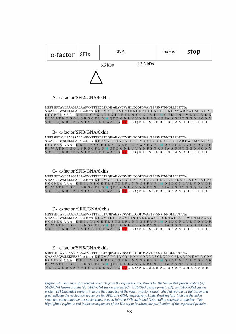
53
A- α-factor/SFI2/GNA/6xHis
MRFPSIFTAVLFAASSALAAPVNTTTEDETAQIPAEAVIGYSDLEGDFDVAVLPFSNSTNNGLLFINTTIA
SIAAKEEGVSLEKREAEA α-factor K E C M A D E T V C Y I H N H N N C C G S C L C L N G P Y A R P W E M L V G N C
K C G P K E A A A D N I L Y S G E T L S T G E F L N Y G S F V F I M Q E D C N L V L Y D V D K
P I W A T N T G G L S R S C F L S M Q T D G N L V V Y N P S N K P I W A S N T G G Q N G N Y
V C I L Q K D R N V V I Y G T D R W A T G tags L E Q K L I S E E D L N S A V D H H H H H H
B- α-factor/SFI3/GNA/6xhis MRFPSIFTAVLFAASSALAAPVNTTTEDETAQIPAEAVIGYSDLEGDFDVAVLPFSNSTNNGLLFINTTIA
SIAAKEEGVSLEKREAEA α-factor K E C M V D G T V C Y I H N H N D C C G S C L C L N G P I A R P W E M M V G N C
K C G P K A A A A D N I L Y S G E T L S T G E F L N Y G S F V F I M Q E D C N L V L Y D V D K
P I W A T N T G G L S R S C F L S M Q T D G N L V V Y N P S N K P I W A S N T G G Q N G N Y
V C I L Q K D R N V V I Y G T D R W A T G tags L E Q K L I S E E D L N S A V D H H H H H H
C- α-factor/SFI5/GNA/6xhis MRFPSIFTAVLFAASSALAAPVNTTTEDETAQIPAEAVIGYSDLEGDFDVAVLPFSNSTNNGLLFINTTIA
SIAAKEEGVSLEKREAEA α-factor K E C M V D G T V C Y I H N H N D C C G S C L C P N G P L A R P W E M L V G N C
K C G P K A A A A D N I L Y S G E T L S T G E F L N Y G S F V F I M Q E D C N L V L Y D V D K
P I W A T N T G G L S R S C F L S M Q T D G N L V V Y N P S N K P I W A S N T G G Q N G N Y
V C I L Q K D R N V V I Y G T D R W A T G tags L E Q K L I S E E D L N S A V D H H H H H H
D- α-factor /SFI6/GNA/6xhis MRFPSIFTAVLFAASSALAAPVNTTTEDETAQIPAEAVIGYSDLEGDFDVAVLPFSNSTNNGLLFINTTIA
SIAAKEEGVSLEKREAEA α-factor K E C M T D E T V C Y I H N H N D C C G S C L C L N G P I A R P W E M M V G N C
K C G P K A A A A D N I L Y S G E T L S T G E F L N Y G S F V F I M Q E D C N L V L Y D V D K
P I W A T N T G G L S R S C F L S M Q T D G N L V V Y N P S N K P I W A S N T G G Q N G N Y
V C I L Q K D R N V V I Y G T D R W A T G tags L E Q K L I S E E D L N S A V D H H H H H H
E- α-factor/SFI8/GNA/6xhis MRFPSIFTAVLFAASSALAAPVNTTTEDETAQIPAEAVIGYSDLEGDFDVAVLPFSNSTNNGLLFINTTIA
SIAAKEEGVSLEKREAEA α-factor K E C M A D G T V C Y I H N H N D C C G S C L C P N G P L A R P W E M L V G N C
K C G P K A A A A D N I L Y S G E T L S T G E F L N Y G S F V F I M Q E D C N L V L Y D V D K
P I W A T N T G G L S R S C F L S M Q T D G N L V V Y N P S N K P I W A S N T G G Q N G N Y
V C I L Q K D R N V V I Y G T D R W A T G tags L E Q K L I S E E D L N S A V D H H H H H H
Figure 3-4: Sequence of predicted products from the expression constructs for the SFI2/GNA fusion protein (A),
SFI3/GNA fusion protein (B), SFI5/GNA fusion protein (C), SFI6/GNA fusion protein (D), and SFI8/GNA fusion
protein (E).Unshaded regions indicate the sequence of the yeast α-factor signal. Shaded regions in light grey and
grey indicate the nucleotide sequences for SFIx and GNA, respectively. Underlined regions indicate the linker
sequence contributed by the nucleotides, used to join the SFIx toxin and GNA coding sequences together. The
highlighted region in red indicates sequences of the His tag to facilitate the purification of the expressed protein.
α-factor SFIx GNA
6xHis stop
6.5 kDa 12.5 kDa
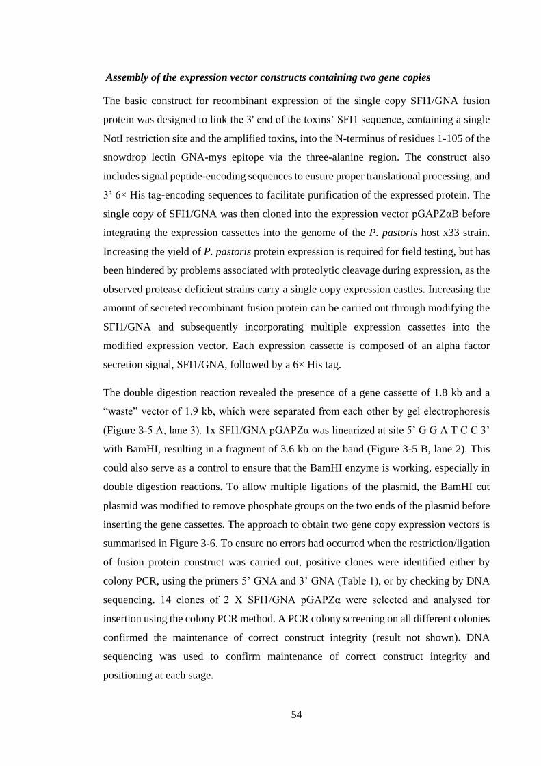
54
Assembly of the expression vector constructs containing two gene copies
The basic construct for recombinant expression of the single copy SFI1/GNA fusion
protein was designed to link the 3' end of the toxins’ SFI1 sequence, containing a single
NotI restriction site and the amplified toxins, into the N-terminus of residues 1-105 of the
snowdrop lectin GNA-mys epitope via the three-alanine region. The construct also
includes signal peptide-encoding sequences to ensure proper translational processing, and
3’ 6× His tag-encoding sequences to facilitate purification of the expressed protein. The
single copy of SFI1/GNA was then cloned into the expression vector pGAPZαB before
integrating the expression cassettes into the genome of the P. pastoris host x33 strain.
Increasing the yield of P. pastoris protein expression is required for field testing, but has
been hindered by problems associated with proteolytic cleavage during expression, as the
observed protease deficient strains carry a single copy expression castles. Increasing the
amount of secreted recombinant fusion protein can be carried out through modifying the
SFI1/GNA and subsequently incorporating multiple expression cassettes into the
modified expression vector. Each expression cassette is composed of an alpha factor
secretion signal, SFI1/GNA, followed by a 6× His tag.
The double digestion reaction revealed the presence of a gene cassette of 1.8 kb and a
“waste” vector of 1.9 kb, which were separated from each other by gel electrophoresis
(Figure 3-5 A, lane 3). 1x SFI1/GNA pGAPZα was linearized at site 5’ G G A T C C 3’
with BamHI, resulting in a fragment of 3.6 kb on the band (Figure 3-5 B, lane 2). This
could also serve as a control to ensure that the BamHI enzyme is working, especially in
double digestion reactions. To allow multiple ligations of the plasmid, the BamHI cut
plasmid was modified to remove phosphate groups on the two ends of the plasmid before
inserting the gene cassettes. The approach to obtain two gene copy expression vectors is
summarised in Figure 3-6. To ensure no errors had occurred when the restriction/ligation
of fusion protein construct was carried out, positive clones were identified either by
colony PCR, using the primers 5’ GNA and 3’ GNA (Table 1), or by checking by DNA
sequencing. 14 clones of 2 X SFI1/GNA pGAPZα were selected and analysed for
insertion using the colony PCR method. A PCR colony screening on all different colonies
confirmed the maintenance of correct construct integrity (result not shown). DNA
sequencing was used to confirm maintenance of correct construct integrity and
positioning at each stage.
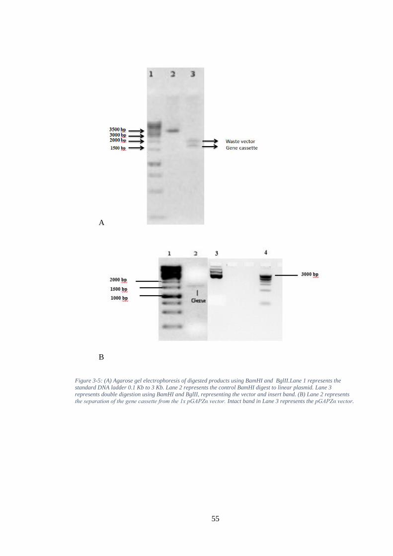
55
A
B
Figure 3-5: (A) Agarose gel electrophoresis of digested products using BamHI and BglII.Lane 1 represents the
standard DNA ladder 0.1 Kb to 3 Kb. Lane 2 represents the control BamHI digest to linear plasmid. Lane 3
represents double digestion using BamHI and BglII, representing the vector and insert band. (B) Lane 2 represents
the separation of the gene cassette from the 1x pGAPZα vector. Intact band in Lane 3 represents the pGAPZα vector.
56
pGAP α-factor SFI1/GNA 6XHis
pGAP α-factor SFI1/GNA 6XHis
Figure 3-6: Diagrammatic representation of the cloning strategy adopted to enable insertion of multiple copies of the
fusion protein cassette into the Pichia yeast genome.
SFI1/GNA expression cassette
SFI1/GNA expression cassette1 SFI1/GNA expression cassette2
pGAP α-factor SFI1/GNA 6XHis
ZeocinR PUC ori
PUC ori
ZeocinR

57
Expression of the recombinant protein SFIx/GNA
Positive and verified plasmids of the expression constructs SFIx /GNA pGAPZα were
linearized and transformed into competent cells of the P. pastoris X 33 strain. Due to
the presence of a constitutive yeast promoter on the expression vector, they were
selected on zeocin-containing plates. After preparation of the SFIx/GNA constructs, the
expression of recombinant proteins were run on a small-scale culture, allowing isolation
high level expression clones. Culture supernatants (grown for four days) were analysed
by Western blotting using antiGNA antibodies (1:3000 dilution). As the fusion protein
SFIx/GNA constructs have been confirmed to have a GNA linked to the C-terminal of
the toxins (Figure 3-7), a GNA fusion protein was used as a positive control.
SFI2/GNA, SFI3/GNA, SFI5/GNA, SFI6/GNA, and SFI8/GNA should show reactivity
at around 17 kDa at time points 48 and 96 hours. The presence of GNA at the correct
size in the positive standard lanes shows the blots have worked successfully. However,
the fusion proteins were not detected through a sensitive technique Western blot at time
point 48 (result not shown). The fusion proteins SFI2/GNA, SFI3/GNA, SFI5/GNA,
SFI6/GNA, and SFI8/GNA were detected at high levels of expression, with a number of
bands seen on the gel between 17 kDa and 14 kDa at time point 96 hours.
Figure 3-7: The presence of GNA of the correct size in the positive standard lanes shows the blots have
worked successfully. However, the fusion proteins SFI2/GNA, SFI3/GNA, SFI5/GNA, SFI6/GNA, and
SFI8/GNA were subjected to high levels of expression, with a number of bands seen on the gel between
17 kDa and 14 kDa.
58
Yeast transformation and gene copy analysis
The original SFI1/GNA (FP2.1) and the two gene copies of construct 2 X SFI1/GNA (2
X FP2.1) were retransformed into P. pastoris using the pGAPZα expression vector.
Small-scale expression and immune blot assays similar to those used previously for
SFIx/GNA pGAPZaB were carried out (Figure 3-8). The three colonies of expressed
proteins for the original SFI1/GNA with a molecular size of 17 kDa, and the three
colonies of the 2 X SFI1/GNA constructs, were examined by Western blotting analysis.
A single band that indicated mol. wt. reactivity and the correct size for both constructs
was present. The original SFI1/GNA fusion protein resulted in lower expression in the
levels of intact fusion proteins in X33 cells compared to the modified construct, as
assessed by Western blotting using an anti-GNA antibody (Figure 3-8). The modified
construct was found to produce a 1:2 ratio of fusion protein (mol. wt. of approximately
17 kDa) as measured by a Western blot.
Figure 3-8: Western blot analysis of the protein expression of SFI1/GNA pGAPZα and 2 x SFI1/GNA pGAPZα using
anti-GNA antibodies derived from the small-scale cultures’ supernatants.
Positive and verified plasmids of the expression constructs SFIx /GNA pGAPZα were
linearized and transformed into competent cells of the P.pastoris X33 strain. Due to the
presence of a constitutive yeast promoter on the expression vector, they were selected
on zeocin-containing plates. The expression of recombinant proteins was run on a
small-scale culture which allowed us to select of the best expressing clone for fusion
protein production by bench-top fermentation. Culture supernatants were analysed by
Western blotting using GNA antibodies. One clone of expressing recombinant proteins
were used for fusion protein production by bench-top fermentation.
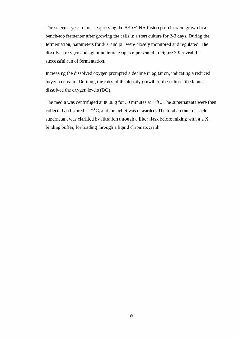
59
The selected yeast clones expressing the SFIx/GNA fusion protein were grown in a
bench-top fermenter after growing the cells in a start culture for 2-3 days. During the
fermentation, parameters for dO2 and pH were closely monitored and regulated. The
dissolved oxygen and agitation trend graphs represented in Figure 3-9 reveal the
successful run of fermentation.
Increasing the dissolved oxygen prompted a decline in agitation, indicating a reduced
oxygen demand. Defining the rates of the density growth of the culture, the lanner
dissolved the oxygen levels (DO).
The media was centrifuged at 8000 g for 30 minutes at 4 OC. The supernatants were then
collected and stored at 4O C, and the pellet was discarded. The total amount of each
supernatant was clarified by filtration through a filter flask before mixing with a 2 X
binding buffer, for loading through a liquid chromatograph.
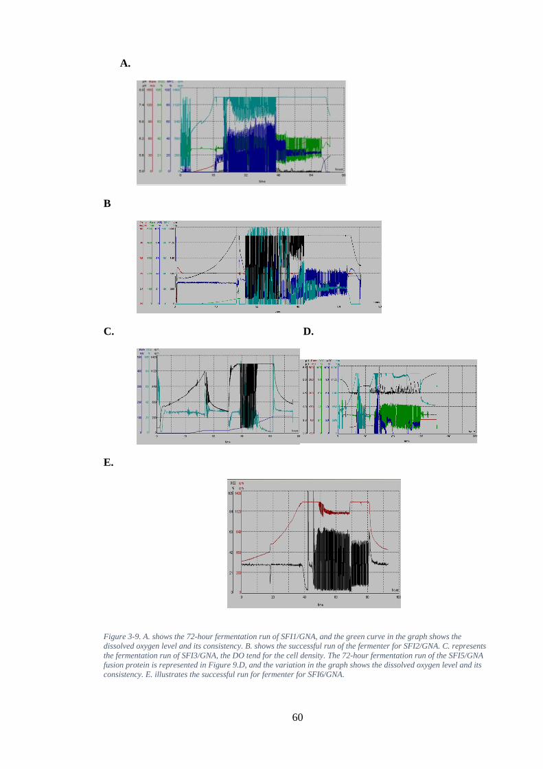
60
A.
B
C. D.
E.
Figure 3-9. A. shows the 72-hour fermentation run of SFI1/GNA, and the green curve in the graph shows the
dissolved oxygen level and its consistency. B. shows the successful run of the fermenter for SFI2/GNA. C. represents
the fermentation run of SFI3/GNA, the DO tend for the cell density. The 72-hour fermentation run of the SFI5/GNA
fusion protein is represented in Figure 9.D, and the variation in the graph shows the dissolved oxygen level and its
consistency. E. illustrates the successful run for fermenter for SFI6/GNA.
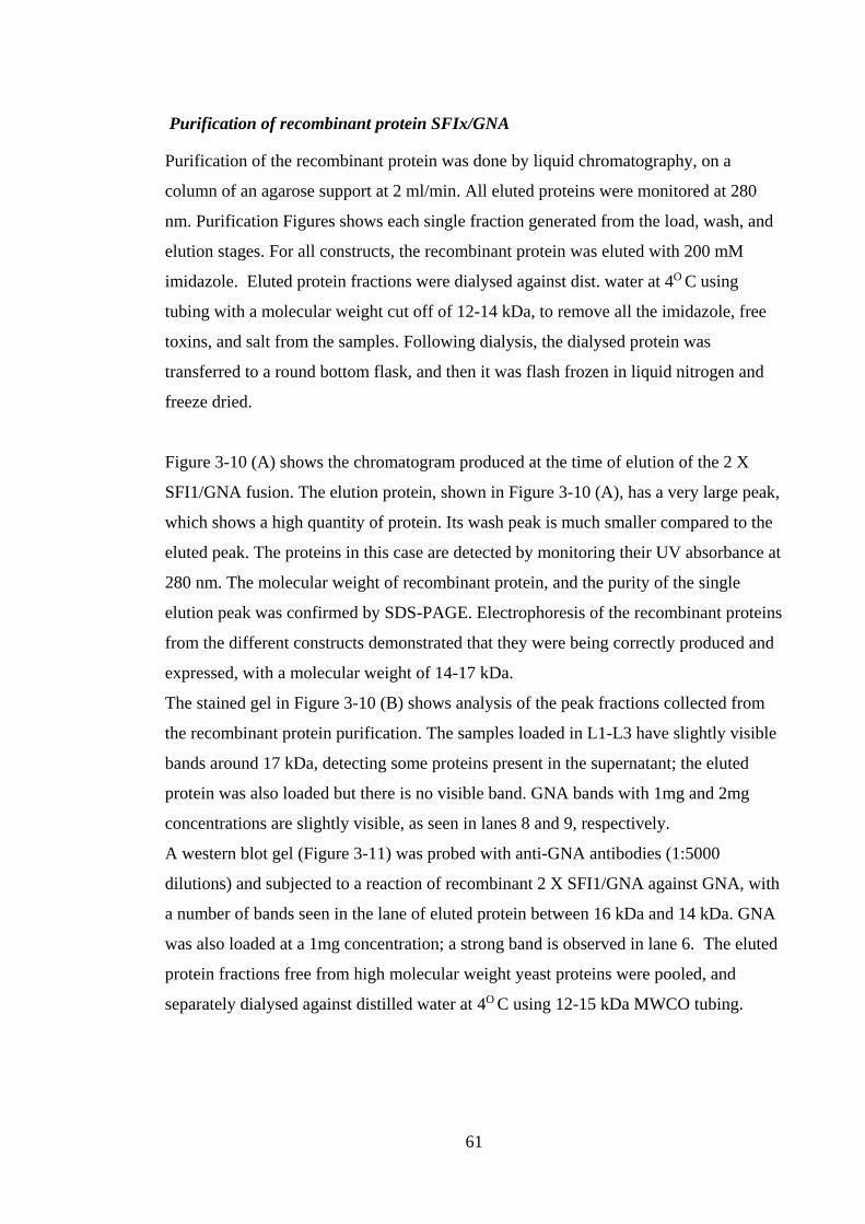
61
Purification of recombinant protein SFIx/GNA
Purification of the recombinant protein was done by liquid chromatography, on a
column of an agarose support at 2 ml/min. All eluted proteins were monitored at 280
nm. Purification Figures shows each single fraction generated from the load, wash, and
elution stages. For all constructs, the recombinant protein was eluted with 200 mM
imidazole. Eluted protein fractions were dialysed against dist. water at 4O C using
tubing with a molecular weight cut off of 12-14 kDa, to remove all the imidazole, free
toxins, and salt from the samples. Following dialysis, the dialysed protein was
transferred to a round bottom flask, and then it was flash frozen in liquid nitrogen and
freeze dried.
Figure 3-10 (A) shows the chromatogram produced at the time of elution of the 2 X
SFI1/GNA fusion. The elution protein, shown in Figure 3-10 (A), has a very large peak,
which shows a high quantity of protein. Its wash peak is much smaller compared to the
eluted peak. The proteins in this case are detected by monitoring their UV absorbance at
280 nm. The molecular weight of recombinant protein, and the purity of the single
elution peak was confirmed by SDS-PAGE. Electrophoresis of the recombinant proteins
from the different constructs demonstrated that they were being correctly produced and
expressed, with a molecular weight of 14-17 kDa.
The stained gel in Figure 3-10 (B) shows analysis of the peak fractions collected from
the recombinant protein purification. The samples loaded in L1-L3 have slightly visible
bands around 17 kDa, detecting some proteins present in the supernatant; the eluted
protein was also loaded but there is no visible band. GNA bands with 1mg and 2mg
concentrations are slightly visible, as seen in lanes 8 and 9, respectively.
A western blot gel (Figure 3-11) was probed with anti-GNA antibodies (1:5000
dilutions) and subjected to a reaction of recombinant 2 X SFI1/GNA against GNA, with
a number of bands seen in the lane of eluted protein between 16 kDa and 14 kDa. GNA
was also loaded at a 1mg concentration; a strong band is observed in lane 6. The eluted
protein fractions free from high molecular weight yeast proteins were pooled, and
separately dialysed against distilled water at 4O C using 12-15 kDa MWCO tubing.
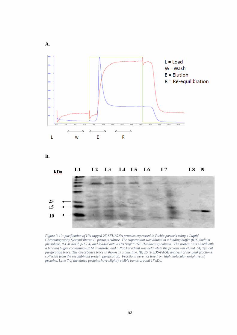
62
A.
B.
Figure 3-10: purification of His-tagged 2X SFI1/GNA proteins expressed in Pichia pastoris using a Liquid
Chromatography SystemFiltered P. pastoris culture. The supernatant was diluted in a binding buffer (0.02 Sodium
phosphate, 0.4 M NaCl, pH 7.4) and loaded onto a HisTrap™ (GE Healthcare) column. The protein was eluted with
a binding buffer containing 0.2 M imidazole, and a NaCl gradient was held while the protein was eluted. (A) Typical
purification trace. The absorbance trace is shown as a blue line. (B) 15 % SDS-PAGE analysis of the peak fractions
collected from the recombinant protein purification. Fractions were not free from high molecular weight yeast
proteins. Lane 7 of the eluted proteins have slightly visible bands around 17 kDa.
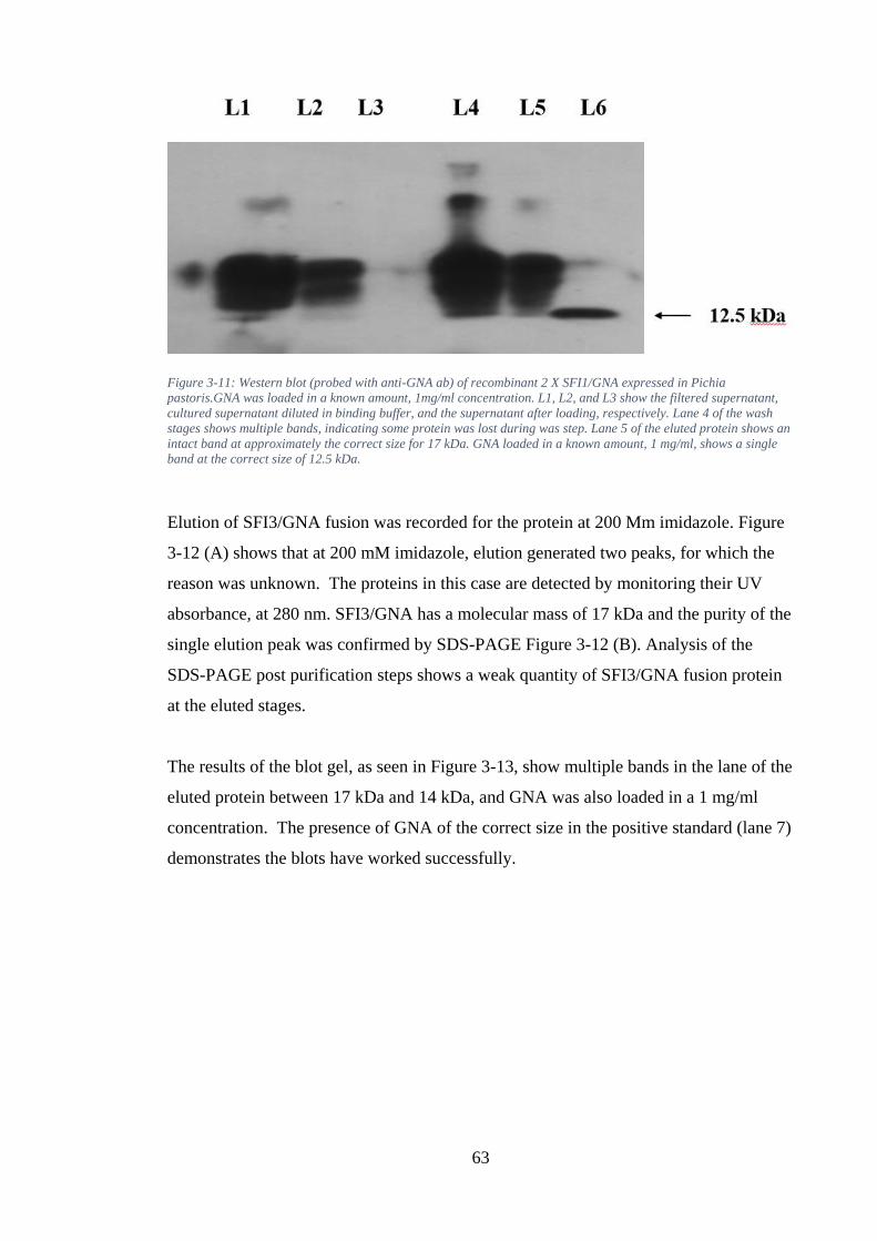
63
Figure 3-11: Western blot (probed with anti-GNA ab) of recombinant 2 X SFI1/GNA expressed in Pichia
pastoris.GNA was loaded in a known amount, 1mg/ml concentration. L1, L2, and L3 show the filtered supernatant,
cultured supernatant diluted in binding buffer, and the supernatant after loading, respectively. Lane 4 of the wash
stages shows multiple bands, indicating some protein was lost during was step. Lane 5 of the eluted protein shows an
intact band at approximately the correct size for 17 kDa. GNA loaded in a known amount, 1 mg/ml, shows a single
band at the correct size of 12.5 kDa.
Elution of SFI3/GNA fusion was recorded for the protein at 200 Mm imidazole. Figure
3-12 (A) shows that at 200 mM imidazole, elution generated two peaks, for which the
reason was unknown. The proteins in this case are detected by monitoring their UV
absorbance, at 280 nm. SFI3/GNA has a molecular mass of 17 kDa and the purity of the
single elution peak was confirmed by SDS-PAGE Figure 3-12 (B). Analysis of the
SDS-PAGE post purification steps shows a weak quantity of SFI3/GNA fusion protein
at the eluted stages.
The results of the blot gel, as seen in Figure 3-13, show multiple bands in the lane of the
eluted protein between 17 kDa and 14 kDa, and GNA was also loaded in a 1 mg/ml
concentration. The presence of GNA of the correct size in the positive standard (lane 7)
demonstrates the blots have worked successfully.
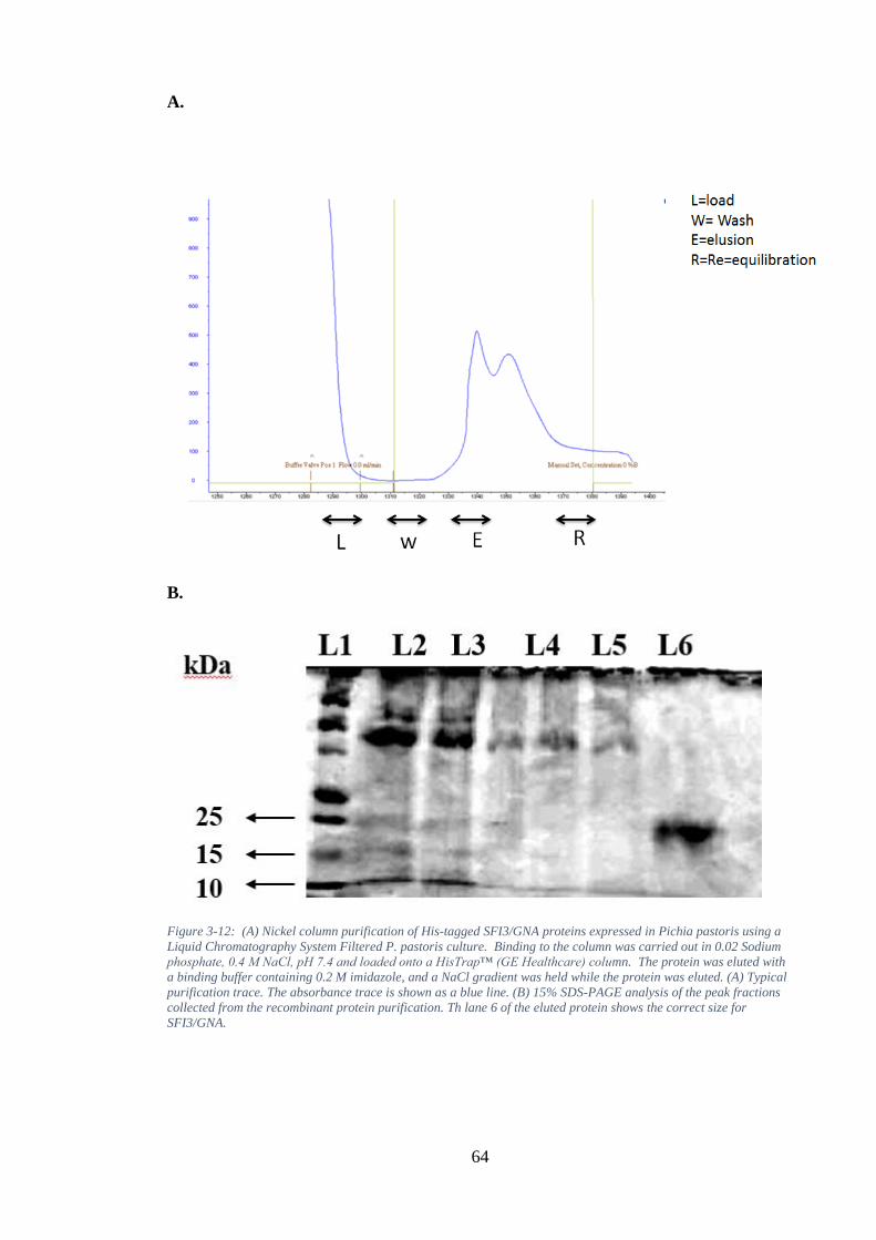
64
A.
B.
Figure 3-12: (A) Nickel column purification of His-tagged SFI3/GNA proteins expressed in Pichia pastoris using a
Liquid Chromatography System Filtered P. pastoris culture. Binding to the column was carried out in 0.02 Sodium
phosphate, 0.4 M NaCl, pH 7.4 and loaded onto a HisTrap™ (GE Healthcare) column. The protein was eluted with
a binding buffer containing 0.2 M imidazole, and a NaCl gradient was held while the protein was eluted. (A) Typical
purification trace. The absorbance trace is shown as a blue line. (B) 15% SDS-PAGE analysis of the peak fractions
collected from the recombinant protein purification. Th lane 6 of the eluted protein shows the correct size for
SFI3/GNA.
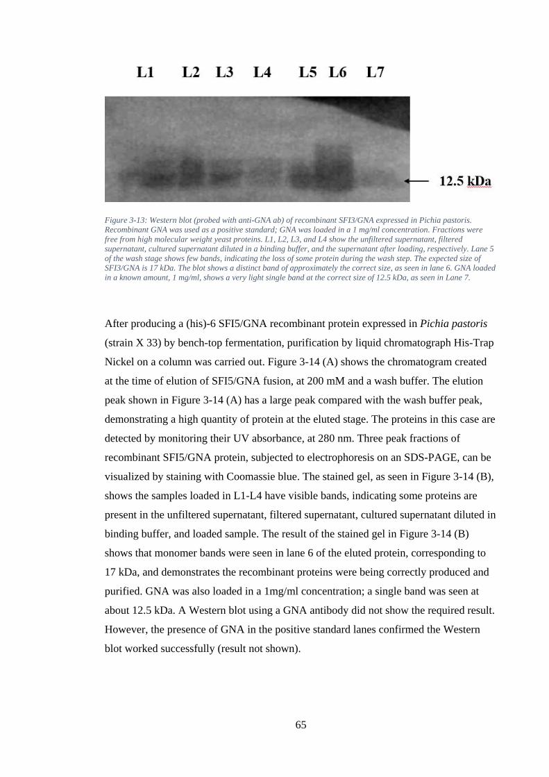
65
Figure 3-13: Western blot (probed with anti-GNA ab) of recombinant SFI3/GNA expressed in Pichia pastoris.
Recombinant GNA was used as a positive standard; GNA was loaded in a 1 mg/ml concentration. Fractions were
free from high molecular weight yeast proteins. L1, L2, L3, and L4 show the unfiltered supernatant, filtered
supernatant, cultured supernatant diluted in a binding buffer, and the supernatant after loading, respectively. Lane 5
of the wash stage shows few bands, indicating the loss of some protein during the wash step. The expected size of
SFI3/GNA is 17 kDa. The blot shows a distinct band of approximately the correct size, as seen in lane 6. GNA loaded
in a known amount, 1 mg/ml, shows a very light single band at the correct size of 12.5 kDa, as seen in Lane 7.
After producing a (his)-6 SFI5/GNA recombinant protein expressed in Pichia pastoris
(strain X 33) by bench-top fermentation, purification by liquid chromatograph His-Trap
Nickel on a column was carried out. Figure 3-14 (A) shows the chromatogram created
at the time of elution of SFI5/GNA fusion, at 200 mM and a wash buffer. The elution
peak shown in Figure 3-14 (A) has a large peak compared with the wash buffer peak,
demonstrating a high quantity of protein at the eluted stage. The proteins in this case are
detected by monitoring their UV absorbance, at 280 nm. Three peak fractions of
recombinant SFI5/GNA protein, subjected to electrophoresis on an SDS-PAGE, can be
visualized by staining with Coomassie blue. The stained gel, as seen in Figure 3-14 (B),
shows the samples loaded in L1-L4 have visible bands, indicating some proteins are
present in the unfiltered supernatant, filtered supernatant, cultured supernatant diluted in
binding buffer, and loaded sample. The result of the stained gel in Figure 3-14 (B)
shows that monomer bands were seen in lane 6 of the eluted protein, corresponding to
17 kDa, and demonstrates the recombinant proteins were being correctly produced and
purified. GNA was also loaded in a 1mg/ml concentration; a single band was seen at
about 12.5 kDa. A Western blot using a GNA antibody did not show the required result.
However, the presence of GNA in the positive standard lanes confirmed the Western
blot worked successfully (result not shown).
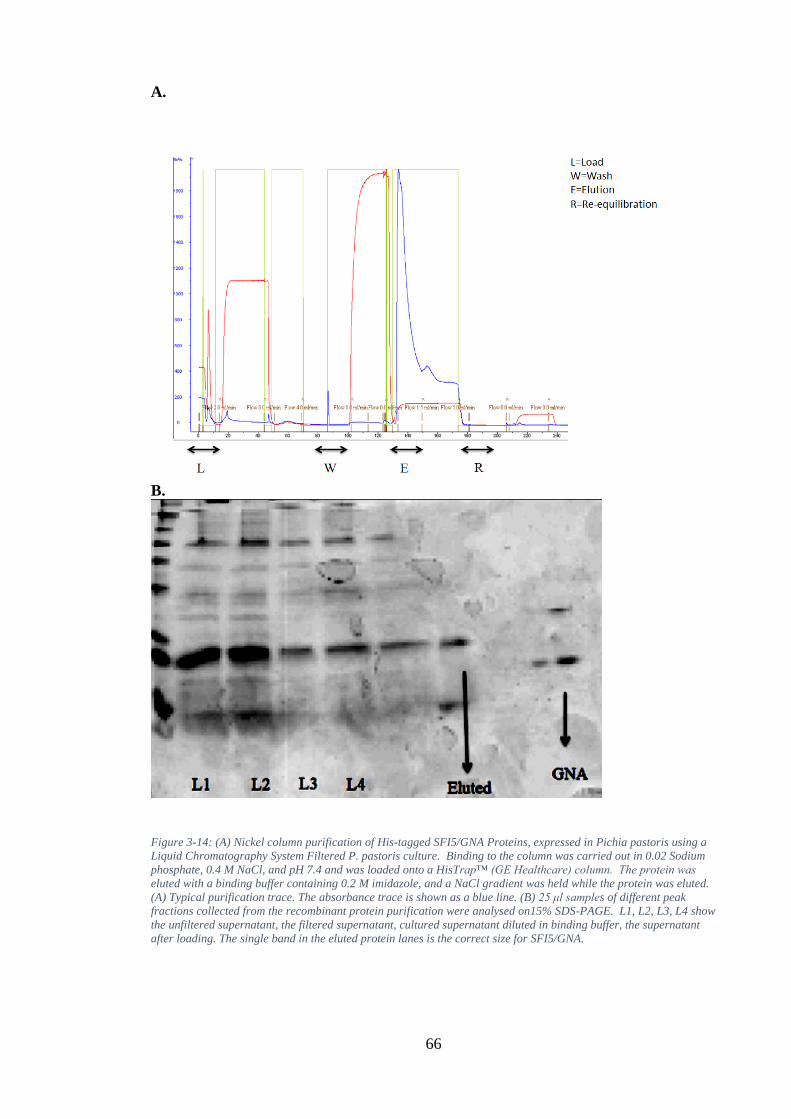
66
A.
B.
Figure 3-14: (A) Nickel column purification of His-tagged SFI5/GNA Proteins, expressed in Pichia pastoris using a
Liquid Chromatography System Filtered P. pastoris culture. Binding to the column was carried out in 0.02 Sodium
phosphate, 0.4 M NaCl, and pH 7.4 and was loaded onto a HisTrap™ (GE Healthcare) column. The protein was
eluted with a binding buffer containing 0.2 M imidazole, and a NaCl gradient was held while the protein was eluted.
(A) Typical purification trace. The absorbance trace is shown as a blue line. (B) 25 μl samples of different peak
fractions collected from the recombinant protein purification were analysed on15% SDS-PAGE. L1, L2, L3, L4 show
the unfiltered supernatant, the filtered supernatant, cultured supernatant diluted in binding buffer, the supernatant
after loading. The single band in the eluted protein lanes is the correct size for SFI5/GNA.
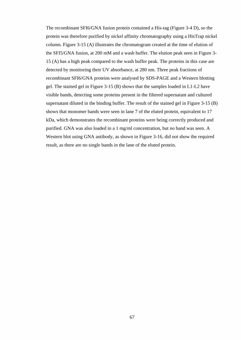
67
The recombinant SFI6/GNA fusion protein contained a His-tag (Figure 3-4 D), so the
protein was therefore purified by nickel affinity chromatography using a HisTrap nickel
column. Figure 3-15 (A) illustrates the chromatogram created at the time of elution of
the SFI5/GNA fusion, at 200 mM and a wash buffer. The elution peak seen in Figure 3-
15 (A) has a high peak compared to the wash buffer peak. The proteins in this case are
detected by monitoring their UV absorbance, at 280 nm. Three peak fractions of
recombinant SFI6/GNA proteins were analysed by SDS-PAGE and a Western blotting
gel. The stained gel in Figure 3-15 (B) shows that the samples loaded in L1-L2 have
visible bands, detecting some proteins present in the filtered supernatant and cultured
supernatant diluted in the binding buffer. The result of the stained gel in Figure 3-15 (B)
shows that monomer bands were seen in lane 7 of the eluted protein, equivalent to 17
kDa, which demonstrates the recombinant proteins were being correctly produced and
purified. GNA was also loaded in a 1 mg/ml concentration, but no band was seen. A
Western blot using GNA antibody, as shown in Figure 3-16, did not show the required
result, as there are no single bands in the lane of the eluted protein.
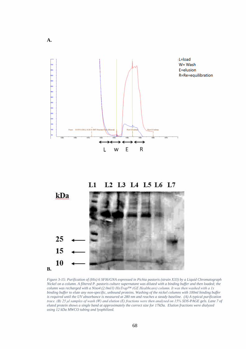
68
A.
B.
Figure 3-15: Purification of (His) 6 SFI6/GNA expressed in Pichia pastoris (strain X33) by a Liquid Chromatograph
Nickel on a column. A filtered P. pastoris culture supernatant was diluted with a binding buffer and then loaded; the
column was recharged with a Niso4 (2.0ml/l) HisTrap™ (GE Healthcare) column. It was then washed with a 1x
binding buffer to elute any non-specific, unbound proteins. Washing of the nickel columns with 100ml binding buffer
is required until the UV absorbance is measured at 280 nm and reaches a steady baseline. (A) A typical purification
trace. (B) 25 μl samples of wash (W) and elution (E) fractions were then analysed on 15% SDS-PAGE gels. Lane 7 of
eluted protein shows a single band at approximately the correct size for 17kDa. Elution fractions were dialyzed
using 12 kDa MWCO tubing and lyophilized.
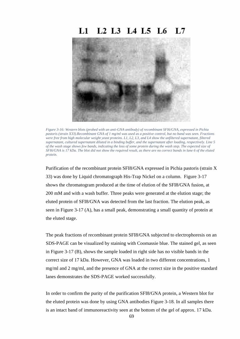
69
Figure 3-16: Western blots (probed with an anti-GNA antibody) of recombinant SFI6/GNA, expressed in Pichia
pastoris (strain X33).Recombinant GNA of 1 mg/ml was used as a positive control, but no band was seen. Fractions
were free from high molecular weight yeast proteins. L1, L2, L3, and L4 show the unfiltered supernatant, filtered
supernatant, cultured supernatant diluted in a binding buffer, and the supernatant after loading, respectively. Line 5
of the wash stage shows few bands, indicating the loss of some protein during the wash step. The expected size of
SFI6/GNA is 17 kDa. The blot did not show the required result, as there are no correct bands in lane 6 of the eluted
protein.
Purification of the recombinant protein SFI8/GNA expressed in Pichia pastoris (strain X
33) was done by Liquid chromatograph His-Trap Nickel on a column. Figure 3-17
shows the chromatogram produced at the time of elution of the SFI8/GNA fusion, at
200 mM and with a wash buffer. Three peaks were generated at the elution stage; the
eluted protein of SFI8/GNA was detected from the last fraction. The elution peak, as
seen in Figure 3-17 (A), has a small peak, demonstrating a small quantity of protein at
the eluted stage.
The peak fractions of recombinant protein SFI8/GNA subjected to electrophoresis on an
SDS-PAGE can be visualized by staining with Coomassie blue. The stained gel, as seen
in Figure 3-17 (B), shows the sample loaded in right side has no visible bands in the
correct size of 17 kDa. However, GNA was loaded in two different concentrations, 1
mg/ml and 2 mg/ml, and the presence of GNA at the correct size in the positive standard
lanes demonstrates the SDS-PAGE worked successfully.
In order to confirm the purity of the purification SFI8/GNA protein, a Western blot for
the eluted protein was done by using GNA antibodies Figure 3-18. In all samples there
is an intact band of immunoreactivity seen at the bottom of the gel of approx. 17 kDa.
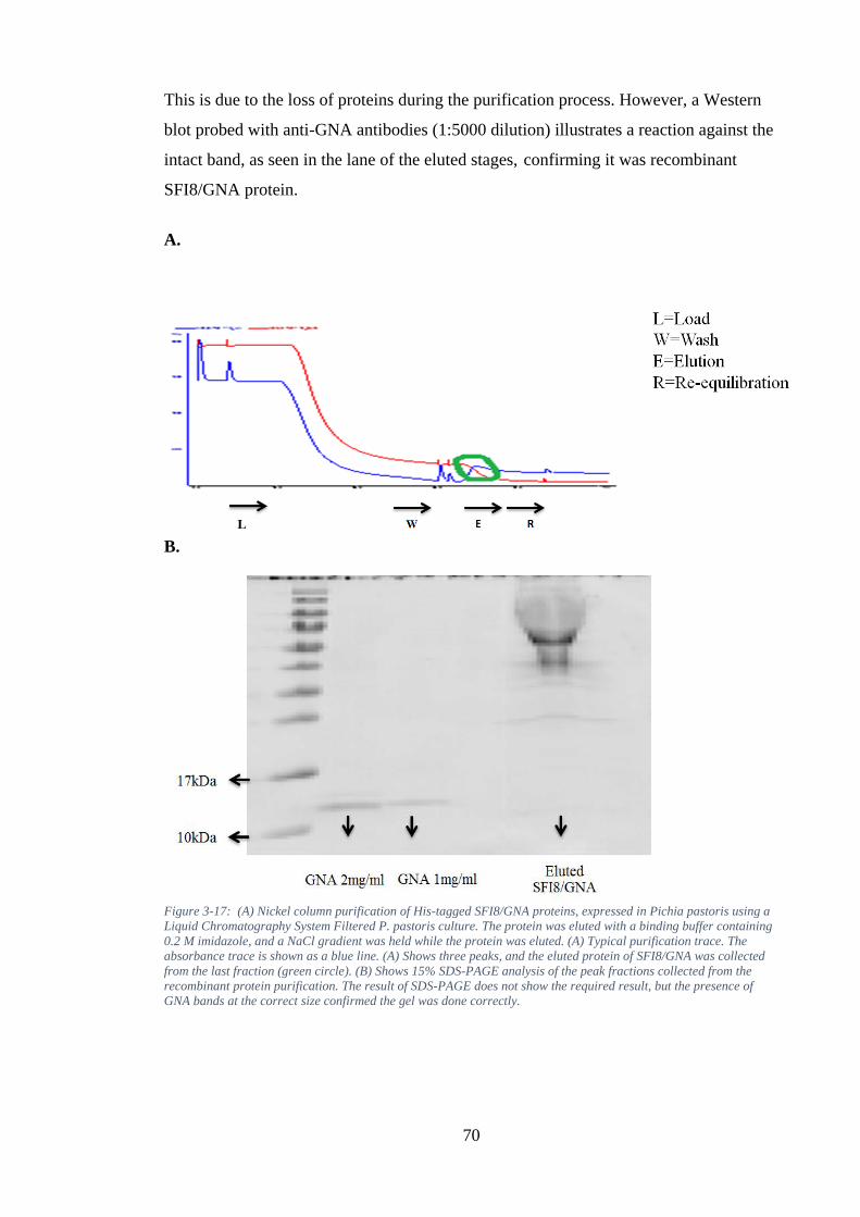
70
This is due to the loss of proteins during the purification process. However, a Western
blot probed with anti-GNA antibodies (1:5000 dilution) illustrates a reaction against the
intact band, as seen in the lane of the eluted stages, confirming it was recombinant
SFI8/GNA protein.
A.
B.
Figure 3-17: (A) Nickel column purification of His-tagged SFI8/GNA proteins, expressed in Pichia pastoris using a
Liquid Chromatography System Filtered P. pastoris culture. The protein was eluted with a binding buffer containing
0.2 M imidazole, and a NaCl gradient was held while the protein was eluted. (A) Typical purification trace. The
absorbance trace is shown as a blue line. (A) Shows three peaks, and the eluted protein of SFI8/GNA was collected
from the last fraction (green circle). (B) Shows 15% SDS-PAGE analysis of the peak fractions collected from the
recombinant protein purification. The result of SDS-PAGE does not show the required result, but the presence of
GNA bands at the correct size confirmed the gel was done correctly.
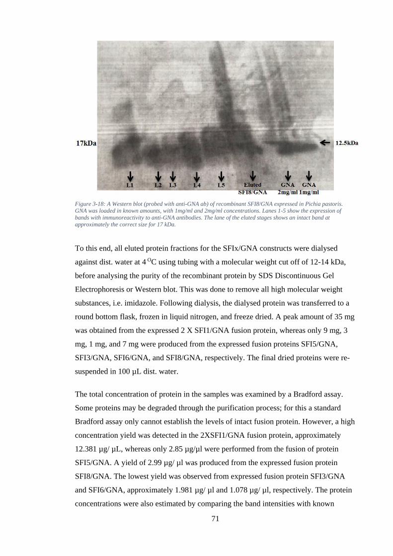
71
Figure 3-18: A Western blot (probed with anti-GNA ab) of recombinant SFI8/GNA expressed in Pichia pastoris.
GNA was loaded in known amounts, with 1mg/ml and 2mg/ml concentrations. Lanes 1-5 show the expression of
bands with immunoreactivity to anti-GNA antibodies. The lane of the eluted stages shows an intact band at
approximately the correct size for 17 kDa.
To this end, all eluted protein fractions for the SFIx/GNA constructs were dialysed
against dist. water at 4 OC using tubing with a molecular weight cut off of 12-14 kDa,
before analysing the purity of the recombinant protein by SDS Discontinuous Gel
Electrophoresis or Western blot. This was done to remove all high molecular weight
substances, i.e. imidazole. Following dialysis, the dialysed protein was transferred to a
round bottom flask, frozen in liquid nitrogen, and freeze dried. A peak amount of 35 mg
was obtained from the expressed 2 X SFI1/GNA fusion protein, whereas only 9 mg, 3
mg, 1 mg, and 7 mg were produced from the expressed fusion proteins SFI5/GNA,
SFI3/GNA, SFI6/GNA, and SFI8/GNA, respectively. The final dried proteins were re-
suspended in 100 µL dist. water.
The total concentration of protein in the samples was examined by a Bradford assay.
Some proteins may be degraded through the purification process; for this a standard
Bradford assay only cannot establish the levels of intact fusion protein. However, a high
concentration yield was detected in the 2XSFI1/GNA fusion protein, approximately
12.381 µg/ µL, whereas only 2.85 µg/µl were performed from the fusion of protein
SFI5/GNA. A yield of 2.99 µg/ µl was produced from the expressed fusion protein
SFI8/GNA. The lowest yield was observed from expressed fusion protein SFI3/GNA
and SFI6/GNA, approximately 1.981 µg/ µl and 1.078 µg/ µl, respectively. The protein
concentrations were also estimated by comparing the band intensities with known
72
amounts of GNA, 2 mg/ml and 1mg/ml, respectively, on a 15% SDS-PAGE. After the
staining and de-staining procedure, unknown protein concentrations were then
determined from the GNA standard protein bands.
3.2.2 Biological Activity of fusion proteins incorporating the toxins from the
venom of the spider segestria (SFIx) and snowdrop lectin (Galanthus nivalis
agglutinin; GNA)
2 X SFI1/GNA, SFI3/GNA, SFI5/GNA, SFI6, and SFI8/GNA were produced in P.
pastoris using a fermenter. The supernatants of all the samples were then purified by a
liquid chromatograph nickel column on an agarose support at 2 ml/min, as described
earlier. The amount of proteins estimated by a standard Bradford assay was
incorporated into an M. persicae artificial diet at 0.1 mg/ml, with diet only used as a
control. GNA was also incorporated into an M. persicae artificial diet at the same
concentration as a positive control. The toxicity of all the protein constructs were
assayed using nymphs (<24 h) M. persicae.
Oral activity of fusion proteins incorporating SFI and GNA against survival trials
set-up over a period of 12 days
To compare the effects of the toxicity among all the recombinant SFI proteins, the
standard bioassays of toxicity for Purified 2 x SFI1/GNA, SFI3/GNA, SFI5/GNA,
SFI6/GNA, and SFI8/GNA were fed to 30 M. persicae (3 repetitions for 10 nymphs).
The whole bioassay process was set up for 12 days under optimal conditions. At 0.1
mg/ml, survival of M. persicae fed either 2 x SFI1/GNA, SFI3/GNA, SFI5/GNA,
SFI6/GNA, or SFI8/GNA was significantly reduced when compared to the control fed
with an artificial diet only (Kaplan–Meier survival curves; log-rank statistics; p<0.001;
data obtained from 30 individuals).
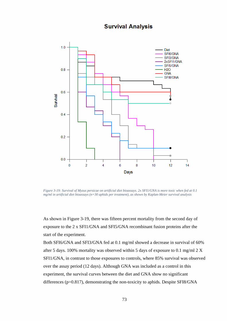
73
Figure 3-19: Survival of Myzus persicae on artificial diet bioassays. 2x SFI1/GNA is more toxic when fed at 0.1
mg/ml in artificial diet bioassays (n=30 aphids per treatment), as shown by Kaplan-Meier survival analysis
As shown in Figure 3-19, there was fifteen percent mortality from the second day of
exposure to the 2 x SFI1/GNA and SFI5/GNA recombinant fusion proteins after the
start of the experiment.
Both SFI6/GNA and SFI3/GNA fed at 0.1 mg/ml showed a decrease in survival of 60%
after 5 days. 100% mortality was observed within 5 days of exposure to 0.1 mg/ml 2 X
SFI1/GNA, in contrast to those exposures to controls, where 85% survival was observed
over the assay period (12 days). Although GNA was included as a control in this
experiment, the survival curves between the diet and GNA show no significant
differences (p=0.817), demonstrating the non-toxicity to aphids. Despite SFI8/GNA
74
showing higher mortality when compared to GNA alone, statistical analysis
demonstrates no significant difference on day 12 (P =0.795).
The surviving M. persicae fed on the SFI3/GNA fusion protein compared with those fed
on the SFI5/GNA fusion protein showed no significance difference at day 12 (p=0.859).
Even though high mortality occurred when M. persicae were fed on the SFI6/GNA
fusion protein at 0.1 mg/ml, the survival curves between SFI6/GNA and SFI5/GNA
show no significant differences (p=0.176). Comparing the toxicity differences between
2 X SFI1/GNA and SFI5/GNA over 12 days indicates no significant difference in
survival curves (p=0.859). However, the results demonstrate that 2 x SFI1/GNA had a
significant effect on the survival and growth of M. persicae.
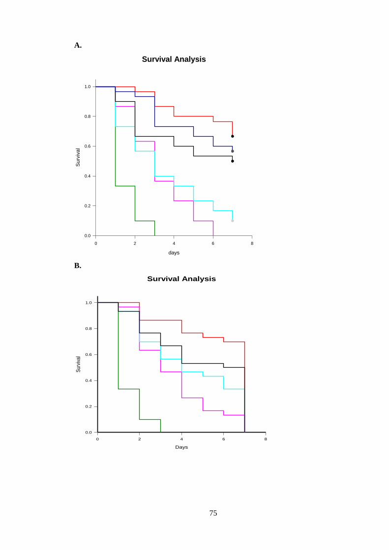
75
A.
B.
Survival Analysis
days
0 2 4 6 8
Su
rviv
al
0.0
0.2
0.4
0.6
0.8
1.0
Survival Analysis
Days
0 2 4 6 8
Sur
viva
l
0.0
0.2
0.4
0.6
0.8
1.0
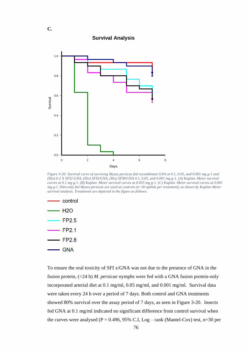
76
C.
Figure 3-20: Survival curve of surviving Myzus persicae fed recombinant GNA at 0.1, 0.05, and 0.001 mg g-1 and
(His) 6-2 X SFI2-GNA, (His) SFI5/GNA, (His) SFI8/GNA 0.1, 0.05, and 0.001 mg g-1. (A) Kaplan–Meier survival
curves at 0.1 mg g-1. (B) Kaplan–Meier survival curves at 0.055 mg g-1. (C) Kaplan–Meier survival curves at 0.001
mg g-1. Diet-only fed Myzus persicae are used as controls (n=30 aphids per treatment), as shown by Kaplan-Meier
survival analysis. Treatments are depicted in the figure as follows:
To ensure the oral toxicity of SFI x/GNA was not due to the presence of GNA in the
fusion protein, (<24 h) M. persicae nymphs were fed with a GNA fusion protein-only
incorporated arterial diet at 0.1 mg/ml, 0.05 mg/ml, and 0.001 mg/ml. Survival data
were taken every 24 h over a period of 7 days. Both control and GNA treatments
showed 80% survival over the assay period of 7 days, as seen in Figure 3-20. Insects
fed GNA at 0.1 mg/ml indicated no significant difference from control survival when
the curves were analysed (P = 0.496, 95% C.I, Log – rank (Mantel-Cox) test, n=30 per
Survival Analysis
Days
0 2 4 6 8
Su
rviv
al
0.0
0.2
0.4
0.6
0.8
1.0
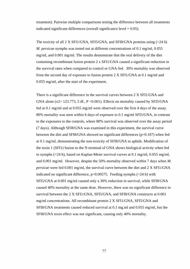
77
treatment). Pairwise multiple comparisons testing the difference between all treatments
indicated significant differences (overall significance level = 0.05).
The toxicity of all 2 X SFI1/GNA, SFI5/GNA, and SFI8/GNA proteins using (<24 h)
M. persicae nymphs was tested out at different concentrations of 0.1 mg/ml, 0.055
mg/ml, and 0.001 mg/ml. The results demonstrate that the oral delivery of the diet
containing recombinant fusion protein 2 x SFI1/GNA caused a significant reduction in
the survival rates when compared to control or GNA-fed. 30% mortality was observed
from the second day of exposure to fusion protein 2 X SFI1/GNA at 0.1 mg/ml and
0.055 mg/ml, after the start of the experiment.
There is a significant difference in the survival curves between 2 X SFI1/GNA and
GNA alone (x2= 123.773, 5 df., P <0.001). Effects on mortality caused by SFI5/GNA
fed at 0.1 mg/ml and at 0.055 mg/ml were observed over the first 4 days of the assay.
80% mortality was seen within 6 days of exposure to 0.1 mg/ml SFI5/GNA, in contrast
to the exposures to the controls, where 80% survival was observed over the assay period
(7 days). Although SFI8/GNA was examined in this experiment, the survival curve
between the diet and SFI8/GNA showed no significant differences (p=0.107) when fed
at 0.1 mg/ml, demonstrating the non-toxicity of SFI8/GNA to aphids. Modification of
the toxin 1 (SFI1) fusion to the N terminal of GNA shows biological activity when fed
to nymphs (<24 h), based on Kaplan-Meier survival curves at 0.1 mg/ml, 0.055 mg/ml,
and 0.001 mg/ml. However, despite the 50% mortality observed within 7 days when M.
persicae were fed 0.001 mg/ml, the survival curve between the diet and 2 X SFI1/GNA
indicated no significant difference, p=0.00575. Feeding nymphs (<24 h) with
SFI5/GNA at 0.001 mg/ml caused only a 30% reduction in survival, while SFI8/GNA
caused 40% mortality at the same dose. However, there was no significant difference in
survival between the 2 X SFI1/GNA, SFI5/GNA, and SFI8/GNA constructs at 0.001
mg/ml concentrations. All recombinant protein 2 X SFI1/GNA, SFI5/GNA and
SFI8/GNA treatments caused reduced survival at 0.1 mg.ml and 0.055 mg/ml, but the
SFI8/GNA toxin effect was not significant, causing only 40% mortality.
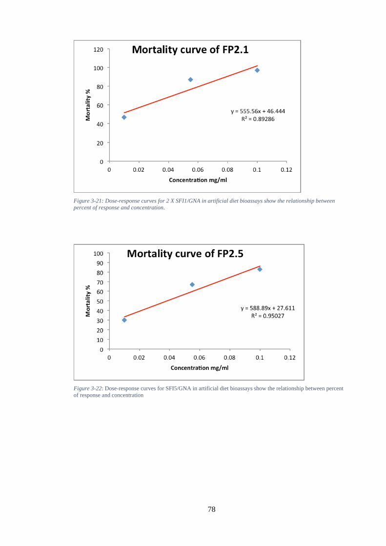
78
Figure 3-21: Dose-response curves for 2 X SFI1/GNA in artificial diet bioassays show the relationship between
percent of response and concentration.
Figure 3-22: Dose-response curves for SFI5/GNA in artificial diet bioassays show the relationship between percent
of response and concentration
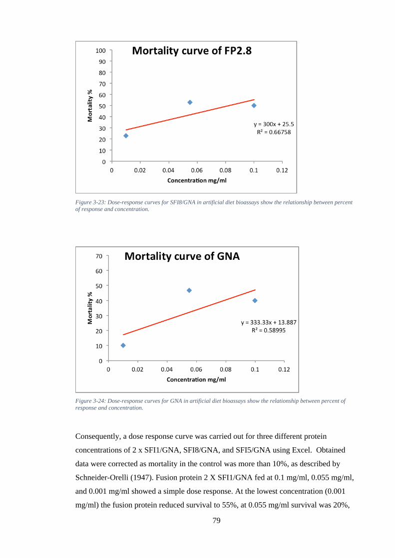
79
Figure 3-23: Dose-response curves for SFI8/GNA in artificial diet bioassays show the relationship between percent
of response and concentration.
Figure 3-24: Dose-response curves for GNA in artificial diet bioassays show the relationship between percent of
response and concentration.
Consequently, a dose response curve was carried out for three different protein
concentrations of 2 x SFI1/GNA, SFI8/GNA, and SFI5/GNA using Excel. Obtained
data were corrected as mortality in the control was more than 10%, as described by
Schneider-Orelli (1947). Fusion protein 2 X SFI1/GNA fed at 0.1 mg/ml, 0.055 mg/ml,
and 0.001 mg/ml showed a simple dose response. At the lowest concentration (0.001
mg/ml) the fusion protein reduced survival to 55%, at 0.055 mg/ml survival was 20%,
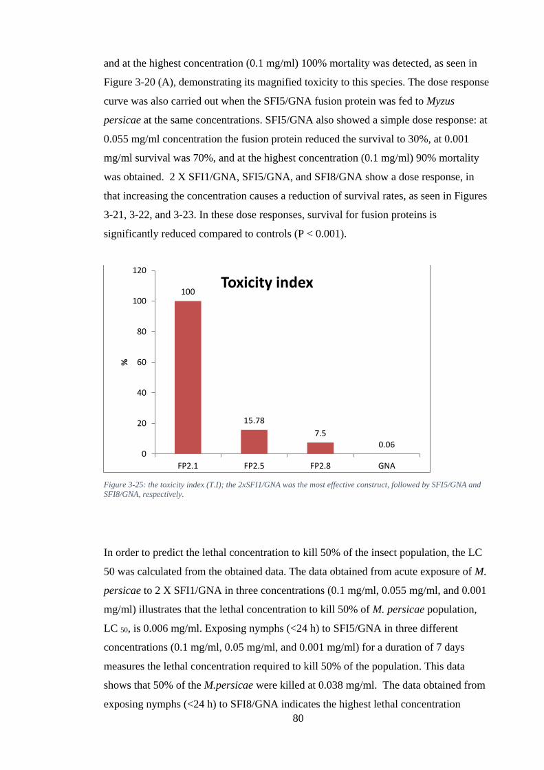
80
and at the highest concentration (0.1 mg/ml) 100% mortality was detected, as seen in
Figure 3-20 (A), demonstrating its magnified toxicity to this species. The dose response
curve was also carried out when the SFI5/GNA fusion protein was fed to Myzus
persicae at the same concentrations. SFI5/GNA also showed a simple dose response: at
0.055 mg/ml concentration the fusion protein reduced the survival to 30%, at 0.001
mg/ml survival was 70%, and at the highest concentration (0.1 mg/ml) 90% mortality
was obtained. 2 X SFI1/GNA, SFI5/GNA, and SFI8/GNA show a dose response, in
that increasing the concentration causes a reduction of survival rates, as seen in Figures
3-21, 3-22, and 3-23. In these dose responses, survival for fusion proteins is
significantly reduced compared to controls (P < 0.001).
Figure 3-25: the toxicity index (T.I); the 2xSFI1/GNA was the most effective construct, followed by SFI5/GNA and
SFI8/GNA, respectively.
In order to predict the lethal concentration to kill 50% of the insect population, the LC
50 was calculated from the obtained data. The data obtained from acute exposure of M.
persicae to 2 X SFI1/GNA in three concentrations (0.1 mg/ml, 0.055 mg/ml, and 0.001
mg/ml) illustrates that the lethal concentration to kill 50% of M. persicae population,
LC 50, is 0.006 mg/ml. Exposing nymphs (<24 h) to SFI5/GNA in three different
concentrations (0.1 mg/ml, 0.05 mg/ml, and 0.001 mg/ml) for a duration of 7 days
measures the lethal concentration required to kill 50% of the population. This data
shows that 50% of the M.persicae were killed at 0.038 mg/ml. The data obtained from
exposing nymphs (<24 h) to SFI8/GNA indicates the highest lethal concentration
100
15.78
7.50.06
0
20
40
60
80
100
120
FP2.1 FP2.5 FP2.8 GNA
%
Toxicity index
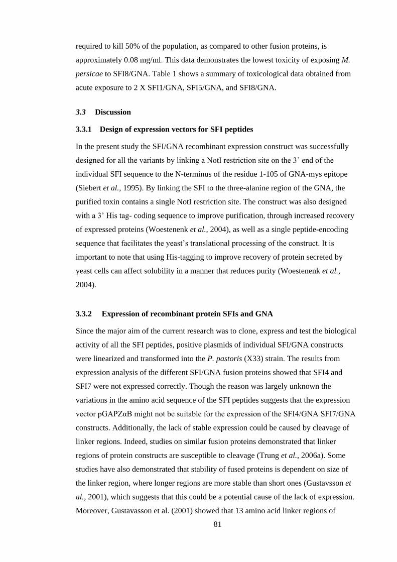
81
required to kill 50% of the population, as compared to other fusion proteins, is
approximately 0.08 mg/ml. This data demonstrates the lowest toxicity of exposing M.
persicae to SFI8/GNA. Table 1 shows a summary of toxicological data obtained from
acute exposure to 2 X SFI1/GNA, SFI5/GNA, and SFI8/GNA.
3.3 Discussion
3.3.1 Design of expression vectors for SFI peptides
In the present study the SFI/GNA recombinant expression construct was successfully
designed for all the variants by linking a NotI restriction site on the 3’ end of the
individual SFI sequence to the N-terminus of the residue 1-105 of GNA-mys epitope
(Siebert et al., 1995). By linking the SFI to the three-alanine region of the GNA, the
purified toxin contains a single NotI restriction site. The construct was also designed
with a 3’ His tag- coding sequence to improve purification, through increased recovery
of expressed proteins (Woestenenk et al., 2004), as well as a single peptide-encoding
sequence that facilitates the yeast’s translational processing of the construct. It is
important to note that using His-tagging to improve recovery of protein secreted by
yeast cells can affect solubility in a manner that reduces purity (Woestenenk et al.,
2004).
3.3.2 Expression of recombinant protein SFIs and GNA
Since the major aim of the current research was to clone, express and test the biological
activity of all the SFI peptides, positive plasmids of individual SFI/GNA constructs
were linearized and transformed into the P. pastoris (X33) strain. The results from
expression analysis of the different SFI/GNA fusion proteins showed that SFI4 and
SFI7 were not expressed correctly. Though the reason was largely unknown the
variations in the amino acid sequence of the SFI peptides suggests that the expression
vector pGAPZαB might not be suitable for the expression of the SFI4/GNA SFI7/GNA
constructs. Additionally, the lack of stable expression could be caused by cleavage of
linker regions. Indeed, studies on similar fusion proteins demonstrated that linker
regions of protein constructs are susceptible to cleavage (Trung et al., 2006a). Some
studies have also demonstrated that stability of fused proteins is dependent on size of
the linker region, where longer regions are more stable than short ones (Gustavsson et
al., 2001), which suggests that this could be a potential cause of the lack of expression.
Moreover, Gustavasson et al. (2001) showed that 13 amino acid linker regions of
82
bacterial origin are more stable than the linker regions of yeast origin. Another
promising candidate for stable peptide fusions is the IgG hinge, which has been shown
to increase stability. However, as the results presented in this chapter reveal SFI4 and
SFI7 produced from P. pastoris might not be stable, the full activity and stability of the
other SFI peptides would still need further investigation. The selection of the P. pastoris
was made based on previous studies that demonstrated that small proteins with
disulphide bonds can be expressed and isolated in this yeast strain (Cereghino and
Cregg, 2000). The yeast has the capability to secret expressed proteins into the culture
due to the yeast alpha-factor sequence incorporated in the expression vector used. This
process reduces any contamination and difficulties associated with isolating the proteins
from the cell lysate. In addition to the benefits of using this yeast strain, proteolysis of
fusion proteins is one of the major drawbacks of directing secretion of the fusion protein
into the medium (Gellissen, 2000; Fitches et al., 2004a). The resultant products after
isolation have been shown to contain some GNA without attached toxin, which could
reduce gut activity (Fitches et al., 2004a). However, the six SFI toxin peptides that were
used in the present study are relatively resistant to proteolysis, suggesting that the
samples used contained small amounts of free GNA. The yeast’s ability to N-
glycosylate proteins is efficient and achieved by passing the proteins through
endoplasmic reticulum, but extra mannose residues can be added during core
glycosylation (Bretthauer and Castellino, 1999; Christou et al., 2006). The addition of
mannose residues could increase gut digestion, but this was not a problem during
bioactivity studies with the purified fusion proteins.
A further experiment to optimize the expression of the SFI peptides using different
expression vectors is important. Although, SFI2/GNA, SFI3/GNA, SFI5/GNA,
SFI6/GNA, and SFI8/GNA were correctly expressed, some were only expressed at very
low concentration. This suggests that the expression vector used in the current study
might not be suitable for all the individual sequences of the SFI peptides. Moreover,
experimental conditions may need to be optimised in future experiments and potential
large-scale production. Additionally, initial efforts to optimise the experimental
conditions by assembling double gene copies of SFI1/GNA into gene cassettes inserted
in the pGAPZa plasmid showed promising results. Preliminary results suggest that high
yields can be achieved by introducing double gene copies of the peptides into the
plasmid.
83
3.3.3 Expression and oral delivery of SF1/GNA, SFI2/GNA, SFI3/GNA,
SFI5/GNA, SFI6/GNA, and SFI8/GNA
Insect resistance to pesticides remains one of the major obstacles to effective pest
control (Chrispeels and Sadava, 2003). The implementation of SFI polypeptides in
pesticide development requires the establishment of a standard production strategy, as
well as an effective oral delivery method. The toxicity of peptides on the insect’s
neurons is dependent on functional access to the central nervous system, through the
haemolymph. Interestingly, GNA has been shown to access the haemolymph through
oral delivery (Powell et al., 1998; Fitches and Gatehouse, 1998; Fitches et al., 2001,
2002), which makes it suitable for this study. Furthermore, experiments on the ability of
GNA to transport fused polypeptides to the central nervous system of insects through
the haemolymph has been established with other potential insecticidal peptides (Fitches
et al., 2004a; Fitches et al., 2004d; Trung et al., 2006a; Fitches et al., 2010). Though the
mechanisms involved in this transport remains poorly described, the ability of GNA to
bind to epithelial surface glycoproteins suggest that endocytosis might be an important
mechanism in this process (Fitches et al., 2004b). In addition to the activity of
membrane receptors, alternative models of GNA transport that involves leaky cell
junctions have been proposed (Fitches et al., 2004b). Therefore, the current study
utilised SFI fused to GNA as a method of delivering SFI to the haemolymph of the
model insect. The expression of 2xSFI1/GNA, SFI2/GNA, SFI3/GNA, SFI5/GNA,
SFI6/GNA, and SFI8/GNA with the GNA fused to the N-terminus of the toxin is
reported in this chapter. Fusion of the GNA to the N-terminus of the toxin can promote
stability and protein folding during production and purification. Indeed, research has
demonstrated that the actual location of the fusion could affect overall stability of a
peptide/GNA fusion production (Fitches et al., 2010).
Five fusion variants of SFI were successfully expressed in P. pastoris X33 and
subsequently purified; the results showed that the expression levels were within the
same range as for other studies that used the same strain of P. pastoris (X33) (Fitches et
al., 2010). It is noteworthy that using this stain to produce a fusion protein specific to
insects can affect purification of the protein, because P. pastoris can incorporate
mannose oligosaccharides to the fusion proteins that can significantly reduce purity.
GNA can bind to mannose, which in turn can introduce high molecular weight yeast
84
proteins and carbohydrates to the fusion proteins. However, all of the purification
results for the fusion proteins showed no presence of high molecular weight proteins
suggesting the absence of yeast proteins or oligosaccharides chains; this finding give
confidence that subsequent experiments were conducted with pure samples. In this light,
the effects of such yeast-derived molecules on peptide function were not investigated
further.
3.3.4 Biological activity of fusion proteins SF1/GNA, SFI2/GNA, SFI3/GNA,
SFI5/GNA, SFI6/GNA, and SFI8/GNA
SFI1, SFI3, SFI5, SFI6, and SFI8 incorporated into GNA (0.1 mg/ml) caused significant
mortality to M. persicae compared to only GNA, which is below the potent
concentration range reported by other studies (6 - 12.5mg/ml) (Fitches et al., 2004a,
2010, Trung et al., 2006). One of the important functional characteristics of fusion
proteins is correct folding, which is dependent on post-translational modification of the
fusion protein (Cregg et al., 2000). Results from the expression of SFI fusion with GNA
suggest that the protein folds with high efficiency, which is essential biological activity.
Furthermore, the results showed that some of the fusion proteins are more active, which
could be explained by their differing stability to proteolytic digestion in the insect gut.
Some of the SFI might be more resistant to gut digestion, thereby increase the amount
of toxins needed to induce mortality. Indeed experiments to determine the highest
concentration needed to induce 50% mortality suggests that partial gut digestion might
occur. The lowest LC 50 value was observed when SFI8/GNA was orally delivered,
suggesting that the SFI8 peptide is the most potent of the toxins, and potentially most
stable. SFI1/GNA caused the most effect at the lowest concentration compared to the
other variants. However, for all the SFI fusion proteins, less than 0.1mg/ml is required
to induce 50% mortality, which suggests that fusion of multiple toxins with different
modes of action may achieve even greater effects.
The data suggests that upon oral delivery some of the SFI variants might be degraded,
which will increase the amount of peptide needed to achieve the LC 50. This hypothesis
is in line with the findings of other research on fusion protein (Fitches et al., 2004),
which demonstrated that fusing toxin to GNA did not eliminate the action of Lacanobia
oleracea’s gut proteases. Future studies should seek to investigate in-vivo gut stability
of the SFI/GNA fusion proteins used in this study; as the eventual success of this
85
strategy is based on both on stability in the insect gut and delivery to the target site.
However, any potential cleavage of some of the toxic peptide failed to completely
remove the SFI peptides, which might be the reason for decrease the toxicity of some
SFI variants. Another potential reason that may cause the variations in toxicity between
the SFI variants is using the Bradford assay to estimate the total concentration of
protein. Some protein may degraded through the purification process; for this a standard
Bradford assay is not suitable mothed as theses assay cannot distinguish between intact
protein and degradation products. In future, a densitometric the fusion protein
quantification based on image analysis of the gel can avoid any potential difference that
may occur.
3.3.5 Increasing stability in gut environments
Small variations in toxicity between the SFI/GNA fusion protein variants may result
from differences stability to proteolytic digestion in the insect gut. Some of the SFI
might be more resistant to gut digestion, thereby increase the amount of toxins needed
to induce mortality. The stability of the SFI fusion protein with GNA was not
investigated in this study. However, further work on gut stability and approaches to
promote effective transport into the haemolymph could increase the potency of the
fusion proteins. Incorporating protease inhibitors into the fusion protein could reduce
the LC 50 even further and fast track commercial application of these fusion proteins.
This may help reduce the cost of producing sufficient amount of peptides to be applied
on a large scale. However, several insect species have been shown to up-regulate
alternative proteases when one is inhibited, which might need to be investigated with
the current fusion protein if it proves useful for a broader range of insect pests (Bolter
and Jongsma, 1995; Oppert et al., 2005).
3.3.6 Functional characteristics of SFI toxins
Segestria florentia venom contains about 25 individual polypeptides (Sagdiev et al.,
1987a). The SFI1 toxin is the most studied of the SFI toxins from S. florentia. It has be
shown to share some evolutionary, functional and structural relationship with other
neurotoxins like PLTX, curtatoxins, APS and SNX325, a toxin from the same species
(Stapleton et al., 1990a; Branton et al., 1993a; Quistad and Skinner, 1994b; Garcíaa et
al., 2015). Interestingly, these neurotoxins primarily act by selective binding to voltage-
dependent calcium channels (Stapleton et al., 1990a; Branton et al., 1993a), which point
86
to similar mode of actions for SFI1 and the other variants. Furthermore, a more recent
study has shown that SFI1 contain a large β-hairpin loop of approximately a third of the
peptide length (Bende et al., 2015). The study showed that this hairpin loop is critical
for SFI1 toxic activity, which facilitates selective inhibition of voltage-gated sodium
channels by pore blocking (Bende et al., 2015). This finding is contrary to the gating
modification mechanisms utilised by other spider purified sodium channels toxins. This
potential of SFI toxins is in line with the finding that they share similar distribution of
cysteine residue (Bende et al., 2015). Unfortunately, the potential mechanisms used by
the other variants evaluated in the present study still need to be investigated. It is
equally plausible that the folding pattern and net charge could target these toxins to the
ion channels of interest.
In summary, results from this Chapter demonstrated that functional SFI/GNA based
fusion proteins can be effectively expressed and purified using p. pastoris (X33). Based
on oral delivery studies the results suggest that the variants tested in the present study
(SFI2, SFI3, SFI5, SFI6, SFI8) were not as toxic to M.persicae compared to previous
reported toxicity studies to SFI1 Down et al. (2006). Given that, SFI1 appears to be the
most toxic of the variants against this aphid species. In the present study double
constructs were prepared for SFI1. As expected this double SFI1/GNA construct was
significantly more toxic compared to the single constructs. Further work to determine
LC50 values for the fusion proteins showed promising results. However, oral activity
against other insects was not investigated, but the potential for proteolytic cleavage
within the insect gut makes this an important field of further study. The presence of
linker regions that might be targeted by insect proteases needs to be established.
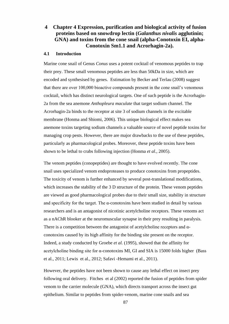
87
4 Chapter 4 Expression, purification and biological activity of fusion
proteins based on snowdrop lectin (Galanthus nivalis agglutinin;
GNA) and toxins from the cone snail (alpha-Conotoxin EI, alpha-
Conotoxin Sm1.1 and Acrorhagin-2a).
4.1 Introduction
Marine cone snail of Genus Conus uses a potent cocktail of venomous peptides to trap
their prey. These small venomous peptides are less than 50kDa in size, which are
encoded and synthesised by genes. Estimation by Becker and Terlau (2008) suggest
that there are over 100,000 bioactive compounds present in the cone snail’s venomous
cocktail, which has distinct neurological targets. One of such peptide is the Acrorhagin-
2a from the sea anemone Anthopleura maculate that target sodium channel. The
Acrorhagin-2a binds to the receptor at site 3 of sodium channels in the excitable
membrane (Honma and Shiomi, 2006). This unique biological effect makes sea
anemone toxins targeting sodium channels a valuable source of novel peptide toxins for
managing crop pests. However, there are major drawbacks to the use of these peptides,
particularly as pharmacological probes. Moreover, these peptide toxins have been
shown to be lethal to crabs following injection (Honma et al., 2005).
The venom peptides (conopeptides) are thought to have evolved recently. The cone
snail uses specialized venom endoproteases to produce conotoxins from propeptides.
The toxicity of venom is further enhanced by several post-translational modifications,
which increases the stability of the 3 D structure of the protein. These venom peptides
are viewed as good pharmacological probes due to their small size, stability in structure
and specificity for the target. The α-conotoxins have been studied in detail by various
researchers and is an antagonist of nicotinic acetylcholine receptors. These venoms act
as a nAChR blooker at the neuromuscular synapse in their prey resulting in paralysis.
There is a competition between the antagonist of acetylcholine receptors and α-
conotoxins caused by its high affinity for the binding site present on the receptor.
Indeed, a study conducted by Groebe et al. (1995), showed that the affinity for
acetylcholine binding site for α-conotoxins MI, GI and SIA is 15000 folds higher (Bass
et al., 2011; Lewis et al., 2012; Safavi -Hemami et al., 2011).
However, the peptides have not been shown to cause any lethal effect on insect prey
following oral delivery. Fitches et al (2002) reported the fusion of peptides from spider
venom to the carrier molecule (GNA), which directs transport across the insect gut
epithelium. Similar to peptides from spider-venom, marine cone snails and sea
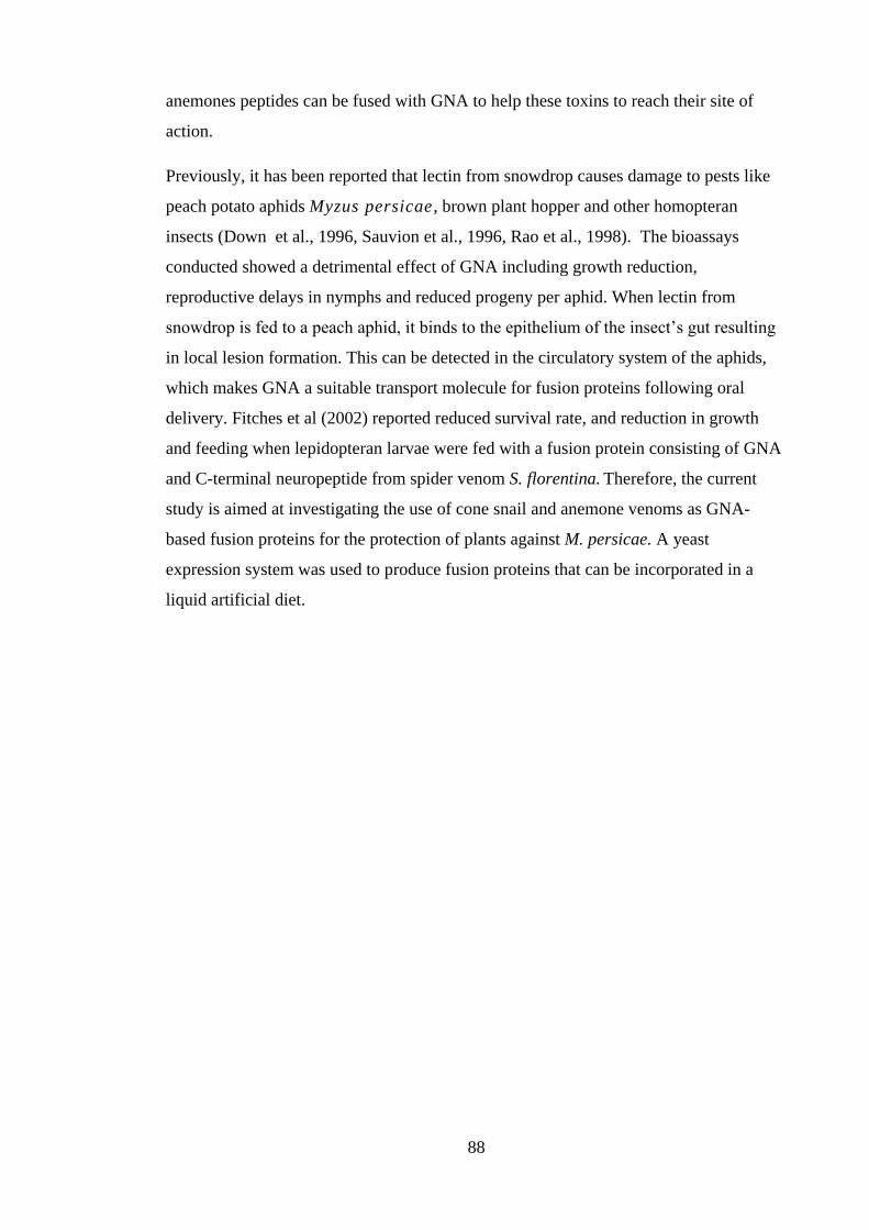
88
anemones peptides can be fused with GNA to help these toxins to reach their site of
action.
Previously, it has been reported that lectin from snowdrop causes damage to pests like
peach potato aphids Myzus persicae , brown plant hopper and other homopteran
insects (Down et al., 1996, Sauvion et al., 1996, Rao et al., 1998). The bioassays
conducted showed a detrimental effect of GNA including growth reduction,
reproductive delays in nymphs and reduced progeny per aphid. When lectin from
snowdrop is fed to a peach aphid, it binds to the epithelium of the insect’s gut resulting
in local lesion formation. This can be detected in the circulatory system of the aphids,
which makes GNA a suitable transport molecule for fusion proteins following oral
delivery. Fitches et al (2002) reported reduced survival rate, and reduction in growth
and feeding when lepidopteran larvae were fed with a fusion protein consisting of GNA
and C-terminal neuropeptide from spider venom S. florentina. Therefore, the current
study is aimed at investigating the use of cone snail and anemone venoms as GNA-
based fusion proteins for the protection of plants against M. persicae. A yeast
expression system was used to produce fusion proteins that can be incorporated in a
liquid artificial diet.

89
4.2 Results:
4.2.1 Production and purification of fusion proteins
Design of expression constructs GNA/Acrorhagin-2a, GNA/alpha-Conotoxin EI and
GNA/ alpha-Conotoxin sm1.1
A synthetic gene encoding the mature alpha-Conotoxin EI, alpha-Conotoxin sm1.1and
Acrorhagin-2a amino acid sequence was successfully assembled with a codon optimised
for expression in P. pastoris using a series of overlapping oligonucleotides. Thereafter
and using a similar cloning strategy to the generation of the SFI/GNA fusion proteins,
the coding sequences of the individual toxins were amplified and ligated to the GNA
moiety previously cloned into the pGAPZα yeast expression vector. Alpha-Conotoxin
EI, sm1.1and Acrorhagin-2a sequences were prepared for cloning by digesting at the
unique XbaI and XhoI enzyme sites. A small fragment was excised and the 5' end of the
toxins sequence containing a single Xho1 restriction site were fused to the c-terminus of
the residues 1-105 of snowdrop lectin GNA-mys epitope via its three-alanine regions.
The GNA/ alpha-Conotoxin EI, GNA/ alpha-Conotoxin sm1.1 and GNA/ Acrorhagin-
2a constructs were predicted to have alpha factor secretary single, 6× His tag and an
additional two amino acid peptides at the N-terminus of GNA, which gave a predicted
molecular weight of 15 kDa. To ensure no errors had occurred when restriction/ligation
of fusion protein construct was carried out, the plasmid was then sent for sequencing.
Sequencing results for selected colonies confirmed the maintenance of correct construct
integrity and positioning at each stage. Sequences were further analysed by in silico
translation to ensure the open reading frames were maintained throughout. A schematic
diagram of the expression constructs, determined nucleotide and predicted protein
sequences for the GNA/Acrorhagin-2a, GNA/ alpha-Conotoxin EI and GNA/ alpha-
Conotoxin sm1.1 are shown in Figures 4-1, 4-2, 4-3. Verified expression constructs for
GNA/ alpha-Conotoxin EI pGAZαB, GNA/ alpha-Conotoxin sm1.1 pGAZαB and
GNA/ Acrorhagin-2a pGAZαB were transformed into competent cells of P. pastoris
and selected on zeocin-containing plates. The initial attempts on transforming the GNA/
alpha-Conotoxin EI pGAPZα, GNA/ alpha-Conotoxin sm1.1 pGAPZα and GNA/
Acrorhagin-2a pGAPZα fusion protein into wild type P. pastoris X33 using the
EasyComp kit resulted in a few colonies after 3 days growth as seen in Appendix E.
90
864 atgagatttccttcaatttttactgctgttttattcgcagcatcc
M R F P S I F T A V L F A A S
819 tccgcattagctgctccagtcaacactacaacagaagatgaaacg
S A L A A P V N T T T E D E T
774 gcacaaattccggctgaagctgtcatcggttactcagatttagaa
A Q I P A E A V I G Y S D L E
729 ggggatttcgatgttgctgttttgccattttccaacagcacaaat
G D F D V A V L P F S N S T N
684 aacgggttattgtttataaatactactattgccagcattgctgct
N G L L F I N T T I A S I A A
639 aaagaagaaggggtatctctcgagaaaagagaggctgaagctgca
K E E G V S L E K R E A E A A
594 gcagacaacattttgtattctggggagaccttaagtacaggggaa
A D N I L Y S G E T L S T G E
549 tttttgaattacggtagcttcgtattcatcatgcaggaagactgt
F L N Y G S F V F I M Q E D C
504 aatctagtgctatacgatgttgacaagccaatctgggcaacgaat
N L V L Y D V D K P I W A T N
459 actggcggactgagcagatcgtgctttctttcaatgcaaaccgat
T G G L S R S C F L S M Q T D
414 ggtaacctcgttgtatataatcctagtaataaaccgatatgggcg
G N L V V Y N P S N K P I W A
369 tccaacactggcggacaaaacggaaactatgtctgtatattacag
S N T G G Q N G N Y V C I L Q
324 aaagatcgaaatgtcgtgatttacgggacggataggtgggctaca
K D R N V V I Y G T D R W A T
279 ggtgcagcagcagactgcagatttgtcggtgctaaatgtacaaaa
G A A A D C R F V G A K C T K
234 gcaaataacccgtgtgtagggaaagtgtgtaatggatatcaattg
A N N P C V G K V C N G Y Q L
189 tactgtcctgttgacgatgatcattgcattatgaagttaactttc
Y C P V D D D H C I M K L T F
144 atacctctagaacaaaaactcatctcagaggatctgaatagcgcc
I P L E Q K L I S E D L N S A
99 gtcgaccatcatcatcatcaccgtccattgtag 67
V D H H H H H R P L *
Figure 4-1: Full GNA/Acrorhagin-2a fusion protein fusion protein construct in yeast pGAPZAP. Full
determined nucleotide sequences, presumed amino acid sequences and schematic representation of fusion
protein construct. Yeast alpha factor signal sequences are highlighted in blue, GNA sequences is
highlighted in red and Acrorhagin toxin sequences are highlighted in green.
Acrorhagin-2a α-factor
GNA 6xHis stop
12.5 kDa 3 kDa
AA AAA
91
818 ttgaacaactatttcgaaacgatgagatttccttcaatttttact
L N N Y F E T M R F P S I F T
773 gctgttttattcgcagcatcctccgcattagctgctccagtcaac
A V L F A A S S A L A A P V N
728 actacaacagaagatgaaacggcacaaattccggctgaagctgtc
T T T E D E T A Q I P A E A V
683 atcggttactcagatttagaaggggatttcgatgttgctgttttg
I G Y S D L E G D F D V A V L
638 ccattttccaacagcacaaataacgggttattgtttataaatact
P F S N S T N N G L L F I N T
593 actattgccagcattgctgctaaagaagaaggggtatctctcgag
T I A S I A A K E E G V S L E
548 aaaagagaggctgaagctgcagcagacaacattttgtattctggg
K R E A E A A A D N I L Y S G
503 gagaccttaagtacaggggaatttttgaattacggtagcttcgta
E T L S T G E F L N Y G S F V
458 ttcatcatgcaggaagactgtaatctagtgctatacgatgttgac
F I M Q E D C N L V L Y D V D
413 aagccaatctgggcaacgaatactggcggactgagcagatcgtgc
K P I W A T N T G G L S R S C
368 tttctttcaatgcaaaccgatggtaacctcgttgtatataatcct
F L S M Q T D G N L V V Y N P
323 agtaataaaccgatatgggcgtccaacactggcggacaaaacgga
S N K P I W A S N T G G Q N G
278 aactatgtctgtatattacagaaagatcgaaatgtcgtgatttac
N Y V C I L Q K D R N V V I Y
233 gggacggataggtgggctacaggtgcagcagcaagagatccgtgt
G T D R W A T G A A A R D P C
188 tgctatcatcctacatgtaacatgagtaatccacaaatatgtcta
C Y H P T C N M S N P Q I C L
143 gaacaaaaactcatctcagaagaggatctgaatagcgccgtcgac
E Q K L I S E E D L N S A V D
98 catcatcatcatcacccttttttttgtagccttagacatgactgt
H H H H H P F F C S L R H D C
53 tcctcagttctaagttgggcactacgagaagaccggtcttgctgg
S S V L S W A L R E D R S C W
8 ataagc 3
I S
Figure 4-2: Full GNA/ alpha-Conotoxin EI fusion protein construct in yeast pGAPZAP. Full determined
nucleotide sequences, presumed amino acid sequences, and schematic representation of fusion protein
construct. The sequence for the yeast alpha-factor signal is highlighted with blue, GNA sequence is
highlighted with red, and alpha-Conotoxin EI toxin sequence is highlighted with green.
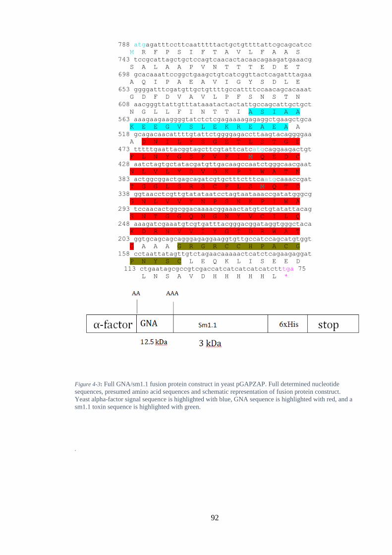
92
788 atgagatttccttcaatttttactgctgttttattcgcagcatcc
M R F P S I F T A V L F A A S
743 tccgcattagctgctccagtcaacactacaacagaagatgaaacg
S A L A A P V N T T T E D E T
698 gcacaaattccggctgaagctgtcatcggttactcagatttagaa
A Q I P A E A V I G Y S D L E
653 ggggatttcgatgttgctgttttgccattttccaacagcacaaat
G D F D V A V L P F S N S T N
608 aacgggttattgtttataaatactactattgccagcattgctgct
N G L L F I N T T I A S I A A
563 aaagaagaaggggtatctctcgagaaaagagaggctgaagctgca
K E E G V S L E K R E A E A A
518 gcagacaacattttgtattctggggagaccttaagtacaggggaa
A D N I L Y S G E T L S T G E
473 tttttgaattacggtagcttcgtattcatcatgcaggaagactgt
F L N Y G S F V F I M Q E D C
428 aatctagtgctatacgatgttgacaagccaatctgggcaacgaat
N L V L Y D V D K P I W A T N
383 actggcggactgagcagatcgtgctttctttcaatgcaaaccgat
T G G L S R S C F L S M Q T D
338 ggtaacctcgttgtatataatcctagtaataaaccgatatgggcg
G N L V V Y N P S N K P I W A
293 tccaacactggcggacaaaacggaaactatgtctgtatattacag
S N T G G Q N G N Y V C I L Q
248 aaagatcgaaatgtcgtgatttacgggacggataggtgggctaca
K D R N V V I Y G T D R W A T
203 ggtgcagcagcagggagaggaaggtgttgccatccagcatgtggt
G A A A G R G R C C H P A C G
158 cctaattatagttgtctagaacaaaaactcatctcagaagaggat
P N Y S C L E Q K L I S E E D
113 ctgaatagcgccgtcgaccatcatcatcatcatctttga 75
L N S A V D H H H H H L *
Figure 4-3: Full GNA/sm1.1 fusion protein construct in yeast pGAPZAP. Full determined nucleotide
sequences, presumed amino acid sequences and schematic representation of fusion protein construct.
Yeast alpha-factor signal sequence is highlighted with blue, GNA sequence is highlighted with red, and a
sm1.1 toxin sequence is highlighted with green.
.

93
Expression of recombinant protein GNA/ alpha-Conotoxin EI, GNA/ alpha-Conotoxin
sm1.1 and GNA/ Acrorhagin-2a
Transformed yeast cells containing the expression constructs were selected on plates
containing the antibiotic zeocin. The obtained clones were screened for the best expression
clone to be used in fusion protein production by bench- top fermentation. Small-scale
culture supernatants (grown for four days) were analysed by Western blotting using anti
GNA antibodies (1:3000 dilution). GNA was used as positive control because the fusion
proteins of GNA/ alpha-Conotoxin EI, GNA/ sm1.1 and GNA/ Acrorhagin-2a were
confirmed to have GNA linked to the N-terminus of the toxins. The expected result for
fusion proteins of GNA/ alpha-Conotoxin EI, GNA/ sm1.1 and GNA/ Acrorhagin-2a
should have reactivity at 15 kDa. The Western blots for the supernatant were performed
over a time course. The result showed that GNA/ alpha-Conotoxin EI were detected after
48 hours and 96 hours (Figure 4-4) of growth. The greatest amount of intact fusion protein
was observed at 96 hour. However, the fusion protein of GNA/ sm1.1 and GNA/
Acrorhagin-2a were not detected through Western blot at both time point (result not
shown). Additionally, the presence of GNA fusion protein at the correct size in the positive
standard lanes indicated that the blotting process was successful.
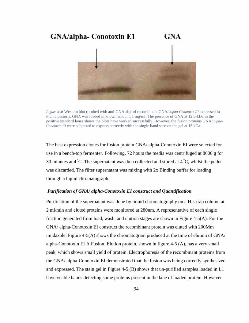
94
Figure 4-4: Western blot (probed with anti-GNA ab): of recombinant GNA/ alpha-Conotoxin EI expressed in
Pichia pastoris. GNA was loaded in known amount, 1 mg/ml. The presence of GNA at 12.5-kDa in the
positive standard lanes shows the blots have worked successfully. However, the fusion proteins GNA/ alpha-
Conotoxin EI were subjected to express correctly with the single band seen on the gel at 15 kDa.
The best expression clones for fusion protein GNA/ alpha-Conotoxin EI were selected for
use in a bench-top fermenter. Following, 72 hours the media was centrifuged at 8000 g for
30 minutes at 4 °C. The supernatant was then collected and stored at 4 °C, whilst the pellet
was discarded. The filter supernatant was mixing with 2x Binding buffer for loading
through a liquid chromatograph.
Purification of GNA/ alpha-Conotoxin EI construct and Quantification
Purification of the supernatant was done by liquid chromatography on a His-trap column at
2 ml/min and eluted proteins were monitored at 280nm. A representative of each single
fraction generated from load, wash, and elution stages are shown in Figure 4-5(A). For the
GNA/ alpha-Conotoxin EI construct the recombinant protein was eluted with 200Mm
imidazole. Figure 4-5(A) shows the chromatogram produced at the time of elution of GNA/
alpha-Conotoxin EI A Fusion. Elution protein, shown in figure 4-5 (A), has a very small
peak, which shows small yield of protein. Electrophoresis of the recombinant proteins from
the GNA/ alpha-Conotoxin EI demonstrated that the fusion was being correctly synthesized
and expressed. The stain gel in Figure 4-5 (B) shows that un-purified samples loaded in L1
have visible bands detecting some proteins present in the lane of loaded protein. However

95
the proteins in the eluted lanes show a high degree of purification. Anti-GNA antibodies
recognized a single 15-kDa band from the GNA/ alpha-Conotoxin EI fusion protein (Figure
4-6). However by comparing the amount of GNA/ alpha-Conotoxin EI in the load fraction
(L4) to the eluted proteins (L3) it also shows that a majority of the recombinant proteins
were lost during the purification process.
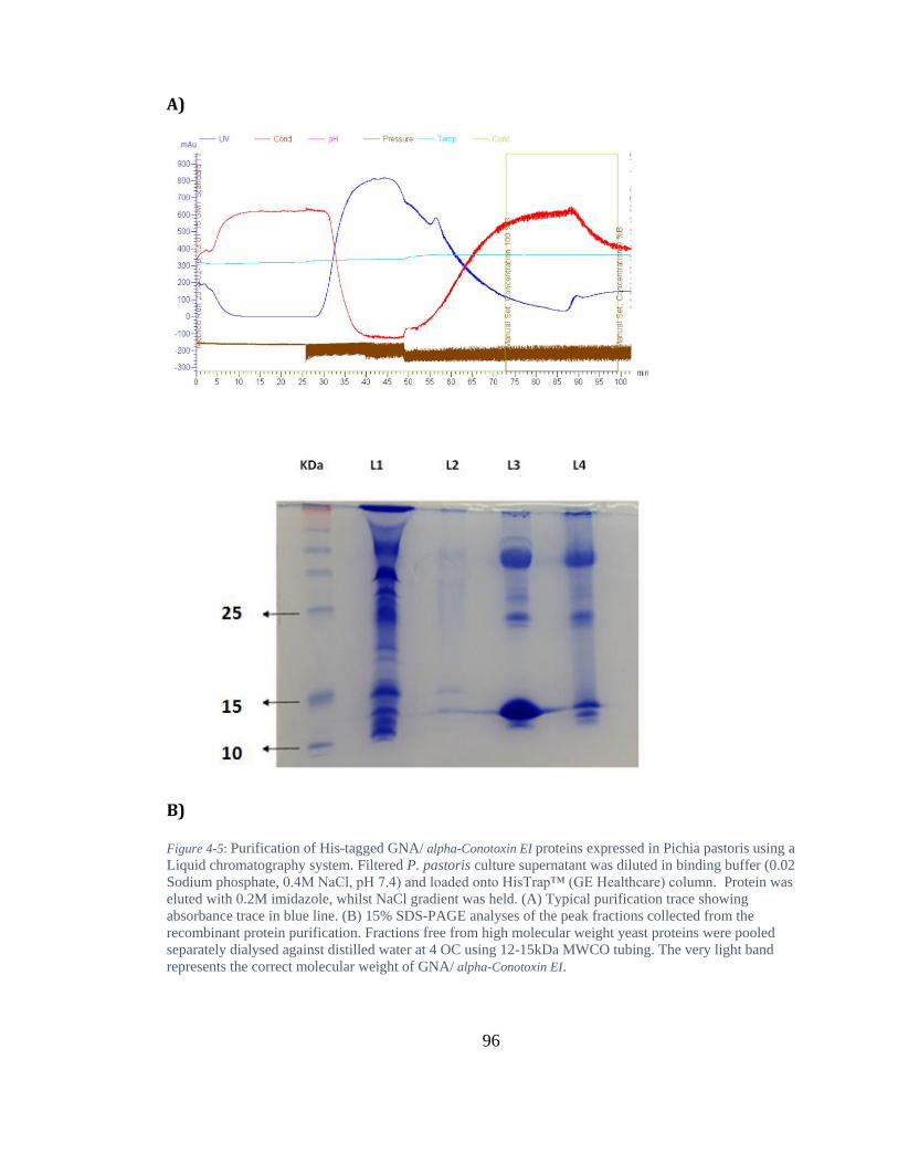
96
A)
B)
Figure 4-5: Purification of His-tagged GNA/ alpha-Conotoxin EI proteins expressed in Pichia pastoris using a
Liquid chromatography system. Filtered P. pastoris culture supernatant was diluted in binding buffer (0.02
Sodium phosphate, 0.4M NaCl, pH 7.4) and loaded onto HisTrap™ (GE Healthcare) column. Protein was
eluted with 0.2M imidazole, whilst NaCl gradient was held. (A) Typical purification trace showing
absorbance trace in blue line. (B) 15% SDS-PAGE analyses of the peak fractions collected from the
recombinant protein purification. Fractions free from high molecular weight yeast proteins were pooled
separately dialysed against distilled water at 4 OC using 12-15kDa MWCO tubing. The very light band
represents the correct molecular weight of GNA/ alpha-Conotoxin EI.
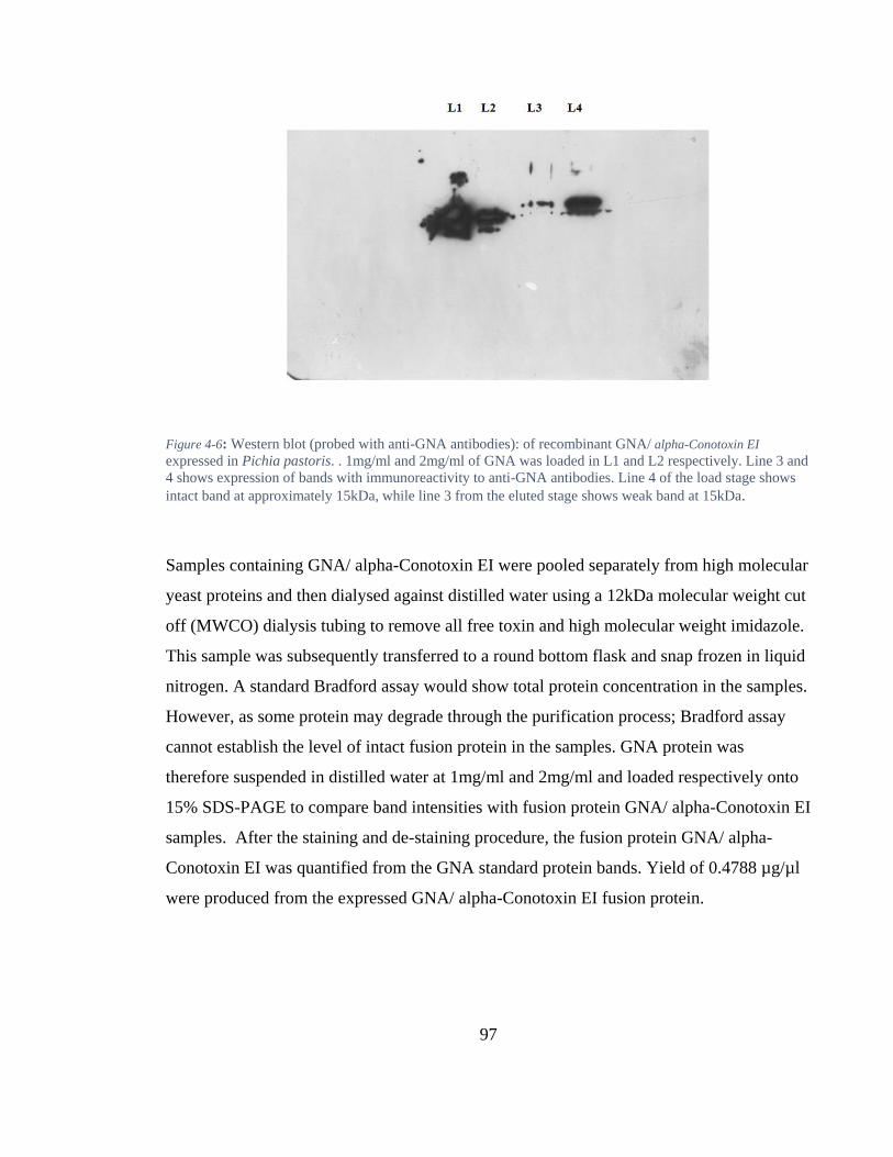
97
Figure 4-6: Western blot (probed with anti-GNA antibodies): of recombinant GNA/ alpha-Conotoxin EI
expressed in Pichia pastoris. . 1mg/ml and 2mg/ml of GNA was loaded in L1 and L2 respectively. Line 3 and
4 shows expression of bands with immunoreactivity to anti-GNA antibodies. Line 4 of the load stage shows
intact band at approximately 15kDa, while line 3 from the eluted stage shows weak band at 15kDa.
Samples containing GNA/ alpha-Conotoxin EI were pooled separately from high molecular
yeast proteins and then dialysed against distilled water using a 12kDa molecular weight cut
off (MWCO) dialysis tubing to remove all free toxin and high molecular weight imidazole.
This sample was subsequently transferred to a round bottom flask and snap frozen in liquid
nitrogen. A standard Bradford assay would show total protein concentration in the samples.
However, as some protein may degrade through the purification process; Bradford assay
cannot establish the level of intact fusion protein in the samples. GNA protein was
therefore suspended in distilled water at 1mg/ml and 2mg/ml and loaded respectively onto
15% SDS-PAGE to compare band intensities with fusion protein GNA/ alpha-Conotoxin EI
samples. After the staining and de-staining procedure, the fusion protein GNA/ alpha-
Conotoxin EI was quantified from the GNA standard protein bands. Yield of 0.4788 µg/µl
were produced from the expressed GNA/ alpha-Conotoxin EI fusion protein.
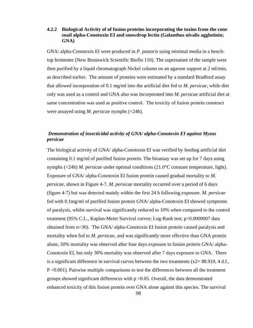
98
4.2.2 Biological Activity of of fusion proteins incorporating the toxins from the cone
snail alpha-Conotoxin EI and snowdrop lectin (Galanthus nivalis agglutinin;
GNA)
GNA/ alpha-Conotoxin EI were produced in P. pastoris using minimal media in a bench-
top fermenter (New Brunswick Scientific Bioflo 110). The supernatant of the sample were
then purified by a liquid chromatograph Nickel column on an agarose support at 2 ml/min,
as described earlier. The amount of proteins were estimated by a standard Bradford assay
that allowed incorporation of 0.1 mg/ml into the artificial diet fed to M. persicae, while diet
only was used as a control and GNA also was incorporated into M. persicae artificial diet at
same concentration was used as positive control. The toxicity of fusion protein construct
were assayed using M. persicae nymphs (<24h).
Demonstration of insecticidal activity of GNA/ alpha-Conotoxin EI against Myzus
persicae
The biological activity of GNA/ alpha-Conotoxin EI was verified by feeding artificial diet
containing 0.1 mg/ml of purified fusion protein. The bioassay was set up for 7 days using
nymphs (<24h) M. persicae under optimal conditions (21.0°C constant temperature, light).
Exposure of GNA/ alpha-Conotoxin EI fusion protein caused gradual mortality to M.
persicae, shown in Figure 4-7. M. persicae mortality occurred over a period of 6 days
(figure 4-7) but was detected mainly within the first 24 h following exposure. M. persicae
fed with 0.1mg/ml of purified fusion protein GNA/ alpha-Conotoxin EI showed symptoms
of paralysis, whilst survival was significantly reduced to 10% when compared to the control
treatment (95% C.L., Kaplan-Meier Survival curves: Log-Rank test; p<0.0000007 data
obtained from n=30). The GNA/ alpha-Conotoxin EI fusion protein caused paralysis and
mortality when fed to M. persicae, and was significantly more effective than GNA protein
alone, 50% mortality was observed after four days exposure to fusion protein GNA/ alpha-
Conotoxin EI, but only 30% mortality was observed after 7 days exposure to GNA. There
is a significant difference in survival curves between the two treatments (x2= 88.910, 4 d.f.,
P <0.001). Pairwise multiple comparisons to test the differences between all the treatment
groups showed significant differences with p <0.05. Overall, the data demonstrated
enhanced toxicity of this fusion protein over GNA alone against this species. The survival
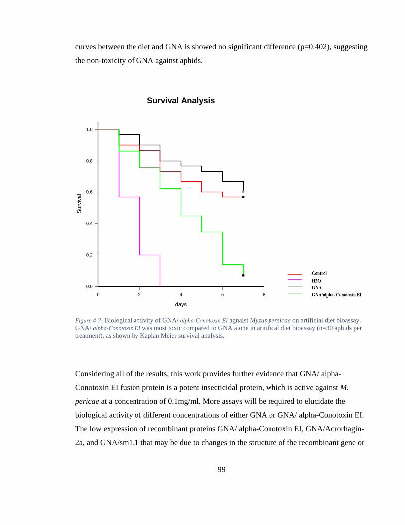
99
curves between the diet and GNA is showed no significant difference (p=0.402), suggesting
the non-toxicity of GNA against aphids.
Figure 4-7: Biological activity of GNA/ alpha-Conotoxin EI agnaist Myzus persicae on artificial diet bioassay.
GNA/ alpha-Conotoxin EI was most toxic compared to GNA alone in aritifical diet bioassay (n=30 aphids per
treatment), as shown by Kaplan Meier survival analysis.
Considering all of the results, this work provides further evidence that GNA/ alpha-
Conotoxin EI fusion protein is a potent insecticidal protein, which is active against M.
pericae at a concentration of 0.1mg/ml. More assays will be required to elucidate the
biological activity of different concentrations of either GNA or GNA/ alpha-Conotoxin EI.
The low expression of recombinant proteins GNA/ alpha-Conotoxin EI, GNA/Acrorhagin-
2a, and GNA/sm1.1 that may be due to changes in the structure of the recombinant gene or
Survival Analysis
days
0 2 4 6 8
Su
rviv
al
0.0
0.2
0.4
0.6
0.8
1.0
100
disappearance of the gene from yeast, would further investigation. Such investigations
would provide possible methods to improve yield during expression and purification.
Expression of foreign proteins at high levels depends on many factors. This study was
carried out with the yeast secretory expression vector PGAPZalph containing the zeocin
resistance cassette for selection and secretion signals alpha-factor. Thus, allows secretion of
heterologous protein into the culture medium, but not every protein produced is secreted in
soluble fractions. Particularly, studies have shown that some proteins are produced
intracellular, which often a cell lysis need process to release the cellular contents and
extraction to remove cell debris.
4.2.3 Transformed P.pastoris on Zeocin plates
The second aim of the study was to demonstrate differential expression of protein
intracellular and extracellular. The GNA/ alpha-Conotoxin EI and GNA/sm1.1 constructs
were subsequently transformed into X33 P. pastoris yeast using the pGAPZα constitutive
vector for expression as described previously. Few colonies were observed after 3 days
growth and 5 colonies were picked for expression screening. A PCR colony screening for
the presence of the toxin sequences was performed on all the unique colonies to confirm
maintenance of correct construct integrity. The presence of a 500 bp amplicon following
PCR in lanes 1-6 shows that all GNA/ alpha-Conotoxin EI clones contained the fusion
cassette. Conversely the lack of amplification in lane 7 demonstrated that the Sm1.1 version
was not successfully transformed into P. pastoris. (Figure 4-8). Single colonies from
various strains were inoculated in 10 mL YPD medium at 30°C for 5 days on shaker until
OD600 reached to 6. Culture supernatant collected during this period was then analysed by
SDS-PAGE gel electrophoresis and Western blotting (probed with anti-GNA antibodies) to
establish levels of expression.
101
Figure 4-8: PCR results from a colony screening of GNA/Sm1.1 pGAPZα and GNA/α-conotoxins EI pGAPZα
transformants. Lane M: 1 kb ladder. Lane 1-6: PCR reaction of the GNA/α-conotoxins EI transformed single
band of about 500 bp. Lane 7 contains PCR reaction of GNA/Sm1.1 showing no expression.. GNA as seen in
lane 8 produced intake band is about 500bp. Negative control was ran in the lane 9 and, has no band.
Expression of the fusion protein
Low value for cell density and dry weight was noted for the GNA/Sm1.1 pGAPZα and
GNA/ alpha-Conotoxin EI pGAPZα transformed yeast compared to GNA transformed
(Figure 4-9). A graph of the difference between all fusion proteins (Figure 4-9) shows
increased the optical density measured at 600 nm (OD600) readings of the yeast culture
growth at the initial 48 h, whilst recording low cell growth rate after 48h.
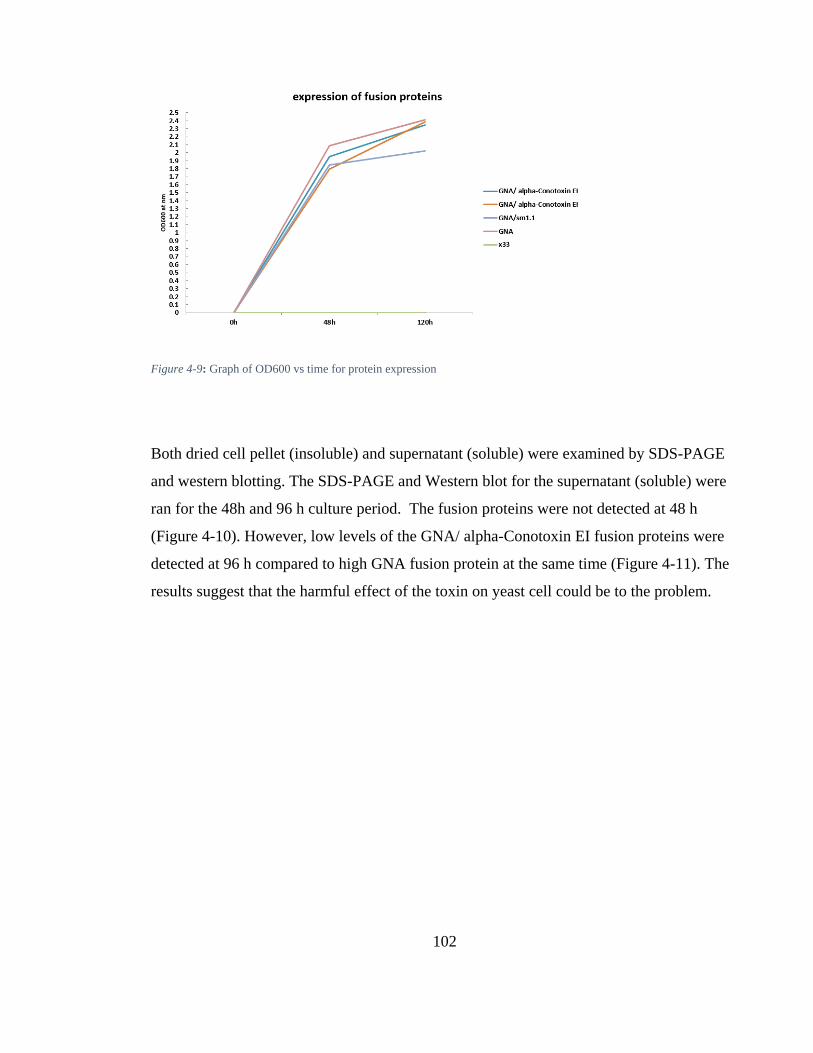
102
Figure 4-9: Graph of OD600 vs time for protein expression
Both dried cell pellet (insoluble) and supernatant (soluble) were examined by SDS-PAGE
and western blotting. The SDS-PAGE and Western blot for the supernatant (soluble) were
ran for the 48h and 96 h culture period. The fusion proteins were not detected at 48 h
(Figure 4-10). However, low levels of the GNA/ alpha-Conotoxin EI fusion proteins were
detected at 96 h compared to high GNA fusion protein at the same time (Figure 4-11). The
results suggest that the harmful effect of the toxin on yeast cell could be to the problem.

103
A)
B)
Figure 4-10: A) SDS- PAGE and Western blot (probed with anti-GNA ab): at 48h for expression screening of
GNA/ alpha-Conotoxin EI fusion protein. 25-μl samples from different colony culture Supernatant was loaded
onto SDS- PAGE gel, recombinant GNA was used as a positive standard. No bands were observed of all
Colonies. The Presence of the GNA at approximately the correct size (12 kDa) was visualised by SDS-PAGE,
followed by western blotting using anti-GNA antibodies (B).

104
Figure 4-11: SDS- PAGE and Western blot (probed with anti-GNA ab): at 96h, for expression screening of GNA/ alpha-
Conotoxin EI fusion protein. 25-μl samples from different colony culture Supernatant was loaded onto SDS- PAGE gel,
recombinant GNA was used as a positive standard. Colonies 1 and 6 show very low expression of fusion proteins, colony
8 shows intact band at approximately the correct size (12 kDa) for GNA, while line 9 represents the strain x33 as negative
control with no band.
In the Western blot image (figure 4-12), the single band on the left represents GNA/ alpha-
Conotoxin EI fusion protein from transformed yeast after cell lysis using the YeastBuster™
protein extraction. A high density band is also seen in the same gel for the GNA fusion
portion after lysis cell. However, Western blot image for the GNA/Sm1.1 fusion protein
transformed yeast after cell lysis using the YeastBuster™ protein extraction showed no
protein. Results not shown demonstrated poor yeast transformation.
Before examining the oral activity of these crude (GNA/ alpha-Conotoxin EI) recombinant
protein extracts the protein concentration was estimated by Bradfoard assay. High
concentration yield was detected from the expressed GNA/ alpha-Conotoxin EI fusion
protein after lysis cell using the YeastBuster™ Protein, approximately 5.874- µg/ µl. A
yield of 6.060 µg/ µl were also produced from the expressed GNA fusion protein after lysis
cell using the YeastBuster™ Protein.
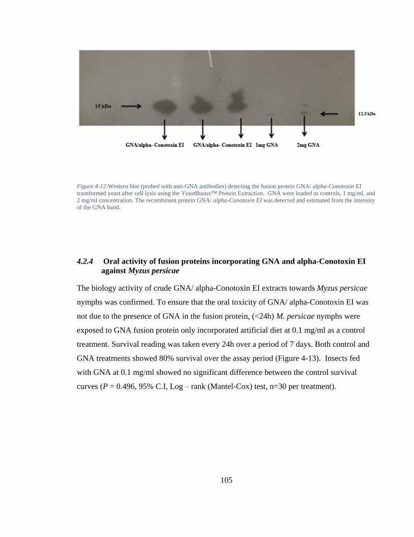
105
Figure 4-12:Western blot (probed with anti-GNA antibodies) detecting the fusion protein GNA/ alpha-Conotoxin EI
transformed yeast after cell lysis using the YeastBuster™ Protein Extraction. GNA were loaded as controls, 1 mg/ml, and
2 mg/ml concentration. The recombinant protein GNA/ alpha-Conotoxin EI was detected and estimated from the intensity
of the GNA band.
4.2.4 Oral activity of fusion proteins incorporating GNA and alpha-Conotoxin EI
against Myzus persicae
The biology activity of crude GNA/ alpha-Conotoxin EI extracts towards Myzus persicae
nymphs was confirmed. To ensure that the oral toxicity of GNA/ alpha-Conotoxin EI was
not due to the presence of GNA in the fusion protein, (<24h) M. persicae nymphs were
exposed to GNA fusion protein only incorporated artificial diet at 0.1 mg/ml as a control
treatment. Survival reading was taken every 24h over a period of 7 days. Both control and
GNA treatments showed 80% survival over the assay period (Figure 4-13). Insects fed
with GNA at 0.1 mg/ml showed no significant difference between the control survival
curves (P = 0.496, 95% C.I, Log – rank (Mantel-Cox) test, n=30 per treatment).
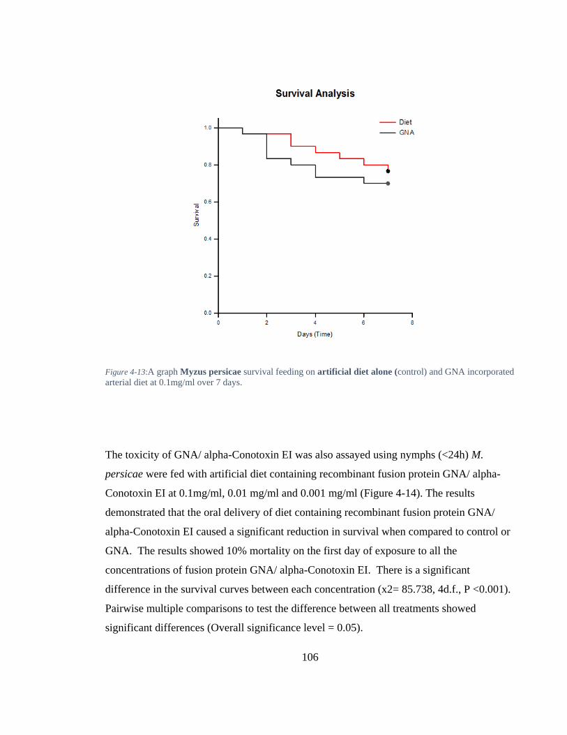
106
Figure 4-13:A graph Myzus persicae survival feeding on artificial diet alone (control) and GNA incorporated
arterial diet at 0.1mg/ml over 7 days.
The toxicity of GNA/ alpha-Conotoxin EI was also assayed using nymphs (<24h) M.
persicae were fed with artificial diet containing recombinant fusion protein GNA/ alpha-
Conotoxin EI at 0.1mg/ml, 0.01 mg/ml and 0.001 mg/ml (Figure 4-14). The results
demonstrated that the oral delivery of diet containing recombinant fusion protein GNA/
alpha-Conotoxin EI caused a significant reduction in survival when compared to control or
GNA. The results showed 10% mortality on the first day of exposure to all the
concentrations of fusion protein GNA/ alpha-Conotoxin EI. There is a significant
difference in the survival curves between each concentration (x2= 85.738, 4d.f., P <0.001).
Pairwise multiple comparisons to test the difference between all treatments showed
significant differences (Overall significance level = 0.05).
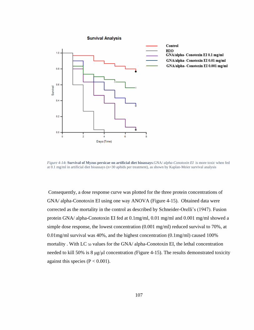
107
Figure 4-14: Survival of Myzus persicae on artificial diet bioassays.GNA/ alpha-Conotoxin EI is more toxic when fed
at 0.1 mg/ml in artificial diet bioassays (n=30 aphids per treatment), as shown by Kaplan-Meier survival analysis
Consequently, a dose response curve was plotted for the three protein concentrations of
GNA/ alpha-Conotoxin EI using one way ANOVA (Figure 4-15). Obtained data were
corrected as the mortality in the control as described by Schneider-Orelli’s (1947). Fusion
protein GNA/ alpha-Conotoxin EI fed at 0.1mg/ml, 0.01 mg/ml and 0.001 mg/ml showed a
simple dose response, the lowest concentration (0.001 mg/ml) reduced survival to 70%, at
0.01mg/ml survival was 40%, and the highest concentration (0.1mg/ml) caused 100%
mortality . With LC 50 values for the GNA/ alpha-Conotoxin EI, the lethal concentration
needed to kill 50% is 8 μg/μl concentration (Figure 4-15). The results demonstrated toxicity
against this species (P < 0.001).
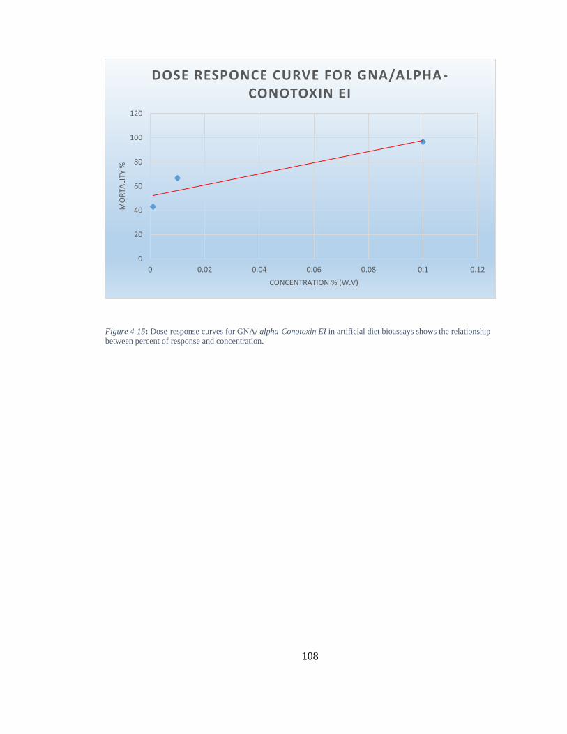
108
Figure 4-15: Dose-response curves for GNA/ alpha-Conotoxin EI in artificial diet bioassays shows the relationship
between percent of response and concentration.
0
20
40
60
80
100
120
0 0.02 0.04 0.06 0.08 0.1 0.12
MO
RTA
LITY
%
CONCENTRATION % (W.V)
DOSE RESPONCE CURVE FOR GNA/ALPHA-CONOTOXIN EI
109
4.3 Discussion
4.3.1 Design of expression vectors for synthetic genes
Three synthetic genes encoding GNA/alpha-conotoxin EI, GNA/sm1.1, or
GNA/Acrorhagin-2a were successfully designed by overlapping oligonucleotides with
modification of codon usage optimised for P pastoris expression. Expressed vectors
(pGAZαB) incorporating the above gene sequences were analysed by restriction digestion
using Xbai that nicks the sequence at the 5’ T C T A G A 3’ and Xhoi that nicks at 5’ T C T
G A G 3’ (Bendezú and de Boer, 2008). The combination of these two enzymes resulted in
a sequence with a single Xho1 restriction site fused to the three-alanine region of the C-
terminus of residues 1-105 of snowdrop lectin GNA-mys epitope. Each construct contained
a 6x His tag-coding sequence to aid subsequent purification of the recombinant protein by
nickel-affinity chromatography (Woestenenk et al., 2004). Constructs also expressed an
alpha factor secretary signal at the C-terminus of the GNA sequence, to direct expression of
the synthesised recombinant proteins (Brake and Van den Berg, 1991).
4.3.2 Expression of recombinant proteins: GNA/alpha-conotoxin EI, GNA/sm1.1,
GNA/Acrorhagin-2a
Since the aim of Chapter 4 was to clone, express and test the biological activity of α-
conotoxins (alpha-conotoxin EI; Sm1.1) and the acrorhagin-2a peptide, positive plasmids
containing GNA/alpha-conotoxin EI, GNA/sm1.1 and GNA/Acrorhagin-2a constructs from
E. coli were linearized and transformed into P. pastoris (X33) strain. Although previous
studies have demonstrated increased stability of fusion proteins when the toxins were fused
to the C-terminus of GNA due to reduced cleavage of the linker regions (Trung et al.,
2006a; Fitches et al., 2010), in the present study Western blot analyses showed that only
GNA/alpha-conotoxin EI was expressed, while GNA/sm1.1 and GNA/Acrorhagin-2a could
not be detected. The absorbance reading at 280nm for the expressed GNA/sm1.1 and
GNA/Acrorhagin-2a showed the presence of small amounts of protein, suggesting that
potential cleavage or degradation of these particular recombinant proteins in P pastoris
might have occurred. Even in the case of GNA/alpha-conotoxin EI, expression levels of the
recombinant protein were very low, which points to potential issues associated with the C-
110
terminus linking of the toxins to GNA. Other studies have shown that linker regions of
fusion proteins are susceptible to cleavage; in particular shorter linker regions are less
stable than longer regions (Gustavsson et al., 2001; Trung et al., 2006a), which could also
be a problem in the current study. Therefore, alternative linking of the toxins to GNA
through the N-terminus, as reported for the SFI toxins, or utilisation of IgG hinge, may
increase stability and hence improve expression; however this would need further
investigation. Another potential cause for the low expression seen in the present study
could be due to the strain of P. pastoris used for the expression of these fusion proteins.
Although all strains of P. pastoris have been shown to be suitable for expression of small
proteins with disulphide bonds (Cereghino and Cregg, 2000), proteolysis of resulting fusion
proteins is one of the major limitations of using this expression system for secretion of
expressed proteins (Gellissen, 2000; Fitches et al., 2004a). As a result, experiments to
optimise the expression of conotoxins using either different expression vectors or different
strains of P. pastoris might be needed. Furthermore, although the construct expressed a
functional signal peptide (alpha-factor) to direct extracellular secretion of the recombinant
protein, not all proteins are properly secreted and may be partially expressed intracellularly;
cell lysis would then be required in an attempt to recover the expressed protein. Initial
attempts to investigate the low protein yield showed that cell lysis did in fact increase the
concentration of GNA/alpha-conotoxin EI protein to 5.874 ug/ul, thus supporting the
hypothesis that the recombinant protein was not being secreted effectively. Additionally,
there could be potential toxic effects of the fusion protein on the yeast cells themselves,
resulting in cell death. Further work would need to be carried out to investigate possible
methods to improve yield during expression and purification.
4.3.3 Insecticidal activity of the recombinant fusion protein GNA/alpha-conotoxin
EI
The potential of using isolated components from marine cone snail venom as insecticidal
agents have received little attention to date since it was not previously possible to target
them to their sites of action within the pest insect. Thus the ability to express such proteins
(for example alpha-conotoxin EI, and sm1.1 from cone snails; Acrorhagin-2a from sea
anemone), as fusion proteins linked to GNA as a carrier molecule provides an opportunity
111
for developing these molecules as bio-insecticides since the GNA will deliver them to their
sites of action. This study demonstrated that oral delivery of GNA/alpha-conotoxin EI
fusion protein to neonate M. pericae induced toxic effects. The alpha-conotoxin EI toxin
fused to the C-terminus of GNA (GNA/alpha-conotoxin EI) presented significant levels of
toxicity when compared to GNA alone in artificial diet. Sub-lethal effects of GNA towards
this aphid species have also been reported by other authors (Nakasu et al., 2014). When fed
to M. pericae at 0.1 mg/ml, GNA caused only 30% mortality, while GNA/alpha-conotoxin
EI caused 90% mortality at the same concentration. Unfortunately, studies to test the effect
of alpha-conotoxin EI on aphids in the absence of GNA have not been carried out to date so
direct comparisons are not possible. However, previous studies have demonstrated that
GNA has a stabilizing effect on the toxin hairpin when fused to the C-terminus, in addition
to transporting the toxin across the midgut epithelium (Nakasu et al., 2014). The fact that
GNA/alpha-conotoxin EI was toxic to the aphids would suggest that the recombinant fusion
protein is being correctly folded, which is dependent on post-translational modification of
the fusion protein (Cregg et al., 2000). Furthermore, the results suggest that the orally
delivered GNA/alpha-conotoxin EI is resistant to gut digestion. Indeed, experiments to
determine the lethal concentration needed to induce 50% mortality suggests that this
molecule can be used to control aphids. The LC50 value for the GNA/ alpha-Conotoxin EI
was less than 0.01mg/ml concentration. However, the findings reported here provide
evidence that the GNA/alpha-conotoxin EI fusion protein is a potent insecticidal protein,
which is active against M. pericae.
4.3.4 Insecticides targeting nicotinic acetylcholine receptors
Most of the conotoxins peptides isolated from the venom of cone snails are encoded by a
large gene familly, which specifically targets voltage-gated sodium, potassium and calcium
ion channels, as well as other membrane receptors like nicotinic acetylcholine receptors,
NMDAR or 5-HT3R (Liu et al., 2012a). By constructing cDNA libaries from the venom
ducts of C. virgo, Conus imperialis, C. eburneus, and C. marmoreus collected from the
South China Sea, Liu et al. (2012) identified several novel conotoxin genes and
characterised the diversity of conotoxin superfamily genes from various Conus species,
112
(reviewed extensively in (Liu et al., 2012a). Neves et al. (2013) have identified disulfide-
rich conopeptides in C. crotchii that belongs to the A-, O1-, O2-, O3-, T- and D- gene
superfamilies. These conopeptides can block Ca2+ channels, act on nicotinic acetylcholine
receptors (nicotinic acetylcholine receptorss) and inhibit K+ channels. Identification of
alpha-5 and alpha-6 subunits which contribute to the nicotinic acetylcholine receptors
expressed on striatal dopaminergic terminals opened the possibility of developing nicotinic
acetylcholine receptor selective ligands active on dopaminergic (Olivera et al., 2008b).
The α - conotoxins contain 12 and 19 amino acids residues and use several disulphide
bonds to maintain the structure integrity. They are highly selective at blocking the nicotinic
acetylcholine receptors. However, inhibition of ligand-gated potassium, sodium, or calcium
ion channels across the synaptic membrane has also been reported. Most of the α-
conotoxins are competitive antagonists for nicotinic acetylcholine receptors, which bind
with high affinity to one or two binding sites of the receptors.
In summary, results from this chapter demonstrated that functional GNA/alpha-conotoxin
EI based fusion proteins can be effectively expressed in P. pastoris, albeit at low levels of
expression. Feeding studies suggested that the toxic effects of this neurotoxin within fusion
proteins were comparable to the correctly folded native conotoxin. However, the expression
of the other toxin peptides reported in this study needs to be investigated and potential
optimisation approaches utilised. Further work on oral activity against other insects was not
investigated, but the potential effect of proteolytic cleavage and digestion within the insect
midgut makes this an important field for further study. The presence of linker regions that
might be targeted by insect proteases needs to be established.
113
5 Chapter 5 General Discussion
5.1 Mechanisms of insecticide resistance in Myzus persicae
As the overall aim of the study was to develop biopesticides for the control of aphids that
are both sustainable and effective, it is important to understand problems encountered with
current synthetic pesticides, particularly the ability of aphids to evolve resistance to such
molecules. Below are the different mechanisms by which aphids are able to overcome the
toxic effects of these synthetic pesticides:
5.1.1 Overproduction of carboxylesterases
One of the major mechanisms of resistance in insect populations to insecticides is the
increased production of carboxylesterases, which has been associated to resistance to
organophosphate-, carbamate- and pyrethroid-based insecticides. It has been demonstrated
that esterases involved in organophosphate resistance in M. persicae can hydrolyse 1-
naphthyl acetates, substrates of the enzyme (Needham and Sawicki, 1971b; Voudouris et al.,
2015b), through the activity of carboxylesterases E4 and/or FE4. Carboxylesterases produced
by M. persicae act by sequestering organophosphate and carbamate insecticides before it is
transported to the insect nervous system (Devonshire and Field, 1991b; Feyereisen et al.,
2015b). Further studies also showed that amplification of the structural carboxylesterases
genes results in the enhanced production of the enzyme (Devonshire and Field, 1991b).
Amplification of the carboxylesterases genes correlates with the resistance phenotype
observed in some M. persicae (Field, 2000). Using fluorescence in-suithybridisation, Field
and Devonshire, demonstrated that autosomal chromosomal translocation 1,3 is linked to
amplification of carboxylesterase genes (Devonshire and Field, 1991b), which plays
important role in carboxylesterase related resistance.
5.1.2 Mutation of the acetylcholinesterase enzyme
Some classes of carbamate insecticides like dimethyl carbamate are resistant to esterase
activity and retain high insecticidal activity against M. persicae (Foster et al., 2002).
However, several populations of M. persicae that exhibit resistance against this insecticide
have been detected around the globe (Moores et al., 1994). Biochemical assay results showed

114
that reduced or lack of sensitivity of the target site of carbamate is the major contributing
factor in this resistance. Carbamate based insecticides target acetylcholinesterase enzyme,
which is the hydrolase regulating impulse transmission by targeting acetylcholine at the
synapses (Moores et al., 1994). Unfortunately, modification of the acetylcholinesterase
enzyme induced by mutation has been shown to cause more than 100 fold insensitivity to
dimethyl carbamate (Moores et al., 1994). Sequencing of the acetylcholinesterase gene
isolated from resistant M. persicae showed the presence of a point mutation that causes the
substitution of a single amino acid at S431F of the enzyme protein sequence (Andrews et al.,
2002; Nabeshima et al., 2003b). Subsequent expression of the modified enzyme in yeast
provided additional evidence that this has functional consequences, caused by reduced
binding of carbamate insecticides to the acetylcholinesterase enzyme (Benting and Nauen,
2004). The point mutation affecting S431F is located in the region of the enzyme sequence
that controls the orientation of ligands bound to the active site. Particularly, the substitution
of a serine with phenylalanine would inhibit the interaction of carbamate insecticides with
the catalytic region of the active site that is caused by change in the space and hydrophobicity
of the enzyme (Andrews et al., 2004, Nabeshima et al., 2003). An interesting aspect of this
substitution is the fact that non-aphid insects naturally have phenylalanine at the same site,
as this mutation reverts the aphid sequence back in line with other insects (Andrews et al.,
2004).
5.1.3 Mutation of the voltage-gated sodium channel
Although the enhanced production of carboxylesterases confers some resistance in aphids
against pyrethroid insecticides, it has been demonstrated that the initial mechanism of
resistance is by a “knockdown resistance” in the target site of the insecticide (Martinez‐
Torres et al., 1999). This knockdown effect is caused by mutations in voltage-dependent
sodium channels (Williamson et al., 1996). The voltage-dependent sodium channel is
important in the initiation and maintenance of membrane action potential in neurons. The
first report of mutation induced knockdown resistance to pyrethroids was the replacement of
leucine with phenylalanine (L1014F) in the trans-membrane segment of the sodium channel
(Martinez‐Torres et al., 1997; Martinez‐Torres et al., 1999). Moreover, M918T has been
115
identified in the intracellular linker of voltage-dependent sodium channel of M. persicae
(Eleftherianos et al., 2008). Computational modelling suggests that this mutation might cause
loss of polar interaction between pyrethroids (O'Reilly et al., 2006; Fontaine et al., 2011).
However, the resistance that might be caused by the two mutations remains to be
characterised.
5.1.4 Enhanced expression of the cytochrome P450, CYP6CY3 and Mutation of the
nicotinic acetylcholine receptor
The increased resistance to organophosphates, carbamates, and pyrethroids by M. persicae
has resulted in increased use of neonicotinoids, which are highly active against most of the
adaptive resistant mechanisms developed to the older insecticides (Nauen and Denholm,
2005). However, low level resistance in the form of natural tolerance subsequently appeared
in some populations of M. persicae, most importantly the tobacco adapted race of the aphid
(Nauen, 1995; Devine et al., 1996). Indeed, a clone of the M. persicae collected from Greece
has more than 60 fold resistance against topical application of different neonicotinoids
compared to wild type strain of the aphid (Philippou et al., 2010; Puinean et al., 2010).
Further investigations identified a P450-mediated detoxification as one of the major
mechanisms of resistance, but other potential mechanisms might exist (Philippou et al., 2009,
Puinean et al., 2010). Microarray and PCR analysis revealed the constitutive expression of
multiple CYP6CY3 genes because of gene amplification (Puinean et l., 2010).
Enhanced CYP6CY3 expression in M. persicae results in a low level of resistance against
neonicotinoids following topical application (Puinean et al., 2010), but the practical
significance of this resistance mechanism is limited because independently the penetration-
associated resistance cannot impair effectiveness of such insecticides when applied at the
correct concentration. Unfortunately, a clone of M. persicae collected from France was
shown to exhibit complete resistance to neonicotinoids (Bass et al., 2011, Slater et al., 2011).
Biochemical assays suggested that this particular clone utilises two major resistance
mechanisms, including cytochrome P450 detoxification, discussed above, and insensitivity
to piperonyl butoxide (Bass et al., 2011a; Slater et al., 2012). Microarray and quantitative
PCR analysis by the same researchers demonstrated CY6CY3 overexpression is significantly
116
similar to levels expressed by clones with low-level resistance to neonicotinoids (Bass et al.,
2011). This suggested that other mechanisms might underlie the complete resistance by the
clone from France. In particular modifications in the neonicotinoid target-site, were thought
to be an obvious mechanism. The nicotinic acetylcholine receptor targeted by neonicotinoids
is a neurotransmitter-gated ion channel that regulates nerve signalling via the post-synaptic
membrane (Bass et al., 2011a), but mutations and modifications to the neurotransmitter-gate
channel remains to be demonstrated.
5.2 Mechanisms of peptide targeting to ion channels
Several toxins derived from invertebrate predators have been shown to specifically target
voltage dependent sodium, potassium or calcium ion channels. Moreover, toxins with unique
targets like ryanodine channels and calcium activated potassium channels have been
identified (Fajloun et al., 2000). These toxins have been shown to interact with ion channels
through neurotoxin binding sites, which results in blocking of conductance or modulation of
gating. These binding sites are the determinants of phyla specificity, a critical aspect of
developing biopesticides. For instance, Hainantoxin, curtatoxins and Tx4 toxins have been
shown to target binding site-1 on sodium channels, binding site-3 on potassium channels and
binding site-4 on sodium channels, respectively (Stapleton et al., 1990a; De Lima et al., 2002;
Liu et al., 2013). In addition, biological activity of several other invertebrate neurotoxins,
(like δ-atracotoxins) on insect and vertebrate ion channels has been demonstrated. The
presence of conserved structural motifs in the domains of these voltage-gated ion channels
might explain this activity observed across phyla (Catterall, 1995). Research efforts are
currently directed towards the identification of molecular determinants of neurotoxin
selectivity and biological activity. However, accurate mapping of binding sites and
characterisation of the mechanisms of action of invertebrate derived toxins is needed before
their effective implementation in pest control. The mechanism and targets of a neurotoxin is
important, because most chemical compounds used for pest control, including those that have
been withdrawn due to environmental damage, are known to target the central nervous
system of crop pests. Chemical compounds like DTT block or inhibit the conductance of ion
channels such as voltage-gated sodium channel and glutamate gated chloride channels, whilst
others, such as pyrethroids act through high affinity binding to receptors like nicotinic

117
acetylcholine receptors and aminobutyric acid receptor (Tedford et al., 2004; Raymond-
Delpech et al., 2005). The use of predator derived toxins that target important and yet under-
utilised ion channels like the voltage gated calcium channels, a known target of the SFI
peptides investigated in this study could improve pest control. On one hand, the utilisation
of voltage dependent calcium channel could reduce insect resistance to pesticides due to
multiple targets (Eberl et al., 1998). While on the other, the essential role of this unique
channel in cell function could independently provide a potent target for pest control
(Kawasaki et al., 2002). Furthermore, the nicotinic acetylcholine receptors also investigated
in this study are known targets for some chemical insecticides such as imidacloprid (IMI).
Thus the ability to replace these synthetic chemical insecticides with equally potent
biopesticides could reduce the impact of pest control on the environment.
5.3 Mechanisms for delivering venom derived neuropeptides
One of the major challenges facing the technology of incorporating toxin peptides in
insecticides is the delivery of the peptides to the specific site of action. In the case of
neurotoxins, accessing the central nervous system of insects has faced many drawbacks,
attributed to the fact that ion channels targeted by such toxins are not accessible through the
conventional methods of applying insecticides. In a natural scenario, the predators producing
these toxins directly deliver their venoms to the central nervous system through injecting
them directly into the haemolymph; the active toxin is then transported systemically to the
ion channels where they cause paralysis and other forms of biological incapacitation.
However, a typical crop protection application relies on topical or oral application. Topically
applied venom peptides lack the ability to interact and move across the insect cuticle, because
the cuticle lacks surface receptors (Quistad et al., 1991b). Oral delivery exposes the toxin
peptides to proteases secreted by gut epithelial cells, whereby they are hydrolysed in the
midgut (Quistad et al., 1991b); furthermore, they also require to be actively transported
across the gut epithelium to the haemolymph. Interestingly, advances in biotechnology over
the past few decades have provided a number of tools to exploit these peptides in pest control.
Vector delivery using naturally occurring viruses and fusion to carrier proteins such as the
lectin GNA, reported in this study, are some of the actively studied mechanisms for toxin

118
delivery to the target sites.
5.4 Fusion with snowdrop lectin (GNA)
Several researchers have successfully incorporated predator venom-derived peptides into a
carrier protein like snowdrop lectin (GNA), and demonstrated comparable biological activity
to the ‘natural’ delivery of such venoms i.e. when injected into the haemolymph (Fitches and
Gatehouse, 1998; Fitches et al., 2004a; Fitches et al., 2004d; Trung et al., 2006a; Fitches et
al., 2010). However, irrespective of the promising biological activity on insects that are
reported by these studies following oral delivery of the toxin as a GNA-based fusion protein,
many research questions relating to mechanisms of action and specificity to targets or phyla
remains unanswered. For instance, the transport of GNA across gut epithelium through
receptor binding and subsequent endocytosis has been reported (Fitches et al., 2001), but the
biochemical mechanisms leading to transport to the haemolymph and subsequent cleavage
of the fused toxins remains poorly described. Moreover, a wide range of other proteins has
been reported to cross the gut epithelium of insects. Bovine serum albumin was reported to
cross the gut epithelium of lepidopteran into the haemolymph (Casartelli et al., 2005; Jeffers
et al., 2005), through a megalin-like receptor activated active transport (Casartelli et al.,
2008). Additionally, peptides of the storage protein, vicilin, have been reported to cross the
gut epithelium of bruchid seed weevils after feeding on seeds containing the peptides (Souza
et al., 2010), which suggests that similar mechanisms in GNA transport is possible.
The effectiveness of GNA/neurotoxin recombinant fusion protein is dependent on the
interaction between gut surface receptors and the carbohydrate-binding motif of GNA. GNA
denatured by boiling is no longer able to bind to these carbohydrate (Fitches and Gatehouse,
1998). Moreover, increased stability to gut digestion might be a critical aspect of GNA
delivery. Another important factor in using GNA as an efficient carrier protein for toxic
peptides is the stability of the recombinant fusion protein during translation and correct post-
translational modifications in expression vectors. In contrast to GNA, garlic lectins, which
are also mannose specific, are not effective carrier molecules since toxin/garlic lectin-based
fusion proteins have been shown not to be biologically active when fed orally to insects
(Fitches et al., 2008). Further, structural investigations have revealed that toxins fused to
119
garlic lectin failed to fold correctly and possess scrambled disulphide bonds (Fitches et al.,
2008). Interestingly, predator derived toxins contain many disulphide bonds, whilst GNA has
cysteine residues at position 29, 52 and 86, and a single intra disulphide brigde between the
cysteine at position 29 and 52 (Hester et al., 1995b; Tedford et al., 2004). It has therefore
been hypothesised that the activity of disulphide isomerases and other protein processing
machineries promotes folding of GNA in the endoplasmic reticulum in plant and yeast
expression vectors, which also enhances proper folding of fused toxin peptides. However,
the evidence to support this hypothesis needs to be established fully and represents a field for
further research. The toxic effects of the GNA based fusions proteins delivered to M. persicae
reduced survival and increased mortality at comparable levels reported for synthetic
insecticides currently utilised for control of this insect pest. These levels of toxicity provide
evidence that the peptides were correctly folded.
It is important to investigate the suitability of other carrier proteins in delivering neurotoxins
orally, which could increase the potential use of these peptides in pest control. The current
study used a GNA based fusion protein to deliver spider toxins, SFI1, SFI3, SFI5, SFI6, and
SFI8 and cone snail toxin, antagonists of nicotinic acetylcholine receptors to the gut of M.
persicae in order to investigate biological activity and toxicity. The results demonstrated that
recombinant proteins SFI/GNA investigated are likely to have insecticidal activity towards
this aphid species. The fusion protein SFI1/GNA caused the highest toxicity to M. persicae
when fed in a diet at 0.1mg/ml. The other four fusion proteins also caused reasonable toxicity,
observed as increased mortality. In general, all the fusion proteins tested in the present study
were able to cause at least 50% mortality at low concentrations (< 0.1mg/ml), making their
use as bioinsectides potentially viable. The nicotinic acetylcholine receptors antagonists
(alpha-conotoxin E1) also caused toxicity, resulting in increased mortality at 0.1mg/ml.
The biological activity of the tested toxins is dependent on stability within the gut
environment. GNA based fusion proteins reported in Chapter 3, and Chapter 4 were stable in
the gut environment and caused oral toxicity to aphids. It will be worthy of further
investigation to determine gut stability of the toxins in other potential pests. The recombinant
protein is only toxic if it is sufficiently resistant to gut proteolysis and is able to be transported
120
intact to the haemolymph. However, some investigators have suggested that once in the
haemolymph the active peptide has to be released for subsequent toxicity, but provided no
evidence to support their hypothesis (REF) In such cases they further speculate that if the
transported fusion proteins are too stable, this will prevent the release of free toxins to the
central nervous system; this possibility needs to be investigated.
5.5 Alternative delivery methods of toxins to insects
Another potential method of delivery is vector-based infection of pest using viruses like
Baculoviruses. Baculoviruses are specific arthropod viruses, which have been shown to
infect insects but not vertebrates or plants (Groner, 1986; Herniou et al., 2003; Zhu et al.,
2015). The virus can be transferred to mammalian cells, but the virus lacks replication
abilities in mammalian cells (Kost and Condreay, 2002). Unfortunately, the integration of
the SFI and other neuropeptides used in the present study (such as those derived from cone
snails or anemones) to any insect virus has not been investigated to date. Other spider toxins
such as ACTX have been demonstrated to cause mortality in insects following viral
expression. Given that these viral vectors have a pathogenic effect on their host insect, the
potency of any incorporated toxins could be enhanced. In addition, multiple expressions of
the SFI toxins could further increase mortality (Tedford et al., 2004). For instance, synergistic
expression of two types of scorpion derived neurotoxins in Baculoviruses caused significant
reduction in survival compared to individual expression of the toxins in the virus (Regev et
al., 2006). However, the results from this study is only limited to some species of insects,
including Diptera, Hymenoptera, and Lepidoptera (Moscardi, 1999; Erlandson et al., 2008).
It would be interesting to evaluate if such as approach would increase the toxicity of the
neuropeptides investigate in the present study to aphids.
5.6 Integrated pest control approaches
Other pest control strategies that may be applied with fusion proteins to reduce the
development of resistance to pesticides or that have been used in the control of M. persicae
are RNA interference, plant expression of toxic peptides and pyramiding toxins (Ferry et
121
al., 2006; Pitino et al., 2011). As with Bt expressing crop plants developed for the control
of Coleoptera and Lepidoptera pests, stacking several toxin-coding genes into a pyramid
for the control of aphid could reduce resistance by increasing the activity of individual
components. For instance, a transgenic corn has been developed expressing six insect
resistance genes, which provides protection against rootworm and herbicides
(AgroSciences, 2007). Additionally, protease inhibitors that might be less effective on their
own could prove invaluable when combined with SFI or other neurotoxic peptides such as
Sm1.1 or alpha-conontoxin E1, in a fusion protein, resulting in increased delivery of intact
molecules to the target site in the neuromuscular system. For example, transgenic cotton
expressing Bt fused to cowpea trypsin inhibitor has been commercially released in China
(He et al., 2008). The expression of semiochemicals in plants to repel or attract insects has
been established (Guerrero et al., 1997). However, application in bait traps to plants
carrying fusion proteins has not been explored, which could be an interesting aspect of
integrated pest control. This could ensure that target aphids feed on the insecticidal fusion
protein incorporating SFI and nicotinic acetylcholine receptors. Finally, RNA interference
is a proven effective strategy for the control of several species of insect pests (Baum et al.,
2007; Scharf, 2015). The double stranded RNA can either be expressed in the target crop
itself, or delivered as a biopesticide. The double stranded RNA interferes with the
expression of the insect target gene when ingested, often resulting in complete knock out of
important gene and causing mortality. This approach increases specificity of pest control
and reduces the risk of any potential non-target activity (Price and Gatehouse, 2008; Zhang
et al., 2015). Unfortunately there is little evidence demonstrating the efficacy of RNAi in
aphids. However, identification of potential genetic targets could be utilized in selecting
toxins for fusion proteins and RNA interference approaches for other insect pest species.
5.7 Conclusions
The amount of biopesticides currently used in pest control is still below 1% of the global
pesticide market, with environmentally damaging products constituting the majority of all
the insecticides (Whalon and Wingerd, 2003). There is increased need for biopesticides,

122
including those from invertebrate venoms. SFI toxins targeting the voltage-gated ion
channels and one of the neuropeptides targeting nicotinic acetylcholine receptors ( -
conotoxin E1) were functionally expressed in yeast cells as GNA-based fusion proteins,
which was significantly toxic to the aphid Myzus persicae following oral delivery. The
results of the current study suggest that these fusion proteins provide attractive and
environmentally friendly candidates for control of insect pests. Conducting further studies
could allow the use of these toxins independently or in combination with other pest control
methods. Biopesticides such as the fusion protein investigated in this study offers many
advantages over synthetic pesticides; they are usually sustainable and more
environmentally friendly than conventional pesticides, many of which are highly damaging
to the environment. Moreover, biopesticides are designed with high specificity, which only
affects particular pests. This is in contrast to chemical compounds that affect a very broad
spectrum of organisms, including beneficial insects, mammals and birds. Biopesticides will
also degrade in the environment with time and thus are less likely to contaminate soils and
waterways. In the past decade the use of biopesticides has increased significantly because
of extensive research efforts to enhance effectiveness and discover new candidates. As
biotechnological techniques for commercial production and storage has been developed, the
application of this category of pest control agents has improved. Although synthetic
products dominate the pesticide market, the use of biopesticides began during the
introduction of Bacillus thuringiensis spores in 1950. Interestingly, the use of biopesticides
as part of integrated pest management strategies has brought about an increase in the
demand for biopesticides. Currently, the drive for organic food and integrated pest control
are the major factors driving the growth of biopesticides. Moreover, the constant pressure
from synthetic compounds has caused an increase in resistance to such molecules within
insect pest populations. Since the protection of commercial crops is very important,
biopesticides represents a sustainable alternative.
In spite of the increasing research on predator venoms, more is yet to be done to
characterize the structure and functional activities of these compounds in a wide range of
insect pests. Combined, the venoms from different spider, snail and sea anemone species
has been estimated to exceed several thousands of compounds, but less than 1% have been
fully characterised. Indeed, many recent studies have described more than 500 peptide
123
venom compounds from these species. As a result, the SFI toxins and nicotinic
acetylcholine receptor antagonists could be particularly valuable in pest control. However,
they are yet to be fully characterized and second, they are a wide group of peptides that
share structural motifs with toxins encountered in other predators, such as snake toxins.
However, research effort is also needed to understand insect ion channels, which will allow
a better understanding of the diversity of the venom derived peptides and functions of the
insect ion channels. Another field of biotechnological importance is toxin gene detection.
The availability of innovations for genome sequencing of venomous animals or spiders in
general, including deep sequencing technologies could facilitate fast and easy discovery of
genes encoding these neurotoxins. Such genomic approaches will shed light on both
evolutionary mechanisms of venom development and genetic processes involved in the
development of novel functionalities. For all such reasons, research on spider, cone snail
and sea anemone venoms in any field is very important. From genetic studies to more
applied toxicological assessment like the current study, these predators should be targets for
future research on bio-insecticide discovery.
124
References:
(IRAG-UK), I.R.A.G. (2008) Guidelines for preventing and managing insecticide
resistance in aphids on potatoes
AgroSciences, D. (2007) 'Monsanto, Dow Agreement paves the way for industry’s first-
ever, eight-gene stacked offering in corn', Dow AgroScience News Room, 14th September.
Andrews, M., Callaghan, A., Bass, C., Williamson, M., Field, L. and Moores, G. (2002)
Proceedings of the XI International Symposium on Cholinergic Mechanisms: Function and
Dysfunction. St Moritz, Switzerland.
Anjum, F. (2014) Relative toxicity of insecticides to crucifer pests and their natural
enemies: interaction of insecticide and insect behaviours. Imperial College London and the
Diploma of Imperial College London
Anthony, N., Unruh, T., Ganser, D. and ffrench-Constant, R. (1998) 'Duplication of the Rdl
GABA receptor subunit gene in an insecticide-resistant aphid, Myzus persicae', Mol Gen
Genet, 260(2-3), pp. 165-75.
Attaran, A., Roberts, D.R., Curtis, C.F. and Kilama, W.L. (2000) 'Balancing risks on the
backs of the poor', Nat Med, 6(7), pp. 729-31.
Baker, J.R. (1994) Insect and related pests of flowers and foliage plants: some important,
common, and potential pests in the southeastern United States. North Carolina Cooperative
Extension Service.
Bass, C., Puinean, A.M., Andrews, M., Cutler, P., Daniels, M., Elias, J., Paul, V.L.,
Crossthwaite, A.J., Denholm, I. and Field, L.M. (2011a) 'Mutation of a nicotinic
acetylcholine receptor β subunit is associated with resistance to neonicotinoid insecticides
in the aphid Myzus persicae', Bmc Neuroscience, 12(1), p. 51.
Bass, C., Puinean, A.M., Andrews, M., Cutler, P., Daniels, M., Elias, J., Paul, V.L.,
Crossthwaite, A.J., Denholm, I., Field, L.M., Foster, S.P., Lind, R., Williamson, M.S. and
Slater, R. (2011b) 'Mutation of a nicotinic acetylcholine receptor beta subunit is associated
with resistance to neonicotinoid insecticides in the aphid Myzus persicae', BMC Neurosci,
12, p. 51.
Bass, C., Puinean, A.M., Andrews, M., Cutler, P., Daniels, M., Elias, J., Paul, V.L.,
Crossthwaite, A.J., Denholm, I., Field, L.M., Foster, S.P., Lind, R., Williamson, M.S. and
Slater, R. (2011c) 'Mutation of a nicotinic acetylcholine receptor beta subunit is associated
with resistance to neonicotinoid insecticides in the aphid Myzus persicae', Bmc
Neuroscience, 12.
Baum, J.A., Bogaert, T., Clinton, W., Heck, G.R., Feldmann, P., Ilagan, O., Johnson, S.,
Plaetinck, G., Munyikwa, T. and Pleau, M. (2007) 'Control of coleopteran insect pests
through RNA interference', Nature biotechnology, 25(11), pp. 1322-1326.
Baumgartner, P., Harper, K., Raemaekers, R.J., Durieux, A., Gatehouse, A.M., Davies,
H.V. and Taylor, M.A. (2003) 'Large-scale production and purification of recombinant
Galanthus nivalis agglutinin (GNA) expressed in the methylotrophic yeast Pichia pastoris',
Biotechnol Lett, 25(15), pp. 1281-5.
Beard, C.B., Dotson, E.M., Pennington, P.M., Eichler, S., Cordon-Rosales, C. and
Durvasula, R.V. (2001) 'Bacterial symbiosis and paratransgenic control of vector-borne
Chagas disease', Int J Parasitol, 31(5-6), pp. 621-7.
125
Beard, J. and Australian Rural Health Research, C. (2006) 'DDT and human health', Sci
Total Environ, 355(1-3), pp. 78-89.
Becker, S. and Terlau, H. (2008) 'Toxins from cone snails: properties, applications and
biotechnological production', Appl Microbiol Biotechnol, 79(1), pp. 1-9.
Beckmann, M. and Haack, K.-J. (2003) 'Insektizide für die Landwirtschaft: Chemische
Schädlingsbekämpfung [Insecticides for agriculture: Chemical pest control]', Chemie in
unserer Zeit, 37, pp. 88-97.
Beleboni, R.D., Pizzo, A.B., Fontana, A.C.K., Carolino, R.D.O.G., Coutinho-Netto, J. and
dos Santos, W.F. (2004) 'Spider and wasp neurotoxins: pharmacological and biochemical
aspects', European Journal of Pharmacology, 493(1-3), pp. 1-17.
Bende, N.S., Dziemborowicz, S., Herzig, V., Ramanujam, V., Brown, G.W., Bosmans, F.,
Nicholson, G.M., King, G.F. and Mobli, M. (2015) 'The insecticidal spider toxin SFI1 is a
knottin peptide that blocks the pore of insect voltage‐gated sodium channels via a large β‐hairpin loop', FEBS Journal, 282(5), pp. 904-920.
Bendezú, F.O. and de Boer, P.A. (2008) 'Conditional lethality, division defects, membrane
involution, and endocytosis in mre and mrd shape mutants of Escherichia coli', Journal of
bacteriology, 190(5), pp. 1792-1811.
Benting, J. and Nauen, R. (2004a) 'Biochemical evidence that an S431F mutation in
acetylcholinesterase-1 of Aphis gossypii mediates resistance to pirimicarb and omethoate',
Pest Management Science, 60(11), pp. 1051-1055.
Benting, J. and Nauen, R. (2004b) 'Biochemical evidence that an S431F mutation in
acetylcholinesterase‐1 of Aphis gossypii mediates resistance to pirimicarb and omethoate',
Pest management science, 60(11), pp. 1051-1055.
Blackman, R.L. and Eastop, V.F. ( 2006) Aphids on the World's Herbaceous Plants and
Shrubs. John Wiley & Sons, Chichester.
Bolter, C.J. and Jongsma, M.A. (1995) 'Colorado potato beetles (Leptinotarsa
decemlineata) adapt to proteinase inhibitors induced in potato leaves by methyl jasmonate',
Journal of Insect Physiology, 41(12), pp. 1071-1078.
Bosmans, F., Rash, L., Zhu, S.Y., Diochot, S., Lazdunski, M., Escoubas, P. and Tytgat, J.
(2006) 'Four novel tarantula toxins as selective modulators of voltage-gated sodium channel
subtypes', Molecular Pharmacology, 69(2), pp. 419-429.
Brake, A.J. and Van den Berg, J.A. (1991) 'DNA constructs containing a Kluyveromyces
alpha factor leader sequence for directing secretion of heterologous polypeptides'. Google
Patents.
Branton, W.D., Rudnick, M.S. and Zhou, Y. (1993a) 'Fatty acylated toxin structure',
Nature, 365, pp. 496-497.
Branton, W.D., Rudnick, M.S., Zhou, Y., Eccleston, E.D., Fields, G.B. and Bowers, L.D.
(1993b) 'Fatty Acylated Toxin Structure', Nature, 365(6446), pp. 496-497.
Bretthauer, R.K. and Castellino, F.J. (1999) 'Glycosylation of Pichia pastoris‐derived
proteins', Biotechnology and applied biochemistry, 30(3), pp. 193-200.
Brooks, E.M., Hines, E.R., (1999) 'Viral biopesticides for heliothine control—fact or
fiction.', Today's Life., pp. 38-44.
Capinera, J.l. (2001) 'Featured creatures. Entomology and Nematology ' February 2014.
07/10/2015. Department, University of Florida. Available at:
http://entnemdept.ufl.edu/creatures/veg/aphid/green_peach_aphid.htm.
126
Casartelli, M., Cermenati, G., Rodighiero, S., Pennacchio, F. and Giordana, B. (2008) 'A
megalin-like receptor is involved in protein endocytosis in the midgut of an insect (Bombyx
mori, Lepidoptera)', American Journal of Physiology-Regulatory, Integrative and
Comparative Physiology, 295(4), pp. R1290-R1300.
Casartelli, M., Corti, P., Leonardi, M.G., Fiandra, L., Burlini, N., Pennacchio, F. and
Giordana, B. (2005) 'Absorption of albumin by the midgut of a lepidopteran larva', Journal
of insect physiology, 51(8), pp. 933-940.
Catterall, W.A. (1995) 'Structure and function of voltage-gated ion channels', Annual
review of biochemistry, 64(1), pp. 493-531.
Catterall, W.A. (2001) 'A 3D view of sodium channels', Nature, 409(6823), pp. 988-9, 991.
Catterall, W.A. and Few, A.P. (2008) 'Calcium channel regulation and presynaptic
plasticity', Neuron, 59(6), pp. 882-901.
Cereghino, J.L. and Cregg, J.M. (2000) 'Heterologous protein expression in the
methylotrophic yeast Pichia pastoris', FEMS microbiology reviews, 24(1), pp. 45-66.
Cestele, S., Yarov-Yarovoy, V., Qu, Y., Sampieri, F., Scheuer, T. and Catterall, W.A.
(2006) 'Structure and function of the voltage sensor of sodium channels probed by a beta-
scorpion toxin', J Biol Chem, 281(30), pp. 21332-44.
Chanda, B. and Bezanilla, F. (2002) 'Tracking voltage-dependent conformational changes
in skeletal muscle sodium channel during activation', J Gen Physiol, 120(5), pp. 629-45.
Chrispeels, M.J. and Sadava, D.E. (2003) Plants, genes, and crop biotechnology. Jones &
Bartlett Learning.
Christou, P., Capell, T., Kohli, A., Gatehouse, J.A. and Gatehouse, A.M. (2006) 'Recent
developments and future prospects in insect pest control in transgenic crops', Trends in
plant science, 11(6), pp. 302-308.
Cordeiro Mdo, N., Diniz, C.R., Valentim Ado, C., von Eickstedt, V.R., Gilroy, J. and
Richardson, M. (1992) 'The purification and amino acid sequences of four Tx2 neurotoxins
from the venom of the Brazilian 'armed' spider Phoneutria nigriventer (Keys)', FEBS Lett,
310(2), pp. 153-6.
Council, E.c.t.t. (2006) EU Policy for a sustainable use of pesticides.
Cregg, J.M., Cereghino, J.L., Shi, J. and Higgins, D.R. (2000) 'Recombinant protein
expression in Pichia pastoris', Molecular biotechnology, 16(1), pp. 23-52.
Daly, R. and Hearn, M.T. (2005) 'Expression of heterologous proteins in Pichia pastoris: a
useful experimental tool in protein engineering and production', J Mol Recognit, 18(2), pp.
119-38.
De Lima, M., Stankiewicz, M., Hamon, A., De Figueiredo, S., Cordeiro, M., Diniz, C.,
Martin-Eauclaire, M.-F. and Pelhate, M. (2002) 'The toxin Tx4 (6-1) from the spider
Phoneutria nigriventer slows down Na+ current inactivation in insect CNS via binding to
receptor site 3', Journal of insect physiology, 48(1), pp. 53-61.
Decourtye, A., Devillers, J., Cluzeau, S., Charreton, M. and Pham-Delegue, M.H. (2004)
'Effects of imidacloprid and deltamethrin on associative learning in honeybees under semi-
field and laboratory conditions', Ecotoxicol Environ Saf, 57(3), pp. 410-9.
Devine, G.J., Harling, Z.K., Scarr, A.W. and Devonshire, A.L. (1996) 'Lethal and sublethal
effects of imidacloprid on nicotine‐tolerant Myzus nicotianae and Myzus persicae',
Pesticide Science, 48(1), pp. 57-62.
Devonshire, A.L. and Field, L.M. (1991a) 'Gene Amplification and Insecticide Resistance',
Annual Review of Entomology, 36, pp. 1-23.
127
Devonshire, A.L. and Field, L.M. (1991b) 'Gene amplification and insecticide resistance',
Annual review of entomology, 36(1), pp. 1-21.
Dill, A., Thomas, S.G., Hu, J., Steber, C.M. and Sun, T.P. (2004) 'The Arabidopsis F-box
protein SLEEPY1 targets gibberellin signaling repressors for gibberellin-induced
degradation', Plant Cell, 16(6), pp. 1392-405.
Diniz, M.R., Paine, M.J., Diniz, C.R., Theakston, R.D. and Crampton, J.M. (1993)
'Sequence of the cDNA coding for the lethal neurotoxin Tx1 from the Brazilian "armed"
spider Phoneutria nigriventer predicts the synthesis and processing of a preprotoxin', J Biol
Chem, 268(21), pp. 15340-2.
Down, R.E., Fitches, E.C., Wiles, D.P., Corti, P., Bell, H.A., Gatehouse, J.A. and Edwards,
J.P. (2006) 'Insecticidal spider venom toxin fused to snowdrop lectin is toxic to the peach-
potato aphid, Myzus persicae (Hemiptera: Aphididae) and the rice brown planthopper,
Nilaparvata lugens (Hemiptera: Delphacidae)', Pest Manag Sci, 62(1), pp. 77-85.
Down, R.E., Gatehouse, A.M.R., Hamilton, W.D.O. and Gatehouse, J.A. (1996) 'Snowdrop
lectin inhibits development and decreases fecundity of the glasshouse potato aphid
(Aulacorthum solani) when administered in vitro and via transgenic plants both in
laboratory and glasshouse trials', Journal of Insect Physiology, 42(11-12), pp. 1035-1045.
Eberl, D.F., Ren, D., Feng, G., Lorenz, L.J., Van Vactor, D. and Hall, L.M. (1998) 'Genetic
and developmental characterization of Dmca1D, a calcium channel α1 subunit gene in
Drosophila melanogaster', Genetics, 148(3), pp. 1159-1169.
Eleftherianos, I., Foster, S., Williamson, M. and Denholm, I. (2008) 'Characterization of the
M918T sodium channel gene mutation associated with strong resistance to pyrethroid
insecticides in the peach-potato aphid, Myzus persicae (Sulzer)', Bulletin of entomological
research, 98(02), pp. 183-191.
Erlandson, M., Mahy, B. and van Regenmortel, M. (2008) 'Insect pest control by viruses',
Encyclopedia of Virology, 3, pp. 125-133.
Escoubas, P., Diochot, S. and Corzo, G. (2000) 'Structure and pharmacology of spider
venom neurotoxins', Biochimie, 82(9-10), pp. 893-907.
Escoubas, P., Sollod, B. and King, G.F. (2006) 'Venom landscapes: Mining the complexity
of spider venoms via a combined cDNA and mass spectrometric approach', Toxicon, 47(6),
pp. 650-663.
Fajloun, Z., Carlier, E., Lecomte, C., Geib, S., di Luccio, E., Bichet, D., Mabrouk, K.,
Rochat, H., De Waard, M. and Sabatier, J.M. (2000) 'Chemical synthesis and
characterization of Pi1, a scorpion toxin from Pandinus imperator active on K+ channels',
European Journal of Biochemistry, 267(16), pp. 5149-5155.
Febvay, G., Delobel, B. and Rahbe, Y. (1988) 'Influence of the Amino-Acid Balance on the
Improvement of an Artificial Diet for a Biotype of Acyrthosiphon-Pisum (Homoptera,
Aphididae)', Canadian Journal of Zoology-Revue Canadienne De Zoologie, 66(11), pp.
2449-2453.
Ferry, N., Edwards, M., Gatehouse, J., Capell, T., Christou, P. and Gatehouse, A. (2006)
'Transgenic plants for insect pest control: a forward looking scientific perspective',
Transgenic research, 15(1), pp. 13-19.
Feyereisen, R. (1995) 'Molecular biology of insecticide resistance', Toxicol Lett, 82-83, pp.
83-90.
128
Feyereisen, R., Dermauw, W. and Van Leeuwen, T. (2015a) 'Genotype to phenotype, the
molecular and physiological dimensions of resistance in arthropods', Pestic Biochem
Physiol, 121, pp. 61-77.
Feyereisen, R., Dermauw, W. and Van Leeuwen, T. (2015b) 'Genotype to phenotype, the
molecular and physiological dimensions of resistance in arthropods', Pesticide
Biochemistry and Physiology.
Field, L. (2000) 'Methylation and expression of amplified esterase genes in the aphid
Myzus persicae (Sulzer)', Biochem. J, 349, pp. 863-868.
Fitches, E., Audsley, N., Gatehouse, J.A. and Edwards, J.P. (2002) 'Fusion proteins
containing neuropeptides as novel insect contol agents: snowdrop lectin delivers fused
allatostatin to insect haemolymph following oral ingestion', Insect Biochemistry and
Molecular Biology, 32(12), pp. 1653-1661.
Fitches, E., Edwards, M.G., Mee, C., Grishin, E., Gatehouse, A.M., Edwards, J.P. and
Gatehouse, J.A. (2004a) 'Fusion proteins containing insect-specific toxins as pest control
agents: snowdrop lectin delivers fused insecticidal spider venom toxin to insect
haemolymph following oral ingestion', Journal of Insect Physiology, 50(1), pp. 61-71.
Fitches, E., Edwards, M.G., Mee, C., Grishin, E., Gatehouse, A.M., Edwards, J.P. and
Gatehouse, J.A. (2004b) 'Fusion proteins containing insect-specific toxins as pest control
agents: snowdrop lectin delivers fused insecticidal spider venom toxin to insect
haemolymph following oral ingestion', J Insect Physiol, 50(1), pp. 61-71.
Fitches, E., Edwards, M.G., Mee, C., Grishin, E., Gatehouse, A.M.R., Edwards, J.P. and
Gatehouse, J.A. (2004c) 'Fusion proteins containing insect-specific toxins as pest control
agents: snowdrop lectin delivers fused insecticidal spider venom toxin to insect
haemolymph following oral ingestion', Journal of Insect Physiology, 50(1), pp. 61-71.
Fitches, E. and Gatehouse, J.A. (1998) 'A comparison of the short and long term effects of
insecticidal lectins on the activities of soluble and brush border enzymes of tomato moth
larvae (Lacanobia oleracea)', Journal of Insect Physiology, 44(12), pp. 1213-1224.
Fitches, E., Wilkinson, H., Bell, H., Bown, D.P., Gatehouse, J.A. and Edwards, J.P.
(2004d) 'Cloning, expression and functional characterisation of chitinase from larvae of
tomato moth (Lacanobia oleracea): a demonstration of the insecticidal activity of insect
chitinase', Insect biochemistry and molecular biology, 34(10), pp. 1037-1050.
Fitches, E., Woodhouse, S.D., Edwards, J.P. and Gatehouse, J.A. (2001) 'In vitro and in
vivo binding of snowdrop (Galanthus nivalis agglutinin; GNA) and jackbean (Canavalia
ensiformis; Con A) lectins within tomato moth (Lacanobia oleracea) larvae; mechanisms of
insecticidal action', Journal of Insect Physiology, 47(7), pp. 777-787.
Fitches, E.C., Bell, H.A., Powell, M.E., Back, E., Sargiotti, C., Weaver, R.J. and
Gatehouse, J.A. (2010) 'Insecticidal activity of scorpion toxin (ButaIT) and snowdrop lectin
(GNA) containing fusion proteins towards pest species of different orders', Pest
management science, 66(1), pp. 74-83.
Fitches, E.C., Pyati, P., King, G.F. and Gatehouse, J.A. (2012) 'Fusion to snowdrop lectin
magnifies the oral activity of insecticidal omega-Hexatoxin-Hv1a peptide by enabling its
delivery to the central nervous system', PLoS One, 7(6), p. e39389.
Fontaine, S., Caddoux, L., Brazier, C., Bertho, C., Bertolla, P., Micoud, A. and Roy, L.
(2011) 'Uncommon associations in target resistance among French populations of Myzus
persicae from oilseed rape crops', Pest management science, 67(8), pp. 881-885.
129
Foster, S.P., Denholm, I. and Devonshire, A.L. (2002) 'Field‐simulator studies of
insecticide resistance to dimethylcarbamates and pyrethroids conferred by metabolic‐and
target site‐based mechanisms in peach‐potato aphids, Myzus persicae (Hemiptera:
Aphididae)', Pest management science, 58(8), pp. 811-816.
Foster, S.P., Tomiczek, M., Thompson, R., Denholm, I., Poppy, G., Kraaijeveld, A.R. and
Powell, W. (2007) 'Behavioural side-effects of insecticide resistance in aphids increase
their vulnerability to parasitoid attack', Animal Behaviour, 74, pp. 621-632.
Froy, O., Zilberberg, N., Chejanovsky, N., Anglister, J., Loret, E., Shaanan, B., Gordon, D.
and Gurevitz, M. (2000) 'Scorpion neurotoxins: structure/function relationships and
application in agriculture', Pest Management Science, 56(5), pp. 472-474.
Fuxa, J.R. (1991) 'Insect control with baculoviruses', Biotechnol Adv, 9(3), pp. 425-42.
Garabrant, D.H., Held, J., Langholz, B., Peters, J.M. and Mack, T.M. (1992) 'DDT and
related compounds and risk of pancreatic cancer', J Natl Cancer Inst, 84(10), pp. 764-71.
Garcíaa, F., Villegasb, E., Ortiza, E. and Corzoa, G. (2015) 'Structural Diversity and
Basic/Acidic Residue Balance of Active Cysteine-Rich Insecticidal Peptides from Spiders'.
Gellissen, G. (2000) 'Heterologous protein production in methylotrophic yeasts', Applied
microbiology and biotechnology, 54(6), pp. 741-750.
Gleeson, M.A., White, C.E., Meininger, D.P. and Komives, E.A. (1998) 'Generation of
protease-deficient strains and their use in heterologous protein expression', Methods Mol
Biol, 103, pp. 81-94.
Goldin, A.L., Barchi, R.L., Caldwell, J.H., Hofmann, F., Howe, J.R., Hunter, J.C., Kallen,
R.G., Mandel, G., Meisler, M.H., Netter, Y.B., Noda, M., Tamkun, M.M., Waxman, S.G.,
Wood, J.N. and Catterall, W.A. (2000) 'Nomenclature of voltage-gated sodium channels',
Neuron, 28(2), pp. 365-368.
Gould, W.P. and Jeanne, R.L. (1984) 'Polistes Wasps (Hymenoptera, Vespidae) as Control
Agents for Lepidopterous Cabbage Pests', Environmental Entomology, 13(1), pp. 150-156.
Gray, W.R., Luque, A., Olivera, B.M., Barrett, J. and Cruz, L.J. (1981) 'Peptide toxins from
Conus geographus venom', J Biol Chem, 256(10), pp. 4734-40.
Grishin, E. (1999) 'Polypeptide neurotoxins from spider venoms', Eur J Biochem, 264(2),
pp. 276-80.
Grishin, E.V., Volkova, T.M., Arseniev, A.S., Reshetova, O.S., Onoprienko, V.V.,
Magazanic, L.G., Antonov, S.M. and Fedorova, I.M. (1986) 'Structure-Functional
Characterization of Argiopine - an Ion Channel Blocker from the Venom of Spider
Argiope-Lobata', Bioorganicheskaya Khimiya, 12(8), pp. 1121-1124.
Groebe, D.R., Dumm, J.M., Levitan, E.S. and Abramson, S.N. (1995) 'Alpha-Conotoxins
Selectively Inhibit One of the 2 Acetylcholine Binding-Sites of Nicotinic Receptors',
Molecular Pharmacology, 48(1), pp. 105-111.
Groner, A. (1986) 'Specificity and safety of baculoviruses'.
Guerrero, A., Feixas, J., Pajares, J., Wadhams, L., Pickett, J. and Woodcock, C. (1997)
'Semiochemically induced inhibition of behaviour of Tomicus destruens
(Woll.)(Coleoptera: Scolytidae)', Naturwissenschaften, 84(4), pp. 155-157.
Gustavsson, M., Lehtiö, J., Denman, S., Teeri, T.T., Hult, K. and Martinelle, M. (2001)
'Stable linker peptides for a cellulose-binding domain–lipase fusion protein expressed in
Pichia pastoris', Protein engineering, 14(9), pp. 711-715.
130
He, H., Wang, Z. and Zhang, Y. (2008) 'Monitoring BT resistance in the Field: China as a
case study. In: Environmental Impact of Genetically Modified/Novel Crop, N. Ferry and
A.M.R. Gatehouse, eds.', CAB International, wallingford, UK, in press.
Heathcote, G.D. (1962) 'Suitability of some plant hosts for the development of the peach-
potato aphid, Myzus persicae', Entomologia Experimentalis et Applicata, 5, pp. 114-118.
Herniou, E.A., Olszewski, J.A., Cory, J.S. and O'Reilly, D.R. (2003) 'The genome sequence
and evolution of baculoviruses', Annual Review of Entomology, 48(1), pp. 211-234.
Hester, G., Kaku, H., Goldstein, I.J. and Wright, C.S. (1995a) 'Structure of Mannose-
Specific Snowdrop (Galanthus-Nivalis) Lectin Is Representative of a New Plant Lectin
Family', Nature Structural Biology, 2(6), pp. 472-479.
Hester, G., Kaku, H., Goldstein, I.J. and Wright, C.S. (1995b) 'Structure of mannose-
specific snowdrop (Galanthus nivalis) lectin is representative of a new plant lectin family',
Nature structural & molecular biology, 2(6), pp. 472-479.
Hodgkin, A.L. and Huxley, A.F. (1952) 'A quantitative description of membrane current
and its application to conduction and excitation in nerve', J Physiol, 117(4), pp. 500-44.
Honma, T., Hasegawa, Y., Ishida, M., Nagai, H., Nagashima, Y. and Shiomi, K. (2005)
'Isolation and molecular cloning of novel peptide toxins from the sea anemone Antheopsis
maculata', Toxicon, 45(1), pp. 33-41.
Honma, T. and Shiomi, K. (2006) 'Peptide toxins in sea anemones: structural and functional
aspects', Mar Biotechnol (NY), 8(1), pp. 1-10.
Isbister, G.K. and White, J. (2004) 'Clinical consequences of spider bites: recent advances
in our understanding', Toxicon, 43(5), pp. 477-492.
Jain, K.K. (2000) 'An evaluation of intrathecal ziconotide for the treatment of chronic pain',
Expert Opin Investig Drugs, 9(10), pp. 2403-10.
Janes, R.W. (2005) 'alpha-Conotoxins as selective probes for nicotinic acetylcholine
receptor subclasses', Curr Opin Pharmacol, 5(3), pp. 280-92.
Jeffers, L.A., Thompson, D.M., Ben‐Yakir, D. and Roe, R.M. (2005) 'Movement of
proteins across the digestive system of the tobacco budworm, Heliothis virescens',
Entomologia experimentalis et applicata, 117(2), pp. 135-146.
Kawai, N., Miwa, A., Shimazaki, K., Sahara, Y., Robinson, H.P. and Nakajima, T. (1991)
'Spider toxin and the glutamate receptors', Comp Biochem Physiol C, 98(1), pp. 87-95.
Kawasaki, F., Collins, S.C. and Ordway, R.W. (2002) 'Synaptic calcium-channel function
in Drosophila: analysis and transformation rescue of temperature-sensitive paralytic and
lethal mutations of cacophony', The Journal of neuroscience, 22(14), pp. 5856-5864.
King, G.F. (2004) 'The wonderful world of spiders: preface to the special Toxicon issue on
spider venoms', Toxicon, 43(5), pp. 471-5.
King, G.F. (2007) 'Modulation of insect Ca-v channels by peptidic spider toxins', Toxicon,
49(4), pp. 513-530.
Kohn, A.J. (1998) 'Superfamily conoidea.', PL Beesley, GJB Ross, A Wells (Eds.),
Mollusca: The southern synthesis. Fauna of Australia, CSIRO Publishing, Melbourne, pp.
846–854.
Kost, T.A. and Condreay, J.P. (2002) 'Recombinant baculoviruses as mammalian cell gene-
delivery vectors', Trends in biotechnology, 20(4), pp. 173-180.
Kozlov, S. and Grishin, E. (2005) 'Classification of spider neurotoxins using structural
motifs by primary structure features. Single residue distribution analysis and pattern
analysis techniques', Toxicon, 46(6), pp. 672-686.
131
Kozlov, S. and Grishin, E. (2012) 'Convenient nomenclature of cysteine-rich polypeptide
toxins from sea anemones', Peptides, 33(2), pp. 240-4.
Kumar, P.S., Kumar, D.S. and Umamaheswari, S. (2015) 'A perspective on toxicology of
Conus venom peptides', Asian Pac J Trop Med, 8(5), pp. 337-51.
Kuroli, G. and Lantos, Z.S. (2006) Long-term study of flight activity and abundance of
wheat colonizing aphid species. Cereal Research Communication.
Lewis, R.J., Dutertre, S., Vetter, I. and Christie, M.J. (2012) 'Conus venom peptide
pharmacology', Pharmacol Rev, 64(2), pp. 259-98.
Lewis, R.J. and Garcia, M.L. (2003) 'Therapeutic potential of venom peptides', Nat Rev
Drug Discov, 2(10), pp. 790-802.
Lipkin, A., Kozlov, S., Nosyreva, E., Blake, A., Windass, J. and Grishin, E. (2002) 'Novel
insecticidal toxins from the venom of the spider Segestria florentina', Toxicon, 40(2), pp.
125-130.
Liu, Z., Cai, T., Zhu, Q., Deng, M., Li, J., Zhou, X., Zhang, F., Li, D., Li, J. and Liu, Y.
(2013) 'Structure and function of hainantoxin-III, a selective antagonist of neuronal
tetrodotoxin-sensitive voltage-gated sodium channels isolated from the Chinese bird spider
Ornithoctonus hainana', Journal of Biological Chemistry, 288(28), pp. 20392-20403.
Liu, Z., Li, H., Liu, N., Wu, C., Jiang, J., Yue, J., Jing, Y. and Dai, Q. (2012a) 'Diversity
and evolution of conotoxins in Conus virgo, Conus eburneus, Conus imperialis and Conus
marmoreus from the South China Sea', Toxicon, 60(6), pp. 982-989.
Liu, Z., Li, H., Liu, N., Wu, C., Jiang, J., Yue, J., Jing, Y. and Dai, Q. (2012b) 'Diversity
and evolution of conotoxins in Conus virgo, Conus eburneus, Conus imperialis and Conus
marmoreus from the South China Sea', Toxicon, 60(6), pp. 982-9.
Loret, E.P., Hammock, B.D. (1993) 'Structure and neurotoxicity of scorpion venom.', In:
Brownell, P.H., Polis, G. (Eds.), Scorpion Biology and Research. Oxford University Press,
Oxford, UK, pp. 389–399.
Luo, S., Zhangsun, D. and Tang, K. (2005) 'Functional GNA expressed in Escherichia coli
with high efficiency and its effect on Ceratovacuna lanigera Zehntner', Appl Microbiol
Biotechnol, 69(2), pp. 184-91.
Martinez-Torres, D., Foster, S.P., Field, L.M., Devonshire, A.L. and Williamson, M.S.
(1999) 'A sodium channel point mutation is associated with resistance to DDT and
pyrethroid insecticides in the peach-potato aphid, Myzus persicae (Sulzer) (Hemiptera:
Aphididae)', Insect Mol Biol, 8(3), pp. 339-46.
Martinez‐Torres, D., Devonshire, A.L. and Williamson, M.S. (1997) 'Molecular studies of
knockdown resistance to pyrethroids: cloning of domain II sodium channel gene sequences
from insects', Pesticide Science, 51(3), pp. 265-270.
Martinez‐Torres, D., Foster, S., Field, L., Devonshire, A. and Williamson, M. (1999) 'A
sodium channel point mutation is associated with resistance to DDT and pyrethroid
insecticides in the peach‐potato aphid, Myzus persicae (Sulzer)(Hemiptera: Aphididae)',
Insect molecular biology, 8(3), pp. 339-346.
Martinez, J.S., Olivera, B.M., Gray, W.R., Craig, A.G., Groebe, D.R., Abramson, S.N. and
McIntosh, J.M. (1995) 'alpha-Conotoxin EI, a new nicotinic acetylcholine receptor
antagonist with novel selectivity', Biochemistry, 34(44), pp. 14519-26.
McCarthy, S. (1993) 'Congress takes a look at estrogenic pesticides and breast cancer', J.
Pesticide Reform, 13 (4), 25.
132
Meehan, C.J., Olson, E.J., Reudink, M.W., Kyser, T.K. and Curry, R.L. (2009) 'Herbivory
in a spider through exploitation of an ant-plant mutualism', Curr Biol, 19(19), pp. R892-3.
Messerli, S.M. and Greenberg, R.M. (2006) 'Cnidarian toxins acting on voltage-gated ion
channels', Marine Drugs, 4(3), pp. 70-81.
Metcalf, R.L. and Luckmann, W.H. (1994
) Introduction to Insect Pest Management. 3rd edn.
Moczydlowski, E., Olivera, B.M., Gray, W.R. and Strichartz, G.R. (1986) 'Discrimination
of muscle and neuronal Na-channel subtypes by binding competition between
[3H]saxitoxin and mu-conotoxins', Proc Natl Acad Sci U S A, 83(14), pp. 5321-5.
Moores, G.D., Devine, G.J. and Devonshire, A.L. (1994) 'Insecticide-insensitive
acetylcholinesterase can enhance esterase-based resistance in Myzus persicae and Myzus
nicotianae', Pesticide Biochemistry and Physiology, 49(2), pp. 114-120.
Moran, Y., Gordon, D. and Gurevitz, M. (2009) 'Sea anemone toxins affecting voltage-
gated sodium channels - molecular and evolutionary features', Toxicon, 54(8), pp. 1089-
1101.
Moran, Y., Kahn, R., Cohen, L., Gur, M., Karbat, I., Gordon, D. and Gurevitz, M. (2007)
'Molecular analysis of the sea anemone toxin Av3 reveals selectivity to insects and
demonstrates the heterogeneity of receptor site-3 on voltage-gated Na+ channels',
Biochemical Journal, 406, pp. 41-48.
Moscardi, F. (1999) 'Assessment of the application of baculoviruses for control of
Lepidoptera', Annual review of entomology, 44(1), pp. 257-289.
Nabeshima, T., Kozaki, T., Tomita, T. and Kono, Y. (2003a) 'An amino acid substitution
on the second acetylcholinesterase in the pirimicarb-resistant strains of the peach potato
aphid, Myzus persicae', Biochem Biophys Res Commun, 307(1), pp. 15-22.
Nabeshima, T., Kozaki, T., Tomita, T. and Kono, Y. (2003b) 'An amino acid substitution
on the second acetylcholinesterase in the pirimicarb-resistant strains of the peach potato
aphid, Myzus persicae', Biochemical and biophysical research communications, 307(1), pp.
15-22.
Nakasu, E.Y., Edwards, M.G., Fitches, E., Gatehouse, J.A. and Gatehouse, A.M. (2014)
'Transgenic plants expressing ω-ACTX-Hv1a and snowdrop lectin (GNA) fusion protein
show enhanced resistance to aphids', Frontiers in plant science, 5.
Nauen, R. (1995) 'Behaviour modifying effects of low systemic concentrations of
imidacloprid on Myzus persicae with special reference to an antifeeding response',
Pesticide Science, 44(2), pp. 145-153.
Nauen, R. and Denholm, I. (2005) 'Resistance of insect pests to neonicotinoid insecticides:
current status and future prospects', Archives of insect biochemistry and physiology, 58(4),
pp. 200-215.
Nault, L.R. (1997) 'Arthropod transmission of plant viruses: A new synthesis', Annals of the
Entomological Society of America, 90(5), pp. 521-541.
Needham, P.H. and Sawicki, R.M. (1971a) 'Diagnosis of Resistance to Organophosphorus
Insecticides in Myzus-Persicae (Sulz)', Nature, 230(5289), pp. 125-&.
Needham, P.H. and Sawicki, R.M. (1971b) 'Diagnosis of resistance to organophosphorus
insecticides in Myzus persicae (Sulz.)'.
Neves, J., Campos, A., Osorio, H., Antunes, A. and Vasconcelos, V. (2013) 'Conopeptides
from Cape Verde Conus crotchii', Marine Drugs, 11(6), pp. 2203-2215.
133
Newcomb, R., Palma, A., Fox, J., Gaur, S., Lau, K., Chung, D., Cong, R., Bell, J.R., Horne,
B., Nadasdi, L. and et al. (1995) 'SNX-325, a novel calcium antagonist from the spider
Segestria florentina', Biochemistry, 34(26), pp. 8341-7.
Nicholson, G.M. (2007) 'Insect-selective spider toxins targeting voltage-gated sodium
channels', Toxicon, 49(4), pp. 490-512.
Nicholson, G.M., Graudins, A., Wilson, H.I., Little, M. and Broady, K.W. (2006) 'Arachnid
toxinology in Australia: From clinical toxicology to potential applications', Toxicon, 48(7),
pp. 872-898.
Norton, R.S. (2009) 'Structures of sea anemone toxins', Toxicon, 54(8), pp. 1075-1088.
Norton, R.S. and Pallaghy, P.K. (1998) 'The cystine knot structure of ion channel toxins
and related polypeptides', Toxicon, 36(11), pp. 1573-1583.
O'Reilly, A., Khambay, B., Williamson, M., Field, L., Wallace, B. and Davies, T. (2006)
'Modelling insecticide-binding sites in the voltage-gated sodium channel', Biochem. J, 396,
pp. 255-263.
Olivera, B.M. (1997) 'E.E. Just Lecture, 1996. Conus venom peptides, receptor and ion
channel targets, and drug design: 50 million years of neuropharmacology', Mol Biol Cell,
8(11), pp. 2101-9.
Olivera, B.M. (2002) 'Conus venom peptides: Reflections from the biology of clades and
species', Annual Review of Ecology and Systematics, 33, pp. 25-47.
Olivera, B.M. (2006) 'Conus peptides: Biodiversity-based discovery and exogenomics',
Journal of Biological Chemistry, 281(42), pp. 31173-31177.
Olivera, B.M., Cruz, L.J., Desantos, V., Lecheminant, G.W., Griffin, D., Zeikus, R.,
Mcintosh, J.M., Galyean, R., Varga, J., Gray, W.R. and Rivier, J. (1987) 'Neuronal
Calcium-Channel Antagonists - Discrimination between Calcium-Channel Subtypes Using
Omega-Conotoxin from Conus-Magus Venom', Biochemistry, 26(8), pp. 2086-2090.
Olivera, B.M., Gray, W.R., Zeikus, R., Mcintosh, J.M., Varga, J., Rivier, J., Desantos, V.
and Cruz, L.J. (1985) 'Peptide Neurotoxins from Fish-Hunting Cone Snails', Science,
230(4732), pp. 1338-1343.
Olivera, B.M., Quik, M., Vincler, M. and McIntosh, J.M. (2008a) 'Subtype-selective
conopeptides targeted to nicotinic receptors - Concerted discovery and biomedical
applications', Channels, 2(2), pp. 143-152.
Olivera, B.M., Quik, M., Vincler, M. and McIntosh, J.M. (2008b) 'Subtype-selective
conopeptides targeted to nicotinic receptors: Concerted discovery and biomedical
applications', Channels, 2(2), pp. 143-152.
Olivera, B.M. and Teichert, R.W. (2007) 'Diversity of the neurotoxic Conus peptides: A
model for concerted pharmacological discovery', Molecular Interventions, 7(5), pp. 251-
260.
Oppert, B., Morgan, T., Hartzer, K. and Kramer, K. (2005) 'Compensatory proteolytic
responses to dietary proteinase inhibitors in the red flour beetle, Tribolium castaneum
(Coleoptera: Tenebrionidae)', Comparative Biochemistry and Physiology Part C:
Toxicology & Pharmacology, 140(1), pp. 53-58.
Pallaghy, P.K., Alewood, D., Alewood, P.F. and Norton, R.S. (1997) 'Solution structure of
robustoxin, the lethal neurotoxin from the funnel-web spider Atrax robustus', Febs Letters,
419(2-3), pp. 191-196.
134
Pallaghy, P.K., Nielsen, K.J., Craik, D.J. and Norton, R.S. (1994) 'A common structural
motif incorporating a cystine knot and a triple-stranded beta-sheet in toxic and inhibitory
polypeptides', Protein Sci, 3(10), pp. 1833-9.
Pan-UK (2003) 'Current Pesticide Spectrum, Global Use and Major Concerns', http:
//www.pan-uk.org/briefing/SIDA_Fil/Chap1.htm [Online].
Park, K.H., Suk, J.E., Jacobsen, R., Gray, W.R., McIntosh, J.M. and Han, K.H. (2001)
'Solution conformation of alpha-conotoxin EI, a neuromuscular toxin specific for the
alpha(1)/delta subunit interface of Torpedo nicotinic acetylcholine receptor', Journal of
Biological Chemistry, 276(52), pp. 49028-49033.
Peccoud, J., Simon, J.C., von Dohlen, C., Coeur d'Acier, A., Plantegenest, M.,
Vanlerberghe-Masutti, F. and Jousselin, E. (2010) 'Evolutionary history of aphid-plant
associations and their role in aphid diversification', Comptes Rendus Biologies, 333(6-7),
pp. 474-487.
Philippou, D., Field, L. and Moores, G. (2010) 'Metabolic enzyme (s) confer imidacloprid
resistance in a clone of Myzus persicae (Sulzer)(Hemiptera: Aphididae) from Greece', Pest
management science, 66(4), pp. 390-395.
Pimentel, D. and Greiner, A. (1997) '‘Environmental and socio-economic costs of pesticide
use', Techniques for Reducing Pesticide Use: Environmental and Economic Benefits,
Chichester, UK, John Wiley & Sons, pp. 51–78.
Pimentel, D., Mclaughlin, L., Zepp, A., Lakitan, B., Kraus, T., Kleinman, P., Vancini, F.,
Roach, W.J., Graap, E., Keeton, W.S. and Selig, G. (1993) 'Environmental and Economic-
Effects of Reducing Pesticide Use in Agriculture (Reprinted from Biosci, Vol 41, Pg 402,
1991)', Agriculture Ecosystems & Environment, 46(1-4), pp. 273-288.
Pitino, M., Coleman, A.D., Maffei, M.E., Ridout, C.J. and Hogenhout, S.A. (2011)
'Silencing of aphid genes by dsRNA feeding from plants', PloS one, 6(10), p. e25709.
Pospischil, R. and Hanke, S. (1994) 'Fly control programmes in pig stables-new strategies',
Proc. of 13th Congr. of the Int. Pig Vet. Soc.(Ed. Prachak Poomvises & Pringsri
Ingkaninun), Bangkok, Thailand, pp. 26-30.
Powell K. S., Gatehouse A. M. R., A., H.V. and A, G.J. ( 1993) ' Antimetabolic effects of
plants lectins and fungal enzymes on the nymphal stages of two important rice pests,'
Nilaparvata lugens and Nephotettix cinciteps. Entomol. Exp. Appl, 66, pp. 119-126.
Powell, K.S., Gatehouse, A.M.R., Hilder, V.A., Vandamme, E.J.M., Peumans, W.J.,
Boonjawat, J., Horsham, K. and Gatehouse, J.A. (1995) 'Different Antimetabolic Effects of
Related Lectins Towards Nymphal Stages of Nilaparvata-Lugens', Entomologia
Experimentalis Et Applicata, 75(1), pp. 61-65.
Powell, K.S., Spence, J., Bharathi, M., Gatehouse, J.A. and Gatehouse, A.M.R. (1998)
'Immunohistochemical and developmental studies to elucidate the mechanism of action of
the snowdrop lectin on the rice brown planthopper, Nilaparvata lugens (Stal).', Journal of
Insect Physiology, 44(7-8), pp. 529-539.
Price, D.R. and Gatehouse, J.A. (2008) 'RNAi-mediated crop protection against insects',
Trends in biotechnology, 26(7), pp. 393-400.
Pringos, E., Vignes, M., Martinez, J. and Rolland, V. (2011) 'Peptide Neurotoxins That
Affect Voltage-Gated Calcium Channels: A Close-Up on omega-Agatoxins', Toxins, 3(1),
pp. 17-42.
Puinean, A.M., Foster, S.P., Oliphant, L., Denholm, I., Field, L.M., Millar, N.S.,
Williamson, M.S. and Bass, C. (2010) 'Amplification of a cytochrome P450 gene is
135
associated with resistance to neonicotinoid insecticides in the aphid Myzus persicae', PLoS
Genet, 6(6), p. e1000999.
Pyati, P., Fitches, E. and Gatehouse, J.A. (2014) 'Optimising expression of the recombinant
fusion protein biopesticide omega-hexatoxin-Hv1a/GNA in Pichia pastoris: sequence
modifications and a simple method for the generation of multi-copy strains', Journal of
Industrial Microbiology & Biotechnology, 41(8), pp. 1237-1247.
Quistad, G.B., Reuter, C.C., Skinner, W.S., Dennis, P.A., Suwanrumpha, S. and Fu, E.W.
(1991a) 'Paralytic and Insecticidal Toxins from the Funnel Web Spider, Hololena-Curta',
Toxicon, 29(3), pp. 329-336.
Quistad, G.B., Reuter, C.C., Skinner, W.S., Dennis, P.A., Suwanrumpha, S. and Fu, E.W.
(1991b) 'Paralytic and insecticidal toxins from the funnel web spider, Hololena curta',
Toxicon, 29(3), pp. 329-336.
Quistad, G.B. and Skinner, W.S. (1994a) 'Isolation and Sequencing of Insecticidal Peptides
from the Primitive Hunting Spider, Plectreurys-Tristis (Simon)', Journal of Biological
Chemistry, 269(15), pp. 11098-11101.
Quistad, G.B. and Skinner, W.S. (1994b) 'Isolation and sequencing of insecticidal peptides
from the primitive hunting spider, Plectreurys tristis (Simon)', Journal of Biological
Chemistry, 269(15), pp. 11098-11101.
Raemaekers, R.J.M., de Muro, L., Gatehouse, J.A. and Fordham-Skelton, A.P. (1999)
'Functional phytohemagglutinin (PHA) and Galanthus nivalis agglutinin (GNA) expressed
in Pichia pastoris - Correct N-terminal processing and secretion of heterologous proteins
expressed using the PHA-E signal peptide', European Journal of Biochemistry, 265(1), pp.
394-403.
Rao, K.V., Rathore, K.S., Hodges, T.K., Fu, X., Stoger, E., Sudhakar, D., Williams, S.,
Christou, P., Bharathi, M., Bown, D.P., Powell, K.S., Spence, J., Gatehouse, A.M.R. and
Gatehouse, J.A. (1998) 'Expression of snowdrop lectin (GNA) in transgenic rice plants
confers resistance to rice brown planthopper', Plant Journal, 15(4), pp. 469-477.
Raymond-Delpech, V., Matsuda, K., Sattelle, B.M., Rauh, J.J. and Sattelle, D.B. (2005)
'Ion channels: molecular targets of neuroactive insecticides', Invertebrate Neuroscience,
5(3-4), pp. 119-133.
Regev, A., Rivkin, H., Gurevitz, M. and Chejanovsky, N. (2006) 'New measures of
insecticidal efficacy and safety obtained with the 39K promoter of a recombinant
baculovirus', FEBS letters, 580(30), pp. 6777-6782.
Reily, M.D., Thanabal, V. and Adams, M.E. (1995) 'The solution structure of omega-Aga-
IVB, a P-type calcium channel antagonist from venom of the funnel web spider,
Agelenopsis aperta', J Biomol NMR, 5(2), pp. 122-32.
Remaudière, G. and Remaudiere, M. (1997) 'Catalogue des Aphididae du Monde
(Catalogue of the world’s Aphididae) Homoptera, Aphidoidea.', INRA editions. Paris.
Robert, Y. (1992) 'Prospects for Improving Potato Crop Protection against Aphid Vectors',
Netherlands Journal of Plant Pathology, 98, pp. 37-45.
Rockel, D., Korn, W. and Kohn, A.J. (1995) Manual of the living Conidae. Verlag Christa
Hemmen.
Safavi-Hemami, H., Siero, W.A., Gorasia, D.G., Young, N.D., MacMillan, D., Williamson,
N.A. and Purcell, A.W. (2011a) 'Specialisation of the Venom Gland Proteome in Predatory
Cone Snails Reveals Functional Diversification of the Conotoxin Biosynthetic Pathway',
Journal of Proteome Research, 10(9), pp. 3904-3919.
136
Safavi-Hemami, H., Siero, W.A., Kuang, Z.H., Williamson, N.A., Karas, J.A., Page, L.R.,
MacMillan, D., Callaghan, B., Kompella, S.N., Adams, D.J., Norton, R.S. and Purcell,
A.W. (2011b) 'Embryonic Toxin Expression in the Cone Snail Conus victoriae PRIMED
TO KILL OR DIVERGENT FUNCTION?', Journal of Biological Chemistry, 286(25), pp.
22546-22557.
Sagdiev, N., Valieva, L., Korneev, A., Sadykov, A. and Salikhov, S. (1987a) '[Toxic
components of the venom of the cellar spider Segestria florentina]', Bioorganicheskaia
khimiia, 13(8), pp. 1013-1018.
Sagdiev, N., Valieva, L.A., Korneev, A.S., Sadykov, A.A. and Salikhov Sh, I. (1987b)
'[Toxic components of the venom of the cellar spider Segestria florentina]', Bioorg Khim,
13(8), pp. 1013-8.
Sambrook, J. and Russell., D.W. (2001) Molecular Cloning: A Laboratory Manual.
Sandall, D.W., Satkunanathan, N., Keays, D.A., Polidano, M.A., Liping, X., Pham, V.,
Down, J.G., Khalil, Z., Livett, B.G. and Gayler, K.R. (2003) 'A novel alpha-conotoxin
identified by gene sequencing is active in suppressing the vascular response to selective
stimulation of sensory nerves in vivo', Biochemistry, 42(22), pp. 6904-11.
Santos, J.P., Maia, J.D.G. and Cruz, I. (1990) 'Damage to Germination of Seed Corn
Caused by Mayze Weevil (Sitophilus-Zeamais) and Angoumois Grain Moth (Sitotroga-
Cerealella)', Pesquisa Agropecuaria Brasileira, 25(12), pp. 1687-1692.
Sauvion, N., Nardon, C., Febvay, G., Gatehouse, A.M.R. and Rahbe, Y. (2004) 'Binding of
the insecticidal lectin Concanavalin A in pea aphid, Acyrthosiphon pisum (Harris) and
induced effects on the structure of midgut epithelial cells', Journal of Insect Physiology,
50(12), pp. 1137-1150.
Sauvion, N., Rahbe, Y., Peumans, W.J., VanDamme, E.J.M., Gatehouse, J.A. and
Gatehouse, A.M.R. (1996) 'Effects of GNA and other mannose binding lectins on
development and fecundity of the peach-potato aphid Myzus persicae', Entomologia
Experimentalis Et Applicata, 79(3), pp. 285-293.
Scharf, M.E. (2015) 'Termites as targets and models for biotechnology', Annual review of
entomology, 60, pp. 77-102.
Sebastian, P.A. and Peter, K.V. (2009) Spiders of India. Universities Press (India).
Siebert, P.D., Chenchik, A., Kellogg, D.E., Lukyanov, K.A. and Lukyanov, S.A. (1995) 'An
improved PCR method for walking in uncloned genomic DNA', Nucleic acids research,
23(6), p. 1087.
Silva, A.X., Bacigalupe, L.D., Luna-Rudloff, M. and Figueroa, C.C. (2012) 'Insecticide
Resistance Mechanisms in the Green Peach Aphid Myzus persicae (Hemiptera: Aphididae)
II: Costs and Benefits', Plos One, 7(6).
Skinner, W.S., Adams, M.E., Quistad, G.B., Kataoka, H., Cesarin, B.J., Enderlin, F.E. and
Schooley, D.A. (1989) 'Purification and characterization of two classes of neurotoxins from
the funnel web spider, Agelenopsis aperta', J Biol Chem, 264(4), pp. 2150-5.
Skinner, W.S., Dennis, P.A., Li, J.P. and Quistad, G.B. (1992) 'Identification of insecticidal
peptides from venom of the trap-door spider, Aptostichus schlingeri (Ctenizidae)', Toxicon,
30(9), pp. 1043-50.
Slater, R., Paul, V.L., Andrews, M., Garbay, M. and Camblin, P. (2012) 'Identifying the
presence of neonicotinoidresistant peach‐potato aphid (Myzus persicae) in the peach‐growing regions of southern France and northern Spain', Pest management science, 68(4),
pp. 634-638.
137
Smith, J.J. and Blumenthal, K.M. (2007) 'Site-3 sea anemone toxins: molecular probes of
gating mechanisms in voltage-dependent sodium channels', Toxicon, 49(2), pp. 159-70.
Sollod, B.L., Wilson, D., Zhaxybayeva, O., Gogarten, J.P., Drinkwater, R. and King, G.F.
(2005) 'Were arachnids the first to use combinatorial peptide libraries?', Peptides, 26(1), pp.
131-9.
Souza, S.M., Uchôa, A.F., Silva, J.R., Samuels, R.I., Oliveira, A.E., Oliveira, E.M.,
Linhares, R.T., Alexandre, D. and Silva, C.P. (2010) 'The fate of vicilins, 7S storage
globulins, in larvae and adult Callosobruchus maculatus (Coleoptera: Chrysomelidae:
Bruchinae)', Journal of insect physiology, 56(9), pp. 1130-1138.
Spence, I., Gillessen, D., Gregson, R.P. and Quinn, R.J. (1977) 'Characterization of the
neurotoxic constituents of Conus geographus (L) venom', Life Sci, 21(12), pp. 1759-69.
Stapleton, A., Blankenship, D.T., Ackermann, B.L., Chen, T.-M., Gorder, G.W., Manley,
G.D., Palfreyman, M.G., Coutant, J.E. and Cardin, A. (1990a) 'Curtatoxins. Neurotoxic
insecticidal polypeptides isolated from the funnel-web spider Hololena curta', Journal of
Biological Chemistry, 265(4), pp. 2054-2059.
Stapleton, A., Blankenship, D.T., Ackermann, B.L., Chen, T.M., Gorder, G.W., Manley,
G.D., Palfreyman, M.G., Coutant, J.E. and Cardin, A.D. (1990b) 'Curtatoxins - Neurotoxic
Insecticidal Polypeptides Isolated from the Funnel-Web Spider Hololena-Curta', Journal of
Biological Chemistry, 265(4), pp. 2054-2059.
SUN, Y.-P. (1950) 'Toxicity index an improved method of comparing the relative toxicity
of insecticides', Journal of Economic Entomology, 43 pp., pp. 45-53.
Tedford, H.W., Sollod, B.L., Maggio, F. and King, G.F. (2004) 'Australian funnel-web
spiders: master insecticide chemists', Toxicon, 43(5), pp. 601-618.
Terlau, H. and Olivera, B.M. (2004) 'Conus venoms: A rich source of novel ion channel-
targeted peptides', Physiological Reviews, 84(1), pp. 41-68.
Terlau, H., Shon, K.J., Grilley, M., Stocker, M., Stuhmer, W. and Olivera, B.M. (1996)
'Strategy for rapid immobilization of prey by a fish-hunting marine snail', Nature,
381(6578), pp. 148-51.
Tomalski, M.D., Bruce, W.A., Travis, J. and Blum, M.S. (1988) 'Preliminary
Characterization of Toxins from the Straw Itch Mite, Pyemotes-Tritici, Which Induce
Paralysis in the Larvae of a Moth', Toxicon, 26(2), pp. 127-132.
Towbin, H., Staehelin, T. and Gordon, J. (1979) 'Electrophoretic transfer of proteins from
polyacrylamide gels to nitrocellulose sheets: procedure and some applications', Proc Natl
Acad Sci U S A, 76(9), pp. 4350-4.
Trung, N.P., Fitches, E. and Gatehouse, J.A. (2006a) 'A fusion protein containing a
lepidopteran-specific toxin from the South Indian red scorpion (Mesobuthus tamulus) and
snowdrop lectin shows oral toxicity to target insects', BMC biotechnology, 6(1), p. 18.
Trung, N.P., Fitches, E. and Gatehouse, J.A. (2006b) 'A fusion protein containing a
lepidopteran-specific toxin from the South Indian red scorpion (Mesobuthus tamulus) and
snowdrop lectin shows oral toxicity to target insects', Bmc Biotechnology, 6.
Turner, M.W. (1996) 'Mannose-binding lectin: The pluripotent molecule of the innate
immune system', Immunology Today, 17(11), pp. 532-540.
Van Damme, E.J.M., Peumans, W.J., Barre, A. and Rouge, P. (1998) 'Plant lectins: A
composite of several distinct families of structurally and evolutionary related proteins with
diverse biological roles', Critical Reviews in Plant Sciences, 17(6), pp. 575-692.
138
Van Emden, H.F., Eastop, V.F., Hughes, R.D. and Way, M.J. (1969) 'The Ecology of
Myzus persicae', Annual Review of Entomology, 14, pp. 197-270.
Van Emden, H.F. and Harrington, R. (2007) Aphids as Crop Pests. CABI.
Vickers, L.H. (2012) Aphid responses to drought: a combined physiological and
transcriptomic approach. University of Birmingham
Voudouris, C.C., Kati, A.N., Sadikoglou, E., Williamson, M., Skouras, P.J., Dimotsiou, O.,
Georgiou, S., Fenton, B., Skavdis, G. and Margaritopoulos, J.T. (2015a) 'Insecticide
resistance status of Myzus persicae in Greece: long-term surveys and new diagnostics for
resistance mechanisms', Pest Manag Sci.
Voudouris, C.C., Kati, A.N., Sadikoglou, E., Williamson, M., Skouras, P.J., Dimotsiou, O.,
Georgiou, S., Fenton, B., Skavdis, G. and Margaritopoulos, J.T. (2015b) 'Insecticide
resistance status of Myzus persicae in Greece: long‐term surveys and new diagnostics for
resistance mechanisms', Pest management science.
W.W., W. and E, S. (2004) 'The various effects of insects on ecosystem functioning',
Insects and Ecosystem Function. Ecological Studies 173, Springer Berlin, Heidelberg, pp.
3–24.
Wang, C.S. and St Leger, R.J. (2007) 'A scorpion neurotoxin increases the potency of a
fungal insecticide', Nature Biotechnology, 25(12), pp. 1455-1456.
Wang, X., Connor, M., Smith, R., Maciejewski, M.W., Howden, M.E., Nicholson, G.M.,
Christie, M.J. and King, G.F. (2000) 'Discovery and characterization of a family of
insecticidal neurotoxins with a rare vicinal disulfide bridge', Nat Struct Biol, 7(6), pp. 505-
13.
Whalon, M.E. and Wingerd, B.A. (2003) 'Bt: mode of action and use', Archives of insect
biochemistry and physiology, 54(4), pp. 200-211.
Williamson, M.S., Martinez-Torres, D., Hick, C.A. and Devonshire, A.L. (1996)
'Identification of mutations in the houseflypara-type sodium channel gene associated with
knockdown resistance (kdr) to pyrethroid insecticides', Molecular and General Genetics
MGG, 252(1-2), pp. 51-60.
Woestenenk, E.A., Hammarström, M., van den Berg, S., Härd, T. and Berglund, H. (2004)
'His tag effect on solubility of human proteins produced in Escherichia coli: a comparison
between four expression vectors', Journal of structural and functional genomics, 5(3), pp.
217-229.
Yang, S., Pyati, P., Fitches, E. and Gatehouse, J.A. (2014) 'A recombinant fusion protein
containing a spider toxin specific for the insect voltage-gated sodium ion channel shows
oral toxicity towards insects of different orders', Insect Biochemistry and Molecular
Biology, 47, pp. 1-11.
Yu, F.H. and Catterall, W.A. (2003) 'Overview of the voltage-gated sodium channel
family', Genome Biol, 4(3), p. 207.
Yu, F.H., Westenbroek, R.E., Silos-Santiago, I., McCormick, K.A., Lawson, D., Ge, P.,
Ferriera, H., Lilly, J., DiStefano, P.S., Catterall, W.A., Scheuer, T. and Curtis, R. (2003)
'Sodium channel beta4, a new disulfide-linked auxiliary subunit with similarity to beta2', J
Neurosci, 23(20), pp. 7577-85.
Yu, K., Liu, C., Kim, B.G. and Lee, D.Y. (2015) 'Synthetic fusion protein design and
applications', Biotechnol Adv, 33(1), pp. 155-64.

139
Zhang, J., Khan, S.A., Hasse, C., Ruf, S., Heckel, D.G. and Bock, R. (2015) 'Full crop
protection from an insect pest by expression of long double-stranded RNAs in plastids',
Science, 347(6225), pp. 991-994.
Zhang, Y., Wang, L.M., Wu, K.M., Wyckhuys, K.A.G. and Heimpel, G.E. (2008) 'Flight
performance of the soybean aphid, Aphis glycines (Hemiptera : Aphididae) under different
temperature and humidity regimens', Environmental Entomology, 37(2), pp. 301-306.
Zhu, S., Li, H. and Guo, C. (2015) '[Research progress on baculovirus encoded inner
nuclear membrane sorting motif--a review]', Wei sheng wu xue bao= Acta microbiologica
Sinica, 55(5), pp. 537-542.
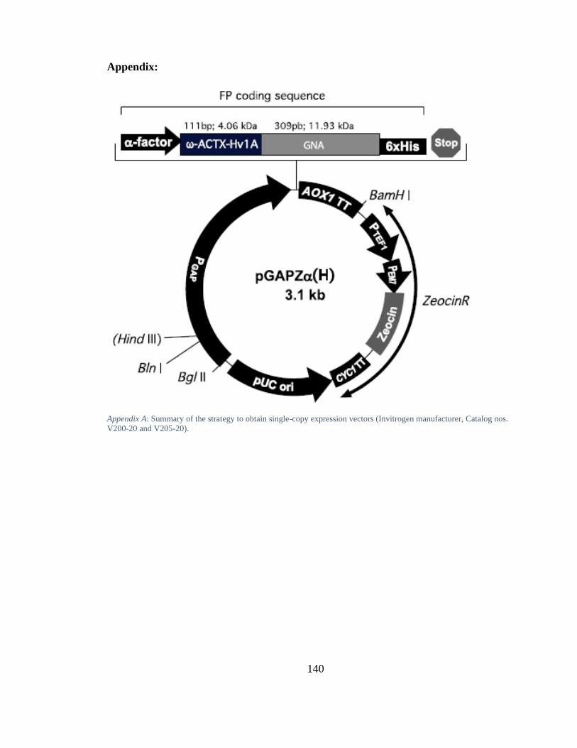
140
Appendix:
Appendix A: Summary of the strategy to obtain single-copy expression vectors (Invitrogen manufacturer, Catalog nos.
V200-20 and V205-20).
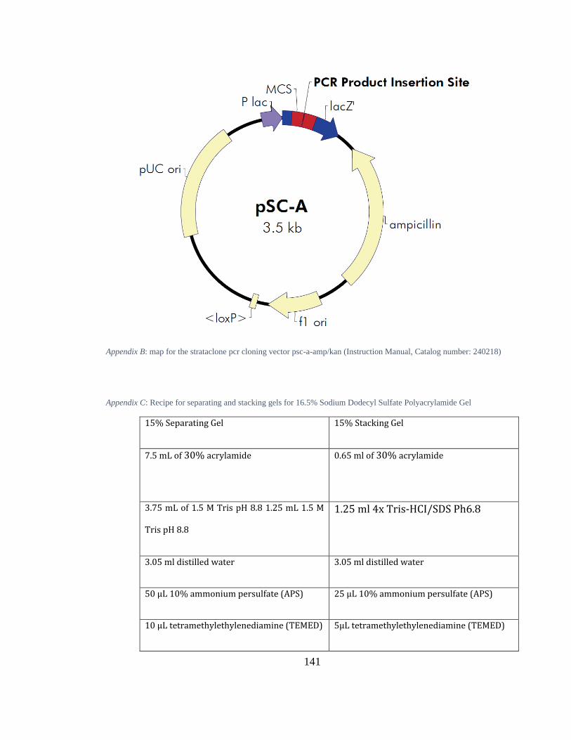
141
Appendix B: map for the strataclone pcr cloning vector psc-a-amp/kan (Instruction Manual, Catalog number: 240218)
Appendix C: Recipe for separating and stacking gels for 16.5% Sodium Dodecyl Sulfate Polyacrylamide Gel
15% Separating Gel 15% Stacking Gel
7.5 mL of 30% acrylamide 0.65 ml of 30% acrylamide
3.75 mL of 1.5 M Tris pH 8.8 1.25 mL 1.5 M
Tris pH 8.8
1.25 ml 4x Tris-HCI/SDS Ph6.8
3.05 ml distilled water 3.05 ml distilled water
50 μL 10% ammonium persulfate (APS) 25 μL 10% ammonium persulfate (APS)
10 μL tetramethylethylenediamine (TEMED) 5μL tetramethylethylenediamine (TEMED)
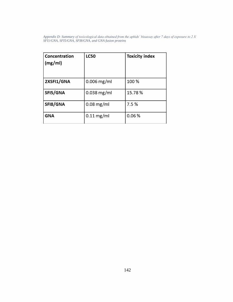
142
Appendix D: Summary of toxicological data obtained from the aphids’ bioassay after 7 days of exposure to 2 X
SFI1/GNA, SFI5/GNA, SFI8/GNA, and GNA fusion proteins
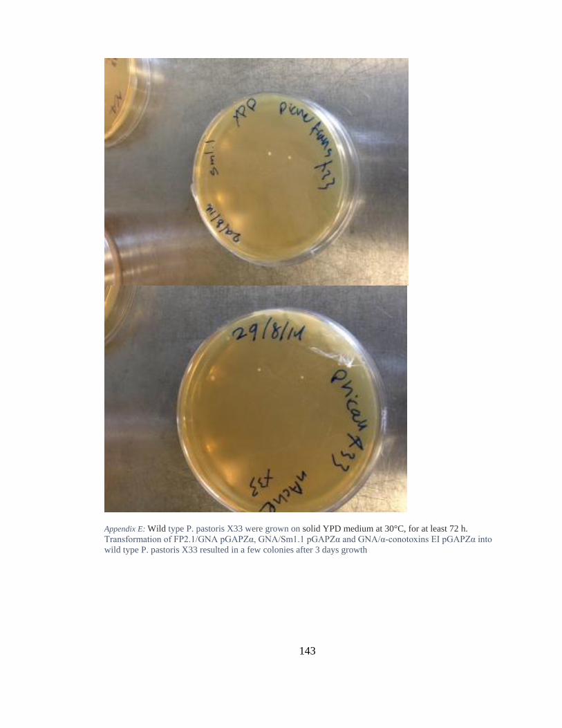
143
Appendix E: Wild type P. pastoris X33 were grown on solid YPD medium at 30°C, for at least 72 h.
Transformation of FP2.1/GNA pGAPZα, GNA/Sm1.1 pGAPZα and GNA/α-conotoxins EI pGAPZα into
wild type P. pastoris X33 resulted in a few colonies after 3 days growth