Embed Size (px)
Citation preview

Introductory Experiments 71Curr. Issues Mol. Biol. (2000) 2(3): 71-85.
© 2000 Caister Academic Press *Corresponding author
Robert C. Tait*
2318 Cezanne Court, Davis, CA 95616, USA
Abstract
Nine practical exercises demonstrate the basicprinciples in recombinant DNA. The exercises explainthe principles that DNA equals genes and that changesin DNA cause changes in genetic properties. The aimis to provide a teaching resource that can be used toillustrate the theory and applications of molecularbiology to highschool students, undergraduatestudents, medics, dentists, doctors, nurses, lifescientists, and anyone learning the basics of DNAtechnology.
Introduction
The exercises contained in this article have been chosento demonstrate the basic principles in recombinant DNA:digestion of DNA with a restriction endonuclease, gelelectrophoresis of DNA samples, insertion of DNA into cellscan change their growth characteristics, and DNA can berearranged to cause changes in genetic properties. Theexercises are intended to be an example of the principlesthat DNA equals genes and that changes in DNA causechanges in genetic properties.
Each exercise is preceded by a short discussion ofthe principles involved and the specific goals of theprocedure. Solutions that must be prepared for eachexercise are detailed in a Materials section. While someof these “recipes” can be changed considerably withoutaffecting the outcome of the exercise, many of the “minor”details are crucial to the success of the protocol. Forexample, a restriction digestion buffer may contain only 6mM NaCl relative to 50 mM TRIS-Cl buffer, an apparentlytrivial amount of salt in the overall scheme of things.However, many enzymes are very specific about saltconcentrations required for activity, and deleting the NaClfrom the buffer may inactivate or actually change therecognition properties of the enzyme. Please do notarbitrarily change recipes and expect exercises to workproperly.
The recipes assume a simple knowledge of chemistryand use standard abbreviations regarding concentrations:
M = moles/liter = molarmM = millimoles/liter = 10-3 moles/liter = millimolarl = literml = milliliter = 10-3 literµl = microliter = 10-6 liter
When pH of a solution is indicated, there is generally anallowance of about 0.5 pH unit. If a pH of 7.5 is indicated,a pH of 7.0 to 8.0 will generally suffice. If possible, use apH meter in the preparation of solutions, otherwise, usepH paper and be as accurate as reasonable.
The instructions often require the addition of water,sometimes indicated specifically as distilled water (dH2O).For electrophoresis, media, and general solutions, tapwater will often suffice. For reactions involving enzymes,distilled water should always be used rather than tap water.Many enzymes can be inhibited by heavy metal saltspresent at low levels in tap water. Distilled water purchasedfor use in a steam iron can be used in the event that thereis no access to a water still or deionizer.
Certain solutions must be sterilized (media for cultureof bacteria, for example). An electric hot plate and astandard pressure cooker can be used as a substitute foran autoclave. Heat media at 16 lb pressure for 20-30minutes to sterilize small volumes of liquids (less than 500ml solution per container). While a microwave oven is usedfor melting agarose in nearly every molecular biologyresearch lab, a hot plate with a boiling water bath or a gasburner will accomplish the same thing. Agarose solutionshave a tendency to superheat and boil violently when theagarose granules are first melting. A solution of agarosethat has been previously melted, then allowed to cool andsolidify is less likely to boil violently when melted the secondtime.
As a note of caution, realize that molecular biologyuses a number of noxious chemicals: organic solvents(phenol, chloroform, ether) are quite toxic and certaincompounds (ethidium bromide and UV light, for example)are known mutagens capable of causing genetic changes.The use of such compounds has been kept to a minimumin these exercises and where such compounds are used,precautions are noted in directions for the exercises. Duringelectrophoresis, although low voltages are used, severeinjury is possible. Electrophoresis boxes should have aninterlock mechanism - a device that prevents the currentfrom being applied to the buffer when the buffer is exposed.Most commercial gel boxes include this safety designfeature, as do the plans that accompany these exercises.COMMON SENSE IS REQUIRED IN ALL LABEXERCISES. Food and drinks should be prohibited fromthe work area and lab coats should be worn. Power unitsshould be turned off while loading gels or handling gelboxes. All spills should be cleaned up when they occur.
Having previously indicated that these exercisesshould not be arbitrarily altered, I now emphasize that theseprotocols should not be considered inviolate. There areabout 113 ways to accomplish the same thing in anymolecular biology exercise. As your understanding of thesemethods increases, you will recognize how protocols canbe altered to suit a particular circumstance. Other protocolswill appear that seem easier or more reproducible in yourown situation. While this is one of the aspects of molecularbiology that makes the methods so powerful, this variabilityalso tends to unnecessarily confuse novices in the field.Before modifying a protocol, be certain that you understandthe changes and that these alterations will not adverselyaffect the outcome of an exercise.
The bacteriophage lambda, pBR322, and pUC19plasmid DNA samples used in these exercises are availablefrom a number of commercial suppliers. The bacterial DNA
Introductory Experiments in Recombinant DNA

72 Tait
can be prepared by a simple procedure (see Appendix)that uses detergent lysis and phenol/chloroform extractionto prepare high quality DNA. Commercially available calfthymus DNA can be substituted. Plasmid recombinantscontaining E. coli DNA in pUC19 were chosen specificallyto work entirely with bacterial DNA and minimize anymisperception of biohazard potential. Competent cells arecommercially available from several sources, butacceptable competent cells can also be prepared with aminimum of expertise.
Exercise 1. Gel electrophoresis of nucleic acids
Nucleic acid samples are often subjected to gelelectrophoresis to characterize the size and number ofdifferent fragments in the sample. In this exercise, DNAsamples that have been digested with restriction enzymesand mixed with a tracking dye will be subjected to agarosegel electrophoresis. The electrophoresis buffer, the saltsolution that both conducts electric current and controlsthe pH of the solution during the separation of the DNAfragments, is TRIS/borate/EDTA or TBE, a commonly usedbuffer system.
When charged molecules are placed in an electric field,the molecules will migrate towards one of the electrodes,depending on the net charge of the molecule. Nucleic acidshave an overall negative charge due to the negativecharges associated with the phosphate backbone of themolecules, so they will migrate towards the positiveelectrode. Since the distribution of phosphate is very regularacross the length of the nucleic acid molecule, nucleic acidshave a constant charge/mass ratio and will thereforemigrate at the same rate in an electric field.
Separation of nucleic acid molecules of different sizeor conformation is achieved by adding a support matrix tothe electric field and forcing the molecules to migratethrough this matrix, typically agarose or polyacrylamide gel.This matrix acts like a sieve that allows small molecules togo faster than large molecules. The result is that moleculesseparate in the matrix according to their relative size andshape.
This exercise will use gel electrophoresis to examinethe fragments present in several DNA samples. Theprinciple goal is to obtain experience in pouring gels,loading DNA samples, and visualizing the DNA bands.
Due to the small volumes of sample to be applied tothe gel, samples are routinely handled with a manualmicropipet device that uses a disposable plastic tip tohandle a 10 to 20 µl sample. Several commercial devicesare available. A substitute sample loader can be assembledfrom a disposable 0.5 or 1 ml syringe fitted with a disposableplastic tip (see Appendix).
Materials• EcoRI- and HindIII-digested bacteriophage lambda
DNA samples containing SM Dye (other DNA samplescan be added or substituted).
• Reaction Stop Mix Dye (SM Dye): 10% glycerol, 0.5%SDS (sodium dodecyl sulphate), 0.025% xylene cyanolFF dye (XC), 0.025% bromphenol-blue WS dye (BPB).This mix is added to a DNA sample after digestion toprepare the sample for electrophoresis. The SDS helpsinactivate DNA binding proteins and releases themfrom the DNA fragments and the glycerol weights the
sample so that it will layer uniformly into the slots inthe gel. The two dyes serve as visual markers of theprogress of the electrophoresis only and do not stainDNA or proteins. The purple BPB dye will migrate withabout twice the relative mobility of the turquoise XCdye.
• TBE electrophoresis buffer: 90 mM TRIS, 2.5 mMNa2EDTA, 89 mM boric acid, final pH 8.2 (can be storedas a 10x stock).
• 1% Agarose: Molten 1% agarose in TBE buffer, heatedto 100oC to melt the agarose and stored in 55oC waterbath until needed. Melt the agarose very carefully tominimize superheating and violoent boiling.
• Ethidium bromide (EtBr): For staining gels to visualizeDNA, approximately 200 ml of 4 µg/ml ethidiumbromide in water in a shallow tray, stored covered andprotected from excessive light exposure. EtBr shouldbe treated as a mutagen - a compound capable ofcausing genetic mutations - and bare skin should notcome in contact with the solution. It is bothphotosensitive and biodegradable and dilute solutionsare often disposed of via the sink. Concentratedsolutions must be saved and disposed of asbiohazardous chemical waste.
Protocol1. Use the molten agarose to pour a minigel as
demonstrated or according to instructions for the gelunit in use. Illustrations for assembling a generic gelunit are included in the Appendix. It is important to becertain that all small agarose particles are completelymelted before use. Allow the gel to completely hardenbefore removing the well-forming comb. This shouldtake about 15 minutes.
2. Remove the comb from the solidified gel and transferthe gel to a running unit. Submerge the gel in TBEbuffer.
3. Use a manual pipettor with a disposable plastic tip toload 10 µl of each sample into one of the wells in thegel. Plasmid and bacteriophage DNA samples shouldbe easy to load, but chromosomal DNA samples maybe quite viscous and difficult to load.
4. Once the samples have been loaded, close the unitand apply power to the electrophoresis chamber.Typical minigels run at 100-130 volts (60-160milliamps), depending on the unit used. During the run,the XC and BPB dyes will resolve into two bands, withthe BPB the faster band. Run the gel until this band isnear the end of the gel, then turn the current off andremove the gel.
5. Transfer the gel to the staining tray containing theethidium bromide solution, and stain the gel for 5minutes. Ethidium bromide is a mutagen, and glovesshould be worn or a spatula used to transfer the gel inand out of the ethidium solution.
6. Transfer the stained gel to a destaining tray containingwater. Destain the gel for 5 minutes to remove excessethidium bromide.
7. The stained gel can be visualized with a UVtransilluminator or a UV mineral lamp. DO NOT LOOKDIRECTLY AT THE UV LIGHT!! UV light causes skinburns, is a mutagen, and will cause severe headachesfrom eye damage with direct exposure. Always use aface mask or shield.

Introductory Experiments 73
Analysis and significance of resultsThe results of a typical gel are shown illustrated below:
If lambda DNA samples digested with different restrictionendonucleases are present on the same gel, note that eachdifferent restriction enzyme produces a distinct, completelyreproducible pattern of DNA fragments. Relative separationof individual fragments is dependent on agaroseconcentration, electrophoresis conditions, and the qualityof the gel preparation.
Many different types of artefact can distort the bandsin a gel. Some of the more common gel artefacts areillustrated below.
a. Gel was crooked in gel box and samples migrated offof the gel.
b. Agarose was not completely melted before gel waspoured. The small specks of high concentrationagarose in the gel cause distortion in DNA bands andbright spots in the gel.
c. The sample well containing the EcoRI-digested lambdaDNA leaked at the right side, causing loss of sample
intensity and blurring of bands.d. The smearing of the EcoRI-digested lambda DNA is
caused by loading too much DNA.
During electrophoresis of DNA fragments through anagarose gel, the fragments separate by size with smallerfragments moving faster than larger fragments. Therelationship between size and mobility is not linear, butcan be approximated by plotting the distance migratedversus the log(Molecular Weight).
Using the HindIII fragments as molecular weightsstandards to estimate the size of other DNA fragments.Mobility of fragments is determined and plotted as log(Size)versus mobility. The line drawn through the lambda DNAfragments can be used as a standard curve fordetermination of sizes of other DNA fragments present onthe same gel.
Exercise 2. Restriction endonuclease digestion ofchromosomal and plasmid DNA
Digestion of DNA with a site-specific restrictionendonuclease will generate a set of specific DNAfragments. In this exercise, a variety of DNA samples willbe digested with the restriction enzyme EcoRI. This enzymerecognizes and cleaves the sequence 5'-GAATTC-3'between the G and A residues to generate the four-basecohesive terminus AATT:
5'-NNNNGAATTCNNNN-3' EcoRI 5'-NNNNG AATTCNNNN-3'3'-NNNNCTTAAGNNNN-5' ----------> 3'-NNNNCTTAA GNNNN-5'
Because EcoRI recognizes and cleaves at a six-basesequence (5'-GAATTC-3'), the number of fragmentsgenerated by digestion of a DNA molecule with this enzyme

74 Tait
is determined approximately by the frequency of occurenceof the recognition site. Since there are four possible basesthat can occur at any position in DNA (A,C,G,T) theprobability of occurence of a specific base is (1/4), whilethat of a specific two-base sequence is (1/4)2, a four-basesequence (1/4)4, and a six-base sequence is (1/4)6. Thesix-base EcoRI site, therefore, should occur about onceevery 46 bases, or once every 4096 base pairs. EcoRIcleaves the 2,800 base pair plasmid pUC19 only once andwill convert the circular molecule to a linear form, cleavesthe linear 50,000 base pair bacteriophage lambda DNAfive times to generate six fragments, cleaves hundreds oftimes in the E. coli DNA genome, and thousands of timesin eukaryotic DNA. The larger the genome of an organismis, the greater the number of fragments generated duringdigestion with a restriction enzyme.
Several DNA samples from genomes of increasing sizewill be digested with EcoRI in this exercise. If both theenzyme digestion and the gel analysis are to be performedin the same day, as soon as the digestion reactions havebeen set up and are incubating, prepare agarose minigelsfor analysis of the digested DNA samples.
Materials• Plasmid pUC19 vector DNA: 100 µg/ml.• Bacteriophage lambda DNA: 100 µg/ml.• Bacterial chromosomal DNA: 100 µg/ml.• EcoRI endonuclease: 3-5 units/µl. Keep this in ice at
all times!!• 10x EcoRI Reaction buffer: 500 mM TRIS-HCl pH 7.0,
750 mM NaCl, 60 mM MgCl2, 60 mM 2-mercaptoethanol. Most enzyme companies supply thecorrect buffer with each enzyme purchased.
• Water (dH2O): Sterile, deionized or distilled.• Manual pipet devices, 1-20 µl and 20-200 µl capability.• Disposable conical 1.5 ml microfuge tubes and pipet
tips.
Protocol1. For each different chromosomal DNA sample, set up
a digestion reaction in a labeled 1.5 ml conical tubeby making the following additions:
10x EcoRI buffer 5 µldH2O 35 µl 1 ug DNA 10 µlTotal volume 50 µl
2. Mix the contents of the tubes gently but thoroughly,then from each tube remove 10 µl and place in a tubelabeled with the sample number and the term “-EcoRI”.Place these 10 µl samples on ice. They are theundigested controls and will be used to compare tosamples following digestion.
3. To the remaining 40 µl of each sample add:
EcoRI, 5-10 units 2 µl
Mix, then incubate at 37oC for 30 minutes.4. From each digestion tube, remove 10 µl of each
sample and add to a tube labeled with the samplename and “+EcoRI”. The remainder of each digestionreaction should be frozen for further use.
5. You should now have two tubes containing 10 µl ofeach DNA sample, one “-EcoRI” and one “+EcoRI”,for a total of six tubes. To each of these tubes add:
SM dye 5 µl
Mix the contents of each tube, and the samples areready for electrophoresis. Samples can be stored on iceor frozen for future gel electrophoresis.
Significance of procedureThis exercise has prepared reaction mixes that containseveral DNA samples of different sizes and conformations,then digested portions of each reaction mix with therestriction enzyme EcoRI. The samples are now ready foranalysis by agarose gel electrophoresis to determine theeffects of the restriction enzyme digestion. The same gelelectrophoresis described in Exercise 1 will be utilized.
Materials• DNA samples prepared in Exercise 2.• Electrophoresis materials, see Exercise 1.
Protocol1. Prepare a 1% agarose gel in TBE buffer as in Exercise
1.2. When the gel has solidified, assemble the gel unit.
Load 15 µl of each of the six samples. You may alsowant to include one lane of the digested lambda DNAused in the first exercise as a DNA standard.Electrophorese as in Exercise 1.
3. Stain the gel with ethidium bromide and visualize theDNA bands with a UV light box. A photograph of thegel is useful, but a sketch of the gel is sufficient forrecording information.

Introductory Experiments 75
Analysis and significance of resultsA typical gel is illustrated below:
Compare each undigested (-EcoRI) with the correspondingdigested (+EcoRI) sample. Note that the migration positionof the undigested circular plasmid pUC19 DNA (a) changeswhen cleaved at the unique EcoRI site to generate a linearmolecule (b). The linear undigested lambda DNA (c)contains five EcoRI cleavage sites, and migrates as sixbands after digestion (two of the bands are of similar sizeand can be difficult to resolve, but this double band will betwice as intense as the other bands) (d). The slowlymigrating, broad, undigested bacterial chromosomal DNAsample (e) is converted upon digestion to hundreds ofsmaller, yet distinct bands (f). Note that as the size of thegenome (pUC19<lambda<E. coli) increases, the numberof cleavage sites also increases, demonstrating that largergenomes are actually composed of larger amounts of DNA.
Exercise 3. Competent cell production
Although some bacteria naturally undergo a growth phasewhen they are capable of taking up DNA, strains of E. coli,the bacteria most commonly used in a molecular biologylab, cannot normally take up DNA. These bacteria mustbe chemically treated to be made competent fortransformation. Competent cells can be purchased from avariety of commercial sources but can be relativelyexpensive for routine classroom demonstrations andlaboratories. Cells can be made by harvesting rapidlygrowing bacteria and exposing them to calcium chlorideon ice. The following protocol can be used to makecompetent E. coli that are of sufficient quality for routineuse.
Materials• 35 ml LB medium (1% Bacto Tryptone, 0.5% Bacto
Yeast Extract, 0.5% NaCl, pH 7.0-7.2) in sterile 100-250 ml flask.
• Fresh 2 ml overnight LB culture of JM83.• Sterile 50 or 35 ml centrifuge tube.• Sterile 30 mM CaCl2.
• Sterile 30 mM CaCl2, 15% glycerol.• Sterile microfuge tubes.• Ice.• Refrigerated centrifuge. If a refrigerated centrifuge is
not available, place a clinical or counter-top centrifugein a refrigerator. Allowing the cells to warm up duringthe centrifugation may affect the competency of thecells.
Protocol1. Competent cells must be made from a rapidly growing
culture of bacteria to obtain good efficiencies oftransformation. Mid-logarithmic phase of growth (3-5x 10⁸ cells/ml) is good for preparation of general-usecompetent cells. With a fresh overnight innoculationculture, a 1:50 dilution into a fresh culture will generallygive mid-log cells in 60-90 minutes of growth at 37oC.If an old overnight culture is used or if insufficientdilution is made, the bacteria will undergo a significantlag phase before growth begins.
2. Use sterile techniques to transfer 0.7 ml of theovernight culture of JM83 to the flask containing 35 mlof LB. Incubate in a 37oC shaker with vigorous shakingto allow growth. Monitor the growth of the culture every20 minutes (A550). When absorbance is approximately0.5, the culture is ready to harvest. This shouldcorrespond to a mid-logarithmic phase of growth. If asemi-log plot of A550 versus time is prepared, harvestof cells should occur in the linear portion of the graph.
3. Although strict sterile technique is not necessary, tryto minimize contamination of the cells throughout thefollowing procedures. Pour the culture into a sterilecentrifuge tube. Spin 7000 rpm, 5 minutes to pelletthe cells.
4. Pour off the supernatant. Add 17.5 ml of cold 30 mMCaCl2 (1/2 of the original culture volume). Gentlyresuspend the cells.
5. Place cells on ice for 20 minutes. Cells will swell toform spheroplasts. Cells can incubate on ice as longas 90 minutes.
6. Centrifuge 7000 rpm, 5 minutes, at 4oC.7. Pour off supernatant. Many strains of E. coli often form
a halo instead of a normal pellet.
76 Tait
8. Add 3.5 ml of cold 30 mM CaCl2, 15% glycerol.GENTLY RESUSPEND THE CELLS. If resuspensionis too harsh, competency may decrease. Return cellsuspension to ice.
9. Label 17 sterile microcentrifuge tubes “cJM83”. Aliquot200 µl of competent cells into each tube. Cap tubes.Cells can be used immediately or stored in a freezerfor future use. To freeze, move the tubes to a pre-chilled rack in the freezer for storage. Cells can bestored at -20oC (normal lab freezer) for several weeksor at -70oC for months without significant loss ofcompetency. Recombination deficient strains (recA-
strains, in particular) rapidly lose viability/competencywhen stored in a freezer.
10. If competent cells are made and stored for use in alab exercise, it is wise to transform one tube with apure plasmid DNA sample prior to use to verify thatthe cells have not lost competency during storage.Larger numbers of competent cells can be preparedby scaling all procedures up as needed. When culturevolumes are increased, be certain that the culture hasa large surface area to maintain adequate aeration inthe growing culture.
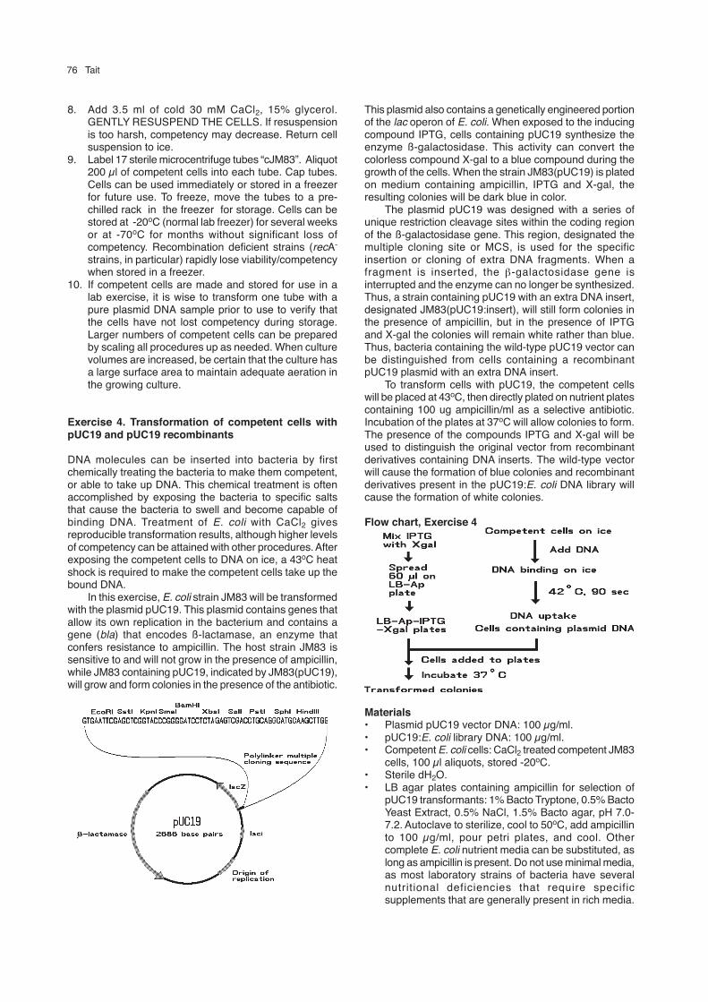
Exercise 4. Transformation of competent cells withpUC19 and pUC19 recombinants
DNA molecules can be inserted into bacteria by firstchemically treating the bacteria to make them competent,or able to take up DNA. This chemical treatment is oftenaccomplished by exposing the bacteria to specific saltsthat cause the bacteria to swell and become capable ofbinding DNA. Treatment of E. coli with CaCl2 givesreproducible transformation results, although higher levelsof competency can be attained with other procedures. Afterexposing the competent cells to DNA on ice, a 43oC heatshock is required to make the competent cells take up thebound DNA.
In this exercise, E. coli strain JM83 will be transformedwith the plasmid pUC19. This plasmid contains genes thatallow its own replication in the bacterium and contains agene (bla) that encodes ß-lactamase, an enzyme thatconfers resistance to ampicillin. The host strain JM83 issensitive to and will not grow in the presence of ampicillin,while JM83 containing pUC19, indicated by JM83(pUC19),will grow and form colonies in the presence of the antibiotic.
This plasmid also contains a genetically engineered portionof the lac operon of E. coli. When exposed to the inducingcompound IPTG, cells containing pUC19 synthesize theenzyme ß-galactosidase. This activity can convert thecolorless compound X-gal to a blue compound during thegrowth of the cells. When the strain JM83(pUC19) is platedon medium containing ampicillin, IPTG and X-gal, theresulting colonies will be dark blue in color.
The plasmid pUC19 was designed with a series ofunique restriction cleavage sites within the coding regionof the ß-galactosidase gene. This region, designated themultiple cloning site or MCS, is used for the specificinsertion or cloning of extra DNA fragments. When afragment is inserted, the β-galactosidase gene isinterrupted and the enzyme can no longer be synthesized.Thus, a strain containing pUC19 with an extra DNA insert,designated JM83(pUC19:insert), will still form colonies inthe presence of ampicillin, but in the presence of IPTGand X-gal the colonies will remain white rather than blue.Thus, bacteria containing the wild-type pUC19 vector canbe distinguished from cells containing a recombinantpUC19 plasmid with an extra DNA insert.
To transform cells with pUC19, the competent cellswill be placed at 43oC, then directly plated on nutrient platescontaining 100 ug ampicillin/ml as a selective antibiotic.Incubation of the plates at 37oC will allow colonies to form.The presence of the compounds IPTG and X-gal will beused to distinguish the original vector from recombinantderivatives containing DNA inserts. The wild-type vectorwill cause the formation of blue colonies and recombinantderivatives present in the pUC19:E. coli DNA library willcause the formation of white colonies.
Flow chart, Exercise 4
Materials• Plasmid pUC19 vector DNA: 100 µg/ml.• pUC19:E. coli library DNA: 100 µg/ml.• Competent E. coli cells: CaCl2 treated competent JM83
cells, 100 µl aliquots, stored -20oC.• Sterile dH2O.• LB agar plates containing ampicillin for selection of
pUC19 transformants: 1% Bacto Tryptone, 0.5% BactoYeast Extract, 0.5% NaCl, 1.5% Bacto agar, pH 7.0-7.2. Autoclave to sterilize, cool to 50oC, add ampicillinto 100 µg/ml, pour petri plates, and cool. Othercomplete E. coli nutrient media can be substituted, aslong as ampicillin is present. Do not use minimal media,as most laboratory strains of bacteria have severalnutritional deficiencies that require specificsupplements that are generally present in rich media.

Introductory Experiments 77
• 10 mM IPTG: 23.8 mg IPTG (isopropyl-β-D-thiogalactopyranoside)/ml dH2O.
• 5% X-gal: 50 mg X-Gal (5-bromo-4-chloro-3-indolyl-β-D- galactopyranoside)/ml dimethylformamide.
• 43oC temperature block or water bath.• 37oC incubator.
Protocol1. Prepare six LB-Ap plates for transformation by adding
IPTG and X-gal to each plate. To a sterile tube add 60µl 10 mM IPTG and 300 µl 5% X-gal. Mix solution.Place 60 µl of this IPTG/X-gal mix on the surface ofan LB-Ap plate. Dip a glass spreader in 95% ethanoland flame to sterilize. Use the spreader to distributethe IPTG/X-gal mix evenly over the surface of the plate.Replace the lid on the plate and invert the plate. Repeatthis process for each of the remaining 5 plates. If youattempt do do all of the plates at the same time, thesolution may soak into the center of the plates and willnot be uniformly distributed over the surface. Allowthe solution to soak into the surface of the plates for15-30 minutes (if possible, place the plates in a 37oCincubator during this time period).
2. Thaw three tubes of competent cells at roomtemperature. As soon as cells are thawed, label onetube “-DNA”, one tube “+pUC19”, and one tube“+library” and place all tubes on ice.
3. To the “-DNA” tube add 10 µl sterile ddH 2O, to the“+pUC19” tube add 10 µl (1 µg) of pUC19 DNA, andto the “+library” tube add 10 µl of pUC19:EC libraryDNA. Place tubes on ice for at least 15 minutes (lengthof time is not crucial here). This allows DNA to bind tothe competent cells. While tubes are on ice, adjust awater bath to 42-43oC.
4. Heat pulse the cells to cause DNA uptake. Transfer allthree of the tubes of competent cells containing DNAto the 42oC bath for 90 seconds, then transfer to aroom temperature rack.
5. Add 10 µl of one of the transformed cell samples to anLB-Ap-IPTG-Xgal plate and the rest (100 µl) to anotherplate. Dip the glass spreader in ethanol, flame, anduse to distribute the cells evenly over the surface ofeach plate. Invert the plates and label with amount ofcells and type of DNA (such as 10 µl, pUC19). Repeatfor each of the tubes of transformed cells (you shouldend up with no tubes of transformed cells left overand have six labeled plates with cells spread on them).Place plates in a 37oC incubator for 12-18 hours toallow colonies to form and color to develop. Platescan be stored in a refrigerator and color will continueto develop.
Analysis and significance of resultsAfter the plates have been allowed to incubate and colonieshave formed and colored, compare all the plates. Thefollowing results are typical:• JM83 transformed with no DNA should give no distinct
colonies but may have some smearing or poor growthin areas of dense numbers of cells.
• JM83 transformed with pUC19 DNA should give manydistinct colonies, nearly all of which (>99%) should beblue. The occasional white colony will be a contaminantor a mutant derivative of pUC19 in which the ß-galactosidase gene is no longer active. By counting
the number of colonies on a plate and dividing by theamount of transforming DNA represented on the plate,it possible to calculate the efficiency of transformationin terms of transformants/µg DNA. Example: 1 µg ofpUC19 DNA was added to 100 µl of competent cells,and 1 µl of this mix was plated on an LB-Ap plate,resulting in 227 colonies. The amount of DNArepresented on the plate is 10 µl/110 µl x 1 µg = 0.1µg. Transformation efficiency is 227 colonies/0.1 µg,or 2.27 x 10³ transformants/µg DNA.
• JM83 transformed with pUC19:E. coli library shouldgive similar numbers of colonies when compared withthe pUC19 transformation, but only a percentage ofthe colonies will be blue. The blue colonies are theresult of transformation with pUC19 containing noadditional DNA insert, and the white colonies arecaused by transformation with recombinant pUC19plasmids. The transformation efficiency can becalculated for the DNA as described above and thepercentage of recombinant molecules can bedetermined by the calculation:
100 x # white colonies/# total colonies.
This exercise demonstrates two fundamental principles ofrecombinant DNA methods:1. DNA can be inserted into bacteria to change the
properties of the cells (ampicillin-sensitive cellsconverted to ampicillin-resistant colonies).
2. DNA fragments can be re-arranged to change thegenetic properties of the DNA molecules (blue coloniescaused by pUC19 are converted to white colonieswhen DNA fragments are inserted into pUC19 DNA).
These principles can be further examined by isolation andexamination of plasmid DNA present in the transformantcolonies.
Exercise 5. Transformation of competent cells withpUC19 and pBR322 plasmid DNA
One of the most important concepts of DNA biology is thatgenes are composed of DNA and that the introduction ofDNA into a cell can change the physical properties of thecell. This can be illustrated with petri plates containing asimple nutrient medium (Luria broth, Tryptone, Nutrientagar, essentially any medium that will allow the growth ofE. coli), a small amount of plasmid DNA, and competentE. coli bacterial cells.
This exercise uses E. coli JM83 cells that have beenmade competent, or able to take up DNA. The cells weregrown to mid-logarithmic phase, then harvested andresuspended in 30 mM CaCl2. After incubation on ice for30 minutes, the cells were again harvested andresuspended in 30 mM CaCl2 with 15% glycerol. Cells weredispensed into 0.2 ml volumes in sterile 0.5 ml tubes. Eachof these tubes contains sufficient cells for at least 4 differenttransformation reactions. Competent cells may slowly losetheir ability to take up DNA if not stored at -70oC. For thisreason, the competent cells are shipped in dry ice to arrivethe week before the exercise and are stored in a freezer atabout -5oC until used.
A DNA sample containing two types of pure plasmidDNA, pUC19 and pBR322, at a concentration of 100 µg/ml will be used to demonstrate that DNA can change cellular

78 Tait
properties. The plasmid pUC19, a common plasmid cloningvector used to carry and propagate other DNA fragmentsin bacteria, has two genes of interest: one confersresistance to the antibiotic ampicillin, and the other encodesa ß-galactosidase gene derivative that produces a proteinthat can help the bacteria metabolize derivatives of thesugar galactose. When the colorless compound X-gal isadded to the growth medium, the ß-galactosidase proteincan metabolize the X-gal into a colored derivative that turnsthe bacterial colonies blue.
The second plasmid DNA provided is pBR322, one ofthe older plasmid cloning vectors. Like pUC19, this plasmidconfers resistance to ampicillin, but it also confersresistance to the protein synthesis inhibitor tetracycline.Just like bacteria containing pUC19, bacteria containingpBR322 will be able to grow in medium containingampicillin. Since pBR322 does not contain the ß-galactosidase gene derivative, cells containing this plasmidwill be unable to metabolize X-gal to a colored compoundand will remain white when exposed to X-gal. Bacteriacontaining pBR322 will, however, be able to grow in thepresence of a concentration of tetracycline that kills cellscontaining pUC19.
The bacterial strain JM83 is sensitive to ampicillin andwill not grow on nutrient plates in the presence of theantibiotic at a concentration of 100 µg/ml. When eitherpUC19 or pBR322 is inserted into the competent JM83cells, the cells become transformed and are able to growin the presence of the antibiotic. These two plasmids canthus cause the same change in bacterial phenotype - abilityto grow in the presence of ampicillin - and this propertycannot be used to distinguish between cells containing oneof these two plasmids.
Genetic maps of the plasmids pUC19 and pBR322
However, if the compounds X-gal and IPTG are alsoadded to the ampicillin plate, the ß-galactosidase genepresent on pUC19 will cause bacteria containing thisplasmid to form blue colonies, while bacteria containingpBR322 will form white colonies. The difference inphenotype associated with these plasmids - ability of cells
containing pUC19 to form a blue colony - allows rapidsorting of colonies formed by cells containing each of thetwo types of plasmid. This demonstrates that two plasmidsthat confer the same phenotypic property (such as abilityto grow in the presence of ampicillin) can be readilydistiguished if the plasmids differ in ability to confer asecond phenotype (such as ability to metabolize X-gal toa blue compound).
This exercise will also demonstrate a second importantprinciple of DNA manipulation: when two genes are bothpresent on the same DNA fragment, selection for one ofthe genes will force maintenance of the second gene aswell. As the transformed bacteria grow and form coloniesin the presence of ampicillin but in the absence oftetracycline, there is no selective pressure that requiresthe expression of the tetracycline resistance gene presenton pBR322. Nevertheless, because the tetracyclineresistance gene is part of pBR322, growth in the presenceof ampicillin forces the maintenance of pBR322 and alsoselects for the covalently attached tetracycline resistancegene. If the blue and white colonies obtained on theampicillin plate containing X-gal and IPTG are transferredto a nutrient plate containing tetracycline at a concentrationof 20 µg/ml, the blue colonies containing pUC19 will beunable to grow and the white colonies containing pBR322will be able to form colonies. This is the fundamentalprinciple that allows a plasmid or a viral DNA molecule tobe used as a vector to carry and propagate exogenous, orextra, DNA fragments. DNA fragments that are covalentlyinserted into a vector by the use of restriction enzymesand DNA ligase will be maintained as the carrier vectormolecule replicates in a host cell.
Materials• Three nutrient agar plates containing 100 µg/ml
ampicillin (NA+Ap plates).• One nutrient agar plates containing 20 µg/ml
tetracycline (NA+Tc plates).• A water bath or temperature block at 43oC.• Incubator for plate culture (this can be performed at
room temperature, but growth of colonies will take twodays).
• Glass spreader bar.• Ethanol or isopropanol for sterilizing glass spreader
bar.• One 0.2 ml tube of competent JM83 (store these in a
normal freezer [not frost-free if possible] until needed,then on ice).
• Ten µl of pUC19/pBR322 mixed plasmid DNA at 100µg/ml.
• 2% X-gal in dimethylformamide (this solvent isnecessary for X-gal).
• 100 mM IPTG in sterile water.• Sterile toothpicks.
Procedure1. Since X-gal and IPTG are not usually added to nutrient
plates when the plates are liquid, it will be necessaryto add these compounds to the surface of the solidplates prior to use. Place 0.1 ml of the X-gal in a 1.5ml microcentrifuge tube (not a polystyrene tube - theX-gal solvent may melt the plastic). Add 20 µl of IPTGto the tube and mix the two solutions to make an X-gal/IPTG mixture.

Introductory Experiments 79
2. Place 60 µl of the X-gal/IPTG mixture on the surfaceof each of two of the plates containing ampicillin(NA+Ap). Save one NA+Ap plate and the NA+Tc platefor later use. Dip a glass spreader in alcohol and thenignite the alcohol in a burner flame to sterilize thespreader. Cool the spreader by touching the agarsurface, then spread the X-gal/IPTG evenly across thesurface of one of the NA+Ap plates. Again sterilizethe spreader and spread the X-gal/IPTG mixture onthe second NA+Ap plate. Invert the plates and label“+Xgal/IPTG”. This step allows the coloring agents tosoak into the surface of the agar. The agar surfacemay cloud as the dimethylformamide solvent dissipatesand the X-gal precipitates. It is important to add the X-gal/IPTG mixture shortly before the plates are usedbecause the X-gal can break down and may not colorwell if too old.
3. Place a tube of competent cells on ice and allow tothaw. Add the 10 µl of mixed plasmid DNA into thethawed cells and gently mix in. Return the cells to theice.
4. Allow the tube to stand in the ice bucket for 15 to 20minutes to allow the DNA to stick to the surface of thecompetent cells.
5. To make the DNA enter the cells, place the tube in a43oC water bath (be careful with the temperature, toohot or too cool will not work as well). Allow the tube toincubate at 43oC for 60 seconds, then remove fromthe water bath and place at room temperature.
6. The transformation is complete. It is now necessaryto plate the transformation mix on selective plates todetect the transformants and inhibit the growth of thenon-transformed cells. Transfer 20 µl of thetransformed cells to one nutrient plate containingampicillin, IPTG, and X-gal. Use a sterile spreader todistribute the cells. Label the plate “20 µl”. Spread theremainder of the transformed cells on the second plateand distribute uniformly with a sterile spreader. Labelthe plate “180 µl”.
7. Incubate the plates overnight at 37oC to allow growthof colonies. Check the plates for colonies after 18 hoursgrowth and transfer the plates to a refrigerator forstorage if the colonies are sufficiently large (the sizeof a typewritten”o”) or are extremely numerous (>300per plate). Although the colonies will stop increasingin size, blue color will continue to develop in the cold.If allowed to remain at 37oC too long, non-transformed,ampicillin-sensitive “feeder” colonies will begin to formas the ampicillin-resistant colonies degrade theampicillin in the medium.
8. If the transformation has been successful, both blueand white colonies should be present on the surfaceof the two plates. The blue colonies are formed as aresult of the transformation of bacteria with pUC19 andthe white colonies are formed as a result of thetransformation of bacteria with pBR322. This can beverified by using the ability of pBR322 to allow growthin the presence of tetracycline. To check transformantsfor this phenotypic property, label the back of theremaining NA+Ap plate and the NA+Tc plate with thenumbers “1” to “40” and your initials as indicatedbelow:
9. Touch a sterile toothpick to a blue colony on one ofthe NA+Ap X-gal/IPTG plates and make a short streakof cells over the number “1” on the NA+Ap plate andthe NA+Tc plate. Repeat this for an additional 19 bluecolonies and for 20 white colonies. Incubate both platesovernight at 37oC to allow the growth of the bacteria.
Analysis and significance of resultsSince both the blue and the white colonies were originallyobtained following growth on an NA+Ap X-gal/IPTG plate,all of these colonies should again grow on the NA+Ap platebut, since X-gal and IPTG are no longer present, all coloniesshould remain white. Only the white colonies obtained fromthe NA+Ap X-gal/IPTG plate should form colonies on theNA+Tc plate, confirming that the transformants thatoriginally formed white colonies on NA+Ap X-gal/IPTGcontained pBR322. Note that although the original selectionfor transformed bacteria involved resistance to ampicillin,resistance to tetracycline was also maintained by nearlyall of the ampicillin-resistant white colonies. Since the genesconferring resistance to ampicillin and tetracycline are bothpresent on the same DNA molecule (the plasmid pBR322),selection for one of the two genes (ampicillin resistance)also selects for the presence of the non-selected gene(tetracycline resistance).
It is possible to obtain ampicillin-resistant whitecolonies that cannot grow on tetracycline and ampicillin-resistant blue colonies that can grow on tetracycline. Whenthe DNA from the plasmids present in these colonies isisolated and examined by digestion with restrictionenzymes, the first type of colony can often be demonstratedto be caused by the occurrence of a deletion that removespart or all of the ß-galactosidase gene of pUC19, causinginability to metabolize X-gal to a blue compound, or adeletion that removes part of the tetracycline resistancegene, causing loss of tetracycline resistance. Thesedeletions occur naturally in bacteria as an aspect of normalDNA replication, recombination, and repair processes.
The second type of colony is most commonly causedby the presence of bacteria containing both pUC19 andpBR322 in the same colony. This can be verified bystreaking the bacteria out on an NA+Ap X-gal/IPTG plateand incubating to obtain isolated colonies that are eitherblue and tetracycline-sensitive or white and tetracycline-resistant. While both plasmids can occassionally beinserted into the same bacterial cell during thetransformation process, pUC19 and pBR322 replicate byvery similar mechanisms and are said to be incompatible

80 Tait
with one another. When incompatible plasmids are insertedinto the same bacterium and the cell is grown in theabsence of selective pressure designed to forcemaintenance of both plasmid types, the plasmids willsegregate during division of the transformed bacterium andthe resulting progeny will contain one or the other of thetwo plasmids.
Exercise 6. Comparison of plasmid and bacteriophagecloning vectors
Much of the scientific utility of recombinant DNA technologyis based on the use of self-replicating DNA elements toserve as carriers for DNA fragments that are not capableof replication in bacteria. Bacterial cloning vectors arebased on either bacterial plasmids or bacteriophage.Plasmids are naturally occurring, circular DNA moleculesthat can replicate as mini-chromosomes in bacteria. Whenisolated from nature, these DNA molecules also generallyencode resistance to one or more antibiotics and maycontain genes that allow transfer of the plasmid DNA fromone cell to another during bacterial mating. Most cloningvectors derived from plasmids retain the plasmid origin ofDNA replication and one or more antibiotic resistancegenes, such as the ß-lactamase gene, which allowssensitive bacteria to form colonies on solid mediumcontaining ampicillin. With plasmid cloning vectors,selection of bacteria containing the vector requires selectionfor a phenotype, or physical characteristic, associated witha gene present on the vector DNA molecule.
Cloning vectors have also been constructed frombacteriophage, viruses that replicate in bacteria. Two typesof bacteriophage cloning vectors, derived from either thedouble-stranded, linear DNA bacteriophage lambda or themalespecific, single-stranded, circular DNA bacteriophageM13, are in common use in E. coli. Detection of cellscontaining either of these types of vector does not requireselection for a phenotypic marker, but is based on the abilityof the bacteriophage to form a plaque in a lawn of hostbacteria, a property associated with the replication of theviral DNA vectors.
Lambda has a large genome (approx. 50 kilobase pairsof double-stranded DNA) that can, on insertion into abacterial host, activate either of two sets of viral genes.One set of genes allows the bacteriophage to undergo alytic cycle during which progeny phage are produced andthe infected bacterial cell bursts open, releasing thedaughter phage into the medium. When activated, the otherset of genes causes the bacteriophage DNA to lysogenize,or insert itself into a specific location in the bacterialgenomic DNA, where it remains silent while the bacterialcell undergoes normal cellular functions. Certain conditions,such as DNA damage, will induce the lysogen, activatingthe lytic genes. The bacteriophage DNA then excises fromthe host chromosome, produces progeny, and lyses thehost cell. When bacteria are grown on the surface of solidmedium from a low density to a uniform lawn of cells, thepresence of lambda can be detected by the appearanceof plaques, circular clear zones in the cloudy lawn of hostbacteria. As the host bacteria grow to form the indicatorlawn, successive cycles of infection/production of progeny/cell lysis cause the death and lysis of all of bacteria in acircular zone surrounding even a single lambdabacteriophage. Cells that are able to lysogenize the lambda
DNA into their own genome become resistant to infectionand may form small colonies within the zone of lysis. Theability of lambda DNA to cause the lysis of bacteria servesas the selective marker for the presence of cloning vectorsderived from this bacteriophage. In marked contrast toplasmid cloning vectors, which require phenotypic selectionto detect cells containing the vector, the replication of alambda cloning vector provides a plaque, the immediatemeans of detection of cells containing the vector. Becausethe lambda plaque is basically a zone of lysed cells,however, the plaque indicates only where the infected cellswere. Lambda vectors and recombinants derived from themmust generally be maintained not as viable bacterial strains,but as stocks of virus particles.
An M13 bacteriophage particle contains a single-stranded, circular DNA genome of about 8 kilobase pairs.These particles will only infect host bacteria that areproducing the F-pilus protein, which is part of the matingapparatus encoded by the large F plasmid. Bacteria thatare male, or F+, can be infected by the M13 phage particle.The entering single-stranded viral DNA is converted to andreplicates as a double-stranded circular DNA. Single-stranded progeny phage identical to the original infectingDNA are produced, coated with viral coat proteins, andextruded through the bacteria cell wall into the mediumwithout lysing the host cell. When propagated in a sensitivehost grown on the surface of a solid medium, cloningvectors derived from M13 will form small, circular zones ofinfected cells that are extruding phage particles. In contrastto plaques formed by lambda vectors, these plaques arecloudy rather than clear and contain viable, infected cellsthat can be propagated as bacterial strains. While theseplaques are biologically quite different than those inducedby strains of lambda, the plaques appear similar to thoseinduced by lambda. M13 vectors and recombinants derivedfrom them can be maintained as either viable infectedbacteria containing the double-stranded circular replicatingform of virus DNA or as stocks of viral particles containingsingle-stranded circular DNA.
This exercise will use JM101, an F+ strain of E. colithat will allow growth of plasmids and M13 to illustrate thesimilarities and differences in the use of these differenttypes of cloning vector.
Materials• Six tubes competent JM101 bacteria.• 11 µl pUC19 DNA, 100 µg/ml (Ampr plasmid vector).• 11 µl mp19 DNA, 100 µg/ml (M13 cloning vector).• Two LB plates containing 100 µg/ml ampicillin (LB-
Ap).• Two LB plates (LB).• Twenty-five ml LB-soft agar (LB containing 0.8% agar).• Sterile 5-10 ml glass or plastic tubes.
Procedure1. Place the tubes of competent cells in an ice bucket to
thaw. Label the two LB-Ap plates “pUC19 1 µl” and“pUC19 10 µl”. Label two LB plates “mp19 1 µl” and“mp19 10 µl”. Leave the plates at room temperatureto warm up for about 10 minutes.
2. When the competent cells have thawed, label the tubesas below:

Introductory Experiments 81
Tube 1 pUC19 1 µlTube 2 pUC19 10 µlTube 3 mp19 1 µlTube 4 mp19 10 µl
3. Add the appropriate amount of each of the DNAsamples to the labeled tubes of competent cells andgently mix in. Allow to incubate on ice for 20 minutes.
4. Label four of the capped sterile glass or plastic tubesas below:
Tube 1 pUC19 1 µlTube 2 pUC19 10 µlTube 3 mp19 1 µlTube 4 mp19 10 µl
5. Use a microwave or boiling water bath to melt the 25ml of LB-soft agar. Place the melted soft agar in a 55oCbath for at least 5 minutes to allow the temperature todecrease from boiling. Use a sterile pipet to dispense3 ml of the liquid agar into the tube labeled “Tube 1pUC19 1 µl”. Immediately use a pipet device and steriletip to transfer the competent cell/DNA mixture in thetube labeled “pUC19 1 µl” into the 3 ml of liquid agar .Pour the mixture onto the surface of the LB-Ap platelabeled “pUC19 1 µl” and gently rotate the plate todistribute evenly over the surface of the plate. Carefullyplace the overlaid plate aside to solidify and do notmove it for 5 minutes. If this process is too slow, theagar will solidify in the tube or in lumps on the surfaceof the plate. If the overlaid plate is inverted too soonafter the pouring process, the soft agar will slide off ofthe agar plate.
6. Repeat this overlay for each of the remaining tubes ofcompetent cells.
7. Incubate the plates at 37oC to allow growth of thebacteria.
Analysis of resultsFollowing 12-18 hours of incubation at 37oC, the bacteriawill have grown sufficiently to compare the difference inthe two types of vectors. For the cells transformed withpUC19, the ampicillin in the LB plate rapidly diffuses intothe soft agar overlay and retards the growth of the non-transformed cells. Cells that contain the plasmid areresistant to the antibiotic and form small colonies in andon the surface of the soft agar overlay.
For each of the bacteriophage vector transformations,the non-transformed bacteria will have formed a fairlyuniform layer of cells called a lawn. The M13 mp19 vectorwill cause numerous holes and depressions in the lawn.These are best viewed by holding the plate up to a brightlight.
Note that as the size of the vector increases, thenumber of transformants decreases (# pUC># mp19). Thisis principally caused by the difference in the sizes of thesevectors (about 3 kb and 8 kb).
Exercise 7. The miniscreen: Rapid isolation of plasmidDNA
The construction of a library of recombinant plasmidsinvolves the digestion of chromosomal and vector DNAwith a restriction enzyme, followed by joining chromosomal
DNA fragments to the linearized vector DNA with theenzyme T4 DNA ligase, followed by insertion of the DNAinto a host cell. Because the presence of restrictionenzymes can interfere with the ligation process, thedigested DNA samples are generally extracted with phenol/chloroform to remove protein. Then, because ligationactivity is not optimal in the buffer used for restriction ofthe DNA’s, samples are precipitated with ethanol, washedwith 70% ethanol to remove salts, dried, and resuspendedin ligation buffer. The enzyme T4 DNA ligase, which usesATP as a co-factor, is added to seal the nicks in the DNAmolecules and generate recombinant molecules. Theligated DNA sample can then be used to transformcompetent cells to give blue and white colonies.
The blue and white colonies obtained aftertransformation of cells with a ligated DNA sample can becultured and the plasmid DNA present in the cells extractedand examined. When purifying circular plasmid DNA froma bacterial cell, it is necessary to separate the plasmidaway from the chromosomal DNA. Differences in thephysical properties of these two types of DNA facilitate thisseparation. While purification of large (milligram) amountsof plasmid can be somewhat time-consuming and involvethe use of large cultures (typically 1 liter) of bacteria, avariety of rapid isolation procedures allow the purificationof a small amount of plasmid DNA (1- 5 µg) from a smallculture (1-10 ml). The DNA obtained by these small-volume,rapid-isolation methods, generally referred to as“miniscreens” or “minipreps”, is not as pure as thatobtained by more elaborate methods, but is suitable formany characterization and cloning procedures.
These miniscreen procedures arose from the need toquickly sort through a number of bacterial transformantsthat contained recombinant DNA molecules of potentialinterest. A typical gene isolation experiment might generate20 candidates for the desired gene construction. The time,expense and labor involved in purifying the DNA from a 1liter culture of each candidate were found to be quiteannoying, and abbreviated protocols were developed toallow the simultaneous screening of many candidates.
Rapid miniscreen procedures take advantage of thedifferent purification properties of chromosomal DNA andsupercoiled plasmid DNA to obtain a partial purification ofthe plasmid from a small culture. Steps of the process aregenerally performed in 1.5 ml plastic tubes, andcentrifugation is in a microcentrifuge that can spin 12-24tubes at 12,000 to 16,000x gravity. Spins used to removeprecipitates are on the order of 5-15 minutes, and the entireminiscreen process will take about 90 minutes to purifythe plasmid DNA from 12 samples. These protocolsfacilitate both the mass screening of transformantscontaining DNA molecules of potential interest and the rapidanalysis and manipulation of DNA samples of particularinterest.
Following transformation of JM83 with the pUC19:E.coli library DNA, incubation of the LB-Ap plates for 12-18hr should result in the formation of bacterial colonies onthe surface of the plates. Those transformants that containonly pUC19 are both Apr and able to metabolize X-gal to ablue compound and form blue colonies, while those cellsthat contain pUC19 plasmids with DNA inserts are Apr butunable to metabolize X-gal and therefore form whitecolonies. The ratio of white to blue colonies is an indicatorof the frequency of recombinant transformants in thepopulation.

82 Tait
To characterize the plasmids in the recombinanttransformants, white colonies are picked with a sterile loopand transferred into 2 ml cultures of LB medium, allowedto grow for 6-18 hr at 37oC with shaking, then subjected toa miniscreen procedure. These cultures can either beinoculated the day before needed or grown several daysin advance and stored in a refrigerator until needed. Longterm storage of cells for DNA extraction is bestaccomplished by harvesting the cells from the cultures (step1 below), adding the SET buffer, and freezing the cellpellets. To proceed with the miniscreen, thaw the pelletsand vortex to resuspend the cells, then resume with step3.
Materials• LB medium: 1% Bacto Tryptone, 0.5% Bacto Yeast
Extract, 0.5% NaCl, pH 7.0-7.2 (sterilized) forpropagation of cultures to be miniscreened. Anycomplete nutrient medium can be substituted.
• One 2 ml LB E. coli JM83 culture, grown at 37oC withshaking for 8-15 hours.
• One 2 ml LB E. coli JM83 culture containing theplasmid pUC19, grown at 37oC with shaking for 8-15hours.
• Four 2 ml LB E. coli JM83 cultures from whiterecombinant colonies obtained from the pUC19:E. colilibrary, grown at 37oC with shaking for 8-15 hours.
• SET buffer: 20% sucrose, 50 mM TRIS-HCl pH 7.6,50 mM EDTA.
• Lytic mix: 1% SDS, 0.2 N NaOH.• Sodium (Na) acetate: 3.0 M, pH 4.8. Make 3 M acetic
acid and 3 M Na acetate, then mix to pH 4.8. If notmade this way, miniscreens will not necessarily workwell. Store at 4oC.
• RNase stock: Pancreatic ribonuclease A (RNase A), 1mg/ml in 0.1 M sodium acetate, 0.3 mM EDTA.
• Isopropanol: Room temp.• Ethanol: 70%, room temp. (70% isopropanol can be
substituted).• Water (dH2O): Sterile, deionized or distilled.• Ice.• Conical microcentrifuge tubes and pipet tips.
Protocol1. Transfer 1.5 ml of each culture to a labeled
microcentrifuge tube. You should have a total of sixtubes. Spin 1 min to pellet cells. Pour off and discardsupernatants.
2. Resuspend cell pellets in 150 µl of SET buffer. Vortexor agitate to resuspend cells.
3. To each tube add 350 µl of Lytic mix. Invert severaltimes to mix. Cells will lyse and solution will clearslightly. Viscosity increases.
4. Place in ice bath and chill approx. 10 min. Solutionwill begin to cloud as SDS precipitates.
5. To each tube, add 250 µl of cold Na acetate buffer.Invert to mix. Return tubes to ice bath and incubate15 min. SDS and chromosomal DNA will precipitateduring this incubation.
6. Centrifuge tubes 10 min at 4oC in a microcentrifuge.Pour supernatants (approx. 700 µl) into clean, labeledmicrocentrifuge tubes. Discard tubes containing thepellets.
7. To each tube containing a supernatant, add 2 µl of
RNase stock. Invert to mix. Incubate 10 min at 37oC.8. Add an equal volume (approx. 700 µl, simply fill the
remainder of the space in the tube) of isopropanol.Invert tubes several times to mix. Immediatelycentrifuge 5 min at room temp in a microcentrifuge.Pour off and discard supernatants.
9. Wash DNA pellets by adding 1 ml of 70% ethanol toeach tube. Invert several times to mix. Centrifuge 3min at room temp. Pour off ethanol and use a Kimwipeto dry the lip of each tube. Vacuum dry the DNA pellets.Resuspend each pellet in 20 µl dH2O. After DNA hasbeen allowed to resuspend for 10 minutes on ice, tapthe tubes gently to help resuspend the DNA, thencentrifuge 20 seconds to collect solution in the bottomof the tubes. The DNA is now ready to be digestedwith restriction enzymes or can be stored frozen untilfurther use.
Flow chart, Exercises 7 and 8
Exercise 8. Characterization of pUC19 recombinantDNAs
The miniscreen DNAs will now be digested with restrictionendonuclease EcoRI, the same enzyme that was used toconstruct the library. Any DNA fragments that were insertedin the unique EcoRI site of pUC19 will be released upondigestion, and agarose gel electrophoresis will allowcharacterization of the size and number of additionalfragments.
Introductory Experiments 83
Materials• Miniscreen DNAs.• Ice.• Conical microcentrifuge tubes and pipet tips.• Endonuclease digestion materials, see Exercise 2.• Gel electrophoresis materials, see Exercise 1.
Protocol1. If you plan to digest more than a few samples, it is
convenient to make a mix containing 10x EcoRI buffer,H2O, and EcoRI enzyme, aliquot this mix into reactiontubes, then to each tube add a separate DNA sample.For example, for each six miniscreens to digest, planon a 20 µl reaction for each digest, with 2 µl ofminiscreen DNA diluted into 18 µl of reaction mix.Prepare the following mix:
10x EcoRI buffer 12 µldH2O 96 µlEcoRI enzyme, 20 units 2-5 µlTotal volume 110-113 µl
Label six tubes for the digestion reactions, anddispense 18 ul of this mix into each tube. Then add 2µl of a miniscreen DNA to each of the tubes, for a totalof six different reactions. This is more convenient thansetting up six individual reactions.
2. Incubate at 37oC for 30-60 min, then remove 8 µl fromeach digestion and place on a Parafilm strip. Add 3 µlof SM dye to each sample.
3. Subject samples to electrophoresis on a 1% agarosegel in TBE buffer. Run a sample of EcoRI- or HindIII-digested lambda DNA as a molecular weight standard.
4. Following electrophoresis, staining and visualizationof the DNA fragments will reveal any additionalfragments present in the pUC19 recombinants. Whilea photographic record of the gel allows convenientmeasurement of fragment position for the purpose ofcalculating mobilities, and hence determiningmolecular weights, photos are expensive. A ruler canbe laid next to the gel on the UV box, and a drawingwith measurements added used to construct a graphof fragment mobilities.
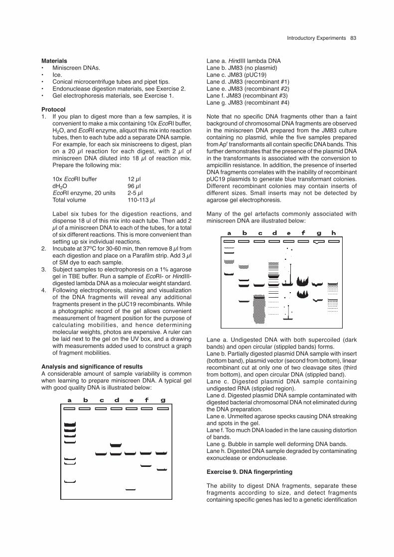
Analysis and significance of resultsA considerable amount of sample variability is commonwhen learning to prepare miniscreen DNA. A typical gelwith good quality DNA is illustrated below:
Lane a. HindIII lambda DNALane b. JM83 (no plasmid)Lane c. JM83 (pUC19)Lane d. JM83 (recombinant #1)Lane e. JM83 (recombinant #2)Lane f. JM83 (recombinant #3)Lane g. JM83 (recombinant #4)
Note that no specific DNA fragments other than a faintbackground of chromosomal DNA fragments are observedin the miniscreen DNA prepared from the JM83 culturecontaining no plasmid, while the five samples preparedfrom Apr transformants all contain specific DNA bands. Thisfurther demonstrates that the presence of the plasmid DNAin the transformants is associated with the conversion toampicillin resistance. In addition, the presence of insertedDNA fragments correlates with the inability of recombinantpUC19 plasmids to generate blue transformant colonies.Different recombinant colonies may contain inserts ofdifferent sizes. Small inserts may not be detected byagarose gel electrophoresis.
Many of the gel artefacts commonly associated withminiscreen DNA are illustrated below:
Lane a. Undigested DNA with both supercoiled (darkbands) and open circular (stippled bands) forms.Lane b. Partially digested plasmid DNA sample with insert(bottom band), plasmid vector (second from bottom), linearrecombinant cut at only one of two cleavage sites (thirdfrom bottom), and open circular DNA (stippled band).Lane c. Digested plasmid DNA sample containingundigested RNA (stippled region).Lane d. Digested plasmid DNA sample contaminated withdigested bacterial chromosomal DNA not eliminated duringthe DNA preparation.Lane e. Unmelted agarose specks causing DNA streakingand spots in the gel.Lane f. Too much DNA loaded in the lane causing distortionof bands.Lane g. Bubble in sample well deforming DNA bands.Lane h. Digested DNA sample degraded by contaminatingexonuclease or endonuclease.
Exercise 9. DNA fingerprinting
The ability to digest DNA fragments, separate thesefragments according to size, and detect fragmentscontaining specific genes has led to a genetic identification

84 Tait
approach called DNA fingerprinting. This method takesadvantage of the fact that a single nucleotide sequencedifference can cause the appearance or disappearance ofa restriction enzyme cleavage site in DNA. The individual-specific differences in restriction enzyme cleavage siteposition relative to a specific gene can cause variation inthe size of the DNA restriction fragment that carries thatgene in different individuals. The variation in the size of arestriction fragment that carries a specific gene, referredto as “restriction fragment length polymorphism” (RFLP),can be used to genetically match DNA samples with thedonors from which they were obtained. Individuals whoappear identical for a specific physical trait can be easilydistinguished if sequence differences that cause changesin DNA restriction fragment sizes are associated with thattrait.
When applied to identification of human DNA samples,DNA fingerprint analysis is complicated by the presenceof hundreds or thousands of DNA fragments followingdigestion of DNA samples with a specific restrictionenzyme. Rather than attempt to analyze all of the DNAfragments, the fragments are generally separated by gelelectrophoresis, transferred to a membrane, and allowedto react with a DNA or RNA hybridization probe that willspecifically anneal to a small number of the many differentDNA fragments. The probe fragment generally contains anucleoside that is either radioactive (such as 32PdATP) andcan be detected by autoradiography or has been modified(such as biotinylated dATP) to be recognized by specificantibodies and detected by a color reaction. The imagethat is obtained by visualization of the probe is a subset ofthe total set of DNA fragments and can be referred to as aDNA fingerprint. When hybridization probes are chosencarefully, the fingerprints can be specific for DNA that hasbeen found to have a high degree of individual-specificvariation. When sufficient information has been obtainedabout the frequency of occurence of a specific DNAfingerprint pattern, the DNA fingerprints of two DNAsamples can be used to calculate the probability of whetherthe two DNA samples were obtained from the sameindividual.
Since humans are diploid and contain two copies ofeach chromosome, one from the mother and the other fromthe father, a human DNA fingerprint can be thought of asthe combination of two fingerprint patterns. Since thesepatterns can be used to link an “unknown” DNA sample toDNA samples obtained from specific individuals, DNAfingerprint analysis is becoming increasingly common ascourtroom evidence in paternity suits and in criminalprosecution involving crimes like rape and murder. Thisexercise will use bacteriophage lambda DNA digested withvarious restriction enzymes to simulate the results of a DNAfingerprint analysis and help determine paternity of a child.
MaterialsBacteriophage lambda DNA at a final concentration of 50µg/ml in gel loading buffer, digested with the followingrestriction enzymes: HindIII (H), EcoRI (E), BamHI (B), KpnI(K), and SalI (S). Each of these digests will be used tosimulate a haploid DNA fingerprint (one set ofchromosomes). To make the test DNA samples obtainedfrom the individuals involved in the paternity suit, thedigested lambda DNA samples have been mixed in thefollowing manner to simulate the diploid chromosomal
fingerprint of each individual:
Mother = 100 µl H + 100 µl H (HH)Baby = 100 µl H + 100 µl E (HE)Rock Star = 100 µl H + 100 µl K (HK)Ex-drummer = 100 µl S + 100 µl B (SB)Mailman = 100 µl S + 100 µl E (SE)
Loading 7 µl of each mix will deliver 350 ng of DNA, givingdistinctive diploid DNA fingerprint patterns for eachindividual.
• Gel box, gel tray and comb.• Power supply.• UV light source.• Dry agarose.• 10 mg/ml ethidium bromide.• 10X TBE gel buffer.
BackgroundWhile living with a rock star, a young woman becomespregnant, whereupon the rock star promptly severs hisrelationship with the woman, leaving her and the resultantbaby completely penniless. The mother of the child suesfor child support, claiming the rock star as the father of herbaby. The following are the summary statements from eachof the individuals who become involved in the resultingpaternity suit:
Mother: Rock Star is the father of my baby. He shouldbe required to financially support us.
Baby: Wah! Burp.Rock Star: The child is not my son. The males in my
family were found through genetic analysisto carry a recessive gene that causesincurable craving for disco music. On theadvice of my doctor, I had a vasectomy whenI was twenty-three to prevent transmissionof this genetic disorder to my children. Ikicked her out because I caught her in bedwith my Drummer, not because she waspregnant. She should talk to him aboutsupport.
Ex-drummer: Who says I’m the father? I wasn’t the onlyother guy she was seeing. I even caught themailman in the bedroom with her. I couldn’tsupport her anyways. I can’t get work sinceRock Star kicked me out of his band and Igot busted with 2 pounds of heroin.
Mailman: Sure I was in her bedroom. I had a packagefor Rock Star and needed a signature. WhenI rang the doorbell, she called out she wassick in bed, but the door was unlocked andshe would sign for the package if I didn’tmind risking the flu. I was leaving herbedroom when Drummer walked in the frontdoor. I think the door was unlocked and shewas in bed waiting for Drummer.
At the request of the court, DNA is extracted from bloodsamples obtained from each of these individuals andprovided to you, who will perform the fingerprint analysisand interpret the results.

Introductory Experiments 85
Procedure1. Weigh out 0.5 gram of the agarose and place in a 100
ml flask or beaker. Add 45 ml of distilled water and 5ml of 10X TBE buffer.
2. Melt the agarose completely in a microwave oven orboiling water bath. The solution must boil to melt theagarose. Agarose has a tendency to superheat andflash-boil, so be careful on the first boiling. The boilingwill be more controlled once some of the agarose hasmelted. Be certain all the small particles are melted toprevent streaks in the gel. The agarose should beallowed to cool to about 55oC. It will solidify at about45oC.
3. Add 2 µl of ethidium bromide to the agarose and mix.This is the stain to detect the DNA. Ethidium bromideis a mutagen (comparable to a pack of cigarettes) andsolutions containing ethidium should not be handledwith bare hands.
4. Pour a gel as demonstrated and allow to solidify.5. Assemble the gel in the gel box. Submerge the gel in
1X TBE running buffer made by diluting 10 ml of 10XTBE Buffer with 90 ml of distilled water.
6. Load the samples in the following order and amount:
Lane DNA Sample Amount
1 Mother 7 µl2 Baby 7 µl3 Rock Star 7 µl4 Drummer 7 µl5 Mailman 7 µl6 Baby 7 µl
7. Plug in the leads and turn on the power. Voltage ofaround 80 to 120 volts is typical, but will vary dependingon the gel box. A very low voltage will slow the rundown, but will enhance resolution of the DNA bands.A very high voltage will heat the buffer and can actuallymelt the gel. The depth of the buffer determines howmuch current flows in the system. Use only enoughbuffer to just submerge the gel to prevent excessiveheating of the gel (deep buffer = high current = highheat production).
8. As the run progresses, two blue bands will resolve inthe gel. These are merely tracking dyes to judge howfar the DNA has migrated. When the faster dye isreaches the bottom of the gel, turn off the power.Remove the gel from the box and expose to the UVlight. Be careful to shield eyes and skin from the UVlight, as it is both a mutagen and a skin and eye burningagent.
9. Either photograph or sketch the resulting bandpatterns. Analyze the DNA band patterns. Rememberthat the DNA fingerprint of each person will containtwo sets of bands, one from each parent. These couldbe either two identical sets of bands, as in the case ofthe Mother (HH), or two different sets of bands, as inthe case of the Baby (HE). The Mother must haveobtained one H set of bands from her mother and oneH set of bands from her father. The Baby must alsohave obtained one set of bands from its mother andone set from its father.
Questions you will need to consider:1. Is Rock Star the father of the Baby? Why or why not?2. Is Drummer the father of the Baby? Why or why not?3. Is Mailman the father of the Baby? Why or why not?4. The probability that a DNA fingerprint will contain a
particular DNA band pattern is dependent on thefrequency of that particular band pattern in the generalpopulation. You are now provided with the informationthat the entire population of the USA has beenscreened with the DNA probes you have just used andthe frequencies of occurrence of the H, E, K, B, and Sband patterns have been determined to be as follows:
Band pattern Frequency in populationH 1 in 100 peopleE 1 in 10 peopleK 1 in 100,000 peopleB 1 in 100,000 peopleS 1 in 50 people
Does this information change your answers toquestions 1-3 above, and why or why not?
5. As the court-appointed expert in DNA fingerprints, whatis your recommendation to the court regarding thepaternity suit and the identity of the father of the Baby?
Further Reading
Tait, R.C. 1997. An Introduction to Molecular Biology.Horizon Scientific Press, Wymondham, UK.
• MALDI-TOF Mass Spectrometry in Microbiology
Edited by: M Kostrzewa, S Schubert (2016) www.caister.com/malditof
• Aspergillus and Penicillium in the Post-genomic Era
Edited by: RP Vries, IB Gelber, MR Andersen (2016) www.caister.com/aspergillus2
• The Bacteriocins: Current Knowledge and Future Prospects
Edited by: RL Dorit, SM Roy, MA Riley (2016) www.caister.com/bacteriocins
• Omics in Plant Disease Resistance
Edited by: V Bhadauria (2016) www.caister.com/opdr
• Acidophiles: Life in Extremely Acidic Environments
Edited by: R Quatrini, DB Johnson (2016) www.caister.com/acidophiles
• Climate Change and Microbial Ecology: Current Research and Future Trends
Edited by: J Marxsen (2016) www.caister.com/climate
• Biofilms in Bioremediation: Current Research and Emerging Technologies
Edited by: G Lear (2016) www.caister.com/biorem
• Microalgae: Current Research and Applications
Edited by: MN Tsaloglou (2016) www.caister.com/microalgae
• Gas Plasma Sterilization in Microbiology: Theory, Applications, Pitfalls and New Perspectives
Edited by: H Shintani, A Sakudo (2016) www.caister.com/gasplasma
• Virus Evolution: Current Research and Future Directions
Edited by: SC Weaver, M Denison, M Roossinck, et al. (2016) www.caister.com/virusevol
• Arboviruses: Molecular Biology, Evolution and Control
Edited by: N Vasilakis, DJ Gubler (2016) www.caister.com/arbo
• Shigella: Molecular and Cellular Biology
Edited by: WD Picking, WL Picking (2016) www.caister.com/shigella
• Aquatic Biofilms: Ecology, Water Quality and Wastewater Treatment
Edited by: AM Romaní, H Guasch, MD Balaguer (2016) www.caister.com/aquaticbiofilms
• Alphaviruses: Current Biology
Edited by: S Mahalingam, L Herrero, B Herring (2016) www.caister.com/alpha
• Thermophilic Microorganisms
Edited by: F Li (2015) www.caister.com/thermophile
• Flow Cytometry in Microbiology: Technology and Applications
Edited by: MG Wilkinson (2015) www.caister.com/flow
• Probiotics and Prebiotics: Current Research and Future Trends
Edited by: K Venema, AP Carmo (2015) www.caister.com/probiotics
• Epigenetics: Current Research and Emerging Trends
Edited by: BP Chadwick (2015) www.caister.com/epigenetics2015
• Corynebacterium glutamicum: From Systems Biology to Biotechnological Applications
Edited by: A Burkovski (2015) www.caister.com/cory2
• Advanced Vaccine Research Methods for the Decade of Vaccines
Edited by: F Bagnoli, R Rappuoli (2015) www.caister.com/vaccines
• Antifungals: From Genomics to Resistance and the Development of Novel Agents
Edited by: AT Coste, P Vandeputte (2015) www.caister.com/antifungals
• Bacteria-Plant Interactions: Advanced Research and Future Trends
Edited by: J Murillo, BA Vinatzer, RW Jackson, et al. (2015) www.caister.com/bacteria-plant
• Aeromonas
Edited by: J Graf (2015) www.caister.com/aeromonas
• Antibiotics: Current Innovations and Future Trends
Edited by: S Sánchez, AL Demain (2015) www.caister.com/antibiotics
• Leishmania: Current Biology and Control
Edited by: S Adak, R Datta (2015) www.caister.com/leish2
• Acanthamoeba: Biology and Pathogenesis (2nd edition)
Author: NA Khan (2015) www.caister.com/acanthamoeba2
• Microarrays: Current Technology, Innovations and Applications
Edited by: Z He (2014) www.caister.com/microarrays2
• Metagenomics of the Microbial Nitrogen Cycle: Theory, Methods and Applications
Edited by: D Marco (2014) www.caister.com/n2
Caister Academic Press is a leading academic publisher of advanced texts in microbiology, molecular biology and medical research. Full details of all our publications at caister.com
Further Reading
Order from caister.com/order


![INDIAN INSTITUTE OF TECHNOLOGY BOMBAY Aerospace Engineering (AE) [Department of Aerospace Engineering] Specialization : Aerodynamics (AE1) Dynamics and Control (AE2)](https://img.dokumen.tips/doc/110x75/5afd3acb7f8b9a944d8d24af/indian-institute-of-technology-bombay-aerospace-engineering-ae-department-of.jpg)