Embed Size (px)
Citation preview

Br J Sports Med. 2014;48(7):532-539
Acute Hamstring Injuries in Swedish Elite Sprinters and Jumpers: A Prospective Randomised Controlled Clinical Trial
Comparing Two Rehabilitation Protocols
Carl M Askling, Magnus Tengvar, Olga Tarassova and Alf Thorstensson
Abstract and Introduction
Abstract
Background: Hamstring strain is a common injury in sprinters and jumpers, and
therefore time to return to sport and secondary prevention become of particular
concern.
Objective: To compare the effectiveness of two rehabilitation protocols after acute
hamstring injury in Swedish elite sprinters and jumpers by evaluating time needed
to return to full participation in the training process.
Study design: Prospective randomised comparison of two rehabilitation protocols.
Methods: Fifty-six Swedish elite sprinters and jumpers with acute hamstring injury,
verified by MRI, were randomly assigned to one of two rehabilitation protocols.
Twenty-eight athletes were assigned to a protocol emphasising lengthening
exercises, L-protocol, and 28 athletes to a protocol consisting of conventional
exercises, C-protocol. The outcome measure was the number of days to return to
full training. Re-injuries were registered during a period of 12 months after return.
Results: Time to return was significantly shorter for the athletes in the L-protocol,
mean 49 days (1SD±26, range 18–107 days), compared with the C-protocol, mean
86 days (1SD±34, range 26–140 days). Irrespective of protocol, hamstring injuries
where the proximal free tendon was involved took a significantly longer time to
return than injuries that did not involve the free tendon, L-protocol: mean 73 vs 31
days and C-protocol: mean 116 vs 63 days, respectively. Two reinjuries were
registered, both in the C-protocol.

Conclusions: A rehabilitation protocol emphasising lengthening type of exercises
is more effective than a protocol containing conventional exercises in promoting
time to return in Swedish elite sprinters and jumpers.
Introduction
Acute hamstring injury is common in track and field athletes, especially in elite
sprinters and jumpers.[1–7]Furthermore, hamstring injuries are a heterogeneous
group consisting of different injury types, locations and sizes, which makes
recommendations regarding rehabilitation and prognosis about healing time
difficult.[8–14]The reinjury rate is high,[15–17] which may indicate inadequate
rehabilitation programmes and/or premature return to sports.[18,19]
There is a lack of clinical research and consensus, based on prospective,
randomised studies, regarding the effectiveness of various rehabilitation protocols
for acute hamstring injuries in elite sprinters and jumpers. Overall, few studies until
now have evaluated the effectiveness of different treatment protocols for acute
hamstring injuries in any type of sport.[20,21] However, recently a study on Swedish
elite football players showed that a protocol aimed at loading the hamstrings during
extensive lengthening mainly during eccentric muscle actions was significantly
more effective compared with a conventional protocol in promoting time to return
to play after acute hamstring injury.[13] It is not possible to generalise these results
to other elite athletes in different sports, with a different training-process and with
other demands on the hamstring muscle group without performing the same
protocol in a new clinical trial. The current investigation uses the same
methodological approach as the study on elite football players.[13]
Aim
The main objective of this study on Swedish elite sprinters and jumpers was to
compare the effectiveness of two rehabilitation protocols for acute hamstring
injuries with varying emphasis on muscle-tendon lengthening by evaluating time
needed to return to full participation in the training process. Other aims were to
study possible associations between injury type, location, size, palpation pain and
time to return.
Material and Methods
Swedish male and female elite sprinters and jumpers were enrolled using our
extensive contacts with medical teams and coaches working with elite track and
field athletes in Sweden. These athletes were juniors (between 15 and 19 years)

and seniors (20 years and older) ranked among the top 20 in each discipline
indoors and/or outdoors. Also, the Swedish Athletic Association took part by
informing all major track and field clubs in Sweden about the study. The total
recruitment time was 38 months, January 2009–February 2012. Forty-six sprinters
and 10 jumpers (of whom 8 were horizontal jumpers) were included, all with clinical
signs of acute hamstring injury, as confirmed by MRI. A randomisation process
was used to assign athletes to either of the two protocols, the L-protocol or the C-
protocol, respectively. Athletes were stratified into subgroups according to gender,
injury type (ie, sprinting-type or stretching-type injury) and proximal free tendon
involvement or not (see below). The first athlete in each stratified subgroup was
randomised to either the L-protocol or C-protocol using a dice. Subsequent athletes
in each subgroup were then alternated between protocols. The allocation of each
athlete in a subgroup was therefore dependent on the randomised allocation of the
first athlete in that subgroup. In addition, eight sprinters and jumpers with clinical
signs of acute hamstring injury, but where the MRI showed no sign of injury, were
followed in parallel. These MRI-negative athletes were all assigned to the L-
protocol. The use of non-steroidal anti-inflammatory drugs (NSAIDs) and/or other
pain reducing medicine during the rehabilitation period was not allowed. All athletes
gave their informed consent prior to participation. Approval of the study was
granted by the Regional Ethics Committee (Dnr: 2008/1320-31/2). There were no
dropouts in the study.
Inclusion/Exclusion Criteria
To be included, the athlete had to have sustained acute sudden pain in the
posterior thigh that immediately forced the athlete to stop the activity, training or
competing. The initial clinical examination had to reveal localised pain when
palpating the hamstring muscles, localised pain when performing a passive straight
leg raise test, and increased pain when adding an isometric hamstring contraction
during that test.[8] Exclusion criteria included verified or suspected earlier hamstring
injury in the same leg during the past 6 months, extrinsic trauma to the posterior
thigh, ongoing or chronic low back problems and pregnancy.
Injury Situation—Type of Injury
At the first visit, the athletes were interviewed by the same test-leader (CMA) about
the injury situation, that is, the movements or exercises at which the acute injury
occurred, whether it was during a training session or competition. In addition, the
type of injury was established, that is, sprint-type injury, happening at high-speed

running[9,12] or stretch-type injury, occurring during slow stretching to extreme
muscle lengths.[10,11]
Clinical Examination
All athletes were examined within 2 days after the injury. The clinical examination
included manual assessment of flexibility and strength of the injured and uninjured
leg. The uninjured leg was always tested before the injured leg, and flexibility
before strength. Flexibility was measured with a passive straight leg raise test (until
the athlete reported pain and/or discomfort) and a standard flexometer.[8] The
isometric strength test was performed with the athlete in a prone position with
resistance applied at the heel at 15° and 45° of knee flexion. The strength was
tested manually by performing a combined knee flexion and hip extension due to
the bi-articular nature of the hamstring muscles. Hamstring strength was also
tested in a lengthened state in a supine position with the knee positioned at 15° of
flexion while resistance was applied to the heel. The foot was maintained in plantar
flexion in order to limit the activation of the gastrocnemius muscle. A bilateral
comparison was performed for each measurement.
The point of peak palpation pain was recorded and the distance between that point
and the ischial tuberosity was measured.[8] The same test-leader (CMA) performed
this clinical examination weekly until there were no signs of injury remaining. The
decision by the test-leader (CMA) that there were no signs of injury remaining had
to be confirmed by an independent colleague by performing the same clinical
examination.
Askling H-test
When the clinical examination at the end of the rehabilitation showed no signs of
injury remaining, the Askling H-test was performed.[22] A simplified version of the H-
test, without an electrogoniometer but with a knee brace to keep the leg in
extension and straps stabilising the upper body and the contralateral leg, was
performed. The instruction to the athlete was to perform a straight leg raise as fast
as possible to the highest point without taking any risk of injury (three trials per leg,
uninjured leg tested first; no warm-up). If the athlete experienced any insecurity
during this voluntary straight leg raising (on a Visual Analogue Scale, from 0 to 10),
he/she was not allowed to go back to full training. Instead, the rehabilitation period
was extended and the H-test repeated with an interval of 3–5 days until insecurity
was eliminated.
MRI
All athletes underwent an MRI investigation within 5 days after the acute injury.
MRI investigations were performed on a 1.5 Tesla superconductive MRI unit
(Magnetom Symphony, Siemens, Erlangen, Germany). Briefly, longitudinal,
sagittal and frontal short tau inversion recovery (STIR) images as well as
transversal T1-weighted and STIR images (5 mm slice thickness and 0.5 mm gap)
were obtained from both legs.[9] All MRI investigations were assessed and reported
by one of the radiologists (MT). A muscle was considered injured when it contained
high-signal intensity (oedema) on the STIR images, as compared with the
uninjured side. If more than one muscle/tendon was injured, the one with the
greatest signal abnormality was considered the 'primary' injury and the second
largest, the 'secondary' injury. In this study, MRI parameters were quantified only
for the primary injury. The free proximal tendon (PT) was deemed injured if 2 of the
3 following parameters were present: the tendon was thickened, and/or had a collar
of high-signal intensity around it, and/or had high intratendinous signal intensity, as
compared with the uninjured side. The maximal longitudinal length of the
muscle/tendon oedema was measured.[9] In addition, the perpendicular distance
between the level of the most proximal pole of the oedema and the level of the
most distal part of the ischial tuberosity was measured.[9]
Specific Rehabilitation Protocols
The time from the date of injury to the date of rehabilitation protocol initiation was
5 days for both protocols. Overall, exercises were chosen that could be performed
in any place and without the use of advanced equipment. The exercises of the L-
protocol specifically aimed at loading the hamstrings during extensive lengthening,
mainly during eccentric muscle actions. In contrast, the C-protocol consisted of
conventional exercises for the hamstrings with less emphasis on lengthening. Each
rehabilitation protocol consisted of three different exercises, where exercise 1 was
aimed mainly at increasing flexibility, exercise 2 was a combined exercise for
strength and trunk/pelvis stabilisation and exercise 3 was more of a specific
strength training exercise.[13] All exercises were performed in the sagittal plane. The
intensity and volume of training were made as equal as possible between the two
protocols. The training sessions were supervised, at least once every week, during
the whole rehabilitation period, and the speed and load were increased over time.
No pain provocation was allowed at any time when performing the exercises. All
exercises included in the two rehabilitation protocols are explained in figures 1–6.
Video demonstrations of all six exercises (L-protocol and C-protocol) showing how
the progression can be performed are included as supplemental files.
(Enlarge Image)
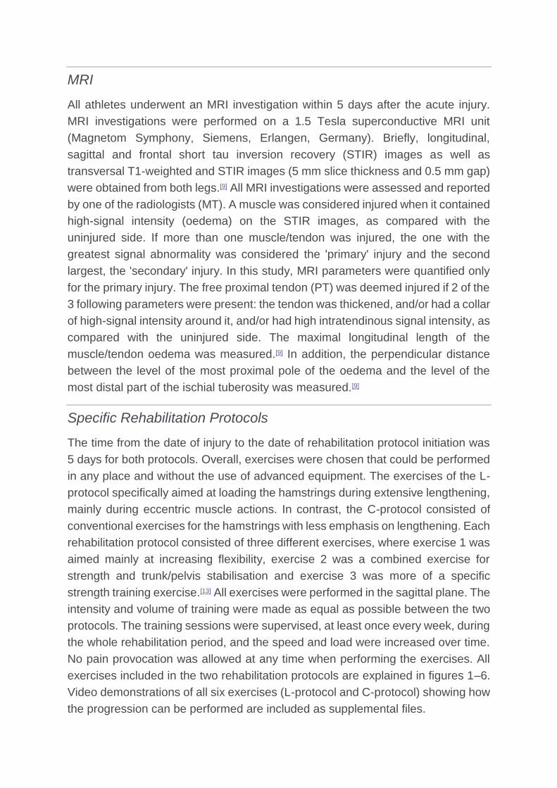
Figure 1.
L-1 'The Extender': the player should hold and stabilise the thigh of the injured leg with the hip
flexed approximately 90° and then perform slow knee extensions to a point just before pain is felt.
Twice every day, 3 sets with 12 repetitions (see online supplementry video 1).
(Enlarge Image)
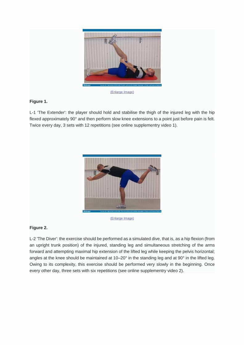
Figure 2.
L-2 'The Diver': the exercise should be performed as a simulated dive, that is, as a hip flexion (from
an upright trunk position) of the injured, standing leg and simultaneous stretching of the arms
forward and attempting maximal hip extension of the lifted leg while keeping the pelvis horizontal;
angles at the knee should be maintained at 10–20° in the standing leg and at 90° in the lifted leg.
Owing to its complexity, this exercise should be performed very slowly in the beginning. Once
every other day, three sets with six repetitions (see online supplementry video 2).

(Enlarge Image)
Figure 3.
L-3 'The Glider': the exercise is started from a position with upright trunk, one hand holding on to
a support and legs slightly split. All the body weight should be on the heel of the injured (here left)
leg with approximately 10–20° flexion in the knee. The motion is started by gliding backwards on
the other leg (note the low friction sock) and stopped before pain is reached. The movement back
to the starting position should be performed by the help of both arms, not using the injured leg.
Progression is achieved by increasing the gliding distance and performing the exercise faster.
Once every third day, three sets with four repetitions (see online supplementry video 3).
(Enlarge Image)
Figure 4.
C-1 Stretching—contract/relax. The heel of the injured leg is placed on a stable support surface in
a high position (close to maximum) with the knee in approximately 10° flexion. The heel is pressed

down for 10 s and then, after relaxation for 10 s, a new position is assumed by flexing the upper
body slowly forwards for 20 s. Twice a day, three sets with four repetitions (see online
supplementry video 4).
(Enlarge Image)
Figure 5.
C-2 Cable-pendulum: A stationary cable-machine or expander is used. With the uninjured leg as
the standing leg, forward–backward hip motions are performed with the injured leg with the knee
in approximately 20–30° flexion. This exercise involves the whole body and should be performed
slowly in the beginning of the rehabilitation period. Once every other day, three sets with six
repetitions (see online supplementry video 5).
(Enlarge Image)
Figure 6.
C-3 Pelvic lift: This exercise is started in a supine position with the body weight on both heels, and
then the pelvis is lifted up and down slowly. Start with the knee in 90° of flexion. The load is
increased by putting more of the body weight on the injured leg and by having a greater extension

in the knee. Ultimately, only the slightly bent injured leg is carrying the load. Every third day, three
sets with eight repetitions (see online supplementry video 6).
General Rehabilitation Programme
A general rehabilitation programme with a subject-specific progression was
followed by all athletes in both specific protocols. The general programme was
implemented by the test-leader (CMA) week by week and supervised by the PT
responsible for the athlete in the track and field club during the entire rehabilitation
period. No pain and/or discomfort was allowed from the injured posterior thigh
during the rehabilitation process. Acutely, the athlete should use crutches if pain
was provoked by walking.
The general programme was performed three times a week and started with
stationary cycling 10 min, 10×20 s fast foot stepping in place, 10×jogging 40 m with
short strides, 10×10 m forward/backward accelerations. When the above part of
the general programme could be performed without pain and/or discomfort, a
progressive running programme was started. This was composed of high-speed
running drills 6×20, 4×40 and 2×60 m, performed three times a week.
In addition to the specific protocol and the general programme, all athletes were
asked to conduct as much as possible of their standard training programme without
experiencing any pain and/or discomfort. This training was supervised by their
regular track and field coaches.
Outcome
The main outcome was time to return, that is, time from injury to full participation
in the training process. Also, occurrence of reinjuries was registered during a 12
month period after return. If a re-injury occurred, the medical team responsible was
to immediately contact the study leader so that the same procedure as for the
original injury could be repeated. The full 1-year follow-up period was completed
by all athletes in the study.
Statistical Analyses
All statistical analyses were conducted with STATISTICA V.11.0 software (StatSoft
Inc). The Shapiro-Wilk W tests showed that the data were not normally distributed.
A Mann-Whitney U test was performed to investigate differences in age, height and
mass as well as MRI and palpation measures. A χ2 test was applied to investigate

differences in proportion of injury type and PT involvement as well as in gender.
The Mann-Whitney U test was also used to assess differences in time to return
between the protocols, between subgroups with respect to PT involvement (with
Bonferroni correction applied), as well as between MRI-negative athletes and
athletes with sprinting-type injury within the L-protocol. The Mann-Whitney U test
(Cohen's d) for independent samples was applied and the χ2 test (p) was used as
a measure of effect size. Spearman's rank order correlations were calculated to
investigate associations between time to return and MRI and palpation parameters.
The significance level was set at p≤0.05.
Results
Injury Situation, Type and Location
Thirty (52%) of all the 56 MRI-verified injuries occurred during competition, 14
(50%) in the L-protocol and 16 (57%) in the C-protocol, respectively. Fifty-two
(93%) of the 56 injuries were sprinting-type and 4 (7%) stretching-type injuries. In
44 of the 56 athletes (79%), the primary injury was located in the long head of
biceps femoris (BFlh) and in 7 of those 44 (16%), there was a secondary injury, in
all cases located in the semitendinosus (ST). In the remaining eight injuries of
sprinting type, the primary injury was located in the ST. In the four athletes with a
stretching-type injury, the injury location was in the semimembranosus.
Characterisation of Protocol Participants
There were no significant differences between groups of athletes in the L-protocol
and the C-protocol with respect to age, height, mass, gender, performance level,
type of injury and involvement of the proximal free tendon (Table 1). Neither were
there any group differences in distance between the most proximal pole of the
oedema and the ischial tuberosity, length of the oedema and distance between the
point of peak palpation pain and the ischial tuberosity (Table 1).
Askling H-test
Eight athletes (29%) in the L-protocol and 19 (68%) in the C-protocol experienced
insecurity when performing the H-test and therefore needed to extend their
rehabilitation period. On average, the time to return was prolonged by 8 days
(1SD±3.0, range 3–14) in the L-protocol and by 10 days (1SD±3.5, range 4–20) in
the C-protocol, respectively.
Time to Return
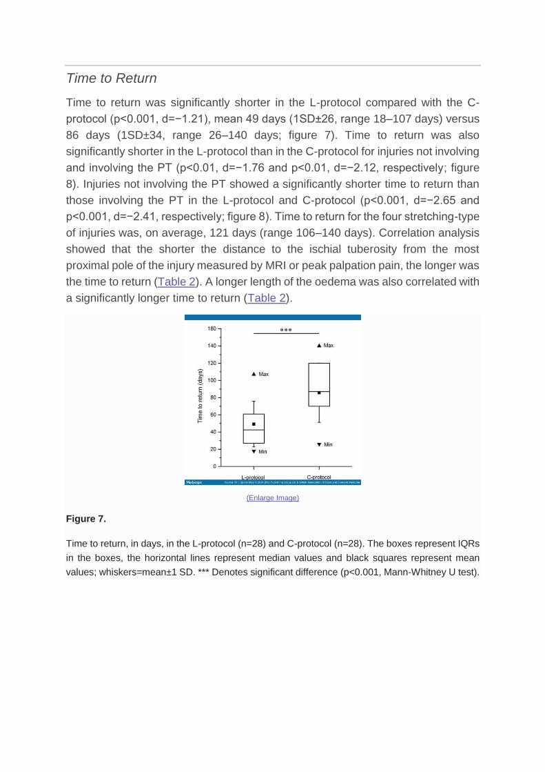
Time to return was significantly shorter in the L-protocol compared with the C-
protocol (p<0.001, d=−1.21), mean 49 days (1SD±26, range 18–107 days) versus
86 days (1SD±34, range 26–140 days; figure 7). Time to return was also
significantly shorter in the L-protocol than in the C-protocol for injuries not involving
and involving the PT (p<0.01, d=−1.76 and p<0.01, d=−2.12, respectively; figure
8). Injuries not involving the PT showed a significantly shorter time to return than
those involving the PT in the L-protocol and C-protocol (p<0.001, d=−2.65 and
p<0.001, d=−2.41, respectively; figure 8). Time to return for the four stretching-type
of injuries was, on average, 121 days (range 106–140 days). Correlation analysis
showed that the shorter the distance to the ischial tuberosity from the most
proximal pole of the injury measured by MRI or peak palpation pain, the longer was
the time to return (Table 2). A longer length of the oedema was also correlated with
a significantly longer time to return (Table 2).
(Enlarge Image)
Figure 7.
Time to return, in days, in the L-protocol (n=28) and C-protocol (n=28). The boxes represent IQRs
in the boxes, the horizontal lines represent median values and black squares represent mean
values; whiskers=mean±1 SD. *** Denotes significant difference (p<0.001, Mann-Whitney U test).
(Enlarge Image)
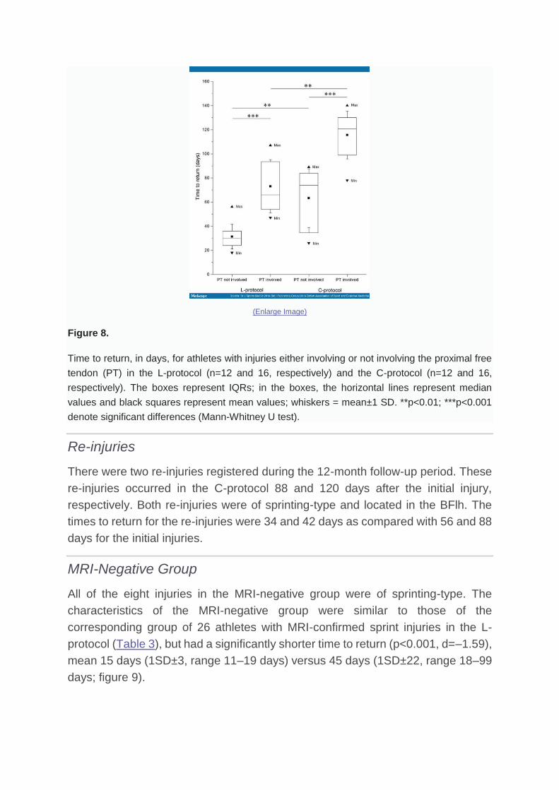
Figure 8.
Time to return, in days, for athletes with injuries either involving or not involving the proximal free
tendon (PT) in the L-protocol (n=12 and 16, respectively) and the C-protocol (n=12 and 16,
respectively). The boxes represent IQRs; in the boxes, the horizontal lines represent median
values and black squares represent mean values; whiskers = mean±1 SD. **p<0.01; ***p<0.001
denote significant differences (Mann-Whitney U test).
Re-injuries
There were two re-injuries registered during the 12-month follow-up period. These
re-injuries occurred in the C-protocol 88 and 120 days after the initial injury,
respectively. Both re-injuries were of sprinting-type and located in the BFlh. The
times to return for the re-injuries were 34 and 42 days as compared with 56 and 88
days for the initial injuries.
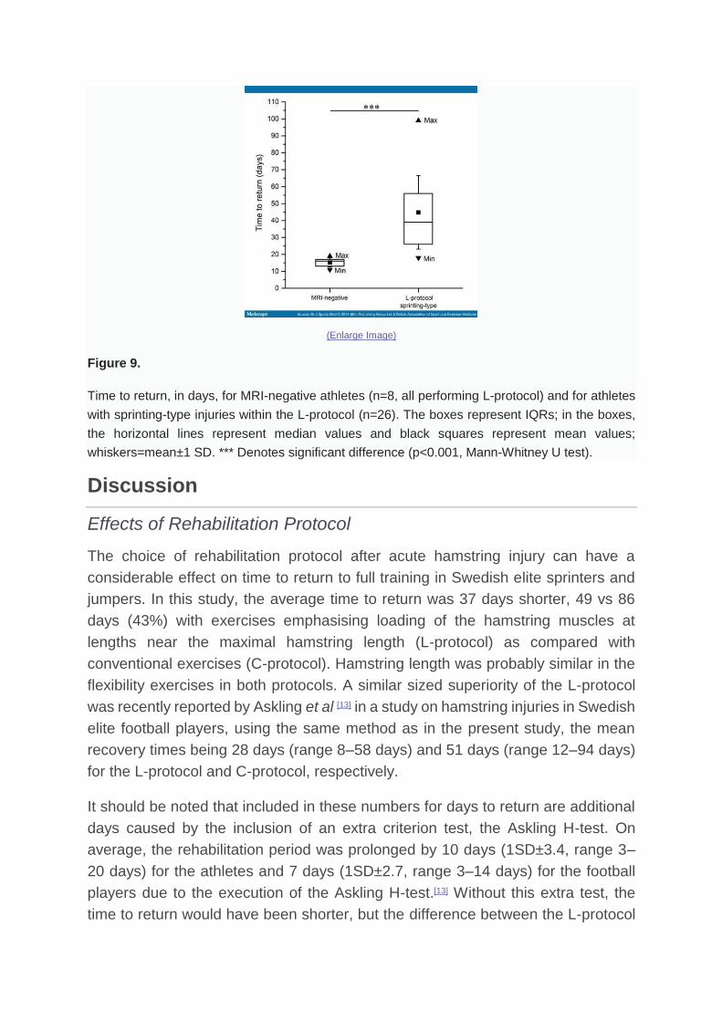
MRI-Negative Group
All of the eight injuries in the MRI-negative group were of sprinting-type. The
characteristics of the MRI-negative group were similar to those of the
corresponding group of 26 athletes with MRI-confirmed sprint injuries in the L-
protocol (Table 3), but had a significantly shorter time to return (p<0.001, d=–1.59),
mean 15 days (1SD±3, range 11–19 days) versus 45 days (1SD±22, range 18–99
days; figure 9).
(Enlarge Image)
Figure 9.
Time to return, in days, for MRI-negative athletes (n=8, all performing L-protocol) and for athletes
with sprinting-type injuries within the L-protocol (n=26). The boxes represent IQRs; in the boxes,
the horizontal lines represent median values and black squares represent mean values;
whiskers=mean±1 SD. *** Denotes significant difference (p<0.001, Mann-Whitney U test).
Discussion
Effects of Rehabilitation Protocol
The choice of rehabilitation protocol after acute hamstring injury can have a
considerable effect on time to return to full training in Swedish elite sprinters and
jumpers. In this study, the average time to return was 37 days shorter, 49 vs 86
days (43%) with exercises emphasising loading of the hamstring muscles at
lengths near the maximal hamstring length (L-protocol) as compared with
conventional exercises (C-protocol). Hamstring length was probably similar in the
flexibility exercises in both protocols. A similar sized superiority of the L-protocol
was recently reported by Askling et al [13] in a study on hamstring injuries in Swedish
elite football players, using the same method as in the present study, the mean
recovery times being 28 days (range 8–58 days) and 51 days (range 12–94 days)
for the L-protocol and C-protocol, respectively.
It should be noted that included in these numbers for days to return are additional
days caused by the inclusion of an extra criterion test, the Askling H-test. On
average, the rehabilitation period was prolonged by 10 days (1SD±3.4, range 3–
20 days) for the athletes and 7 days (1SD±2.7, range 3–14 days) for the football
players due to the execution of the Askling H-test.[13] Without this extra test, the
time to return would have been shorter, but the difference between the L-protocol

and the C-protocol would still have remained highly significant, for example, mean
47 days vs 79 days for the athletes. It seems most likely that the requirement of a
secure H-test for everyone before being allowed to return to full
training/competition would have functioned to prevent re-injuries. In this study, only
two re-injuries occurred among the 56 athletes (3%) during the 12 month follow-up
(in the footballers: only 1 of 75).[13] This is considerably lower that the recurrence
rates of 14–25% reported earlier for these types of sports.[16,23,24] It is worth noting
that should the hamstring injury recur, the second injury is usually more severe
than the first, typically requiring a longer time away from sport than the original
one.[16,23,24]
The exercises in the L-protocol and C-protocol were selected based on practical
experience. This includes a number of exercises, joint excursions and speed.
Progression was steered by the avoidance of pain. Considering the lack of
objective data, we have chosen to describe the protocols in quite some detail for
the readers/users to form their own opinions. It is our belief that the rather
remarkable difference in outcome between the two protocols is mainly due to the
more systematic attempts to put load on the hamstrings during maximal dynamic
lengthening in the L-programme, involving movements at the hip and the knee.
Otherwise, the two protocols were made as similar as possible in terms of early
start after injury, thorough instruction and regular follow-up and progression in
load/speed/excursion based on the avoidance of the pain criterion.
It is proposed that neuromuscular inhibition of hamstring voluntary activation
occurs following acute hamstring injury, and that this inhibition has a negative effect
on hamstring recovery by limiting hamstring load during lengthening
exercises.[18,19] This limited exposure to eccentric stimuli at long hamstring muscle
lengths could potentially lead to eccentric hamstring weakness and selective
hamstring atrophy, possibly in combination with selective hypertrophy of the short
head of biceps femoris,[18] resulting in a shift in the torque–angle relationship.[19] The
L-protocol used in the present study was aimed to stress the injured hamstrings
from day five after injury occurrence and during the entire rehabilitation process.
One possible explanation of the positive result of the L-protocol could be that the
type of exercises included in the protocol was beneficial to voluntary activation of
the injured hamstrings compared with the C-protocol.
Effects of Factors Not Related to Rehabilitation Protocol
Earlier investigations have demonstrated that the type of injury, involvement of the
free muscle tendon, location of pain and injury in relation to the ischial tuberosity
and the size (length) of the injury are important factors associated with the duration
of the time to return.[8–13] The current study showed that increased recovery time
can be expected with peak palpation pain closer to the ischial tuberosity, MRI
documented involvement of the free muscle tendon, oedema closer to the ischial
tuberosity and longer overall oedema length. Time to return to sport (average 15
days) for the MRI-negative group was clearly the shortest. This is in accordance
with earlier studies demonstrating that MRI-negative cases have better prognosis
for recovery than those showing injury signs on MRI.[13,14] There were too few
stretching type of injuries (4 of 56) to allow a statistical comparison, but actual times
to return for athletes with stretching mechanisms were all longer than the longest
for the sprint injuries. Otherwise, the data confirmed earlier findings from sprinters,
dancers and a group of athletes from different sports as well as from football
players.[8–13] A notable difference was that the injury length was smaller and its
distance to tuber longer in the current athletes than in the football players despite
a longer time to return.[13] One factor that may explain the longer time to return to
sport for sprinters and jumpers compared with football players is that in order to
prevent re-injury elite sprinters and jumpers probably need 100% restored function,
but an elite football player can possibly play again without 100% restored function.
This indicates that such associations should preferably be made within the same
category of sport.
Finally, a couple of additional practical observations concerning the injury situation
and training layout might be worth highlighting. Almost half, 26 of 56, of the injuries
occurred during training sessions, about equally distributed between the two
protocols: 14 (50%) in the L-protocol and 12 (43%) in the C-protocol, respectively.
The athletes witnessed that a clear majority (20; 77%) of these injuries happened
at the very end of a training session with high-speed drills, typically during the last
of a total of 10 planned repetitions of 120 m sprints to maximal speed. Of the four
stretching-type of injuries, all occurring during initial warm-up, two happened when
the coach manually applied force to make the athlete reach a more extreme
movement excursion in a unilateral straight leg raise.
Strengths and Weaknesses
Following the initial clinical examination and the MRI investigation, the test-leader
(CMA) randomised the athletes to the L-protocol or C-protocol, respectively. One
of the authors (CMA) was responsible for supervising all athletes' rehabilitation
protocols once a week and also for performing the clinical examinations and the
Askling H-test. This provided consistency for instructions, examinations and
testing. However, it prevented blinding and increased the risk of bias. To decrease
bias, an independent, blinded test leader had to verify absence of clinical injury
signs before the Askling H-test. Furthermore, the performance in this test was
judged by the athlete in terms of absence of insecurity. Finally, it needs to be
pointed out that the cohort consisted of elite athletes eager to perform well and
return to sport as soon as possible.
Conclusions
A rehabilitation protocol consisting of mainly lengthening type of exercises is more
effective than a conventional protocol in promoting return to full training in Swedish
elite sprinters and jumpers after acute hamstring injury. On this basis, it is
recommended that hamstring injury rehabilitation protocols should be preferentially
based on strength and flexibility exercises that primarily involve high loads at long
muscle-tendon lengths. Further studies are needed to verify the possible role of
applying the Askling H-test to reduce the commonly high rate of recurrence of
hamstring injury.
References
1. Alonso JM, Junge A, Renstrom P, et al. Sports injuries surveillance during 2007 IAAF World Athletics Championships. Clin J Sports Med 2009;19:26–32.
2. Alonso JM, Tscholl PM, Engebretsen L, et al. Occurrence of injuries and illnesses during the 2009 IAAF World Athletics Championships. Br J Sports Med 2010;44:1100–5.
3. Alonso JM, Edouard P, Fischetto G, et al. Determination of future prevention strategies in elite track and field: analysis of Daegu 2011 IAAF Championships injuries and illnesses surveillance. Br J Sports Med 2012;46:505–14.
4. Bennell KL, Crossley K. Musculoskeletal injuries in track and field: incidence, distribution and risk factors. Aust J Sci Med Sport 1996;28:69–75.
5. Edouard P, Depiesse F, Herbert P, et al. Injuries and illness during the 2011 Paris European Athletics Indoor Championship. Scand J Med Sci Sports 2013;23:213–18.
6. Jacobsson J, Timpka T, Kowalski J, et al. Injury patterns in Swedish elite athletics: annual incidence, injury types and risk factors. Br J Sports Med 2013;47:941–52.
7. Malliaropoulos N, Papacostas E, Kiristi O, et al. Posterior thigh muscle injuries in elite track and field athletes. Am J Sports Med 2010;38:1813–19.
8. Askling C, Saartok T, Thorstensson A. Type of acute hamstring strain affects flexibility, strength, and time to return to pre-injury level. Br J Sports Med 2006;40:40–4.
9. Askling C, Tengvar M, Saartok T, et al. Acute first-time hamstring strains during high-speed running. A longitudinal study including clinical and magnetic resonance imaging findings. Am J Sports Med 2007;35:197–206.
10. Askling C, Tengvar M, Saartok T, et al. Acute first-time hamstring strains during slow-speed stretching. Clinical, magnetic resonance imaging, and recovery characteristics. Am J Sports Med 2007;35:1716–24.

11. Askling C, Tengvar M, Saartok T, et al. Proximal hamstring strains of stretching type in different sports. Injury situations, clinical and magnetic resonance characteristics, and return to sport. Am J Sports Med 2008a;36:1799–804.
12. Askling C, Thorstensson A. Hamstring muscle strain in sprinters. New Stud Athletics 2008b;23:67–79.
13. Askling CM, Tengvar M, Thorstensson A. Acute hamstring injuries in Swedish elite football: a prospective randomized clinical trial comparing two rehabilitation protocols. Br J Sports 2013;47:953–9.
14. Ekstrand J, Jeremiah HC, Waldén M, et al. Hamstring muscle injuries in professional football: the correlation of MRI findings with return to play. Br J Sports Med 2012;46:112–17.
15. Ekstrand J, Hägglund M, Walden M. Epidemiology of muscle injuries in professional football. Am J Sports Med2011;13:1226–32.
16. Malliaropoulos N, Isinkaye T, Tsitas K, et al. Re-injury after acute posterior thigh muscle injuries in elite track and field athletes. Am J Sports Med 2011;39:304–10.
17. Orchard JW. Intrinsic and extrinsic risk factors for muscle strains in Australian football. Am J Sports Med2001;29:300–3.
18. Opar DA, Williams MD, Shield AJ. Hamstring strain injuries. Factors that lead to injury and re-injury. Sports Med2012;42:209–26.
19. Opar DA, Williams MD, Timmins RG, et al. Knee flexor strength and bicep femoris electromyographical activity is lower in previously strained hamstrings. J Electromyogr Kinesiol 2013;23:696–703.
20. Sherry MA, Best TM. A compariason of 2 rehabilitation programs in the treatment of acute hamstring strains. J Orthop and Sports Phys Ther 2004;34:116–25.
21. Silder A, Sherry MA, Sanfilippo J, et al. Clinical and morphological changes following 2 rehabilitation programs for acute hamstring strain injuries: a randomized clinical trial. J Orthop Sports Phys Ther 2013;43:284–99.
22. Askling CM, Nilsson J, Thorstensson A. A new hamstring test to complement the common clinical examination before return to sport after injury. Knee Surg SportsTraumatol Arthrosc 2010;18:1798–803.
23. Petersen J, Thorborg K, Nielsen MB, et al. Acute hamstring injuries in Danish elite football: a 12-month prospective registration study among 374 players. Scand JMed Sci Sports 2010;20:588–92.
24. Woods C, Hawkins RD, Maltby S, et al. The Football Association Medical Research Programme: an audit of injuries in professional football—analysis of hamstring injuries. Br J Sports Med 2004;38:36–41.