Embed Size (px)
Citation preview
Acute and Chronic inflammation
Fatima Obeidat, MD
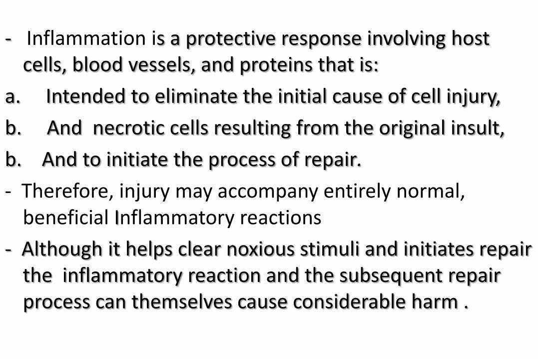
- Inflammation is a protective response involving host cells, blood vessels, and proteins that is:
a. Intended to eliminate the initial cause of cell injury,
b. And necrotic cells resulting from the original insult,
b. And to initiate the process of repair.
- Therefore, injury may accompany entirely normal, beneficial Inflammatory reactions
- Although it helps clear noxious stimuli and initiates repair the inflammatory reaction and the subsequent repair process can themselves cause considerable harm .
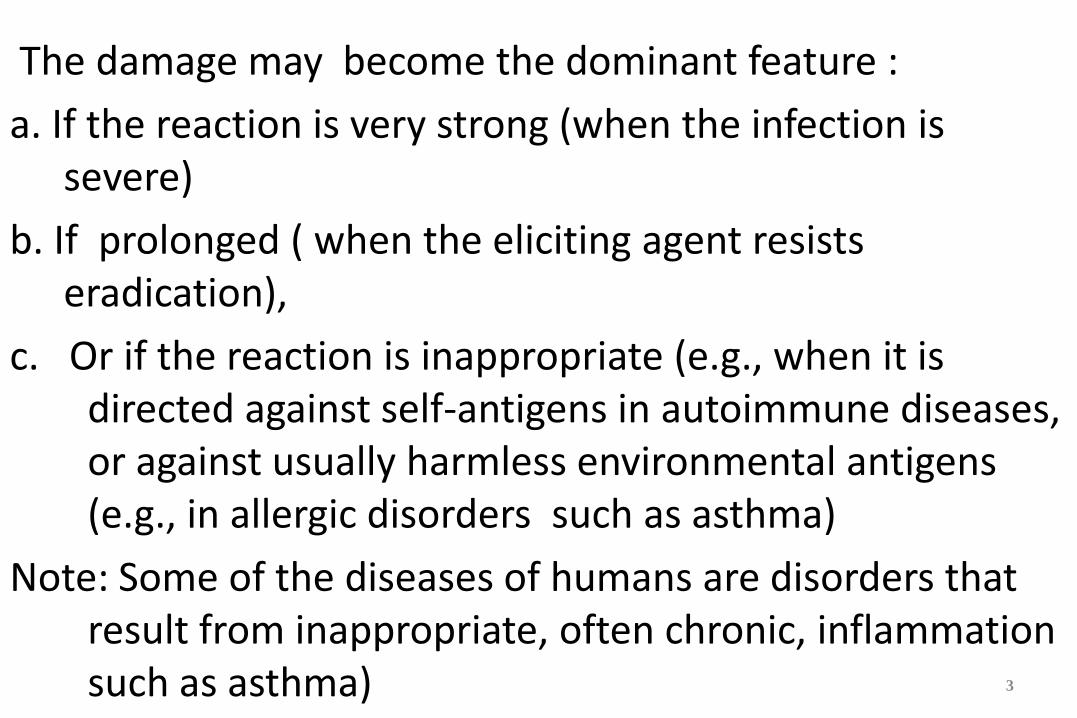
The damage may become the dominant feature :
a. If the reaction is very strong (when the infection is severe)
b. If prolonged ( when the eliciting agent resists eradication),
c. Or if the reaction is inappropriate (e.g., when it is directed against self-antigens in autoimmune diseases, or against usually harmless environmental antigens (e.g., in allergic disorders such as asthma)
Note: Some of the diseases of humans are disorders that result from inappropriate, often chronic, inflammation such as asthma)
3
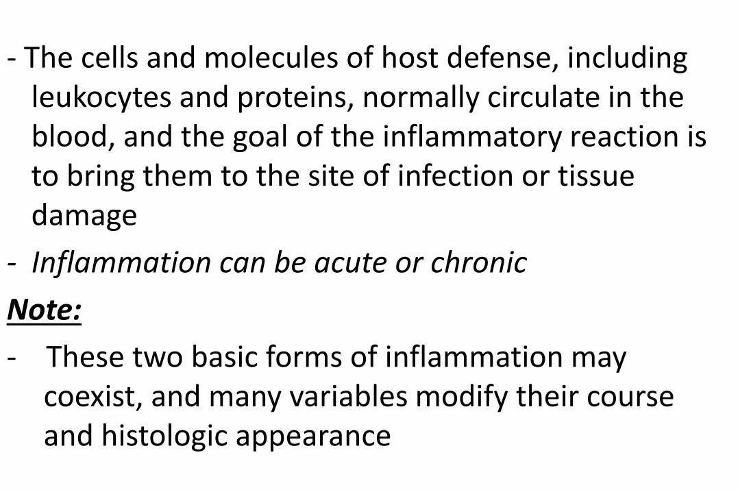
- The cells and molecules of host defense, including leukocytes and proteins, normally circulate in the blood, and the goal of the inflammatory reaction is to bring them to the site of infection or tissue damage
- Inflammation can be acute or chronic
Note:
- These two basic forms of inflammation may coexist, and many variables modify their course and histologic appearance
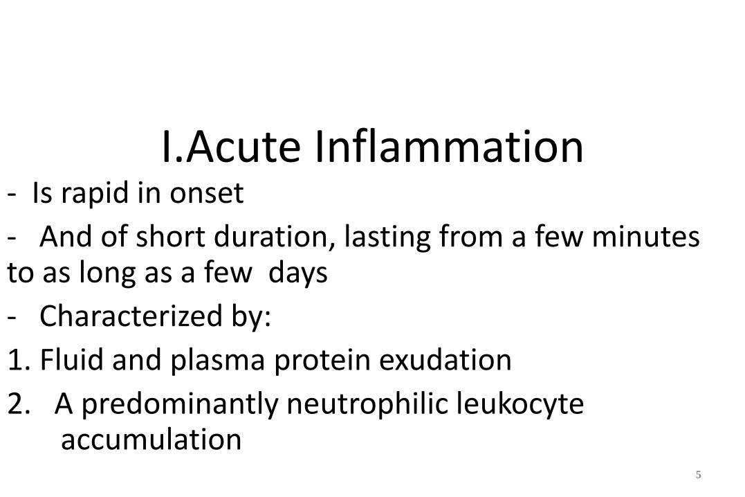
I.Acute Inflammation - Is rapid in onset
- And of short duration, lasting from a few minutes to as long as a few days
- Characterized by:
1. Fluid and plasma protein exudation
2. A predominantly neutrophilic leukocyte accumulation
5
The external manifestations of inflammation
- Often called its cardinal signs
1.Heat (calor)
2. Redness (rubor)
3. Swelling (tumor )
4. Pain (dolor)
5. Loss of function
Inflammation is normally controlled and self-limited
a. The mediators of inflammation and cells are activated only in response to the injury
b. The mediators are short lived because are degraded or become inactive as the injurious agent is eliminated
c. Activation of anti-inflammatory mechanisms
Stimuli for Acute Inflammation
1. Infections are among the most common and medically important causes of inflammation.
2. Trauma (blunt and penetrating) , Physical and chemical agents
3. Foreign bodies (splinters, dirt, sutures).
4. Tissue injury
6. Immune reactions
1.Recognition of Microbes, and Foreign Substances
- The phagocytes, dendritic cells (cells in connective tissue), and epithelial cells, express receptors that are designed to sense the presence of infectious pathogen and substances released from dead cells
- These receptors called "pattern recognition receptors " because they recognize structures ( molecular patterns) that are common to many microbes or to dead cells.
A. Toll-like receptors (TLRs) :- Are microbial sensors that are named for the founding member called Toll.
- There are ten TLRs, which recognize products of bacteria
8
Erythema and swelling
(such as endotoxin) and viruses
- Recognition of microbes by these receptors activates transcription factors that stimulate the production of a number of secreted and membrane proteins
- These proteins include mediators of inflammation ,antiviral cytokines(interferons), and proteins that promote lymphocyte activation
B. The inflammasome:
- Is a multi-protein cytoplasmic complex that recognizes uric acid crystals and some microbial products
.
- Triggering of the inflammasome results in activation of caspase-1, that cleaves precursors of interleukin-1β into its active form- IL-1 which is a mediator of leukocyte recruitment and phagocytosis.
- Gout, is caused by deposition of urate crystals, which are ingested by phagocytes activating inflammasome, resulting in IL-1 production and acute inflammation.
- IL-1 antagonists are effective treatments in cases of gout that are resistant to anti-inflammatory therapy

2. Vascular Changes in acute inflammation
A. Changes in Vascular Caliber and Flow
1. Transient vasoconstriction (lasting only for seconds)
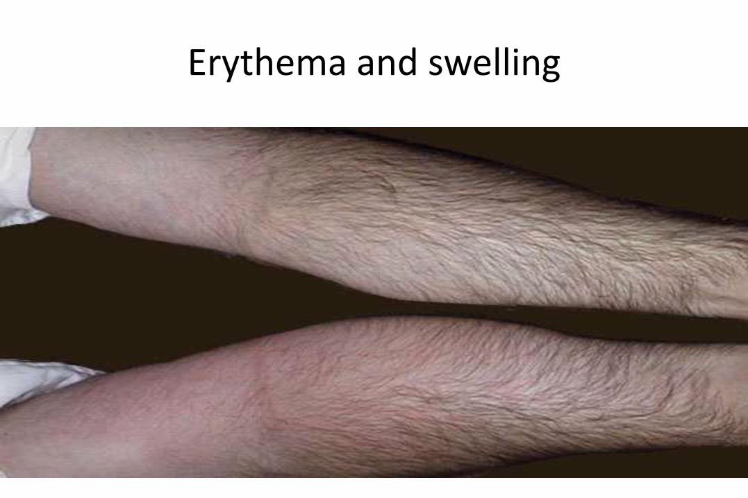
2. Arteriolar vasodilation : which results in locally increased blood flow and engorgement of the down-stream capillary beds and this vascular expansion is the cause of the redness ( erythema) and (warmth) of acute inflammation.
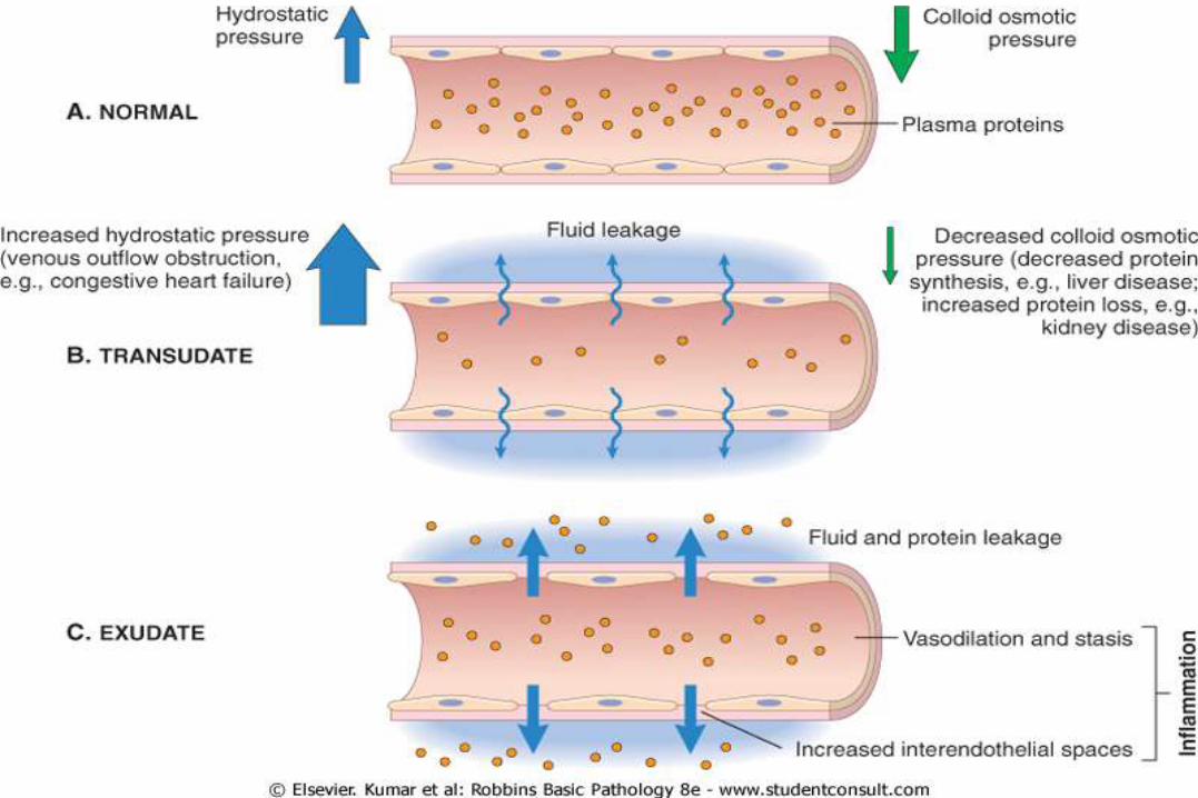
B. Increased Vascular Permeability
- Causes movement of the protein-rich fluid to the extravascular tissues which in turn increases the osmotic pre ssure of the interstitial fluid , leading to more
outflow of water from the blood into the tissues and the resulting protein- rich fluid accumulation is called an exudate and must be distinguished from transudates
Transudates:
a. Are caused by increased hydrostatic pressure, usually a consequence of reduced venous return.
b. have low concentrations of protein with no or few red blood cells
c. and accumulate in various non-inflammatory conditions
Note: Fluid accumulation in extravascular spaces, whether exudate or a transudate, produces tissue edema.
13
Mechansims of increased vascular permeability in acute inflammatory reactions
a-Endothelial cell contraction :
- Causes Immediate transient response
- Leads to intercellular gaps in postcapillary venules
- Is the most common cause of increased vascular permeability and is a reversible process
- It occurs rapidly after binding of histamine, bradykinin, leukotrienes, to specific receptors
- Is usually short-lived (15 to 30 minutes)
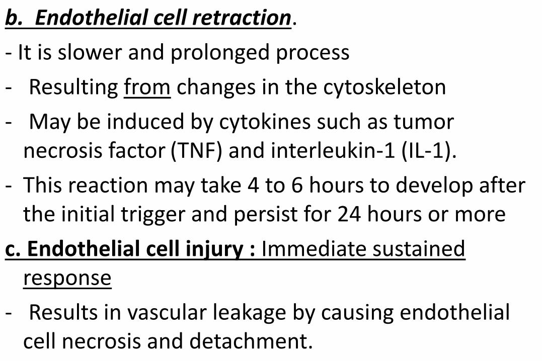
b. Endothelial cell retraction.
- It is slower and prolonged process
- Resulting from changes in the cytoskeleton
- May be induced by cytokines such as tumor necrosis factor (TNF) and interleukin-1 (IL-1).
- This reaction may take 4 to 6 hours to develop after the initial trigger and persist for 24 hours or more
c. Endothelial cell injury : Immediate sustained response
- Results in vascular leakage by causing endothelial cell necrosis and detachment.
- Endothelial cells are damaged after severe injury such as with burns and Some infections
- In most cases, leakage begins immediately after the injury
- The leakage persists for several hours (or days) until the damaged vessels are thrombosed or repaired Venules, capillaries, and arterioles can all be affected, depending on the site of the injury
Note: Direct injury to endothelial cells may induce a delayed prolonged leakage that begins after a delay of 2 to 12hours, lasts for several hours or even days
- The response is called delayed prolonged leakage
16
d. Leukocyte-mediated endothelial injury - May occur as a consequence of leukocyte
accumulation along the vessel wall - This process is largely restricted to venules and
pulmonary and glomerular capillaries where leukocytes adhere for prolonged periods to the endothelium
e. Increased transcytosis - It occurs via an intracellular vesicular pathway
augments venular permeability ,especially after exposure to certain mediators like vascular endothelial growth factors (VEGF)
- it occurs via channels formed by fusion of intracellular
vesicles

Exudate
f. Leakage from new blood vessels - The process is called angiogenesis - These vessel sprouts remain leaky until the
proliferating endothelial cells mature sufficiently to form
intercellular junctions New endothelial cells have increased expression of
receptors for factors that induce angiogenesis like VEGF (vascular endothelial growth factor) directly induce vascular permeability
C. Stasis : Increased permeability causes the red cells in the flowing blood to become concentrated,so increasing blood viscosity and slowing the circulation and these changes are reflected microscopically by dilated small vessels packed the red blood cells, called stasis.
D- Margination:- As stasis develops, leukocytes (principally neutrophils) begin to accumulate along the vascular
endothelial surface-a process called margination Responses of Lymphatic Vessels
- In inflammation, lymph flow is increased and helps drain edema fluid, leukocytes, and cell debris from the
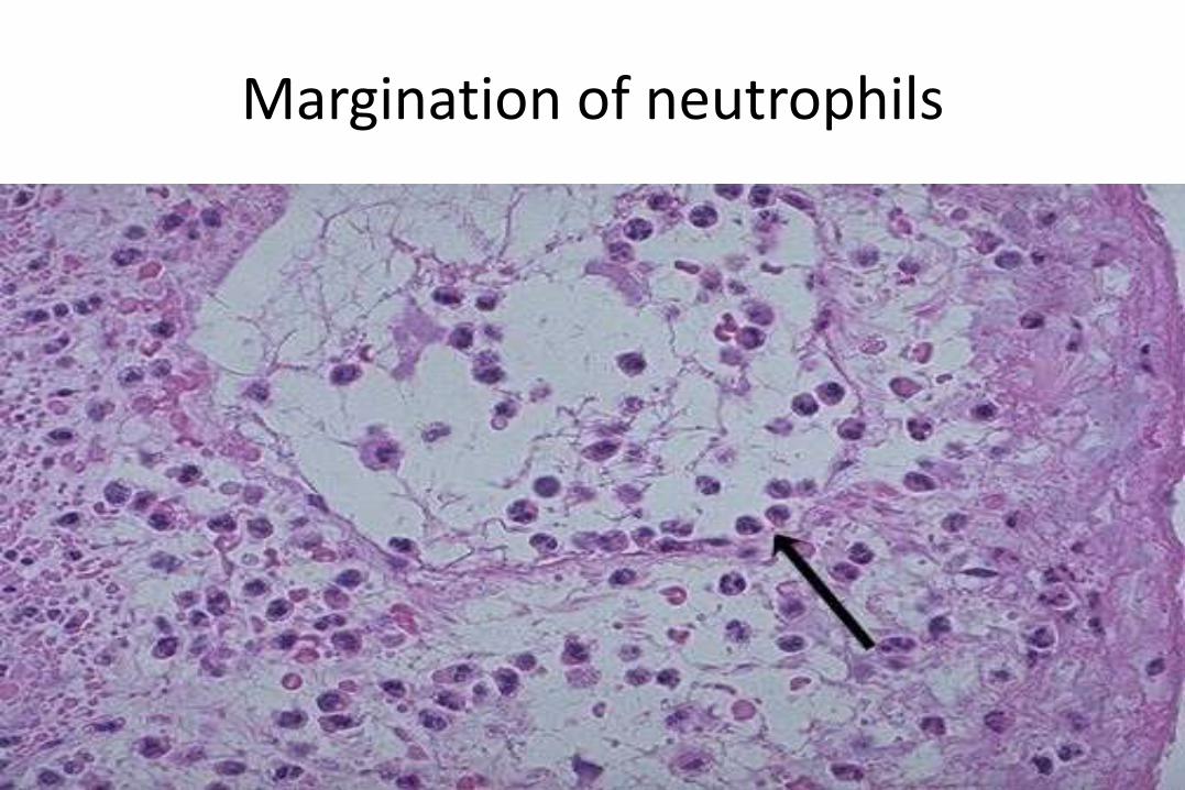
Margination of neutrophils
extravascular space.
- In severe inflammation by microbes, the lymphatics may transport the offending agent, causing its dissemination
- The lymphatics may become secondarily inflamed (lymphangitis), as may the draining lymph nodes (lymphadenitis)
- For clinicians, the presence of red streaks near a skin wound is a sign of an infection in the wound.
- This streaking follows the course of the lymphatic channels and is diagnostic of lymphangitis ; and may be accompanied by painful enlargement of the lymph nodes
3.. Cellular Events: Leukocyte Recruitment and Activation
- Leukocytes ingest offending agents, kill microbes, and eliminate necrotic tissue and foreign substances
- Once, the leucocytes are activated, they may induce tissue damage and prolong inflammation,.
- Therefore, host defense mechanisms include checks and balances that ensure that leukocytes are recruited and activated only when and where they are needed
- Leukocytes normally flow rapidly in the blood, and in inflammation, they have to be stopped and brought to the
offending agent or the site of tissue damage, which are typically outside the vessels.
- The sequence of recruitment of leukocytes from the vascular lumen to the extravascular space consists of:
1. Margination and Rolling
- As blood flows from capillaries into venules, circulating cells are swept by laminar flow against the vessel wall.
- Because the smaller red cells tend to move faster thanthe larger white cells, leukocytes are pushed out of the central axial column and thus have a better opportunity to interact with endothelium.
a. Margination : Means the process of leukocyte accumulation at the periphery of vessels
b. Rolling : It is weak and transient binding between leukocytes and endothelial cells
- Rolling is mediated by the selectin family of adhesion molecules which are receptors expressed on leukocytes and endothelium that contain an extracellular domain that binds sugars
Types of selectins
a. E-selectin (also called CD62E), expressed on endothelium
b. P-selectin (CD62P), present on platelets and endothelium
26
c. L-selectin (CD62L), present on the surface of leukocytes.
- Selectins bind sialylated oligosaccharides (e.g., sialyl-Lewis X on leukocytes) that are attached to mucin-like glycoproteins on cells
- The endothelial selectins are expressed at low levels or are not present at all on un-activated endothelium, and are up– regulated after stimulation by mediators
- Therefore, binding of leukocytes is largely restricted to endothelium at sites of infection or tissue injury (where the mediators are produced) and for examples
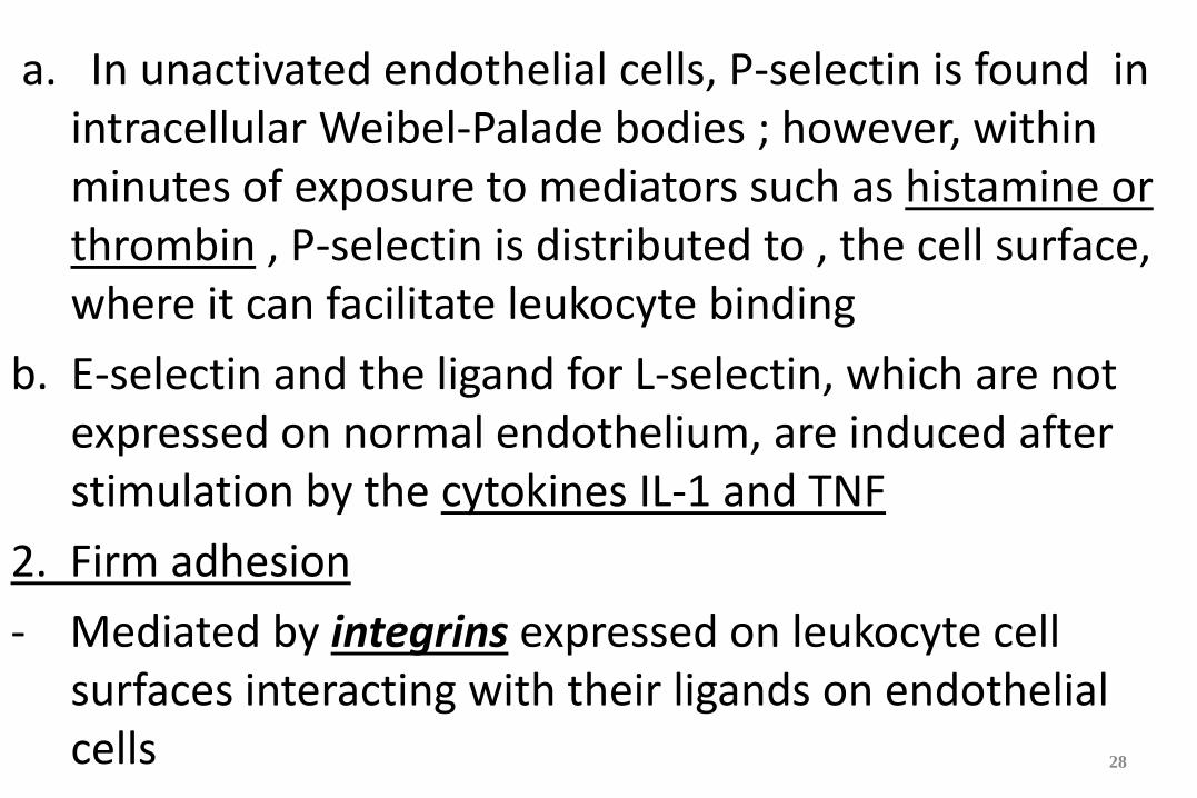
a. In unactivated endothelial cells, P-selectin is found in intracellular Weibel-Palade bodies ; however, within minutes of exposure to mediators such as histamine or thrombin , P-selectin is distributed to , the cell surface, where it can facilitate leukocyte binding
b. E-selectin and the ligand for L-selectin, which are not expressed on normal endothelium, are induced after stimulation by the cytokines IL-1 and TNF
2. Firm adhesion
- Mediated by integrins expressed on leukocyte cell surfaces interacting with their ligands on endothelial cells
28
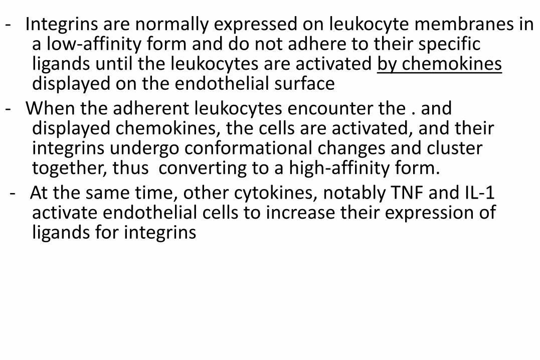
- Integrins are normally expressed on leukocyte membranes in a low-affinity form and do not adhere to their specific ligands until the leukocytes are activated by chemokines displayed on the endothelial surface
- When the adherent leukocytes encounter the . and displayed chemokines, the cells are activated, and their integrins undergo conformational changes and cluster together, thus converting to a high-affinity form.
- At the same time, other cytokines, notably TNF and IL-1 activate endothelial cells to increase their expression of ligands for integrins
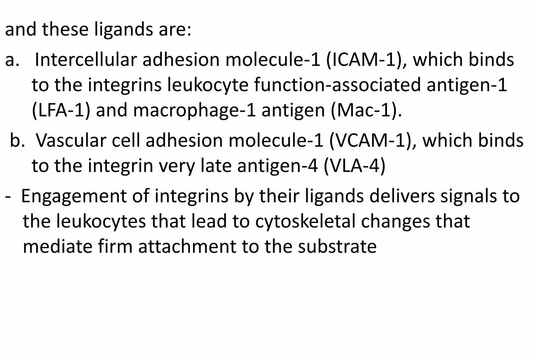
and these ligands are:
a. Intercellular adhesion molecule-1 (ICAM-1), which binds to the integrins leukocyte function-associated antigen-1 (LFA-1) and macrophage-1 antigen (Mac-1).
b. Vascular cell adhesion molecule-1 (VCAM-1), which binds to the integrin very late antigen-4 (VLA-4)
- Engagement of integrins by their ligands delivers signals to the leukocytes that lead to cytoskeletal changes that mediate firm attachment to the substrate
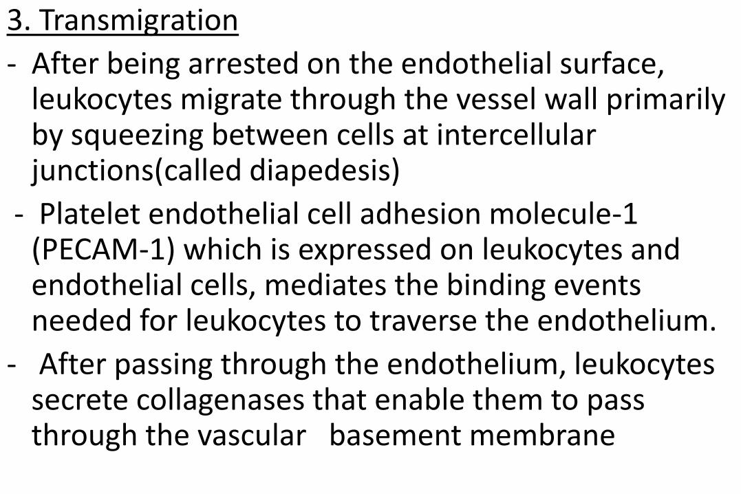
3. Transmigration
- After being arrested on the endothelial surface, leukocytes migrate through the vessel wall primarily by squeezing between cells at intercellular junctions(called diapedesis)
- Platelet endothelial cell adhesion molecule-1 (PECAM-1) which is expressed on leukocytes and endothelial cells, mediates the binding events needed for leukocytes to traverse the endothelium.
- After passing through the endothelium, leukocytes secrete collagenases that enable them to pass through the vascular basement membrane
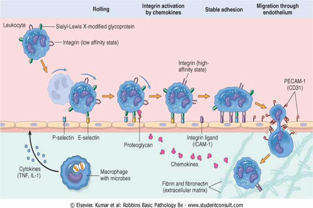
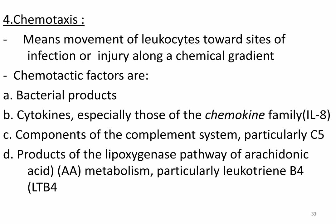
4.Chemotaxis :
- Means movement of leukocytes toward sites of infection or injury along a chemical gradient
- Chemotactic factors are:
a. Bacterial products
b. Cytokines, especially those of the chemokine family(IL-8)
c. Components of the complement system, particularly C5
d. Products of the lipoxygenase pathway of arachidonic acid) (AA) metabolism, particularly leukotriene B4 (LTB4
33
- Chemotactic molecules bind to cell surface receptors which triggers the assembly of cytoskeletal contractile elements necessary for movement and leukocytes move by extending pseudopods that anchor to the ECM and pull the cell in the direction off the extension
- The direction of such movement is specified by a higher density of chemokine receptors at the leading edge of the cell
- Thus, leukocytes move to and are retained at the site where they are needed
The type of emigrating leukocyte varies with the age of the inflammatory response and with the type of stimulus
- In most forms of acute inflammation, neutrophils predominate in the inflammatory infiltrate during the first 6 to 24 hours and are replaced by monocytes in 24 to 48 hours
- Several factors account for this early abundance of neutrophils:
a. Are the most numerous leukocytes in the blood
b. They respond more rapidly to chemokines
c. They may attach more firmly to the adhesion molecules , such as P- and E-selectins
d. Are short-lived-they die by apoptosis and disappear within 24 to 48 hours-while monocytes survive longer.
- There are exceptions to this pattern of cellular infiltration
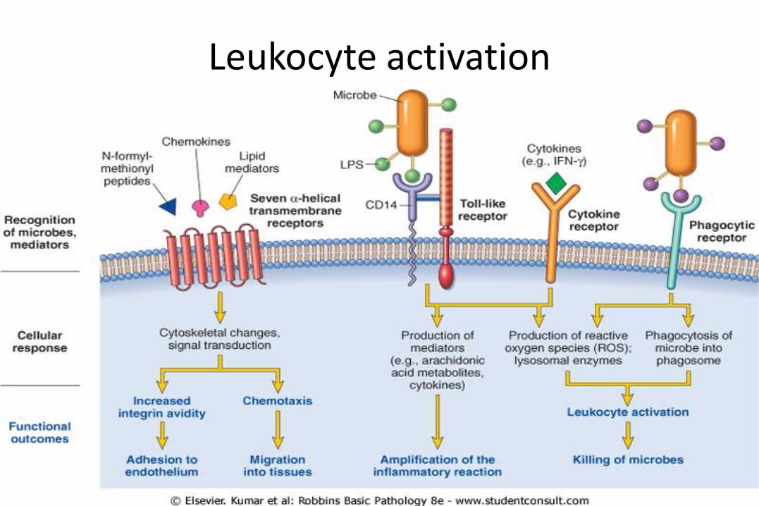
5. Leukocyte Activation
- Once leukocytes have been recruited to the site of infection or tissue necrosis, they must be activated to perform their functions
- Stimuli for activation include microbes, products of necrotic cells, and several mediators
- Leukocytes use various receptors to sense the presence of microbes, dead cells, and foreign substances
- Engagement of these cellular receptors induces leukocyte activation
- Leukocyte activation results in the enhancement of the following functions:
A. Phagocytosis of particles
B. Intracellular destruction of phagocytosed microbes and dead cells by substances produced in phagosomes, including reactive oxygen and lysosomal enzymes
37
C. Liberation of substances that destroy extracellular microbes and dead tissues, which are largely the same as the substances produced within phagocytic vesicles
D. Production of mediators, including arachidonic acid metabolites and cytokines, that amplify the inflammatory reaction, by recruiting and activating more leukocytes
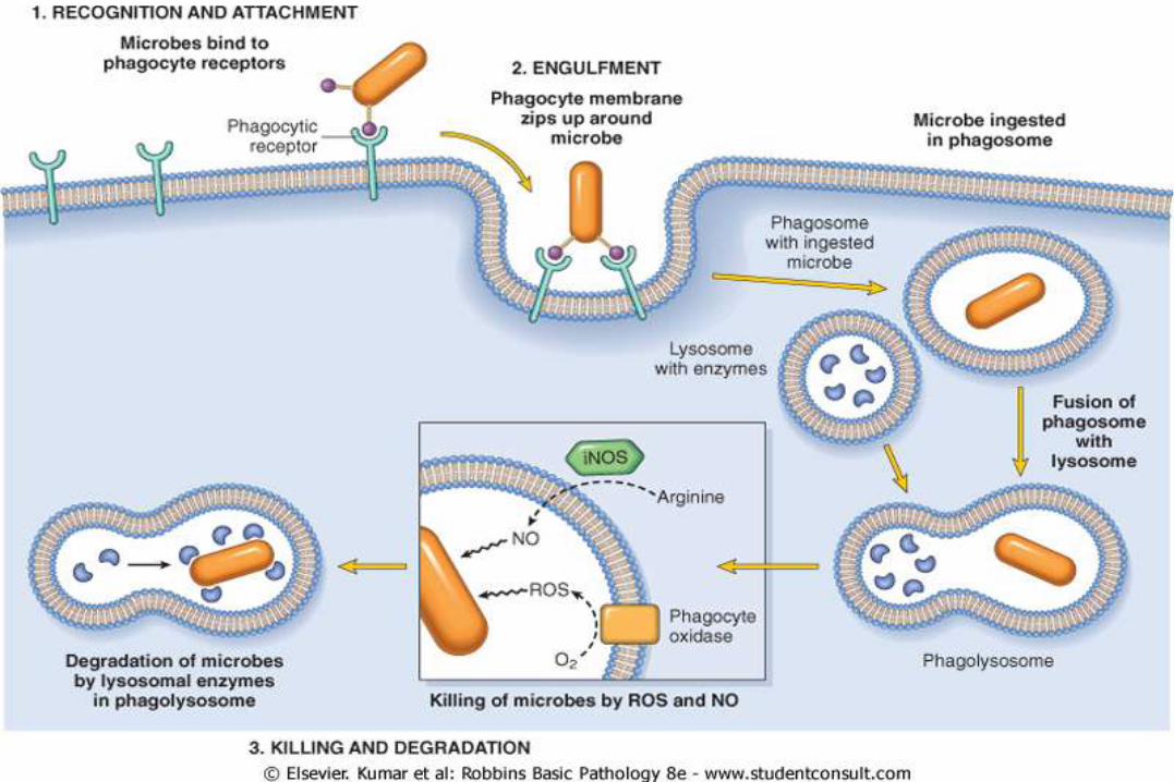
A. Phagocytosis consists of three steps
1. Recognition and attachment of the particle to the ingesting leukocyte;
- Occurs by means of specific receptors
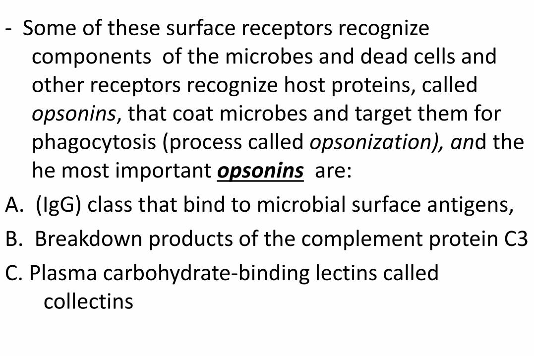
- Some of these surface receptors recognize components of the microbes and dead cells and other receptors recognize host proteins, called opsonins, that coat microbes and target them for phagocytosis (process called opsonization), and the he most important opsonins are:
A. (IgG) class that bind to microbial surface antigens,
B. Breakdown products of the complement protein C3
C. Plasma carbohydrate-binding lectins called collectins
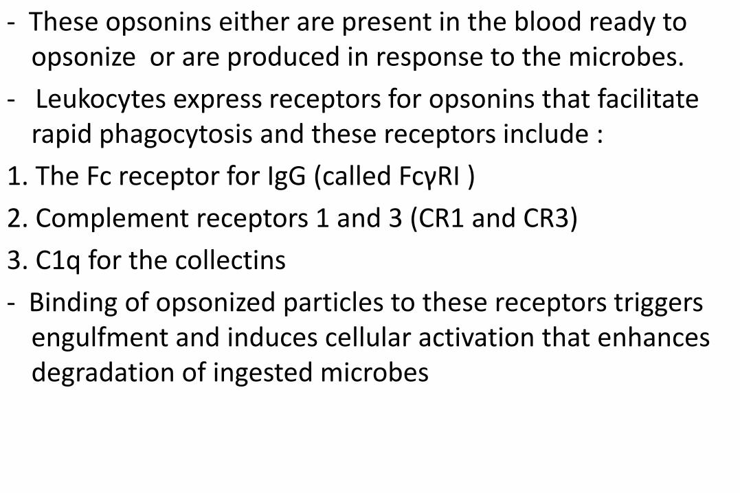
- These opsonins either are present in the blood ready to opsonize or are produced in response to the microbes.
- Leukocytes express receptors for opsonins that facilitate rapid phagocytosis and these receptors include :
1. The Fc receptor for IgG (called FcγRI )
2. Complement receptors 1 and 3 (CR1 and CR3)
3. C1q for the collectins
- Binding of opsonized particles to these receptors triggers engulfment and induces cellular activation that enhances degradation of ingested microbes

2. Engulfment : In this process, pseudopods are extended
around the object, forming a phagocytic vacuole.
- The membrane of the vacuole then fuses with the membrane of a lysosomal granule, resulting in discharge of the granule's contents into the phagolysosome
3. Killing and Degradation of Phagocytosed microbes
- The steps include the production of microbicidal substances within lysosomes and fusion of the lysosomes with phagosomes, so exposing the ingested particles to the destructive mechanisms of the Leukocytes
-
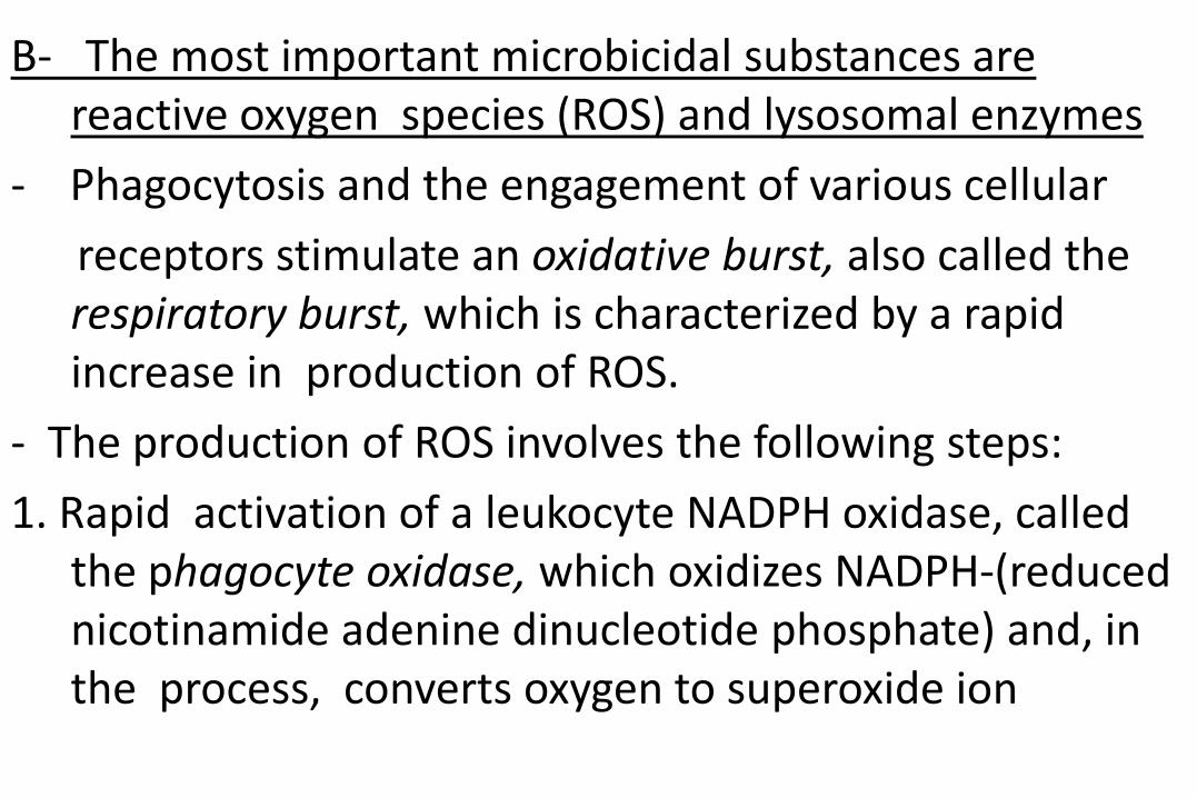
B- The most important microbicidal substances are reactive oxygen species (ROS) and lysosomal enzymes
- Phagocytosis and the engagement of various cellular
receptors stimulate an oxidative burst, also called the respiratory burst, which is characterized by a rapid increase in production of ROS.
- The production of ROS involves the following steps:
1. Rapid activation of a leukocyte NADPH oxidase, called the phagocyte oxidase, which oxidizes NADPH-(reduced nicotinamide adenine dinucleotide phosphate) and, in the process, converts oxygen to superoxide ion
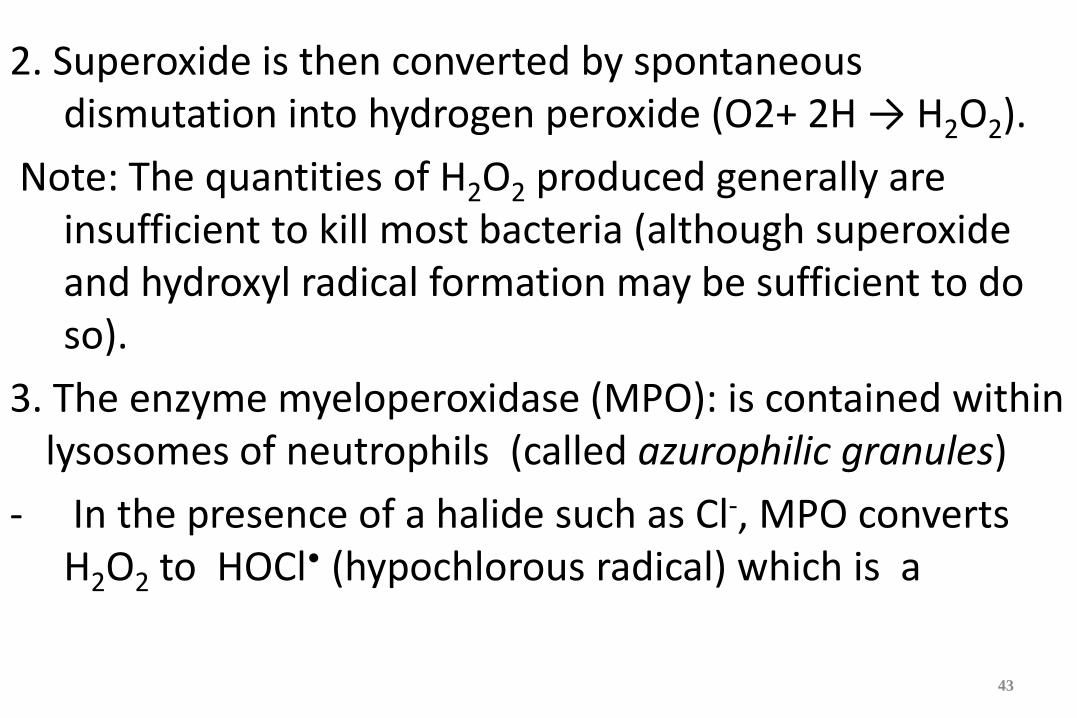
2. Superoxide is then converted by spontaneous dismutation into hydrogen peroxide (O2+ 2H → H2O2).
Note: The quantities of H2O2 produced generally are insufficient to kill most bacteria (although superoxide and hydroxyl radical formation may be sufficient to do so).
3. The enzyme myeloperoxidase (MPO): is contained within lysosomes of neutrophils (called azurophilic granules)
- In the presence of a halide such as Cl-, MPO converts H2O2 to HOCl• (hypochlorous radical) which is a
43
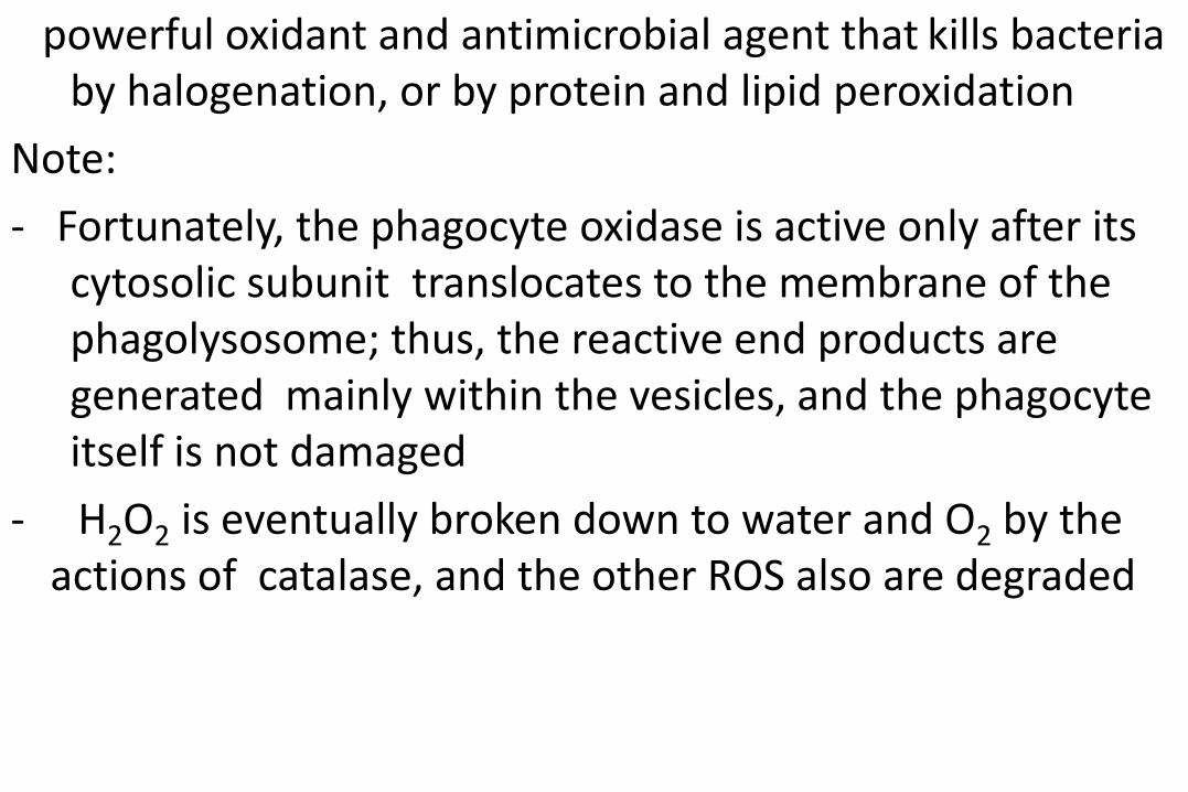
powerful oxidant and antimicrobial agent that kills bacteria by halogenation, or by protein and lipid peroxidation
Note:
- Fortunately, the phagocyte oxidase is active only after its cytosolic subunit translocates to the membrane of the phagolysosome; thus, the reactive end products are generated mainly within the vesicles, and the phagocyte itself is not damaged
- H2O2 is eventually broken down to water and O2 by the actions of catalase, and the other ROS also are degraded

- The dead microorganisms are then degraded by the action of lysosomal acid hydrolases and perhaps the most important lysosomal enzyme involved in bacterial killing is elastase
- Several other constituents of leukocyte granules are capable of killing infectious pathogens and these include:
45
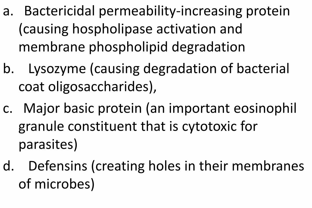
a. Bactericidal permeability-increasing protein (causing hospholipase activation and membrane phospholipid degradation
b. Lysozyme (causing degradation of bacterial coat oligosaccharides),
c. Major basic protein (an important eosinophil granule constituent that is cytotoxic for parasites)
d. Defensins (creating holes in their membranes of microbes)
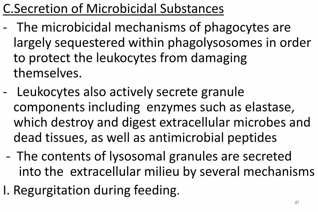
C.Secretion of Microbicidal Substances
- The microbicidal mechanisms of phagocytes are largely sequestered within phagolysosomes in order to protect the leukocytes from damaging themselves.
- Leukocytes also actively secrete granule components including enzymes such as elastase, which destroy and digest extracellular microbes and dead tissues, as well as antimicrobial peptides
- The contents of lysosomal granules are secreted into the extracellular milieu by several mechanisms
I. Regurgitation during feeding.
47
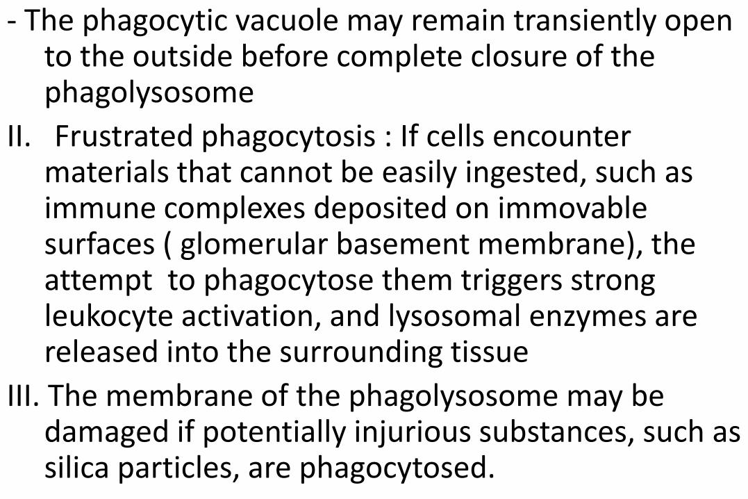
- The phagocytic vacuole may remain transiently open to the outside before complete closure of the phagolysosome
II. Frustrated phagocytosis : If cells encounter materials that cannot be easily ingested, such as immune complexes deposited on immovable surfaces ( glomerular basement membrane), the attempt to phagocytose them triggers strong leukocyte activation, and lysosomal enzymes are released into the surrounding tissue
III. The membrane of the phagolysosome may be damaged if potentially injurious substances, such as silica particles, are phagocytosed.
Leukocyte activation
Leukocyte-Induced Tissue Injury - Because leukocytes are capable of secreting
potentially harmful substances such as enzymes and ROS, they are important causes of injury to normal cells and tissues under several circumstances:
a. In certain infections that are difficult to eradicate, such as tuberculosis and some viral diseases, the host response contributes more to the pathologic process than does the microbe itself.
b. As a normal attempt to clear damaged and dead tissues ( after a myocardial infarction), In an infarct, inflammation
may exacerbate the injurious consequences of the ischemia, especially upon reperfusion
c. When the inflammatory response is inappropriately directed against host tissues, as in certain autoimmune diseases, or when the host reacts excessively against nontoxic environmental substances, such as allergic diseases including asthma
- In all of these situations, the mechanisms by which leukocytes damage normal tissues are the same as the 52
mechanisms involved in the clearance of microbes and dead tissues, because once the leukocytes are activated, their effector mechanisms do not distinguish between offender and host
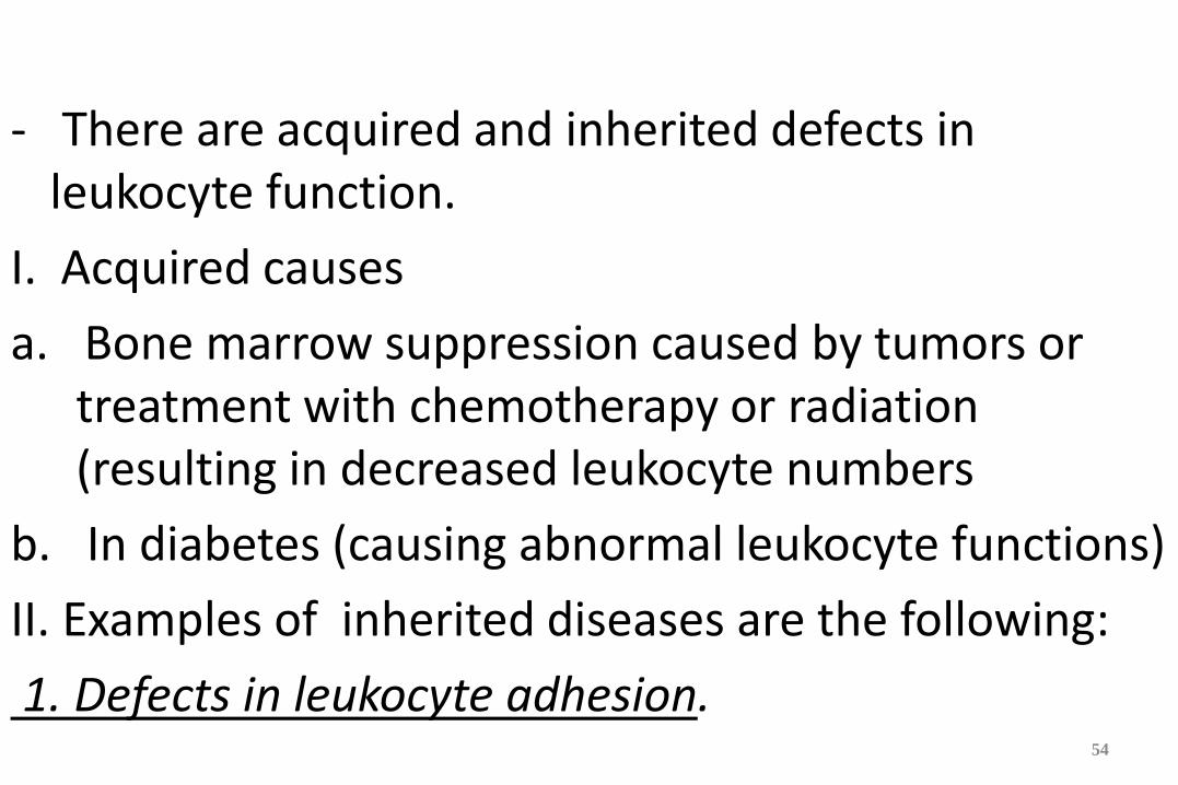
Defects in Leukocyte Function - There are acquired and inherited defects in
leukocyte function.
I. Acquired causes
a. Bone marrow suppression caused by tumors or treatment with chemotherapy or radiation (resulting in decreased leukocyte numbers
b. In diabetes (causing abnormal leukocyte functions)
II. Examples of inherited diseases are the following:
1. Defects in leukocyte adhesion.
54
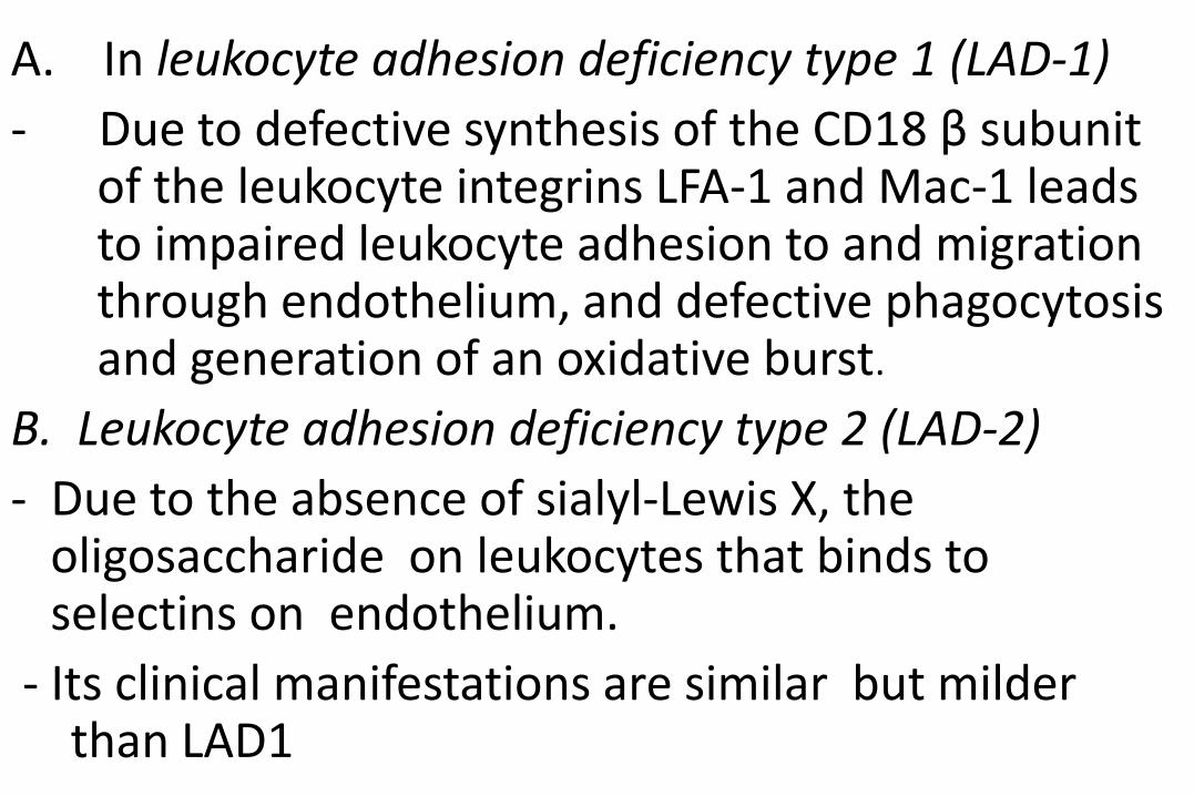
A. In leukocyte adhesion deficiency type 1 (LAD-1)
- Due to defective synthesis of the CD18 β subunit of the leukocyte integrins LFA-1 and Mac-1 leads to impaired leukocyte adhesion to and migration through endothelium, and defective phagocytosis and generation of an oxidative burst.
B. Leukocyte adhesion deficiency type 2 (LAD-2)
- Due to the absence of sialyl-Lewis X, the oligosaccharide on leukocytes that binds to selectins on endothelium.
- Its clinical manifestations are similar but milder than LAD1
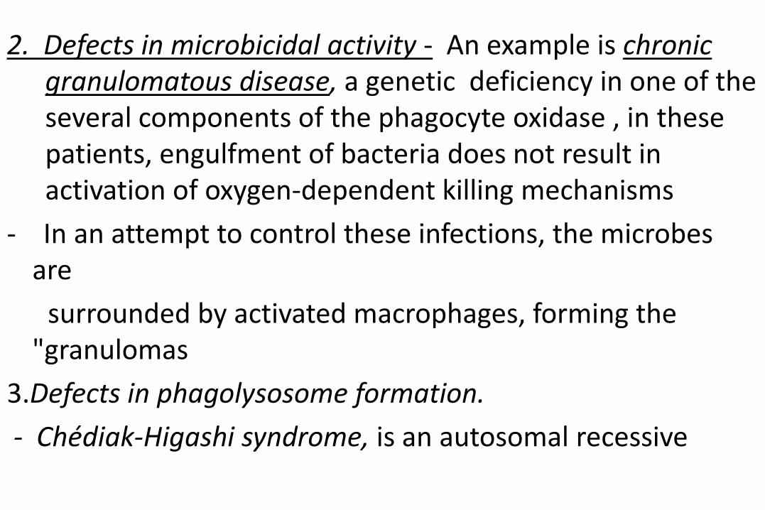
2. Defects in microbicidal activity - An example is chronic granulomatous disease, a genetic deficiency in one of the several components of the phagocyte oxidase , in these patients, engulfment of bacteria does not result in activation of oxygen-dependent killing mechanisms
- In an attempt to control these infections, the microbes are
surrounded by activated macrophages, forming the "granulomas
3.Defects in phagolysosome formation.
- Chédiak-Higashi syndrome, is an autosomal recessive
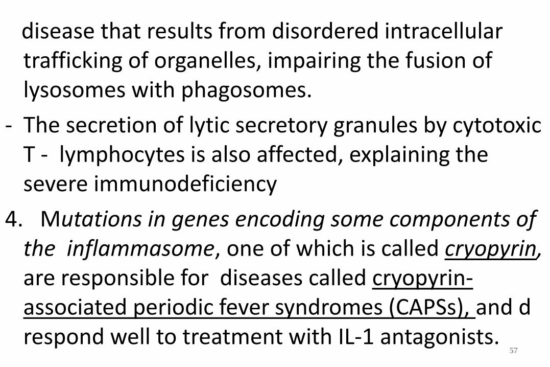
disease that results from disordered intracellular trafficking of organelles, impairing the fusion of lysosomes with phagosomes.
- The secretion of lytic secretory granules by cytotoxic T - lymphocytes is also affected, explaining the severe immunodeficiency
4. Mutations in genes encoding some components of the inflammasome, one of which is called cryopyrin, are responsible for diseases called cryopyrin-associated periodic fever syndromes (CAPSs), and d respond well to treatment with IL-1 antagonists.
57
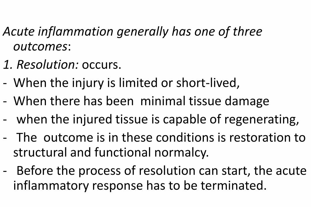
Outcomes of Acute Inflammation
Acute inflammation generally has one of three outcomes:
1. Resolution: occurs.
- When the injury is limited or short-lived,
- When there has been minimal tissue damage
- when the injured tissue is capable of regenerating,
- The outcome is in these conditions is restoration to structural and functional normalcy.
- Before the process of resolution can start, the acute inflammatory response has to be terminated.
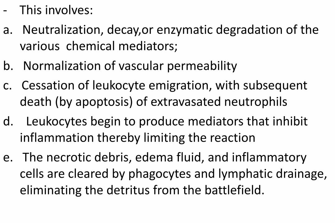
- This involves:
a. Neutralization, decay,or enzymatic degradation of the various chemical mediators;
b. Normalization of vascular permeability
c. Cessation of leukocyte emigration, with subsequent death (by apoptosis) of extravasated neutrophils
d. Leukocytes begin to produce mediators that inhibit inflammation thereby limiting the reaction
e. The necrotic debris, edema fluid, and inflammatory cells are cleared by phagocytes and lymphatic drainage, eliminating the detritus from the battlefield.
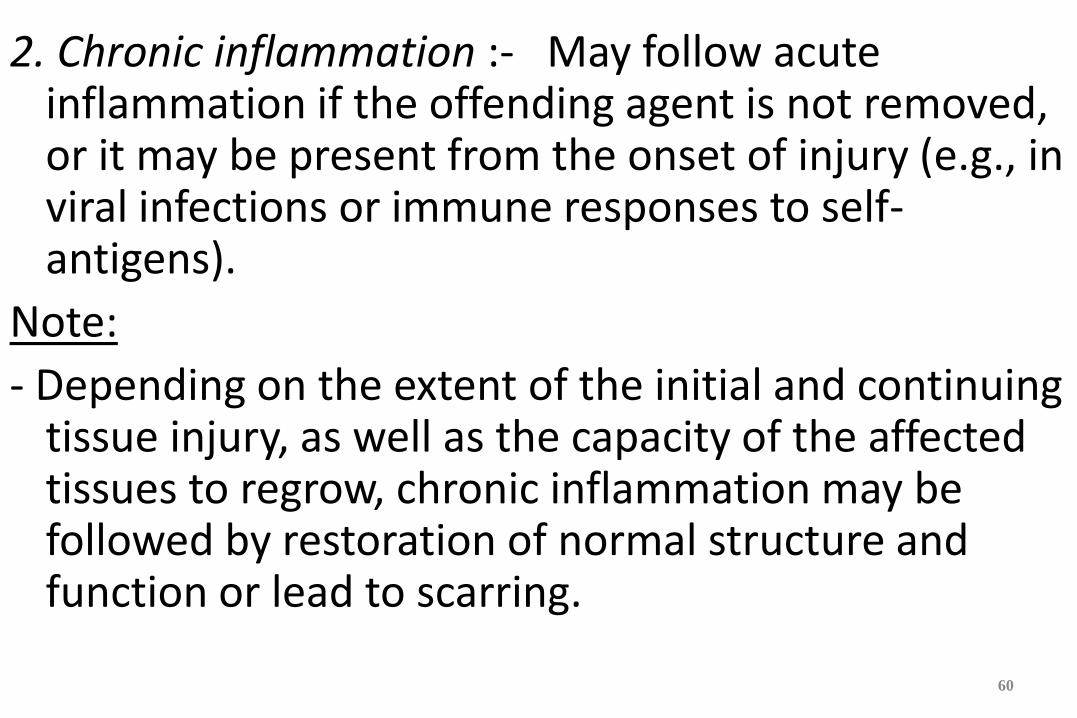
2. Chronic inflammation :- May follow acute inflammation if the offending agent is not removed, or it may be present from the onset of injury (e.g., in viral infections or immune responses to self-antigens).
Note:
- Depending on the extent of the initial and continuing tissue injury, as well as the capacity of the affected tissues to regrow, chronic inflammation may be followed by restoration of normal structure and function or lead to scarring.
60
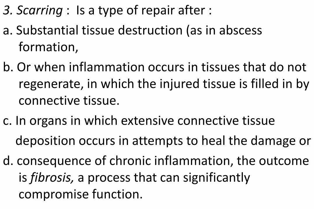
3. Scarring : Is a type of repair after :
a. Substantial tissue destruction (as in abscess formation,
b. Or when inflammation occurs in tissues that do not regenerate, in which the injured tissue is filled in by connective tissue.
c. In organs in which extensive connective tissue
deposition occurs in attempts to heal the damage or
d. consequence of chronic inflammation, the outcome is fibrosis, a process that can significantly compromise function.
MORPHOLOGIC PATTERNS OF ACUTE INFLAMMATION
- The importance of recognizing these patterns is that they are associated with different clinical situations.
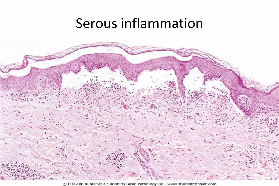
1. Serous inflammation :- Characterized by the outpouring of a watery, relatively protein-poor fluid that, derives either from the plasma or from the secretions of mesothelial cells lining the peritoneal, pleural, and pericardial cavities.
- The skin blister resulting from a burn or viral infection is an example of the accumulation of a serous effusion either within or immediately beneath the epidermis of the skin
- Fluid in a serous cavity is called an effusion.
2. Fibrinous inflammation :- Occurs as a consequence of more severe injuries, resulting in greater vascular permeability that allows large molecules (such as fibrinogen) to pass the endothelial barrier.
- Histologically, the accumulated extravascular fibrin appears as an eosinophilic meshwork of threads
- A fibrinous exudate is characteristic of inflammation in the the meninges, pericardium, and pleura
- Such exudates:
a. May be degraded by fibrinolysis, and the accumulated
Serous inflammation
debris may be removed by macrophages, resulting in restoration of the normal tissue structure (resolution).
b. Extensive fibrin-rich exudates may not be completely removed, and are replaced by an ingrowth of fibroblasts and blood vessels (organization), leading to scarring that may have significant clinical consequences.
- For example, organization of a fibrinous pericardial exudate forms dense fibrous scar tissue that obliterates the pericardial space and restricts myocardial function
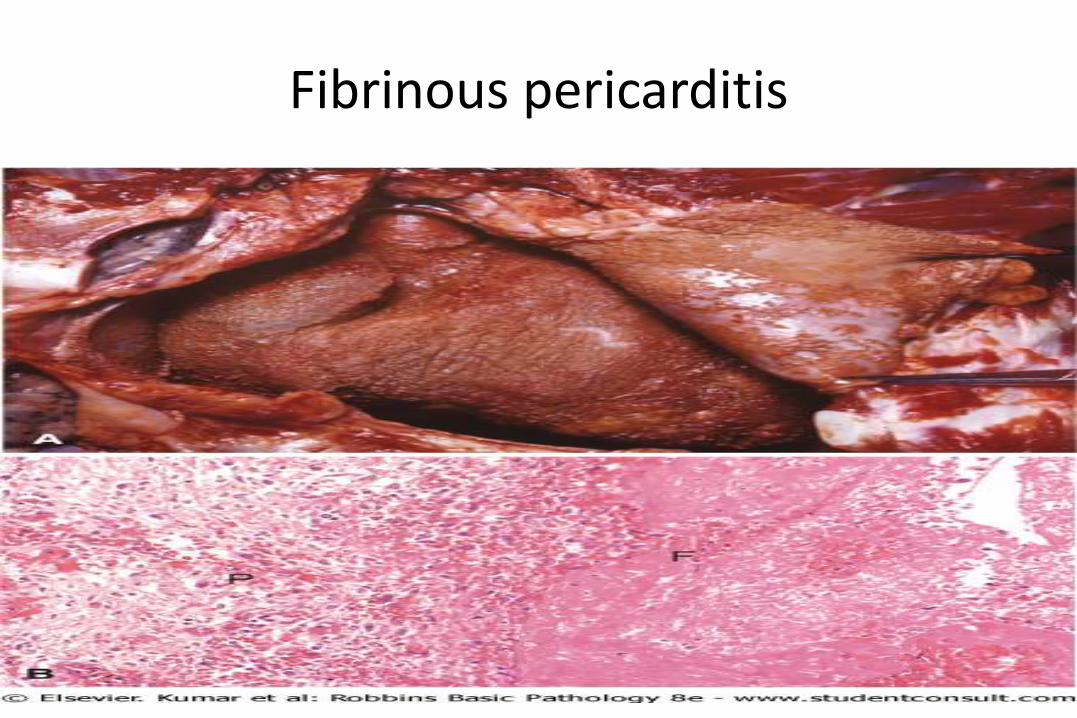
Fibrinous pericarditis
3. Suppurative (purulent) inflammation and abscess
- Characterized by collection of large amounts of purulent exudate consisting of neutrophils, necrotic cells, and edema fluid.and certain organisms (e.g., staphylococci) are more likely to induce such localized suppuration and are therefore referred to as pyogenic (pus-forming }
67
Abscesses :
- Are focal collections of pus that may be caused by seeding of pyogenic organisms into a tissue or by secondary infections of necrotic foci.
- Abscesses typically have a central, largely necrotic region rimmed by a layer of preserved neutrophils with a surrounding zone of dilated vessels and fibroblast proliferation indicative of attempted repair.

Chemical Mediators of inflammation
1. Mediators may be produced:
a. Locally by cells at the site of inflammation,
- Cell-derived mediators are normally sequestered in intracellular granules and are rapidly secreted upon cellular activation (e.g., histamine in mast cells) or are synthesized de novo in response to a stimulus (e.g., prostaglandins and cytokines produced by leukocytes)
b. May be derived from circulating inactive precursors synthesized by the liver and activated at the site of inflammation
- Plasma protein-derived mediators (complement proteins, kinins) circulate in an inactive form and undergo proteolytic cleavage to acquire their biologic activities.
2. Most mediators act by binding to specific receptors on different target cells.
-. Such mediators may act on only one or a very few cell types, or they may have diverse actions
- Other mediators (lysosomal proteases, ROS) have direct enzymatic activities that do not require binding to specific receptors.
3. The actions of most mediators are tightly regulated and short-lived.
- Once activated and released from the cell,
a. Some mediators quickly decay (e.g., arachidonic acid metabolites)
b. Some are inactivated by enzymes (e.g., kininase inactivates bradykinin
c. Some are eliminated (e.g., antioxidants scavenge toxic oxygen metabolites),
d. Or are inhibited (e.g., Complement regulatory proteins block complement activation.
I. Cell-Derived Mediators
- Produced by tissue macrophages, mast cells, leukocytes and endothelial cells at the site of inflammation,
A Vasoactive Amines : histamine and serotonin, are stored as preformed molecules and are among the first mediators to be released in acute inflammatory reactions.
- Histamine: Produced mainly by mast cells , basophils and platelets
- Preformed histamine is released from mast cell granules in response to a variety of stimuli:
1. Physical injury such as trauma or heat;
72
2. Immune reactions involving binding of IgE antibodies to Fc receptors on mast cells
3. C3a and C5a fragments of complement, the so-called anaphylatoxins
5. Neuropeptides (e.g., substance P)
6. Cytokines like IL-1 and IL-8
- In humans, histamine causes :
a. Arteriolar dilation and
b. Rapidly increases vascular permeability
- Histamine is inactivated by histaminase