Embed Size (px)
Citation preview

Olivia M. Farr,1 Jagriti Upadhyay,1 Anna Gavrieli,1 Michelle Camp,1
Nikolaos Spyrou,1 Harper Kaye,1 Hannah Mathew,2 Maria Vamvini,1
Anastasia Koniaris,1 Holly Kilim,1 Alexandra Srnka,1 Alexandra Migdal,1
and Christos S. Mantzoros1
Lorcaserin Administration DecreasesActivation of Brain Centers inResponse to Food Cues and TheseEmotion- and Salience-RelatedChanges Correlate With Weight LossEffects: A 4-Week-Long Randomized,Placebo-Controlled, Double-BlindClinical TrialDiabetes 2016;65:2943–2953 | DOI: 10.2337/db16-0635
Lorcaserin is a serotonin 5-hydroxytryptamine 2c recep-tor agonist effective in treating obesity. Studies in rodentshave shown that lorcaserin acts in the brain to exert itsweight-reducing effects, but this has not yet been stud-ied in humans. We performed a randomized, placebo-controlled, double-blind trial with 48 obese participantsand used functional MRI to study the effects of lorcaserinon the brain. Subjects taking lorcaserin had decreasedbrain activations in the attention-related parietal andvisual cortices in response to highly palatable food cuesat 1 week in the fasting state and in the parietal cortex inresponse to any food cues at 4 weeks in the fed state.Decreases in emotion- and salience-related limbic ac-tivity, including the insula and amygdala, were attenu-ated at 4 weeks. Decreases in caloric intake, weight,and BMI correlated with activations in the amygdala,parietal, and visual cortices at baseline. These data sug-gest that lorcaserin exerts its weight-reducing effectsby decreasing attention-related brain activations to foodcues (parietal and visual cortices) and emotional andlimbic activity (insula, amygdala). Results indicating thatbaseline activation of the amygdala relates to increased
efficacy suggest that lorcaserin would be of particularbenefit to emotional eaters.
Obesity is a growing problem in industrialized countries,where approximately one-third of the population is obese,and has been associated with a number of health problems,including diabetes, heart disease, different types of cancer,and decreased life expectancy (1). Thus, there is a clear needfor the development of new treatments for obesity. Onesuch recent treatment is lorcaserin, a selective serotonin5-hydroxytryptamine 2c (5-HT2c) receptor agonist.
Previous use of serotonergic agonists, such as fenflur-amine, was linked to nonselective activation of 5-HT2receptors that led to various cardiac problems (2). How-ever, because a 5-HT2c receptor antagonist blocked theweight-reducing effects of fenfluramine, the more selec-tive activation of 5-HT2c receptors by lorcaserin wouldproduce this antiobesity benefit seemingly without cardiacrisk (3). Knockout mice for the 5-HT2c receptors have ahigher body weight as a result of abnormal food consump-tion, further confirming the role of this receptor in obesity
1Division of Endocrinology, Beth Israel Deaconess Medical Center and HarvardMedical School, Boston, MA2Boston University Medical Center, Boston, MA
Corresponding author: Christos S. Mantzoros, [email protected].
Received 18 May 2016 and accepted 30 June 2016.
Clinical trial reg. no. NCT02400359, clinicaltrials.gov.
This article contains Supplementary Data online at http://diabetes.diabetesjournals.org/lookup/suppl/doi:10.2337/db16-0635/-/DC1.
This article is featured in a podcast available at http://www.diabetesjournals.org/content/diabetes-core-update-podcasts.
© 2016 by the American Diabetes Association. Readers may use this article aslong as the work is properly cited, the use is educational and not for profit, and thework is not altered. More information is available at http://www.diabetesjournals.org/content/license.
Diabetes Volume 65, October 2016 2943
OBESITYSTUDIES

(4). These animals exhibit changes in metabolic hormones,developing insulin and leptin resistance and, moreover,impaired glucose tolerance in addition to prolonged mealduration and frequency (4,5). A similar mechanism maycontribute to the significant improvement in the levels offasting serum glucose, total cholesterol, triglycerides, andblood pressure seen in patients treated with lorcaserin (6–8).
5-HT2c receptors are located almost exclusively in thecentral nervous system (CNS), including the thalamus andhypothalamus, areas that are known to be involved infeeding regulation, but also in more cortical areas involvedin higher thought and top-down processes (9–11). Activa-tion of 5-HT2c receptors in the rodent brain, predo-minantly in the hypothalamus, initiates a cascade thatstimulates release of a-melanocortin2stimulating hor-mone, which acts on melanocortin-4 receptors to regulateappetite (12–14). This pathway may also activate corticalareas, because there is evidence of melanocortin-4 receptorsacting in the cortex of mice to reduce food intake (15,16).
The exact mechanisms underlying weight loss fromlorcaserin have not yet been fully elucidated in humans.Several large studies have demonstrated that lorcaserin is aneffective weight-loss agent, with total body mass lossaveraging greater than 5% (6,17), but significant interindivid-ual variability exists in the magnitude of the weight lost.Because 5-HT2c receptors are a novel target for obesity, fur-ther work is required to determine whether the weight-reducing effects of lorcaserin are caused by actions in theCNS, and if yes, which specific brain centers are involved.Furthermore, whether there are CNS centers whose activationmay predict which individuals respond the most to lorcaserin,and thus provide additional clinical insight, is not yet known.
We performed a randomized, placebo-controlled, double-blind study to examine potential CNS targets for lorcaserinby using functional MRI (fMRI) to study how lorcaserinalters brain center activations in response to food imagesin the short-term (1 week) and longer-term (4 weeks). Inaddition, we explored how individual differences in thebrain’s response to food cues at baseline may predict themagnitude of future weight loss and decreased caloric in-take with lorcaserin therapy.
RESEARCH DESIGN AND METHODS
Forty-eight men and women provided written informedconsent to participate in this randomized, placebo-controlled, double-blind study (full details are available inSupplementary Fig. 1). Participants first had a screeningvisit at the Beth Israel Deaconess Medical Center ClinicalResearch Center to ensure that they met the inclusion andexclusion criteria for the study. No prior studies withlorcaserin in the human brain existed on which to basepower calculations. We thus enrolled 48 subjects, with ana priori plan to replace up to 8 subjects that would dropout (assuming up to 20% attrition). Sample size was se-lected to be similar to prior randomized trials of otherpharmaceuticals studied using similar neuroimaging pro-tocols. With 80% power and 2 groups of 20 participants,
we estimated that we would be able to detect an effectsize difference of 0.9 at the a = 0.05 level.
After the screening visit, participants were randomized1:1 to receive oral lorcaserin (10 mg, twice daily) orplacebo, which was identical in appearance to lorcaserin.Randomization tables were produced by the HarvardCatalyst biostatisticians with SAS using blocks of fourand delivered directly to the Research Pharmacy for usesuch that study staff would remain blinded. The study tookplace over 4 weeks; participants visited the Clinical Re-search Center on weeks 0, 1, 2, and 4. The first study visitwas a baseline overnight visit, which consisted of at least a12-h fast, followed by a blood draw, vital signs, physicalexamination, anthropometry (waist and hip measurements),resting metabolic rate (measured with SensorMedics VmaxSpectra), two fMRI scans (one in the fasting state andanother in the fed state), and neurocognitive testing. Inaddition, at each visit, all participants were given thestandard of care for obesity, where they met with aregistered dietitian to be counseled about weight loss,with the recommendation of decreasing caloric intake by500 kcal/day and exercising 30 min 3 days/week. Patientswere also given the Modified Scale for Suicidal Ideation(MSSI) by a physician at each visit to ensure they did notdevelop suicidal thoughts, because manipulating serotoninlevels could potentially lead to changes in mood andsuicidal ideation. They returned after 1 and 4 weeks forthe same overnight visits.
Participants also attended an outpatient follow-up visitat 2 weeks, which consisted of a physical examination andblood draw. In between visits, patients continued to taketheir medication at home and kept a detailed food diary,which was discussed with the patients at each visit andanalyzed by registered dietitians. Fasting blood was drawnby venipuncture by a registered nurse. Samples were an-alyzed by LabCorp, a Clinical Laboratory ImprovementAmendments–certified laboratory.
Data AnalysisData were analyzed using SPSS 19 software and firstsummarized with descriptive statistics. Data for categoricalvariables are presented as numbers and/or percentages.Kolmogorov-Smirnov test and frequency histograms wereused to check the normality of distribution of the contin-uous variables. Repeated-measures ANOVA were performedacross time points with lorcaserin or placebo as a between-subjects factor. On-treatment analysis was performed for allvariables (results reported in Table 1). Intention-to-treatanalysis was done with the anthropometric data only, toconfirm on-treatment analysis, using the last observationcarried forward method (results reported in Table 2).
fMRI Protocol and AnalysisParticipants viewed food and nonfood items within a3-Tesla GE MRI scanner at the MRI center at Beth IsraelDeaconess Medical Center in the fasting and fed stateswith an InVivo Therapeutics 8-channel high-definitionreceiver head coil. Scanning was done using a protocol
2944 Lorcaserin and the Brain Diabetes Volume 65, October 2016

Tab
le1—
Res
ults
from
stud
yvisits
ove
r1monthforthelorcas
erin
(n=17
)an
dplace
bo(n
=19
)group
s
Place
bo(m
ean6
SE)
Lorcas
erin
(mea
n6
SE)
F†P†
Wee
k0
Wee
k1
Wee
k2
Wee
k4
Wee
k0
Wee
k1
Wee
k2
Wee
k4
Anthrop
ometry
BMI(kg/m
2)
34.8
61.2
34.8
61.2
34.6
61.2
34.7
61.2
40.4
61.3
40.2
61.3
39.9
61.3
39.6
61.3
8.74
0.00
6*WC
iliac
(cm)
113.14
62.99
114.68
62.83
113.51
62.55
115.76
2.86
128.82
63.08
512
8.18
62.91
125.08
62.63
127.01
62.95
11.25
0.00
2*WC
umbilica
l(cm
)11
1.92
62.64
112.62
62.56
113.68
62.51
115.76
62.42
126.54
62.81
127.44
62.72
125.32
62.67
126.59
62.57
13.52
0.00
1*Hip
(cm)
118.32
63.10
118.70
62.99
117.09
46
3.08
119.78
62.88
130.40
63.30
129.22
63.18
129.15
63.28
129.76
63.07
6.7
0.01
5*SBP(m
mHg)
130.47
63.10
127.95
62.75
133.90
63.18
126.32
62.73
124.18
63.28
119.71
62.91
123.94
63.37
120.94
62.88
6.22
0.01
8*DBP(m
mHg)
77.376
2.23
75.376
2.26
75.536
2.28
75.686
2.28
74.886
2.36
72.416
2.39
77.066
2.41
74.536
2.41
0.28
0.60
0
Ene
rgyex
pen
ditu
reVO2(L/m
in)
0.26
60.13
ND
ND
0.25
46
0.01
10.26
26
0.01
4ND
ND
0.24
66
0.01
20.03
0.85
9VO2/kg
(mL/kg
/min)
2.49
60.08
ND
ND
2.49
60.09
2.29
60.09
ND
ND
2.20
60.10
4.15
0.05
*VCO2(L/m
in)
0.21
56
0.01
1ND
ND
0.21
16
0.00
90.21
56
0.01
2ND
ND
0.19
86
0.01
0.21
0.65
Res
piratory
quo
tient
0.82
96
0.01
ND
ND
0.82
96
0.01
10.81
86
0.01
1ND
ND
0.80
26
0.01
22.38
0.13
2Res
tingen
ergy
expen
ditu
re(kca
l/day
)1,77
9.94
685
.14
ND
ND
1,74
8.21
674
.23
1,79
8.25
692
.87
ND
ND
1,68
3.50
680
.98
0.04
0.83
9Predictedba
sal
metab
olic
rate
(kca
l/day
)1,87
7.52
668
.91
ND
ND
1,87
1.31
669
.13
1,95
4.18
675
.09
ND
ND
1,95
2.81
675
.33
0.61
0.44
1
Metab
olic
profile
Gluco
se(m
g/dL)
92.826
2.14
94.356
2.28
96.656
2.74
94.066
2.14
93.946
2.14
92.296
2.28
95.416
2.74
90.536
2.14
0.32
80.57
1Creatinine(m
g/dL)
0.88
60.08
0.90
60.06
0.94
60.05
0.93
60.06
0.88
60.08
0.83
60.06
0.85
60.05
0.84
60.06
0.61
40.43
9ALT
(IU/L)
24.776
2.97
26.246
3.76
24.126
3.02
22.596
3.14
17.356
2.97
17.066
3.76
18.126
3.02
17.826
3.14
2.41
30.13
AST(IU
/L)
24.416
2.04
27.656
4.47
27.716
4.25
21.716
2.23
18.006
2.04
16.946
4.47
18.536
4.25
17.186
2.23
3.01
90.09
2GGT(IU
/L)
33.386
5.22
39.256
8.79
42.696
11.26
36.196
7.91
22.186
5.07
20.826
8.52
22.656
11.12
19.656
7.67
2.10
50.15
7Bilirubin(m
g/dL)
0.51
60.08
0.46
60.07
0.47
60.08
0.44
60.07
0.48
60.07
0.46
60.07
0.45
60.07
0.47
60.07
0.00
60.94
1INR
1.16
60.04
1.16
60.03
1.08
60.02
1.08
60.07
1.06
60.04
1.11
60.03
1.08
60.02
1.16
60.07
0.18
80.66
7Prothrombin
(time)
12.136
0.41
12.066
0.33
11.266
0.16
11.346
0.77
11.016
0.42
11.566
0.34
11.346
0.16
12.196
0.79
0.16
80.68
4Apolipop
rotein
A1(m
g/dL)
153.94
66.21
156.65
66.99
155.18
66.03
156.71
66.51
139.24
66.21
130.47
66.99
131.65
66.51
126.41
66.51
7.80
60.00
9*B
(mg/dL)
88.786
4.51
87.226
6.16
85.176
6.06
90.946
4.55
89.896
4.64
88.006
6.34
87.656
6.24
84.886
4.68
0.00
40.95
Totalc
holesterol
(mg/dL)
175.76
66.68
176.47
67.32
176.00
66.97
178.70
66.91
175.31
66.89
167.00
67.54
170.06
67.18
164.12
67.13
0.67
0.41
9
CANTA
Btest
resu
ltsIED
totale
rrors
20.106
4.04
12.606
2.24
ND
11.006
1.36
14.756
4.52
13.256
2.50
ND
7.25
61.52
1.00
40.33
1SSPsp
anleng
th5.53
60.44
6.00
60.34
ND
6.10
60.39
5.41
60.46
5.06
60.36
ND
5.23
60.41
1.63
0.21
Con
tinue
don
p.29
46
diabetes.diabetesjournals.org Farr and Associates 2945

similar to that previously described (18). First, in each ofthe scanning sessions, a T1-weighted magnetization pre-pared rapid gradient echo structural MRI was acquired.Next, five 7-min gradient-echo T2-weighted echo planarimages depicting blood oxygenation level–dependent (BOLD)contrast were acquired from noncontiguous near axialplanes (repetition time = 3.5 s, echo time = 25 ms, in-planeresolution = 2.5 3 2.5 mm, matrix size = 96 3 96, field ofview = 24 3 24 cm, voxel bandwidth = 83.33 kHz, slicethickness = 3 mm). E-Prime software controlled stimuluspresentation. Images were presented in blocks, and eachblock was presented in a counterbalanced order and in-terspersed with periods of visual fixation.
The fMRI protocol consisted of five runs, during whichsubjects viewed blocks of highly desirable (high-calorieor high-fat images such as cakes, onion rings, and othersimilar foods), less desirable (low-calorie or low-fat imagessuch as vegetables and fruits), or nonfood images (examplesincluded flowers, rocks, and trees) and provided responseson how well they could imagine or visualize each imageusing a response box held in their right hand, as previouslydescribed (19,20). Approximately 150 images were used inrandomized order, presented during both the fasting andfed states. Blocks consisted of 5 images each, where eachimage was shown for 3 s (15 s total for each block), with10 s of fixation/rest between blocks, and 16 blocks wereshown during each of the 5 runs.
BOLD data were preprocessed using SPM8 software(Statistical Parametric Mapping; The Wellcome Trust Cen-tre of Neuroimaging, London, U.K.). Briefly, images of eachindividual subject were flipped, realigned (motion-corrected),normalized to an EPI template with affine registration,followed by nonlinear transformation, and smoothed witha Gaussian kernel of 6 mm. A general linear model wasconstructed for each subject, using the onsets of the foodor nonfood image blocks with realignment parameters insix dimensions. The data were high-pass filtered to removelow-frequency signal drifts. The contrast images (highlydesirable . less desirable food images; all food (highlyand less desirable) . nonfood images) of the first-levelanalysis were used for the second-level group statistics.Flexible factorials were used to compare the two groups atweeks 1 and 4, controlling for baselines (week 0). Whole-brain regressions were used to examine how brain activa-tions at baseline (week 0) related to changes in weight,BMI, and caloric intake. Given the multiple areas studied,activations that passed a corrected threshold of P , 0.05,family-wise error (FWE) corrected for multiple compari-sons for the cluster, and/or peak activation are reported.
On the basis of an a priori hypothesis for the hypothal-amus, owing to findings in rodents of lorcaserin’s weight-reducing efficacy being mediated by the hypothalamus, weperformed a region of interest analysis for the hypothalamususing a 10-mm radius sphere, as defined previously (19).Effect size data for hypothalamus and for activations, whichwere significantly different for week 1 . week 0 and forweek 1 . week 4, contrasts were extracted using MarsBaR
Tab
le1—
Continue
d
Place
bo(m
ean6
SE)
Lorcas
erin
(mea
n6
SE)
F†P†
Wee
k0
Wee
k1
Wee
k2
Wee
k4
Wee
k0
Wee
k1
Wee
k2
Wee
k4
SST(last
half)
SSD
(50%
)35
8.01
641
.46
370.38
646
.92
ND
313.66
645
.22
313.16
643
.83
314.84
649
.60
ND
258.11
647
.81
0.81
80.37
2SSRT
169.94
624
.23
165.72
625
.52
ND
177.24
627
.60
225.55
625
.62
228.60
626
.98
ND
224.30
629
.18
2.76
20.10
6SWM
betw
een
errors
44.116
5.02
41.066
4.69
ND
46.176
6.55
55.236
5.17
51.886
4.82
ND
50.716
6.74
1.63
20.21
SWM
strategy
27.566
1.14
27.726
1.42
ND
27.396
1.20
30.296
1.17
30.476
1.46
ND
28.716
1.23
2.13
10.15
4VRM
totalc
orrect
6.79
60.56
8.10
60.44
ND
8.32
60.52
6.29
60.59
6.76
60.46
ND
7.18
60.55
2.58
10.11
7
fMRItes
tingtook
plac
eat
wee
ks0,
1,an
d4.
Therewas
aninterm
ediate
visitat
wee
k2forpatient
chec
k-in
with
thephy
sician
.ALT
,alan
ineam
inotrans
ferase
;AST,
aspartate
aminotrans
ferase
;CANTA
B,Cam
brid
geNeu
rops
ycho
logica
lTes
tAutom
ated
Battery;DBP,diastolic
blood
press
ure;
GGT,
g-glutamyl
tran
sferas
e;IED,intra-/ex
tra-dim
ension
alse
tsh
ift;
INR,internationa
lnormalized
ratio
;ND,no
data(som
eas
sess
men
tsweredon
eat
select
visits);SBP,sy
stolic
blood
press
ure;
SSD,stop
sign
aldelay
;SSP,sp
atials
pan
;SSRT,
stop
sign
alreac
tiontim
e;SST,
stop
sign
altask
;SWM,s
patia
lworking
mem
ory;
VRM,v
erbal
mem
ory;
WC,w
aist
circum
ferenc
e.†Th
eFan
dPva
lues
arefrom
repea
ted-m
easu
resANOVAov
ertim
ebetwee
nthetw
ogrou
ps.
*P,
0.05
,FW
Eco
rrec
tedforpea
k.
2946 Lorcaserin and the Brain Diabetes Volume 65, October 2016
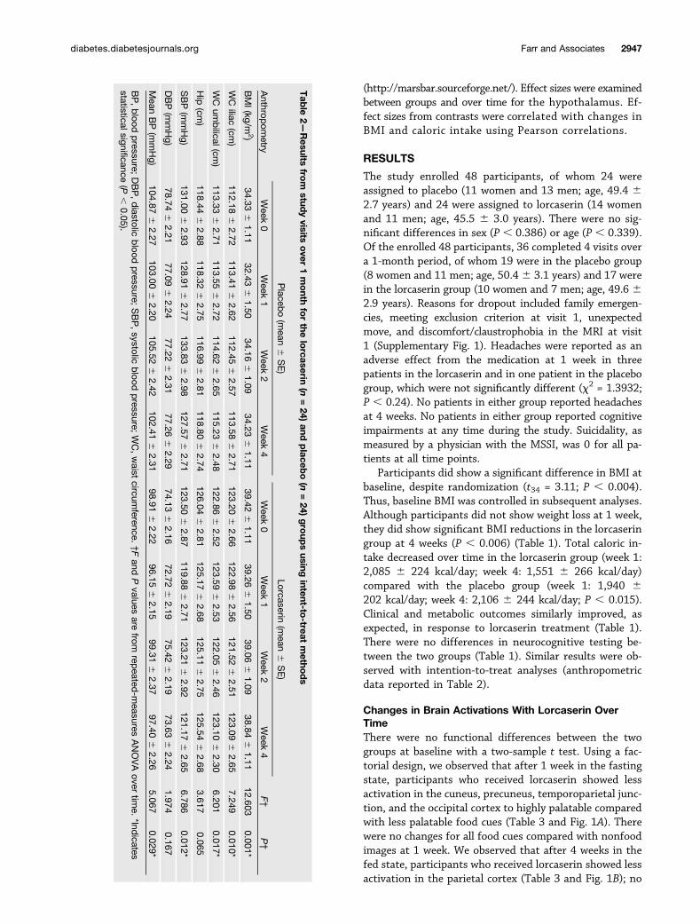
(http://marsbar.sourceforge.net/). Effect sizes were examinedbetween groups and over time for the hypothalamus. Ef-fect sizes from contrasts were correlated with changes inBMI and caloric intake using Pearson correlations.
RESULTS
The study enrolled 48 participants, of whom 24 wereassigned to placebo (11 women and 13 men; age, 49.4 62.7 years) and 24 were assigned to lorcaserin (14 womenand 11 men; age, 45.5 6 3.0 years). There were no sig-nificant differences in sex (P , 0.386) or age (P , 0.339).Of the enrolled 48 participants, 36 completed 4 visits overa 1-month period, of whom 19 were in the placebo group(8 women and 11 men; age, 50.46 3.1 years) and 17 werein the lorcaserin group (10 women and 7 men; age, 49.6 62.9 years). Reasons for dropout included family emergen-cies, meeting exclusion criterion at visit 1, unexpectedmove, and discomfort/claustrophobia in the MRI at visit1 (Supplementary Fig. 1). Headaches were reported as anadverse effect from the medication at 1 week in threepatients in the lorcaserin and in one patient in the placebogroup, which were not significantly different (x2 = 1.3932;P , 0.24). No patients in either group reported headachesat 4 weeks. No patients in either group reported cognitiveimpairments at any time during the study. Suicidality, asmeasured by a physician with the MSSI, was 0 for all pa-tients at all time points.
Participants did show a significant difference in BMI atbaseline, despite randomization (t34 = 3.11; P , 0.004).Thus, baseline BMI was controlled in subsequent analyses.Although participants did not show weight loss at 1 week,they did show significant BMI reductions in the lorcaseringroup at 4 weeks (P , 0.006) (Table 1). Total caloric in-take decreased over time in the lorcaserin group (week 1:2,085 6 224 kcal/day; week 4: 1,551 6 266 kcal/day)compared with the placebo group (week 1: 1,940 6202 kcal/day; week 4: 2,106 6 244 kcal/day; P , 0.015).Clinical and metabolic outcomes similarly improved, asexpected, in response to lorcaserin treatment (Table 1).There were no differences in neurocognitive testing be-tween the two groups (Table 1). Similar results were ob-served with intention-to-treat analyses (anthropometricdata reported in Table 2).
Changes in Brain Activations With Lorcaserin OverTimeThere were no functional differences between the twogroups at baseline with a two-sample t test. Using a fac-torial design, we observed that after 1 week in the fastingstate, participants who received lorcaserin showed lessactivation in the cuneus, precuneus, temporoparietal junc-tion, and the occipital cortex to highly palatable comparedwith less palatable food cues (Table 3 and Fig. 1A). Therewere no changes for all food cues compared with nonfoodimages at 1 week. We observed that after 4 weeks in thefed state, participants who received lorcaserin showed lessactivation in the parietal cortex (Table 3 and Fig. 1B); no
Tab
le2—Results
from
studyvisits
over
1month
forthe
lorcaserin
(n=24)
andplaceb
o(n
=24)
group
susing
intent-to-treat
metho
ds
Anthrop
ometry
Placeb
o(m
ean6
SE)
Lorcaserin(m
ean6
SE)
F†
P†
Week
0Week
1Week
2Week
4Week
0Week
1Week
2Week
4
BMI(kg/m
2)34.33
61.11
32.436
1.5034.16
61.09
34.236
1.1139.42
61.11
39.266
1.5039.06
61.09
38.846
1.1112.603
0.001*
WC
iliac(cm
)112.18
62.72
113.416
2.62112.45
62.57
113.586
2.71123.20
62.66
122.986
2.56121.52
62.51
123.096
2.657.249
0.010*
WC
umbilical(cm
)113.33
62.71
113.556
2.72114.62
62.65
115.236
2.48122.86
62.52
123.596
2.53122.05
62.46
123.106
2.306.201
0.017*
Hip
(cm)
118.446
2.88118.32
62.75
116.996
2.81118.80
62.74
126.046
2.81125.17
62.68
125.116
2.75125.54
62.68
3.6170.065
SBP(m
mHg)
131.006
2.93128.91
62.77
133.836
2.98127.57
62.71
123.506
2.87119.88
62.71
123.216
2.92121.17
62.65
6.7860.012*
DBP(m
mHg)
78.746
2.2177.09
62.24
77.226
2.3177.26
62.29
74.136
2.1672.72
62.19
75.426
2.1973.63
62.24
1.9740.167
Mean
BP(m
mHg)
104.876
2.27103.00
62.20
105.526
2.42102.41
62.31
98.916
2.2296.15
62.15
99.316
2.3797.40
62.26
5.0670.029*
BP,blood
pressure;
DBP,diastolic
blood
pressure;
SBP,systolic
blood
pressure;
WC,waist
circumference.
†Fand
Pvalues
arefrom
repeated
-measures
ANOVAover
time.
*Indicates
statisticalsignificance(P
,0.05).
diabetes.diabetesjournals.org Farr and Associates 2947

Tab
le3—
Brain
activa
tions
from
who
le-b
rain
factorial
analys
esforplace
bo>lorcas
erin†
Cluster
size
(mm
3)
FWE-corrected
Pva
lue
Vox
elzva
lue
MNIco
ordinates
(mm)
Side
Iden
tified
region
Gen
eral
brain
area
XY
Z
Wee
k1–
wee
k0:
high
ly.
less
des
irable
food
cues
Fastingstate
8,60
60.00
14.77
*264
236
16L
Sup
eriortempo
ralG
Temporop
arietaljun
ction
23,456
0.00
14.18
252
264
10L
Middle
temporal
GVisua
lcortic
es4.15
214
274
28L
Precu
neus
Precu
neus
20,850
0.00
14.17
26
286
14C
Cun
eus
Cun
eus
8,47
50.00
14.17
18262
216
RCereb
ellum
Cereb
ellum
6,50
60.00
53.82
44260
6R
Middle
temporal
GVisua
lcortic
es3.67
36268
6R
Middle
occipita
lGVisua
lcortic
es3,73
10.04
63.69
4256
28
CCereb
ellum
Cereb
ellum
Wee
k4–
wee
k0:
allfoo
dcu
es.
nonfoo
dcu
esFe
dstate
9,90
00.03
83.76
240
270
52L
Sup
eriorparietalG
Parietalc
ortex
3.74
258
242
48L
Inferio
rpa
rietalG
Parietalc
ortex
Wee
k1–
wee
k4:
high
ly.
less
des
irable
food
cues
Fastingstate
34,950
0.00
14.25
246
262
22
LOcc
ipita
lGVisua
lcortic
es55
,519
0.00
13.95
32262
36R
Ang
ular
GParietalc
ortex
3.94
32268
20R
Occ
ipita
lGVisua
lcortic
es25
,406
0.00
13.82
52226
34R
Pos
tcen
tral
GParietalc
ortex
3.6
42232
32R
Inferio
rpa
rietalG
Parietalc
ortex
9,99
40.04
43.5
5628
28
RInsu
laInsu
la3.37
42218
216
RParah
ippoc
ampal
GHippoc
ampus
/amyg
dala
C,c
enter;G,g
yrus
;L,left;MNI,Mon
trea
lNeu
rologica
lIns
titute;
R,right. †
Activations
show
nsu
rviveco
rrec
tions
forP,
0.05
,FWEco
rrec
tedforclus
ter(P
valuesh
ownin
theap
propria
teco
lumn).Pea
kssh
ownforclus
ters
arethemos
tsign
ifica
ntalon
gthesa
meiden
tified
region
.*Pas
sesP,
0.05
,FW
Eco
rrec
tedforpe
ak.
2948 Lorcaserin and the Brain Diabetes Volume 65, October 2016

significant differences were found at 4 weeks in the fastingstate. There were no changes for highly desirable comparedwith less desirable food cues at 4 weeks. In a comparisonof short- and long-term (1 vs. 4 weeks) treatment, partic-ipants who received lorcaserin showed less activations inthe insula, parietal cortex, visual cortices, hippocampus,and amygdala in the fasting state at 1 week than at4 weeks, suggesting attenuation at 4 weeks (Table 3 andFig. 1C). There were no differences in activation for thehypothalamus, as was suggested by previous studies inrodents to be the site of action for lorcaserin in the brain,but this, similar to prior studies in humans, may havebeen caused by artifacts in the fMRI and/or its smallsize (see DISCUSSION).
Across participants, changes in activation in the cuneusbetween week 1 and week 0 and between week 1 and week4 in the fasting state to highly desirable compared withless desirable food cues correlated with changes in BMIat 4 weeks (r = 0.60, P , 0.0001; r = 0.37, P , 0.023,respectively). Changes in the visual cortices in the fastingstate to highly desirable compared with less desirable foodcues at week 1–week 0 also correlated with changes inBMI (r = 0.35, P , 0.039). Reported changes in caloricintake did not significantly correlate with changes in brainactivations.
When we examine those individuals in the lorcaseringroup, changes in activation in the cuneus between week1 and week 0 in the fasting state to highly desirablecompared with less desirable food cues correlated with
changes in BMI at 4 weeks (r = 0.58, P , 0.014). Changesin the parietal cortices in the fasting state to highly de-sirable compared with less desirable food cues at week1–week 4 also correlated with changes in BMI (r = 0.57,P , 0.016). Changes in caloric intake correlated withchanges in the visual and parietal cortices and insula atweek 1–week 0 (r = 0.64, P , 0.005; r = 0.53, P , 0.029;r = 0.70, P , 0.002, respectively) and with changes in theprecuneus, temporoparietal junction, visual and parietalcortices, and insula at week 1–week 4 (r = 0.81, P, 0.0001;r = 0.88, P , 0.0001; r = 0.86, P , 0.0001; r = 0.77, P ,0.0001; r = 0.67, P , 0.003, respectively).
Baseline Predictors of Efficacy With LorcaserinTo determine whether there are baseline predictors of theefficacy of lorcaserin, we performed whole-brain regres-sions with baseline brain activations to food cues and themetrics of efficacy, including weight loss, decreases in BMI,and decreases in caloric intake. Greater weight lost at4 weeks is correlated with activation of the amygdala tohighly desirable compared with less desirable food cuesduring the fed state in a regression analysis on the brainof the patients who received lorcaserin (n = 17) at baseline(cluster size = 2,663 mm3; peak at 220, 210, 218; z =4.58) (Fig. 2A). Activations in visual cortices of the occip-ital lobe during the fed state to highly desirable comparedwith less desirable food images correlate significantly withdecreases in BMI (cluster size = 40,200 mm3; peak at 8,238,24; z = 4.45) (Fig. 2B), as well as the amygdala, which does
Figure 1—Changes in brain activations over time with lorcaserin. Shown are brain activations for placebo > lorcaserin to highly desirablecompared with less desirable food images in the fasting state after 1 week (A), to all food compared with nonfood images in the fed stateafter 4 weeks (B), and to highly desirable images compared with less desirable food images in the fasting state for short-term (1 week)compared with long-term (4 weeks) treatments (C). BOLD contrasts are superimposed on a T1 structural image in axial sections from z =215to z = 60, in neurological orientation.
diabetes.diabetesjournals.org Farr and Associates 2949

not reach a corrected threshold (cluster size = 2,438 mm3;peak at 218, 28, 216; z = 3.55). For participants onlorcaserin who submitted their detailed food logs (n = 13),we observe that baseline activation in the prefrontal cortexto all food images compared with nonfood images during thefed state indicating lower baseline activations are associatedwith more decreases in total caloric consumption (clustersize = 13,069 mm3; peak at 26, 58, 10; z = 4.26) (Fig. 2C).
DISCUSSION
We report decreases in brain activations to food cues withrelatively short-term (1 week) and longer-term (4 weeks)therapy with lorcaserin. More specifically, we report deac-tivations with lorcaserin at 1 week during the fastingstate and before weight loss with decreased attention-related parietal and visual activations to highly desirablefood cues. In a direct comparison between 1 and 4 weeks,we see significant decreases with lorcaserin at 1 week tohighly desirable food cues in attention-related brain areasand the amygdala compared with the longer-term 4-weektime point, indicating attenuation of deactivations bylorcaserin over time. After 4 weeks and modest weightloss, we report decreases in the attention-related parietalcortex in the fed state.
When baseline predictors of success with lorcaserin wereexamined, weight loss at 4 weeks correlated with baselineactivation of amygdala, part of the emotional/limbic system,to highly desirable food cues after a meal in a whole-brainregression analysis, suggesting that increased activation of
the amygdala indicates which participants would benefitmost from the use of lorcaserin. Decreases in BMI correlatedwith amygdala and occipital activations at baseline for thelorcaserin group in the fed state, suggesting that lorcaserinmay be helpful for individuals who find highly desirable andless healthful food cues more salient. Decreases in totalcaloric consumption correlated inversely with activations inthe prefrontal cortex to all food cues compared with non-food cues during the fed state, suggesting poorer baselinecognitive control may suggest later benefit of lorcaserinon reported caloric consumption. Altogether, our findingssuggest that lorcaserin decreases cortical and limbic activa-tions to food cues over time, with greater effects in theshort-term or 1 week. Furthermore, the regressions withchanges in weight and BMI suggest that lorcaserin may beof the most benefit to emotional eaters or those who havedysfunctional activations at baseline.
Changes in Brain Activations With Lorcaserin OverTimeAt 1 week, we observe decreased activation in the parietalcortex, cuneus, precuneus, and visual cortices to highly de-sirable food cues. These areas are involved in many cognitiveprocesses but may be representing changes in attention andsaliency; that is, attending to cues of relative importance,processing, indicating that short-term treatment withlorcaserin decreases the importance and attention to highlypalatable foods. In the fasting state, activations of areasinvolved in salience and attention to high-calorie comparedwith low-calorie food cues have been previously observed in
Figure 2—Baseline predictors of efficacy with lorcaserin. Shown are results from a whole-brain regression analysis with weight loss (A),BMI decrease (B), and caloric intake (C) at 4 weeks with brain activations at baseline (week 0). Greater activation of areas shown in red atbaseline (week 0) are correlated with greater weight loss, BMI decreases, or decreases in caloric intake at 4 weeks. BOLD contrasts aresuperimposed on a T1 structural image in axial sections from z = 215 to z = 60, in neurological orientation.
2950 Lorcaserin and the Brain Diabetes Volume 65, October 2016

participants without obesity because these are generallymore attention-grabbing foods (21). Other studies havealso shown similarly increased activation in the occipitalcortex for food images (22,23). These typically increasedactivations are due to the inherent importance and emo-tional value of food cues; indeed, others have shown thathigher occipital lobe activation is linked with exposure toemotional images (24,25). The parietal cortex is a well-known component of the attention and salience systemthat increases activity to important stimuli (26–28).Thus, our data indicate that highly desirable food cuesappear less important with lorcaserin treatment. Alto-gether, these results suggest that lorcaserin decreases theemotional significance of highly desirable food cues, whichleads to the observed decrease in food consumption.
Regarding changes over time, we report attenuation ofbrain deactivations between 1 and 4 weeks of lorcaserintherapy, where there is less deactivation of attention-related circuitry at 4 weeks than at 1 week. Indeed, adirect comparison showed the same visual and parietalcortices as well as the insula and amygdala, which mayalso be involved in the saliency network, are more de-activated at 1 week than at 4 weeks for lorcaserin com-pared with placebo. Among their involvement in other CNSsystems, the insula and amygdala are both involved insalience and emotional processing (29–33). Although theinsula is also involved in proprioception and taste (34), itsactivation in this study could be related to salience or tochanges in gut motility and cue priming to involve theenteric nervous system resulting from the posterior loca-tion of the activation (35). This relative increase between1 and 4 weeks may represent attenuation at 4 weeks,where brain activations begin to return to control/base-line levels. Indeed, at 4 weeks compared with baseline, weobserve only decreased activation of the parietal cortex tofood cues, regardless of desirability, in the fed state. Thislikely demonstrates a continued decreasing “value” of foodcues with lorcaserin treatment in the longer-term, whichhas become less pronounced or attenuated over time.
Unlike studies with rodents, we did not observe anychanges in hypothalamic activation with lorcaserin (9–11).Rodents and humans have a different brain structure. Eat-ing behaviors in rodents are primarily controlled by thehomeostatic system (hypothalamus), and the rodent cortexis not comparable in structure or function to the cortexcontrolling food intake in humans. Indeed, in humans,eating is often controlled by higher cortical processes, in-cluding reward, saliency, and other networks, and not sim-ply the homeostatic processes of the hypothalamus (36).The lack of a finding in the hypothalamus may also be dueto the limitations of fMRI, because the hypothalamus issmall and susceptible to artifacts from its proximity to thesinuses (36). Thus, hypothalamic activation by lorcaserinshould not be ruled out but cannot be confirmed from thisfMRI study in humans.
It could be argued that differences in brain activa-tions over time may indicate confounding effects, such as
interacting changes with weight loss over time and/orhabituation to the effects of the medication. That patientscould have habituated to the task over time is also possible.Considering that participants in the placebo group had thesame number of fMRI scans, these potential confoundersshould have been adequately controlled.
Baseline Predictors of Efficacy With LorcaserinGiven the variability of responses to lorcaserin in the clinicand to determine predictors of efficacy with lorcaserin, weperformed whole-brain regression analyses of baseline ac-tivations with metrics of clinical success, including weightloss, BMI changes, and caloric intake. We observed that theactivation of the amygdala at baseline in response to highlydesirable food cues, an indicator of emotional eating, cor-related with greater weight lost at 4 weeks. The amygdala isa component of the saliency network, particularly respond-ing to emotionally salient stimuli (31–33), that has alsobeen implicated directly in the emotional eating system(37,38). Thus, individuals who find highly palatable foodsemotionally more salient would be the ones to mainly ob-tain the most weight loss/change in brain activations overtime when treated with lorcaserin. This also indicates thatlorcaserin may be most helpful for individuals who areemotional eaters and will need to be confirmed with fur-ther studies, including not only fMRIs but also question-naires specific for emotional eating. In addition, we reportthat decreases in BMI correlate not only with activation ofthe amygdala but also with occipital activations at baselinefor the lorcaserin group in the fed state. This providesfurther support for the notion that lorcaserin may behelpful for individuals who find highly desirable and lesshealthful food cues more salient. Furthermore, prefrontalactivation during the fed state to all food cues comparedwith nonfood cues correlates inversely with decreases intotal caloric consumption. Prefrontal activations may in-dicate a number of cognitive processes, including emotion,salience, memory, and top-down processes (39). Most fre-quently with regards to food cues, prefrontal activationsare thought to be related to cognitive control and to anindividual’s ability to stop him- or herself from eating inexcess or eating unhealthy foods (29,40,41). Thus, individ-uals who had less cognitive control–related prefrontal ac-tivations at baseline showed less decreases in caloric intake,indicating that participants who already have less corticalinhibition to food cues receive the greatest benefit fromlorcaserin in food consumption. This is a plausible hypoth-esis, because these individuals would have the most roomfor improvement. This needs to be tested by future specif-ically designed studies focusing on emotional eaters andany differences in the efficacy and brain responses tofood with lorcaserin.
Strengths, Limitations, and Other ConsiderationsAlthough we do not observe any changes in cognition withup to 1 month of lorcaserin treatment, animal studiessuggest that there may be cognitive benefits. Whether theeffects observed previously in mice were a result of changes
diabetes.diabetesjournals.org Farr and Associates 2951
in body weight or the medication per se remain unclear, be-cause they are not confirmed in our study with participantson the medication before significant weight loss. A priorstudy in mice demonstrated decreased spatial/workingmemory with diet-induced obesity, which was improvedwith lorcaserin therapy (42), but this may simply highlighteffects of weight loss or simply the differences betweenrodent and human brains. Weight loss itself typically re-sults in improved performance on cognitive tasks in hu-mans, including memory and executive function (43,44).Because we do not observe any changes on cognition withlorcaserin in this study, this may indicate that any longer-term benefits may be the result of weight loss and not themedication itself and/or that such changes may be weakeror take longer to manifest in humans. Longer-term studieswould be required to determine whether this might be thecase. One could argue that it is a limitation of the studythat our randomization did not eliminate differences inBMI between the two groups at baseline. However, wecontrolled for such baseline differences in our analysesand observed no differences in brain activations to foodcues between the two groups at baseline. One would alsoquestion, given that no prior data exist on which to basepower calculations, whether an adequate number of partic-ipants was included. The number of participants was sim-ilar to recent studies with other medications and MRI(45,46). Furthermore, although we have examined multiplefMRI contrasts over time, we report results that reach athreshold corrected for multiple comparisons with FWEcorrections, according to the standard with fMRI studies.Thus, multiple comparisons should not have affected ourfindings.
Studies of longer duration would also be warranted tolook at continued success and longer-term effects on foodcue processing, such as at 12 weeks, when weight lossis expected to plateau. Indeed, studies have shown thatweight loss by week 12 with lorcaserin is a strong predictorof longer-term (52 weeks) success (47), which may indicatea good time at which to determine individual differences inresponse to lorcaserin. Overall, these results are strong andindicate clear changes in brain responses to food cues withlorcaserin therapy. In addition, we observe baseline differ-ences in activation that correlate to behavioral changes inresponse to lorcaserin and that may suggest that lorcaserinis of particular benefit to individuals who are emotionaleaters and/or place importance on high-calorie or high-fat foods. This is of particular importance in the clinic,given the variable responses observed to lorcaserin therapy.If confirmed and correlated with appropriately designedquestionnaire data, these findings can not only providemechanistic explanations but also lead to developmentsof clinical tools to select which individuals would respondbest clinically to treatment with lorcaserin.
Acknowledgments. Drug supply (lorcaserin and placebo) was providedby Arena Pharmaceuticals GmbH through Eisai, Inc., to investigators.
Funding. Eisai, Inc., supported the study through an investigator-initiatedstudy grant. Eisai, Inc., approved the design of the study but had no role in studydesign; conduct of the study; collection, management, analysis, and interpre-tation of the data; or the preparation, review, or approval of the manuscript.The project was supported by National Institute of Child Health and HumanDevelopment and Harvard Clinical and Translational Science Center grantUL1-RR-025758 from the National Center for Research Resources. O.M.F. issupported by 5T32HD052961. This study is also partly supported by NationalInstitutes of Health grant DK-081913.Duality of Interest. No potential conflicts of interest relevant to this articlewere reported.Author Contributions. O.M.F. and C.S.M. designed the experiment.O.M.F., J.U., A.G., M.C., H.Ka., H.M., M.V., A.K., H.Ki., A.S., A.M., and C.S.M.conducted the experiments and acquired data. O.M.F. and N.S. analyzed thedata. O.M.F. wrote the manuscript with input from all of the other authors. O.M.F.is the guarantor of this work and, as such, had full access to all the data in thestudy and takes responsibility for the integrity of the data and the accuracy of thedata analysis.
References1. Fontaine KR, Redden DT, Wang C, Westfall AO, Allison DB. Years of life lostdue to obesity. JAMA 2003;289:187–1932. Loke YK, Derry S, Pritchard-Copley A. Appetite suppressants and valvularheart disease - a systematic review. BMC Clin Pharmacol 2002;2:63. Vickers SP, Dourish CT, Kennett GA. Evidence that hypophagia induced byd-fenfluramine and d-norfenfluramine in the rat is mediated by 5-HT2C recep-tors. Neuropharmacology 2001;41:200–2094. Tecott LH, Sun LM, Akana SF, et al. Eating disorder and epilepsy in micelacking 5-HT2c serotonin receptors. Nature 1995;374:542–5465. Nonogaki K, Strack AM, Dallman MF, Tecott LH. Leptin-independent hy-perphagia and type 2 diabetes in mice with a mutated serotonin 5-HT2C receptorgene. Nat Med 1998;4:1152–11566. Fidler MC, Sanchez M, Raether B, et al.; BLOSSOM Clinical Trial Group. Aone-year randomized trial of lorcaserin for weight loss in obese and over-weight adults: the BLOSSOM trial. J Clin Endocrinol Metab 2011;96:3067–30777. Rueda-Clausen CF, Padwal RS, Sharma AM. New pharmacological ap-proaches for obesity management. Nat Rev Endocrinol 2013;9:467–4788. Smith SR, Weissman NJ, Anderson CM, et al.; Behavioral Modification andLorcaserin for Overweight and Obesity Management (BLOOM) Study Group.Multicenter, placebo-controlled trial of lorcaserin for weight management. N EnglJ Med 2010;363:245–2569. Abramowski D, Rigo M, Duc D, Hoyer D, Staufenbiel M. Localization of the5-hydroxytryptamine2C receptor protein in human and rat brain using specificantisera. Neuropharmacology 1995;34:1635–164510. Hoffman BJ, Mezey E. Distribution of serotonin 5-HT1C receptor mRNA inadult rat brain. FEBS Lett 1989;247:453–46211. Mengod G, Nguyen H, Le H, Waeber C, Lübbert H, Palacios JM. The dis-tribution and cellular localization of the serotonin 1C receptor mRNA in the rodentbrain examined by in situ hybridization histochemistry. Comparison with receptorbinding distribution. Neuroscience 1990;35:577–59112. Heisler LK, Cowley MA, Tecott LH, et al. Activation of central melanocortinpathways by fenfluramine. Science 2002;297:609–61113. Lam DD, Przydzial MJ, Ridley SH, et al. Serotonin 5-HT2C receptor agonistpromotes hypophagia via downstream activation of melanocortin 4 receptors.Endocrinology 2008;149:1323–132814. Morton GJ, Cummings DE, Baskin DG, Barsh GS, Schwartz MW. Centralnervous system control of food intake and body weight. Nature 2006;443:289–29515. Benoit SC, Schwartz MW, Lachey JL, et al. A novel selective melanocortin-4receptor agonist reduces food intake in rats and mice without producing aversiveconsequences. J Neurosci 2000;20:3442–3448
2952 Lorcaserin and the Brain Diabetes Volume 65, October 2016

16. Krude H, Biebermann H, Schnabel D, et al. Obesity due to proopiomelanocortindeficiency: three new cases and treatment trials with thyroid hormone andACTH4-10. J Clin Endocrinol Metab 2003;88:4633–464017. O’Neil PM, Smith SR, Weissman NJ, et al. Randomized placebo-controlledclinical trial of lorcaserin for weight loss in type 2 diabetes mellitus: theBLOOM-DM study. Obesity (Silver Spring) 2012;20:1426–143618. Farooqi IS, Bullmore E, Keogh J, Gillard J, O’Rahilly S, Fletcher PC. Leptinregulates striatal regions and human eating behavior. Science 2007;317:135519. Farr OM, Fiorenza C, Papageorgiou P, et al. Leptin therapy alters appetiteand neural responses to food stimuli in brain areas of leptin-sensitive subjectswithout altering brain structure. J Clin Endocrinol Metab 2014;99:E2529–E253820. Alonso-Alonso M, Ziemke F, Magkos F, et al. Brain responses to foodimages during the early and late follicular phase of the menstrual cycle in healthyyoung women: relation to fasting and feeding. Am J Clin Nutr 2011;94:377–38421. Frank S, Laharnar N, Kullmann S, et al. Processing of food pictures: in-fluence of hunger, gender and calorie content. Brain Res 2010;1350:159–16622. Führer D, Zysset S, Stumvoll M. Brain activity in hunger and satiety: anexploratory visually stimulated FMRI study. Obesity (Silver Spring) 2008;16:945–95023. Schur EA, Kleinhans NM, Goldberg J, Buchwald D, Schwartz MW, MaravillaK. Activation in brain energy regulation and reward centers by food cues varieswith choice of visual stimulus. Int J Obes 2009;33:653–66124. Lang PJ, Bradley MM. Emotion and the motivational brain. Biol Psychol2010;84:437–45025. Lang PJ, Bradley MM, Fitzsimmons JR, et al. Emotional arousal and acti-vation of the visual cortex: an fMRI analysis. Psychophysiology 1998;35:199–21026. Corbetta M, Shulman GL. Control of goal-directed and stimulus-driven at-tention in the brain. Nat Rev Neurosci 2002;3:201–21527. McFadden KL, Cornier MA, Melanson EL, Bechtell JL, Tregellas JR. Effectsof exercise on resting-state default mode and salience network activity inoverweight/obese adults. Neuroreport 2013;24:866–87128. Capotosto P, Tosoni A, Spadone S, et al. Anatomical segregation of visualselection mechanisms in human parietal cortex. J Neurosci 2013;33:6225–622929. Hendrick OM, Luo X, Zhang S, Li CS. Saliency processing and obesity: apreliminary imaging study of the stop signal task. Obesity (Silver Spring) 2012;20:1796–180230. Farr OM, Hu S, Zhang S, Li CS. Decreased saliency processing as a neuralmeasure of Barratt impulsivity in healthy adults. Neuroimage 2012;63:1070–107731. Berns GS, Capra CM, Moore S, Noussair C. Three studies on the neuro-economics of decision-making when payoffs are real and negative. Adv HealthEcon Health Serv Res 2008;20:1–2932. Garrido MI, Barnes GR, Sahani M, Dolan RJ. Functional evidence for a dualroute to amygdala. Curr Biol 2012;22:129–134
33. Vuilleumier P, Armony JL, Driver J, Dolan RJ. Distinct spatial frequencysensitivities for processing faces and emotional expressions. Nat Neurosci 2003;6:624–63134. Rolls ET. Taste, olfactory, and food reward value processing in the brain.Prog Neurobiol 2015;127-128:64–9035. Coss-Adame E, Rao SS. Brain and gut interactions in irritable bowel syn-drome: new paradigms and new understandings. Curr Gastroenterol Rep 2014;16:37936. Salem V, Dhillo WS. Imaging in endocrinology: the use of functional MRI tostudy the endocrinology of appetite. Eur Endocrinol 2015;173:R59–R6837. Ulrich-Lai YM, Christiansen AM, Wang X, Song S, Herman JP. Statisticalmodeling implicates neuroanatomical circuit mediating stress relief by ‘comfort’food. Brain Struct Funct 2016;221:3141–135638. van Bloemendaal L, Veltman DJ, ten Kulve JS, et al. Emotional eating isassociated with increased brain responses to food-cues and reduced sensitivityto GLP-1 receptor activation. Obesity (Silver Spring) 2015;23:2075–208239. Leisman G, Moustafa AA, Shafir T. Thinking, walking, talking: integratorymotor and cognitive brain function. Front Public Health 2016;4:9440. Hendrick OM, Ide JS, Luo X, Li CS. Dissociable processes of cognitivecontrol during error and non-error conflicts: a study of the stop signal task. PLoSOne 2010;5:e1315541. Batterink L, Yokum S, Stice E. Body mass correlates inversely with inhibitorycontrol in response to food among adolescent girls: an fMRI study. Neuroimage2010;52:1696–170342. Yang H, Huang F, Ni M, et al. Cognitive function is impaired by obesity andalleviated by lorcaserin treatment in mice. CNS Neurosci Ther 2015;21:472–47443. Siervo M, Arnold R, Wells JC, et al. Intentional weight loss in overweight andobese individuals and cognitive function: a systematic review and meta-analysis.Obes Rev 2011;12:968–98344. Pursey KM, Stanwell P, Callister RJ, Brain K, Collins CE, Burrows TL. Neuralresponses to visual food cues according to weight status: a systematic review offunctional magnetic resonance imaging studies. Front Nutr 2014;1:745. van Bloemendaal L, Veltman DJ, Ten Kulve JS, et al. Brain reward-systemactivation in response to anticipation and consumption of palatable food is alteredby glucagon-like peptide-1 receptor activation in humans. Diabetes Obes Metab2015;17:878–88646. ten Kulve JS, Veltman DJ, van Bloemendaal L, et al. Endogenous GLP-1mediates postprandial reductions in activation in central reward and satiety areasin patients with type 2 diabetes. Diabetologia 2015;58:2688–269847. Smith SR, O’Neil PM, Astrup A, et al. Early weight loss while on lorcaserin,diet and exercise as a predictor of week 52 weight-loss outcomes. Obesity (SilverSpring) 2014;22:2137–2146
diabetes.diabetesjournals.org Farr and Associates 2953