Embed Size (px)
Citation preview
Accelerated Partial Breast Irradiation
Dr Patricia Lillis MD, MHA,MSS
Marshfield Clinic
Radiation Oncology

Outline
1. Rationale
2. Review of selected literature
3. Technical aspects
4. Selection criteria
5. Ongoing questions and trials
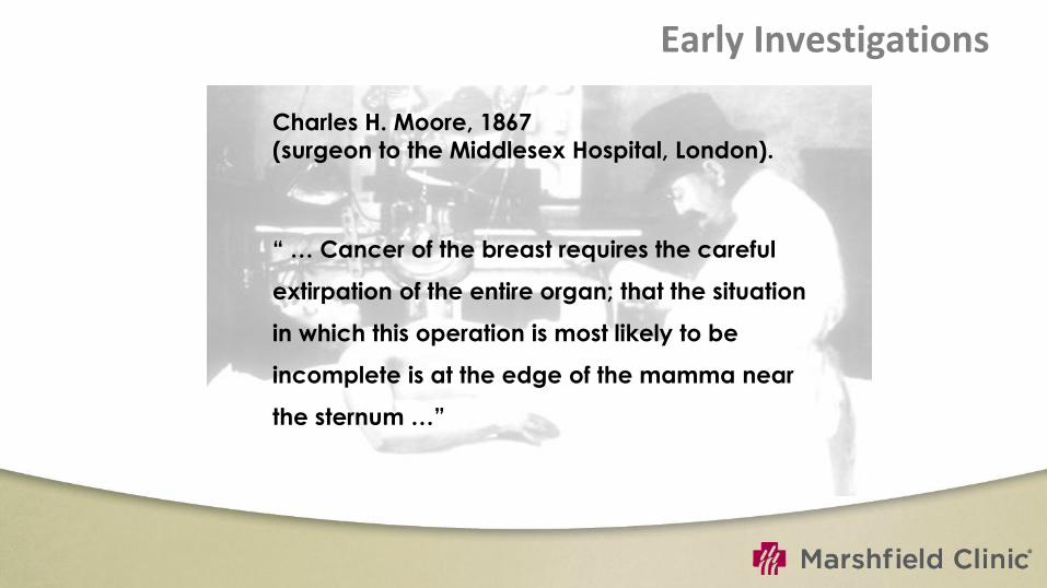
Early Investigations
Charles H. Moore, 1867
(surgeon to the Middlesex Hospital, London).
“ … Cancer of the breast requires the careful
extirpation of the entire organ; that the situation
in which this operation is most likely to be
incomplete is at the edge of the mamma near
the sternum …”

Early Investigations
William Halsted, 1852-1922
(surgeon to the Johns Hopkins Hospital, Baltimore).
“ Most of us have heard our teacher in surgery admit
that they never cured a case of cancer of the breast …
Everyone knows how dreadful the end-results were
before cleaning out the axilla became recognized as
an essential part of the operation.”
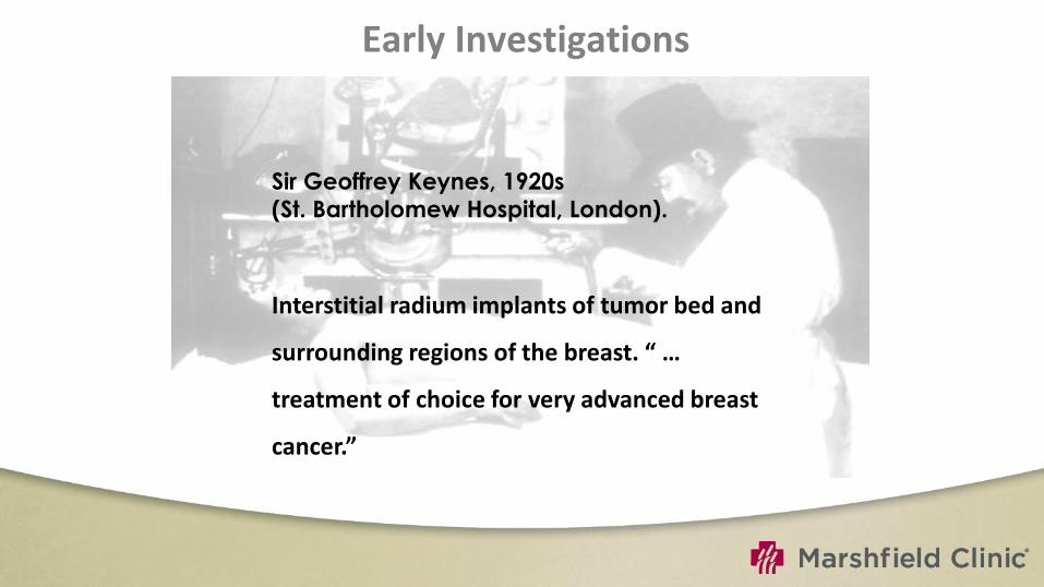
Early Investigations
Sir Geoffrey Keynes, 1920s
(St. Bartholomew Hospital, London).
Interstitial radium implants of tumor bed and
surrounding regions of the breast. “ …
treatment of choice for very advanced breast
cancer.”
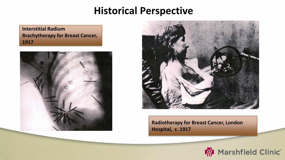
Historical Perspective
Radiotherapy for Breast Cancer, London Hospital, c. 1917
Interstitial Radium Brachytherapy for Breast Cancer, 1917
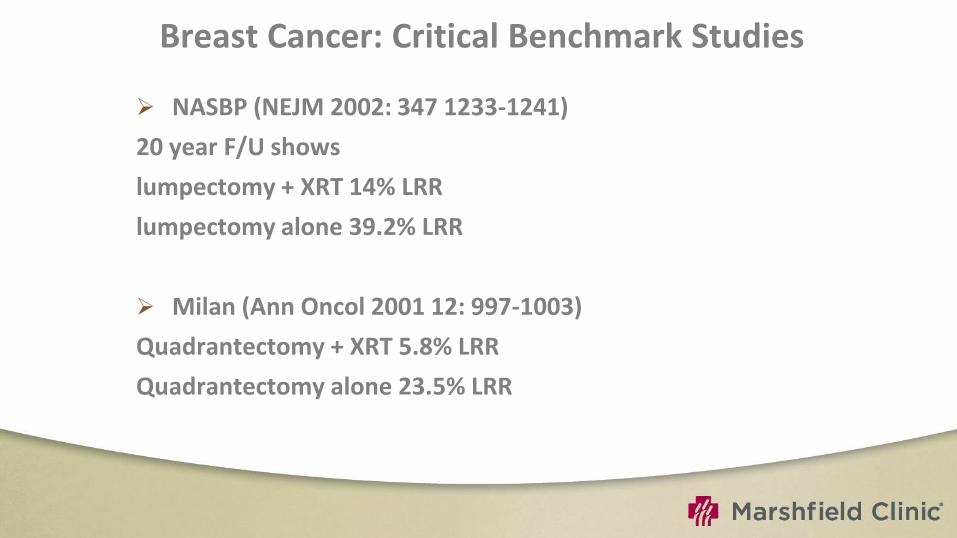
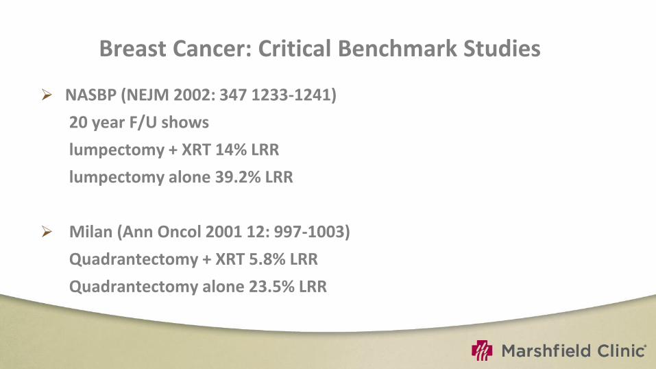
Breast Cancer: Critical Benchmark Studies
NASBP (NEJM 2002: 347 1233-1241)
20 year F/U shows
lumpectomy + XRT 14% LRR
lumpectomy alone 39.2% LRR
Milan (Ann Oncol 2001 12: 997-1003)
Quadrantectomy + XRT 5.8% LRR
Quadrantectomy alone 23.5% LRR
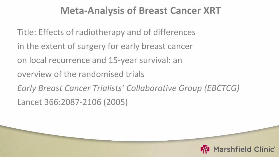
Meta-Analysis of Breast Cancer XRT
Title: Effects of radiotherapy and of differences
in the extent of surgery for early breast cancer
on local recurrence and 15-year survival: an
overview of the randomised trials
Early Breast Cancer Trialists’ Collaborative Group (EBCTCG)
Lancet 366:2087-2106 (2005)
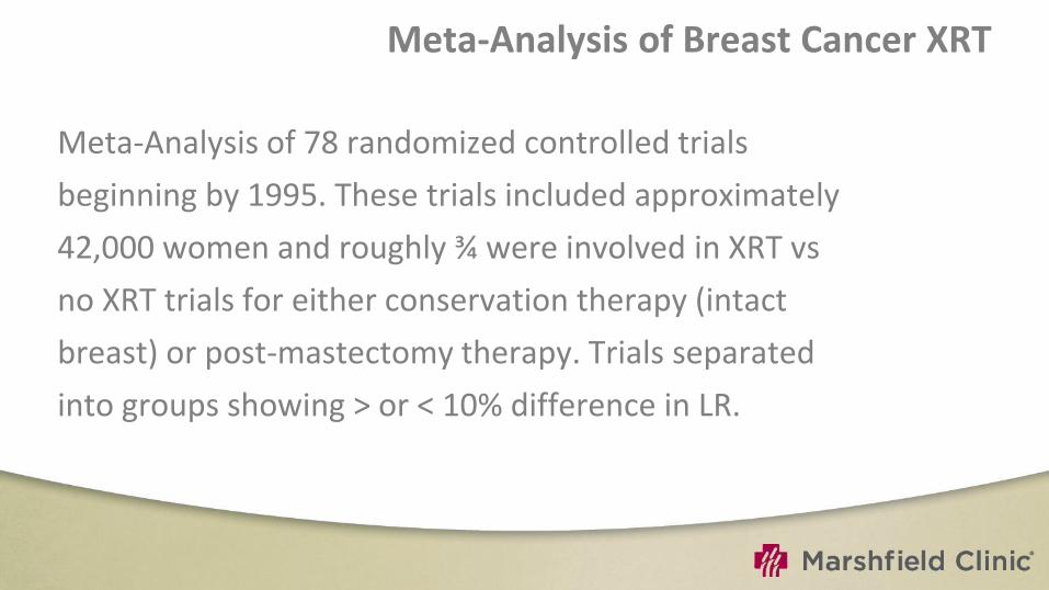
Meta-Analysis of Breast Cancer XRT
Meta-Analysis of 78 randomized controlled trials
beginning by 1995. These trials included approximately
42,000 women and roughly ¾ were involved in XRT vs
no XRT trials for either conservation therapy (intact
breast) or post-mastectomy therapy. Trials separated
into groups showing > or < 10% difference in LR.
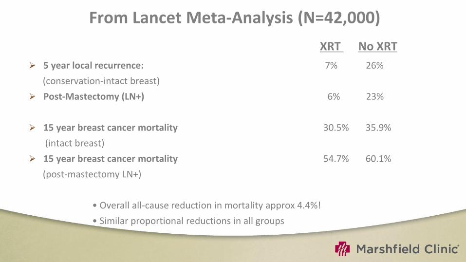
From Lancet Meta-Analysis (N=42,000)
XRT No XRT
5 year local recurrence: 7% 26%
(conservation-intact breast)
Post-Mastectomy (LN+) 6% 23%
15 year breast cancer mortality 30.5% 35.9%
(intact breast)
15 year breast cancer mortality 54.7% 60.1%
(post-mastectomy LN+)
• Overall all-cause reduction in mortality approx 4.4%!
• Similar proportional reductions in all groups

Techniques of APBI
Interstitial implant brachytherapy
Intra-Operative Radiotherapy
External beam radiotherapy
Intracavitary Brachytherapy
Breast Cancer: Critical Benchmark Studies
NASBP (NEJM 2002: 347 1233-1241)
20 year F/U shows
lumpectomy + XRT 14% LRR
lumpectomy alone 39.2% LRR
Milan (Ann Oncol 2001 12: 997-1003)
Quadrantectomy + XRT 5.8% LRR
Quadrantectomy alone 23.5% LRR
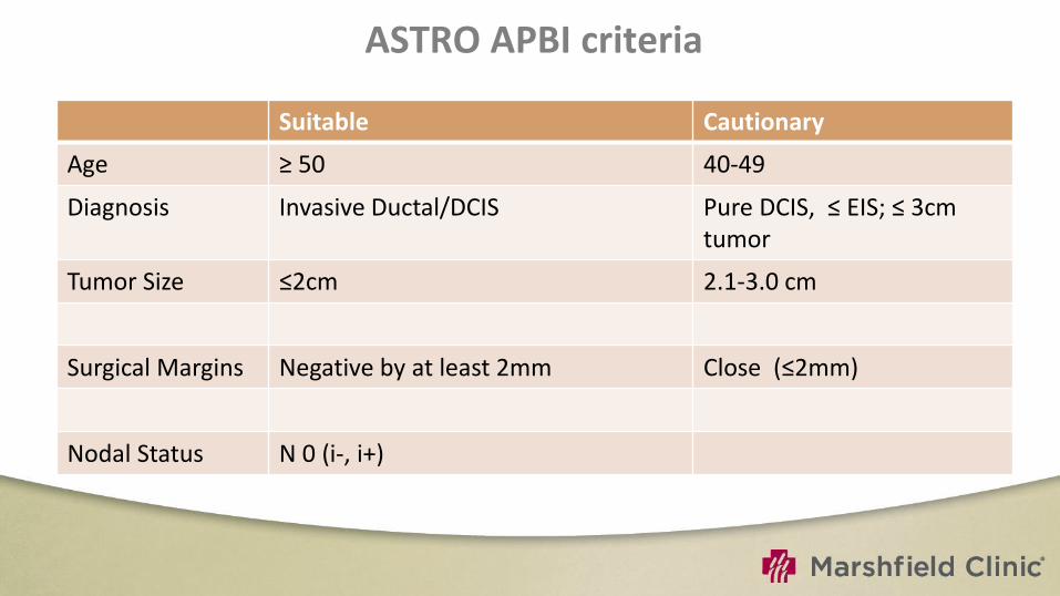
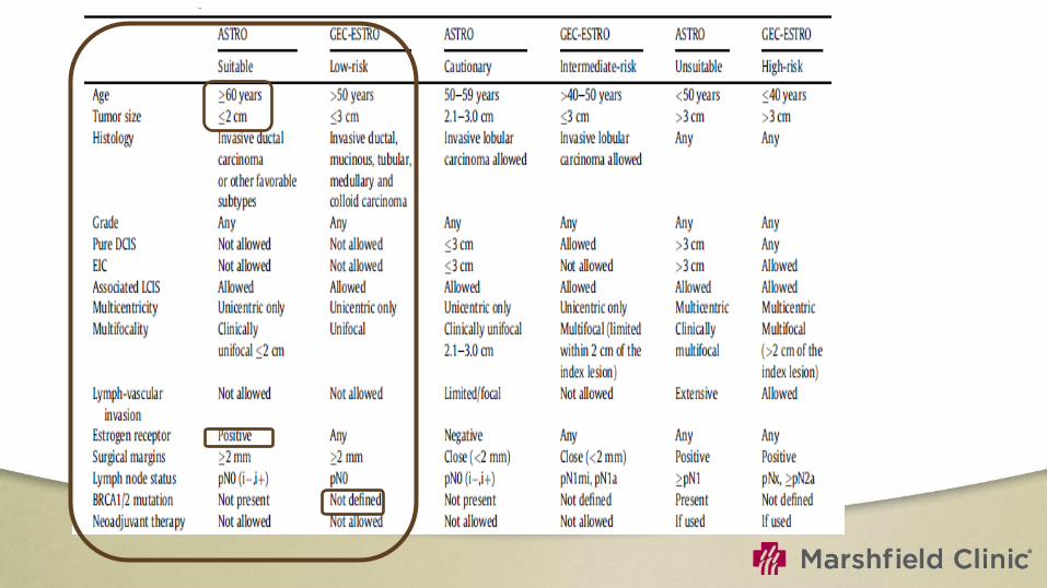
ASTRO APBI criteria
Suitable Cautionary
Age ≥ 50 40-49
Diagnosis Invasive Ductal/DCIS Pure DCIS, ≤ EIS; ≤ 3cm tumor
Tumor Size ≤2cm 2.1-3.0 cm
Surgical Margins Negative by at least 2mm Close (≤2mm)
Nodal Status N 0 (i-, i+)
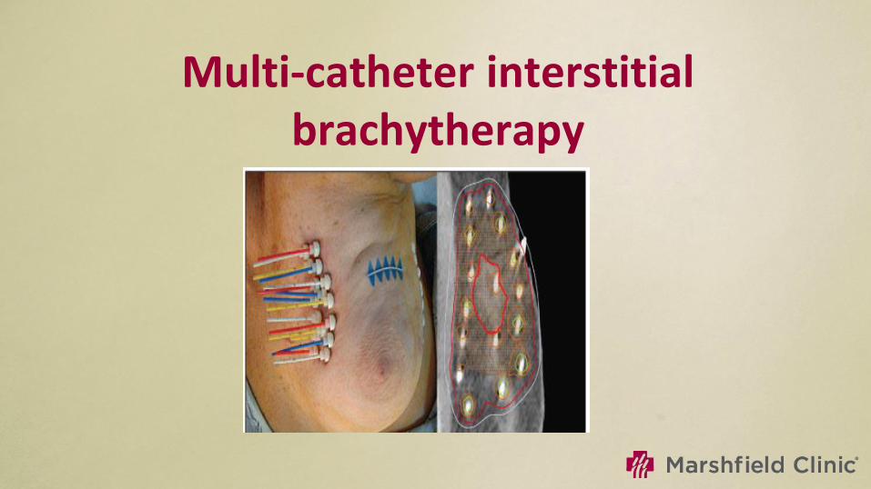
Multi-catheter interstitial brachytherapy
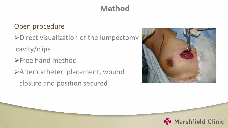
Method
Open procedure
Direct visualization of the lumpectomy
cavity/clips
Free hand method
After catheter placement, wound
closure and position secured
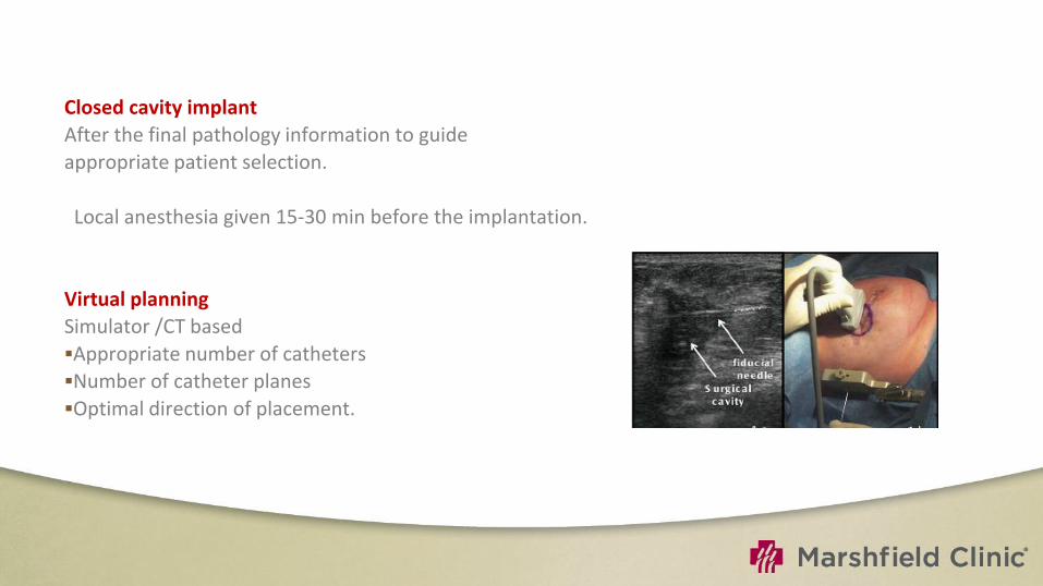
Closed cavity implant
After the final pathology information to guide
appropriate patient selection.
Local anesthesia given 15-30 min before the implantation.
Virtual planning
Simulator /CT based
Appropriate number of catheters
Number of catheter planes
Optimal direction of placement.
Target volume
Lumpectomy cavity plus a 2-cm margin
Near chest wall and skin -1 to 1.5 cm
Dose
HDR-34 Gy/10# bid in 5 days
LDR- 45 Gy @ 50 cGy/hr over 4.5 days
Arthur et al. IJROBP 2003 vol 56, 681-9
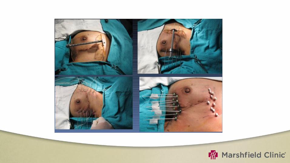
Catheter implantation
Free hand technique
Template to ensure even needle spacing
Using image guidance
Ultrasound
Fluoroscopy
CT Scan
Design of the implant geometry
Needles are implanted parallel and equidistance from each other (Paris system).
In most cases inserted in a mediolateral direction.
In very medially or laterally located tumor sites, needles should be implanted in a craniocaudal direction to enable separate target area from skin points.
In some rare cases, the upper outer quadrant has to be implanted with needles orientated in a 45° angle to avoid overlap of source positions and skin
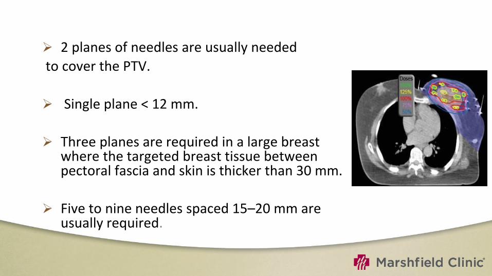
2 planes of needles are usually needed
to cover the PTV.
Single plane < 12 mm.
Three planes are required in a large breast where the targeted breast tissue between pectoral fascia and skin is thicker than 30 mm.
Five to nine needles spaced 15–20 mm are usually required.
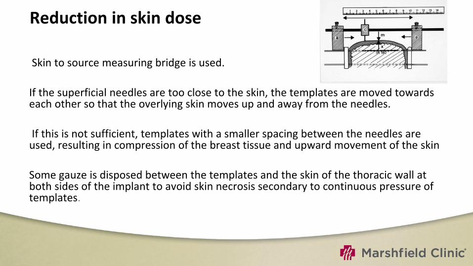
Reduction in skin dose
Skin to source measuring bridge is used.
If the superficial needles are too close to the skin, the templates are moved towards each other so that the overlying skin moves up and away from the needles.
If this is not sufficient, templates with a smaller spacing between the needles are used, resulting in compression of the breast tissue and upward movement of the skin
Some gauze is disposed between the templates and the skin of the thoracic wall at both sides of the implant to avoid skin necrosis secondary to continuous pressure of templates.
Advantages
Has the longest follow-up.
Better control and tailoring of radiation-dose delivery to variations in lumpectomy cavity, shape, or location within the breast.
Limits toxicity to healthy tissue while delivering the maximum dose to at-risk tissue.
Critical structures can be avoided by differential loading of the catheters
Limitations
Considerable training and experience
Appearance and patient acceptance of multiple catheter implants in the breast
high skin dose: great care is required to ensure adequate source-to-skin distance in patients treated with brachytherapy
Therefore, may not be a viable treatment option for patients with superficial tumors or small breasts
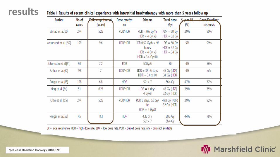
results
Njeh et al. Radiation Oncology 2010,5:90
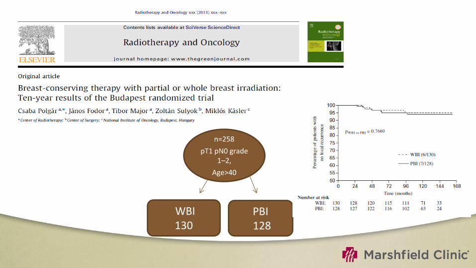
n=258
pT1 pN0 grade 1–2,
Age>40
WBI 130
PBI 128
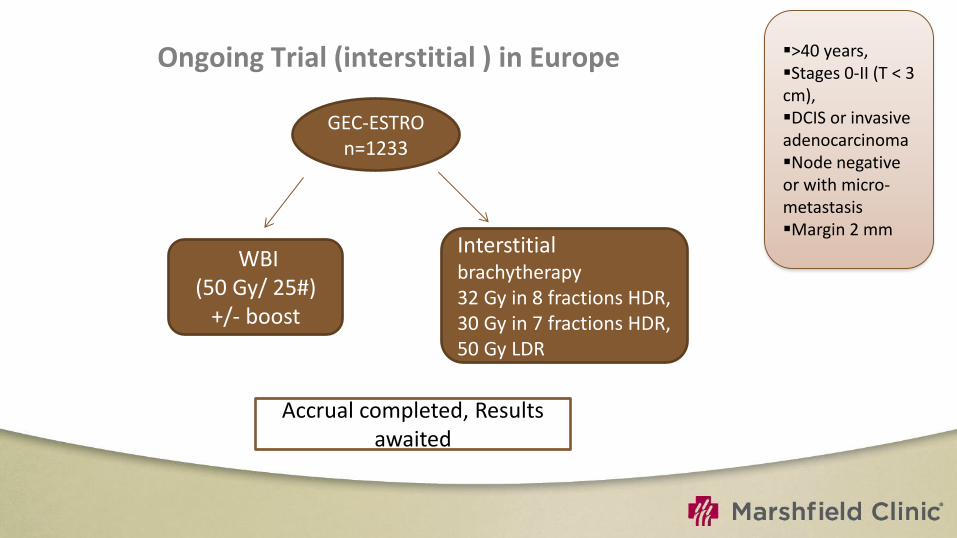
Ongoing Trial (interstitial ) in Europe
GEC-ESTRO n=1233
WBI (50 Gy/ 25#)
+/- boost
Interstitial brachytherapy 32 Gy in 8 fractions HDR, 30 Gy in 7 fractions HDR, 50 Gy LDR
>40 years, Stages 0-II (T < 3 cm), DCIS or invasive adenocarcinoma Node negative or with micro-metastasis Margin 2 mm
Accrual completed, Results awaited

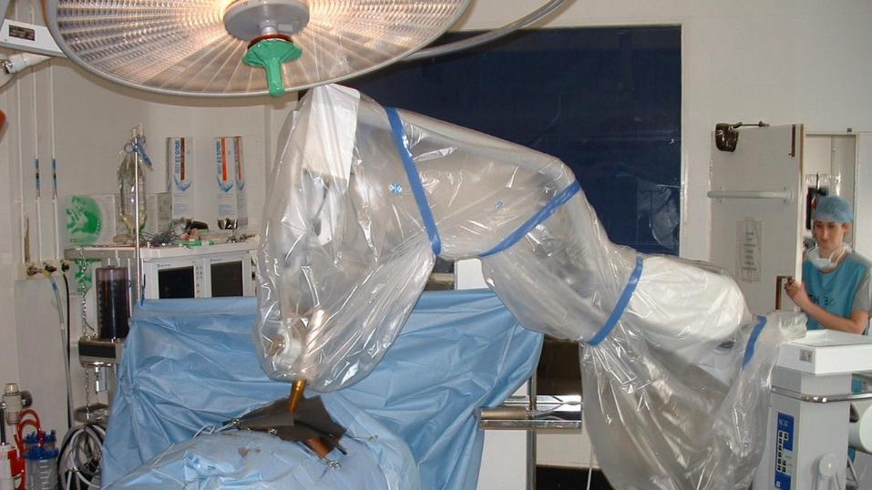
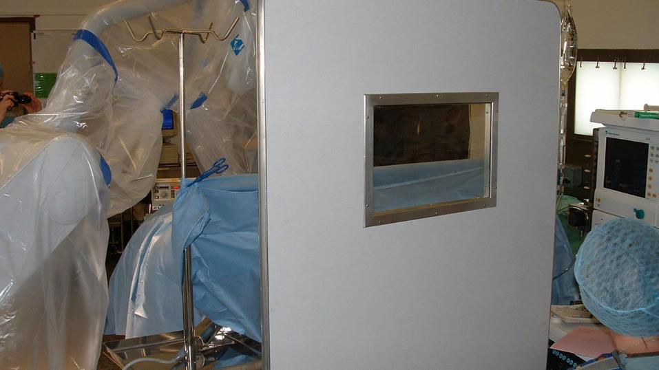
Intra-Operative Breast Irradiation
Intraoperative Radiation Therapy (IORT) for PBI
TARGIT trial is comparing whole breast irradiation to IORT delivering a single dose of 20 Gy. Primary accrual is in Europe
Using the Intrabeam Photon Radiosurgery System, 50 kV x-rays.
Trial has enrolled 900 patients with target of 2200 patients.
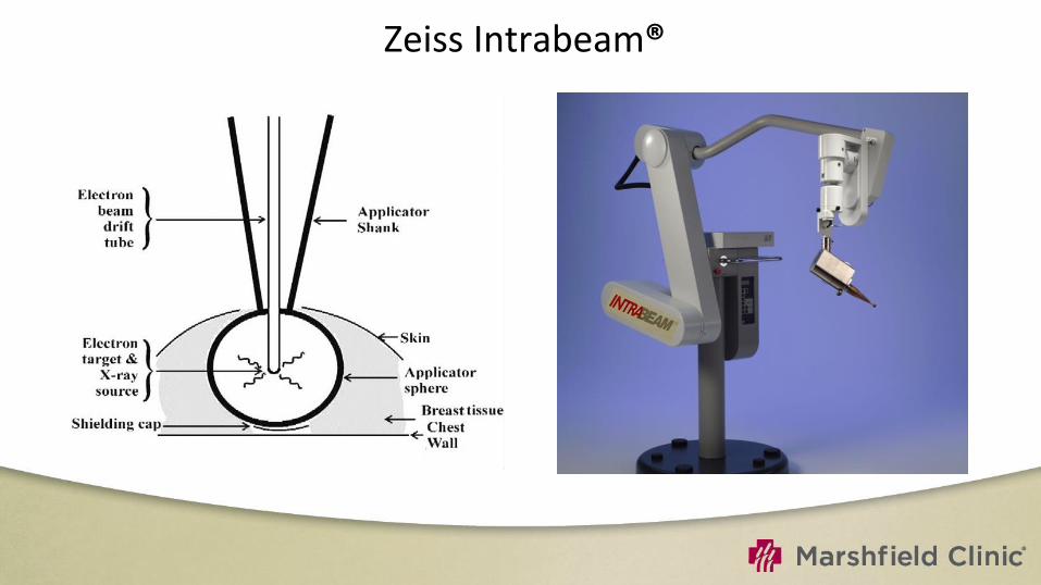
Zeiss Intrabeam®
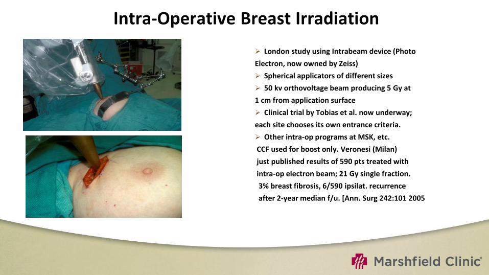
Intra-Operative Breast Irradiation
London study using Intrabeam device (Photo
Electron, now owned by Zeiss)
Spherical applicators of different sizes
50 kv orthovoltage beam producing 5 Gy at
1 cm from application surface
Clinical trial by Tobias et al. now underway;
each site chooses its own entrance criteria.
Other intra-op programs at MSK, etc.
CCF used for boost only. Veronesi (Milan)
just published results of 590 pts treated with
intra-op electron beam; 21 Gy single fraction.
3% breast fibrosis, 6/590 ipsilat. recurrence
after 2-year median f/u. [Ann. Surg 242:101 2005
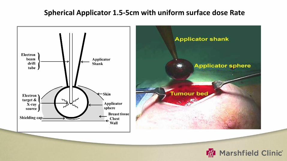
Spherical Applicator 1.5-5cm with uniform surface dose Rate

Intracavitary Brachytherapy
Balloon based brachytherapy include:
Mammosite
Contura
SAVI
Axxent electronic brachytherapy
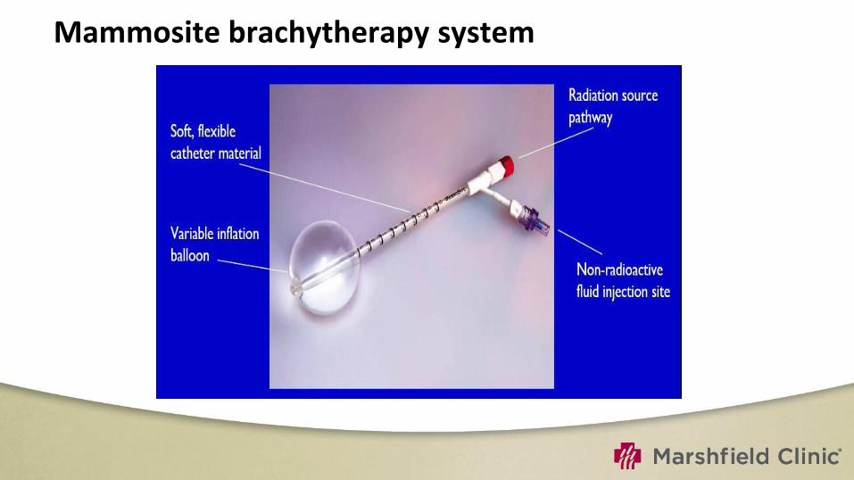
Mammosite brachytherapy system
structure
Silicone balloon
Double-lumen catheter (15 cm length and 6 mm in diameter)
Inflation channel:- saline solution mixed with a small amount of contrast material to aid visualization.
Source channel:- for passage of an Ir-192 high dose rate (HDR) brachytherapy source.
Source channel runs centrally through the length of the balloon.
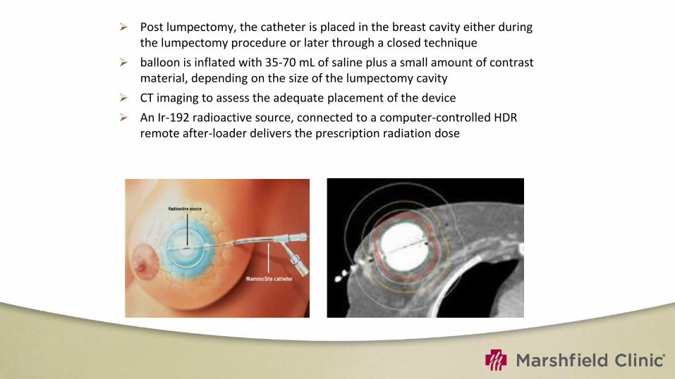
Post lumpectomy, the catheter is placed in the breast cavity either during the lumpectomy procedure or later through a closed technique
balloon is inflated with 35-70 mL of saline plus a small amount of contrast material, depending on the size of the lumpectomy cavity
CT imaging to assess the adequate placement of the device
An Ir-192 radioactive source, connected to a computer-controlled HDR remote after-loader delivers the prescription radiation dose
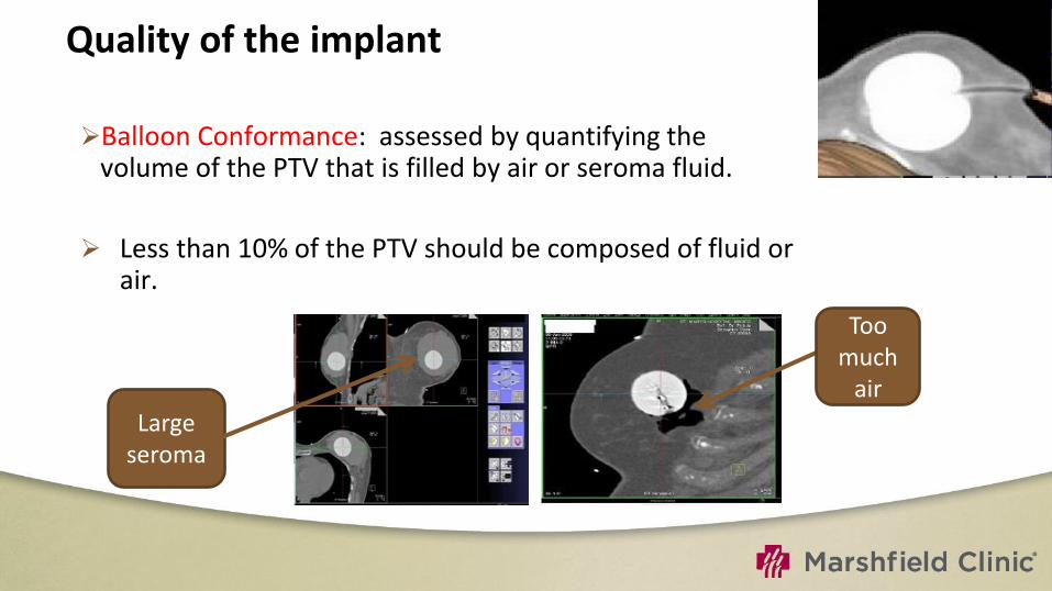
Quality of the implant Balloon Conformance: assessed by quantifying the
volume of the PTV that is filled by air or seroma fluid.
Less than 10% of the PTV should be composed of fluid or air.
Too much
air
Large seroma
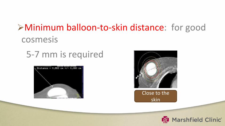
Minimum balloon-to-skin distance: for good cosmesis
5-7 mm is required
Close to the skin
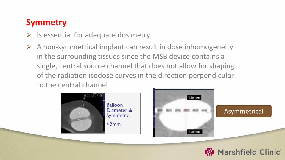
Symmetry
Is essential for adequate dosimetry.
A non-symmetrical implant can result in dose inhomogeneity in the surrounding tissues since the MSB device contains a single, central source channel that does not allow for shaping of the radiation isodose curves in the direction perpendicular to the central channel
Asymmetrical
Dose
34 Gy over 10 fractions
Minimum 6 hours between fractions
D 90> 90%
V150< 50cc
V200< 20 cc
Skin dose Max <145%
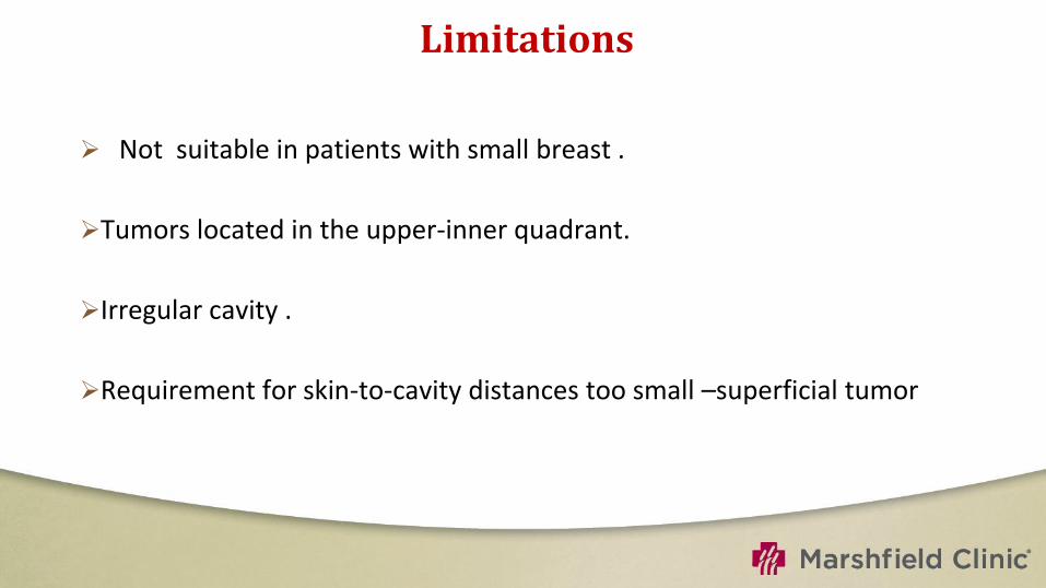
Limitations
Not suitable in patients with small breast .
Tumors located in the upper-inner quadrant.
Irregular cavity .
Requirement for skin-to-cavity distances too small –superficial tumor
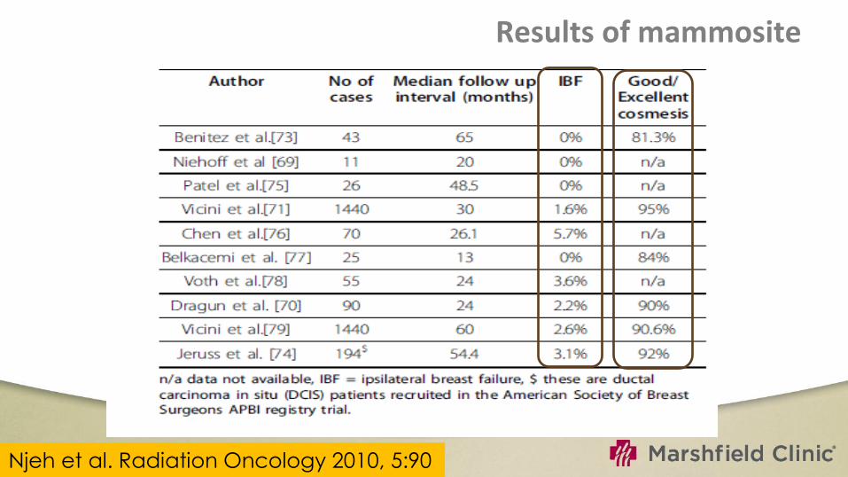
Results of mammosite
Njeh et al. Radiation Oncology 2010, 5:90
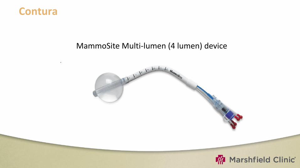
Contura
MammoSite Multi-lumen (4 lumen) device
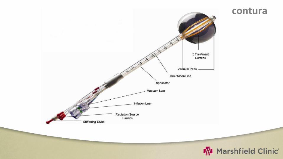
contura
Balloon based brachytherapy include:
Mammosite
Axxent electronic brachytherapy
Contura
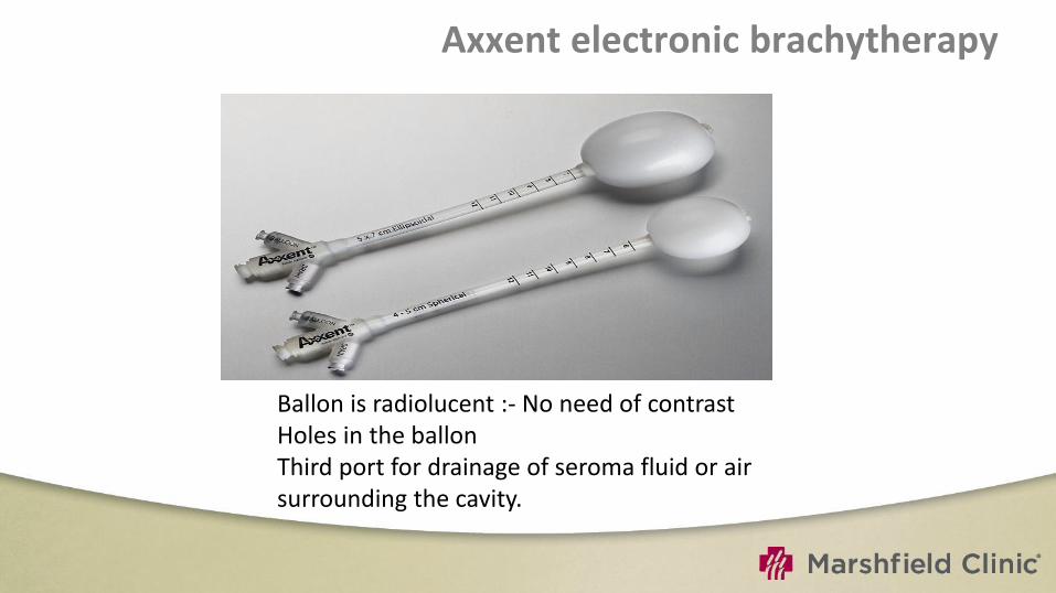
Axxent electronic brachytherapy
Ballon is radiolucent :- No need of contrast Holes in the ballon Third port for drainage of seroma fluid or air surrounding the cavity.
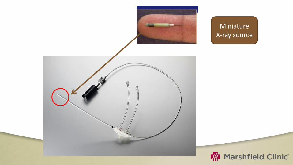
Miniature X-ray source
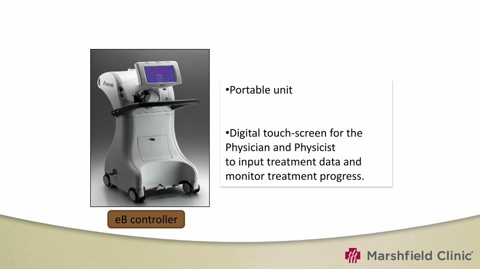
eB controller
•Portable unit
•Digital touch-screen for the Physician and Physicist to input treatment data and monitor treatment progress.
Advantages
Specifically shielded radiation room or an HDR afterloader unit are not required.
This reduces costs and allows for portability of the system, which can lead to greater access for patients particularly in more remote or rural locations.
Can be used intraoperatively
Electronic 50 Kv x-ray source
Low energy spectrum that results in more rapid dose falloff with depth in tissue.
Radiobiologic effect (RBE) for low-energy photons is higher on the order of 1.2-2.
This has currently not been taken into account in the prescribed dose for EBB, which uses the same prescription of 34.0 Gy in 10 fractions as used with 192Ir.
Dose to structures proximal to the point(1 cm) is higher and the dose to structures beyond this point is lower with EBB
Careful clinical evaluation is needed to determine the clinical impact of these factors with respect to late tissue affects and cosmesis.
In addition to a central lumen, the Contura balloon has four surrounding channels to accommodate the HDR source.
Additional source positions allows increased dose flexibility compared with a single-catheter approach.
Vacuum port to remove fluid or air around the lumpectomy cavity.
Reduce the dose to normal tissues (chest wall and skin) better protection of organs at risk such as the heart and lungs.
Possible to account for asymmetric balloon implant with respect to the central channel.
Hybrid brachytherapy devices
Struts Adjusted Volume Implant (SAVI)
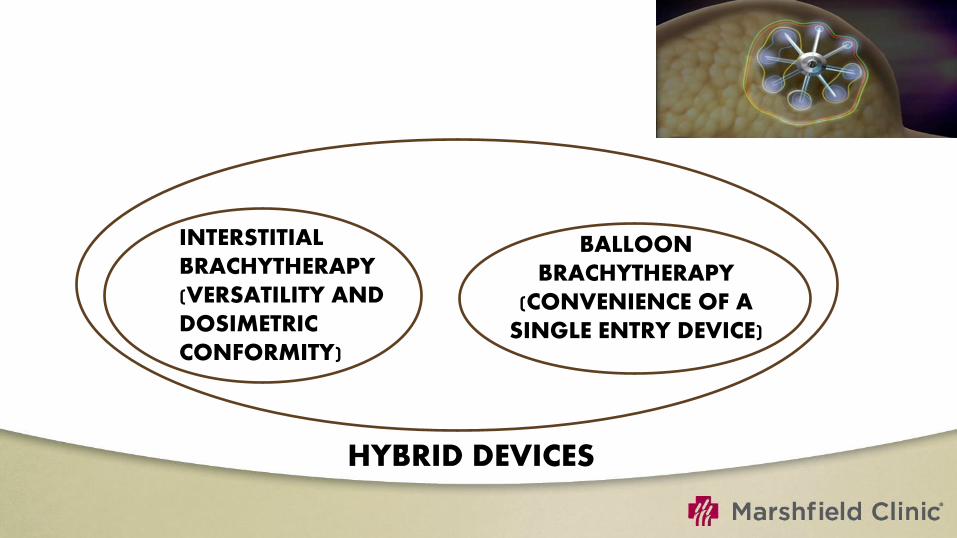
INTERSTITIAL BRACHYTHERAPY (VERSATILITY AND DOSIMETRIC CONFORMITY)
BALLOON BRACHYTHERAPY
(CONVENIENCE OF A SINGLE ENTRY DEVICE)
HYBRID DEVICES
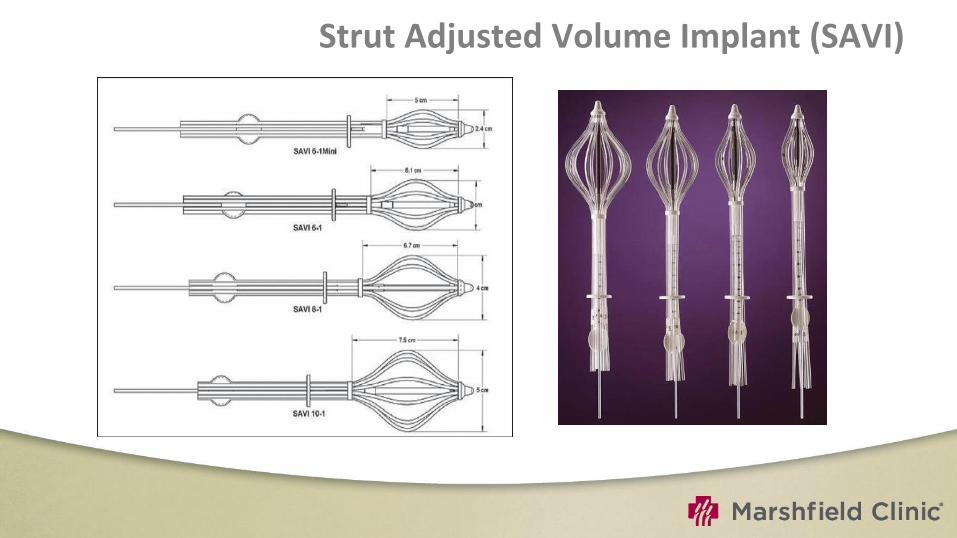
Strut Adjusted Volume Implant (SAVI)
Contains six outer expandable plastic tubes to displace the tissue
Central catheter surrounded by six additional catheters that allow the passage of an HDR iridium-192 source
The radiation source is not in direct contact with the breast tissue
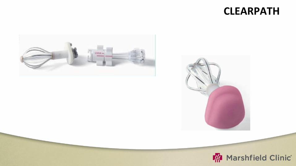
CLEARPATH
Consists of a central strut Surrounded by 6, 8 or 10 peripheral struts
Can be differentially loaded with HDR source
Insertion done in collapsed form through an incision (freehand; USG guided)
Then expanded to fit the cavity
CT required (verification and planning)
EBRT (IMRT)
advantages
Non-invasive (complications of surgery like seroma and infection can be avoided)
Widespread availability
Technically less demanding
Treatment results with external beam may be more uniform between radiation oncologists
Greater dose homogeneity
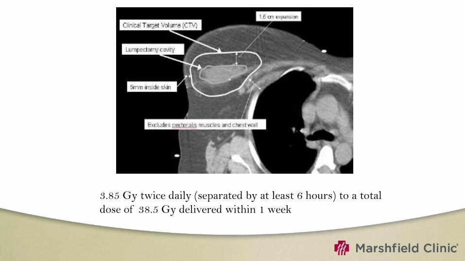
3.85 Gy twice daily (separated by at least 6 hours) to a total
dose of 38.5 Gy delivered within 1 week
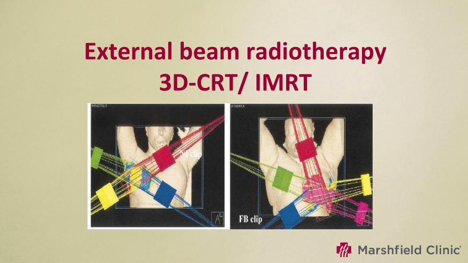
External beam radiotherapy 3D-CRT/ IMRT
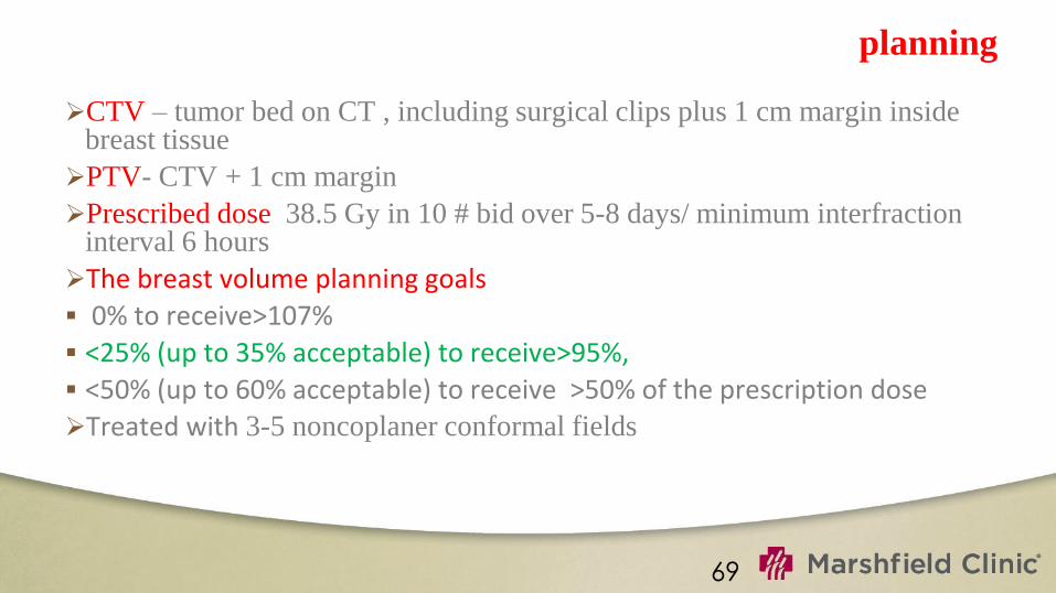
planning
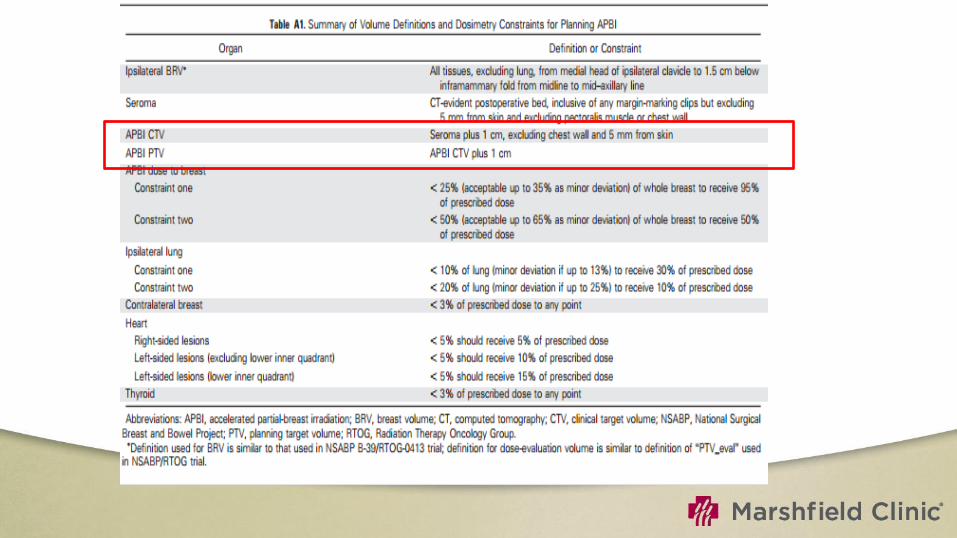
CTV – tumor bed on CT , including surgical clips plus 1 cm margin inside breast tissue
PTV- CTV + 1 cm margin
Prescribed dose 38.5 Gy in 10 # bid over 5-8 days/ minimum interfraction interval 6 hours
The breast volume planning goals
0% to receive>107%
<25% (up to 35% acceptable) to receive>95%,
<50% (up to 60% acceptable) to receive >50% of the prescription dose
Treated with 3-5 noncoplaner conformal fields
69
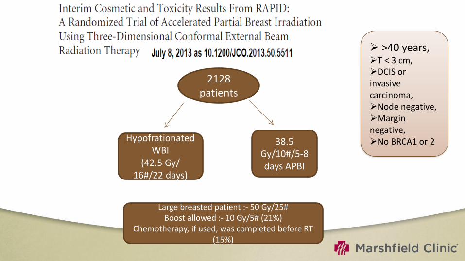
2128 patients
Hypofrationated WBI
(42.5 Gy/ 16#/22 days)
38.5 Gy/10#/5-8 days APBI
Large breasted patient :- 50 Gy/25# Boost allowed :- 10 Gy/5# (21%)
Chemotherapy, if used, was completed before RT (15%)
>40 years, T < 3 cm, DCIS or invasive carcinoma, Node negative, Margin negative, No BRCA1 or 2
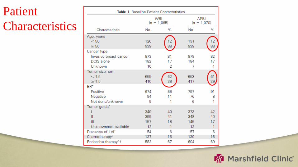
Patient
Characteristics
Cosmetic Analysis
By EORTC Cosmetic Rating System.
At baseline, assessed by a trained nurse.
Patient questionnaire
Assessed by two panels of three radiation oncologists using the digital photographs on follow-up.
The treated breast was compared with size and shape, location of the areola and nipple, appearance of the surgical scar, presence of telangiectasia, and global cosmetic score
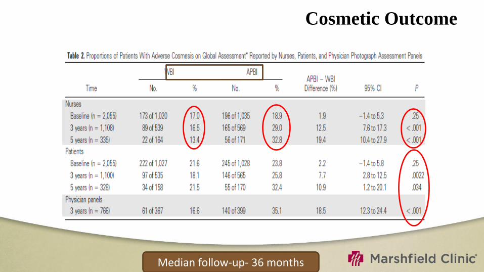
Cosmetic Outcome
Median follow-up- 36 months
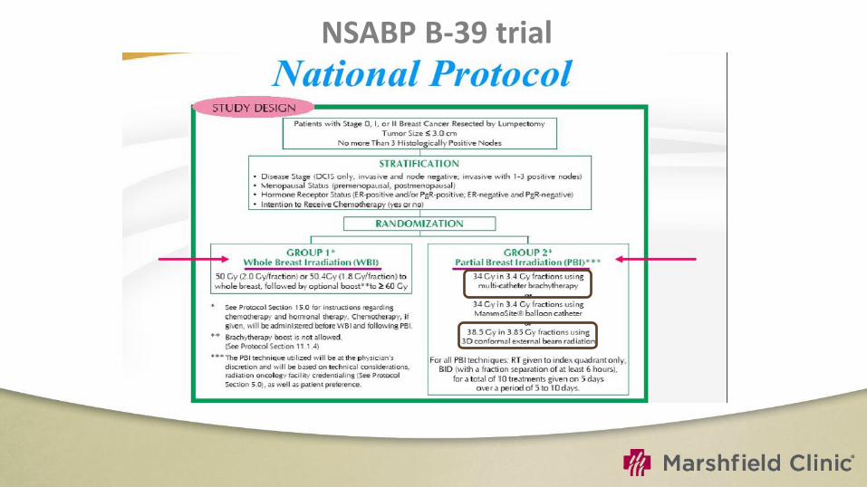
NSABP B-39 trial

5y
European Journal of Cancer (2015)
51, 451– 463
>40 years
T < 2.5cm
LVI allowed
DCIS allowed
ALN positive allowed
520 patients
2005-2013
n=1233
Conventional WBI (260)
(50 Gy/ 25#/Boost 10 Gy )
APBI(260) 30 Gy/5#
Nonconsecutive days
5 year follow -up
ASTRO Recommendations
Low risk - APBI outside the context of a clinical trial is an acceptable treatment option
High-risk group- APBI is considered C/I
Intermediate risk group- APBI is considered acceptable only in the context of prospective clinical trials.

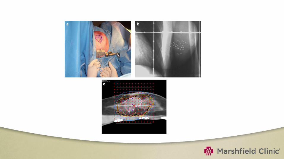
Novel methods
Percutaneous insertion of radioactive seeds (palladium-103 ) under US guidance
Use of LDR sources has the potential for improving the therapeutic ratio
A preplan is generated with optimal seed position and spacing to deliver the prescribed dose of 90 Gy to cover the lumpectomy cavity with a 1.5-cm margin.
Using a grid template 103Pd seeds are placed according to the preplan needle and seed distribution.
Permanent breast seed implant
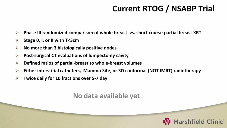
Current RTOG / NSABP Trial
Phase III randomized comparison of whole breast vs. short-course partial breast XRT
Stage 0, I, or II with T<3cm
No more than 3 histologically positive nodes
Post-surgical CT evaluations of lumpectomy cavity
Defined ratios of partial-breast to whole-breast volumes
Either interstitial catheters, Mammo Site, or 3D conformal (NOT IMRT) radiotherapy
Twice daily for 10 fractions over 5-7 day
No data available yet

CONCLUSIONS
In about 5-8 years, the ongoing studies will hopefully answer the questions related to patient selection, long-term outcome, and toxicity of the different techniques.
A modest reduction in initial treatment efficacy cannot be justified in patients with early breast cancer, who have an excellent prognosis with standard BCT including WBI.
For now, patients should be carefully selected for APBI and closely followed with accurate documentation of outcomes

THANK YOU