Embed Size (px)
Citation preview

ACC/AHA TASK FORCE REPORT
ACC/AHA Guidelines for the Management of Patients With ValvularHeart Disease
A Report of the American College of Cardiology/American Heart AssociationTask Force on Practice Guidelines (Committee on Management of Patients WithValvular Heart Disease)
COMMITTEE MEMBERSROBERT O. BONOW, MD, FACC, Chair
BLASE CARABELLO, MD, FACCANTONIO C. DE LEON, JR., MD, FACCL. HENRY EDMUNDS, JR., MD, FACCBRADLEY J. FEDDERLY, MD, FAAFPMICHAEL D. FREED, MD, FACCWILLIAM H. GAASCH, MD, FACC
CHARLES R. MCKAY, MD, FACCRICK A. NISHIMURA, MD, FACCPATRICK T. O’GARA, MD, FACCROBERT A. O’ROURKE, MD, FACCSHAHBUDIN H. RAHIMTOOLA, MD, FACC
TASK FORCE MEMBERSJAMES L. RITCHIE, MD, FACC, Chair
MELVIN D. CHEITLIN, MD, FACCKIM A. EAGLE, MD, FACCTIMOTHY J. GARDNER, MD, FACCARTHUR GARSON, JR., MD, MPH, FACCRAYMOND J. GIBBONS, MD, FACC
ROBERT A. O’ROURKE, MD, FACCRICHARD O. RUSSELL, MD, FACCTHOMAS J. RYAN, MD, FACCSIDNEY C. SMITH, JR., MD, FACC
TABLE OF CONTENTS
PreambleI. Introduction ...............................................................................1489
II. General Principles ...................................................................1490A. Evaluation of the Patient With a Cardiac
Murmur................................................................................14901. Introduction .................................................................1490
2. Classification of Murmurs..........................................1491a. Dynamic Cardiac Auscultation ............................1492b. Other Physical Findings ........................................1492c. Associated Symptoms ..........................................1492d. Electrocardiography and Chest
Roentgenography .................................................1492e. Echocardiography .................................................1493f. Cardiac Catheterization .......................................1494
3. Approach to the Patient ...........................................1494B. Endocarditis and Rheumatic Fever
Prophylaxis ..........................................................................14951. Endocarditis Prophylaxis ...........................................14952. Rheumatic Fever Prophylaxis ...................................1498
a. General Considerations .......................................1498b. Primary Prevention ..............................................1498c. Secondary Prevention ..........................................1498
III. Specific Valve Lesions ............................................................1498A. Aortic Stenosis ...................................................................1498
1. Introduction ................................................................1498a. Grading the Degree of Stenosis .........................1498
2. Pathophysiology ..........................................................14993. Natural History ..........................................................1499
When citing this document, the American College of Cardiology and theAmerican Heart Association recommend that the following format be used:Bonow RO, Carabello B, de Leon AC Jr, Edmunds LH Jr, Fedderly BJ, FreedMD, Gaasch WH, McKay CR, Nishimura RA, O’Gara PT, O’Rourke RA,Rahimtoola SH. ACC/AHA guidelines for the management of patients with valvularheart disease: a report of the American College of Cardiology/American HeartAssociation Task Force on Practice Guidelines (Committee on Management ofPatients With Valvular Heart Disease). J Am Coll Cardiol. 1998;32:1486–588.
Address for reprints: A single reprint of this document (the completeGuidelines) is available by calling 800-253-4636 (US only) or writing theAmerican College of Cardiology, Educational Services, 9111 Old GeorgetownRoad, Bethesda, MD 20814-1699. Ask for reprint No. 71-0153. To obtain areprint of the shorter version (Executive Summary and Summary of Recommen-dations) published in the November 3 issue of Circulation, ask for reprint No.71-0154. To purchase additional reprints, specify version and reprint number: upto 999 copies, call 800-611-6083 (US only) or fax 413-665-2671; 1000 or morecopies, call 214-706-1466, fax 214-691-6342, or E-mail [email protected]
JACC Vol. 32, No. 5November 1, 1998:1486–588
1486
©1998 by the American College of Cardiology and the American Heart Association, Inc. 0735-1097/98/$19.00Published by Elsevier Science Inc. PII S0735-1097(98)00454-9

4. Management of the Asymptomatic Patient ............1500a. Initial Evaluation ..................................................1500b. Serial Testing ........................................................1501c. Medical Therapy ...................................................1501d. Physical Activity and Exercise ............................1501
5. Indications for Cardiac Catheterization ..................1501a. Low-Gradient Aortic Stenosis ............................1502
6. Indications for Aortic Valve Replacement .............1502a. Symptomatic Patients ...........................................1502b. Asymptomatic Patients ........................................1502c. Patients Undergoing Coronary Artery
Bypass Surgery ......................................................15027. Aortic Balloon Valvotomy ........................................15038. Medical Therapy for the Inoperable Patient .........15039. Evaluation After Aortic Valve Replacement .........1503
10. Special Considerations in the Elderly .....................1504B. Aortic Regurgitation .........................................................1504
1. Etiology .......................................................................15042. Acute Aortic Regurgitation ......................................1504
a. Pathophysiology ....................................................1504b. Diagnosis ...............................................................1504c. Treatment ..............................................................1505
3. Chronic Aortic Regurgitation ..................................1505a. Pathophysiology ....................................................1505b. Natural History .....................................................1505c. Diagnosis and Initial Evaluation of the
Asymptomatic Patient ..........................................1508d. Medical Therapy ...................................................1509e. Physical Activity and Exercise ............................1511f. Serial Testing ........................................................1511g. Indications for Cardiac Catheterization ............1512h. Indications for Aortic Valve Replacement .......1512
4. Concomitant Aortic Root Disease ..........................15145. Evaluation of Patients After Aortic Valve
Replacement ...............................................................15146. Special Considerations in the Elderly .....................1514
C. Mitral Stenosis ...................................................................15151. Pathophysiology and Natural History .....................15152. Evaluation and Management of the
Asymptomatic Patient ...............................................1515a. Initial Workup ......................................................1515b. Medical Therapy: General ..................................1517c. Medical Therapy: Atrial Fibrillation ..................1518d. Medical Therapy: Prevention of Systemic
Embolization .........................................................1518e. Recommendations Regarding Physical
Activity and Exercise ...........................................1519f. Serial Testing ........................................................1519
3. Evaluation of the Symptomatic Patient ..................1519a. Initial Workup ......................................................1519b. Indications for Cardiac Catheterization ............1519
4. Indications for Surgical or PercutaneousValvotomy ...................................................................1521
5. Indications for Mitral Valve Replacement .............15246. Management of Patients After Valvotomy
or Commissurotomy ..................................................15247. Special Considerations ..............................................1525
a. Pregnant Patients .................................................1525b. Older Patients .......................................................1525
D. Mitral Valve Prolapse .......................................................15251. Pathophysiology and Natural History .....................1525
2. Evaluation and Management of theAsymptomatic Patient ...............................................1527
3. Evaluation and Management of theSymptomatic Patient ..................................................1528
4. Surgical Considerations .............................................1529E. Mitral Regurgitation .........................................................1530
1. Etiology .......................................................................15302. Acute Severe Mitral Regurgitation .........................1530
a. Pathophysiology ....................................................1530b. Diagnosis ...............................................................1530c. Medical Therapy ...................................................1530
3. Chronic Asymptomatic Mitral Regurgitation ........1530a. Pathophysiology ....................................................1530b. Diagnosis ...............................................................1531c. Serial Testing ........................................................1531d. Guidelines for Physical Activity and Exercise ..1531e. Medical Therapy ...................................................1531f. Indications for Cardiac Catheterization ............1532
4. Indications for Surgery ..............................................1532a. Types of Surgery ...................................................1532b. Timing of Surgery for Symptomatic Patients
With Normal Left Ventricular Function ...........1533c. Timing of Surgery in Asymptomatic or
Symptomatic Patients With Left VentricularDysfunction ...........................................................1533
d. Asymptomatic Patients with NormalLeft Ventricular Function ...................................1534
e. Atrial Fibrillation .................................................1534f. Feasibility of Repair Versus Replacement .......1535
5. Ischemic Mitral Regurgitation .................................15356. Evaluation of Patients After Mitral Valve
Replacement or Repair .............................................15357. Special Considerations in the Elderly .....................1535
F. Multiple Valve Disease ....................................................15351. Introduction ................................................................15352. Mixed Single Valve Disease .....................................1536
a. Pathophysiology ....................................................1536b. Diagnosis ...............................................................1536c. Management ..........................................................1536
3. Combined Mitral Stenosis and AorticRegurgitation ..............................................................1537a. Pathophysiology ....................................................1537b. Management ..........................................................1537
4. Combined Mitral Stenosis and TricuspidRegurgitation ..............................................................1537a. Pathophysiology ....................................................1537b. Diagnosis ...............................................................1537c. Management ..........................................................1537
5. Combined Mitral and Aortic Regurgitation ..........1537a. Pathophysiology ....................................................1537b. Diagnosis and Therapy .........................................1537
6. Combined Mitral and Aortic Stenosis ....................1537a. Pathophysiology ....................................................1537b. Diagnosis and Therapy ........................................1537
7. Combined Aortic Stenosis and MitralRegurgitation ..............................................................1537a. Pathophysiology ....................................................1537b. Diagnosis and Therapy ........................................1537
G. Tricuspid Valve Disease ...................................................15381. Pathophysiology ..........................................................15382. Diagnosis .....................................................................1538
1487JACC Vol. 32, No. 5 BONOW ET AL.November 1, 1998:1486–588 ACC/AHA TASK FORCE REPORT

3. Management ...............................................................1538H. Valvular Heart Disease Associated With
Anorectic Drugs ................................................................1539
IV. Evaluation and Management of Infective Endocarditis ......1541A. Antimicrobial Therapy .....................................................1541B. Culture-Negative Endocarditis ........................................1544C. Endocarditis in HIV-Seropositive Patients ....................1544D. Indications for Echocardiography in Endocarditis .......1544E. Outpatient Treatment .......................................................1544F. Indications for Surgery in Patients With Active
Infective Endocarditis .......................................................1545
V. Management of Valvular Disease in Pregnancy .................1545A. Physiological Changes of Pregnancy ...............................1545B. Physical Examination ........................................................1546C. Echocardiography ..............................................................1546D. General Management Guidelines ...................................1546E. Specific Lesions .................................................................1547
1. Mitral Stenosis ...........................................................15472. Mitral Regurgitation ..................................................15473. Aortic Stenosis ...........................................................15484. Aortic Regurgitation .................................................15485. Pulmonary Valve Stenosis ........................................15486. Tricuspid Valve Disease ...........................................15487. Marfan Syndrome ......................................................1548
F. Endocarditis Prophylaxis ..................................................1548G. Cardiac Valve Surgery ......................................................1549H. Anticoagulation .................................................................1549
1. Warfarin ......................................................................15492. Heparin .......................................................................1549
VI. Management of Valvular Heart Disease inAdolescents and Young Adults .............................................1550A. Aortic Stenosis ...................................................................1550B. Aortic Regurgitation .........................................................1552C. Mitral Regurgitation .........................................................1552D. Mitral Stenosis ...................................................................1552E. Tricuspid Valve Disease ...................................................1553F. Pulmonic Stenosis .............................................................1554
1. Pathophysiology ..........................................................15542. Diagnosis .....................................................................15543. Clinical Course ...........................................................1554
G. Pulmonary Regurgitation .................................................1555
VII. Management of Patients With ProstheticHeart Valves ............................................................................1555A. Classification of Prosthetic Heart Valves ......................1555
1. Mechanical Valves .....................................................1555a. Ball Valves ............................................................1555b. Disk Valves ...........................................................1556c. Bileaflet Valves .....................................................1556
2. Biological Valves ........................................................1556a. Autograft Valves ...................................................1556b. Autologous Pericardial Valves ............................1556c. Homograft (or Allograft) Valves .......................1556d. Porcine Heterograft (or Xenograft)
Valves .....................................................................1557e. Bovine Pericardial Valves ...................................1557
B. Complications of Prosthetic Heart Valves .....................15571. Guidelines for Reporting Clinical Results .............1557
a. AATS/STS Guidelines for Clinical Reporting ..15572. Valve-Related Complications ...................................1557
a. Major Randomized Trials ...................................1558C. Management of Patients With Prosthetic
Heart Valves ......................................................................15581. Antibiotic Prophylaxis ...............................................1558
a. Infective Endocarditis ..........................................1558b. Recurrence of Rheumatic Carditis ....................1558
2. Antithrombotic Therapy ...........................................1558a. Mechanical Valves ................................................1558b. Biological Valves ..................................................1559c. Difficulties in Maintaining
Anticoagulant Therapy ........................................1560d. Embolic Events During Adequate
Antithrombotic Therapy ......................................1560e. Excessive Anticoagulation ...................................1560f. Antithrombotic Therapy in Patients Requiring
Noncardiac Surgery/Dental Care .......................1560g. Antithrombotic Therapy in Patients Needing
Cardiac Catheterization/Angiography ................1561h. Thrombosis of Prosthetic Heart Valves ............1561
3. Follow-up Visits .........................................................1562a. First Outpatient Postoperative Visit ..................1562b. Follow-up Visits in Patients Without
Complications .......................................................1562c. Follow-up Visits in Patients With
Complications .......................................................15634. Reoperation to Replace a Prosthetic Valve ...........1563
D. Major Criteria for Valve Selection .................................1563
VIII. Evaluation and Treatment of Coronary ArteryDisease in Patients With Valvular Heart Disease ..............1565A. Probability of Coronary Artery Disease in
Patients With Valvular Heart Disease ...........................1565B. Diagnosis of Coronary Artery Disease ...........................1566C. Treatment of Coronary Artery Disease at the
Time of Aortic Valve Replacement ...............................1566D. Aortic Valve Replacement in Patients Undergoing
Coronary Artery Bypass Surgery .....................................1566E. Management of Concomitant Mitral Valve
Disease and Coronary Artery Disease ...........................1567
1488 BONOW ET AL. JACC Vol. 32, No. 5ACC/AHA TASK FORCE REPORT November 1, 1998:1486–588

ACC/AHA Guidelines for theManagement of Patients With
Valvular Heart Disease
PreambleIt is important that the medical profession play a significant
role in critically evaluating the use of diagnostic proceduresand therapies in the management or prevention of diseasestates. Rigorous and expert analysis of the available datadocumenting relative benefits and risks of those proceduresand therapies can produce helpful guidelines that improve theeffectiveness of care, optimize patient outcomes, and favorablyimpact the overall cost of care by focusing resources on themost effective strategies.
The American College of Cardiology (ACC) and the Amer-ican Heart Association (AHA) have jointly engaged in theproduction of such guidelines in the area of cardiovasculardisease since 1980. This effort is directed by the ACC/AHATask Force on Practice Guidelines. Its charge is to develop andrevise practice guidelines for important cardiovascular diseasesand procedures. Experts in the subject under consideration areselected from both organizations to examine subject-specificdata and write guidelines. The process includes additionalrepresentatives from other medical specialty groups whenappropriate. Writing groups are specifically charged to per-form a formal literature review, weigh the strength of evidencefor or against a particular treatment or procedure, and includeestimates of expected health outcomes where data exist.Patient-specific modifiers, comorbidities, and issues of patientpreference that might influence the choice of particular tests ortherapies are considered as well as frequency of follow-up andcost-effectiveness.
These practice guidelines are intended to assist physiciansin clinical decision making by describing a range of generallyacceptable approaches for the diagnosis, management, orprevention of specific diseases or conditions. These guidelinesattempt to define practices that meet the needs of mostpatients in most circumstances. The ultimate judgment regard-ing care of a particular patient must be made by the physicianand patient in light of all the circumstances presented by thatpatient.
The Committee on Management of Patients With ValvularHeart Disease was chaired by Robert O. Bonow, MD, FACC,and included the following members: Blase Carabello, MD,FACC; Antonio C. de Leon, Jr., MD, FACC; L. HenryEdmunds, Jr., MD, FACC; Bradley J. Fedderly, MD, FAAFP;Michael D. Freed, MD, FACC; William H. Gaasch, MD,FACC; Charles R. McKay, MD, FACC; Rick A. Nishimura,MD, FACC; Patrick T. O’Gara, MD, FACC; Robert A.O’Rourke, MD, FACC; and Shahbudin H. Rahimtoola, MD,FACC. In August 1998, the full text of the guidelines wasapproved for publication in the November issue of the Journalof the American College of Cardiology and the executive sum-
mary for publication in the November 3 issue of Circulation.Reprints of both the full text and the executive summary areavailable from both organizations.
I. IntroductionThe American College of Cardiology and the American
Heart Association (ACC/AHA) have long been involved in thejoint development of practice guidelines designed to assistphysicians in the management of selected cardiovascular dis-orders or the selection of certain cardiovascular procedures.The determination of the disorders or procedures for which todevelop guidelines is based on several factors, including im-portance to physicians and whether there are sufficient datafrom which to derive accepted guidelines. One importantcategory of cardiac disorders that affect a large number ofpatients who require diagnostic procedures and decisionsregarding long-term management is valvular heart disease.
During the past 2 decades, major advances have occurred indiagnostic techniques, the understanding of natural history,and interventional cardiological and surgical procedures forpatients with valvular heart disease. These advances haveresulted in enhanced diagnosis, more scientific selection ofpatients for surgery or catheter-based intervention versusmedical management, and increased survival of patients withthese disorders. The information base from which to makeclinical management decisions has greatly expanded in recentyears, yet in many situations management issues remain con-troversial or uncertain. Unlike many other forms of cardiovas-cular disease, there is a scarcity of large-scale multicenter trialsaddressing the diagnosis and treatment of patients with valvu-lar disease from which to derive definitive conclusions, and theinformation available in the literature represents primarily theexperiences reported by single institutions in relatively smallnumbers of patients.
The Committee on Management of Patients With ValvularHeart Disease was given the task of reviewing and compilingthis information base and making recommendations for diag-nostic testing, treatment, and physical activity. For topics inwhich there is an absence of multiple randomized controlledtrials, the preferred basis for medical decision making inclinical practice (evidence-based medicine), the committee’srecommendations were based on data derived from singlerandomized trials or nonrandomized studies or were based ona consensus opinion of experts. Where no or few data exist, thisis identified in the text.
The committee membership consisted of cardiovasculardisease specialists as well as representatives of the cardiacsurgery and family practice fields; both the academic andprivate practice sectors were represented. This document wasreviewed by 3 outside reviewers nominated by the ACC and 3outside reviewers nominated by the AHA, as well as numerouscontent reviewers and individuals nominated by the AmericanAcademy of Family Physicians and the Society of ThoracicSurgeons.
The guidelines follow the format established in previous
1489JACC Vol. 32, No. 5 BONOW ET AL.November 1, 1998:1486–588 ACC/AHA TASK FORCE REPORT

ACC/AHA guidelines for classifying indications for diagnosticand therapeutic procedures:
Class I: Conditions for which there is evidence and/orgeneral agreement that a given procedure or treatment isuseful and effective.
Class II: Conditions for which there is conflicting evidenceand/or a divergence of opinion about the usefulness/efficacy ofa procedure or treatment.
IIa. Weight of evidence/opinion is in favor of useful-ness/efficacy.
IIb. Usefulness/efficacy is less well established byevidence/opinion.
Class III: Conditions for which there is evidence and/orgeneral agreement that the procedure/treatment is not usefuland in some cases may be harmful.
The reference list is not exhaustive or all-inclusive, as thiswould be beyond the scope of this publication, but includesthose papers that the committee believes represent the mostcomprehensive or convincing data and are necessary to sup-port its conclusions.
The guidelines attempt to deal with general issues oftreatment of patients with heart valve disorders, such asevaluation of patients with heart murmurs, prevention andtreatment of endocarditis, management of valve disease inpregnancy, and treatment of patients with concomitant coro-nary artery disease (CAD) as well as more specialized issuesthat pertain to specific valve lesions. The guidelines focusprimarily on valvular heart disease in the adult, with a separatesection dealing with specific recommendations for valve disor-ders in adolescents and young adults. The diagnosis andmanagement of infants and young children with congenitalvalvular abnormalities are significantly different from those ofthe adolescent or adult and are beyond the scope of theseguidelines.
This task force report overlaps with several previouslypublished ACC/AHA guidelines about cardiac imaging anddiagnostic testing, including the Guidelines for Clinical Use ofCardiac Radionuclide Imaging (1), the Guidelines for ClinicalApplication of Echocardiography (2), the Guidelines for Ex-ercise Testing (3), and the Guidelines for Coronary Angiogra-phy (4). Although these guidelines are not intended to include
detailed information covered in previous guidelines on the useof imaging and diagnostic testing, an essential component ofthis report is the discussion of indications for these tests in theevaluation and treatment of patients with valvular heart dis-ease.
The committee emphasizes the fact that many factorsultimately determine the most appropriate treatment of indi-vidual patients with valvular heart disease within a givencommunity. These include the availability of diagnostic equip-ment and expert diagnosticians, the expertise of interventionalcardiologists and surgeons, and notably the wishes of well-informed patients. Therefore, deviation from these guidelinesmay be appropriate in some circumstances. These guidelinesare written with the assumption that a diagnostic test can beperformed and interpreted with skill levels consistent withpreviously reported ACC training and competency statementsand ACC/AHA guidelines, that interventional cardiologicaland surgical procedures can be performed by highly trainedpractitioners within acceptable safety standards, and that theresources necessary to perform these diagnostic proceduresand provide this care are readily available. This is not true in allgeographic areas, which further underscores the committee’sposition that its recommendations are guidelines and not rigidrequirements.
II. General PrinciplesA. Evaluation of the Patient With aCardiac Murmur
1. Introduction. Cardiac auscultation remains the mostwidely used method of screening for heart disease. The pro-duction of murmurs is due to 3 main factors: (1) high bloodflow rate through normal or abnormal orifices; (2) forwardflow through a narrowed or irregular orifice into a dilatedvessel or chamber; or (3) backward or regurgitant flow throughan incompetent valve, septal defect, or patent ductus arterio-sus. Often, several of these factors are operative (5–7).
A heart murmur may have no pathological significance ormay be an important clue to the presence of valvular, congen-ital, or other structural abnormalities of the heart (8). Mostsystolic heart murmurs do not signify cardiac disease, andmany are related to physiological increases in blood flowvelocity (9). In other instances, a heart murmur may be animportant clue to the diagnosis of undetected cardiac disease(eg, valvular aortic stenosis) that may be important even whenasymptomatic or that may define the reason for cardiacsymptoms. In these situations, various noninvasive or invasivecardiac tests may be necessary to establish a firm diagnosis andform the basis for rational treatment of an underlying disorder.Two-dimensional (2-D) and Doppler echocardiography is par-ticularly useful in this regard, as discussed in the ACC/AHAGuidelines for the Clinical Application of Echocardiography(2). Diastolic murmurs virtually always represent pathologicalconditions and require further cardiac evaluation, as do most
Abbreviations used in these guidelines:
AR 5 aortic regurgitationAS 5 aortic stenosisAVR 5 aortic valve replacementCAD 5 coronary artery diseaseECG 5 electrocardiogramLV 5 left ventricularMR 5 mitral regurgitationMS 5 mitral stenosisMVP 5 mitral valve prolapseMVR 5 mitral valve replacementNYHA 5 New York Heart AssociationTR 5 tricuspid regurgitation
1490 BONOW ET AL. JACC Vol. 32, No. 5ACC/AHA TASK FORCE REPORT November 1, 1998:1486–588

continuous murmurs. Continuous “innocent” murmurs includevenous hums and mammary souffles.
The traditional auscultation method of assessing cardiacmurmurs has been based on their timing in the cardiac cycle,configuration, location and radiation, pitch, intensity (grades 1through 6), and duration (5–9). The configuration of a murmurmay be crescendo, decrescendo, crescendo-decrescendo(diamond-shaped), or plateau. The precise times of onset andcessation of a murmur associated with cardiac pathologydepend on the point in the cardiac cycle at which an adequatepressure difference between 2 chambers appears and disap-pears (5–9). A classification of cardiac murmurs is listed inTable 1.
2. Classification of Murmurs. Holosystolic (pansystolic)murmurs are generated when there is flow between chambersthat have widely different pressures throughout systole, such asthe left ventricle and either the left atrium or right ventricle.With an abnormal regurgitant orifice, the pressure gradientand regurgitant jet begin early in contraction and last untilrelaxation is almost complete.
Midsystolic (systolic ejection) murmurs, often crescendo-decrescendo in configuration, occur when blood is ejectedacross the aortic or pulmonic outflow tracts. The murmurs startshortly after S1, when the ventricular pressure rises sufficientlyto open the semilunar valve. As ejection increases, the murmuris augmented, and as ejection declines, it diminishes.
In the presence of normal semilunar valves, this murmurmay be caused by an increased flow rate such as that whichoccurs with elevated cardiac output (eg, pregnancy, thyrotoxi-cosis, anemia, arteriovenous fistula), ejection of blood into adilated vessel beyond the valve, or increased transmission ofsound through a thin chest wall. Most benign innocent mur-murs occurring in children and young adults are midsystolicand originate either from the aortic or pulmonic outflow tracts.Valvular or subvalvular obstruction (stenosis) of either ventri-cle may also cause a midsystolic murmur, the intensity depend-ing in part on the velocity of blood flow across the narrowedarea. Midsystolic murmurs also occur in certain patients withmitral regurgitation (MR) or, less frequently, tricuspid regur-gitation (TR) resulting from papillary muscle dysfunction.Echocardiography is often necessary to separate a prominentand exaggerated (Grade 3 or greater) benign midsystolicmurmur from one due to valvular aortic stenosis (AS).
Early systolic murmurs are less common; they begin with
the first sound and end in midsystole. An early systolic murmuris often due to TR occurring in the absence of pulmonaryhypertension and in other patients with acute MR. In largeventricular septal defects with pulmonary hypertension andsmall muscular ventricular septal defects, the shunting at theend of systole may be insignificant, with the murmur limited toearly and midsystole.
Late systolic murmurs are soft or moderately loud, high-pitched murmurs at the left ventricular (LV) apex that startwell after ejection and end before or at S2. They are often dueto ischemia or infarction of the mitral papillary muscles or totheir dysfunction due to LV dilatation. Late systolic murmursin patients with midsystolic clicks result from late systolicregurgitation due to prolapse of the mitral leaflet(s) into theleft atrium. Such late systolic murmurs can also occur in theabsence of clicks.
Early immediate diastolic murmurs begin with or shortlyafter S2, when the associated ventricular pressure drops suffi-ciently below that in the aorta or pulmonary artery. High-pitched murmurs of aortic regurgitation (AR) or pulmonicregurgitation due to pulmonary hypertension are generallydecrescendo, consistent with the rapid decline in volume orrate of regurgitation during diastole. The diastolic murmur ofpulmonic regurgitation without pulmonary hypertension is lowto medium pitch, and the onset of this murmur is slightlydelayed because regurgitant flow is minimal at pulmonic valveclosure, when the reverse pressure gradient responsible for theregurgitation is minimal.
Middiastolic murmurs usually originate from the mitral andtricuspid valves, occur early during ventricular filling, and aredue to a relative disproportion between valve orifice size anddiastolic blood flow volume. Although they are usually due tomitral or tricuspid stenosis, middiastolic murmurs may also bedue to increased diastolic blood flow across the mitral ortricuspid valve when such valves are severely regurgitant,across the normal mitral valve in patients with ventricularseptal defect or patent ductus arteriosus, and across the normaltricuspid valve in patients with atrial septal defect. In severe,long-term AR, a low-pitched diastolic murmur (Austin-Flintmurmur) is often present at the LV apex; it may be eithermiddiastolic or presystolic.
Presystolic murmurs begin during the period of ventricularfilling that follows atrial contraction and therefore occur insinus rhythm. They are usually due to mitral or tricuspidstenosis. A right or left atrial myxoma may cause eithermiddiastolic or presystolic murmurs similar to tricuspid ormitral stenosis (MS).
Continuous murmurs arise from high- to low-pressureshunts that persist through the end of systole and the beginningof diastole. Thus, they begin in systole, peak near S2, andcontinue into all or part of diastole. There are many causes ofcontinuous murmurs, but they are uncommon in patients withvalvular heart disease (5–9).
a. Dynamic Cardiac Auscultation. Attentive cardiac auscul-tation during dynamic changes in cardiac hemodynamics oftenenables the careful observer to deduce the correct origin and
Table 1. Classification of Cardiac Murmurs
1. Systolic murmursa. Holosystolic (pansystolic) murmursb. Midsystolic (systolic ejection) murmursc. Early systolic murmursd. Mid to late systolic murmurs
2. Diastolic murmursa. Early high-pitched or low-pitched diastolic murmursb. Middiastolic murmursc. Presystolic murmurs
3. Continuous murmurs
1491JACC Vol. 32, No. 5 BONOW ET AL.November 1, 1998:1486–588 ACC/AHA TASK FORCE REPORT

significance of a cardiac murmur (10–13). Changes in theintensity of heart murmurs during various maneuvers areindicated in Table 2.
b. Other Physical Findings. The presence of other physicalfindings, either cardiac or noncardiac, may provide importantclues to the significance of a cardiac murmur and the need forfurther testing (Figure 1). For example, a right heart murmurin early to midsystole at the lower left sternal border likelyrepresents TR without pulmonary hypertension in an intrave-nous drug user who presents with fever, petechiae, Osler’snode, and Janeway lesion.
Associated cardiac findings frequently provide importantinformation about cardiac murmurs. Fixed splitting of thesecond heart sound during inspiration and expiration in apatient with a grade 2/6 midsystolic murmur in the pulmonic
area and left sternal border should suggest the possibility of anatrial septal defect. A soft or absent A2 or reversed splitting ofS2 may denote severe AS. An early aortic systolic ejectionsound heard during inspiration and expiration suggests abicuspid aortic valve, whereas an ejection sound heard only inthe pulmonic area and left sternal border during expirationusually denotes pulmonic valve stenosis. LV dilatation onprecordial palpation and bibasilar pulmonary rales favor thediagnosis of MR in a patient with a grade 2/6 holosystolicmurmur at the cardiac apex. A slow-rising, diminished arterialpulse suggests severe AS in a patient with a grade 2/6 midsys-tolic murmur at the upper intercostal spaces. The typicalpulsus parvus and tardus may be absent in the elderly, evenwith severe AS secondary to the effects of aging on thevasculature. Pulsus parvus may also occur with severely lowoutput from any cause. Factors that aid in the diagnosis of LVoutflow tract obstruction are listed in Table 3.
c. Associated Symptoms. An important consideration in apatient with a cardiac murmur is the presence or absence ofsymptoms (14) (Figure 1). For example, symptoms of syncope,angina pectoris, or congestive heart failure in a patient with amidsystolic murmur will usually result in a more aggressiveapproach than in patients with a similar midsystolic murmurwho have none of these symptoms. 2-D and Doppler echocar-diography to rule in or out the presence of significant AS willlikely be obtained. A history of thromboembolism or possibleinfective endocarditis will also usually result in a more exten-sive workup. In patients with cardiac murmurs and clinicalfindings suggestive of endocarditis, 2-D and Doppler echocar-diography is usually indicated (2).
Conversely, many asymptomatic children and young adultswith grade 2/6 midsystolic murmurs and no other cardiacphysical findings need no further cardiac workup after theinitial history and physical examination (Figure 1). A particu-larly important group is the large number of asymptomaticelderly patients, many with systemic hypertension, who havemidsystolic murmurs related to sclerotic aortic valve leaflets;flow into tortuous, noncompliant great vessels; or a combina-tion of these. Such murmurs must be distinguished from thosecaused by mild to severe valvular AS, which is prevalent in thisage group. The absence of LV hypertrophy on electrocardiog-raphy is reassuring, and this test is considerably less costly thanroutine echocardiography.
d. Electrocardiography and Chest Roentgenography. Al-though echocardiography usually provides more specific andoften quantitative information about the significance of a heartmurmur and may be the only test needed, the electrocardio-gram (ECG) and chest x-ray are readily available and may havebeen obtained already. The absence of ventricular hypertro-phy, atrial abnormality, arrhythmias, conduction abnormali-ties, prior myocardial infarction, and evidence of active isch-emia on the ECG provides useful negative information at arelatively low cost. Abnormal findings on the ECG, such asventricular hypertrophy or a prior infarction, should lead to amore extensive evaluation including 2-D and Doppler echo-cardiography (Figure 1).
Table 2. Interventions Used to Alter the Intensity ofCardiac Murmurs
RespirationRight-sided murmurs generally increase with inspiration. Left-sidedmurmurs usually are louder during expiration.
Valsalva maneuverMost murmurs decrease in length and intensity. Two exceptions are thesystolic murmur of HCM, which usually becomes much louder, and that ofMVP, which becomes longer and often louder. Following release of theValsalva, right-sided murmurs tend to return to baseline intensity earlierthan left-sided murmurs.
ExerciseMurmurs caused by blood flow across normal or obstructed valves (eg, PS,MS) become louder with both isotonic and submaximal isometric(handgrip) exercise. Murmurs of MR, VSD, and AR also increase withhandgrip exercise. However, the murmur of HCM often decreases withnear-maximum handgrip exercise.
Positional changesWith standing, most murmurs diminish, 2 exceptions being the murmur ofHCM, which becomes louder, and that of MVP, which lengthens and oftenis intensified. With prompt squatting, most murmurs become louder, butthose of HCM and MVP usually soften and may disappear. Passive legraising usually produces the same results as prompt squatting.
Postventricular premature beat or atrial fibrillationMurmurs originating at normal or stenotic semilunar valves increase inintensity during the cardiac cycle following a VPB or in the beat after along cycle length in AF. By contrast, systolic murmurs due toatrioventricular valve regurgitation do not change, diminish (papillarymuscle dysfunction), or become shorter (MVP).
Pharmacological interventionsDuring the initial relative hypotension following amyl nitrite inhalation,murmurs of MR, VSD, and AR decrease, while murmurs of AS increasebecause of increased stroke volume. During the later tachycardia phase,murmurs of MS and right-sided lesions also increase. This intervention maythus distinguish the murmur of the Austin-Flint phenomenon from that ofMS. The response in MVP often is biphasic (softer then louder thancontrol).
Transient arterial occlusionTransient external compression of both arms by bilateral cuff inflation to20 mm Hg greater than peak systolic pressure augments the murmurs ofMR, VSD, and AR but not murmurs due to other causes.
Abbreviations: AF 5 atrial fibrillation, AR 5 aortic regurgitation, AS 5aortic stenosis, HCM 5 hypertrophic cardiomyopathy, MR 5 mitral regurgita-tion, MS 5 mitral stenosis, MVP 5 mitral valve prolapse, PS 5 pulmonicstenosis, VPB 5 ventricular premature beat, VSD 5 ventricular septal defect.
1492 BONOW ET AL. JACC Vol. 32, No. 5ACC/AHA TASK FORCE REPORT November 1, 1998:1486–588

Posteroanterior and lateral chest roentgenograms oftenyield qualitative information on cardiac chamber size, pulmo-nary blood flow, pulmonary venous pressures, pulmonaryvascular redistribution, and cardiac calcification in patientswith cardiac murmurs. When abnormal findings are present onchest x-ray, 2-D and Doppler echocardiography should beperformed (Figure 1). A normal chest x-ray and ECG are likelyin patients with insignificant midsystolic cardiac murmurs,particularly in younger age groups and when the murmur is lessthan grade 3 in intensity (15–17). Many asymptomatic patientsneed neither an ECG nor a chest x-ray when a careful cardiacexamination indicates an insignificant vibratory midsystolicheart murmur and no other abnormal findings.
e. Echocardiography. Echocardiography is an importantnoninvasive method for assessing the significance of cardiacmurmurs by imaging cardiac structure and function and thedirection and velocity of blood flow through cardiac valves andchambers. 2-D echocardiography may indicate abnormal val-
vular motion and morphology but usually does not indicatethe severity of valvular stenosis or regurgitation except inMS. With Doppler echocardiography, a change or shift inultrasound frequency indicates the direction and velocity offlow in relation to transducers. The direction of flow isdisplayed as a spectral velocity profile of blood flowingtoward or away from the transducer. The velocity reflectsthe pressure gradient across stenotic and regurgitant valves.The presence of an abnormal regurgitant jet on color flowimaging detects valvular regurgitation and provides semi-quantitative information about its severity.
Although 2-D echocardiography and color flow Dopplerimaging can provide important information on patients with
Table 3. Factors That Differentiate the Various Causes of Left Ventricular Outflow Tract Obstruction
Valvular Supravalvular Discrete Subvalvular HOCM
Valve calcification Common after age 40 No No NoDilated ascending aorta Common Rare Rare RarePP after VPB Increased Increased Increased DecreasedValsalva effect on SM Decreased Decreased Decreased IncreasedMurmur of AR Common Rare Sometimes NoFourth heart sound (S4) If severe Uncommon Uncommon CommonParadoxic splitting Sometimes* No No Rather common*Ejection click Most (unless valve calcified) No No Uncommon or noneMaximal thrill and murmur 2nd RIS 1st RIS 2nd RIS 4th LISCarotid pulse Normal to anacrotic* (parvus et tardus) Unequal Normal to anacrotic Brisk, jerky, systolic rebound
*Depends on severity. Abbreviations: AR 5 aortic regurgitation, HOCM 5 hypertrophic obstructive cardiomyopathy, LIS 5 left intercostal space, PP 5 pulsepressure, RIS 5 right intercostal space, SM 5 systolic murmur, VPB 5 ventricular premature beat. From Marriott HJL. Bedside Cardiac Diagnosis. Philadelphia, PA:JB Lippincott Co; 1993:116. With permission.
Figure 1. Strategy for evaluating heart murmurs. *If an ECG or chestx-ray has been obtained and is abnormal, an echocardiogram isrecommended.
1493JACC Vol. 32, No. 5 BONOW ET AL.November 1, 1998:1486–588 ACC/AHA TASK FORCE REPORT

cardiac murmurs, these tests are not necessary for allpatients with cardiac murmurs and usually add little butexpense in the evaluation of asymptomatic patients withshort grade 1 to 2 midsystolic murmurs and otherwisenormal physical findings. Alternatively, if the diagnosis isstill questionable after transthoracic echocardiography, trans-esophageal echocardiography or cardiac catheterizationmay be appropriate.
It is important to consider that many recent studies indicatethat Doppler ultrasound devices are very sensitive and maydetect valvular regurgitation through the tricuspid and pul-monic valves in a large percentage of young, healthy subjectsand through left-sided valves (particularly the mitral) in avariable but lower percentage (18–22).
General recommendations for performing 2-D and Dopp-ler echocardiography in asymptomatic and symptomatic pa-tients with heart murmurs follow. Of course, individual excep-tions to these indications may exist.
Recommendations for Echocardiography in Asymptomatic PatientsWith Cardiac Murmurs
Indication Class
1. Diastolic or continuous murmurs. I2. Holosystolic or late systolic murmurs. I3. Grade 3 or midsystolic murmurs. I4. Murmurs associated with abnormal physical findings
on cardiac palpation or auscultation.IIa
5. Murmurs associated with an abnormal ECG or chestx-ray.
IIa
6. Grade 2 or softer midsystolic murmur identified asinnocent or functional by an experienced observer.
III
7. To detect “silent” AR or MR in patients withoutcardiac murmurs, then recommend endocarditis prophylaxis.
III
Recommendations for Echocardiography in Symptomatic PatientsWith Cardiac Murmurs
Indication Class
1. Symptoms or signs of congestive heart failure, myocardialischemia, or syncope.
I
2. Symptoms or signs consistent with infective endocarditis orthromboembolism.
I
3. Symptoms or signs likely due to noncardiac disease withcardiac disease not excluded by standard cardiovascularevaluation.
IIa
4. Symptoms or signs of noncardiac disease with anisolated midsystolic “innocent” murmur.
III
f. Cardiac Catheterization. Cardiac catheterization can pro-vide important information about the presence and severity ofvalvular obstruction, valvular regurgitation, and intracardiacshunting. It is not necessary in most patients with cardiacmurmurs and normal or diagnostic echocardiograms but pro-vides additional information on some patients in whom there isa discrepancy between echocardiographic and clinical findings.Indications for cardiac catheterization for hemodynamic as-sessment of specific valve lesions are given in sections III.A.through III.F. of these guidelines. Specific indications for
coronary arteriography to assess the presence of coronarydisease are given in section VIII.
3. Approach to the Patient. The evaluation of the patientwith a heart murmur may vary greatly, depending on many ofthe considerations discussed above (17,23). These include theintensity of the cardiac murmur, its timing in the cardiac cycle,its location and radiation, and its response to various physio-logical maneuvers (Table 2). Also of importance is the pres-ence or absence of cardiac and noncardiac symptoms andwhether other cardiac or noncardiac physical findings suggestthat the cardiac murmur is clinically significant (Figure 1).
Patients with definite diastolic heart murmurs or continu-ous murmurs not due to a cervical venous hum or a mammarysouffle during pregnancy are candidates for 2-D and Dopplerechocardiography. If the results of echocardiography indi-cate significant heart disease, further evaluation may beindicated. An echocardiographic examination is also recom-mended for most patients with apical or left sternal edgeholosystolic or late systolic murmurs, for patients withmidsystolic murmurs of grade 3 or greater intensity, and forpatients with softer systolic murmurs in whom dynamiccardiac auscultation suggests a definite cardiac diagnosis(eg, hypertrophic cardiomyopathy).
More specifically, further evaluation including echocardiog-raphy is recommended for patients in whom the intensity of asystolic murmur increases during the Valsalva maneuver,becomes louder when the patient assumes the upright position,and decreases in intensity when the patient squats. Theseresponses suggest the diagnosis of either hypertrophic cardio-myopathy or mitral valve prolapse (MVP). Additionally, fur-ther assessment is indicated when a systolic murmur increasesin intensity during transient arterial occlusion, becomes louderduring sustained handgrip exercise, or does not increase inintensity either in the cardiac cycle following a prematureventricular contraction or after a long R-R interval in patientswith atrial fibrillation. The diagnosis of MR or ventricularseptal defect is likely.
In many patients with grade 1 to 2 midsystolic murmurs, anextensive workup is not necessary. This is particularly true forchildren and young adults who are asymptomatic, have anotherwise normal cardiac examination, and have no otherphysical findings associated with cardiac disease.
However, echocardiography is indicated in certain patientswith grade 1 to 2 midsystolic murmurs, including patients withsymptoms or signs consistent with infective endocarditis orthromboembolism and those with symptoms or signs consis-tent with congestive heart failure, myocardial ischemia, orsyncope. Echocardiography also usually provides an accu-rate diagnosis in patients with other abnormal physicalfindings on cardiac palpation or auscultation, the latterincluding widely split second heart sounds, systolic ejectionsounds, and specific changes in intensity of the systolicmurmur during certain physiological maneuvers as de-scribed in Table 2.
Although 2-D and Doppler echocardiography is an impor-tant test for those with a moderate to high likelihood of a
1494 BONOW ET AL. JACC Vol. 32, No. 5ACC/AHA TASK FORCE REPORT November 1, 1998:1486–588

clinically important cardiac murmur, it must be reemphasizedthat trivial, minimal, or physiological valvular regurgitation,especially affecting the mitral, tricuspid, or pulmonic valves, isdetected by color flow imaging techniques in many otherwisenormal patients and includes many patients who have no heartmurmur at all (18–22). This must be considered when theresults of echocardiography are used to guide decisions con-cerning asymptomatic patients in whom echocardiography wasused to assess the clinical significance of an isolated murmur.
Very few data address the cost-effectiveness of variousapproaches to the patient undergoing medical evaluation of acardiac murmur. Optimal auscultation by well-trained ex-aminers who can recognize an insignificant midsystolicmurmur with confidence (by dynamic cardiac auscultation asindicated) results in less frequent use of expensive addi-tional testing to define murmurs that do not indicate cardiacpathology.
Many murmurs in asymptomatic adults are innocent andhave no functional significance. Such murmurs have the fol-lowing characteristics: (1) grade 1 to 2 intensity at the leftsternal border; (2) a systolic ejection pattern; (3) normalintensity and splitting of the second heart sound; (4) no otherabnormal sounds or murmurs; and (5) no evidence of ventric-ular hypertrophy or dilatation and the absence of increasedmurmur intensity with the Valsalva maneuver (10). Suchmurmurs are especially common in high-output states such aspregnancy (24,25). When the characteristic features of individ-ual murmurs are considered together with information ob-tained from the history and physical examination, the correctdiagnosis can usually be established (17). In patients withambiguous clinical findings, the echocardiogram can oftenprovide a definite diagnosis, rendering a chest x-ray and/orECG unnecessary.
In the evaluation of heart murmurs, the purposes of echo-cardiography are to (1) define the primary lesion in terms ofetiology and severity; (2) define hemodynamics; (3) definecoexisting abnormalities; (4) detect secondary lesions; (5)evaluate cardiac chamber size and function; (6) establish areference point for future comparisons; and (7) reevaluate thepatient after an intervention.
As valuable as echocardiography may be, the basic cardio-vascular physical examination is still the most appropriatemethod of screening for cardiac disease and will establish manyclinical diagnoses. Echocardiography should not replace thecardiovascular examination but can be useful in determiningthe etiology and severity of lesions, particularly in elderlypatients.
B. Endocarditis and Rheumatic Fever Prophylaxis1. Endocarditis Prophylaxis. Endocarditis is a serious ill-
ness associated with significant mortality. Its prevention byappropriate administration of antibiotics before proceduresexpected to produce bacteremia merits serious consideration.Experimental studies suggest that endothelial damage leads to
Recommendations for Endocarditis Prophylaxis
Indication Class
High-Risk Category Iz Prosthetic heart valves, including bioprosthetic homograft and
allograft valves.z Previous bacterial endocarditis.z Complex cyanotic congenital heart disease, (eg, single
ventricle states, transposition of the great arteries, tetralogy ofFallot).
z Surgically constructed systemic-pulmonary shunts or conduits.Moderate-Risk Category I
z Most other congenital cardiac malformations (other thanabove or below).
z Acquired valvular dysfunction (eg, rheumatic heart disease).z Hypertrophic cardiomyopathy.*z MVP with auscultatory evidence of valvular regurgitation and/
or thickened leaflets.†Low- or Negligible-Risk Category III
z Isolated secundum atrial septal defect.z Surgical repair of atrial septal defect, ventricular septal
defect, or patent ductus arteriosus (without residua >6 mo).z Previous coronary artery bypass graft surgery.z MVP without valvular regurgitation.†z Physiological, functional, or innocent heart murmurs.‡z Previous Kawasaki disease without valvular dysfunction.z Cardiac pacemakers and implanted defibrillators.
Adapted from Dajani et al (36) with permission.*This committee recommends prophylaxis in hypertrophic cardiomyopathy
only when there is latent or resting obstruction.†Patients with MVP without regurgitation require additional clinical judg-
ment. Indications for antibiotic prophylaxis in MVP are discussed in sectionIII.D.2. of these guidelines. Patients who do not have MR but do haveechocardiographic evidence of thickening and/or redundancy of the valve leafletsand especially men >245 years may be at increased risk for bacterial endocarditis(36). Additionally, approximately one third of patients with MVP without MR atrest may have exercise-induced MR (37). Some patients may exhibit MR at reston 1 occasion and not on others. There are no data available to address this latterissue, and at present, the decision must be left to clinical judgment, taking intoaccount the nature of the invasive procedure, the previous history of endocarditis,and the presence or absence of valve thickening and/or redundancy.
‡In patients with echocardiographic evidence of physiological MR in theabsence of a murmur and with structurally normal valves, prophylaxis is notrecommended. The committee also does NOT recommend prophylaxis forphysiological tricuspid and pulmonary regurgitation detected by Doppler in theabsence of a murmur, as such findings occur in a large number of normalindividuals and the risk of endocarditis is extremely low. Recommendationsregarding Doppler echocardiography for purposes of antibiotic prophylaxis inpatients who have received anorectic drugs are given in section III.H. of theseguidelines.
platelet and fibrin deposition and thus a nonbacterial throm-botic endocardial lesion. In the presence of bacteremia, theorganisms adhere to these lesions and multiply within theplatelet-fibrin complex, leading to an infective vegetation(26,27). Valvular and congenital abnormalities, especiallythose that result in abnormal high-velocity jet streams, candamage the endothelial lining and predispose to plateletaggregation and fibrin deposition at those sites, which are thusat higher risk for bacterial colonization.
Several issues must be considered in generating recommen-dations for endocarditis prophylaxis (28). Evidence supportingprophylaxis consists of the following:
1495JACC Vol. 32, No. 5 BONOW ET AL.November 1, 1998:1486–588 ACC/AHA TASK FORCE REPORT

1. Clinical experience documents endocarditis following bac-teremia.
2. Bacteremia by organisms known to produce endocarditisfollows various procedures such as dental procedures, en-doscopy, cystoscopy, etc.
3. Antibiotics to which known offending organisms are sensi-tive are available.
4. In laboratory animal models of endocarditis, antibioticprophylaxis has been shown to be effective.
5. Small clinical studies in humans appear to show benefitfrom prophylaxis against endocarditis (29,30).
The following evidence raises questions about the value ofprophylaxis:
1. Lack of any sufficiently large, controlled clinical trials tosupport the application of the results of laboratory animalstudies to humans.
2. Clinical reports of failure of antibiotic prophylaxis againstendocarditis (28,31) or studies that appear to show thatprophylaxis is not protective (32).
3. The evidence that dental and other procedures causeendocarditis is circumstantial. With the incidence of bacte-remia (positive blood culture) varying from 8% (urethralcatheterization) to as high as 88% (periodontal surgery)(33), the actual incidence of endocarditis is low (10 to 60cases/1 million persons per year) (28).
4. In specific circumstances, such as prophylaxis for all cases of
MVP, the risk of death from penicillin prophylaxis isestimated to be greater than the risk for infective endocar-ditis (34,35).
In view of these issues, it has been suggested that the risk ofendocarditis in patients with preexisting cardiac disorders beclassified as relatively high, moderate, and low or negligible, asdetermined by the cardiac disorder. Guidelines for the preven-tion of endocarditis have been issued by the American HeartAssociation (36), and the recommendations made here arebased on those guidelines.
Various dental and/or surgical procedures are associatedwith varying degrees and frequencies of bacteremia. Thefrequency of bacteremia is highest with dental and oral proce-
Table 4. Endocarditis Prophylaxis for Dental Procedures (36)
A. Endocarditis prophylaxis recommendedz Dental extractionsz Periodontal procedures, including surgery, scaling and root planing,
probing, recall maintenancez Dental implant placement and reimplantation of avulsed teethz Endodontic (root canal) instrumentation or surgery only beyond the apexz Subgingival placement of antibiotic fibers/stripsz Initial placement of orthodontic bands but not bracketsz Intraligamentary local anesthetic injections*z Prophylactic cleaning of teeth or implants where bleeding is anticipated
B. Endocarditis prophylaxis not recommendedz Restorative dentistry† (operative and prosthodontic) with/without
retraction cordz Local anesthetic injections (nonintraligamentary)*z Intracanal endodontic treatment; postplacement and buildupz Placement of rubber damsz Postoperative suture removalz Placement of removable prosthodontic/orthodontic appliancesz Taking of oral impressionsz Fluoride treatmentsz Taking of oral radiographsz Orthodontic appliance adjustmentz Shedding of primary teeth
*Intraligamentary injections are directed between the root and bone todeliver anesthetic agents to the periosteum of the bone.
†Includes filling cavities and replacement of missing teeth. In selectedcircumstances, especially with significant bleeding, antibiotic use may be indi-cated. From Dajani et al (36) with permission.
Table 5. Endocarditis Prophylaxis for Nondental Procedures (36)
A. Endocarditis prophylaxis recommendedRespiratory tract
z Tonsillectomy/adenoidectomyz Surgical operations involving respiratory mucosaz Bronchoscopy with rigid bronchoscope
Gastrointestinal tract (prophylaxis for high-risk patients; optimal formoderate risk)z Sclerotherapy for esophageal varicesz Esophageal stricture dilationz Endoscopic retrograde cholangiography with biliary obstructionz Biliary tract surgeryz Surgical operations involving intestinal mucosa
Genitourinary tractz Prostatic surgeryz Cystoscopyz Urethral dilation
B. Endocarditis prophylaxis not recommendedRespiratory tract
z Endotracheal intubationz Bronchoscopy with a flexible bronchoscope, with or without biopsy*z Tympanostomy tube insertion
Gastrointestinal tractz Transesophageal echocardiography*z Endoscopy with or without gastrointestinal biopsy*
Genitourinary tractz Vaginal hysterectomy*z Vaginal delivery*z Caesarean sectionz In uninfected tissue:
Urethral catheterizationUterine dilation and curettageTherapeutic abortionSterilization proceduresInsertion or removal of intrauterine devices
Otherz Cardiac catheterization, including balloon angioplastyz Implantation of cardiac pacemakers, implantable defibrillators, and
coronary stentsz Incision or biopsy of surgically scrubbed skinz Circumcision
*Prophylaxis is optional for high-risk patients. From Dajani et al (36) withpermission.
1496 BONOW ET AL. JACC Vol. 32, No. 5ACC/AHA TASK FORCE REPORT November 1, 1998:1486–588
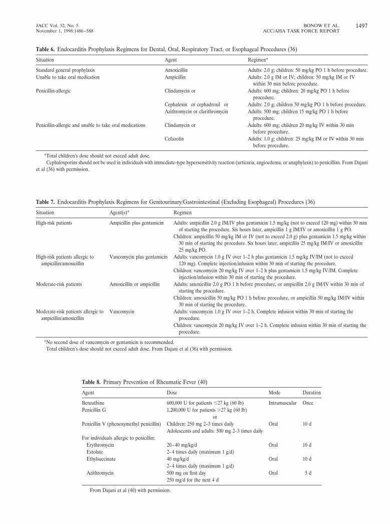
Table 6. Endocarditis Prophylaxis Regimens for Dental, Oral, Respiratory Tract, or Esophageal Procedures (36)
Situation Agent Regimen*
Standard general prophylaxis Amoxicillin Adults: 2.0 g; children: 50 mg/kg PO 1 h before procedure.Unable to take oral medication Ampicillin Adults: 2.0 g IM or IV; children: 50 mg/kg IM or IV
within 30 min before procedure.Penicillin-allergic Clindamycin or Adults: 600 mg; children: 20 mg/kg PO 1 h before
procedure.Cephalexin† or cephadroxil† or Adults: 2.0 g; children 50 mg/kg PO 1 h before procedure.Azithromycin or clarithromycin Adults: 500 mg; children 15 mg/kg PO 1 h before
procedure.Penicillin-allergic and unable to take oral medications Clindamycin or Adults: 600 mg; children 20 mg/kg IV within 30 min
before procedure.Cefazolin† Adults: 1.0 g; children: 25 mg/kg IM or IV within 30 min
before procedure.
*Total children’s dose should not exceed adult dose.†Cephalosporins should not be used in individuals with immediate-type hypersensitivity reaction (urticaria, angioedema, or anaphylaxis) to penicillins. From Dajani
et al (36) with permission.
Table 7. Endocarditis Prophylaxis Regimens for Genitourinary/Gastrointestinal (Excluding Esophageal) Procedures (36)
Situation Agent(s)* Regimen†
High-risk patients Ampicillin plus gentamicin Adults: ampicillin 2.0 g IM/IV plus gentamicin 1.5 mg/kg (not to exceed 120 mg) within 30 minof starting the procedure. Six hours later, ampicillin 1 g IM/IV or amoxicillin 1 g PO.
Children: ampicillin 50 mg/kg IM or IV (not to exceed 2.0 g) plus gentamicin 1.5 mg/kg within30 min of starting the procedure. Six hours later, ampicillin 25 mg/kg IM/IV or amoxicillin25 mg/kg PO.
High-risk patients allergic toampicillin/amoxicillin
Vancomycin plus gentamicin Adults: vancomycin 1.0 g IV over 1–2 h plus gentamicin 1.5 mg/kg IV/IM (not to exceed120 mg). Complete injection/infusion within 30 min of starting the procedure.
Children: vancomycin 20 mg/kg IV over 1–2 h plus gentamicin 1.5 mg/kg IV/IM. Completeinjection/infusion within 30 min of starting the procedure.
Moderate-risk patients Amoxicillin or ampicillin Adults: amoxicillin 2.0 g PO 1 h before procedure, or ampicillin 2.0 g IM/IV within 30 min ofstarting the procedure.
Children: amoxicillin 50 mg/kg PO 1 h before procedure, or ampicillin 50 mg/kg IM/IV within30 min of starting the procedure.
Moderate-risk patients allergic toampicillin/amoxicillin
Vancomycin Adults: vancomycin 1.0 g IV over 1–2 h. Complete infusion within 30 min of starting theprocedure.
Children: vancomycin 20 mg/kg IV over 1–2 h. Complete infusion within 30 min of starting theprocedure.
*No second dose of vancomycin or gentamicin is recommended.†Total children’s dose should not exceed adult dose. From Dajani et al (36) with permission.
Table 8. Primary Prevention of Rheumatic Fever (40)
Agent Dose Mode Duration
Benzathine 600,000 U for patients ,227 kg (60 lb) Intramuscular OncePenicillin G 1,200,000 U for patients .27 kg (60 lb)
orPenicillin V (phenoxymethyl penicillin) Children: 250 mg 2–3 times daily Oral 10 d
Adolescents and adults: 500 mg 2–3 times dailyFor individuals allergic to penicillin:
Erythromycin 20–40 mg/kg/d Oral 10 dEstolate 2–4 times daily (maximum 1 g/d)Ethylsuccinate 40 mg/kg/d Oral 10 d
2–4 times daily (maximum 1 g/d)Azithromycin 500 mg on first day Oral 5 d
250 mg/d for the next 4 d
From Dajani et al (40) with permission.
1497JACC Vol. 32, No. 5 BONOW ET AL.November 1, 1998:1486–588 ACC/AHA TASK FORCE REPORT

dures, intermediate with procedures involving the genitouri-nary tract, and lowest with gastrointestinal diagnostic proce-dures (28). Recommendations for endocarditis prophylaxis, asdetermined by dental, surgical, and other procedures, arelisted in Tables 4 through 7.
The procedure—thus the portal of entry—is a determinantof the type of organism involved in the resulting bacteremia.This is usually the determinant of the antibiotic chosen forprophylaxis. Because streptococci are normal inhabitants ofthe oral cavity, the antibiotic prophylaxis regimen for dentaland oral procedures is directed against these organisms. Forgenitourinary and lower gastrointestinal procedures, the anti-biotic prophylactic regimen is designed to cover enterococciand other gram-negative organisms.
2. Rheumatic Fever Prophylaxis. a. General Considerations.Rheumatic fever is an important cause of valvular heartdisease. In the United States (and Western Europe), cases ofacute rheumatic fever have been uncommon since the 1970s.However, starting in 1987, an increase in cases has beenobserved (38,39). With the enhanced understanding of thecausative organism, group A streptococcus, their rheumatoge-nicity is attributed to the prevalence of M protein serotypes inthe offending organism. This has resulted in the developmentof kits that allow rapid detection of group A streptococci withspecificity .295% and more rapid identification of their pres-ence in upper respiratory infection. Because the test has a lowsensitivity, the negative test requires a throat culture confir-mation (39). Prompt recognition and treatment representprimary rheumatic fever prevention. For patients who havehad a previous episode of rheumatic fever, continuous anti-streptococcal prophylaxis results in secondary prevention.
b. Primary Prevention. Rheumatic fever prevention treat-ment guidelines have been established by the American HeartAssociation (40) (Table 8).
c. Secondary Prevention. Patients who have had an episodeof rheumatic fever are at high risk of developing recurrentepisodes of acute rheumatic fever. Patients who developcarditis are especially prone to similar episodes with subse-quent attacks. Secondary prevention of rheumatic fever recur-
rence is thus of great importance. Continuous antimicrobialprophylaxis has been shown to be effective. Anyone who hashad rheumatic fever with or without carditis (including MS)should have prophylaxis for recurrent rheumatic fever. TheAHA guidelines for secondary prevention are shown in Table9. The AHA guidelines for duration of secondary preventionare shown in Table 10.
III. Specific Valve LesionsA. Aortic Stenosis
1. Introduction. The most common cause of AS in adults isa degenerative-calcific process that produces an immobiliza-tion of the aortic valve cusps. This calcific disease progressesfrom the base of the cusps to the leaflets, eventually causing areduction in the effective valve area; true commissural fusionmay not occur. A congenital malformation of the valve mayalso result in stenosis and is the more common cause in youngadults. The management of congenital AS in adolescents andyoung adults is discussed in section VI.A. of these guidelines.Over several decades, progressive fibrosis and calcification ofthe congenitally abnormal valve (often bicuspid) produce adeformity that resembles the degenerative-calcific lesion.Rheumatic fever results in AS due to fusion of the commis-sures with scarring and eventual calcification of the cusps.Thus, calcification is a common feature of AS in older adultsregardless of the primary cause (41–43).
An ejection systolic murmur may be heard in the presenceof a normal valve, one that is thickened and minimallycalcified, and one that is stenotic (41,44). The 3 conditionsmust be distinguished.
a. Grading the Degree of Stenosis. The aortic valve areamust be reduced to one fourth its normal size before significantchanges in the circulation occur. Because the orifice area of thenormal adult valve is '3.0 to 4.0 cm2, an area .0.75 to 1.0 cm2
is usually not considered severe AS (44,45). Historically, thedefinition of severe AS is based on the hydraulic orifice-areaformulae developed by Gorlin and Gorlin, which indicate thatlarge pressure gradients accompany only modest increments inflow when the valve area is ,0.75 cm2 (46). However, in large
Table 9. Secondary Prevention of Rheumatic Fever (40)
Agent Dose Mode
Benzathine 1,200,000 U every 4 wk IntramuscularPenicillin G (every 3 wk for high-risk* pts such as
those with residual carditis)Penicillin V 250 mg twice daily Oral
orSulfadiazine 0.5 g once daily for pts ,227 kg (60 lb) Oral
1.0 g once daily for pts .27 kg (60 lb)For individuals allergic
to penicillin andsulfadiazine:
Erythromycin 250 mg twice daily Oral
Abbreviations: Pts 5 patients. *High-risk patients include patients withresidual rheumatic carditis as well as patients from economically disadvantagedpopulations. From Dajani et al (40) with permission.
Table 10. Duration of Secondary Rheumatic Fever Prophylaxis (40)
Category Duration
Rheumatic fever with carditis andresidual heart disease(persistent valvular disease)
.210 y since last episode and at leastuntil age 40 y, sometimes lifelongprophylaxis*
Rheumatic fever with carditis butno residual heart disease (novalvular disease)
10 y or well into adulthood, whichever islonger
Rheumatic fever without carditis 5 y or until age 21 y, whichever is longer
*The committee’s interpretation of “lifelong” prophylaxis refers to patientswho are at high risk and likely to come in contact with populations with a highprevalence of streptococcal infection, ie, teachers, day-care workers. FromDajani et al (40) with permission.
1498 BONOW ET AL. JACC Vol. 32, No. 5ACC/AHA TASK FORCE REPORT November 1, 1998:1486–588

patients, a valve area of 1.0 cm2 may be severely stenotic,whereas a valve area of 0.7 cm2 may be adequate for a smallerpatient.
On the basis of a variety of hemodynamic and naturalhistory data, in these guidelines we graded the degree of AS asmild (area .1.5 cm2), moderate (area .1.0 to 1.5 cm2), orsevere (area ,21.0 cm2) (46a). When stenosis is severe andcardiac output is normal, the mean transvalvular pressuregradient is generally .50 mm Hg. Some patients with severeAS remain asymptomatic, whereas others with only moderatestenosis develop symptoms. Therapeutic decisions, particularlythose related to corrective surgery, are based largely on thepresence or absence of symptoms. Thus, the absolute valvearea (or transvalvular pressure gradient) is not usually theprimary determinant of the need for aortic valve replacement(AVR).
2. Pathophysiology. In adults with AS, the obstructiondevelops gradually—usually over decades. During this time,the left ventricle adapts to the systolic pressure overloadthrough a hypertrophic process that results in increased LVwall thickness while a normal chamber volume is maintained(47–49). The resulting increase in relative wall thickness isusually enough to counter the high intracavitary systolic pres-sure, and as a result, LV systolic wall stress (afterload) remainswithin the range of normal. The inverse relation betweensystolic wall stress and ejection fraction is maintained; as longas wall stress is normal, the ejection fraction is preserved (50).However, if the hypertrophic process is inadequate and rela-tive wall thickness does not increase in proportion to pressure,wall stress increases and the high afterload causes a decrease inejection fraction (50–52). The depressed contractile state ofthe myocardium may also be responsible for a low ejectionfraction, but a combination of excessive afterload and de-pressed contractility contributes to a low ejection fraction inmany patients (53). When low ejection fraction is caused bydepressed contractility, corrective surgery will be less beneficialthan in patients with a low ejection fraction caused by highafterload (54).
As a result of increased wall thickness, low volume/massratio, and diminished compliance of the chamber, LV end-diastolic pressure increases without chamber dilatation (55–58). Thus, increased end-diastolic pressure usually reflectsdiastolic dysfunction rather than systolic dysfunction or failure(59). A forceful atrial contraction that contributes to anelevated end-diastolic pressure plays an important role inventricular filling without increasing mean left atrial or pulmo-nary venous pressure (60). Loss of atrial contraction such asthat which occurs with atrial fibrillation is often followed byserious clinical deterioration.
The development of concentric hypertrophy appears to bean appropriate and beneficial adaptation to compensate forhigh intracavitary pressures. Unfortunately, this adaptationoften carries adverse consequences. The hypertrophied heartmay have reduced coronary blood flow per gram of muscle andalso exhibit a limited coronary vasodilator reserve, even in theabsence of epicardial CAD (61,62). The hemodynamic stress of
exercise or tachycardia can produce a maldistribution ofcoronary blood flow and subendocardial ischemia, which cancontribute to systolic or diastolic dysfunction of the leftventricle. Hypertrophied hearts also exhibit an increased sen-sitivity to ischemic injury, with larger infarcts and highermortalities than are seen in the absence of hypertrophy(63–65). Another problem that is particularly common inelderly patients, especially women, is an excessive or inappro-priate degree of hypertrophy; wall thickness is greater thannecessary to counterbalance the high intracavitary pressures(66–69). As a result, systolic wall stress is low, ejection fractionis high, and the ventricle resembles that seen in patients withhypertensive hypertrophic cardiomyopathy of the elderly (70).Such inappropriate LV hypertrophy has been associated withhigh perioperative morbidity and mortality (66,68).
3. Natural History. The natural history of AS in the adultconsists of a prolonged latent period during which morbidityand mortality are very low. The rate of progression of thestenotic lesion has been estimated in a variety of hemodynamicstudies performed largely in patients with moderate AS (71).Cardiac catheterization studies indicate that some patientshave a decrease in valve area of 0.1 to 0.3 cm2 per year; thesystolic pressure gradient across the valve may increase by asmuch as 10 to 15 mm Hg per year (72–78). However, morethan half of the reported patients show little or no progressionover a 3- to 9-year period. Doppler echocardiographic dataobtained over several years are consistent with those obtainedwith cardiac catheterization. Some patients exhibit a significantincrease in transvalvular pressure gradient ('15 to 19 mm Hgper year) and a decrease in valve area; others show little or nochange (79–83). The average rate of change is '0.12 cm2 peryear (84). Although it appears that the progression of AS canbe more rapid in patients with degenerative calcific diseasethan in those with congenital or rheumatic disease (41,73), it isnot possible to predict the rate of progression in an individualpatient. For this reason, careful clinical follow-up is mandatoryin all patients with moderate to severe AS.
Eventually, symptoms of angina, syncope, or heart failuredevelop after a long latent period, and the outlook changesdramatically. After the onset of symptoms, average survival isless than 2 to 3 years (85–90). Thus, the development ofsymptoms identifies a critical point in the natural history of AS.Management decisions are based largely on these naturalhistory data; many clinicians treat asymptomatic patients con-servatively, whereas corrective surgery is generally recom-mended in patients with symptoms thought to be due to AS.
Sudden death is known to occur in patients with severe ASand rarely has been documented to occur without priorsymptoms (85,88,91). These older retrospective studies empha-size the possibility of sudden death in asymptomatic patients.However, prospective echocardiographic studies provide im-portant data on the rarity of sudden death in asymptomaticpatients (Table 11). In one report, 51 asymptomatic patientswith severe AS were followed for an average of 17 months. Inthis study, 2 patients died; symptoms preceded death in bothcases (90). In another report of 113 patients followed for 20
1499JACC Vol. 32, No. 5 BONOW ET AL.November 1, 1998:1486–588 ACC/AHA TASK FORCE REPORT

months, there were no cases of sudden death without preced-ing symptoms; in this study, survival was no different from thatof an age- and sex-matched control group (92). In this latterstudy, all patients had Doppler velocities across the aortic valve.24 m/s. However, only one third had aortic valve velocities.25 m/s, and a number of patients were not included in thefollow-up analysis because they underwent AVR at the discre-tion of the clinician. In a third report of 123 patients followedfor an average of 30 months, there were no cases of suddendeath (84). These findings were similar in 2 smaller studies(77,81). Therefore, although sudden death occasionally doesoccur in the absence of preceding symptoms in patients withAS (85,88,93), it must be an uncommon event—probably ,1%per year.
4. Management of the Asymptomatic Patient. Manyasymptomatic patients with severe AS develop symptomswithin a few years and require surgery. In one series, theincidence of angina, dyspnea, or syncope in 113 asymptomaticpatients with Doppler outflow velocities .24 m/s was 14% after1 year and 38% after 2 years (92). In another report of 123asymptomatic patients, the rate of symptom development was38% at 3 years for the total group but 79% at 3 years inpatients with Doppler outflow velocity .24 m/s (84). Therefore,patients with severe AS require careful monitoring for devel-opment of symptoms and progressive disease.
a. Initial Evaluation. The diagnosis of severe AS can usuallybe made on the basis of the systolic outflow murmur, delayedand diminished carotid upstrokes, sustained LV impulse, andreduced intensity of the aortic component of the second heartsound. Paradoxical splitting of the second sound may bepresent. In the elderly, the pulsus tardus and parvus may beabsent because of the effects of aging on the vasculature.Patients presenting with the physical findings of AS shouldundergo selected laboratory examinations, including an ECG,a chest x-ray, and an echocardiogram. The 2-D echocardio-gram is valuable for confirming the presence of aortic valvedisease and determining the LV response to pressure overload.In most patients, the severity of the stenotic lesion can bedefined with Doppler echocardiographic measurements of amean transvalvular pressure gradient and a derived valve area,as discussed in the ACC/AHA Guidelines for the Clinical
Application of Echocardiography (2). The mean pressuregradient may be underestimated if the Doppler beam is notparallel to the velocity jet; however, it may occasionallyoverestimate the transvalvular gradient, especially in the pa-tient with a small aortic root and/or high cardiac output. Thus,the pressure gradient and derived valve area require meticu-lous attention to measurement of LV outflow tract area andvelocity. Echocardiography is also used to assess LV size andfunction, degree of hypertrophy, and presence of other asso-ciated valvular disease.
Recommendations for Echocardiography in Aortic Stenosis
Indication Class
1. Diagnosis and assessment of severity of AS. I2. Assessment of LV size, function, and/or hemodynamics. I3. Reevaluation of patients with known AS with changing symptoms
or signs.I
4. Assessment of changes in hemodynamic severity and ventricularfunction in patients with known AS during pregnancy.
I
5. Reevaluation of asymptomatic patients with severe AS. I6. Reevaluation of asymptomatic patients with mild to moderate AS
and evidence of LV dysfunction or hypertrophy.IIa
7. Routine reevaluation of asymptomatic adult patients with mildAS having stable physical signs and normal LV size and function.
III
From the ACC/AHA Guidelines for the Clinical Application ofEchocardiography (2).
In some patients, it may be necessary to proceed withcardiac catheterization and coronary angiography at the timeof initial evaluation. For example, this is appropriate if there isa discrepancy between clinical and echocardiographic exami-nations or if the patient is symptomatic and AVR is planned.
Exercise testing in adults with AS has been discouragedlargely because of concerns about safety. Furthermore, whenused to assess the presence or absence of CAD, the test haslimited diagnostic accuracy. Presumably, this is due to thepresence of an abnormal baseline ECG, LV hypertrophy, andlimited coronary flow reserve. Certainly, exercise testingshould not be performed in symptomatic patients. However, inasymptomatic patients, exercise testing is safe and may provideinformation that is not uncovered during the initial clinical
Table 11. Studies of the Natural History of Asymptomatic Patients With Aortic Stenosis
Study, yNumber
of patients
Meanfollow-up,
yearSeverity of aorticstenosis
Sudden deathwithout symptoms
(number of patients) Comments
Chizner et al 1980 (91) 8 5.7 AVA ,1.1 cm2 0 retrospective studyTurina et al 1987 (77) 17 2.0 AVA ,0.9 cm2 0 retrospective studyHorstkotte and Loogen 1988 (88) 35 “years” AVA 5 0.4–0.8 cm2 3 retrospective studyKelly et al 1988 (90) 51 1.5 PV 5 3.5–5.8 m/s 0 prospective studyPellikka et al 1990 (92) 113 1.7 PV .4.0 m/s 0 prospective studyFaggiano et al 1992 (81) 37 2.0 AVA 5 0.85 6 0.15 cm2 0 prospective studyOtto et al 1997 (84) 114 2.5 PV 5 3.6 6 0.6 m/s 0 prospective study
Total 375 2.1 3 average risk of sudden death '0.4%/y
Abbreviations: AVA 5 aortic valve area; PV 5 peak instantaneous velocity
1500 BONOW ET AL. JACC Vol. 32, No. 5ACC/AHA TASK FORCE REPORT November 1, 1998:1486–588

evaluation (94–97). Exercise testing in asymptomatic patientsshould be performed only under the supervision of an experi-enced physician with close monitoring of blood pressure andthe ECG. Such testing can identify patients with a limitedexercise capacity or even exercise-induced symptoms despite anegative medical history. Although the prognostic significanceof electrocardiographic ST depression is unknown, an abnor-mal hemodynamic response (eg, hypotension) in a patient withsevere AS is sufficient reason to consider AVR. Finally, inselected patients, the observations made during exercise mayprovide a basis for advice about physical activity.
The frequency of follow-up visits to the physician dependson the severity of the valvular stenosis and in part on thepresence of comorbid conditions. Recognizing that an optimalschedule for repeated medical examinations has not beendefined, many physicians perform an annual history and phys-ical examination on patients with mild AS. Those with moder-ate and severe AS should be examined more frequently.Patients should be advised to promptly report the developmentof any exertional chest discomfort, dyspnea, lightheadedness,or syncope.
b. Serial Testing. Echocardiographic studies can be animportant part of an integrated approach that includes adetailed history, physical examination, and in some patients acarefully monitored exercise test. Recognizing that the rate ofprogression varies considerably, clinicians often perform anannual echocardiogram on patients known to have moderateto severe AS. However, current understanding of the naturalhistory of AS and indications for surgical intervention do notsupport the use of annual echocardiographic studies to assesschanges in valve area as such. However, serial echocardio-grams are helpful for assessing changes in LV hypertrophy andfunction. Therefore, in patients with severe AS, an echocar-diogram every year may be appropriate. In patients withmoderate AS, serial studies performed every 2 years or so aresatisfactory, and in patients with mild AS, serial studies can beperformed every 5 years. Echocardiograms should be per-formed more frequently if there is a change in clinical findings.In patients with echocardiograms of suboptimal quality, car-diac magnetic resonance imaging may be used to assess LVvolume, wall thickness, mass, and systolic function (98–102) aswell as severity of AS (103,104). In centers with specificexpertise in cardiac magnetic resonance imaging, serial mag-netic resonance imaging may be performed in place of serialechocardiograms.
c. Medical Therapy. Antibiotic prophylaxis is indicated forprevention of infective endocarditis and, in those with rheu-matic AS, recurrent episodes of rheumatic fever. Patients withassociated systemic arterial hypertension should be treatedcautiously with appropriate antihypertensive agents. Withthese exceptions, there is no specific medical therapy forpatients who have not yet developed symptoms, and patientswho develop symptoms require surgery, not medical therapy.Most asymptomatic patients lead a normal life, althoughrestriction of physical activity should be advised in mostpatients with moderate or severe AS.
d. Physical Activity and Exercise. Recommendations forphysical activity are based on the clinical examination, withspecial emphasis on the hemodynamic severity of the stenoticlesion. The severity can usually be judged by Doppler echocar-diography, but in borderline cases, diagnostic cardiac catheter-ization may be necessary to accurately define the degree ofstenosis.
Recommendations on participation in competitive sportshave been published by the Task Force on Acquired ValvularHeart Disease of the 26th Bethesda Conference (105). Physicalactivity is not restricted in asymptomatic patients with mild AS;these patients can participate in competitive sports. Patientswith moderate AS should avoid competitive sports that involvehigh dynamic and static muscular demands. Other forms ofexercise can be performed safely, but it is advisable to evaluatesuch patients with an exercise test before they begin an exerciseor athletic program. Patients with severe AS should be advisedto limit their activity to relatively low levels.
5. Indications for Cardiac Catheterization. In patients withAS, the indications for cardiac catheterization and angiogra-phy are essentially the same as in other conditions, namely toassess the coronary circulation and confirm or clarify theclinical diagnosis. In preparation for AVR, coronary angiog-raphy is indicated in patients suspected of having CAD, asdiscussed in detail in section VIII of these guidelines. If theclinical and echocardiographic data are typical of severeisolated AS, coronary angiography may be all that is neededbefore AVR. A complete left- and right-heart catheterizationmay be necessary to assess the hemodynamic severity of the ASif there is a discrepancy between clinical and echocardio-graphic data or there is evidence of associated valvular orcongenital disease or pulmonary hypertension.
Recommendations for Cardiac Catheterization in Aortic Stenosis
Indication Class
1. Coronary angiography before AVR in patients at risk for CAD(see section VIII.B. of these guidelines).
I
2. Assessment of severity of AS in symptomatic patients when AVRis planned or when noninvasive tests are inconclusive or thereis a discrepancy with clinical findings regarding severity of ASor need for surgery.
I
3. Assessment of severity of AS before AVR when noninvasive testsare adequate and concordant with clinical findings andcoronary angiography is not needed.
IIb
4. Assessment of LV function and severity of AS in asymptomaticpatients when noninvasive tests are adequate.
III
The pressure gradient across a stenotic valve is related tothe valve orifice area and the transvalvular flow (106). Thus, inthe presence of depressed cardiac output, relatively low pres-sure gradients are frequently obtained in patients with severeAS. On the other hand, during exercise or other high flowstates, systolic gradients can be measured in minimally stenoticvalves. For these reasons, complete assessment of AS requires(1) measurement of transvalvular flow, (2) determination ofthe transvalvular pressure gradient, and (3) calculation of theeffective valve area. Careful attention to detail with accurate
1501JACC Vol. 32, No. 5 BONOW ET AL.November 1, 1998:1486–588 ACC/AHA TASK FORCE REPORT

measurements of pressure and flow is important, especially inpatients with low cardiac output or a low transvalvular pressuregradient.
a. Low-Gradient Aortic Stenosis. Patients with severe ASand low cardiac output often present with only modest trans-valvular pressure gradients (ie, ,30 mm Hg). Such patients canbe difficult to distinguish from those with low cardiac outputand only mild to moderate AS. In the former (true anatomi-cally severe AS), the stenotic lesion contributes to an elevatedafterload, decreased ejection fraction, and low stroke volume.In the latter, primary contractile dysfunction is responsible forthe decreased ejection fraction and low stroke volume; theproblem is further complicated by reduced valve openingforces that contribute to limited valve mobility and apparentstenosis. In both situations, the low-flow state and low-pressuregradient contribute to a calculated effective valve area that canmeet criteria for severe AS. The standard valve area formula isless accurate and is known to underestimate the valve area inlow-flow states, and under such conditions, it should beinterpreted with caution. In theory, Doppler-derived valveareas should be less susceptible to low flow, but this does notappear to be borne out in clinical practice. It has beensuggested that valve resistance might provide a better separa-tion between critical and noncritical AS, particularly in pa-tients with low transvalvular pressure gradients (107,108).Although valve resistance is less sensitive to flow than valvearea, the resistance calculations have not been proved to besubstantially better than valve area calculations.
In patients with low-gradient stenosis and what appears tobe moderate to severe AS, it may be useful to determine thetransvalvular pressure gradient and to calculate valve area andresistance during a baseline state and again during exercise orpharmacological (ie, dobutamine infusion) stress (97,109–111). This approach is based on the notion that patients whodo not have true, anatomically severe stenosis exhibit anincrease in the valve area during an increase in cardiac output(109,110). Thus, if a dobutamine infusion produces an incre-ment in stroke volume, an increase in valve area, and adecrease in valve resistance, it is likely that the baselinecalculations overestimated the severity of the stenosis. Inpatients with severe AS, these changes may result in a calcu-lated valve area that is higher than the baseline calculation butone that remains in the severe range, whereas in patientswithout severe AS, the calculated valve area with dobutaminewill fall outside the severe range and indicate that severe AS isnot present.
6. Indications for Aortic Valve Replacement. In the vastmajority of adults, AVR is the only effective treatment forsevere AS. However, younger patients may be candidates forvalvotomy (see section VI.A. of these guidelines). Althoughthere is some lack of agreement about the optimal timing ofsurgery, particularly in asymptomatic patients, it is possible todevelop rational guidelines for most patients. Particular con-sideration should be given to the natural history of symptom-atic and asymptomatic patients and to operative risks andoutcomes after surgery.
a. Symptomatic Patients. Patients with angina, dyspnea, orsyncope exhibit symptomatic improvement and an increase insurvival after AVR (86,112–116). These salutary results ofsurgery are partly dependent on the state of LV function. Theoutcome is similar in patients with normal LV function and inthose with moderate depression of contractile function. Thedepressed ejection fraction in many of the patients in this lattergroup is caused by excessive afterload (afterload mismatch[52]), and LV function improves after AVR in such patients. IfLV dysfunction is not caused by afterload mismatch, thenimprovement in LV function and resolution of symptoms maynot be complete after valve replacement (116). Survival is stillimproved in this setting (112), with the possible exception ofpatients with severe LV dysfunction caused by CAD (116).Therefore, in the absence of serious comorbid conditions,AVR is indicated in virtually all symptomatic patients withsevere AS. However, patients with severe LV dysfunction,particularly those with so-called low gradient AS, create adifficult management decision (117) (see above). AVR shouldnot be performed in such patients if they do not have anatom-ically severe AS. In patients who do have severe AS, even thosewith a low transvalvular pressure gradient, AVR results inhemodynamic improvement and better functional status.
b. Asymptomatic Patients. Many clinicians are reluctant toproceed with AVR in an asymptomatic patient (118), whereasothers are concerned about following a patient with severe AS.Although insertion of a prosthetic aortic valve is associatedwith low perioperative morbidity and mortality, long-termmorbidity and mortality can be appreciable for mechanical andbioprosthetic valves. Significant complications occur at the rateof at least 2% to 3% per year, and death due directly to theprosthesis occurs at the rate of '1% per year (119–124). Thus,even if surgical mortality can be minimized, the combined riskof surgery and the late complications of a prosthesis exceed thepossibility of preventing sudden death and prolonging survivalin all asymptomatic patients, as discussed previously. Despitethese considerations, some difference of opinion persistsamong clinicians regarding the indications for corrective sur-gery in asymptomatic patients. Some argue that irreversiblemyocardial depression and/or fibrosis may develop during aprolonged asymptomatic stage and that this may preclude anoptimal outcome. Such irreversibility has not been proved, butthis concept has been used to support early surgery (114,125).Still others attempt to identify patients who may be at espe-cially high risk of sudden death without surgery, although datasupporting this approach are limited. Patients in this subgroupinclude those who have an abnormal response to exercise (eg,hypotension), those with LV systolic dysfunction or marked/excessive LV hypertrophy, or those with evidence of verysevere AS. However, it should be recognized that such “high-risk” patients are rarely asymptomatic.
c. Patients Undergoing Coronary Artery Bypass Surgery. Pa-tients with severe AS, with or without symptoms, who areundergoing coronary artery bypass surgery should undergoAVR at the time of the revascularization procedure. Similarly,patients with severe AS undergoing surgery on other valves
1502 BONOW ET AL. JACC Vol. 32, No. 5ACC/AHA TASK FORCE REPORT November 1, 1998:1486–588

(such as mitral valve repair) or the aortic root should alsoundergo AVR as part of the surgical procedure, and it isgenerally accepted practice to perform AVR in patients withmoderate AS (for example, gradient .230 mm Hg) who areundergoing mitral valve or aortic root surgery, as discussed insections III.F.6. and III.F.7. of these guidelines. Such patientswith moderate AS may also warrant AVR at the time ofcoronary artery bypass surgery, but there are limited data tosupport this policy. Greater controversy persists regarding theindications for concomitant AVR at the time of coronaryartery bypass surgery in patients with milder forms of AS, asdiscussed in section VIII.D. of these guidelines.
Recommendations for Aortic Valve Replacement in Aortic Stenosis
Indication Class
1. Symptomatic patients with severe AS. I2. Patients with severe AS undergoing coronary artery bypass surgery. I3. Patients with severe AS undergoing surgery on the aorta or other
heart valves.I
4. Patients with moderate AS undergoing coronary artery bypasssurgery or surgery on the aorta or other heart valves (see sectionsIII.F.6., III.F.7., and VIII.D. of these guidelines).
IIa
5. Asymptomatic patients with severe AS andz LV systolic dysfunction IIaz Abnormal response to exercise (eg, hypotension) IIaz Ventricular tachycardia IIbz Marked or excessive LV hypertrophy (>215 mm) IIbz Valve area <0.6 cm2 IIb
6. Prevention of sudden death in asymptomatic patients with none ofthe findings listed under indication 5.
III
7. Aortic Balloon Valvotomy. Percutaneous balloon aorticvalvotomy is a procedure in which one or more balloons areplaced across a stenotic valve and inflated to decrease theseverity of stenosis (126–128). This procedure has an impor-tant role in treating adolescents and young adults with AS (seesection VI.A.) but a very limited role in older adults. Themechanism underlying relief of the stenotic lesion in olderadults is fracture of calcific deposits within the valve leafletsand to some degree stretching of the annulus and separation ofthe calcified or fused commissures (129–131). Immediatehemodynamic results include a moderate reduction in thetransvalvular pressure gradient, but the postvalvotomy valvearea rarely exceeds 1.0 cm2. Despite the modest change invalve area, an early symptomatic improvement is usually seen.However, serious complications occur with a frequency .10%(132–138); restenosis and clinical deterioration occur within 6to 12 months in most patients (133,138–141). Therefore, inadults with AS, balloon valvotomy is not a substitute for AVR(141–144).
Despite the procedural morbidity and mortality and limitedlong-term results, balloon valvotomy can have a temporary rolein the management of some symptomatic patients who are notinitially candidates for AVR (144). For example, patients withsevere AS and refractory pulmonary edema or cardiogenicshock may benefit from aortic valvuloplasty as a “bridge” tosurgery; an improved hemodynamic state may reduce the risks
of surgery. The indications for palliative valvotomy in patientswith serious comorbid conditions are less well established, butmost patients can expect temporary relief of symptoms despitea very limited life expectancy. Asymptomatic patients withsevere AS who require urgent noncardiac surgery may becandidates for valvotomy, but most such patients can besuccessfully treated with more conservative measures(145,146).
Recommendations for Aortic Balloon Valvotomy in Adults WithAortic Stenosis*
Indication Class
1. A “bridge” to surgery in hemodynamically unstable patients whoare at high risk for AVR.
IIa
2. Palliation in patients with serious comorbid conditions. IIb3. Patients who require urgent noncardiac surgery. IIb4. An alternative to AVR. III
*Recommendations for aortic balloon valvotomy in adolescents and youngadults with AS are provided in section VI.A. of these guidelines.
8. Medical Therapy for the Inoperable Patient. Comorbidconditions (eg, malignancy) or, on occasion, patient prefer-ences may preclude corrective surgery. Under such circum-stances, limited medical therapies are available to controlsymptoms. Patients with evidence of pulmonary congestion canbenefit from treatment with digitalis, diuretics, and angiotensinconverting enzyme (ACE) inhibitors. Indeed, a cautious reduc-tion in central blood volume and LV preload can be efficaciousin some patients with heart failure symptoms. It should berecognized, however, that excessive preload reduction candepress cardiac output and reduce systemic arterial pressure;patients with severe AS are especially subject to this untowardeffect. Digitalis should be reserved for patients with depressedsystolic function or atrial fibrillation. Atrial fibrillation andother atrial arrhythmias have an adverse effect on atrial pumpfunction and ventricular rate; if prompt cardioversion is un-successful, pharmacological control of the ventricular rate withdigitalis or perhaps amiodarone is essential. Efforts should bemade to prevent atrial fibrillation, especially development of arapid ventricular response. b-Adrenergic receptor blockingagents as well as other drugs with negative inotropic effectsshould not be used in patients with heart failure caused by AS.If angina is the predominant symptom, cautious use of nitratesand b-blockers can provide relief. There is no specific medicaltherapy for syncope unless it is caused by a bradyarrhythmia ortachyarrhythmia.
9. Evaluation After Aortic Valve Replacement. AVRshould be considered a form of palliative therapy in that aprosthetic valve with its attendant complications is substitutedfor a diseased native valve (119–124). Patients with prostheticheart valves therefore require periodic clinical and selectedlaboratory examinations. A complete history and physicalexamination should be performed at least once a year. Indica-tions for echocardiography are discussed in section VII.C.3. ofthese guidelines.
1503JACC Vol. 32, No. 5 BONOW ET AL.November 1, 1998:1486–588 ACC/AHA TASK FORCE REPORT

10. Special Considerations in the Elderly. Because there isno effective medical therapy and balloon valvotomy is not anacceptable alternative to surgery, AVR must be considered inall elderly patients who have symptoms caused by AS. Valvereplacement is technically possible at any age (147), but thedecision to proceed with such surgery depends on manyfactors, including the patient’s wishes and expectations. Olderpatients with symptoms due to severe AS, normal coronaryarteries, and preserved LV function can expect a betteroutcome than those with coronary disease or ventriculardysfunction (87). Certainly advanced cancer and permanentneurological defects as a result of stroke make cardiac surgeryinappropriate. Deconditioned and debilitated patients oftendo not return to an active existence, and the presence of theother comorbid disorders may have a major impact on out-come.
In addition to the confounding effects of CAD and thepotential for stroke, other considerations are peculiar to olderpatients. For example, a narrow LV outflow tract and a smallaortic annulus sometimes present in elderly women mayrequire enlargement of the annulus. Heavy calcification of thevalve, annulus, and aortic root may require debridement.Occasionally, a composite valve-aortic graft is needed. Like-wise, excessive or inappropriate hypertrophy associated withvalvular stenosis can be a marker for perioperative morbidityand mortality (66,68). Preoperative recognition of elderlypatients with marked LV hypertrophy followed by appropriateperioperative management may substantially reduce this mor-bidity and mortality. There is no perfect method for weighingall of the relevant factors and identifying specifically high- andlow-risk elderly patients (148). The decision to proceed withvalve replacement depends on an imprecise analysis thatconsiders the balance between the potential for improvedsymptoms and survival and the morbidity and mortality ofsurgery.
B. Aortic Regurgitation1. Etiology. There are a number of common causes of AR.
These include idiopathic dilatation, congenital abnormalitiesof the aortic valve (most notably bicuspid valves), calcificdegeneration, rheumatic disease, infective endocarditis, sys-temic hypertension, myxomatous proliferation, dissection ofthe ascending aorta, and Marfan syndrome. Less commonetiologies include traumatic injuries to the aortic valve, anky-losing spondylitis, syphilitic aortitis, rheumatoid arthritis, os-teogenesis imperfecta, giant cell aortitis, Ehlers-Danlos syn-drome, Reiter’s syndrome, discrete subaortic stenosis, andventricular septal defects with prolapse of an aortic cusp.Recently, anorectic drugs have also been reported to cause AR(see section III.H. of these guidelines). The majority of theselesions produce chronic AR with slow, insidious LV dilatationand a prolonged asymptomatic phase. Other lesions, in partic-ular infective endocarditis, aortic dissection, and trauma, moreoften produce acute severe AR, which can result in sudden
catastrophic elevation of LV filling pressures and reduction incardiac output.
2. Acute Aortic Regurgitationa. Pathophysiology. In acute severe AR, the sudden large
regurgitant volume is imposed on a left ventricle of normal sizethat has not had time to accommodate the volume overload.With an abrupt increase in end-diastolic volume, the ventricleoperates on the steep portion of a normal diastolic pressure-volume relationship, and LV end-diastolic and left atrialpressures may increase rapidly and dramatically. The Frank-Starling mechanism is used, but the inability of the ventricle todevelop compensatory chamber dilatation acutely results in adecrease in forward stroke volume. Although tachycardiadevelops as a compensatory mechanism to maintain cardiacoutput, this is often insufficient. Hence, patients frequentlypresent with pulmonary edema and/or cardiogenic shock.Acute AR creates especially marked hemodynamic changes inpatients with preexisting pressure overload hypertrophy, inwhom the small, noncompliant LV cavity is set on an evensteeper diastolic pressure-volume relationship and has reducedpreload reserve. Examples of this latter situation include aorticdissection in patients with systemic hypertension, infectiveendocarditis in patients with preexisting AS, and acute regur-gitation after balloon valvotomy or surgical commissurotomyfor congenital AS.
b. Diagnosis. Many of the characteristic physical findings ofchronic AR are modified or absent when valvular regurgitationis acute, which may lead to underestimation of its severity. LVsize may be normal on physical examination and cardiomegalymay be absent on chest x-ray. Pulse pressure may not beincreased because systolic pressure is reduced and the aorticdiastolic pressure equilibrates with the elevated LV diastolicpressure. Because this diastolic pressure equilibration betweenaorta and ventricle may occur before the end of diastole, thediastolic murmur may be short and/or soft and thereforepoorly heard. The elevated LV diastolic pressure may close themitral valve prematurely, reducing the intensity of the firstheart sound. An apical diastolic rumble may be present, but itis usually brief and without presystolic accentuation. Tachycar-dia is invariably present.
Echocardiography is indispensable in confirming the pres-ence and severity of the valvular regurgitation, in determiningits etiology, in estimating the degree of pulmonary hyperten-sion (if TR is present), and in determining whether there israpid equilibration of aortic and LV diastolic pressure. Evi-dence for rapid pressure equilibration includes a short ARdiastolic half-time (,300 ms), a short mitral deceleration time(,150 ms), or premature closure of the mitral valve.
Acute AR caused by aortic root dissection is a surgicalemergency that requires particularly prompt identification andmanagement. Transesophageal echocardiography is indicatedwhen aortic dissection is suspected (149–151). If the diagnosisremains uncertain, cardiac catheterization and aortographyshould be performed. Coronary angiography is an importantcomponent of the evaluation of aortic dissection and acute ARand should be performed, provided that it does not delay
1504 BONOW ET AL. JACC Vol. 32, No. 5ACC/AHA TASK FORCE REPORT November 1, 1998:1486–588

urgent surgery. In some patients, other diagnostic imagingmethods, such as computed tomographic imaging or magneticresonance imaging, may be required if echocardiography doesnot provide the diagnosis and angiography is not planned(149,150,152).
c. Treatment. Death from pulmonary edema, ventriculararrhythmias, electromechanical dissociation, or circulatory col-lapse is common in acute severe AR, even with intensivemedical management. Early surgical intervention is recom-mended. Nitroprusside and possibly inotropic agents such asdopamine or dobutamine to augment forward flow and reduceLV end-diastolic pressure may be helpful to manage thepatient temporarily before operation. Intra-aortic ballooncounterpulsation is contraindicated. Although b-blockers areoften used in treating aortic dissection, these agents should beused very cautiously if at all in the setting of acute AR becausethey will block the compensatory tachycardia. In patients withacute severe AR resulting from infective endocarditis, surgeryshould not be delayed, especially if there is hypotension,pulmonary edema, or evidence of low output. In patients withmild acute AR, antibiotic treatment may be all that is necessaryif the patient is hemodynamically stable. Exceptions to thislatter recommendation are discussed in section IV.E. of theseguidelines.
3. Chronic Aortic Regurgitationa. Pathophysiology. The left ventricle responds to the vol-
ume load of chronic AR with a series of compensatorymechanisms, including an increase in end-diastolic volume, anincrease in chamber compliance that accommodates the in-creased volume without an increase in filling pressures, and acombination of eccentric and concentric hypertrophy. Thegreater diastolic volume permits the ventricle to eject a largetotal stroke volume to maintain forward stroke volume in thenormal range. This is accomplished through rearrangement ofmyocardial fibers with the addition of new sarcomeres anddevelopment of eccentric LV hypertrophy (153). As a result,preload at the sarcomere level remains normal or near-normal,and the ventricle retains its preload reserve. The enhancedtotal stroke volume is achieved through normal performanceof each contractile unit along the enlarged circumference(154). Thus, LV ejection performance is normal, and ejectionphase indexes such as ejection fraction and fractional shorten-ing remain in the normal range. However, the enlargedchamber size, with the associated increase in systolic wallstress, also results in an increase in LV afterload and is astimulus for further concentric hypertrophy (153,155). Thus,AR represents a condition of combined volume overload andpressure overload (156). As the disease progresses, recruit-ment of preload reserve and compensatory hypertrophypermit the ventricle to maintain normal ejection perfor-mance despite the elevated afterload (157,158). The major-ity of patients remain asymptomatic throughout this com-pensated phase, which may last for decades. Vasodilatortherapy has the potential to reduce the hemodynamicburden in such patients.
For purposes of the subsequent discussion, patients with
normal LV systolic function will be defined as those withnormal LV ejection fraction at rest. It is recognized that overallLV function is usually not “normal” in chronic severe AR andthat the hemodynamic abnormalities noted above may beconsiderable. It is also recognized that the transition to LVsystolic dysfunction represents a continuum and that there isno single hemodynamic measurement that represents theabsolute boundary between normal LV systolic function andLV systolic dysfunction.
In a large subset of patients, the balance between afterloadexcess, preload reserve, and hypertrophy cannot be maintainedindefinitely. Preload reserve may be exhausted (158) and/orthe hypertrophic response may be inadequate (48), so thatfurther increases in afterload result in a reduction in ejectionfraction, first into the low normal range and then belownormal. Impaired myocardial contractility may also contributeto this process. Patients often develop dyspnea at this point inthe natural history, which is related to declining systolicfunction or elevated filling pressures. In addition, diminishedcoronary flow reserve in the hypertrophied myocardium mayresult in exertional angina (159). However, this transitionmay be much more insidious, and it is possible for patientsto remain asymptomatic until severe LV dysfunction hasdeveloped.
LV systolic dysfunction (defined as an ejection fractionbelow normal at rest) is initially a reversible phenomenonrelated predominantly to afterload excess, and full recovery ofLV size and function is possible with AVR (160–171). Withtime, during which the ventricle develops progressive chamberenlargement and a more spherical geometry, depressedmyocardial contractility predominates over excessive load-ing as the cause of progressive systolic dysfunction. This canprogress to the extent that the full benefit of surgicalcorrection of the regurgitant lesion, in terms of recovery ofLV function and improved survival, can no longer beachieved (169,172–181).
A large number of studies have identified LV systolicfunction and end-systolic size as the most important deter-minants of survival and postoperative LV function in pa-tients undergoing AVR for chronic AR (160 –170,172–189).Studies of predictors of surgical outcome are listed in Table12.
Among patients undergoing valve replacement for chronicAR with preoperative LV systolic dysfunction (defined as anejection fraction below normal at rest), several factors areassociated with worse functional and survival results afteroperation. These are listed in Table 13.
b. Natural History. (1) ASYMPTOMATIC PATIENTS WITH NOR-MAL LV FUNCTION. There are no truly large-scale studies eval-uating the natural history of asymptomatic patients in whomLV systolic function was known to be normal as determined byinvasive or noninvasive testing. The current recommendationsare derived from 7 published series (190–197) involving a totalof 490 such patients (range, 27 to 104 patients/series) with amean follow-up period of 6.4 years (Table 14). This analysis issubject to the usual limitations of comparing different clinical
1505JACC Vol. 32, No. 5 BONOW ET AL.November 1, 1998:1486–588 ACC/AHA TASK FORCE REPORT

series with different patient selection factors and different endpoints. For example, 1 series (192) represents patients receiv-ing placebo in a randomized drug trial (198) that includedsome patients with “early” New York Heart Association(NYHA) functional Class II symptoms (although none had“limiting” symptoms), and another (196) represents patientsreceiving digoxin in a long-term study comparing the effects ofnifedipine with digoxin. In another study (197), 20% of pa-tients were not asymptomatic but had “early” NYHA func-tional Class II symptoms, and the presence of these symptoms
was a significant predictor of death, LV dysfunction, or devel-opment of more severe symptoms. Some patients in this latterseries had evidence of LV systolic dysfunction (fractionalshortening as low as 18%).
The results of these 7 studies are summarized in Tables 14and 15. The rate of progression to symptoms and/or LVsystolic dysfunction averaged 4.3% per year. Sudden deathoccurred in 6 of the 490 patients, an average mortality rate of,0.2% per year. Six of the 7 studies reported the rate ofdevelopment of asymptomatic LV dysfunction (191–194,196,197); 36 of a total of 463 patients developed depressedsystolic function at rest without symptoms during a mean5.9-year follow-up period, a rate of 1.3% per year.
Despite the low likelihood of patients developing asymp-tomatic LV dysfunction, it should also be emphasized thatmore than one fourth of patients who die or develop systolicdysfunction do so before the onset of warning symptoms(191–194,196). Thus, careful questioning of patients regardingsymptomatic status is not sufficient in the serial evaluation of
Table 12. Preoperative Predictors of Surgical Outcome in Aortic Regurgitation
Study, year Study designNumber
of patients Outcome assessed Findings
Forman et al 1980 (175) Retrospective 90 Survival High-risk group identified by preoperative angiographic LV EF ,0.50.Henry et al 1980 (182) Prospective 50 Survival High-risk group identified by preoperative echocardiographic LV FS ,0.25 and/
or ESD .55 mm.Cunha et al 1980 (176) Retrospective 86 Survival High-risk group identified by preoperative echocardiographic LV FS ,0.30.
Mortality also significantly associated with preoperative ESD. Among patientswith FS ,0.30, mortality higher in NYHA FC III–IV than in FC I–II.
Greves et al 1981 (177) Retrospective 45 Survival High-risk group identified by preoperative angiographic LV EF ,0.45 and/or CI,2.5 L/min. Among patients with EF ,0.45, mortality higher in NYHA FCIII–IV than in FC I–II.
Kumpuris et al 1982 (183) Prospective 43 Survival, heart failure,LV function
Persistent LV dilatation after AVR predicted by preoperative echocardiographicLV ESD, radius/thickness mean and end-systolic wall stress. All deathsoccurred in patients with persistent LV dilatation.
Gaasch et al 1983 (178) Prospective 32 Symptoms, LVfunction
Persistent LV dilatation after AVR predicted by echocardiographic LV ESD.2.6 cm/m2, EDD .3.8 cm/m2 and radius/thickness ratio .3.8. Trend towardworse survival in patients with persistent LV dilatation.
Fioretti et al 1983 (184) Retrospective 47 LV function Persistent LV dysfunction predicted by preoperative EDD .275 mm and/or ESD.255 mm.
Stone et al 1984 (185) Prospective 113 LV function Normal LV function after AVR predicted by preoperative LV FS .0.26, ESD,55 mm, and EDD ,80 mm. No preoperative variable predictedpostoperative LV function.
Bonow et al 1985, 1988(179, 170)
Prospective 80 Survival, LV function Postoperative survival and LV function predicted by preoperative LV EF, FS,ESD. High-risk group identified by subnormal EF at rest. Among patientswith subnormal EF, poor exercise tolerance and prolonged duration of LVdysfunction identified the highest-risk group.
Daniel et al 1985 (186) Retrospective 84 Survival, symptoms,LV function
Outcome after AVR predicted by preoperative LV FS and ESD. Survival at 2.5 ywas 90.5% with FS .0.25 and ESD ,255 mm but only 70% with ESD.55 mm and FS ,225%.
Cormier et al 1986 (187) Prospective 73 Survival High-risk group identified by preoperative LV EF ,0.40 and ESD .255 mm.Sheiban et al 1986 (188) Retrospective 84 Survival High-risk group identified by preoperative LV EF ,0.50 and ESD .55 mm.Carabello et al 1987 (168) Retrospective 14 LV function Postoperative LV EF predicted by preoperative ESD, FS, EDD, radius/thickness
ratio.Taniguchi et al 1987 (169) Retrospective 62 Survival High-risk group identified by preoperative ESV .200 mL/m2 and/or EF ,0.40.Michel et al 1995 (181) Retrospective 286 LV function Postoperative LV dysfunction predicted by preoperative LV EF, FS, ESD, EDD.Klodas et al 1996 (189) Retrospective 219 Survival High-risk group identified by preoperative EF ,0.50.
Abbreviations: EDD 5 end-diastolic dimension, EF 5 ejection fraction, ESD 5 end-systolic dimension, ESV 5 end-systolic volume, FC 5 functional class, FS 5fractional shortening, LV 5 left ventricular, NYHA 5 New York Heart Association.
Table 13. Factors Predictive of Reduced Postoperative Survival andRecovery of Left Ventricular Function in Patients With AorticRegurgitation and Preoperative Left VentricularSystolic Dysfunction
Severity of preoperative symptoms or reduced exercise toleranceSeverity of depression of LV ejection fractionDuration of preoperative LV systolic dysfunction
1506 BONOW ET AL. JACC Vol. 32, No. 5ACC/AHA TASK FORCE REPORT November 1, 1998:1486–588

asymptomatic patients, and quantitative evaluation of LVfunction is also indispensable. Moreover, patients at risk offuture symptoms, death, or LV dysfunction can also be iden-tified on the basis of noninvasive testing. Three of the naturalhistory studies provide concordant information on the vari-ables associated with higher risk (192–194). These are age, LVend-systolic dimension (or volume), and LV end-diastolic
dimension (or volume). The LV ejection fraction duringexercise, which is also identified in these studies, may not be anindependent risk factor as the direction and magnitude ofchange in ejection fraction from rest to exercise is related notonly to myocardial contractility (199) but also severity ofvolume overload (193,200–202) and exercise-induced changesin preload and peripheral resistance (203). In a multivariateanalysis (193), only age and end-systolic dimension on initialstudy were independent predictors of outcome, as were therate of increase in end-systolic dimension and decrease inresting ejection fraction during serial longitudinal studies.During a mean follow-up period of 8 years, patients withinitial end-systolic dimensions .50 mm had a likelihood ofdeath, symptoms, and/or LV dysfunction of 19% per year. Inthose with end-systolic dimensions of 40 to 50 mm, the likelihoodwas 6% per year, and when the dimension was ,40 mm, it waszero (193).
Table 14. Studies of the Natural History of Asymptomatic Patients With Aortic Regurgitation
Study, yearNumber of
patientsMean
follow-up, y
Progression tosymptoms, death,
or LV dysfunction,rate/y
Progression toasymptomatic
LVdysfunction
Mortality(no. of patients) Comments(n) (rate/y)
Bonow et al 1983, 1991(190, 193)
104 8.0 3.8% 4 0.5% 2 Outcome predicted by LV ESD, EDD,change in EF with exercise, and rateof change in ESD and EF at restwith time
Scognamiglio et al 1986(191)*
30 4.7 2.1% 3 2.1% 0 3 patients developing asymptomaticLV dysfunction initially had lowerPAP/ESV ratios and trend towardhigher LV ESD and EDD andlower FS
Siemienczuk et al 1989(192)
50 3.7 4.0% 1 0.5% 0 Patients included those receivingplacebo and medical dropouts in arandomized drug trial; includedsome patients with NYHA FC IIsymptoms; outcome predicted byLV ESV, EDV, change in EF withexercise, and end-systolic wall stress
Tornos et al 1995 (194) 101 4.6 3.0% 6 1.3% 0 Outcome predicted by pulse pressure,LV ESD, EDD, and EF at rest
Ishii et al 1996 (195) 27 14.2 3.6% — — 0 Development of symptoms predictedby systolic BP, LV ESD, EDD, massindex, and wall thickness
Scognamiglio et al 1994(196)*
74 6.0 5.7% 15 3.4% 0 All patients received digoxin in arandomized drug trial
Borer et al 1998 (197) 104 7.3 6.2% 7 0.9% 4 20% of patients in NYHA FC II;outcome predicted by initial FC IIsymptoms, change in LV EF withexercise, LV ESD, and LV FS
Average 490 6.4 4.3% 36 1.3% (0.19%/y)
Average inasymptomatic series
336 6.5 3.7% 28 1.6% (0.09%/y) Excludes series of Borer et al andSiemienczuk et al
Abbreviations: BP 5 blood pressure, EDD 5 end-diastolic dimension, EDV 5 end-diastolic volume, EF 5 ejection fraction, ESD 5 end-systolic dimension, ESV 5end-systolic volume, FC 5 functional class, FS 5 fractional shortening, LV 5 left ventricular, NYHA 5 New York Heart Association, PAP 5 pulmonary arterypressure. *Two studies by same authors involved separate patient groups.
Table 15. Natural History of Aortic Regurgitation
Asymptomatic patients with normal LV systolic function (190–197):z Progression to symptoms and/or LV dysfunction ,6%/yz Progression to asymptomatic LV dysfunction ,3.5%/yz Sudden death ,0.2%/y
Asymptomatic patients with LV systolic dysfunction (204–206):z Progression to cardiac symptoms .25%/y
Symptomatic patients (207–209):z Mortality rate .10%/y
1507JACC Vol. 32, No. 5 BONOW ET AL.November 1, 1998:1486–588 ACC/AHA TASK FORCE REPORT

(2) ASYMPTOMATIC PATIENTS WITH DEPRESSED SYSTOLIC FUNC-TION. The limited data in asymptomatic patients with de-pressed LV ejection fraction indicate that the majority developsymptoms warranting operation within 2 to 3 years (204–206).The average rate of symptom onset in such patients is .25%per year (Table 15).
(3) SYMPTOMATIC PATIENTS. There are no recent large-scalestudies of the natural history of symptomatic patients withchronic AR because the onset of angina or significant dyspneais usually an indication for valve replacement. The datadeveloped in the presurgical era indicate that patients withdyspnea, angina, or overt heart failure have a poor outcomewith medical therapy, analogous to that of patients withsymptomatic AS. Mortality rates of .10% per year have beenreported in patients with angina pectoris and .20% per year inthose with heart failure (207–209). LV function was notmeasured in these patients, so it is unclear whether symptom-atic patients with normal ejection fractions have the sameadverse outcome as symptomatic patients with LV dysfunction.However, more recent data indicate a poor outcome of symp-tomatic patients with medical therapy, even among those withpreserved LV systolic function (195,210).
c. Diagnosis and Initial Evaluation of the AsymptomaticPatient. The diagnosis of chronic severe AR can usually bemade on the basis of the diastolic murmur, displaced LVimpulse, wide pulse pressure, and the characteristic peripheralfindings reflecting wide pulse pressure. A third heart sound isoften heard as a manifestation of the volume load and is notnecessarily an indication of heart failure. An Austin-Flintrumble is a specific finding for severe AR (211,212). In manypatients with more mild to moderate AR, the physical exami-nation will identify the regurgitant lesion but will be less2accurate in determining its severity. When the diastolic murmurof AR is louder in the third and fourth right intercostal spacescompared with the third and fourth left intercostal spaces, the ARlikely results from aortic root dilatation rather than from adeformity of the leaflets alone (213). The chest x-ray and ECGare helpful in evaluating overall heart size and rhythm, evidenceof LV hypertrophy, and evidence of conduction disorders.
Echocardiography is indicated to confirm the diagnosis ofAR if there is an equivocal diagnosis based on physicalexamination; assess the cause of AR as well as valve morphol-ogy; provide a semiquantitative estimate of the severity ofregurgitation; assess LV dimension, mass, and systolic func-tion; and assess aortic root size. In asymptomatic patients withpreserved systolic function, these initial measurements repre-sent the baseline information with which future serial measure-ments can be compared. Quantitative measurements of LVcavity size and systolic function from 2-D–guided M-modetracings are more reproducible than and hence preferable toquantitative measurements made directly from 2-D images. Inaddition to semiquantitative assessment of the severity ofregurgitation by color flow jet area and width by Dopplerechocardiography, indirect measures of severity of regurgi-tation are helpful, using the rate of decline in regurgitantgradient measured by the slope of diastolic flow velocity, the
degree of reversal in pulse wave velocity in the descendingaorta, and the magnitude of LV outflow tract velocity(2,214,215). Comparison of stroke volumes at the aorticvalve compared with another uninvolved valve may provide aquantitative measurement of regurgitant fraction (216), but thismeasurement should be made only in experienced laboratories.
LV wall stress may also be estimated from blood pressureand echocardiographic measurements. However, such wallstress measurements are difficult to reproduce, have method-ological and conceptual problems, and should not be usedfor diagnosis or management decision making in clinicalpractice.
For purposes of the subsequent discussion of managementof patients with AR, severe AR is defined as clinical and Dopplerevidence of severe regurgitation (with the semiquantitative meth-ods noted above) in addition to LV cavity dilatation.
Recommendations for Echocardiography in Aortic Regurgitation
Indication Class
1. Confirm presence and severity of acute AR. I2. Diagnosis of chronic AR in patients with equivocal physical
findings.I
3. Assessment of etiology of regurgitation (including valvemorphology and aortic root size and morphology).
I
4. Assessment of LV hypertrophy, dimension (or volume), andsystolic function.
I
5. Semiquantitative estimate of severity of AR. I6. Reevaluation of patients with mild, moderate, or severe
regurgitation with new or changing symptoms.I
7. Reevaluation of LV size and function in asymptomaticpatients with severe regurgitation (recommended timing ofreevaluation is given in Figure 2).
I
8. Reevaluation of asymptomatic patients with mild, moderate,or severe regurgitation and enlarged aortic root.
I
9. Yearly reevaluation of asymptomatic patients with mild tomoderate regurgitation with stable physical signs and normalor near-normal LV chamber size.
III
If the patient is asymptomatic and leads an active lifestyleand the echocardiogram is of good quality, no other testingis necessary. If the patient has severe AR and is sedentary orhas equivocal symptoms, exercise testing is helpful to assessfunctional capacity, symptomatic responses, and hemo-dynamic effects of exercise (Figure 2). If the echocardio-gram is of insufficient quality to assess LV function, radio-nuclide angiography should be used in asymptomaticpatients to measure LV ejection fraction at rest and esti-mate LV volumes. In patients who are symptomatic oninitial evaluation, it is reasonable to proceed directly tocardiac catheterization and angiography if the echocardio-gram is of insufficient quality to assess LV function orseverity of AR.
The exercise ejection fraction and the change in ejectionfraction from rest to exercise are often abnormal, even inasymptomatic patients (190,192–194,197,200 –202,206,217–222). However, these have not been proved to have indepen-dent diagnostic or prognostic value when LV function at rest
1508 BONOW ET AL. JACC Vol. 32, No. 5ACC/AHA TASK FORCE REPORT November 1, 1998:1486–588

and severity of LV volume overload by echocardiography arealready known. One study that did identify the LV ejectionfraction response to exercise as a predictor of symptomaticdeterioration or LV dysfunction (197) included many patientswith NYHA functional Class II symptoms, LV systolic dysfunc-tion (fractional shortening as low as 18%), and severe LVdilatation (end-diastolic and end-systolic dimensions as high as87 mm and 65 mm, respectively). Hence, the predictive natureof this response in asymptomatic patients with normal LVsystolic function and without severe LV dilatation has not beendemonstrated.
Recommendations for Exercise Testing in ChronicAortic Regurgitation*
Indication Class
1. Assessment of functional capacity and symptomatic responses inpatients with a history of equivocal symptoms.
I
2. Evaluation of symptoms and functional capacity beforeparticipation in athletic activities.
IIa
3. Prognostic assessment before AVR in patients with LV dysfunction. IIa4. Exercise hemodynamic measurements to determine the effect of AR
on LV function.IIb
5. Exercise radionuclide angiography for assessing LV function inasymptomatic or symptomatic patients.
IIb
6. Exercise echocardiography or dobutamine stress echocardiographyfor assessing LV function in asymptomatic or symptomaticpatients.
III
*These recommendations differ from the ACC/AHA Guidelines for ExerciseTesting (3). The committee believes that indications 1, 2, and 3 above warrant ahigher recommendation than IIb.
Recommendations for Radionuclide Angiography inAortic Regurgitation
Indication Class
1. Initial and serial assessment of LV volume and function at restin patients with suboptimal echocardiograms or equivocalechocardiographic data.*
I
2. Serial assessment of LV volume and function at rest when serialechocardiograms are not used.*
I
3. Assessment of LV volume and function in asymptomatic patientswith moderate to severe regurgitation when echocardiographicevidence of declining LV function is suggestive but notdefinitive.*
I
4. Confirmation of subnormal LV ejection fraction beforerecommending surgery in an asymptomatic patient withborderline echocardiographic evidence of LV dysfunction.*
I
5. Assessment of LV volume and function in patients withmoderate to severe regurgitation when clinical assessment andechocardiographic data are discordant.*
I
6. Routine assessment of exercise ejection fraction. IIb7. Quantification of AR in patients with unsatisfactory
echocardiograms.IIb
8. Quantification of AR in patients with satisfactoryechocardiograms.
III
9. Initial and serial assessment of LV volume and function at restin addition to echocardiography.
III
*In centers with expertise in cardiac magnetic resonance imaging (MRI),cardiac MRI may be used in place of radionuclide angiography for theseindications.
d. Medical Therapy. Therapy with vasodilating agents isdesigned to improve forward stroke volume and reduce regur-gitant volume. These effects should translate into reductions inLV end-diastolic volume, wall stress, and afterload, resulting inpreservation of LV systolic function and reduction in LV mass.The acute administration of sodium nitroprusside, hydralazine,or nifedipine reduces peripheral vascular resistance and resultsin an immediate augmentation in forward cardiac output and adecrease in regurgitant volume (223–231). With nitroprussideand hydralazine, these acute hemodynamic changes lead to aconsistent reduction in end-diastolic volume and an increase inejection fraction (223–226). This is an inconsistent finding witha single oral dose of nifedipine (228–231). Reduced end-diastolic volume and increased ejection fraction have also beenobserved in small numbers of patients receiving long-termoral therapy with hydralazine and nifedipine for periods of1 to 2 years (198,232); with nifedipine, these effects areassociated with a reduction in LV mass (196,232). Lessconsistent results have been reported with ACE inhibitors,depending on the degree of reduction in arterial pressureand end-diastolic volume (233–235). Reduced blood pres-sure with enalapril and quinapril has been associated withdecreases in end-diastolic volume and mass but no change inejection fraction (234,235).
There are 3 potential uses of vasodilating agents in chronicAR. It should be emphasized that these criteria apply only topatients with severe AR. The first is long-term treatment ofpatients with severe AR who have symptoms and/or LVdysfunction who are considered poor candidates for surgerybecause of additional cardiac or noncardiac factors. Thesecond is improvement in the hemodynamic profile ofpatients with severe heart failure symptoms and severe LVdysfunction with short-term vasodilator therapy before pro-ceeding with AVR. In such patients, vasodilating agents withnegative inotropic effects should be avoided. The third isprolongation of the compensated phase of asymptomaticpatients who have volume-loaded left ventricles but normalsystolic function.
Only 1 study, which compared long-acting nifedipine withdigoxin therapy in a total of 143 patients followed for 6 years,has evaluated whether vasodilating therapy alters the long-term natural history of chronic asymptomatic AR in a favor-able manner (196). Patients receiving nifedipine had a moregradual rate of attrition due to onset of symptoms and/orLV dysfunction; long-acting nifedipine reduced the need forvalve replacement over 6 years from 34% to 15%. Moreover,when patients receiving nifedipine did undergo AVR be-cause of symptoms or impaired systolic function, all survivedsurgery, and LV size and function improved considerably inall patients (196). Thus, nifedipine does not appear toobscure the development of important signs and symptomsthat precede the development of irreversible LV dys-function. Whether ACE inhibitors would provide similarlong-term results is unclear because plasma renin andperipheral ACE activity may not be increased in asymptom-
1509JACC Vol. 32, No. 5 BONOW ET AL.November 1, 1998:1486–588 ACC/AHA TASK FORCE REPORT

atic patients with uncomplicated chronic AR with normalLV function.
The goal of vasodilator therapy is to reduce systolic bloodpressure, and drug dosage should be increased until there is ameasurable decrease in systolic blood pressure or the patientdevelops side effects. It is rarely possible to decrease systolicblood pressure to normal because of the increased LV strokevolume, and drug dosage should not be increased excessively inan attempt to achieve this goal. Vasodilator therapy is ofunknown benefit and is not indicated in patients with normalblood pressure and/or normal LV cavity size.
Vasodilator therapy is not recommended for asymptomaticpatients with mild AR and normal LV function in the absenceof systemic hypertension, as these patients have an excellentoutcome with no therapy. In patients with severe AR, vasodi-lator therapy is not an alternative to surgery in asymptomaticor symptomatic patients with LV systolic dysfunction; suchpatients should be considered surgical candidates ratherthan candidates for long-term medical therapy unless AVRis not recommended because of additional cardiac or non-cardiac factors. Whether symptomatic patients who havepreserved systolic function can be treated safely with aggres-
sive medical management and whether aggressive medicalmanagement is as good or better than AVR have not beendetermined. It is recommended that symptomatic patientsundergo surgery rather than long-term medical therapy.
Recommendations for Vasodilator Therapy for ChronicAortic Regurgitation
Indication Class
1. Chronic therapy in patients with severe regurgitation who havesymptoms and/or LV dysfunction when surgery is notrecommended because of additional cardiac or noncardiacfactors.
I
2. Long-term therapy in asymptomatic patients with severeregurgitation who have LV dilatation but normal systolicfunction.
I
3. Long-term therapy in asymptomatic patients with hypertensionand any degree of regurgitation.
I
4. Long-term ACE inhibitor therapy in patients with persistent LVsystolic dysfunction after AVR.
I
5. Short-term therapy to improve the hemodynamic profile ofpatients with severe heart failure symptoms and severe LVdysfunction before proceeding with AVR.
I
6. Long-term therapy in asymptomatic patients with mild tomoderate AR and normal LV systolic function.
III
Figure 2. Management strategy for patientswith chronic severe aortic regurgitation. Pre-operative coronary angiography should beperformed routinely as determined by age,symptoms, and coronary risk factors. Cardiaccatheterization and angiography may alsobe helpful when there is discordance be-tween clinical findings and echocardiogra-phy. In some centers, serial follow-up maybe performed with RVG or MRI ratherthan echocardiography to assess LV vol-ume and systolic function. Abbreviations:DD 5 end-diastolic dimension, RVG 5radionuclide ventriculography, SD 5 end-systolic dimension.
1510 BONOW ET AL. JACC Vol. 32, No. 5ACC/AHA TASK FORCE REPORT November 1, 1998:1486–588

7. Long-term therapy in asymptomatic patients with LV systolicdysfunction who are otherwise candidates for valve replacement.
III
8. Long-term therapy in symptomatic patients with either normalLV function or mild to moderate LV systolic dysfunction who areotherwise candidates for valve replacement.
III
There is scant information about long-term therapy withdrugs other than vasodilators in asymptomatic patients withsevere AR and normal LV function. Thus, there are no data tosupport the long-term use of digoxin, diuretics, nitrates, orpositive inotropic agents in asymptomatic patients.
e. Physical Activity and Exercise. There are no data suggest-ing that exercise, in particular strenuous periodic exercise, willcontribute to or accelerate the progression of LV dysfunctionin AR. Asymptomatic patients with normal LV systolic func-tion may participate in all forms of normal daily physicalactivity, including mild forms of exercise and in some casescompetitive athletics. Isometric exercise should be avoided.Recommendations regarding participation in competitiveathletics were published by the Task Force on AcquiredValvular Heart Disease of the 26th Bethesda Conference(105). Before participation in athletics, exercise testing to atleast the level of exercise required by the proposed activityis recommended so that the patient’s tolerance for thisdegree of exercise can be evaluated. This does not neces-sarily evaluate the long-term effects of strenuous exercise,which are unknown.
f. Serial Testing. The aim of serial evaluation of asymptom-atic patients with chronic AR is to detect the onset ofsymptoms and objectively assess changes in LV size andfunction that can occur in the absence of symptoms. In general,the stability and chronicity of the regurgitant lesion and the LVresponse to volume load need to be established when thepatient first presents to the physician, especially if AR ismoderate to severe. If the chronic nature of the lesion isuncertain and the patient does not present initially with one ofthe indications for surgery, repeat physical examination andechocardiography should be performed within 2 to 3 monthsafter the initial evaluation to ensure that a subacute processwith rapid progression is not under way. Once the chronicityand stability of the process has been established, the frequencyof clinical reevaluation and repeat noninvasive testing dependson the severity of the valvular regurgitation, the degree of LVdilatation, the level of systolic function, and whether previousserial studies have revealed progressive changes in LV size orfunction (Figure 2). In most patients, serial testing during thelong-term follow-up period should include a detailed history,physical examination, and echocardiography. Serial chestx-rays and ECGs have less value but are helpful in selectedpatients.
Asymptomatic patients with mild AR, little or no LVdilatation, and normal LV systolic function can be seen on ayearly basis with instructions to alert the physician if symptomsdevelop in the interim. Yearly echocardiography is not neces-sary unless there is clinical evidence that regurgitation has
worsened. Routine echocardiography can be performed every2 to 3 years in such patients.
Asymptomatic patients with normal systolic function butsevere AR and significant LV dilatation (end-diastolic dimen-sion .60 mm) require more frequent and careful reevaluation,with a history and physical examination every 6 months andechocardiography every 6 to 12 months, depending on theseverity of dilatation and stability of measurements. If stable,echocardiographic measurements are not required more fre-quently than every 12 months. In patients with more advancedLV dilatation (end-diastolic dimension .70 mm or end-systolic dimension .50 mm), for whom the risk of developingsymptoms or LV dysfunction ranges between 10% and 20%per year (193,194), it is reasonable to perform serial echocar-diograms as frequently as every 4 to 6 months. Serial chestx-rays and ECGs have less value but are helpful in selectedpatients.
Chronic AR may develop from disease processes involvingthe proximal ascending aorta. In patients with aortic rootdilatation, serial echocardiograms are indicated to evaluateaortic root size as well as LV size and function. This isdiscussed in section III.B.4. of these guidelines.
Repeat echocardiograms are also recommended when thepatient has onset of symptoms, there is an equivocal history ofchanging symptoms or exercise tolerance, or there are clinicalfindings suggesting worsening regurgitation or progressive LVdilatation. Patients with echocardiographic evidence of pro-gressive ventricular dilatation or declining systolic functionhave a greater likelihood of developing symptoms or LVdysfunction (193) and should have more frequent follow-upexaminations (every 6 months) than those with stable LVfunction.
In some centers with expertise in nuclear cardiology, serialradionuclide ventriculograms to assess LV volume and func-tion at rest may be an accurate and cost-effective alternative toserial echocardiograms. However, there is no justification forroutine serial testing with both an echocardiogram and aradionuclide ventriculogram. Serial radionuclide ventriculo-grams are also recommended in patients with suboptimalechocardiograms, patients with suggestive but not definiteechocardiographic evidence of LV systolic dysfunction, andpatients for whom there is discordance between clinical assess-ment and echocardiographic data. In centers with specificexpertise in cardiac magnetic resonance imaging, serial mag-netic resonance imaging may be performed in place of radio-nuclide angiography for the indications listed above. In addi-tion to accurate assessment of LV volume, mass, wallthickness, and systolic function (98–102), cardiac magneticresonance imaging may also be used to quantify the severity ofvalvular regurgitation (236–240).
Serial exercise testing is also not recommended routinely inasymptomatic patients with preserved systolic function. How-ever, exercise testing may be invaluable to assess functionalcapacity and symptomatic responses in patients with equivocalchanges in symptomatic status. Serial exercise imaging studies
1511JACC Vol. 32, No. 5 BONOW ET AL.November 1, 1998:1486–588 ACC/AHA TASK FORCE REPORT

to assess LV functional reserve are not indicated in asymptom-atic patients or those in whom symptoms develop.
g. Indications for Cardiac Catheterization. Cardiac catheter-ization is not required in patients with chronic AR unless thereare questions about the severity of AR, hemodynamic abnor-malities, or LV systolic dysfunction that persist despite physicalexamination and noninvasive testing or unless AVR is contem-plated and there is a need to assess coronary anatomy. Theindications for coronary arteriography are discussed in sectionVIII of these guidelines. In some patients undergoing left-heart catheterization for coronary angiography, additionalaortic root angiography and hemodynamic measurements mayprovide useful supplementary data.
Recommendations for Cardiac Catheterization in ChronicAortic Regurgitation
Indication Class
1. Coronary angiography before AVR in patients at risk for CAD(see section VIII.B. of these guidelines).
I
2. Assessing severity of regurgitation when noninvasive tests areinconclusive or discordant with clinical findings regardingseverity of regurgitation or need for surgery.
I
3. Assessing LV function when noninvasive tests are inconclusiveor discordant with clinical findings regarding LV dysfunctionand need for surgery in patients with severe AR.
I
4. Assessment of LV function and severity of regurgitation beforeAVR when noninvasive tests are adequate and concordant withclinical findings and coronary angiography is not needed.
IIb
5. Assessment of LV function and severity of regurgitation inasymptomatic patients when noninvasive tests are adequate.
III
Hemodynamic and angiographic assessment of severity ofAR and LV function may be necessary in some patients beingconsidered for surgery when there are conflicting data betweenclinical assessment and noninvasive tests. Less commonly,other asymptomatic patient subgroups may also require inva-sive measurement of hemodynamics and/or determination ofseverity of AR for occupational purposes or for providingrecommendations for physical activity and exercise when thisinformation cannot be obtained accurately from noninvasivetests.
Hemodynamic measurements during exercise are occasion-ally helpful for determining the effect of AR on LV function ormaking decisions regarding medical or surgical therapy. Inselected patients with severe AR, borderline or normal LVsystolic function, and LV chamber enlargement that is ap-proaching the threshold for operation (defined below), mea-surement of cardiac output and LV filling pressures at rest andduring exercise with a right-heart catheter may be valuable foridentifying patients with severe hemodynamic abnormalities inwhom surgery is warranted.
h. Indications for Aortic Valve Replacement. In patients withpure, chronic AR, AVR should be considered only if AR issevere. Patients with only mild AR are not candidates for valvereplacement, and if such patients have symptoms or LVdysfunction, other etiologies should be considered, such asCAD, hypertension, or cardiomyopathic processes. If the
severity of AR is uncertain after a review of clinical andechocardiographic data, additional information may beneeded, such as invasive hemodynamic and angiographic data.The following discussion applies only to those patients withpure, severe AR.
(1) SYMPTOMATIC PATIENTS WITH NORMAL LV SYSTOLIC FUNC-TION. AVR is indicated in patients with normal systolic func-tion (defined as ejection fraction .20.50 at rest) who haveNYHA functional Class III or IV symptoms. Patients withCanadian Heart Association functional Class II to IV anginapectoris should also be considered for surgery. In manypatients with NYHA functional Class II dyspnea, the etiologyof symptoms is often unclear, and clinical judgment is required.Patients with well-compensated AR often have chronic milddyspnea or fatigue, and it may be difficult to differentiate theeffects of deconditioning or aging from true cardiac symptoms.In such patients, exercise testing may be valuable. If theetiology of these mild symptoms is uncertain and they arenot severe enough to interfere with the patient’s lifestyle, aperiod of observation may be reasonable. However, newonset of mild dyspnea has different implications in severeAR, especially in patients with increasing LV chamber sizeor evidence of declining LV systolic function into the lownormal range. Thus, even if patients have not achieved thethreshold values of LV size and function recommended forsurgery in asymptomatic patients, development of mildsymptoms is an indication for operation in a patient who isnearing these values.
(2) SYMPTOMATIC PATIENTS WITH LV DYSFUNCTION. Patientswith NYHA functional Class II, III, or IV symptoms and withmild to moderate LV systolic dysfunction (ejection fraction0.25 to 0.49) should undergo AVR. Patients with functionalClass IV symptoms have worse postoperative survival rates andlower likelihood of recovery of systolic function compared withpatients with less severe symptoms (170,176,177,179), butAVR will improve ventricular loading conditions and expeditesubsequent management of LV dysfunction (163).
Symptomatic patients with advanced LV dysfunction (ejec-tion fraction ,0.25 and/or end-systolic dimension .60 mm)present difficult management issues. Some patients will mani-fest meaningful recovery of LV function after operation, butmany will have developed irreversible myocardial changes. Themortality associated with valve replacement approaches 10%,and postoperative mortality over the subsequent few years ishigh. Valve replacement should be considered more strongly inpatients with NYHA functional Class II and III symptoms,especially if (1) symptoms and evidence of LV dysfunction areof recent onset and (2) intensive short-term therapy withvasodilators, diuretics, and/or intravenous positive inotropicagents results in substantial improvement in hemodynamics orsystolic function. However, even in patients with NYHAfunctional Class IV symptoms and ejection fraction ,0.25, thehigh risks associated with AVR and subsequent medical man-agement of LV dysfunction are usually a better alternativethan the higher risks of long-term medical management alone(241).
1512 BONOW ET AL. JACC Vol. 32, No. 5ACC/AHA TASK FORCE REPORT November 1, 1998:1486–588

(3) ASYMPTOMATIC PATIENTS. AVR in asymptomatic pa-tients remains a controversial topic, but it is generally agreed(158,242–246) that valve replacement is indicated in patientswith LV systolic dysfunction. As noted previously, for thepurposes of these guidelines, LV systolic dysfunction is definedas an ejection fraction below normal at rest. The lower limit ofnormal will be assumed to be 0.50, realizing that this lowerlimit is technique dependent and may vary among institutions.The committee also realizes that there may be variability inany given measured LV dimension or ejection fraction.Therefore, the committee recommends that 2 consecutivemeasurements be obtained before proceeding with a deci-sion to recommend surgery in the asymptomatic patient.These consecutive measurements could be obtained with thesame test repeated in a short time period (for example, asecond echocardiogram after an initial echocardiogram) orwith a separate independent test (for example, a radionu-clide ventriculogram or a contrast left ventriculogram afteran initial echocardiogram).
Valve replacement is also recommended in patients withsevere LV dilatation (end-diastolic dimension .75 mm orend-systolic dimension .55 mm), even if ejection fraction isnormal. The majority of patients with this degree of dilatationwill have already developed systolic dysfunction because ofafterload mismatch and will thus be candidates for valvereplacement on the basis of the depressed ejection fraction.The elevated end-systolic dimension in this regard is often asurrogate for systolic dysfunction. The relatively small numberof asymptomatic patients with preserved systolic functiondespite severe increases in end-systolic and end-diastolicchamber size should be considered for surgery, as theyappear to represent a high risk group with an increasedincidence of sudden death (193,247), and the results of valvereplacement in such patients have thus far been excellent(189). In contrast, postoperative mortality is considerableonce patients with severe LV dilatation develop symptomsand/or LV systolic dysfunction (189). The data regarding therisk of sudden death and postoperative outcome with severeLV dilatation have been developed with an LV end-diastolicdimension .280 mm, but the committee recommends surgerybefore the left ventricle achieves this degree of dilatationand recommends AVR for patients with LV end-diastolicdimension .75 mm.
Patients with severe AR in whom the degree of dilatationhas not reached but is approaching these threshold values (forexample, LV end-diastolic dimension of 70 to 75 mm orend-systolic dimension of 50 to 55 mm) should be followedcarefully with frequent echocardiograms every 4 to 6 months,as noted previously (Figure 2). In addition, it is reasonable torecommend AVR in such patients if there is evidence ofdeclining exercise tolerance or abnormal hemodynamic re-sponses to exercise, for example, an increase in pulmonaryartery wedge pressure $25 mm Hg with exercise.
Several patient subgroups develop LV systolic dysfunctionwith less marked LV dilatation than observed in the majority ofpatients with uncomplicated AR. These include patients with
long-standing hypertension in whom the pressure-overloadedventricle has reduced compliance and a limited potential toincrease its chamber size; patients with concomitant CAD, inwhom myocardial ischemia may develop with increasing myo-cardial wall stress, resulting in ventricular dysfunction; andpatients with concomitant MS, in whom the left ventricle willnot dilate to the same extent as in patients with pure AR (248).In such patients, it is particularly important that systolicfunction and not merely systolic dimension be monitored.Women also tend to develop symptoms and/or LV dysfunc-tion with less LV dilatation than men (249); this appears tobe related to body size as these differences are not apparentwhen LV dimensions are corrected for body surface area.Hence, LV dimensions alone may be misleading in smallpatients of either gender, and the threshold values ofend-diastolic and end-systolic dimension recommendedabove for AVR in asymptomatic patients (75 mm and55 mm, respectively) may need to be reduced in suchpatients. There are no data with which to derive guidelinesfor LV dimensions corrected for body size, and clinicaljudgment is required.
A decrease in ejection fraction during exercise should notbe used as an indication for AVR in asymptomatic patientswith normal systolic function at rest, because the exerciseejection fraction response is multifactorial and the strength ofevidence is limited. The ejection fraction response to exercisehas not proved to have independent prognostic value inpatients undergoing surgery (179). The change in ejectionfraction with exercise is a relatively nonspecific responserelated to both severity of volume load (193,200–202) andexercise-induced changes in preload and peripheral resistance(203) that develop early in the natural history of AR. Valvereplacement should also not be recommended in asymptom-atic patients with normal systolic function merely because ofevidence of LV dilatation as long as the dilatation is not severe(end-diastolic dimension ,75 mm or end-systolic dimension,55 mm).
Patients who demonstrate progression of LV dilatation orprogressive decline in ejection fraction on serial studiesrepresent a higher-risk group who require careful monitor-ing (193), but such patients often reach a new steady stateand may do well for extended periods of time. Hence, valvereplacement is not recommended until the threshold valuesnoted above are reached or symptoms or LV systolicdysfunction develop.
The surgical options for treating AR are expanding, withgrowing experience in aortic homografts, pulmonary au-tografts, unstented tissue valves, and aortic valve repair. Ifthese techniques are ultimately shown to improve long-termsurvival or reduce postoperative valve complications, it isconceivable that the thresholds for recommending operationmay be reduced. Until such data are available, the indicationsfor operation for AR should not vary with the operativetechnique to be used.
1513JACC Vol. 32, No. 5 BONOW ET AL.November 1, 1998:1486–588 ACC/AHA TASK FORCE REPORT

Recommendations for Aortic Valve Replacement in Chronic SevereAortic Regurgitation
Indication Class
1. Patients with NYHA functional Class III or IV symptoms andpreserved LV systolic function, defined as normal ejectionfraction at rest (ejection fraction >20.50).
I
2. Patients with NYHA functional class II symptoms andpreserved LV systolic function (ejection fraction >20.50 atrest) but with progressive LV dilatation or declining ejectionfraction at rest on serial studies or declining effort toleranceon exercise testing.
I
3. Patients with Canadian Heart Association functional Class IIor greater angina with or without CAD.
I
4. Asymptomatic or symptomatic patients with mild to moderateLV dysfunction at rest (ejection fraction 0.25 to 0.49).
I
5. Patients undergoing coronary artery bypass surgery orsurgery on the aorta or other heart valves.
I
6. Patients with NYHA functional Class II symptoms andpreserved LV systolic function (ejection fraction >20.50 atrest) with stable LV size and systolic function on serialstudies and stable exercise tolerance.
IIa
7. Asymptomatic patients with normal LV systolic function(ejection fraction >0.50) but with severe LV dilatation (end-diastolic dimension >75 mm or end-systolic dimension>55 mm).*
IIa
8. Patients with severe LV dysfunction (ejection fraction <0.25). IIb9. Asymptomatic patients with normal systolic function at rest
(ejection fraction >0.50) and progressive LV dilatation whenthe degree of dilatation is moderately severe (end-diastolicdimension 70 to 75 mm, end-systolic dimension 50 to 55 mm).
IIb
10. Asymptomatic patients with normal systolic function at rest(ejection fraction >0.50) but with decline in ejection fractionduringz Exercise radionuclide angiography IIbz Stress echocardiography III
11. Asymptomatic patients with normal systolic function at rest(ejection fraction >0.50) and LV dilatation when degree ofdilatation is not severe (end-diastolic dimension <70 mm,end-systolic dimension <50 mm).
III
*Consider lower threshold values for patients of small stature of eithergender. Clinical judgment is required.
4. Concomitant Aortic Root Disease. In addition to causingacute AR, diseases of the proximal aorta may also contributeto chronic AR. The valvular regurgitation may be less impor-tant in decision making than the primary disease of the aorta,such as Marfan syndrome, dissection, or chronic dilatation ofthe aortic root caused by hypertension. In such patients, if theAR is mild and/or the left ventricle is only mildly dilated,management should focus on treating the underlying aorticroot disease, which is beyond the scope of these guidelines. Inmany patients, however, AR may be severe and associated withsevere LV dilatation and/or systolic dysfunction, in which casedecisions regarding medical therapy and timing of the opera-tion must consider both conditions. In general, AVR andaortic root reconstruction are indicated in patients with diseaseof the proximal aorta and AR of any severity when the degreeof aortic root dilatation reaches or exceeds 50 mm by echocar-diography (250).
5. Evaluation of Patients After Aortic Valve Replacement.After AVR, careful follow-up is necessary during the early andlong-term postoperative course to evaluate prosthetic valvefunction and assess LV function, as discussed in detail insection VII.C.3. An echocardiogram should be performed soonafter surgery to assess the results of surgery on LV size andfunction and to serve as a baseline against which subsequentechocardiograms may be compared. This could be performedeither before hospital discharge or preferably at the firstoutpatient reevaluation. Within the first few weeks of surgery,there is little change in LV systolic function, and ejectionfraction may even deteriorate compared with preoperativevalues because of the reduced preload (251), even thoughejection fraction may increase over the subsequent severalmonths. Thus, persistent or more severe systolic dysfunctionearly after operation is a poor predictor of subsequent im-provement in LV function in patients with preoperative LVdysfunction. A better predictor of subsequent LV systolicfunction is the reduction in LV end-diastolic dimension, whichdeclines significantly within the first week or two of operation(165,170,252). This is an excellent marker of the functionalsuccess of valve replacement because 80% of the overallreduction in end-diastolic dimension observed during thelong-term postoperative course occurs within the first 10 to 14days after AVR (165,170,252), and the magnitude of reductionin end-diastolic dimension after surgery correlates with themagnitude of increase in ejection fraction (170).
After the initial postoperative reevaluation, the patientshould be seen and examined again at 6 months and 12 monthsand then on a yearly basis if the clinical course is uncompli-cated. If the patient is asymptomatic and the early postopera-tive echocardiogram demonstrates substantial reduction in LVend-diastolic dimension and LV systolic function is normal,serial postoperative echocardiograms after the initial earlypostoperative study are usually not indicated. However, repeatechocardiography is warranted at any point at which there isevidence of a new murmur, questions of prosthetic valveintegrity, or concerns about LV function. Patients with persis-tent LV dilatation on the initial postoperative echocardiogramshould be treated as any other patient with symptomatic orasymptomatic LV dysfunction, including treatment with ACEinhibitors. In such patients, repeat echocardiography to assessLV size and systolic function is warranted at the 6- and12-month reevaluations. If LV dysfunction persists beyond thistime frame, repeat echocardiograms should be performed asclinically indicated. Management of patients after AVR isdiscussed in greater detail in section VII.C.3. of these guide-lines.
6. Special Considerations in the Elderly. The vast majorityof elderly patients with aortic valve disease have AS orcombined AS and AR, and pure AR is uncommon (253).Elderly patients with AR generally fare less well than patientsin young or middle age. Patients older than 75 are more likelyto develop symptoms or LV dysfunction at earlier stages of LVdilatation, have more persistent ventricular dysfunction andheart failure symptoms after surgery, and have worse postop-
1514 BONOW ET AL. JACC Vol. 32, No. 5ACC/AHA TASK FORCE REPORT November 1, 1998:1486–588

erative survival rates than their younger counterparts. Manysuch patients have concomitant CAD, which must be consid-ered in the evaluation of symptoms, LV dysfunction, andindications for surgery. Because the goal of therapy is toimprove the quality of life rather than longevity, symptoms arethe most important guide to determining whether or not AVRshould be performed. Nonetheless, asymptomatic or mildlysymptomatic patients who develop LV dysfunction (as definedpreviously) should be considered for AVR if the risks ofsurgery are balanced in otherwise healthy patients against theexpected improvement in long-term outcome.
C. Mitral Stenosis1. Pathophysiology and Natural History. MS is an obstruc-
tion to LV inflow at the level of the mitral valve as a result ofa structural abnormality of the mitral valve apparatus, prevent-ing proper opening during diastolic filling of the left ventricle.The predominant cause of MS is rheumatic carditis. IsolatedMS occurs in 40% of all patients presenting with rheumaticheart disease, and a history of rheumatic fever can be elicitedfrom '60% of patients presenting with pure MS (254,255).The ratio of women to men presenting with isolated MS is 2:1(254–256). Congenital malformation of the mitral valve occursrarely and is observed mainly in infants and children (257).
In patients with MS from rheumatic fever, the pathologicalprocess causes leaflet thickening and calcification, commissuralfusion, chordal fusion, or a combination of these processes(257,258). The result is a funnel-shaped mitral apparatus inwhich the orifice of the mitral opening is decreased in size.Interchordal fusion obliterates the secondary orifices andcommissural fusion narrows the principal orifice (257,258).
The normal mitral valve area is 4.0 to 5.0 cm2. Narrowing ofthe valve area to ,2.5 cm2 must occur before the developmentof symptoms (106). With a reduction in valve area by therheumatic process, blood can flow from the left atrium to theleft ventricle only if propelled by a pressure gradient. Thisdiastolic transmitral gradient is the fundamental expression ofMS (259) and results in elevation of left atrial pressure, whichis reflected back into the pulmonary venous circulation. In-creased pressure and distension of the pulmonary veins andcapillaries can lead to pulmonary edema as pulmonary venouspressure exceeds that of plasma oncotic pressure. The pulmo-nary arterioles react with vasoconstriction, intimal hyperplasia,and medial hypertrophy, which lead to pulmonary arterialhypertension.
A mitral valve area .1.5 cm2 usually does not producesymptoms at rest (260). However, if there is an increase intransmitral flow or a decrease in the diastolic filling period,there will be a rise in left atrial pressure and development ofsymptoms. From hydraulic considerations, at any given orificesize, the transmitral gradient is a function of the square of thetransvalvular flow rate and dependent on the diastolic fillingperiod (106). Thus, the first symptoms of dyspnea in patientswith mild MS are usually precipitated by exercise, emotionalstress, infection, pregnancy, or atrial fibrillation with a rapid
ventricular response (260). As the obstruction across the mitralvalve increases, there will be increasing symptoms of dyspneaas the left atrial and pulmonary venous pressures increase.
Several other factors influence symptoms in patients withMS. As the severity of stenosis increases, cardiac outputbecomes subnormal at rest (260) and fails to increase duringexercise (261). The degree of pulmonary vascular disease isalso an important determinant of symptoms in patients withMS (260,262,263). A second obstruction to flow develops fromincreased pulmonary arteriolar resistance (262,263), whichmay protect the lungs from pulmonary edema (262,263). Insome patients, an additional reversible obstruction develops atthe level of the pulmonary veins (264,265). The low cardiacoutput and increased pulmonary arteriolar resistance, com-bined with adaptation of the lungs (alveolar basement mem-brane thickening, adaptation of neuroreceptors, and increasedlymphatic drainage), contribute to the ability of a patient withsevere MS to remain minimally symptomatic for prolongedperiods of time (260,262,263).
The natural history of patients with untreated MS has beendefined from studies in the 1950s and 1960s (254–256). MS isa continuous, progressive, lifelong disease, usually consisting ofa slow, stable course in the early years followed by a progres-sive acceleration later in life (254–256,266). In developedcountries, there is a long latent period of 20 to 40 years fromthe occurrence of rheumatic fever to the onset of symptoms.Once symptoms develop, there is another period of almost adecade before symptoms become disabling (254). Overall, the10-year survival of untreated patients presenting with MS is50% to 60%, depending on symptoms at presentation(255,256). In the asymptomatic or minimally symptomaticpatient, survival is .80% at 10 years, with 60% of patientshaving no progression of symptoms (255,256,266). However,once significant limiting symptoms occur, there is a dismal 0 to15% 10-year survival (254–256,266,267). Once there is severepulmonary hypertension, mean survival drops to ,3 years(268). The mortality of untreated patients with MS is due toprogressive heart failure in 60% to 70%, systemic embolism in20% to 30%, pulmonary embolism in 10%, and infection in 1%to 5% (256,257). In North America and Europe, this classichistory of MS has been replaced by an even milder delayedcourse with the decline in incidence of rheumatic fever(266,269). The mean age of presentation is now in the fifth tosixth decade (266,269); more than one third of patients under-going valvotomy are older than 65 years (270). In somegeographic areas, MS progresses more rapidly, presumably dueto either a more severe rheumatic insult or repeated episodesof rheumatic carditis due to new streptococcal infections,resulting in severe symptomatic MS in the late teens and earlytwenties (266).
2. Evaluation and Management of the Asymptomatic Pa-tient. a. Initial Workup. The diagnosis of MS should be madeon the basis of the history, physical examination, chest x-ray,and ECG (Figure 3). Patients may present with no symptomsbut have an abnormal physical examination (266,269). Al-though some patients may present with fatigue, dyspnea, or
1515JACC Vol. 32, No. 5 BONOW ET AL.November 1, 1998:1486–588 ACC/AHA TASK FORCE REPORT

frank pulmonary edema, in others, the initial manifestationof MS is the onset of atrial fibrillation or an embolic event(254).
The diagnostic tool of choice in the evaluation of a patientwith MS is 2-D and Doppler echocardiography (271–276).Echocardiography is able to identify restricted diastolic open-ing of the mitral valve leaflets due to “doming” of the anteriorleaflet and immobility of the posterior leaflet (271–274). Otherentities that can simulate the clinical features of rheumatic MS,such as left atrial myxoma, cor triatriatum, and a parachutemitral valve can be readily identified by 2-D echocardiography.Planimetry of the orifice area may be possible from theshort-axis view. 2-D echocardiography can be used to assessthe morphological appearance of the mitral valve apparatus,including leaflet mobility, leaflet thickness, leaflet calcification,subvalvular fusion, and the appearance of commissures (277–280). These features may be important when considering thetiming and type of intervention to be performed (277–280).Patients with mobile noncalcified leaflets, no commissuralcalcification, and little subvalvular fusion may be candidates
for either balloon catheter or surgical commissurotomy/valvotomy (277–280). Chamber size and function as well asother structural valvular, myocardial, or pericardial abnor-malities can be assessed with the 2-D echocardiographicstudy.
Doppler echocardiography can be used to assess the hemo-dynamic severity of the obstruction (275,276,281). The meantransmitral gradient can be accurately and reproducibly mea-sured from the continuous wave Doppler signal across themitral valve with the modified Bernoulli equation (275,276).The mitral valve area can be noninvasively derived fromDoppler echocardiography with either the diastolic half-timemethod (281–284) or the continuity equation (282). Thehalf-time may be inaccurate in patients with abnormalities ofleft atrial or LV compliance, those with associated AR, andthose who have had mitral valvotomy (283,284). Dopplerechocardiography should also be used to estimate pulmonaryartery systolic pressure from the TR velocity signal (285) andto assess severity of concomitant MR or AR. Formal hemody-namic exercise testing can be done noninvasively with either a
Figure 3. Management strategy for patients withmitral stenosis. Abbreviations: CXR 5 chestx-ray, LA 5 left atrial, MVA 5 mitral valve area,PMBV 5 percutaneous mitral balloon valvot-omy, PAP 5 pulmonary artery pressure,PAWP 5 pulmonary artery wedge pressure.*The committee recognizes that there may bevariability in the measurement of MVA and thatthe mean transmitral gradient, PAWP, and PAPshould also be taken into consideration. †Thereis controversy as to whether patients with severeMS (MVA ,1.0 cm2) and severe pulmonaryhypertension (PAP .60–80 mm Hg) shouldundergo MVR to prevent right ventricular fail-ure. ††Assuming no other cause for pulmonaryhypertension is present.
1516 BONOW ET AL. JACC Vol. 32, No. 5ACC/AHA TASK FORCE REPORT November 1, 1998:1486–588

supine bicycle or upright treadmill with Doppler recordings oftransmitral and tricuspid velocities (286–289). This allowsmeasurement of both the transmitral gradient (286–288) andpulmonary artery systolic pressure (288,289) at rest and withexercise. Dobutamine stress with Doppler recordings may alsobe performed (290).
Recommendations for Echocardiography in Mitral Stenosis
Indication Class
1. Diagnosis of MS, assessment of hemodynamic severity (meangradient, mitral valve area, pulmonary artery pressure), andassessment of right ventricular size and function.
I
2. Assessment of valve morphology to determine suitability forpercutaneous mitral balloon valvotomy.
I
3. Diagnosis and assessment of concomitant valvular lesions. I4. Reevaluation of patients with known MS with changing
symptoms or signs.I
5. Assessment of hemodynamic response of mean gradient andpulmonary artery pressures by exercise Dopplerechocardiography in patients when there is a discrepancybetween resting hemodynamics and clinical findings.
IIa
6. Reevaluation of asymptomatic patients with moderate tosevere MS to assess pulmonary artery pressure.
IIb
7. Routine reevaluation of the asymptomatic patient with mildMS and stable clinical findings.
III
Recommendations for Transesophageal Echocardiography inMitral Stenosis
Indication Class
1. Assess for presence or absence of left atrial thrombus in patientsbeing considered for percutaneous mitral balloon valvotomy orcardioversion.
IIa
2. Evaluate mitral valve morphology and hemodynamics whentransthoracic echocardiography provides suboptimal data.
IIa
3. Routine evaluation of mitral valve morphology and hemodynamicswhen complete transthoracic echocardiographic data aresatisfactory.
III
In the patient who presents with asymptomatic MS, aninitial clinical history, physical examination, ECG, and chestx-ray should be performed. 2-D and Doppler echocardiogra-phy should also be performed to confirm the diagnosis of MSand rule out other concomitant problems that would requirefurther therapy, ie, myocardial or other valvular heart disease.The morphology of the mitral valve apparatus should beassessed. The severity of MS should be determined by usingboth the mean transmitral gradient and valve area from theDoppler echocardiogram, and pulmonary artery pressureshould be estimated when possible. A transesophageal echo-cardiogram is not required unless a question about diagnosisremains after transthoracic echocardiography.
In the asymptomatic patient who has documented mild MS(valve area .1.5 cm2 and mean gradient ,5 mm Hg), nofurther evaluation is needed on the initial workup (Figure 3).These patients usually remain stable for years (255,256,266). Ifthere is more significant MS, a decision to proceed furthershould be based on the suitability of the patient for mitralvalvotomy. In patients with pliable, noncalcified valves with no
or little subvalvular fusion and no calcification in the commis-sures, percutaneous mitral valvotomy can be performed with alow complication rate and may be indicated if symptomsdevelop. Due to the slowly progressive course of MS, patientsmay remain “asymptomatic” with severe stenosis merely byreadjusting their lifestyle to a more sedentary level. Elevatedpulmonary vascular resistance and/or low cardiac output mayalso play an adaptive role in preventing symptoms fromoccurring in patients with severe MS (260,262,263). Elevationof pulmonary vascular resistance is an important physiologicalevent in MS (262), and the level of pulmonary pressure is anindicator of the overall hemodynamic consequence. Patientswith moderate pulmonary hypertension at rest (pulmonaryartery systolic pressure .50 mm Hg) and pliable mitral valveleaflets may be considered for percutaneous mitral valvotomyeven if they deny symptoms. In patients who lead a sedentarylifestyle, a hemodynamic exercise test with Doppler echocar-diography is useful (286–289). Objective limitation of exercisetolerance with a rise in transmitral gradient .15 mm Hg and inpulmonary artery systolic pressure .60 mm Hg may be anindication for percutaneous valvotomy if the mitral valvemorphology is suitable. There is a subset of asymptomaticpatients with severe MS (valve area ,1.0 cm2) and severepulmonary hypertension (pulmonary artery systolic pressure.75% of systemic pressure either at rest or with exercise). Ifthese patients do not have a valve morphology favorable forpercutaneous mitral balloon valvotomy or surgical valve repair,it is controversial whether mitral valve replacement (MVR)should be performed in the absence of symptoms to preventright ventricular failure, but surgery is generally recommendedin such patients.
b. Medical Therapy: General. In the patient with MS, themajor problem is mechanical obstruction to inflow at the levelof the mitral valve, and no medical therapy will specificallyrelieve the fixed obstruction. The left ventricle is protectedfrom a volume or pressure overload, and thus no specificmedical therapy is required in the asymptomatic patient innormal sinus rhythm who has mild MS. Because rheumaticfever is the primary cause of MS, prophylaxis against rheu-matic fever is recommended. Infective endocarditis is uncom-mon but does occur in isolated MS (255,256), and appropriateendocarditis prophylaxis is also recommended.
In the patient who has more than a mild degree of MS,counseling on avoidance of unusual physical stresses is advised.Increased flow and a shortening of the diastolic filling periodby tachycardia increase left atrial pressure against an ob-structed mitral valve. Agents with negative chronotropic prop-erties such as b-blockers or calcium channel blockers may be ofbenefit in patients in sinus rhythm who have exertional symp-toms if these symptoms occur with high heart rates (291,292).Salt restriction and intermittent administration of a diureticare useful if there is evidence of pulmonary vascular conges-tion. Digitalis does not benefit patients with MS in sinusrhythm unless there is left and/or right ventricular dysfunction(293).
1517JACC Vol. 32, No. 5 BONOW ET AL.November 1, 1998:1486–588 ACC/AHA TASK FORCE REPORT

c. Medical Therapy: Atrial Fibrillation. Patients with MS areprone to developing atrial arrhythmias, particularly atrialfibrillation and atrial flutter. Thirty to forty percent of patientswith symptomatic MS develop atrial fibrillation (254,255).Structural changes from the pressure and volume overloadalter the electrophysiological properties of the left atrium(266), and the rheumatic process itself may lead to fibrosis ofthe internodal tracts and damage to the sinoatrial node. Theremay be significant hemodynamic consequences resulting fromthe acute development of atrial fibrillation, with loss of atrialcontribution to LV filling, and from the rapid ventricular rate,which shortens the diastolic filling period and causes elevationof left atrial pressure. Atrial fibrillation occurs more commonlyin older patients (254) and is associated with a poorer prog-nosis, with a 10-year survival rate of 25% compared with 46%in patients who remain in sinus rhythm (256). The risk ofarterial embolization, especially stroke, is significantly in-creased in patients with atrial fibrillation (254,255,294–296).
Treatment of an acute episode of rapid atrial fibrillationconsists of anticoagulation with heparin and control of theheart rate response. Intravenous digoxin, calcium channelblockers, or b-blockers should be used to control ventricularresponse by slowing conduction through the atrioventricularnode. If there is hemodynamic instability, electrical cardiover-sion should be undertaken urgently, with intravenous heparinbefore, during, and after the procedure. Patients who havebeen in atrial fibrillation longer than 24 to 48 hours withoutanticoagulation are at an increased risk for embolic eventsafter cardioversion, but embolization may occur with ,24hours of atrial fibrillation. The decision to proceed withelective cardioversion is dependent on multiple factors, includ-ing duration of atrial fibrillation, hemodynamic response to theonset of atrial fibrillation, a documented history of priorepisodes of atrial fibrillation, and a history of prior embolicevents. If the decision has been made to proceed with electivecardioversion in a patient who has had documented atrialfibrillation for longer than 24 to 48 hours and who has not beenon long-term anticoagulation, 1 of 2 approaches is recom-mended, based on data from patients with nonrheumatic atrialfibrillation. The first is anticoagulation with warfarin for.23 weeks, followed by elective cardioversion (297). The sec-ond is anticoagulation with heparin and transesophageal echo-cardiography to look for left atrial thrombus. In the absence ofleft atrial thrombus, cardioversion is performed with intrave-nous heparin before, during, and after the procedure (298). Itis important to continue anticoagulation after cardioversion toprevent thrombus formation due to atrial mechanical inactiv-ity and then continue long-term warfarin.
Recurrent paroxysmal atrial fibrillation may be treated withantiarrhythmic drugs consisting of group IC agents, group IAagents (in conjunction with a negative dromotropic agent), oramiodarone to try to prevent further episodes. Eventually,atrial fibrillation becomes resistant to prevention or cardiover-sion (266), and control of ventricular response becomes themainstay of therapy. Digoxin slows the heart rate response inpatients with atrial fibrillation and MS (293). However, cal-
cium channel blockers or b-blockers are more effective forpreventing exercise-induced increases in heart rate. Patientswith either paroxysmal or sustained atrial fibrillation should betreated with long-term anticoagulation with warfarin to pre-vent embolic events if they do not have a strong contraindica-tion to anticoagulation (295,299). It is controversial whetherpercutaneous mitral valvotomy should be performed in pa-tients with new-onset atrial fibrillation and moderate to severeMS who are otherwise asymptomatic.
d. Medical Therapy: Prevention of Systemic Embolization.Systemic embolization may occur in 10% to 20% of patientswith MS (254,255,294). The risk of embolization is related toage and the presence of atrial fibrillation (254,255,294–296).One third of embolic events occur within 1 month of the onsetof atrial fibrillation and two thirds occur within 1 year. Thefrequency of embolic events does not seem to be related to theseverity of MS, cardiac output, size of the left atrium, or eventhe presence or absence of heart failure symptoms(254,295,300). An embolic event may thus be the initialmanifestation of MS (254). In patients who have experiencedan embolic event, the frequency of recurrence is as high as 15to 40 events per 100 patient months (295,299).
There are no randomized trials examining the efficacy ofanticoagulation in preventing embolic events specifically inpatients with MS. Retrospective studies have shown a 4- to15-fold decrease in the incidence of embolic events withanticoagulation in these patients (295,299). This benefit ap-plies to both systemic and pulmonary embolism. Most trialsinvolved patients who had .21 embolus before the onset ofanticoagulation therapy (299). However, large randomizedtrials have demonstrated a significant reduction in embolicevents by treatment with anticoagulation in subsets of patientswith atrial fibrillation not associated with MS (301,302). Inthese randomized trials, the subset of patients who benefitedmost from anticoagulation were those with the highest risk ofembolic events (301,302). Patients with MS at the highest riskfor future embolic events are those with prior embolic eventsand those with paroxysmal or persistent atrial fibrillation(254,255,294–296,299). Paroxysmal atrial fibrillation may bedifficult to detect; ambulatory ECG monitoring is valuable inpatients with palpitations. There are no data to support theconcept that oral anticoagulation is beneficial in patients withMS who have not had atrial fibrillation or an embolic event. Itis controversial whether patients without atrial fibrillation oran embolic event who might be at higher risk for futureembolic events (ie, severe stenosis or an enlarged left atrium)should be considered for long-term warfarin therapy (303,304).
Although embolic events are thought to originate from leftatrial thrombi (295,296), the presence or absence of a left atrialthrombus does not seem to correlate with embolic events(254,294). Left atrial thrombi are found during surgery in 15%to 20% of patients with prior embolic events and a similarnumber of patients without embolic events (254,294). Thus,the decision to anticoagulate a patient with MS should not bebased solely on the echocardiographic demonstration of a leftatrial thrombus.
1518 BONOW ET AL. JACC Vol. 32, No. 5ACC/AHA TASK FORCE REPORT November 1, 1998:1486–588

It has been suggested that surgical commissurotomy re-duces the incidence of future embolic events (267). There areno randomized trial data to support this hypothesis, and theretrospective studies that have been reported were performedbefore the availability of standardized anticoagulation regi-mens. Other retrospective studies have concluded that surgerydoes not decrease the incidence of systemic emboli(266,305,306).
Recommendations for Anticoagulation in Mitral Stenosis
Indication Class
1. Patients with atrial fibrillation, paroxysmal or chronic. I2. Patients with a prior embolic event. I3. Patients with severe MS and left atrial dimension >255 mm by
echocardiography.*IIb
4. All other patients with MS. III
*Based on grade C recommendation given this indication by AmericanCollege of Chest Physicians Fourth Consensus Conference on AntithromboticTherapy (303). The Working Group of the European Society of Cardiologyrecommended a lower threshold of left atrial dimension (>50 mm) for recom-mending anticoagulation (304).
e. Recommendations Regarding Physical Activity and Exer-cise. Many patients with mild MS will remain asymptomaticeven with strenuous exercise. In more severe MS, exercise cancause sudden marked increases in pulmonary venous pressurefrom the increase in heart rate and cardiac output, at timesresulting in pulmonary edema (261,263). The long-term effectsof repeated exertion-related increases in pulmonary venousand pulmonary artery pressures on the lung or right ventricleremain unknown (105). MS rarely causes sudden death (254–256). These factors must be considered when recommendingphysical activity and exercise for the patient with MS.
In the majority of patients with MS, recommendations forexercise are symptom limited. Patients should be encouragedto pursue a low-level aerobic exercise program for mainte-nance of cardiovascular fitness. Exertional symptoms of dys-pnea are the limiting factors in terms of exercise tolerance.However, there is a subset of asymptomatic patients who wishto participate in competitive athletics who may deny symptoms.The 26th Bethesda Conference on Recommendations forDetermining Eligibility for Competition in Athletes with Car-diovascular Abnormalities has published guidelines for pa-tients with MS who wish to engage in competitive athletics(105).
f. Serial Testing. Serial follow-up testing of a patient withMS should be based on whether the results of a test will dictateeither a change in therapy or a recommendation for a proce-dure. Patients with MS usually have years without symptomsbefore the onset of deterioration (254,266). All patients shouldbe informed that any change in symptoms warrants reevalua-tion. In the asymptomatic patient, yearly reevaluation is rec-ommended (Figure 3). At the time of the yearly evaluation, ahistory, physical examination, chest x-ray, and ECG should beobtained. An echocardiogram is not recommended yearlyunless there is a change in clinical status. Ambulatory ECG
monitoring to detect paroxysmal atrial fibrillation is indicatedin patients with palpitations.
3. Evaluation of the Symptomatic Patient. a. InitialWorkup. Patients who develop symptoms should undergoevaluation with a history, physical examination, ECG, chestx-ray, and echocardiogram (Figures 4 and 5). 2-D and Dopplerechocardiography is indicated to evaluate mitral valve mor-phology, mitral valve hemodynamics, and pulmonary arterypressure. Patients with NYHA functional Class II symptomsand moderate or severe stenosis (mitral valve area ,21.5 cm2 ormean gradient .25 mm Hg) may be considered for mitralballoon valvotomy if they have suitable mitral valve morphol-ogy. Patients who have NYHA functional Class III or IVsymptoms and evidence of severe MS have a poor prognosis ifleft untreated (254–256) and should be considered for inter-vention with either balloon valvotomy or surgery.
A subset of patients has significant limiting symptoms yetresting hemodynamics that do not indicate moderate to severeMS. If there is a discrepancy between symptoms and hemody-namic data, formal exercise testing or dobutamine stress maybe useful to differentiate symptoms due to MS from othercauses of symptoms. Exercise tolerance, heart rate and bloodpressure response, transmitral gradient, and pulmonary arterypressure can be obtained at rest and during exercise. This canusually be accomplished with either supine bicycle or uprightexercise with Doppler recording of TR and transmitral veloc-ities (286–289). Right- and left-heart catheterization withexercise may also be helpful (307). Patients who are symptom-atic with a significant elevation of pulmonary artery pressure(.60 mm Hg), mean transmitral gradient (.15 mm Hg), orpulmonary artery wedge pressure (.225 mm Hg) on exertion(261,286–288,308) have hemodynamically significant MS andshould be considered for further intervention. Alternatively,patients who do not manifest elevation in either pulmonaryartery, pulmonary artery wedge, or transmitral pressures coin-cident with development of exertional symptoms most likelywould not benefit from intervention on the mitral valve.
b. Indications for Cardiac Catheterization. Cardiac catheter-ization has been considered the standard for determining theseverity of MS. Direct measurements of left atrial and LVpressure determine the transmitral gradient, which is thefundamental expression of the severity of MS (259). Becausethe severity of obstruction is dependent on both flow andgradient (263), the hydraulic Gorlin equation has been used inthe catheterization laboratory to derive a calculated valve area(106). Pulmonary artery pressures and resistance can be ob-tained to examine the effect of MS on the pulmonary circula-tion.
With the advent of Doppler echocardiography, cardiaccatheterization is no longer required for assessment of hemo-dynamics in the majority of patients with isolated MS. Reliablemeasurements of the transmitral gradient may be obtainedwith the modified Bernoulli equation (275,276). The potentialproblems of angle dependence, pressure recovery, proximalacceleration, and inadequate velocity signals that occur in theevaluation of other valve lesions are not present with MS.
1519JACC Vol. 32, No. 5 BONOW ET AL.November 1, 1998:1486–588 ACC/AHA TASK FORCE REPORT

There is often overestimation of the transmitral gradient whencatheterization is performed with pulmonary artery wedgepressure as a substitute for left atrial pressure, even aftercorrection for phase delay. Thus, the transmitral gradientderived by Doppler echocardiography may be more accuratethan that obtained by cardiac catheterization with pulmonaryartery wedge pressure (309).
Mitral valve area is derived from either the half-timemethod or continuity equation by Doppler echocardiography.These measurements correlate well in most instances withvalve areas from cardiac catheterization (281,282). The Dopp-ler half-time method may be inaccurate if there are changes incompliance of the left atrium or left ventricle (282,283),especially after mitral balloon valvotomy, or if there is con-comitant AR. There are limitations to mitral valve areacalculations derived from catheter measurements, because theGorlin equation may not be valid under varying hemodynamicconditions and the empirical coefficient of discharge may beinaccurate with different orifice shapes (265,284). Calculationof valve area by catheterization is also dependent on measure-ment of transmitral gradient and cardiac output. Gradientsmay be inaccurate when pulmonary artery wedge pressure isused, as may cardiac output derived by the thermodilutionmethod. Thus, there may be inaccuracies with both Dopplerand catheter-derived valve areas, and a single valve area should
not be the sole measure of MS severity. Estimates of theseverity of MS should be based on all data, including transmi-tral gradient, mitral valve area, pulmonary artery wedge pres-sure, and pulmonary artery pressure.
In most instances Doppler measurements of transmitralgradient, valve area, and pulmonary pressure will correlatewell with each other. Catheterization is indicated to assesshemodynamics when there is a discrepancy between Doppler-derived hemodynamics and the clinical status of a symptomaticpatient. Absolute left- and right-side pressure measurementsshould be obtained by catheterization when there is elevationof pulmonary artery pressure out of proportion to meangradient and valve area. Catheterization including left ven-triculography (to evaluate severity of MR) is indicated whenthere is a discrepancy between the Doppler-derived meangradient and valve area. Aortic root angiography may benecessary to evaluate severity of AR. If symptoms appear to beout of proportion to noninvasive assessment of resting hemo-dynamics, right- and left-heart catheterization with exercisemay be useful. Transseptal catheterization may rarely berequired for direct measurement of left atrial pressure if thereis doubt about the accuracy of pulmonary artery wedge pres-sure. Coronary angiography may be required in selectedpatients who may need intervention (see section VIII of theseguidelines).
Figure 4. Management strategy for patients with mitralstenosis and mild symptoms. Abbreviations: CXR 5 chestx-ray, LA 5 left atrial, MVA 5 mitral valve area, PMBV 5percutaneous mitral balloon valvotomy, PAP 5 pulmonaryartery pressure, PAWP 5 pulmonary artery wedge pressure.*The committee recognizes that there may be variability inthe measurement of MVA and that the mean transmitralgradient, PAWP, and PAP should also be taken into consid-eration. †There is controversy as to whether patients withsevere MS (MVA ,1.0 cm2) and severe pulmonary hyper-tension (PAP .60–80 mm Hg) should undergo MVR toprevent right ventricular failure.
1520 BONOW ET AL. JACC Vol. 32, No. 5ACC/AHA TASK FORCE REPORT November 1, 1998:1486–588

Recommendations for Cardiac Catheterization in Mitral Stenosis
Indication Class
1. Perform percutaneous mitral balloon valvotomy inproperly selected patients.
I
2. Assess severity of MR in patients being considered forpercutaneous mitral balloon valvotomy when clinical andechocardiographic data are discordant.
IIa
3. Assess pulmonary artery, left atrial, and LV diastolicpressures when symptoms and/or estimated pulmonaryartery pressure are discordant with the severity of MS by2-D and Doppler echocardiography.
IIa
4. Assess hemodynamic response of pulmonary artery andleft atrial pressures to stress when clinical symptoms andresting hemodynamics are discordant.
IIa
5. Assess mitral valve hemodynamics when 2-D and Dopplerechocardiographic data are concordant with clinicalfindings.
III
4. Indications for Surgical or Percutaneous Valvotomy.The concept of mitral commissurotomy was first proposed byBrunton in 1902, and the first successful surgical mitral com-missurotomy was performed in the 1920s. By the late 1940s and1950s, both transatrial and transventricular closed surgicalcommissurotomy were accepted clinical procedures. With thedevelopment of cardiopulmonary bypass in the 1960s, openmitral commissurotomy and replacement of the mitral valve
became the surgical procedures of choice for the treatment ofMS. Percutaneous mitral balloon valvotomy emerged in themid 1980s. This procedure, in which one or more largeballoons is inflated across the mitral valve by a catheter-basedapproach, has become an accepted alternative to surgicalapproaches in selected patients.
The mechanism of improvement from surgical commissur-otomy or percutaneous valvotomy is related to the successfulopening of commissures that were fused by the rheumaticprocess. This results in a decrease in gradient and increase inthe calculated mitral valve area, with resulting improvement inclinical symptomatology. The extent of hemodynamic andclinical improvement is dependent on the underlying morphol-ogy of the mitral valve apparatus. Patients with pliable, non-calcified valves and minimal fusion of the subvalvular appara-tus achieve the best immediate and long-term results.
Closed surgical commissurotomy with either a transatrial ortransventricular approach was popularized in the 1950s and1960s. Early and long-term postoperative follow-up studiesshowed that patients had a significant improvement in symp-toms and survival compared with those treated medically(310–312). Closed commissurotomy remains the surgical tech-nique of choice in many developing countries. Open commis-surotomy has now become the accepted surgical procedure in
Figure 5. Management strategy for patientswith mitral stenosis and moderate to severesymptoms. Abbreviations: CXR 5 chest x-ray,LA 5 left atrial, MV 5 mitral valve, MVA 5mitral valve area, PMBV 5 percutaneous mi-tral balloon valvotomy; PAP 5 pulmonaryartery pressure; PAWP 5 pulmonary arterywedge pressure. *The committee recognizesthat there may be variability in the measure-ment of MVA and that the mean transmitralgradient, PAWP, and PAP should also betaken into consideration. †It is controversial asto which patients with less favorable valvemorphology should undergo PMBV ratherthan mitral valve surgery (see text).
1521JACC Vol. 32, No. 5 BONOW ET AL.November 1, 1998:1486–588 ACC/AHA TASK FORCE REPORT

most institutions in the United States (313–316), because itallows direct inspection of the mitral valve apparatus and,under direct vision, division of the commissures, splitting offused chordae tendineae and papillary muscles, and debride-ment of calcium deposits. Amputation of the left atrial ap-pendage is recommended to reduce the likelihood of postop-erative thromboembolic events (317). The results of theoperation are dependent on the morphology of the mitral valveapparatus and the surgeon’s skill and experience. In patientswith marked deformity of the mitral valve apparatus, a decisionfor MVR can be made at the time of operation. The risk ofoperation is between 1% and 3%, depending on the concom-itant medical status of the patient (313–316). Although there isan inherent bias in the large reported surgical series, the 5-yearreoperation rate is 4% to 7% and the 5-year complication-freesurvival rate ranges from 80% to 90%.
Percutaneous mitral balloon valvotomy was first performedin the mid 1980s and became a clinically approved technique in1994. In the past decade, there have been major advances intechniques and equipment as well as changes in patientselection. A double balloon technique was the initial procedureused by most investigators. Today, an hourglass-shaped singleballoon (Inoue balloon) is used by most centers performing thetechnique. The procedure itself is technically challenging andinvolves a steep learning curve. There is a higher success rateand lower complication rate in experienced high-volume cen-ters (318). Thus, the results of the procedure are highlydependent on the experience of the operators involved, whichmust be considered when making recommendations for pro-ceeding with this technique.
The immediate results of percutaneous mitral valvotomyare similar to those of mitral commissurotomy (318–323). Themean valve area usually doubles (from 1.0 cm2 to 2.0 cm2), witha 50% to 60% reduction in transmitral gradient. Overall, 80%to 95% of patients may have a successful procedure, which isdefined as a mitral valve area .1.5 cm2 and a decrease in leftatrial pressure to ,218 mm Hg in the absence of complications.The most common acute complications reported in large seriesinclude severe MR, which occurs in 2% to 10%, and a residualatrial septal defect. A large atrial septal defect (.1.5:1 left-to-right shunt) occurs in ,212% of patients with the doubleballoon technique and ,5% with the Inoue balloon technique.Smaller atrial septal defects may be detected by transesopha-geal echocardiography in larger numbers of patients. Lessfrequent complications include perforation of the left ventricle(0.5% to 4.0%), embolic events (0.5% to 3%), and myocardialinfarction (0.3% to 0.5%). The mortality of balloon valvotomyin larger series has ranged from 1% to 2% (318–321); however,with increasing experience with the procedure, percutaneousmitral valvotomy can be done in selected patients with amortality of ,1% (322).
Follow-up information after percutaneous balloon valvot-omy is limited. Event-free survival (freedom from death,repeat valvotomy, or MVR) overall is 50% to 65% over 3 to 7years, with an event-free survival of 80% to 90% in patientswith favorable mitral valve morphology (280,320,322–324).
More than 90% of patients free of events remain in NYHAfunctional Class I or II after percutaneous mitral valvotomy.Randomized trials have compared percutaneous balloon val-votomy with both closed and open surgical commissurotomy(325–329). These trials, summarized in Table 16, consisted ofyounger patients (aged 10 to 30 years) with pliable mitral valveleaflets. There was no significant difference in acute hemody-namic results or complication rate between percutaneousmitral valvotomy and surgery, and early follow-up data indicateno difference in hemodynamics, clinical improvement, or ex-ercise time. However, longer-term follow-up studies at 3 to 7years (327,329) indicate more favorable hemodynamic andsymptomatic results with percutaneous balloon valvotomy thanwith closed commissurotomy and results equivalent to those ofopen commissurotomy.
The immediate results, acute complications, and follow-upresults of percutaneous balloon valvotomy are dependent onmultiple factors. It is of utmost importance that this procedurebe performed in centers with skilled and experienced opera-tors. Other factors include age, NYHA functional class, steno-sis severity, LV end-diastolic pressure, cardiac output, andpulmonary artery wedge pressure (320,322,323). The underly-ing mitral valve morphology is the factor of greatest impor-tance in determining outcome (277–280,320,323,324,330), andimmediate post-valvotomy hemodynamics are predictive oflong-term clinical outcome (322). Patients with valvular calci-fication, thickened fibrotic leaflets with decreased mobility,and subvalvular fusion have a higher incidence of acutecomplications and a higher rate of recurrent stenosis onfollow-up (Table 17). Because the success of the procedureis dependent on the ability to split fused commissures, thepresence of marked fusion and severe calcification of com-missures is associated with an increased complication rateand higher incidence of recurrent symptoms (279,280).Alternatively, in patients with noncalcified pliable valvesand no calcium in the commissures, the procedure can beperformed with a high success rate (.90%), low complica-tion rate (,3%), and sustained improvement in 80% to 90%over a 3- to 7-year follow-up period (280,320,322,324).
Relative contraindications to percutaneous balloon valvot-omy include the presence of a left atrial thrombus andsignificant (31 to 41) MR. Transesophageal echocardiogra-phy is frequently performed before the procedure to determinethe presence of left atrial thrombus, specifically examining theleft atrial appendage. If a thrombus is found, 3 months ofanticoagulation with warfarin may result in resolution of thethrombus.
In centers with skilled, experienced operators, percutane-ous balloon valvotomy should be considered the initial proce-dure of choice for symptomatic patients with moderate tosevere MS who have a favorable valve morphology in theabsence of significant MR or left atrial thrombus. In asymp-tomatic patients with a favorable valve morphology, percuta-neous mitral valvotomy may be considered if there is evidenceof a hemodynamic effect on left atrial pressure (new-onsetatrial fibrillation) or pulmonary circulation (pulmonary artery
1522 BONOW ET AL. JACC Vol. 32, No. 5ACC/AHA TASK FORCE REPORT November 1, 1998:1486–588

pressure .50 mm Hg at rest or .60 mm Hg with exercise);the strength of evidence for this recommendation is lowbecause there are no data comparing the results of percu-taneous balloon valvotomy and those of medical therapy insuch asymptomatic patients. It is controversial whetherseverely symptomatic patients with less favorable valvemorphology should undergo this catheter-based procedure(331) (see Figure 5). Although there is a higher acutecomplication rate and a lower event-free survival rate('50% at 5 years in these patients, compared with 80% to90% in patients with favorable valve morphology), this mustbe weighed against the risks and potential complications ofsurgical MVR.
Patients who are being considered for an interventionshould undergo evaluation with a history, physical examina-tion, and 2-D and Doppler echocardiographic examination.The appearance and mobility of the mitral valve apparatus andcommissures should be evaluated by 2-D echocardiography,and the transmitral gradient, mitral valve area, and pulmonaryartery pressure should be obtained from the Doppler exami-nation. If there is a discrepancy between symptoms andhemodynamics, a formal hemodynamic exercise test may be
performed. Patients thought to be candidates for percutaneousmitral valvotomy should undergo transesophageal echocardi-ography to rule out left atrial thrombus and to examine theseverity of MR. If a left atrial thrombus is present, a repeattransesophageal echocardiogram can be performed after sev-eral months of anticoagulation. Percutaneous mitral balloonvalvotomy may be safely performed if there has been resolu-tion of the thrombus. If there is a suspicion that the severity ofMR is 31 or 41 based on the physical examination and/orechocardiogram, a left ventriculogram should be performed.Mitral balloon valvotomy should not be performed in patientswho have grade 31 or 41 MR. Percutaneous mitral balloonvalvotomy should be performed only by skilled operators atinstitutions with extensive experience in performing the tech-nique (318,321). Thus, the decision to proceed with percuta-neous balloon valvotomy or surgical commissurotomy is de-pendent on the experience of the operator and institution. Dueto the less invasive nature of percutaneous balloon valvotomycompared with surgical intervention, appropriate patientswithout symptoms or those with NYHA functional Class IIsymptoms may be considered for catheter-based therapy (Fig-ures 3 and 4).
Table 16. Randomized Trials of Percutaneous Mitral Balloon Valvotomy and Surgical Commissurotomy
Study, yearMean
follow-up ProcedureNumber of
patients AgeAverage
score
Mitral gradient MVA
Restenosis
Freedomfrom
reinterventionNYHA
FC IPre Post Pre Post
Patel et al 1991 (325) Immediate PMBV 23 30 6 11 6.0 12 6 4 4 6 3 0.8 6 0.3 2.1 6 0.7* — — 91%CC 22 26 6 26 6.0 12 6 5 6 6 4 0.7 6 0.2 1.3 6 0.3 — — —
Turi et al 1991 (328) 7 mo PMBV 20 27 6 8 7.2 18 6 4 10 6 2 0.8 6 0.2 1.6 6 0.2 — — —CC 20 28 6 1 8.4 20 6 6 12 6 2 0.9 6 0.4 1.7 6 0.2 — — —
Arora et al 1993 22 mo PMBV 100 19 6 5 — — — 0.8 6 0.3 2.3 6 0.1 5% — —(326) CC 100 20 6 6 — — — 0.8 6 0.2 2.1 6 0.4 4%
Reyes et al 1994 3 y PMBV 30 30 6 9 6.7 — — 0.9 6 0.3 2.4 6 0.6* 10% — 72%(327) OC 30 31 6 9 7.0 — — 0.9 6 0.3 1.8 6 0.4 13% — 57%
Ben Farhat et al 1998 7 y PMBV 30 29 6 12 6.0 — — 0.9 6 0.2 1.8 6 0.4 — 90% 87%(329) OC 30 27 6 9 6.0 — — 0.9 6 0.2 1.8 6 0.3 — 93% 90%
CC 30 28 6 10 6.0 — — 0.9 6 0.2 1.3 6 0.3 — 50% 33%
Abbreviations: CC 5 closed commissurotomy, FC 5 functional class, MVA 5 mitral valve area, NYHA 5 New York Heart Association, OC 5 opencommissurotomy, PMBV 5 percutaneous mitral balloon valvotomy. *Significant difference (P , 0.05) in increased MVA by PMBV compared with surgicalcommissurotomy.
Table 17. Echocardiographic Prediction of Outcome of Percutaneous Mitral Balloon Valvotomy
Study, yearMean
follow-up, moEcho
criteriaNumber of
patients Age SurvivalSurvival free
of events Events
Cohen et al 1992 (320) 36 6 20 score ,28 84 — — 68% at 5 y Death, MVR, repeat PMBVscore .8 52 — — 28% at 5 y
Palacios et al 1995 (324) 20 6 12 score ,28 211 48 6 14 98% at 4 y 98% at 4 y Death, MVR, NYHA FC III–IV symptomsscore .8 116 64 6 11 72% at 4 y 39% at 4 y
Dean et al 1996 (323) 38 6 16 score ,8 272 49 6 13 95% at 4 y — Deathscore 8–12 306 58 6 15 83% at 4 y —score .12 24 58 6 15 24% at 4 y —
Cannan et al 1997 (280) 22 6 10 Com Ca2 120 — — 86% at 3 y Death, MVR, repeat PMBVCom Ca1 29 — — 40% at 3 y
Abbreviations: Com Ca 5 commissural calcification, echo 5 echocardiographic, FC 5 functional class, MVR 5 mitral valve replacement, NYHA 5 New YorkHeart Association, PMBV 5 percutaneous mitral balloon valvotomy.
1523JACC Vol. 32, No. 5 BONOW ET AL.November 1, 1998:1486–588 ACC/AHA TASK FORCE REPORT

Recommendations for Percutaneous Mitral Balloon Valvotomy
Indication Class
1. Symptomatic patients (NYHA functional Class II, III, orIV), moderate or severe MS (mitral valve area <21.5 cm2),*and valve morphology favorable for percutaneous balloonvalvotomy in the absence of left atrial thrombus ormoderate to severe MR.
I
2. Asymptomatic patients with moderate or severe MS (mitralvalve area <21.5 cm2)* and valve morphology favorable forpercutaneous balloon valvotomy who have pulmonaryhypertension (pulmonary artery systolic pressure>50 mm Hg at rest or 60 mm Hg with exercise) in theabsence of left atrial thrombus or moderate to severe MR.
IIa
3. Patients with NYHA functional Class III-IV symptoms,moderate or severe MS (mitral valve area <21.5 cm2),* anda nonpliable calcified valve who are at high risk for surgeryin the absence of left atrial thrombus or moderate to severeMR.
IIa
4. Asymptomatic patients, moderate or severe MS (mitral valvearea <21.5 cm2)* and valve morphology favorable forpercutaneous balloon valvotomy who have new onset ofatrial fibrillation in the absence of left atrial thrombus ormoderate to severe MR.
IIb
5. Patients in NYHA functional Class III-IV, moderate orsevere MS (MVA <21.5 cm2), and a nonpliable calcified valvewho are low-risk candidates for surgery.
IIb
6. Patients with mild MS. III
*The committee recognizes that there may be variability in the measurementof mitral valve area and that the mean transmitral gradient, pulmonary arterywedge pressure, and pulmonary artery pressure at rest or during exercise shouldalso be taken into consideration.
Recommendations for Mitral Valve Repair for Mitral Stenosis
Indication Class
1. Patients with NYHA functional Class III-IV symptoms,moderate or severe MS (mitral valve area <21.5 cm2),*and valve morphology favorable for repair ifpercutaneous mitral balloon valvotomy is not available.
I
2. Patients with NYHA functional Class III-IV symptoms,moderate or severe MS (mitral valve area <21.5 cm2),*and valve morphology favorable for repair if a leftatrial thrombus is present despite anticoagulation.
I
3. Patients with NYHA functional Class III-IV symptoms,moderate or severe MS (mitral valve area <21.5 cm2),*and a nonpliable or calcified valve with the decision toproceed with either repair or replacement made at thetime of the operation.
I
4. Patients in NYHA functional Class I, moderate orsevere MS (mitral valve area <21.5 cm2),* and valvemorphology favorable for repair who have hadrecurrent episodes of embolic events on adequateanticoagulation.
IIb
5. Patients with NYHA functional Class I-IV symptomsand mild MS.
III
*The committee recognizes that there may be a variability in the measure-ment of mitral valve area and that the mean transmitral gradient, pulmonaryartery wedge pressure, and pulmonary artery pressure at rest or during exerciseshould also be considered.
5. Indications for Mitral Valve Replacement. MVR is anaccepted surgical procedure for patients with severe MS whoare not candidates for surgical commissurotomy or percutane-
ous mitral valvotomy. The risk of MVR is dependent onmultiple factors, including functional status, age, LV function,cardiac output, concomitant medical problems, and concomi-tant CAD. In the young, healthy person, MVR can be per-formed with a risk of ,5%. However, in the older patient withconcomitant medical problems or pulmonary hypertension atsystemic levels, the risk of MVR may be as high as 10% to20%. Complications of MVR include valve thrombosis, valvedehiscence, valve infection, valve malfunction, and embolicevents. These are discussed in detail in section VII of theseguidelines. There is also the known risk of long-term antico-agulation.
If there is significant calcification, fibrosis, and subvalvularfusion of the mitral valve apparatus, commissurotomy orpercutaneous balloon valvotomy is less likely to be successful,and MVR will be necessary. Given the risk of MVR and thepotential long-term complications of a prosthetic valve, thereare stricter indications for mitral valve operation in thesepatients with calcified fibrotic valves. In the patient withNYHA functional Class III symptoms due to severe MS orcombined MS/MR, MVR results in excellent symptomaticimprovement. Postponement of surgery until the patientreaches the functional Class IV symptomatic state should beavoided because operative mortality is high and long-termoutcome is suboptimal. However, if the patient presents inNYHA functional Class IV heart failure, surgery should not bedenied because the outlook without surgical intervention isgrave. It is controversial whether asymptomatic or mildlysymptomatic patients with severe MS (valve area ,1 cm2) andsevere pulmonary hypertension (pulmonary artery systolicpressure .60 to 80 mm Hg) should undergo MVR to preventright ventricular failure, but surgery is generally recommendedin such patients. It is recognized that patients with such severepulmonary hypertension are rarely asymptomatic.
Recommendations for Mitral Valve Replacement for Mitral Stenosis
Indication Class
1. Patients with moderate or severe MS (mitral valve area<21.5 cm2)* and NYHA functional Class III-IV symptomswho are not considered candidates for percutaneous balloonvalvotomy or mitral valve repair.
I
2. Patients with severe MS (mitral valve area <21 cm2)* andsevere pulmonary hypertension (pulmonary artery systolicpressure >60 to 80 mm Hg) with NYHA functional ClassI-II symptoms who are not considered candidates forpercutaneous balloon valvotomy or mitral valve repair.
IIa
*The committee recognizes that there may be a variability in the measure-ment of mitral valve area and that the mean transmitral gradient, pulmonaryartery wedge pressure, and pulmonary artery pressure should also be consid-ered.
6. Management of Patients After Valvotomy or Commis-surotomy. Symptomatic improvement occurs immediately af-ter successful percutaneous balloon valvotomy or surgicalcommissurotomy, although objective measurement of maxi-mum oxygen consumption may continue to improve overseveral months postoperatively due to slowly progressive im-
1524 BONOW ET AL. JACC Vol. 32, No. 5ACC/AHA TASK FORCE REPORT November 1, 1998:1486–588

provement in skeletal muscle metabolism (332). Hemody-namic measurements before and after either percutaneousvalvotomy or surgical commissurotomy have confirmed a de-crease in left atrial pressure, pulmonary artery pressure, andpulmonary arteriolar resistance and an improvement in cardiacoutput (333–336). Gradual regression of pulmonary hyperten-sion over months has been demonstrated (333,334,336).
Recurrent symptoms after successful surgical commissurot-omy have been reported to occur in as many as 60% of patientsafter 9 years (285,310,337). However, recurrent stenosis ac-counts for symptoms in ,20% of patients (337). In patientswith an adequate initial result, progressive MR and develop-ment of other valvular or coronary problems are more fre-quently responsible for recurrent symptoms (337). Thus, inpatients presenting with symptoms late after commissurotomy,a comprehensive evaluation is required to look for othercauses. Patients undergoing percutaneous mitral valvotomyhave a higher incidence of recurrent symptoms at 1- to 2-yearfollow-up if there was an unfavorable mitral valve morphology,due to either an initial inadequate result or restenosis (338).
The management of patients after successful percutaneousballoon valvotomy or surgical commissurotomy is similar tothat of the asymptomatic patient with MS. A baseline echo-cardiogram should be performed after the procedure to obtaina baseline measurement of postoperative hemodynamics aswell as to exclude significant complications such as MR, LVdysfunction, or atrial septal defect (in the case of percutaneousvalvotomy). This echocardiogram should be performed at least72 hours after the procedure because acute changes in atrialand ventricular compliance immediately after the procedureaffect the reliability of the half-time in calculation of valve area(282,283). Patients with severe MR or a large atrial septaldefect should be considered for early operation. However, themajority of small left-to-right shunts at the atrial level will closespontaneously over the course of 6 months. In patients with ahistory of atrial fibrillation, warfarin should be restarted 1 to 2days after the procedure.
A history, physical examination, chest x-ray, and ECGshould be obtained at yearly intervals in the patient whoremains asymptomatic or minimally symptomatic. Prophylaxisagainst infective endocarditis and recurrence of rheumaticfever should be followed. If the patient is in atrial fibrillation orhas a history of atrial fibrillation, anticoagulation is recom-mended, as would be the case for all patients with MS. Withrecurrent symptoms, extensive 2-D and Doppler echocardiog-raphy should be performed to evaluate the mitral valve hemo-dynamics and pulmonary artery pressure as well as to rule outsignificant MR or a left-to-right shunt. As with all patients withMS, exercise hemodynamics may be indicated in the patientwith a discrepancy in clinical and hemodynamic findings.
Repeat percutaneous balloon valvotomy can be performedin the patient in whom there is restenosis after either a priorsurgical commissurotomy or balloon valvotomy (277,339). Theresults of these procedures are less satisfactory than the overallresults of initial valvotomy because there is usually more valvedeformity, calcification, and fibrosis than with the initial pro-
cedure (277,339). MVR should be considered in those patientswith recurrent severe symptoms and severe deformity of themitral apparatus.
7. Special Considerations. a. Pregnant Patients. MS oftenaffects young women who are in their childbearing years. Theincreased intravascular volume, increased cardiac output, andtachycardia associated with pregnancy may raise complexissues in the patient with MS and are reviewed in section V ofthese guidelines.
b. Older Patients. An increasing number of older patientsnow present with symptomatic MS, most likely due to a changein the natural history of the disease (269,270). Older patientsare more likely to have heavy calcification and fibrosis of themitral valve leaflets, with significant subvalvular fusion. Inpatients older than 65, the success rate of percutaneousvalvotomy is lower (,50%) than in prior reports of youngerpatients. Procedural mortality is 3%, and there is an increasedrisk of complications, including pericardial tamponade in 5%and thromboembolism in 3%. However, in selected patientswith favorable valve morphology, the procedure may be donesafely with good intermediate-term results (270).
D. Mitral Valve Prolapse1. Pathophysiology and Natural History. MVP refers to a
systolic billowing of one or both mitral leaflets into the leftatrium with or without MR. It is the most common form ofvalvular heart disease and occurs in 2% to 6% of the popula-tion. MVP often occurs as a clinical entity with little or no MRbut is also the most common cause of significant MR in theUnited States. MR stemming from MVP is frequently associ-ated with unique clinical characteristics when compared withother causes of MR (340,341).
The mitral valve apparatus is a complex structure composedof the mitral annulus; valve leaflets; chordae tendineae; pap-illary muscles; and the supporting LV, left atrial, and aorticwalls (342). Disease processes involving any of these compo-nents may result in dysfunction of the valve apparatus andprolapse of the mitral leaflets toward the left atrium duringsystole, when LV pressure exceeds left atrial pressure. Aclassification of MVP is shown in Table 18 (343,344).
In primary MVP, there is interchordal hooding due toleaflet redundancy that includes both the rough and clearzones of the involved leaflets (345). The height of the inter-chordal hooding is usually .4 mm and involves at least onehalf of the anterior leaflet or two thirds of the posterior leaflet.The basic microscopic feature of primary MVP is markedproliferation of the spongiosa, the delicate myxomatous con-nective tissue between the atrialis (a thick layer of collagen andelastic tissue forming the atrial aspect of the leaflet) and thefibrosa or ventricularis (dense layers of collagen that form thebasic support of the leaflet). In primary MVP, myxomatousproliferation of the acid mucopolysaccharide-containing spon-giosa tissue causes focal interruption of the fibrosa. Secondaryeffects of the primary MVP syndrome include fibrosis of thesurfaces of the mitral valve leaflets, thinning and/or elongation
1525JACC Vol. 32, No. 5 BONOW ET AL.November 1, 1998:1486–588 ACC/AHA TASK FORCE REPORT

of chordae tendineae, and ventricular friction lesions. Fibrindeposits often form at the mitral valve–left atrial angle.
The primary form of MVP usually occurs as isolated casesbut may be familial and transmitted as an autosomal dominanttrait (346,347). It occurs with increased frequency in patientswith Marfan syndrome and in other connective tissue diseases(345,348–350). It has been speculated that the primary MVPsyndrome represents a generalized disease of connective tis-sue. Thoracic skeletal abnormalities such as a straight thoracicspine and pectus excavatum are commonly associated withMVP (351). The increased incidence of primary MVP inpatients with von Willebrand’s disease and other coagulopa-thies, primary hypomastia, and various connective tissue dis-eases has been used to support the concept that MVP is aresult of defective embryogenesis of cell lines of mesenchymalorigin (352).
Secondary forms of MVP occur in which myxomatousproliferation of the spongiosa portion of the mitral valve leafletis absent. Serial studies in patients with known ischemic heartdisease have occasionally documented unequivocal MVP afteran acute coronary syndrome that was previously absent (353–355). In most patients with CAD and MVP, however, the 2entities are coincident but unrelated.
Several recent studies indicate that valvular regurgitationcaused by MVP may result from postinflammatory changes,including those after rheumatic fever (356–358). In histologi-cal studies of surgically excised valves, fibrosis with vascular-ization and scattered infiltration of round cells includinglymphocytes and plasmacytes were found without myxomatousproliferation of the spongiosa. With rheumatic carditis theanterior mitral leaflet is more likely to prolapse.
MVP has been observed in patients with hypertrophiccardiomyopathy in whom posterior MVP may result from adisproportionally small LV cavity, altered papillary musclealignment, or a combination of factors (359). The mitral valveleaflet is usually normal. MVP may occur secondary to rup-tured chordae tendineae as a “flail” or partial flail mitralleaflet, whether spontaneous or due to infective endocarditis.
Patients with primary and secondary MVP must be distin-guished from normal variants by cardiac auscultation and/orechocardiography; these variations can result in an incorrectdiagnosis of MVP, particularly in patients whose hearts arehyperkinetic or who are dehydrated (360). Other auscultatoryfindings may be misinterpreted as midsystolic clicks or latesystolic murmurs. Patients with mild to moderate billowing ofone or both nonthickened leaflet(s) toward the left atrium withthe leaflet coaptation point on the LV side of the mitralannulus and with minimal or no MR by Doppler echocardiog-raphy are probably normal (361). Unfortunately, many suchpatients are overdiagnosed as having the MVP syndrome.
In patients with MVP, there may be left atrial dilatation andLV enlargement, depending on the presence and severity ofMR. The supporting apparatus is often involved, and inpatients with connective tissue syndromes such as Marfansyndrome, the mitral annulus is usually dilated and sometimescalcified and does not decrease its circumference by the usual30% during LV systole.
Many studies suggest autonomic nervous system dysfunc-tion in many patients with primary MVP. In several studies,measurements of serum and 24-hour urine epinephrine ornorepinephrine levels were increased in patients with symp-tomatic MVP compared with age-matched controls (362–365).
Tricuspid valve prolapse with similar interchordal hoodingand histological evidence of mucopolysaccharide proliferationand collagen dissolution occurs in '40% of patients with MVP(346). Pulmonic valve prolapse and aortic valve prolapse occurin '10% and 2% of patients with MVP, respectively (345).There is an increased incidence of secundum atrial septaldefect in patients with MVP as well as an increased incidenceof left-sided atrioventricular bypass tracts and supraventriculararrhythmias (345).
In most patient studies, the MVP syndrome is associatedwith a benign prognosis (366,367). The age-adjusted survivalrate of both men and women with MVP is similar to that ofindividuals without this common clinical entity (346). Thegradual progression of MR in patients with MVP may result inthe progressive dilatation of the left atrium and ventricle. Leftatrial dilatation may result in atrial fibrillation, and moderateto severe MR may eventually result in LV dysfunction anddevelopment of congestive heart failure (340). Pulmonaryhypertension may occur with associated right ventricular dys-function. In some patients, after an initially prolonged asymp-tomatic interval, the entire process may enter an acceleratedphase as a result of left atrial and LV dysfunction, atrialfibrillation, and in certain instances ruptured mitral valvechordae (340).
Several long-term prognostic studies suggest that complica-
Table 18. Classification of Mitral Valve Prolapse
Primary MVPFamilialNonfamilialMarfan’s syndromeOther connective tissue diseases
Secondary MVPCADRheumatic heart diseaseReduced LV dimensions*
Hypertrophic cardiomyopathyAtrial septal defectPulmonary hypertensionAnorexia nervosaDehydrationStraight-back syndrome/pectus excavatum
“Flail” mitral valve leaflet(s)Normal variant
Inaccurate auscultation“Echocardiographic heart disease”
Adapted from O’Rourke RA (343). The mitral valve prolapse syndrome. In:Chizner MA, ed. Classic Teachings in Clinical Cardiology: A Tribute to W ProctorHarvey, M.D. New Jersey: Laennec Publ Co; 1996:1049–1070. *From Levine HJ,Isner JM, Salem DN (344). Primary versus secondary mitral valve prolapse:clinical features and implications. Clin Cardiol. 1982;5:371–375.
1526 BONOW ET AL. JACC Vol. 32, No. 5ACC/AHA TASK FORCE REPORT November 1, 1998:1486–588

tions occur most commonly in patients with a mitral systolicmurmur, those with thickened redundant mitral valve leaflets,and those with increased LV or left atrial size, especially inmen older than 45 years (340,368–372).
Sudden death is a rare complication of MVP occurring in,2% of known cases during long-term follow-up (366–373),with annual mortality rates ,1% per year. The likely cause isa ventricular tachyarrhythmia based on the finding of anincreased incidence of complex ventricular ectopy on ambula-tory ECG recordings in patients with MVP who had suddencardiac death (374,375). Although infrequent, the highestincidence of sudden death has been reported in the familialform of MVP; some of these patients have also been noted tohave QT prolongation (340,376).
Infective endocarditis is a serious complication of MVP,which is the leading predisposing cardiovascular diagnosis inmost series of patients reported with endocarditis(340,350,377). Because the absolute incidence of endocarditisis extremely low for the entire MVP population, there has beenmuch controversy about the risk of endocarditis in MVP (378).
As indicated above, progressive MR occurs frequently inpatients with long-standing MVP. Fibrin emboli are responsi-ble in some patients for visual symptoms consistent withinvolvement of the ophthalmic or posterior cerebral circulation(379). Several studies have indicated an increased likelihood ofcerebrovascular accidents in patients under age 45 who haveMVP over what would have been expected in a similarpopulation without MVP (380).
2. Evaluation and Management of the Asymptomatic Pa-tient. The diagnosis of MVP is most commonly made bycardiac auscultation in asymptomatic patients or echocardiog-raphy performed for another purpose. The patient may beevaluated because of a family history of cardiac disease oroccasionally may be referred because of an abnormal restingECG.
The primary diagnostic evaluation of the patient with MVPis a careful physical examination (340,381). The principalcardiac auscultatory feature of this syndrome is the midsystolicclick, a high-pitched sound of short duration. One or moreclicks may vary considerably in intensity and timing in systoleaccording to LV loading conditions and contractility. Clicksresult from sudden tensing of the mitral valve apparatus as theleaflets prolapse into the left atrium during systole. Themidsystolic click(s) is frequently followed by a late systolicmurmur, usually medium- to high-pitched and loudest at thecardiac apex. Occasionally, the murmur has a musical orhonking quality. The character and intensity of the murmuralso vary under certain conditions, from brief and almostinaudible to holosystolic and loud. Dynamic auscultation isoften useful for establishing the clinical diagnosis of the MVPsyndrome (381). Changes in LV end-diastolic volume result inchanges in the timing of the midsystolic click(s) and murmur.When end-diastolic volume is decreased (such as with stand-ing), the critical volume is achieved earlier in systole and theclick-murmur complex occurs shortly after the first heartsound. By contrast, any maneuver that augments the volume of
blood in the ventricle (eg, squatting), reduces myocardialcontractility, or increases LV afterload lengthens the time fromonset of systole to initiation of MVP, and the systolic clickand/or murmur move toward the second heart sound.
Although the ECG may provide some information inpatients with MVP, it is most often normal. Nonspecific ST-Twave changes, T-wave inversions, prominent U waves, andprolongation of the QT interval also occur. Continuous ambu-latory ECG recordings or event monitors may be useful fordocumenting arrhythmias in patients with palpitations. Theyare not indicated as a routine test for asymptomatic patients.Most of the arrhythmias detected are not life-threatening, andpatients often complain of palpitations when the ambulatoryECG recording shows no abnormalities.
Posterior-anterior and lateral chest roentgenograms usuallyshow normal cardiopulmonary findings. The skeletal abnor-malities described above, such as pectus excavatum, are oftenseen (351). When severe MR is present, both left atrial and LVenlargement often result. Various degrees of pulmonary ve-nous congestion are evident when left-heart failure results.Calcification of the mitral annulus may be seen, particularly inadults with Marfan syndrome (381). In asymptomatic patientswith MVP, a chest x-ray usually provides no additional infor-mation.
2-D and Doppler echocardiography is the most usefulnoninvasive test for defining MVP. The M-mode echocardio-graphic definition of MVP includes .22 mm posterior displace-ment of one or both leaflets or holosystolic posterior “ham-mocking” .3 mm. On 2-D echocardiography, systolicdisplacement of one or both mitral leaflets in the parasternallong-axis view, particularly when they coapt on the atrial sideof the annular plane, indicates a high likelihood of MVP.There is disagreement concerning the reliability of echocardio-graphic diagnosis of MVP when observed in only the apical4-chamber view (382,383). The diagnosis of MVP is even morecertain when the leaflet thickness is .5 mm. Leaflet redun-dancy is often associated with an enlarged mitral annulus andelongated chordae tendineae (340). On Doppler echocardiog-raphy, the presence or absence of MR is an important consid-eration, and MVP is more likely when MR is detected as ahigh-velocity eccentric jet in late systole (361).
At present, there is no consensus on the 2-D echocardio-graphic criteria for MVP. Because echocardiography is atomographic cross-sectional technique, no single view shouldbe considered diagnostic. The parasternal long-axis view per-mits visualization of the medial aspect of the anterior mitralleaflet and middle scallop of the posterior leaflet. If thefindings of prolapse are localized to the lateral scallop in theposterior leaflet, they would be best visualized by the apical4-chamber view. All available echocardiographic views shouldbe used with the provision that billowing of the anterior leafletalone in the 4-chamber apical view is not evidence of prolapse;however, a displacement of the posterior leaflet or the coap-tation point in any view, including the apical view, suggests thediagnosis of prolapse. The echocardiographic criteria for MVP
1527JACC Vol. 32, No. 5 BONOW ET AL.November 1, 1998:1486–588 ACC/AHA TASK FORCE REPORT

should include structural changes such as leaflet thickening,redundancy, annular dilatation, and chordal elongation.
Patients with echocardiographic evidence for MVP butwithout evidence of thickened/redundant leaflets or definiteMR are more difficult to classify. If such patients have clinicalauscultatory findings of MVP, then the echocardiogram usuallyconfirms the diagnosis.
Recommendations for Echocardiography in Mitral Valve Prolapse*
Indication Class
1. Diagnosis, assessment of hemodynamic severity of MR, leafletmorphology, and ventricular compensation in patients withphysical signs of MVP.
I
2. To exclude MVP in patients who have been given the diagnosiswhen there is no clinical evidence to support the diagnosis.
I
3. To exclude MVP in patients with first-degree relatives withknown myxomatous valve disease.
IIa
4. Risk stratification in patients with physical signs of MVP orknown MVP.
IIa
5. To exclude MVP in patients in the absence of physical findingssuggestive of MVP or a positive family history.
III
6. Routine repetition of echocardiography in patients with MVPwith mild or no regurgitation and no changes in clinical signsor symptoms.
III
*From the ACC/AHA Guidelines for the Clinical Application of Echocardi-ography (2).
Although the echocardiogram is a confirmatory test fordiagnosing MVP, it is not always abnormal. Nevertheless,echocardiography is useful for defining left atrial size, LV sizeand function, and the extent of mitral leaflet redundancy fordetecting patients at high risk for complications and fordetecting associated lesions such as secundum atrial septaldefect. Doppler echocardiography is helpful for detection andsemiquantitation of MR. Although there is controversy con-cerning the need for echocardiography in patients with classicauscultatory findings of MVP, the usefulness of echocardiog-raphy for risk stratification in patients with MVP has beendemonstrated in .26 published studies (Table 19) (368,382,384–387). All patients with MVP should have an initialechocardiogram. Serial echocardiograms are not usually nec-essary in the asymptomatic patient with MVP unless there areclinical indications of severe or worsening MR.
The use of echocardiography as a screening test for MVP inpatients with and without symptoms who have no systolic clickor murmur on serial, carefully performed auscultatory exami-nations is not recommended. The likelihood of finding aprolapsing mitral valve in such patients is extremely low. Mostpatients with or without symptoms who have a negativedynamic cardiac auscultation and “mild MVP” by echocardi-ography should not be diagnosed as having MVP.
Reassurance is a major part of the management of patientswith MVP, most of whom are asymptomatic or have no cardiacsymptoms and lack a high-risk profile. These patients with mildor no symptoms and findings of milder forms of prolapseshould be reassured of the benign prognosis. A normal lifestyleand regular exercise is encouraged (340,381).
Antibiotic prophylaxis for the prevention of infective endo-carditis during procedures associated with bacteremia is rec-ommended for most patients with a definite diagnosis of MVP(388) as indicated in section II.B. of these guidelines. Therehas been some disagreement concerning whether patients withan isolated systolic click and no systolic murmur shouldundergo endocarditis prophylaxis. Patients with only a systolicclick who have echocardiographic evidence of a higher-riskprofile for endocarditis, such as leaflet thickening, elongatedchordae, left atrial enlargement, or LV dilatation, shouldreceive endocarditis prophylaxis (368,382,384–387).
Recommendations for Antibiotic Endocarditis Prophylaxis forPatients With Mitral Valve Prolapse Undergoing ProceduresAssociated With Bacteremia*
Indication Class
1. Patients with characteristic systolic click-murmur complex. I2. Patients with isolated systolic click and echocardiographic
evidence of MVP and MR.I
3. Patients with isolated systolic click, echocardiographicevidence of high-risk MVP.
IIa
4. Patients with isolated systolic click and equivocal or noevidence of MVP.
III
*These procedures are listed in Tables 4 and 5.
3. Evaluation and Management of the Symptomatic Pa-tient. Some patients consult their physicians about one ormore of the common symptoms that occur with this syndrome;palpitations, often reported at a time when continuous ambu-latory ECG recordings show no arrhythmias; atypical chestpain that rarely resembles classic angina pectoris; dyspnea andfatigue, when objective exercise testing often fails to show anyimpairment in exercise tolerance; and neuropsychiatric com-plaints, with many patients having panic attacks and similarsyndromes (340). Transient cerebral ischemic episodes occurwith increased incidence in patients with MVP, and somepatients develop stroke syndromes. Reports of amaurosisfugax, homonymous field loss, and retinal artery occlusion havebeen described; occasionally the visual loss persists (380,389–391).
The roles of cardiac auscultation and echocardiography inthe assessment of symptomatic patients with MVP are thesame as for patients without symptoms. The indications forantibiotic prophylaxis to prevent endocarditis are also un-changed.
Patients with MVP and palpitations associated with mildtachyarrhythmias or increased adrenergic symptoms and thosewith chest pain, anxiety, or fatigue often respond to therapywith b-blockers (392). In many cases, however, the cessation ofstimulants such as caffeine, alcohol, and cigarettes may besufficient to control symptoms. In patients with recurrentpalpitations, continuous or event-activated ambulatory ECGrecordings may reveal the presence or absence of arrhythmiasat the time of symptoms and indicate appropriate treatment ofexisting arrhythmias. The indications for electrophysiologicaltesting are similar to those in the general population (eg,
1528 BONOW ET AL. JACC Vol. 32, No. 5ACC/AHA TASK FORCE REPORT November 1, 1998:1486–588

aborted sudden death, recurrent syncope of unknown cause,and symptomatic or sustained ventricular tachycardia) (393).
Cardiac catheterization is not required for the diagnosis ofMVP. It is helpful in evaluating associated conditions (eg,CAD and atrial septal defect) and may be needed to assess thehemodynamic effects of severe MR (as well as coronary arteryanatomy) before consideration for valve repair or replacement.
Orthostatic symptoms due to postural hypotension andtachycardia are best treated with volume expansion, preferablyby liberalizing fluid and salt intake. Mineralocorticoid therapyor clonidine may be needed in severe cases, and wearingsupport stockings may be beneficial.
Daily aspirin therapy (80 to 325 mg/d) is recommended forMVP patients with documented focal neurological events whoare in sinus rhythm with no atrial thrombi. Such patients alsoshould avoid cigarettes and oral contraceptives. Long-termanticoagulation therapy with warfarin is recommended forpost-stroke patients with MVP and MVP patients with recur-rent transient ischemic attacks on aspirin therapy (INR 2 to 3).In MVP patients with atrial fibrillation, warfarin therapy isindicated in patients aged $65 years and those with MR,hypertension, or a history of heart failure (INR 2 to 3). Aspirintherapy is satisfactory in patients with atrial fibrillation who are,65 years, have no MR, and have no history of hypertension orheart failure (394,395). Daily aspirin therapy is often recom-mended for patients with high-risk echocardiographic charac-teristics.
Recommendations for Aspirin and Oral Anticoagulants in MitralValve Prolapse
Indication Class
1. Aspirin therapy for cerebral transient ischemic attacks. I2. Warfarin therapy for patients aged >265 years, in atrial
fibrillation with hypertension, MR murmur, or history of heartfailure.
I
3. Aspirin therapy for patients aged <65 years in atrial fibrillationwith no history of MR, hypertension, or heart failure.
I
4. Warfarin therapy for poststroke patients. I
5. Warfarin therapy for transient ischemic attacks despite aspirintherapy.
IIa
6. Aspirin therapy for poststroke patients with contraindications toanticoagulants.
IIa
7. Aspirin therapy for patients in sinus rhythm withechocardiographic evidence of high-risk MVP.
IIb
A normal lifestyle and regular exercise are encouraged formost patients with MVP, especially those who are asymptom-atic (370,396). Restriction from competitive sports is recom-mended when moderate LV enlargement, LV dysfunction,uncontrolled tachyarrhythmias, long QT interval, unexplainedsyncope, prior sudden death, or aortic root enlargement ispresent individually or in combination (340). A familial occur-rence of MVP should be explained to the patient and isparticularly important in those with associated disease who areat greater risk for complications. There is no contraindicationto pregnancy based on the diagnosis of MVP alone.
Asymptomatic patients with MVP and no significant MRcan be evaluated clinically every 3 to 5 years. Serial echocar-diography is not necessary in most patients and is obtainedonly in patients who have high-risk characteristics on the initialechocardiogram and those who develop symptoms consistentwith cardiovascular disease or have a change in physicalfindings suggesting development of significant MR. Patientswho have high-risk characteristics, including those with mod-erate to severe MR, should be followed once a year.
Patients with severe MR with symptoms and/or impairedLV systolic function require cardiac catheterization and eval-uation for mitral valve surgery. The thickened, redundantmitral valve can often be repaired rather than replaced with alow operative mortality and excellent short- and long-termresults (391–393,397,398). Follow-up studies also suggest lowerthrombotic and endocarditis risk with valve repair than withprosthetic valves.
4. Surgical Considerations. Management of MVP mayrequire valve surgery, particularly in those patients who de-velop a flail mitral leaflet due to rupture of chordae tendineae
Table 19. Use of Echocardiography for Risk Stratification in Mitral Valve Prolapse
Study, yearNumber of
patients Features examined Outcome P value
Nishimura et al 1985 (368) 237 MV leaflet .25 mm 1 sum of sudden death, endocarditis, and cerebral embolus P , 0.02LVID .260 mm 1 MVR (26% vs 3.1%) P , 0.001
Zuppiroli et al 1994 (384) 119 MV leaflet .5 mm 1 complex ventricular arrhythmia P , 0.001Babuty et al 1994 (385) 58 undefined MV thickening no relation to complex ventricular arrhythmias NSTakamoto et al 1991 (386) 142 MV leaflet .23 mm, redundant,
low echo density1 ruptured chordae (48% vs 5%)
Marks et al 1989 (382) 456 MV leaflet .25 mm 1 endocarditis (3.5% vs 0%) P , 0.021 moderate-severe MR (11.9% vs 0%) P , 0.0011 MVR (6.6% vs 0.7%) P , 0.021 stroke (7.5% vs 5.8%) NS
Chandraratna et al 1984 (387) 86 MV leaflets .5.1 mm 1 cardiovascular abnormalities (60% vs 6%) (Marfansyndrome, TVP, MR, dilated ascending aorta)
P , 0.001
Abbreviations: MV 5 mitral valve, LVID 5 left ventricular internal diameter, MVR 5 mitral valve replacement, MR 5 mitral regurgitation, TVP 5 tricuspid valveprolapse. From the ACC/AHA Guidelines for the Clinical Application of Echocardiography (2).
1529JACC Vol. 32, No. 5 BONOW ET AL.November 1, 1998:1486–588 ACC/AHA TASK FORCE REPORT

or their marked elongation. Most such valves can be repairedsuccessfully by surgeons experienced in mitral valve repair,especially when the posterior leaflet of the mitral valve ispredominantly affected. Symptoms of heart failure, severity ofMR, presence or absence of atrial fibrillation, LV systolicfunction, LV end-diastolic and end-systolic volumes, and pul-monary artery pressure (rest and exercise) all influence thedecision to recommend mitral valve surgery. Recommenda-tions for surgery in patients with MVP and MR are the sameas for those with other forms of nonischemic severe MR, asindicated in section III.E.4. of these guidelines.
E. Mitral Regurgitation1. Etiology. The common etiologies for MR include MVP
syndrome, rheumatic heart disease, CAD, infective endocardi-tis, and collagen vascular disease. Recently, anorectic drugshave also been reported to cause MR (see section III.H. ofthese guidelines). In some cases, such as ruptured chordaetendineae or infective endocarditis, MR may be acute andsevere. Alternatively, MR may worsen gradually over a pro-longed period of time. These 2 ends of the spectrum have quitedifferent clinical presentations.
2. Acute Severe Mitral Regurgitation.a. Pathophysiology. In acute severe MR, a sudden volume
overload is imposed on the left ventricle. Acute volumeoverload increases LV preload, allowing for a modest increasein total LV stroke volume (399). However, in the absence ofcompensatory eccentric hypertrophy (which has had no time todevelop), forward stroke volume and cardiac output are re-duced. At the same time, the unprepared left atrium and leftventricle cannot accommodate the regurgitant volume, result-ing in pulmonary congestion. In this phase of the disease, thepatient has both reduced forward output (even shock) andsimultaneous pulmonary congestion. In severe MR, the hemo-dynamic overload often cannot be tolerated, and mitral valverepair or replacement must often be performed urgently.
b. Diagnosis. The patient with acute severe MR is almostalways symptomatic. Physical examination of the precordiummay be misleading because a normal-sized left ventricle doesnot produce a hyperdynamic apical impulse. The systolicmurmur of MR, which may or may not be holosystolic, and athird heart sound may be the only abnormal physical findingspresent. A fourth heart sound is also common in acute MRbecause the patient is usually still in sinus rhythm. Trans-thoracic echocardiography may demonstrate the disruption ofthe mitral valve and help provide semiquantitative informationon lesion severity. However, transthoracic echocardiographymay underestimate lesion severity by inadequate imaging ofthe color flow jet. Because transesophageal echocardiographycan more accurately assess the color flow jet (400), transesoph-ageal imaging should be performed if mitral valve morphologyand regurgitant severity are still in question after transthoracicechocardiography. Transesophageal echocardiography is alsohelpful in demonstrating the anatomic cause of MR and
directing successful surgical repair. Indeed, assessment of valveanatomy is a major goal of transesophageal imaging.
If ischemia is not the cause of MR and there is no reason tosuspect CAD, mitral valve repair can usually be performedwithout the need for cardiac catheterization. However, if CADis suspected or there are risk factors for CAD (see SectionVIII. B.), coronary arteriography is necessary before surgerybecause myocardial revascularization should be performedduring mitral valve surgery in those patients with concomitantCAD (401,402).
c. Medical Therapy. In acute severe MR, the goal ofnonsurgical therapy is to diminish the amount of MR, in turnincreasing forward output and reducing pulmonary congestion.In the normotensive patient, administration of nitroprussidemay effectively accomplish all 3 goals. Nitroprusside increasesforward output not only by preferentially increasing aortic flowbut also by partially restoring mitral valve competence as LVsize diminishes (403,404). In the patient rendered hypotensivebecause of a severe reduction in forward output, nitroprussideshould not be administered alone, but combination therapywith an inotropic agent (such as dobutamine) and nitroprus-side is of benefit in some patients. In such patients, aorticballoon counterpulsation increases forward output and meanarterial pressure while diminishing regurgitant volume and LVfilling pressure and can be used to stabilize the patient whilepreparing for surgery. If infective endocarditis is the cause ofacute MR, identification and treatment of the infectiousorganism are essential.
3. Chronic Asymptomatic Mitral Regurgitation.a. Pathophysiology. In chronic severe MR, there has been
time for development of eccentric cardiac hypertrophy inwhich new sarcomeres are laid down in series, increasing thelength of individual myocardial fibers (153,399). The resultingincrease in LV end-diastolic volume is compensatory becauseit permits an increase in total stroke volume, allowing forrestoration of forward cardiac output (405). At the same time,the increase in LV and left atrial size allows accommodation ofthe regurgitant volume at a lower filling pressure, and thesymptoms of pulmonary congestion abate. In this phase ofcompensated MR, the patient may be entirely asymptomatic,even during vigorous exercise. It should be noted that in thecompensatory phase, augmented preload and reduced or nor-mal afterload (provided by the unloading of the left ventricleinto the left atrium) facilitate LV ejection, resulting in a largetotal stroke volume and a normal forward stroke volume.
The duration of the compensated phase of MR is variablebut may last for many years. However, the prolonged burden ofvolume overload may eventually result in LV dysfunction. Inthis phase, contractile dysfunction impairs ejection and end-systolic volume increases. There may be further LV dilatationand increased LV filling pressure. These hemodynamic eventsresult in reduced forward output and pulmonary congestion.However, the still favorable loading conditions often maintainejection fraction in the low normal range (0.50 to 0.60) despitethe presence of significant muscle dysfunction (399,406,407).
1530 BONOW ET AL. JACC Vol. 32, No. 5ACC/AHA TASK FORCE REPORT November 1, 1998:1486–588

Correction of MR should occur before the advanced phases ofLV decompensation.
b. Diagnosis. In evaluating the patient with chronic MR, acareful history is invaluable. A well-established estimation ofbaseline exercise tolerance is important in gauging the subtleonset of symptoms at subsequent evaluations. Physical exami-nation should demonstrate displacement of the LV apicalimpulse, which indicates that MR is severe and chronic,producing cardiac enlargement. A third heart sound is usuallypresent and does not necessarily indicate heart failure. Find-ings consistent with pulmonary hypertension are worrisomebecause they indicate advanced disease with worsened prog-nosis (408). An ECG and chest x-ray are useful in establishingrhythm and heart size, respectively. An initial echocardiogramincluding Doppler interrogation of the mitral valve is indis-pensable in the management of the patient with MR. Theechocardiogram provides a baseline estimation of LV and leftatrial volume, an estimation of LV ejection fraction, andapproximation of the severity of regurgitation. Changes fromthese baseline values are subsequently used to guide the timingof mitral valve surgery. In addition, the echocardiogram canoften disclose the anatomic cause of the patient’s condition. Inthe presence of even mild TR, interrogation of the tricuspidvalve yields an estimate of pulmonary artery pressure bymeasurement of the gradient from the right ventricle to theright atrium (409).
In some patients, Doppler studies show that MR worsenswith exercise, possibly reconciling exercise-induced symptomswith resting echocardiograms that show only mild or moderateregurgitation (410).
c. Serial Testing. The aim of serial follow-up of the patientwith MR is to subjectively assess changes in symptomatic statusand objectively assess changes in LV function and exercisetolerance that can occur in the absence of symptoms. Asymp-tomatic patients with mild MR and no evidence of LVenlargement, LV dysfunction, or pulmonary hypertension canbe followed on a yearly basis with instructions to alert thephysician if symptoms develop in the interim. Yearly echocar-diography is not necessary unless there is clinical evidence thatregurgitation has worsened. In patients with moderate MR,clinical evaluations should be performed annually, and echo-cardiograms are not necessary more than once per year.
Asymptomatic patients with severe MR should be followedwith history, physical examination, and echocardiography every6 to 12 months to assess symptoms or transition to asymptom-atic LV dysfunction. Serial chest x-rays and ECGs have lessvalue but are helpful in selected patients. Exercise stresstesting may be used to add objective evidence regardingsymptoms and changes in exercise tolerance. Exercise testing isespecially important if a good history of the patient’s exercisecapacity cannot be obtained.
Assessment of LV function in the patient with MR is madedifficult because the loading conditions present in MR facili-tate ejection and increase ejection fraction, the standard guideto LV function. Nonetheless, several studies indicated that thepreoperative ejection fraction is an important predictor of
postoperative survival in patients with chronic MR(406,408,411,412). Ejection fraction in a patient with MR withnormal LV function is usually .20.60. Consistent with thisconcept, postoperative survival is reduced in patients with apreoperative ejection fraction ,0.60 compared with patientswith higher ejection fractions (412).
Alternatively or in concert, echocardiographic LV end-systolic dimension (or volume) can be used in the timing ofmitral valve surgery. End-systolic dimension, which may be lessload-dependent than ejection fraction (413), should be,45 mm preoperatively to ensure normal postoperative LVfunction (405,413). If patients become symptomatic, theyshould undergo mitral valve surgery even if LV function isnormal.
Recommendations for Transthoracic Echocardiography inMitral Regurgitation
Indication Class
1. For baseline evaluation to quantify severity of MR and LVfunction in any patient suspected of having MR.
I
2. For delineation of mechanism of MR. I3. For annual or semiannual surveillance of LV function
(estimated by ejection fraction and end-systolicdimension) in asymptomatic severe MR.
I
4. To establish cardiac status after a change in symptoms. I5. For evaluation after MVR or mitral valve repair to
establish baseline status.I
6. Routine follow-up evaluation of mild MR with normal LVsize and systolic function.
III
Recommendations for Transesophageal Echocardiography inMitral Regurgitation
Indication Class
1. Intraoperative transesophageal echocardiography to establish theanatomic basis for MR and to guide repair.
I
2. For evaluation of MR patients in whom transthoracicechocardiography provides nondiagnostic images regardingseverity of MR, mechanism of MR, and/or status of LV function.
I
3. In routine follow-up or surveillance of patients with native valveMR.
III
d. Guidelines for Physical Activity and Exercise. Recommen-dations regarding participation in competitive athletics werepublished by the Task Force on Acquired Valvular HeartDisease of the 26th Bethesda Conference (105). Asymptomaticpatients with MR in sinus rhythm who have normal LVvolumes may exercise without restriction (105). For mildlysymptomatic patients, those with LV dilatation or atrial fibril-lation, exercise should be limited to activities with low tomoderate dynamic and low to moderate static cardiovasculardemands (105).
e. Medical Therapy. In the asymptomatic patient withchronic MR, there is no generally accepted medical therapy.Although intuitively the use of vasodilators may appear to belogical for the same reasons that they are effective in acute MRand chronic AR, there are no large long-term studies toindicate that they are beneficial. Furthermore, because MR
1531JACC Vol. 32, No. 5 BONOW ET AL.November 1, 1998:1486–588 ACC/AHA TASK FORCE REPORT

with normal ejection fraction is a disease in which afterload isnot increased (155,405,414,415), drugs that reduce afterloadmight produce a physiological state of chronic low afterloadwith which there is very little experience. However, in patientswith MR resulting from increased preload (ie, CAD or dilatedcardiomyopathy), there is reason to believe that preloadreduction may be beneficial (223), and in a small series ofpatients with chronic MR in NYHA functional Class I to III, 1year of quinapril therapy reduced LV volumes and mass andimproved functional class (416). These data do not appear tobe applicable to asymptomatic patients. Thus, in the absence ofsystemic hypertension, there is no known indication for the useof vasodilating drugs in asymptomatic patients with preservedLV function.
In patients with MR who develop symptoms but havepreserved LV function, surgery is the most appropriate ther-apy. If atrial fibrillation develops, heart rate should be con-trolled with digitalis, rate-lowering calcium channel blockers,b-blockers, or, rarely, amiodarone. Although the risk of em-bolism with the combination of MR and atrial fibrillation wasformerly considered similar to that of MS and atrial fibrilla-tion, more recent studies suggest that embolic risk may be lessin MR (417,418). Although this suggests that the intensity ofanticoagulation in such patients can probably be reduced, firmguidelines are not yet established, and it is recommended thatthe INR be maintained at 2 to 3 in this population.
f. Indications for Cardiac Catheterization. Cardiac catheter-ization, with or without exercise, is necessary when there is adiscrepancy between clinical and noninvasive findings. Cathe-terization is also performed when surgery is contemplated incases where there is still some doubt about the severity of MRafter noninvasive testing or when there is a need to assessextent and severity of CAD preoperatively. In patients withMR who have risk factors for CAD (advanced age, hypercho-lesterolemia, hypertension, etc) or when there is a suspicionthat MR is ischemic in etiology (either because of knownmyocardial infarction or suspected ischemia), coronary angiog-raphy should be performed before surgery. In cases in whichno reasonable suspicion of CAD exists, coronary angiographycan be avoided.
Obviously, patients should not undergo valve surgery unlessthe valve lesion is severe. In cases of chronic MR, noninvasiveimaging should demonstrate anatomic disruption of the valveor its apparatus, and color flow Doppler should indicate severeMR. Both the left atrium and left ventricle should be enlarged.Discordance between chamber enlargement and the presumedseverity of regurgitation (ie, supposedly chronic severe MRwithout cardiac enlargement) raises questions about the accu-racy of the diagnosis. Such questions should be resolved duringventriculography at cardiac catheterization. Although the stan-dard semiquantitative approach to determining the severity ofMR from ventriculography has its own limitations (419),ventriculography does provide an additional method to assessLV dilatation and function and gauge the severity of MR.Exercise hemodynamics and quantitative angiography mayprovide additional information helpful in decision making.
During the catheterization procedure, a right-heart cathe-terization should be performed if the severity of MR isuncertain to obtain right-sided pressures to quantify the in-crease in left atrial pressure (pulmonary artery wedge pres-sure) and pulmonary artery pressure. Although much has beenwritten about the presence of a large V wave in the pulmonaryartery wedge pressure tracing, the presence or absence of alarge V wave has little diagnostic impact when combined withdata from the rest of the catheterization (420).
Recommendations for Coronary Angiography inMitral Regurgitation
Indication Class
1. When mitral valve surgery is contemplated in patientswith angina or previous myocardial infarction.
I
2. When mitral valve surgery is contemplated in patientswith >21 risk factor for CAD (see section VIII.B. of theseguidelines).
I
3. When ischemia is suspected as an etiologic factor in MR. I4. To confirm noninvasive tests in patients not suspected of
having CAD.IIb
5. When mitral valve surgery is contemplated in patientsaged <35 years and there is no clinical suspicion of CAD.
III
Recommendations for Left Ventriculography andHemodynamic Measurements in Mitral Regurgitation
Indication Class
1. When noninvasive tests are inconclusive regarding severity ofMR, LV function, or the need for surgery.
I
2. When there is a discrepancy between clinical and noninvasivefindings regarding severity of MR.
I
3. In patients in whom valve surgery is not contemplated. III
4. Indications for Surgery. a. Types of Surgery. Three dif-ferent mitral valve operations are currently used for correctionof MR: mitral valve repair, MVR with preservation of part orall of the mitral apparatus, and MVR with removal of themitral apparatus. Each procedure has its advantages anddisadvantages, and therefore the indications for each proce-dure are somewhat different. In most cases, mitral valve repairis the operation of choice when the valve is suitable for repairand appropriate surgical skill and expertise are available. Thisprocedure preserves the patient’s native valve without a pros-thesis and therefore avoids the risk of chronic anticoagulation(except in patients in atrial fibrillation) or prosthetic valvefailure late after surgery. Additionally, preservation of themitral apparatus leads to better postoperative LV function andsurvival than in cases in which the apparatus is removed(421–427). Improved postoperative function occurs with repairbecause the mitral apparatus is an integral part of the leftventricle that is essential for maintenance of normal shape,volume, and function of the left ventricle (428). However, mitralvalve repair is technically more demanding than MVR, mayrequire longer extracorporeal circulation time, and may occasion-ally fail. Valve calcification, rheumatic involvement, and anterior
1532 BONOW ET AL. JACC Vol. 32, No. 5ACC/AHA TASK FORCE REPORT November 1, 1998:1486–588

leaflet involvement decrease the likelihood of repair, whereasuncalcified posterior leaflet disease is almost always reparable.
The advantage of MVR with preservation of the chordalapparatus is that this operation ensures postoperative mitralvalve competence, preserves LV function, and enhances post-operative survival compared with MVR in which the apparatusis disrupted (423,429–432). The disadvantage is the use of aprosthetic valve, with the risks of deterioration inherent intissue valves or the need for anticoagulation inherent inmechanical valves.
MVR in which the mitral valve apparatus is destroyedshould almost never be performed. It should only be per-formed in those circumstances in which the native valve andapparatus are so distorted by the preoperative pathology(rheumatic disease, for example) that the mitral apparatuscannot be spared.
The advantages of mitral valve repair make it applicable inthe 2 extremes of the spectrum of MR. Valve repair might bepossible in patients with far-advanced symptomatic MR anddepressed LV function because it preserves LV function at thepreoperative level (425); MVR with disruption of the appara-tus in such patients could lead to worsened or even fatal LVdysfunction after surgery. At the other extreme, in the rela-tively asymptomatic patient with well-preserved LV function,repair of a severely regurgitant valve might be contemplated.However, failed mitral valve repair would result in a prostheticvalve; this would represent a clear complication, as it would
impose the risks of a prosthesis on a patient who did notpreviously require it. Hence, most cardiologists would notrecommend “prophylactic” surgery in an asymptomatic patientwith MR and normal LV function.
b. Timing of Surgery for Symptomatic Patients With NormalLeft Ventricular Function. Patients with symptoms of conges-tive heart failure despite normal LV function on echocardiog-raphy (ejection fraction .0.60 and end-systolic dimension,45 mm) require surgery. Surgery should be performed inpatients with mild symptoms and severe MR (Figure 6),especially if it appears that mitral valve repair rather thanreplacement can be performed. The feasibility of repair isdependent on several factors, including valve anatomy andsurgical expertise. Successful surgical repair improves symp-toms, preserves LV function, and avoids the problems of aprosthetic valve. When repair is not feasible, MVR withchordal preservation should relieve symptoms and maintainLV function.
c. Timing of Surgery for Asymptomatic or SymptomaticPatients With Left Ventricular Dysfunction. Preoperative vari-ables that are predictive of postoperative survival, symptomatic
Figure 6. Management strategy for patients with chronic severe mitralregurgitation. Abbreviations: AF 5 atrial fibrillation, EF 5 ejectionfraction, ESD 5 end-systolic diameter, FC 5 functional class, MV 5mitral valve, NYHA 5 New York Heart Association, PHT 5 pulmo-nary hypertension.
1533JACC Vol. 32, No. 5 BONOW ET AL.November 1, 1998:1486–588 ACC/AHA TASK FORCE REPORT

improvement, and postoperative LV function are summarizedin Table 20. The timing of surgery for asymptomatic patientswas controversial, but most would now agree that mitral valvesurgery is indicated with the appearance of echocardiographicindicators of LV dysfunction. These include LV ejectionfraction ,20.60 and/or LV end-systolic dimension .245 mm(Figure 6). Surgery performed at this time will likely preventfurther deterioration in LV function and improve longevity.This is true whether repair or replacement is performed (412),although repair is clearly preferred. Although some recom-mend a slightly lower threshold ejection fraction (0.55), it mustbe emphasized that, unlike timing of AVR for AR, LV ejectionfraction should not be allowed to fall into the lower limit of thenormal range in patients with chronic MR (412,433–435). Thedata regarding postoperative survival are much stronger withLV ejection fraction than end-systolic dimension(408,411,412), whereas both ejection fraction and end-systolicdimension strongly influence postoperative LV function andheart failure (405,406,408,413,436). Outcome is also influencedby LV wall thickness–to–radius ratio (436,437).
Mitral valve surgery should also be recommended forsymptomatic patients with evidence of LV systolic dysfunction(ejection fraction ,20.60, end-systolic dimension .245 mm).
Determining the surgical candidacy of the symptomaticpatient with MR and far-advanced LV dysfunction is a com-mon clinical dilemma. The question that often arises iswhether the patient with MR has such advanced LV dysfunc-tion that he or she is no longer a candidate for surgery. Oftensuch cases present difficulty in distinguishing primary cardio-myopathy with secondary MR from primary MR with second-ary myocardial dysfunction. In the latter case, if mitral valverepair appears likely, surgery should still be contemplated,provided ejection fraction is .20.30 (Figure 6). Even thoughsuch a patient is likely to have persistent LV dysfunction,
surgery is likely to improve symptoms and prevent furtherdeterioration of LV function (241).
d. Asymptomatic Patients With Normal Left VentricularFunction. As noted previously, repair of a severely regurgitantvalve may be contemplated in an asymptomatic patient withnormal LV function in order to preserve LV size and functionand prevent the sequelae of chronic MR. Although there areno data with which to recommend this approach to all patients,the committee recognizes that some experienced centers aremoving in this direction for patients for whom the likelihood ofsuccessful repair is high (see below). This approach is oftenrecommended in hemodynamically stable patients with newlyacquired severe MR, such as might occur with rupturedchordae. Surgery is also recommended in an asymptomaticpatient with chronic MR with recent onset of episodic orchronic atrial fibrillation in whom there is a high likelihood ofsuccessful valve repair (see below).
e. Atrial Fibrillation. Atrial fibrillation is a common, poten-tially morbid arrhythmia associated with MR. Preoperativeatrial fibrillation is an independent predictor of reduced long-term survival after mitral valve surgery for chronic MR (412).The persistence of atrial fibrillation after mitral valve surgerycan lead to thromboembolism and partially nullifies an advan-tage of mitral repair by requiring anticoagulation. Predictors ofthe persistence of atrial fibrillation after successful valvesurgery are the presence of atrial fibrillation for .1 year andleft atrial size .50 mm (438). In one study, an even shorterduration of preoperative atrial fibrillation (3 months) was apredictor of persistent atrial fibrillation after mitral valverepair (439); persistent atrial fibrillation after surgery occurredin 80% of patients with preoperative atrial fibrillation .23months but in no patient with preoperative atrial fibrillation,3 months. Although patients who develop atrial fibrillation alsousually manifest other symptomatic or functional changes that
Table 20. Preoperative Predictors of Surgical Outcome in Mitral Regurgitation
Study, year Study designType ofsurgery
Number ofpatients Outcome assessed Findings
Schuler et al 1979 (406) retrospective MVR 20 LV function 12 patients with average LV EF 0.70 had normal postoperativeEF; 4 patients with average EF 0.58 had postoperative EF0.25.
Phillips et al 1981 (411) retrospective MVR 105 survival EF ,0.50 predicted poor survival.Zile et al 1984 (405) prospective MVR 16 heart failure, LV function LV ESD index .2.6 cm/m2 (45 mm) and LV FS ,0.32
predicted poor outcome.Crawford et al 1990 (408) prospective MVR 48 survival, LV function LV EF ,0.50 predicted survival; ESV .50 mL/m2 predicted
persistent LV dilatation.Reed et al 1991 (437) prospective MVR 176 survival LA area .7.0 cm2/m2, end-systolic LV thickness/radius ,0.4,
and PAWP .9 mm Hg predicted poor survival.Wisenbaugh et al 1994 registry MVR 26 survival, LV function ESD, EDD, and FS predicted poor survival and LV function;
(413) MVR-CP 35 only ESD significant in multivariate analysis.Enriquez-Sarano et al 1994 retrospective MVR 214 survival LV EF ,20.60 predicted poor survival whether MVR or CP
(412) repair 195 was performed; EF estimated by echo FS or visual analysis.Enriquez-Sarano et al 1994 retrospective MVR 104 LV function EF, ESD, LV diameter/thickness ratio, and end-systolic wall
(436) repair 162 stress predicted outcome; EF estimated by echo FS or visualanalysis
Abbreviations: CP 5 chordal sparing procedure, EDD 5 end-diastolic dimension; EF 5 ejection fraction, ESD 5 end-systolic dimension, ESV 5 end-systolicvolume, FS 5 fractional shortening, LA 5 left atrial, LV 5 left ventricular, MVR 5 mitral valve replacement, PAWP 5 pulmonary artery wedge pressure.
1534 BONOW ET AL. JACC Vol. 32, No. 5ACC/AHA TASK FORCE REPORT November 1, 1998:1486–588

would warrant mitral valve repair or MVR, many clinicians wouldconsider the onset of episodic or chronic atrial fibrillation to be anindication in and of itself for surgery (Figure 6) (432,439).
f. Feasibility of Repair Versus Replacement. As noted above,in many cases the type of operation, repair versus MVR, isimportant in timing surgery. In fact, although the type ofsurgery to be performed is never actually established until theoperation, many situations lend themselves to preoperativeprediction of the operation that can be performed. Thisprediction is based on the skill and experience of the surgeonin performing repair and on the location and type of mitralvalve disease that caused MR. Nonrheumatic posterior leafletprolapse due to degenerative mitral valve disease or a rupturedchordae tendineae can usually be repaired (440,441). Involve-ment of the anterior leaflet diminishes the likelihood of repair,and consequently the skill and experience of the surgeon areprobably the most important determinants of the eventualoperation that will be performed. In general, rheumatic andischemic involvement of the mitral valve and calcification ofthe mitral valve leaflets or annulus diminish the likelihood ofrepair even in experienced hands.
Recommendations for Mitral Valve Surgery in Nonischemic SevereMitral Regurgitation
Indication Class
1. Acute symptomatic MR in which repair is likely. I2. Patients with NYHA functional Class II, III, or IV symptoms
with normal LV function defined as ejection fraction >0.60and end-systolic dimension <45 mm.
I
3. Symptomatic or asymptomatic patients with mild LVdysfunction, ejection fraction 0.50 to 0.60, and end-systolicdimension 45 to 50 mm.
I
4. Symptomatic or asymptomatic patients with moderate LVdysfunction, ejection fraction 0.30 to 0.50, and/or end-systolicdimension 50 to 55 mm.
I
5. Asymptomatic patients with preserved LV function andatrial fibrillation.
IIa
6. Asymptomatic patients with preserved LV function andpulmonary hypertension (pulmonary artery systolic pressure>50 mm Hg at rest or >60 mm Hg with exercise).
IIa
7. Asymptomatic patients with ejection fraction 0.50 to 0.60and end-systolic dimension <45 mm and asymptomaticpatients with ejection fraction >0.60 and end-systolicdimension 45 to 55 mm.
IIa
8. Patients with severe LV dysfunction (ejection fraction <0.30and/or end-systolic dimension >55 mm) in whom chordalpreservation is highly likely.
IIa
9. Asymptomatic patients with chronic MR with preserved LVfunction in whom mitral valve repair is highly likely.
IIb
10. Patients with MVP and preserved LV function who haverecurrent ventricular arrhythmias despite medical therapy.
IIb
11. Asymptomatic patients with preserved LV function in whomsignificant doubt about the feasibility of repair exists.
III
5. Ischemic Mitral Regurgitation. The outlook for thepatient with ischemic MR is substantially worse than that forregurgitation from other causes (402,442). A worse prognosisaccrues from the fact that ischemic MR is usually caused by LVdysfunction resulting from myocardial infarction. Further-
more, the mitral valve itself is usually anatomically normal andMR is secondary to papillary muscle dysfunction and/or dis-placement that make repair of the valve more difficult. On theother hand, coronary artery bypass graft surgery may improveLV function and reduce ischemic MR. In many patients withtransient severe MR due to ischemia, myocardial revascular-ization can eliminate episodes of severe MR.
In severe MR secondary to acute myocardial infarction,hypotension and pulmonary edema often occur. Treatment isaimed at hemodynamic stabilization, usually with insertion ofan intra-aortic balloon pump. Occasionally, revascularizationof the coronary artery supplying an ischemic papillary musclecan lead to improvement in mitral valve competence. How-ever, such improvement is rare, and correction of acute severeischemic MR usually requires valve surgery. Unlike nonisch-emic MR, in which mitral repair is clearly the operation ofchoice, the best operation for ischemic MR is controversial(443,444). In one recent report, MVR had a better outcomethan repair, especially when annular dilatation rather thanchordal or papillary muscle rupture was the cause of MR (444).
6. Evaluation of Patients After Mitral Valve Replacementor Repair. After mitral valve surgery, follow-up is necessary todetect late surgical failure and assess LV function, as discussedin detail in section VII.C.3. of these guidelines. For patients inwhom a bioprosthesis has been inserted, the specter of even-tual deterioration is always present and must be anticipated. Ifa mechanical valve has been inserted, anticoagulation is re-quired, and chronic surveillance of prothrombin time and INRis necessary. After valve repair, follow-up to assess the effec-tiveness of the repair is indicated early, especially because mostrepair failures are detected soon after surgery.
7. Special Considerations in the Elderly. Elderly patientswith MR fare more poorly with valve surgery than do theircounterparts with AS. In general, operative mortality increasesand survival is reduced in patients .75, especially if MVRmust be performed or if the patient has concomitant CAD(427). In such patients, the goal of therapy is to improve thequality of life rather than prolong it. Thus, surgery may beperformed in asymptomatic younger patients to preserve LVfunction, but it is hard to argue this position in patients olderthan 75. For such patients, symptoms are an important guide indeciding whether or not surgery is necessary. Under mostcircumstances, asymptomatic patients or patients with mildsymptoms should be treated medically.
F. Multiple Valve Disease1. Introduction. Remarkably few data exist to objectively
guide the management of mixed valve disease. The largenumber of combined hemodynamic disturbances (and theirvaried severity) yield a large number of potential combinationsto consider, and few data exist for any specific category. Hence,each case must be considered individually and managementbased on understanding the potential derangements in hemo-dynamics and LV function and the probable benefit of medical
1535JACC Vol. 32, No. 5 BONOW ET AL.November 1, 1998:1486–588 ACC/AHA TASK FORCE REPORT

versus surgical therapy. The committee has developed nospecific recommendations in this section.
2. Mixed Single-Valve Disease. a. Pathophysiology. Inmixed mitral or aortic valve disease, one lesion usually pre-dominates over the other, and the pathophysiology resemblesthat of the pure dominant lesion. Thus, for the patient withmixed AS and AR where stenosis predominates, the patho-physiology and management resemble that of pure AS. Theleft ventricle develops concentric hypertrophy rather thandilatation. The timing of AVR is based on symptomatic status.However, if the attendant regurgitation is more than mild, itcomplicates the pathophysiology by placing the concentricallyhypertrophied and noncompliant left ventricle on a steeperportion of its diastolic pressure-volume curve, in turn causingpulmonary congestion. The effect is that neither lesion by itselfmight be considered severe enough to warrant surgery, but bothtogether produce substantial hemodynamic compromise requir-ing intervention.
In patients with severe AR and mild AS, the high totalstroke volume due to extensive regurgitation may produce asubstantial transvalvular gradient. Because the transvalvulargradient varies with the square of the transvalvular flow (106),a high gradient in predominant regurgitation may be predi-cated primarily on excess transvalvular flow rather than on aseverely compromised orifice area.
In mixed mitral disease, predominant MS produces a leftventricle of normal volume, whereas predominant MR cham-ber dilatation occurs. A substantial transvalvular gradient mayexist in regurgitation-predominant disease because of hightransvalvular flow, but (as in mixed aortic valve disease withpredominant regurgitation) the gradient does not representsevere orifice stenosis.
b. Diagnosis. (1) 2-D AND DOPPLER ECHOCARDIOGRAPHIC
STUDIES. As noted above, chamber geometry is importantin assessing the dominant lesion (stenotic versus regur-gitant), which in turn is important in management. Forinstance, a small left ventricle is inconsistent withchronic severe regurgitation. Doppler interrogation ofthe aortic and mitral valves with mixed disease shouldprovide a reliable estimate of the transvalvular meangradient. However, there may be a significant discrep-ancy between the Doppler-derived maximum instanta-neous gradient and catheter peak gradient with mixedaortic valve disease. Exercise hemodynamics derived byDoppler echocardiography have been helpful in man-agement of mixed valve disease. Mitral valve area can bemeasured accurately by the half-time method in mixedMS/MR. Aortic valve area would be measured inaccu-rately at the time of cardiac catheterization in mixedAS/AR if cardiac output is measured by either thermodi-lution or the Fick method. The valve area can be measuredmore accurately by the continuity equation from Dopplerechocardiography in mixed AS/AR. However, the continu-ity equation calculation of valve area may not be com-pletely independent of flow (445). Although these valvearea measurements by Doppler echocardiography are
more accurate than those obtained at cardiac catheteriza-tion, in general, the confusing nature of mixed valve diseasemakes cardiac catheterization necessary to obtain addi-tional hemodynamic information in most patients.
(2) CARDIAC CATHETERIZATION. Catheterization is oftennecessary to fully assess hemodynamics. The diagnosis of“moderate” mixed disease is frequently made on the basis ofnoninvasive tests alone. This term suggests that the valvedisease is not severe enough to mandate surgery. However, asnoted previously, the nondominant lesion may exacerbate thepathophysiology of the dominant lesion and produce symp-toms. In this context, a complete hemodynamic evaluationincluding exercise hemodynamics may be important. For ex-ample, resting hemodynamics in mixed mitral disease mightshow a transmitral gradient of 5 mm Hg, a valve area of 1.5cm2, and 21 MR with a resting pulmonary artery wedgepressure of 15 mm. However, with exercise, the wedge pressuremay increase dramatically, identifying a hemodynamic causefor the patient’s symptoms and suggesting that mechanicalcorrection will be of benefit. Many cases of mixed valve diseaserequire hemodynamic exercise testing to delineate properassessment (446).
Hemodynamic estimation of valve area requires determina-tion of total valve flow and transvalvular gradient. The pres-ence of valvular regurgitation in a primarily stenotic valvecauses forward cardiac output to underestimate total valveflow, which is the sum of forward plus regurgitant flow. Thus,if standard measures of forward cardiac output (thermodilu-tion, Fick, etc) are used to calculate valve area, the area will beunderestimated. One approach to this problem is to use totalstroke volume (angiographic end-diastolic volume–end systolicvolume) in place of forward stroke volume (Fick or thermodi-lution cardiac output/heart rate) in the Gorlin formula. Al-though this approach is logically valid, it has not been clinicallytested or vetted against a gold standard. Furthermore, angio-graphic stroke volume is dependent on accurate calculation ofcardiac volumes, which may be difficult in the very large and/orspherical left ventricles encountered in valvular regurgitation(447). In general, the utility of this approach is limited.Doppler pressure half-time may be very useful in this situation.
c. Management. Unlike the management of a severe purevalve lesion, solid guidelines for mixed disease are difficult toestablish. The most logical approach is to surgically correctdisease that produces more than mild symptoms or, in the caseof AS-dominant aortic valve disease, to operate in the presenceof even mild symptoms. In regurgitant dominant lesions,surgery can be delayed until symptoms develop or asymptom-atic LV dysfunction (as gauged by markers used in pureregurgitant disease) becomes apparent. The use of vasodilatorsto forestall surgery in patients with asymptomatic mixed dis-ease is untested. Anticoagulants should be used in mixed mitraldisease if atrial fibrillation is present. In mixed mitral diseasewith moderate or severe (31 to 41) regurgitation, percutane-ous mitral balloon valvotomy is contraindicated because regur-gitation may worsen.
1536 BONOW ET AL. JACC Vol. 32, No. 5ACC/AHA TASK FORCE REPORT November 1, 1998:1486–588

3. Combined Mitral Stenosis and Aortic Regurgitation. a.Pathophysiology. When both AR and MS coexist, severe MSusually coexists with mild AR with pathophysiology similar tothat of isolated MS. However, the coexistent AR is occasion-ally severe. The combination of coexistent severe MS andsevere AR may present confusing pathophysiology and oftenleads to misdiagnosis. MS restricts LV filling, blunting theimpact of AR on LV volume (248). Thus, even severe AR mayfail to cause a hyperdynamic circulation, so that typical signs ofAR are absent during physical examination. Likewise, echo-cardiographic LV cavitary dimensions may be only mildlyenlarged. Doppler half-time measurements of mitral valve areamay be inaccurate in the presence of significant AR. Thepicture presented by this complex combination of lesionsusually requires all diagnostic modalities, including cardiaccatheterization, for resolution.
b. Management. Mechanical correction of both lesions iseventually necessary in most patients. Development of symp-toms or pulmonary hypertension is the usual indication forintervention.
When mechanical correction is anticipated in predominantMS, balloon mitral valvotomy followed by AVR obviates theneed for double valve replacement, which has a higher risk ofcomplications than single valve replacement. In most cases, itis advisable to perform mitral valvotomy first and then followthe patient for symptomatic improvement. If symptoms disap-pear, correction of AR can be delayed.
4. Combined Mitral Stenosis and Tricuspid Regurgitation.a. Pathophysiology. When TR coexists with MS, some ele-ments of pulmonary hypertension are also usually present.Thus, the issue arises whether TR will or will not improve whenMS is corrected and pulmonary artery pressure decreases(448). Unfortunately, the status of the tricuspid valve aftercorrection of MS is difficult to predict. In general, if pulmonaryhypertension is severe and the tricuspid valve anatomy is notgrossly distorted, improvement in TR can be expected aftercorrection of MS (449). On the other hand, if there is severerheumatic deformity of the tricuspid valve, competence islikely to be restored only by surgery.
b. Diagnosis. Once TR is suspected by physical examinationto coexist with MS, both can be further evaluated by Dopplerechocardiographic studies. The presence of TR almost guar-antees that an estimation of pulmonary artery pressure can bemade by Doppler interrogation of the tricuspid valve. Anevaluation of the anatomy of both the mitral and tricuspidvalves can be made.
c. Management. If the mitral valve anatomy is favorable forpercutaneous balloon valvotomy and there is concomitantpulmonary hypertension, valvotomy should be performed re-gardless of symptom status. After successful mitral valvotomy,pulmonary hypertension and TR almost always diminish (449).
If mitral valve surgery is performed, concomitant tricuspidannuloplasty should be considered, especially if there arepreoperative signs or symptoms of right-heart failure, ratherthan risking severe persistent TR, which may necessitate asecond operation (450). However, TR that seems severe on
echocardiography but does not cause elevation of right atrialor right ventricular diastolic pressure will generally improvegreatly after MVR. If intraoperative assessment suggests thatTR is functional without significant dilatation of the tricuspidannulus, it may not be necessary to perform an annuloplasty.
5. Combined Mitral and Aortic Regurgitation. a. Patho-physiology. As noted in the previous discussions of isolatedMR and AR, these are 2 very different diseases with differentpathophysiological effects and different guidelines for thetiming of surgery. Thus, in the patient with double valveregurgitation, proper management becomes problematic. Themost straightforward approach is the same as for mixed singlevalve disease, ie, to determine which lesion is dominant and totreat primarily according to that lesion. Although both lesionsproduce LV dilatation, AR will produce modest systemicsystolic hypertension and a mild increase in LV wall thickness.
b. Diagnosis. Doppler echocardiographic interrogationshows bivalve regurgitation and an enlarged left ventricle. 2-Dechocardiography is usually performed to assess severity of ARand MR, LV size and function, left atrial size, pulmonaryartery pressure, and feasibility of mitral valve repair.
6. Combined Mitral and Aortic Stenosis. a. Pathophysiol-ogy. Combined stenotic disease is almost always secondary torheumatic heart disease. Obstruction of flow at the mitral valvediminishes aortic valve flow as well. Thus, the problem ofevaluating aortic valve severity in a low flow–low gradientsituation often exists.
b. Diagnosis and Therapy. In patients with significant ASand MS, the physical findings of AS generally dominate, andthose of MS may be overlooked, whereas the symptoms areusually those of MS. Noninvasive evaluation should be per-formed with 2-D and Doppler echocardiographic studies toevaluate severity of AS and MS, paying special attention tosuitability for mitral balloon valvotomy in symptomatic pa-tients, and to assess ventricular size and function. If the degreeof AS appears to be mild and the mitral valve is acceptable forballoon valvotomy, this should be attempted first. If mitralballoon valvotomy is successful, the aortic valve should then bereevaluated.
7. Combined Aortic Stenosis and Mitral Regurgitation. a.Pathophysiology. Combined AS and MR often develop sec-ondary to rheumatic heart disease. However, congenital ASand MVP may occur in combination in younger patients, asmay degenerative AS and MR in the elderly. If severe, AS willworsen the degree of MR. In addition, MR may cause difficultyin assessing severity of AS because of reduced forward flow.MR will also enhance LV ejection performance, therebymasking the early development of LV systolic dysfunctioncaused by AS. Development of atrial fibrillation and loss ofatrial systole may further reduce forward output because ofimpaired filling of the hypertrophied left ventricle.
b. Diagnosis and Therapy. Noninvasive evaluation shouldbe performed with 2-D and Doppler echocardiography toevaluate the severity of both AS and MR. Attention should bepaid to LV size, wall thickness and function, left atrial size,right-heart function, and pulmonary artery pressure. Particular
1537JACC Vol. 32, No. 5 BONOW ET AL.November 1, 1998:1486–588 ACC/AHA TASK FORCE REPORT

attention should be paid to mitral valve morphology in patientswith these combined lesions. Patients with severe AS andsevere MR (with abnormal mitral valve morphology) withsymptoms, LV dysfunction, or pulmonary hypertension shouldundergo combined AVR and MVR or mitral valve repair.However, in patients with severe AS and lesser degrees of MR,the severity of MR may improve greatly after isolated AVR,particularly when there is normal mitral valve morphology.Intraoperative transesophageal echocardiography and, if nec-essary, visual inspection of the mitral valve should be per-formed at the time of AVR to determine whether additionalmitral valve surgery is warranted in these patients.
In patients with mild to moderate AS and severe MR inwhom surgery on the mitral valve is indicated because ofsymptoms, LV dysfunction, or pulmonary hypertension, pre-operative assessment of the severity of AS may be difficultbecause of reduced forward stroke volume. If the mean aorticvalve gradient is .230 mm Hg, AVR should be performed. Inpatients with less severe aortic valve gradients, inspection ofthe aortic valve and its degree of opening on 2-D or trans-esophageal echocardiography as well as visual inspection bythe surgeon may be important in determining the need forconcomitant AVR.
G. Tricuspid Valve Disease1. Pathophysiology. Tricuspid valve dysfunction can occur
with normal or abnormal valves. When normal tricuspid valvesdevelop dysfunction, the resulting hemodynamic abnormalityis almost always pure regurgitation. This occurs with elevationof right ventricular systolic and/or diastolic pressure, rightventricular cavity enlargement, and tricuspid annular dilatation(451,452); right ventricular systolic hypertension occurs in MS,pulmonic valve stenosis, and the various causes of pulmonaryhypertension. Right ventricular diastolic hypertension occursin dilated cardiomyopathy and right ventricular failure of anycause (451,452).
Abnormalities of the tricuspid valve leading to TR canoccur with rheumatic valvulitis, infective endocarditis, carci-noid, rheumatoid arthritis, radiation therapy, trauma, Marfansyndrome, tricuspid valve prolapse, papillary muscle dysfunc-tion, or congenital disorders such as Ebstein’s anomaly (451)or a cleft tricuspid valve as part of atrioventricular canalmalformations. Anorectic drugs may also cause TR, as indi-cated in section III.H. of these guidelines.
Tricuspid stenosis is most commonly rheumatic in origin.On very rare occasions, infective endocarditis (with large bulkyvegetations), congenital abnormalities, carcinoid, Fabry’s dis-ease, Whipple’s disease, or previous methysergide therapy maybe implicated (453). Right atrial mass lesions represent anonvalvular cause of obstruction to the tricuspid orifice andmay also over time destroy the leaflets and cause regurgitation.Rheumatic tricuspid involvement usually results in both steno-sis and regurgitation.
2. Diagnosis. The clinical features of tricuspid stenosisinclude a giant a wave and diminished rate of y descent in the
jugular venous pulse, a tricuspid opening snap, and a murmurthat is presystolic as well as middiastolic and that increases oninspiration (454). Because acute rheumatic fever is the mostcommon cause of tricuspid stenosis, there is usually associatedmitral and/or aortic disease, and the clinical findings includethose associated with the other 2 valves, especially the mitralvalve.
The clinical features of TR include abnormal systolic c andv waves in the jugular venous pulse, a lower left parasternalsystolic murmur (holosystolic or less than holosystolic, depend-ing on the severity of hemodynamic derangement) that mayincrease on inspiration (Carvallo’s sign), a middiastolic mur-mur in severe regurgitation, and systolic hepatic pulsation. Inrare instances, severe TR may produce systolic propulsion ofthe eyeballs (455), pulsatile varicose veins (456), or a venoussystolic thrill and murmur in the neck (457). Other associatedclinical features are related to the cause of TR.
Echocardiography is valuable in assessing tricuspid valvestructure and motion, measuring annular size, and identifyingother cardiac abnormalities that might influence tricuspidvalve function. Doppler echocardiography permits estimationof the severity of TR (458), right ventricular systolic pressure,and the tricuspid valve diastolic gradient. Although echocardi-ography is a valuable diagnostic tool, it should be pointed outthat clinically insignificant TR is detected by color Dopplerimaging in many normal persons (18–22). This is not anindication for either routine follow-up or prophylaxis againstbacterial endocarditis. Thus, clinical correlation and judgmentmust accompany the echocardiographic results. Systolic pul-monary artery pressures .255 mm Hg are likely to cause TRwith anatomically normal tricuspid valves, whereas TR occur-ring with systolic pulmonary artery pressures ,40 mm Hg islikely to reflect a structural abnormality of the valve apparatus.Systolic pulmonary artery pressure estimation combined withinformation about annular circumference will further improvethe accuracy of clinical assessment (452).
3. Management. The patient’s clinical status and the etiol-ogy of the tricuspid valve abnormality usually determine theappropriate therapeutic strategy. Medical and/or surgical man-agement may be required. For example, in the patient withsevere MS and pulmonary hypertension with resulting rightventricular dilatation and TR, relief of MS and the resultingdecrease in pulmonary artery pressure may result in substantialdiminution of the degree of TR. The timing of surgicalintervention for TR remains controversial as do the surgicaltechniques. To some extent, this controversy has diminishedsince the advent of 2-D and Doppler echocardiography forpreoperative diagnosis and assessment. Intraoperative trans-esophageal Doppler echocardiography allows refinement ofannuloplasty techniques to optimize outcome (459–461). Atpresent, surgery on the tricuspid valve for TR occurs com-monly at the time of mitral valve surgery. However, there areno long-term data regarding the value of such an approach.
Tricuspid valve balloon valvotomy has been advocated fortricuspid stenosis of various etiologies (462–464). However,
1538 BONOW ET AL. JACC Vol. 32, No. 5ACC/AHA TASK FORCE REPORT November 1, 1998:1486–588

severe TR is a common consequence of this procedure, andresults are poor when severe TR develops.
Patients with severe TR of any cause have a poor long-termoutcome because of RV dysfunction and/or systemic venouscongestion (465). Tricuspid valve and chordal reconstructioncan be attempted in some cases of TR resulting from endo-carditis and trauma (466–468). In recent years, annuloplastyhas become an established surgical approach to significant TR(469–473).
When the valve leaflets themselves are diseased, abnormal,or destroyed, valve replacement with a low-profile mechanicalvalve or bioprosthesis is often necessary (474). A biologicalprosthesis is preferred because of the high rate of thrombo-embolic complications with mechanical prostheses in the tri-cuspid position. In patients with associated conduction defects,insertion of a permanent epicardial pacing electrode at thetime of valve replacement can avoid the later need to pass atransvenous lead across the prosthetic valve.
Recommendations for Surgery for Tricuspid Regurgitation
Indication Class
1. Annuloplasty for severe TR and pulmonary hypertension inpatients with mitral valve disease requiring mitral valve surgery.
I
2. Valve replacement for severe TR secondary to diseased/abnormaltricuspid valve leaflets not amenable to annuloplasty or repair.
IIa
3. Valve replacement or annuloplasty for severe TR with meanpulmonary artery pressure <60 mm Hg when symptomatic.
IIa
4. Annuloplasty for mild TR in patients with pulmonaryhypertension secondary to mitral valve disease requiring mitralvalve surgery
IIb
5. Valve replacement or annuloplasty for TR with pulmonaryartery systolic pressure <60 mm Hg in the presence of a normalmitral valve, in asymptomatic patients, or in symptomaticpatients who have not received a trial of diuretic therapy.
III
H. Valvular Heart Disease Associated WithAnorectic Drugs
In addition to the common causes of the valvular lesionsdescribed in the preceding sections, there are a number ofuncommon causes of valvular heart disease related to systemicdiseases, drugs, and toxins. It is beyond the scope of theseguidelines to discuss the specific pathology and natural historyof valve disease stemming from each of these many etiologies.In general, the management strategies for patients with thesedisorders are directed toward management of the underlyingdisease process and diagnosis and management of the associ-ated valvular disease according to the guidelines developed foreach of the valvular lesions in sections III.A. through III.G.
However, it is appropriate to address the issue of valvularheart disease associated with anorectic agents because of thecurrent widespread concern of patients and healthcare profes-sionals that has developed since this association was reportedin the summer of 1997. Investigators at the Mayo Clinic andthe MeritCare Medical Center in Fargo, ND, reported 24patients receiving the combination of fenfluramine and phen-termine in whom unusual valve morphology and associated
regurgitation were identified in both left-sided and right-sidedheart valves (475); all had AR and/or MR, and 12 had TR.Eight patients had associated pulmonary hypertension. Fivepatients underwent valve replacement surgery, and the his-topathological findings of the excised valves included plaque-like encasement of the leaflets and chordal structures withintact valve architecture. The echocardiographic and his-topathological findings were similar to those described inpatients with carcinoid or ergotamine-induced valvular heartdisease (476–480). All 24 patients were symptomatic and hadheart murmurs; thus, the frequency of valvular pathologyin asymptomatic patients receiving the combination offenfluramine-phentermine could not be determined. Whenthis initial series was published, it was accompanied by a letterto the editor from the Food and Drug Administration (481)reporting additional cases of valvular heart disease in 28patients taking the fenfluramine-phentermine combination, aswell as a few patients taking a combination of dexfenfluramineand phentermine, fenfluramine alone, or dexfenfluraminealone. A left-sided heart valve was involved in all cases. A totalof 85 single cases were reported to the FDA by August 1997. Inaddition, the FDA also reported 5 echocardiographic preva-lence surveys (482) in which 86 of 271 patients (32%) receivingcombination fenfluramine-phentermine for 6 to 24 months hadevidence of significant AR and/or MR, as did 6 of 20 patients(30%) receiving dexfenfluramine with or without phentermine.The prevalence of valvular regurgitation was consistent amongthe 5 reporting centers (range, 29% to 36%).
In light of this information, the drugs fenfluramine anddexfenfluramine were withdrawn from the market in Septem-ber 1997. However, a lower prevalence of valvular abnormal-ities was reported in a survey of 21 centers that performedechocardiography in a total of 746 patients (483); in thissurvey, 21 patients (8%) were reported to have valvularregurgitation with the same threshold definitions as in theFDA report.
The risk of valvular heart disease associated with exposureto fenfluramine or dexfenfluramine, alone or in combinationwith phentermine, has been addressed in 3 recent peer-reviewed studies, one of which was a case-control study (483a),one a population-based study (483b), and one a randomized,double-blind placebo-controlled clinical trial (483c). The prev-alence of AR and/or MR in patients exposed to these drugsvaried widely among the 3 studies (from as high as 26% to aslow as ,1%), related primarily to differences in patientselection and study design. The 2 studies which used Dopplerechocardiography to detect valvular regurgitation (483a, 483c)differed considerably in terms of the prevalence of the valvelesions and its statistical significance in comparison to controlgroups, which may be related to differences in the anorecticagents and the duration of exposure. It does appear that theprevalence of significant valvular regurgitation may be relatedto the duration of exposure to the anorectic agents (483b,483d) and that patients exposed for only brief periods of timehave less risk of developing valvular regurgitation.
In addition to the uncertainties regarding the prevalence of
1539JACC Vol. 32, No. 5 BONOW ET AL.November 1, 1998:1486–588 ACC/AHA TASK FORCE REPORT

valvular disease in patients receiving combination- or single-drug therapy, the natural history of the valve disease duringanorectic drug treatment and the natural history after drugwithdrawal are unknown and await further clinical investiga-tion. Thus, the risk of valvular heart disease relative to thebenefit of weight reduction in patients with morbid obesity isunknown.
Considering these unknown variables and the rapidly evolv-ing information linking fenfluramine and dexfenfluramine(with or without phentermine) to valvular heart disease, it isnot possible to derive definitive diagnostic and treatmentguidelines for patients who have received these anorecticdrugs. Hence, clinical judgment is important. The USDepartment of Health and Human Services (DHHS),through the Centers for Disease Control and Preventionand the National Institutes of Health, published interimrecommendations on November 14, 1997 (484,485). TheDHHS recommended that,
1. All persons exposed to fenfluramine or dexfenfluraminefor any period of time, either alone or in combinationwith other agents, should undergo a medical history andcardiovascular examination by their physicians to deter-mine the presence or absence of cardiopulmonary signsor symptoms.
2. An echocardiography evaluation should be performed onall persons who were exposed to fenfluramine or dexfenflu-ramine for any period of time, either alone or in combina-tion with other agents, and who exhibit cardiopulmonarysigns (including a new murmur) or symptoms suggestive ofvalvular disease (eg, dyspnea).
3. Although the clinical importance of asymptomatic valvularregurgitation in exposed patients and the risk for developingbacterial endocarditis in these patients are unknown, prac-titioners should strongly consider performing echocardiog-raphy on all persons—regardless of whether they havecardiopulmonary signs or symptoms—who have been ex-posed to fenfluramine or dexfenfluramine for any period oftime, either alone or in combination with other agents,BEFORE the patient undergoes any invasive procedure forwhich antimicrobial prophylaxis is recommended by the1997 AHA guidelines. Any echocardiographic findings thatmeet the AHA criteria for prophylaxis—regardless ofwhether they are attributable to possible fenfluramine ordexfenfluramine use—should be recognized as indicationsfor antibiotic prophylaxis. The invasive procedures includecertain medical or dental procedures in which antibioticprophylaxis is recommended as defined by the 1997 AHAguidelines. For emergency procedures for which cardiacevaluation cannot be performed, empirical antibiotic pro-phylaxis should be administered according to the 1997 AHAguidelines.
4. Because of the prevalence of minimal degrees of regurgita-tion in the general population, the current case definition ofdrug-associated valvulopathy should include exposed pa-tients with echocardiographically demonstrated AR of mild
or greater severity and/or MR of moderate or greaterseverity, based on published criteria (486,487).
The Committee on Management of Patients With ValvularHeart Disease adopted the majority of the DHHS recommen-dations. However, the committee recommends that certainDHHS statements remain open to interpretation by individualphysicians because of the lack of conclusive scientific data forappropriate care of patients who have taken these drugs.Specifically, the committee interprets the DHHS statementthat practitioners should “strongly consider” performing echo-cardiography on all persons before they undergo invasiveprocedures, such as dental procedures, regardless of whethersigns or symptoms are present, as the need for the physician toconsider the findings of a patient’s cardiovascular physicalexamination and any other pertinent data before ordering thetest.
All patients with a history of use of fenfluramine ordexfenfluramine should undergo a careful history and thor-ough cardiovascular physical examination. The physical exam-ination should include auscultation with the patient in theupright position at end-expiration to detect AR and in the leftlateral decubitus position to detect MR. 2-D and Dopplerechocardiography should be performed in patients with symp-toms, cardiac murmurs, or other signs of cardiac involvement(eg, widened pulse pressure or regurgitant cv waves in thejugular venous pulse). Patients whose body size preventsadequate cardiac auscultation should also undergo 2-D andDoppler echocardiography. For example, mild AR may bedifficult to detect on auscultation in an obese patient. Patientswith clinical and echocardiographic evidence of valvular heartdisease should then undergo treatment and/or further testingaccording to the recommendations developed for the specificvalve lesions addressed in the earlier sections of these guide-lines. Modification of these recommendations may be neces-sary as more information on the natural history of thesespecific valve lesions becomes available.
In light of the current evidence, echocardiographicscreening of all patients with a history of fenfluramine ordexfenfluramine use, especially asymptomatic patients with-out murmurs or associated findings, is not recommended.However, because of possible progression of subclinicalvalvular disease, asymptomatic patients without murmursshould undergo repeat physical examinations in 6 to 8months.
Recommendations for Patients Who Have Used Anorectic Drugs*
Indication Class
1. Discontinuation of the anorectic drug(s). I2. Cardiac physical examination. I3. Echocardiography in patients with symptoms, heart murmurs, or
associated physical findings.I
4. Doppler echocardiography in patients for whom cardiacauscultation cannot be performed adequately because of bodyhabitus.
I
1540 BONOW ET AL. JACC Vol. 32, No. 5ACC/AHA TASK FORCE REPORT November 1, 1998:1486–588

5. Repeat physical examination in 6 to 8 months for those withoutmurmurs.
IIa
6. Echocardiography in all patients before dental procedures in theabsence of symptoms, heart murmurs, or associated physicalfindings.
IIb
7. Echocardiography in all patients without heart murmurs. III
*Fenfluramine or dexfenfluramine or the combination of fenfluramine-phentermine or dexfenfluramine-phentermine.
IV. Evaluation and Management ofInfective Endocarditis
Clinical suspicion of infective endocarditis may be raised bythe presence of fever and other systemic symptoms coupledwith physical findings such as Osler’s nodes, petechiae, Jane-way lesions, Roth spots, splenomegaly, and a cardiac murmur.At present, these physical signs are less commonly encoun-tered, and definitive diagnosis is made by demonstrating anoffending organism by blood culture. Three sets of bloodcultures, obtained at intervals .21 hour within the first 24hours, is the norm; however, in selected patients, 5 to 6 setsof blood cultures may be needed, and some patients haveculture-negative endocarditis (see below). Additionally,echocardiography has provided an important tool for rec-ognizing both valvular structural abnormality and vegeta-tions. The yield for visualization of vegetations for trans-thoracic echocardiography is '60% to 77% and increases to'96% with transesophageal imaging. The latter techniqueusually provides better delineation of valvular anatomy andfunction. This is especially the case for evaluation ofprosthetic valves (488). However, the absence of vegetationson echocardiography does not exclude the diagnosis ofinfective endocarditis, and the complete clinical conditionshould be considered.
Criteria for the diagnosis of infective endocarditis wereproposed by Von Reyn and colleagues (489) with the results ofblood cultures, clinical signs, and symptoms. Subsequently, theDuke criteria were developed to include evidence of endocar-dial involvement (490). Table 21 shows the modified Dukecriteria by Bansal (27).
A. Antimicrobial TherapyAntimicrobial therapy in endocarditis is guided by identifi-
cation of the causative organism. Eighty percent of cases ofendocarditis are due to streptococci and staphylococci. Themajority of native valve endocarditis is caused by Streptococcusviridans (50%) and Staphylococcus aureus (20%). The latterorganism is also the most frequent organism in endocarditisresulting from intravenous drug abuse. Eighty percent oftricuspid valve infection is by S aureus; this organism is also afrequent cause of infective endocarditis in patients withinsulin-dependent diabetes mellitus. With prosthetic valveendocarditis, a wide spectrum of organisms can be responsiblewithin the first year of operation. However, in “early” pros-thetic valve endocarditis, usually defined as endocarditis dur-
ing the first 2 months after surgery, Staphylococcus epidermidisis the frequent offending organism. Late-onset prosthetic valveendocarditis follows the profile of native valve endocarditis: ie,streptococci (viridans) and staphylococci. Enterococcus faecalisand Enterococcus faecium account for 90% of enterococcalendocarditis, usually associated with malignancy or manipula-tion of the genitourinary or gastrointestinal tract. Gram-positive and gram-negative bacilli are relatively uncommoncauses of endocarditis. In recent years, the HACEK group oforganisms (Haemophilus, Actinobacillus, Cardiobacterium,Eikenella, and Kingella species) have become important causesof endocarditis. They cause large vegetations (.1 cm), large-vessel emboli, and congestive heart failure. They should beconsidered along with fungal endocarditis when large vegeta-tions are noted. Fungi, especially candida, are importantcauses of endocarditis in patients with prosthetic valves, com-promised immune systems, and intravenous drug abuse. TheAHA recommendations for antimicrobial regimens are givenin Tables 22 through 27 (491).
Table 21. Duke Criteria* for Clinical Diagnosis ofIneffective Endocarditis
Major CriteriaPersistently positive blood cultures
Typical organisms for endocarditis: Streptococcus viridans, S bovis,“HACEK” group, community-acquired Staphylococcus aureus orenterococci in the absence of primary focus
Persistent bacteremia: $2 positive cultures separated by $12 h or $3positive cultures $1 h apart or 70% blood culture samples positive if$4 are drawn
Evidence of endocardial involvementPositive echocardiogram
Oscillating vegetationAbscessesValve perforationNew partial dehiscence of prosthetic valve
New valvular regurgitationMinor Criteria
Predisposing heart conditionMVP, bicuspid aortic valve, rheumatic or congenital heart disease,
intravenous drug abuseFeverVascular phenomena
Major arterial emboli, septic pulmonary emboli, mycotic aneurysm,intracranial hemorrhage, Janeway lesions
Immunologic phenomenaGlomerulonephritis, Osler’s nodes, Roth spots, rheumatoid factor
Positive blood cultures: not meeting major criteriaEchocardiogram: positive but not meeting major criteria
Diagnosis2 major criteria or1 major criterion plus 3 minor criteria or5 minor criteria
*From Bansal RC (27). Infective endocarditis. Med Clin North Am.1995;79:1205–40. With permission. Modified from Durack DT, Lukes AS, BrightDK (490). New criteria for diagnosis of infective endocarditis. Am J Med.1994;96:200–9. With permission.
1541JACC Vol. 32, No. 5 BONOW ET AL.November 1, 1998:1486–588 ACC/AHA TASK FORCE REPORT

Table 22. Native Valve Endocarditis Involving Penicillin-Susceptible Streptococcus viridans and Streptococcus bovis (Minimum InhibitoryConcentration ,20.1 mg/mL)*
Antibiotic Dosage and RouteDuration,
wk Comments
Aqueous crystalline penicillinG sodium or
12–18 million U/24 h IV either continuouslyor in 6 equally divided doses
4 Preferred in most patients .65 y and those with impairment of theeighth nerve or renal function.
Ceftriaxone sodium 2 g once daily IV or IM† 4
Aqueous crystalline penicillinG sodium
12–18 million U/24 h IV either continuouslyor in 6 equally divided doses
2 When obtained 1 h after a 20–30 min IV infusion or IM injection,serum concentration of gentamicin of approximately 3 mg/mL is
With gentamicin sulfate‡ 1 mg/kg IM or IV every 8 h 2 desirable; trough concentration should be ,1 mg/mL.
Vancomycin hydrochloride§ 30 mg/kg per 24 h IV in 2 equally divideddoses, not to exceed 2 g/24 h unlessserum levels are monitored
4 Vancomycin therapy is recommended for patients allergic to b-lactams; peak serum concentrations of vancomycin should beobtained 1 h after completion of infusion and should be in therange of 30–45 mg/mL for twice-daily dosing.
Abbreviations: IV 5 intravenous; IM 5 intramuscular. *Dosages recommended are for patients with normal renal function. For nutritionally variant streptococci,see Table 24. †Patients should be informed that IM injection of ceftriaxone is painful. ‡Dosing of gentamicin on a mg/kg basis will produce higher serum concentrationsin obese patients than in lean patients. Therefore, in obese patients, dosing should be based on ideal body weight. (Ideal body weight for men is 50 kg 1 2.3 kg/in. .5 ft;ideal body weight for women is 45.5 kg 1 2.3 kg/in. .5 ft.) Relative contraindications to use of gentamicin are age .65 y, renal impairment, or impairment of the eighthnerve. Other potentially nephrotoxic agents (eg, nosteroidal anti-inflammatory drugs) should be used cautiously in patients receiving gentamicin. §Vancomycin dosageshould be reduced in patients with impaired renal function. Vancomycin given on a mg/kg basis will produce higher serum concentrations in obese patients than in leanpatients. Therefore, in obese patients, dosing should be based on ideal body weight. Each dose of vancomycin should be infused over .21 h to reduce the risk of thehistamine-release “red man” syndrome. From Wilson et al (491) with permission.
Table 23. Native Valve Endocarditis Involving Streptococcus viridans and Streptococcus bovis Relatively Resistant to Penicillin G (MinimumInhibitory Concentration .0.1 mg/mL and ,0.5 mg/mL)*
Antibiotic Dosage and RouteDuration,
wk Comments
Aqueous crystallinepenicillin G sodium
18 million U/24 h IV either continuously or in6 equally divided doses
4 Cefazolin or other first-generation cephalosporins may be substitutedfor penicillin in patients whose penicillin hypersensitivity is not of
With gentamicin sulfate† 1 mg/kg IM or IV every 8 h 2 the immediate type.
Vancomycinhydrochloride‡
30 mg/kg per 24 h IV in 2 equally divideddoses, not to exceed 2 g/24 h unless serumlevels are monitored
4 Vancomycin therapy is recommended for patients allergic to b-lactams.
Abbreviations: IV 5 intravenous; IM 5 intramuscular. *Dosages recommended are for patients with normal renal function. †For specific dosing adjustment andissues concerning gentamicin (obese patients, relative contraindications), see Table 22 footnotes. ‡For specific dosing adjustment and issues concerning vancomycin(obese patients, length of infusion), see Table 22 footnotes. From Wilson et al (491) with permission.
Table 24. Standard Therapy for Endocarditis Due to Enterococci*
Antibiotic Dosage and RouteDuration,
wk Comments
Aqueous crystallinepenicillin G sodium
18–30 million U/24 h IV either continuously orin 6 equally divided doses
4–6 4-wk therapy recommended for patients with symptoms ,3 mo induration; 6-wk therapy recommended for pts with symptoms
With gentamicin sulfate† 1 mg/kg IM or IV every 8 h 4–6 .3 mo in duration.
Ampicillin sodium 12 g/24 h IV either continuously or in 6 equallydivided doses
4–6
With gentamicin sulfate† 1 mg/kg IM or IV every 8 h 4–6
Vancomycinhydrochloride‡
30 mg/kg per 24 h IV in 2 equally divideddoses, not to exceed 2 g/24 h unless serumlevels are monitored
4–6 Vancomycin therapy is recommended for patients allergic to b-lactams; cephalosporins are not acceptable alternatives forpatients allergic to penicillin.
With gentamicin sulfate† 1 mg/kg IM or IV every 8 h 4–6
Abbreviations: IV 5 intravenous; IM 5 intramuscular. *All enterococci causing endocarditis must be tested for antimicrobial susceptibility to selected optimaltherapy. This table is for endocarditis due to gentamicin- or vancomycin-susceptible enterococci, Streptococci viridans with a minimum inhibitory concentration of.0.5 mg/mL, nutritionally variant S viridans, or prosthetic valve endocarditis caused by S viridans or Streptococcus bovis. Antibiotic dosages are for patients with normalrenal function. †For specific dosing adjustment and issues concerning gentamicin (obese patients, relative contraindications), see Table 22 footnotes. ‡For specificdosing adjustment and issues concerning vancomycin (obese patients, length of infusion), see Table 22 footnotes. From Wilson et al (491) with permission.
1542 BONOW ET AL. JACC Vol. 32, No. 5ACC/AHA TASK FORCE REPORT November 1, 1998:1486–588

Table 25. Endocarditis Due to Staphylococcus in the Absence of Prosthetic Material*
Antibiotic Dosage and Route Duration Comments
Methicillin-susceptible staphylococci
Regimens for non–b-lactam-allergic patientsNafcillin sodium or oxacillin sodium 2 g IV every 4 h 4–6 wk Benefit of additional aminoglycosides has not been
established.With optional addition of gentamicin sulfate† 1 mg/kg IM or IV every 8 h 3–5 d
Regimens for b-lactam-allergic patientsCefazolin (or other first-generationcephalosporins in equivalent dosages)
2 g IV every 8 h 4–6 wk Cephalosporins should be avoided in patients withimmediate-type hypersensitivity to penicillin.
With optimal addition of gentamicin† 1 mg/kg IM or IV every 8 h 3–5 d
Vancomycin hydrochloride‡ 30 mg/kg per 24 h IV in 2 equally divideddoses, not to exceed 2 g/24 h unlessserum levels are monitored
4–6 wk Recommended for patients allergic to penicillin.
Methicillin-resistant staphylococci
Vancomycin hydrochloride‡ 30 mg/kg per 24 h IV in 2 equally divideddoses, not to exceed 2 g/24 h unlessserum levels are monitored
4–6 wk
Abbreviations: IV 5 intravenous, IM 5 intramuscular. *For treatment of endocarditis due to penicillin-susceptible staphylococci (minimum inhibitoryconcentration ,20.1 mg/mL), aqueous crystalline penicillin G sodium (Table 22, first regimen) can be used for 4 to 6 wk instead of nafcillin or oxacillin. Shorter antibioticcourses have been effective in some drug addicts with right-sided endocarditis due to Staphylococcus aureus. †For specific dosing adjustment and issues concerninggentamicin (obese patients, relative contraindications), see Table 22 footnotes. ‡For specific dosing adjustment and issues concerning vancomycin (obese patients,length of infusion), see Table 22 footnotes. From Wilson et al (491) with permission.
Table 26. Endocarditis Due to Staphylococcus in the Presence of a Prosthetic Valve or Other Prosthetic Material*
Antibiotic Dosage and RouteDuration,
wk Comments
Regimen for methicillin-resistant staphylococciVancomycin hydrochloride† 30 mg/kg per 24 h IV in 2 or 4 equally divided
doses, not to exceed 2 g/24 h unless serumlevels are monitored
.26
With rifampin‡ and 300 mg orally every 8 h .26 Rifampin increases the amount of warfarin sodiumWith gentamicin sulfate§\ 1.0 mg/kg IM or IV every 8 h 2 required for antithrombotic therapy.
Regimen for methicillin-susceptible staphylococciNafcillin sodium or oxacillin sodium 2 g IV every 4 h .26 First-generation cephalosporins or vancomycin
should be used in patients allergic to b-lactam.With rifampin‡ and 300 mg orally every 8 h .26 Cephalosporins should be avoided in patients withWith gentamicin sulfate§\ 1.0 mg/kg IM or IV every 8 h 2 immediate-type hypersensitivity to penicillin or
with methicillin-resistant staphylococci.
Abbreviations: IV 5 intravenous; IM 5 intramuscular. *Dosages recommended are for patients with normal renal function. †For specific dosing adjustment andissues concerning vancomycin (obese patients, length of infusion), see Table 22 footnotes. ‡Rifampin plays a unique role in the eradication of staphylococcal infectioninvolving prosthetic material; combination therapy is essential to prevent rifampin resistance. §For specific dosing adjustment and issues concerning gentamicin (obesepatients, relative contraindications), see Table 22 footnotes. \Use during initial 2 wk. From Wilson et al (491) with permission.
Table 27. Therapy for Endocarditis Due to HACEK Microorganisms (Haemophilus parainfluenzae, Haemophilus aphrophilus, Actinobacillusactinomycetemcomitans, Cardiobacterium Hominis, Eikenella corrodens, and Kingella kingae)*
Antibiotic Dosage and RouteDuration,
wk Comments
Ceftriaxone sodium† 2 g once daily IV or IM† 4 Cefotaxime sodium or other third-generation cephalosporinsmay be substituted
Ampicillin sodium‡ 12 g/24 h IV either continuously or in6 equally divided doses
4
With gentamicin sulfate§ 1 mg/kg IM or IV every 8 h 4
Abbreviations: IV 5 intravenous, IM 5 intramuscular. *Antibiotic dosages are for patients with normal renal function. †Patients should be informed that IMinjection of ceftriaxone is painful. ‡Ampicillin should not be used if laboratory tests show b-lactamase production. §For specific dosing adjustment and issues concerninggentamicin (obese patients, relative contraindications), see Table 22 footnotes. From Wilson et al (491) with permission.
1543JACC Vol. 32, No. 5 BONOW ET AL.November 1, 1998:1486–588 ACC/AHA TASK FORCE REPORT
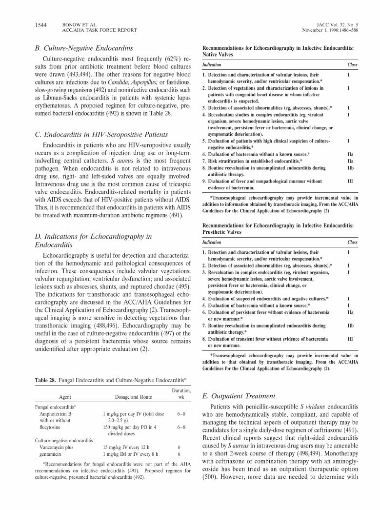
B. Culture-Negative EndocarditisCulture-negative endocarditis most frequently (62%) re-
sults from prior antibiotic treatment before blood cultureswere drawn (493,494). The other reasons for negative bloodcultures are infections due to Candida; Aspergillus; or fastidious,slow-growing organisms (492) and noninfective endocarditis suchas Libman-Sacks endocarditis in patients with systemic lupuserythematosus. A proposed regimen for culture-negative, pre-sumed bacterial endocarditis (492) is shown in Table 28.
C. Endocarditis in HIV-Seropositive PatientsEndocarditis in patients who are HIV-seropositive usually
occurs as a complication of injection drug use or long-termindwelling central catheters. S aureus is the most frequentpathogen. When endocarditis is not related to intravenousdrug use, right- and left-sided valves are equally involved.Intravenous drug use is the most common cause of tricuspidvalve endocarditis. Endocarditis-related mortality in patientswith AIDS exceeds that of HIV-positive patients without AIDS.Thus, it is recommended that endocarditis in patients with AIDSbe treated with maximum-duration antibiotic regimens (491).
D. Indications for Echocardiography inEndocarditis
Echocardiography is useful for detection and characteriza-tion of the hemodynamic and pathological consequences ofinfection. These consequences include valvular vegetations;valvular regurgitation; ventricular dysfunction; and associatedlesions such as abscesses, shunts, and ruptured chordae (495).The indications for transthoracic and transesophageal echo-cardiography are discussed in the ACC/AHA Guidelines forthe Clinical Application of Echocardiography (2). Transesoph-ageal imaging is more sensitive in detecting vegetations thantransthoracic imaging (488,496). Echocardiography may beuseful in the case of culture-negative endocarditis (497) or thediagnosis of a persistent bacteremia whose source remainsunidentified after appropriate evaluation (2).
Recommendations for Echocardiography in Infective Endocarditis:Native Valves
Indication Class
1. Detection and characterization of valvular lesions, theirhemodynamic severity, and/or ventricular compensation.*
I
2. Detection of vegetations and characterization of lesions inpatients with congenital heart disease in whom infectiveendocarditis is suspected.
I
3. Detection of associated abnormalities (eg, abscesses, shunts).* I4. Reevaluation studies in complex endocarditis (eg, virulent
organism, severe hemodynamic lesion, aortic valveinvolvement, persistent fever or bacteremia, clinical change, orsymptomatic deterioration).
I
5. Evaluation of patients with high clinical suspicion of culture-negative endocarditis.*
I
6. Evaluation of bacteremia without a known source.* IIa7. Risk stratification in established endocarditis.* IIa8. Routine reevaluation in uncomplicated endocarditis during
antibiotic therapy.IIb
9. Evaluation of fever and nonpathological murmur withoutevidence of bacteremia.
III
*Transesophageal echocardiography may provide incremental value inaddition to information obtained by transthoracic imaging. From the ACC/AHAGuidelines for the Clinical Application of Echocardiography (2).
Recommendations for Echocardiography in Infective Endocarditis:Prosthetic Valves
Indication Class
1. Detection and characterization of valvular lesions, theirhemodynamic severity, and/or ventricular compensation.*
I
2. Detection of associated abnormalities (eg, abscesses, shunts).* I3. Reevaluation in complex endocarditis (eg, virulent organism,
severe hemodynamic lesion, aortic valve involvement,persistent fever or bacteremia, clinical change, orsymptomatic deterioration).
I
4. Evaluation of suspected endocarditis and negative cultures.* I5. Evaluation of bacteremia without a known source.* I6. Evaluation of persistent fever without evidence of bacteremia
or new murmur.*IIa
7. Routine reevaluation in uncomplicated endocarditis duringantibiotic therapy.*
IIb
8. Evaluation of transient fever without evidence of bacteremiaor new murmur.
III
*Transesophageal echocardiography may provide incremental value inaddition to that obtained by transthoracic imaging. From the ACC/AHAGuidelines for the Clinical Application of Echocardiography (2).
E. Outpatient TreatmentPatients with penicillin-susceptible S viridans endocarditis
who are hemodynamically stable, compliant, and capable ofmanaging the technical aspects of outpatient therapy may becandidates for a single daily-dose regimen of ceftriaxone (491).Recent clinical reports suggest that right-sided endocarditiscaused by S aureus in intravenous drug users may be amenableto a short 2-week course of therapy (498,499). Monotherapywith ceftriaxone or combination therapy with an aminogly-coside has been tried as an outpatient therapeutic option(500). However, more data are needed to determine with
Table 28. Fungal Endocarditis and Culture-Negative Endocarditis*
Agent Dosage and RouteDuration,
wk
Fungal endocarditis*Amphotericin Bwith or without
1 mg/kg per day IV (total dose2.0–2.5 g)
6–8
flucytosine 150 mg/kg per day PO in 4divided doses
6–8
Culture-negative endocarditis†Vancomycin plus 15 mg/kg IV every 12 h 6gentamicin 1 mg/kg IM or IV every 8 h 6
*Recommendations for fungal endocarditis were not part of the AHArecommendations on infective endocarditis (491). †Proposed regimen forculture-negative, presumed bacterial endocarditis (492).
1544 BONOW ET AL. JACC Vol. 32, No. 5ACC/AHA TASK FORCE REPORT November 1, 1998:1486–588

more certainty whether such outpatient regimens havetherapeutic effectiveness equivalent to the established4-week regimens.
F. Indications for Surgery in Patients With ActiveInfective Endocarditis
Surgery is indicated in patients with life-threatening con-gestive heart failure or cardiogenic shock due to surgicallytreatable valvular heart disease with or without proven infec-tive endocarditis if the patient has reasonable prospects ofrecovery with satisfactory quality of life after the operation(442,501–525). Surgery should not be delayed in the setting ofacute infective endocarditis when congestive heart failureintervenes. Surgery is not indicated if complications (severeembolic cerebral damage) or comorbid conditions make theprospect of recovery remote.
The indications for surgery for infective endocarditis inpatients with stable hemodynamics are less clear. Early con-sultation with a cardiovascular surgeon is recommended assoon as the diagnosis of aortic or mitral valve endocarditis ismade so that the surgical team is aware of the patient who maysuddenly need surgery. Surgery is recommended in patientswith annular or aortic abscesses, those with infections resistantto antibiotic therapy, and those with fungal endocarditis. It isrecognized that the presence of valvular vegetations poses athreat of embolic events. Echocardiography, especially withtransesophageal imaging, identifies vegetations and pro-vides size estimation in many instances. Patients with avegetation diameter .10 mm have a significantly higherincidence of embolization than those with a vegetationdiameter ,210 mm (488), and this risk appears to be higherin patients with mitral valve endocarditis than aortic valveendocarditis. However, operation on the basis of vegetationsize alone is controversial.
Patients with prosthetic valves receiving warfarin anticoag-ulation who develop endocarditis should have their warfarindiscontinued and replaced with heparin. This recommendationis less related to the possibility of hemorrhagic complicationsof endocarditis (526) than the possibility of urgent surgery.If surgery is required, the effects of warfarin will havedissipated, and heparin can easily be reversed. Likewise,aspirin, if part of the medical regimen, should also bediscontinued. If neurological symptoms develop, anticoagu-lation should be discontinued until an intracranial hemor-rhagic event is excluded by magnetic resonance imaging orcomputed tomographic scanning.
Recommendations for Surgery for Native Valve Endocarditis*
Indication Class
1. Acute AR or MR with heart failure. I2. Acute AR with tachycardia and early closure of the mitral valve. I3. Fungal endocarditis. I4. Evidence of annular or aortic abscess, sinus or aortic true or false
aneurysm.I
5. Evidence of valve dysfunction and persistent infection after aprolonged period (7 to 10 days) of appropriate antibiotic therapy,as indicated by presence of fever, leukocytosis, and bacteremia,provided there are no noncardiac causes for infection.
I
6. Recurrent emboli after appropriate antibiotic therapy. IIa7. Infection with gram-negative organisms or organisms with a poor
response to antibiotics in patients with evidence of valvedysfunction.
IIa
8. Mobile vegetations >10 mm. IIb9. Early infections of the mitral valve that can likely be repaired. III
10. Persistent pyrexia and leukocytosis with negative blood cultures. III
*Criteria also apply to repaired mitral and aortic allograft or autograftvalves. Endocarditis defined by clinical criteria with or without laboratoryverification; there must be evidence that function of a cardiac valve is impaired.
Recommendations for Surgery for Prosthetic Valve Endocarditis*
Indication Class
1. Early prosthetic valve endocarditis (first 2 months or less aftersurgery).
I
2. Heart failure with prosthetic valve dysfunction. I3. Fungal endocarditis. I4. Staphylococcal endocarditis not responding to antibiotic therapy. I5. Evidence of paravalvular leak, annular or aortic abscess, sinus or
aortic true or false aneurysm, fistula formation, or new-onsetconduction disturbances.
I
6. Infection with gram-negative organisms or organisms with a poorresponse to antibiotics.
I
7. Persistent bacteremia after a prolonged course (7 to 10 days) ofappropriate antibiotic therapy without noncardiac causes forbacteremia.
IIa
8. Recurrent peripheral embolus despite therapy. IIa9. Vegetation of any size on or near the prosthesis. IIb
*Criteria exclude repaired mitral valves or aortic allograft or autograftvalves. Endocarditis is defined by clinical criteria with or without laboratoryverification.
V. Management of Valvular Disease inPregnancy
A. Physiological Changes of PregnancyThe evaluation and management of valvular heart disease
in the pregnant patient requires an understanding of thenormal physiological changes associated with gestation, labor,delivery, and the early postpartum period. On average, there isa 50% increase in circulating blood volume during pregnancythat is accompanied by a commensurate increase in cardiacoutput that usually peaks between the midportion of thesecond and third trimesters. The augmented cardiac outputderives from an increase in the stroke volume, although thereis also a smaller increase in heart rate, averaging 10 to 20 beatsper minute. Because of the effects of uterine circulation andendogenous hormones, systemic vascular resistance falls with adisproportionately greater lowering of diastolic blood pressureand a wide pulse pressure. Inferior vena caval obstruction froma gravid uterus in the supine position can result in an abruptdecrease in cardiac preload, leading to hypotension withweakness and lightheadedness. These symptoms resolvequickly with a change in position (527).
1545JACC Vol. 32, No. 5 BONOW ET AL.November 1, 1998:1486–588 ACC/AHA TASK FORCE REPORT

There is a further abrupt increase in cardiac output duringlabor and delivery related in part to the associated anxiety andpain. Uterine contractions can lead to marked increases inboth systolic and diastolic blood pressure. After delivery, thereis an initial surge in preload related to the autotransfusion ofuterine blood into the systemic circulation and to caval decom-pression (527).
Pregnancy is also associated with a hypercoagulable stateowing to relative decreases in protein S activity, stasis, andvenous hypertension (528). Estrogens may interfere with col-lagen deposition within the media of the medium- and large-sized muscular arteries. Circulating elastase can break up theelastic lamellae and weaken the aortic media during preg-nancy. Weakening of the vascular wall may in turn predisposeto dissection with or without an underlying connective tissuedisorder.
B. Physical ExaminationThe physical examination of the normal parturient is nota-
ble for a slightly fast resting heart rate, bounding pulses(although not truly water-hammer), a widened pulse pressurewith a low normal peak systolic pressure, and warm extremi-ties. Venous pressure is usually elevated above the normalrange for nonpregnant women but rarely in a clearly abnormalrange. The thyroid gland may be enlarged in the absence ofclinical hyperthyroidism. Depending on the stage of pregnancy,the lung volumes may be low because of the raised diaphragms.The precordial impulse is hyperkinetic, and the first heartsound may be louder than normal, with prominent splitting.The second heart sound is usually physiologically split but mayalso widen and appear fixed during the later stages of preg-nancy. Third heart sounds are common. A soft grade 1 to 2midsystolic murmur that is best heard along the mid to upperleft sternal edge is a frequent finding (24). A continuousmurmur, reflecting either a venous hum or a mammary souffle,may sometimes be heard during careful auscultation. Thecervical venous hum is best appreciated in the right supracla-vicular fossa and can be obliterated by movement of the chintoward the stethoscope or digital pressure over the ipsilateraljugular vein. The mammary souffle is a systolic or continuoussound over the engorged breast that can usually be obliteratedwith firm pressure applied to the diaphragm of the stetho-
scope. It is heard in the supine position and attenuates ordisappears when standing. It is appreciated in the late stages ofpregnancy or early in the puerperium. Diastolic heart murmursare unusual. The increased blood volume and enhanced car-diac output associated with normal pregnancy can accentuatethe murmurs associated with stenotic heart valve lesions (eg,MS, AS). On the other hand, murmurs of AR or MR mayactually attenuate in the face of lowered systemic vascularresistance (529).
C. EchocardiographyNormal pregnancy is accompanied by echocardiographic
evidence of mild ventricular chamber enlargement. Pulmonicand tricuspid valvular regurgitation, as assessed by Dopplerinterrogation, is the rule rather than the exception (530). Alarge minority of women will demonstrate Doppler evidence of“physiological” MR in the absence of structural valve disease.Atrioventricular valve incompetence may derive from theannular dilatation that accompanies ventricular enlargement.Appreciation of these echocardiographic and Doppler findingsin normal individuals is an important foundation for thenoninvasive evaluation of subjects with suspected valvulardisease. The use of ultrasound during pregnancy poses no riskto the mother or fetus.
D. General Management GuidelinesClinical experience has shown that there are several cardiac
conditions in which the physiological changes of pregnancy arepoorly tolerated. Most experts would agree that, for someconditions, such as cyanotic heart disease, Eisenmenger syn-drome, or severe pulmonary hypertension, pregnancy shouldbe discouraged. Valvular heart lesions associated with highmaternal and fetal risk during pregnancy are listed in Table 29.Lesions associated with low risk during pregnancy are listed inTable 30.
Individual counseling usually requires a multidisciplinaryapproach and should include information regarding contracep-tion, maternal and fetal risks of pregnancy, and expectedlong-term outcomes. However, many patients with valvularheart disease can be successfully managed throughout preg-nancy and during labor and delivery with conservative medical
Table 29. Valvular Heart Lesions Associated With High Maternaland/or Fetal Risk During Pregnancy
1. Severe AS with or without symptoms2. AR with NYHA functional Class III–IV symptoms3. MS with NYHA functional Class II–IV symptoms4. MR with NYHA functional Class III–IV symptoms5. Aortic and/or mitral valve disease resulting in severe pulmonary
hypertension (pulmonary pressure .75% of systemic pressures)6. Aortic and/or mitral valve disease with severe LV dysfunction (EF ,0.40)7. Mechanical prosthetic valve requiring anticoagulation8. AR in Marfan syndrome
Abbreviations: EF 5 ejection fraction.
Table 30. Valvular Heart Lesions Associated With Low Maternaland Fetal Risk During Pregnancy
1. Asymptomatic AS with low mean gradient (,50 mm Hg) in presence ofnormal LV systolic function (EF .0.50)
2. NYHA functional Class I or II AR with normal LV systolic function3. NYHA functional Class I or II MR with normal LV systolic function4. MVP with no MR or with mild to moderate MR and with normal LV
systolic function5. Mild to moderate MS (MVA .1.5 cm2, gradient ,5 mm Hg) without
severe pulmonary hypertension6. Mild to moderate pulmonary valve stenosis
Abbreviations: EF 5 ejection fraction, MVA 5 mitral valve area.
1546 BONOW ET AL. JACC Vol. 32, No. 5ACC/AHA TASK FORCE REPORT November 1, 1998:1486–588

measures designed to optimize intravascular volume and sys-temic loading conditions.
Simple interventions such as bed rest and avoidance of thesupine position should not be overlooked. Whenever possible,symptomatic or severe valvular lesions should be addressedand rectified before conception and pregnancy. Contempora-neous management with a dedicated obstetrical team accus-tomed to working with high-risk patients is encouraged. Drugsshould generally be avoided whenever possible (Table 31).
E. Specific Lesions1. Mitral Stenosis. Young pregnant women with a previous
history of acute rheumatic fever and carditis should continue toreceive penicillin prophylaxis as indicated in the nonpregnantstate. The most common rheumatic lesion in this age groupremains MS. Patients with mild to moderate MS can almostalways be managed with judicious use of diuretics andb-blockade. Diuretics are given to relieve pulmonary andexcess systemic venous congestion, but care must be taken toavoid vigorous volume depletion to protect against uretero-placental hypoperfusion. b-blockers are chiefly indicated totreat or prevent tachycardia to optimize diastolic filling. Al-though the nonselective b-blocker propranolol has been in usefor decades, some authorities recommend a cardioselectiveb-blocker such as metoprolol or atenolol to prevent thepotential deleterious effects of epinephrine blockade on myo-metrial activity.
Patients with severe MS who are symptomatic beforeconception will not predictably tolerate the hemodynamicburden of pregnancy and should be considered for percutane-ous balloon mitral valvotomy before conception, provided thevalve is anatomically suitable. Patients with severe MS whodevelop NYHA functional Class III-IV symptoms during preg-nancy should undergo percutaneous balloon valvotomy.
For rare patients with MS who fail medical managementduring pregnancy with repetitive or persistent heart failure,there is now a nearly 10-year experience with balloon mitralvalvotomy, either with very limited fluoroscopy (less than 1 to2 minutes exposure with both pelvic and abdominal shielding)or echocardiographic guidance. The reported results withmitral balloon valvotomy have been excellent, with few mater-nal and/or fetal complications, although caution is advised ininterpreting outcomes from individual centers reporting rela-tively few patients (531–540). Percutaneous mitral balloonvalvotomy should only be performed in experienced centersand only after aggressive medical measures have been ex-hausted. In developing countries, there is a long history ofsuccessful surgical closed commissurotomy for pregnantwomen (541).
2. Mitral Regurgitation. MVP is the most common causeof MR in pregnant women. The physical findings pertinent toMVP may be obscured or varied by the physiological changesof pregnancy, especially the increased blood volume andreduced systemic vascular resistance. Associated MR canusually be managed medically, although on rare occasions,
Table 31. Effects of Cardiovascular Drugs Taken During Pregnancy
Drug Potential Fetal Adverse Effects Safety
Warfarin Crosses placental barrier, fetal hemorrhage in utero, embryopathy, central nervous system abnormalities UnsafeHeparin None reported Probably safeDigoxin Low birth weight SafeQuinidine Toxic dose may induce premature labor and cause damage to fetal eighth cranial nerve SafeProcainamide None reported SafeDisopyramide May initiate uterine contractions *Lidocaine High blood levels and fetal acidosis may cause central nervous system depression SafeMexiletine Fetal bradycardia, IUGR, low Apgar score, neonatal hypoglycemia, neonatal bradycardia, and neonatal
hyperthyroidism*
Flecainide 1 reported fetal death *Propafenone None reported *Adenosine None reported. Use during first trimester limited to a few patients. SafeAmiodarone IUGR, prematurity, hypothyroidism UnsafeCalcium channel blocking agents Fetal distress due to maternal hypotension *b-adrenergic blocking agents IUGR, apnea at birth, bradycardia, hypoglycemia, hyperbilirubinemia; b2-blockade blocking agents may
initiate uterine contractionsSafe
Hydralazine None reported SafeSodium nitroprusside Potential thiocyanate toxicity with high dose, fetal mortality with nitroprusside in animal studies Potentially unsafeOrganic nitrates Fetal heart rate deceleration and bradycardia *ACE inhibitors Skull ossification defect, IUGR, premature deliveries, low birth weight, oligohydramnios, neonatal renal
failure, anemia and death, limb contractures, patent ductus arteriosusUnsafe
Diuretic agents Impairment of uterine blood flow and danger of placental hypoperfusion, thrombocytopenia, jaundicehyponatremia, bradycardia
Potentially unsafe
Abbreviation: IUGR 5 intrauterine growth retardation. *To date, only limited information is available, and safety during pregnancy cannot be established. Adaptedfrom Elkayam U (527). Pregnancy and cardiovascular disease. In: Braunwald E, ed. Heart Disease. A Textbook of Cardiovascular Medicine. 5th ed. Philadelphia, Pa:WB Saunders; 1997:1857, with permission. The guidelines committee added warfarin, heparin, and hydralazine to this list.
1547JACC Vol. 32, No. 5 BONOW ET AL.November 1, 1998:1486–588 ACC/AHA TASK FORCE REPORT

mitral valve surgery is required because of ruptured chordaeand acute, severe worsening of the regurgitant lesion. Medicalmanagement includes diuretics for the rare patient with pul-monary congestion. Vasodilator therapy is indicated only inthe presence of concomitant systemic hypertension and shouldnot be advised in the setting of normal or low systemic bloodpressure. ACE inhibitors are considered unsafe because oftheir multiple adverse effects on fetal development. There iswide experience with hydralazine, an agent generally consid-ered safe. When mitral valve surgery is required, repair isalways preferred, as would be the case for any young patientbut especially in relation to the desirability of avoiding thepotential need for anticoagulation.
3. Aortic Stenosis. The most common cause of AS inpregnant women is congenital bicuspid disease. Patients withmild to moderate obstruction and normal LV systolic functioncan usually be managed conservatively through the entirepregnancy. Patients with more severe obstruction (pressuregradient .50 mm Hg) or symptoms should be advised to delayconception until relief of AS can be obtained. For those rarewomen with severe AS whose disease is first appreciatedduring pregnancy, consideration may have to be given to eitherpercutaneous aortic balloon valvotomy (542,543) or surgery(depending on the anatomic findings) before labor anddelivery if heart failure has developed or syncope hasoccurred. These procedures are fraught with danger to boththe mother and fetus, although successful outcomes havebeen reported. Neither is to be undertaken without cautionand forewarning. There is an association between the pres-ence of a bicuspid aortic valve and cystic medial necrosis,which may predispose to spontaneous aortic dissection,usually in the third trimester.
4. Aortic Regurgitation. Isolated AR, like MR, can usuallybe managed medically with a combination of diuretics and, ifnecessary, vasodilator therapy (544). Women with symptomsand/or signs of LV failure should be carefully monitoredthroughout labor and delivery with strict attention to volumestatus and blood pressure. As is true for MR, surgery duringpregnancy should be contemplated only for control of refrac-tory Class III or IV symptoms. Consideration regarding LVsize or systolic function in less symptomatic patients should notapply.
5. Pulmonary Valve Stenosis. Pulmonary valve stenosismay exist in isolation but frequently accompanies other con-genital heart lesions. In general, patients with cyanotic congen-ital heart disease tolerate the stresses of pregnancy far less wellthan those with acyanotic lesions. Isolated pulmonic stenosis israrely a significant impediment to a successful pregnancy. Thislesion can be approached with percutaneous valvotomy underechocardiographic guidance when necessary.
6. Tricuspid Valve Disease. Tricuspid valve disease may becongenital (Ebstein’s anomaly, tricuspid atresia) or acquired(endocarditis, myxomatous replacement/proliferation, carci-noid). The approach to the patient with tricuspid valve involve-ment as part of a more complex congenital heart diseasesyndrome is predicated on the features of the associated
lesions. Isolated TR should not pose a significant problem,although greater care may be necessary to protect againstdiuretic-induced hypoperfusion.
7. Marfan Syndrome. The Marfan syndrome is an inherit-able disorder of connective tissue that stems from abnormali-ties in the fibrillin gene on chromosome 15. It is transmitted inan autosomal dominant fashion and is recognized clinically byits ocular, skeletal, and cardiovascular expressions. Spontane-ous aortic dissection and/or rupture are the most fearedcardiovascular complications associated with pregnancy(545,546). Dissection can occur at any point along the aorta butmost commonly originates in the ascending portion. Enlarge-ment of the aortic root to .4.0 cm identifies a particularlyhigh-risk group, although a normal dimension is by no meansa guarantee against this catastrophic complication. Aortic rootenlargement may or may not be accompanied by regurgitationand an audible heart murmur. MVP with regurgitation is alsofrequently detected.
Any woman with Marfan syndrome who is contemplatingpregnancy should have a screening transthoracic echocardio-gram with careful assessment of aortic root dimensions. En-largement .25.0 cm is considered an indication for electiverepair before conception, usually with a composite valve-graftconduit and reimplantation of the coronary arteries. If aorticroot enlargement (.4.0 cm) is first detected during pregnancy,some authorities recommend termination with prompt aorticrepair, especially if serial echocardiographic studies demon-strate progressive dilatation over time. Dissection and ruptureare most likely to occur during the third trimester or near thetime of delivery. Special care must be taken to provideadequate analgesia to prevent wide surges in blood pressureand its rate of rise (dP/dt) during labor and delivery. Obstet-rical techniques to shorten the second stage of labor areappropriate. General anesthesia and caesarean section mayallow more optimal hemodynamic control. The use of prophy-lactic b-blockade throughout the pregnancy is strongly recom-mended. Such treatment has been shown to slow the rate ofaortic dilatation and reduce the cumulative incidence of car-diovascular complications in nonpregnant adolescents andadults (547). Finally, it should be pointed out that patients withMarfan syndrome and no identifiable cardiovascular abnor-malities on examination or echocardiographic study can besafely shepherded through pregnancy and a normal vaginaldelivery.
F. Endocarditis ProphylaxisThe Committee on Rheumatic Fever, Endocarditis, and
Kawasaki Disease of the American Heart Association does notrecommend routine antibiotic prophylaxis in patients withvalvular heart disease undergoing uncomplicated vaginal de-livery or caesarean section unless infection is suspected. Anti-biotics are optional for high-risk patients with prosthetic heartvalves, a previous history of endocarditis, complex congenitalheart disease, or a surgically constructed systemic-pulmonaryconduit (36). Many practitioners routinely provide antibiotics.
1548 BONOW ET AL. JACC Vol. 32, No. 5ACC/AHA TASK FORCE REPORT November 1, 1998:1486–588

G. Cardiac Valve SurgeryThe performance of cardiac valve surgery is a difficult and
complex undertaking in the pregnant patient. Even under idealconditions, including the use of cardiopulmonary bypass tech-niques that promote high flow rates and warm perfusiontemperatures, there is a high incidence of fetal distress, growthretardation, or wastage (548–550). If possible, it is alwayspreferable to delay surgery until the time the fetus is viableand a caesarean section can be performed as part of aconcomitant procedure (551,552). Surgery should be pur-sued only in the setting of medically refractory symptoms(pulmonary congestion), especially if a low output syndromeintervenes.
For suitable valve lesions, repair is always preferred overreplacement. If valve replacement is necessary, the choice of aheart valve substitute can be problematic. Bioprosthetic valvesdegenerate more quickly in younger patients, a process thatcan be further accelerated during pregnancy (553). Althoughsuch valves may not require longer-term anticoagulation, theydo expose the young patient to an earlier risk of failure andneed for reoperation. Mechanical valve substitutes are moredurable, but the obligate need for anticoagulation may com-plicate current and future pregnancies. For aortic valve dis-ease, homograft valves or pulmonary autografts should beconsidered (554).
H. AnticoagulationPregnant patients with mechanical valve prostheses have an
obligate need for anticoagulation. Unfortunately, there remainsignificant problems and an impressive risk to both the motherand fetus of either hemorrhage or thrombosis with the use ofeither warfarin or heparin. For example, maternal thrombo-embolic phenomena may complicate from 4% to 14% ofpregnancies in women with mechanical prostheses despiteadequate anticoagulation as measured by the serum INR oraPTT (555,556).
Guidelines for the management of the pregnant patientwith a mechanical prosthesis have been difficult to formulatedue to the lack of adequate prospective randomized controlledtrial data. Practice patterns vary widely and no consensus exists, asmight be expected from the disparate results and claims of theretrospective and selective case series that have been reported todate. Appropriately designed trials are needed.
1. Warfarin. Warfarin crosses the placenta and has beenassociated with an increased incidence of spontaneous abor-tion, prematurity, and stillbirth. The manufacturer considersits use during pregnancy to be strictly contraindicated by virtueof its association with fetal deformity (embryopathy) andcentral nervous system abnormalities. The true incidence ofwarfarin embryopathy has been difficult to ascertain from thereported case series and has ranged from ,5% to 67%(556,557). An estimate of 4% to 10% seems reasonable,based on recent reports (558,559). Its risk may be doserelated and appears to be highest if exposure occurs duringthe 6th to 12th week of gestation. Fetal cerebral hemorrhage
can complicate labor and delivery, especially if forcepsevacuation is necessary.
2. Heparin. Heparin does not cross the placenta and isgenerally considered safer (558,560). Its longer-term use,however, is complicated by sterile abscesses, osteoporosis (albeitwith a small risk of fracture), thrombocytopenia, and bleeding.Although heparin was previously considered the preferred anti-thrombotic agent, numerous case series and patient registriesattest to an unacceptable incidence of thromboembolic compli-cations, including fatal valve thrombosis, in high-risk pregnantwomen managed with subcutaneous heparin (12% to 24%)(558,561–564). Criticisms have been levied against thesestudies because of the inclusion of a predominant popula-tion of women with older-generation and more thrombo-genic prostheses, inadequate heparin dosing, and/or the lackof meticulous monitoring strategies. Unfortunately, theefficacy of adjusted-dose subcutaneous heparin has not beendefinitively established.
The choice of anticoagulant must be carefully consideredand should derive from a consideration of maternal prefer-ences regarding the competing risks to the mother and to thefetus. For many women, a 4% to 10% risk of warfarinembryopathy is unacceptable. These women may be unwill-ing to take warfarin at any time during the first trimester orthroughout the entire pregnancy. Implicit in their choice ofheparin, however, is the acceptance of increased risk of maternalhemorrhage or prosthetic valve thrombosis. Certainly, a fulldiscussion of these issues is indicated before conception. Thehazards of anticoagulation are considered by many to be soodious as to argue that mechanical prosthetic valves are relativelycontraindicated in women of child-bearing potential.
Recommendations for Anticoagulation During Pregnancy in PatientsWith Mechanical Prosthetic Valves: Weeks 1 Through 35
Indication Class
1. The decision whether to use heparin during the first trimesteror to continue oral anticoagulation throughout pregnancyshould be made after full discussion with the patient and herpartner; if she chooses to change to heparin for the firsttrimester, she should be made aware that heparin is less safefor her, with a higher risk of both thrombosis and bleeding,and that any risk to the mother also jeopardizes the baby.*
I
2. High-risk women (a history of thromboembolism or an older-generation mechanical prosthesis in the mitral position) whochoose not to take warfarin during the first trimester shouldreceive continuous unfractionated heparin intravenously in adose to prolong the midinterval (6 hours after dosing) aPTT to 2to 3 times control. Transition to warfarin can occur thereafter.
I
3. In patients receiving warfarin, INR should be maintainedbetween 2.0 and 3.0 with the lowest possible dose of warfarin,and low-dose aspirin should be added.
IIa
4. Women at low risk (no history of thromboembolism, newerlow-profile prosthesis) may be managed with adjusted-dosesubcutaneous heparin (17,500 to 20,000 U BID) to prolong themid-interval (6 hours after dosing) aPTT to 2 to 3 times control.
IIb
*From the European Society of Cardiology Guidelines for Prevention ofThromboembolic Events in Valvular Heart Disease (304).
1549JACC Vol. 32, No. 5 BONOW ET AL.November 1, 1998:1486–588 ACC/AHA TASK FORCE REPORT

Recommendations for Anticoagulation During Pregnancy inPatients With Mechanical Prosthetic Valves: After the 36th Week
Indication Class
1. Warfarin should be stopped no later than week 36 and heparinsubstituted in anticipation of labor.
IIa
2. If labor begins during treatment with warfarin, a caesariansection should be performed.
IIa
3. In the absence of significant bleeding, heparin can be resumed4 to 6 hours after delivery and warfarin begun orally.
IIa
Low-molecular-weight heparins offer several potential ad-vantages over unfractionated heparin, including greater bio-availability, ease of administration, lack of need for laboratorymonitoring, and a lower incidence of thrombocytopenia andosteoporosis. They do not cross the placenta. Although theyhave been used to treat deep venous thrombosis in pregnantpatients, there are no data to guide their use in the manage-ment of patients with mechanical heart valves (563). Therecommendation to add low-dose aspirin to a regimen of warfarinor intravenous or adjusted-dose subcutaneous heparin is based onan extrapolation of the results of a single randomized controlledtrial of antithrombotic therapy in high-risk patients with pros-thetic heart valves (564). Dipyridamole should not be consideredas an alternative antiplatelet agent because of its harmful effectson the fetus. Neither warfarin nor heparin is contraindicated inpostpartum mothers who breast-feed (560).
VI. Management of Valvular Heart Diseasein Adolescents and Young Adults
Although the majority of valvular heart disease in olderadults is acquired, the predominant etiology is congenital inchildren, adolescents, and young adults. It has been estimatedthat the prevalence of congenital heart disease is '440,000 inthe United States (exclusive of bicuspid aortic valves; seebelow) (565). Many patients with congenital heart disease havesome valvular involvement. Frequently, it is part of a morecomplex congenital cardiac anomaly, ie, tricuspid stenosis inchildren with pulmonary atresia and an intact ventricularseptum or AS from aortic valve atresia as part of a hypoplasticleft-heart syndrome. The management of these complex dis-eases with multiple valve involvement is beyond the scope ofthese guidelines. Rather, this section concerns isolated valveinvolvement where it is the primary anatomic abnormality.
In evaluating valvular stenosis in children, the severity ofvalvular obstruction is usually reported as peak-to-peak systolicgradient at cardiac catheterization or maximum instantaneousgradient by Doppler rather than valve area. In the catheteriza-tion laboratory, the variation in body size from the neonate tothe adult, difficulties in measuring cardiac output (especially inyoung children), and the relatively rare patient with lowcardiac output have made peak ventricular–to–peak great-vessel pressure gradients for semilunar valves and mean pres-sure gradients for atrioventricular valves the reference stan-dards. With the development of Doppler echocardiographicassessment of valvular obstruction, most pediatric cardiologists
have continued to rely on gradients calculated from peakvelocity using the formula gradient54V2 for the semilunarvalves and mean gradients for the atrioventricular valves ratherthan on valve area. The peak gradient measured by Dopplervelocity (based on maximum instantaneous velocity) is higherthan the peak-to-peak gradient measured at catheterization. Incontrast to children and adolescents, valve area is used bymany centers in evaluation of the young adult.
Ventricular end-systolic or end-diastolic diameter or vol-umes used in evaluating patients with valvular regurgitationare frequently corrected for the large variations in body sizeamong children, adolescents, and young adults. Chamber size iscorrected for body surface area (m2) or commonly by the numberof standard deviations (Z score) above or below the mean withstandard nomograms that correct for body size (566).
The management of the neonate, infant, and young childdiffers significantly from that of the adolescent and youngadult. This section will deal exclusively with adolescents andyoung adults.
A. Aortic StenosisAlthough most adults with aortic valve stenosis have a
degenerative-calcific process that produces immobilization ofthe valve cusps, adolescents and young adults with isolatedaortic valve stenosis almost always have congenital fusion ofone or more commissures resulting in a bicuspid or unicuspidvalve. Although the prevalence of bicuspid and unicuspidvalves may be as high as 2%, only 1 of 50 children born withthese abnormalities will actually have significant obstruction orregurgitation by adolescence.
Much of what has been written in these guidelines for adultswith acquired AS may be transferred to the adolescent oryoung adult. However, certain important differences must beemphasized. Throughout childhood, the aortic annulus andaortic valve must grow parallel with somatic growth. If growthof either the annulus or valve leaflets lags, increased obstruc-tion may occur. Therefore, the rate of progression duringchildhood and adolescent growth may be different from that inthe adult with acquired heart disease. The report from the jointstudy on the Natural History of Congenital Heart Defects (567)followed 473 patients (before the advent of echocardiography),60% of whom were initially evaluated between 2 and 11 years ofage and 34% between 11 and 21 years of age. One third of thechildren had an increase in the transaortic gradient measured bycardiac catheterization during the 4- to 8-year follow-up period.However, the 54 patients .12 years of age showed very smallincreases. Those with higher initial gradients had a greaterlikelihood of demonstrating an increase in the gradient.
Recently, long-term results of the original cohort have beenreported (568), with a mean follow-up period of 20 years. Only20% of those with initial peak LV–to–peak aortic pressuregradients ,25 mm Hg at initial catheterization had anyintervention. However, in those with an initial peak gradient.50 mm Hg, arrhythmias, sudden death, and other morbidevents (including endocarditis, congestive heart failure, syn-
1550 BONOW ET AL. JACC Vol. 32, No. 5ACC/AHA TASK FORCE REPORT November 1, 1998:1486–588

cope, angina, myocardial infarction, stroke, and pacemakerinsertion) occurred at a rate of '1.2% per year. Suddencardiac death occurred in 25 of the 370 patients followed over'8,000 patient years, an incidence of '0.3% per year. Theseverity of obstruction in those who died could not be deter-mined, and a higher-risk subgroup could not be excluded.
The diagnosis of AS can usually be made clinically, withseverity estimated by ECG and Doppler echocardiographic stud-ies. Diagnostic cardiac catheterization is occasionally required ifthere is a discrepancy among clinical evaluation, ECG, and/orDoppler echocardiographic findings. Exercise testing may beuseful, especially in those interested in athletic participation.
Recommendations for Diagnostic Evaluation of the Adolescent orYoung Adult With Aortic Stenosis*
Indication Class
1. ECG.* I2. Echo-Doppler study.* I3. Graded exercise test.† IIa4. Cardiac catheterization† for evaluation of gradient. IIa5. Chest x-ray.* IIb6. Coronary arteriography in the absence of history suggestive
of concomitant CAD.III
*Yearly if echo-Doppler gradient >36 mm Hg (velocity >23 m/s). Every 2years if echo-Doppler gradient <36 mm Hg (peak velocity <3 m/s). †Ifecho-Doppler gradient >36 mm Hg (velocity >3 m/s) and patient interested inathletic participation or if clinical findings and echo-Doppler are disparate.
Balloon valvotomy for calcific AS in older adults has been atbest very short-term palliation. In contrast, the results ofballoon valvotomy in children and adolescents with obstructiondue to fusion of commissures have been considerably moreefficacious. In a large collaborative registry involving 606patients from 23 institutions, the peak LV–to–peak aorticpressure gradients at catheterization were reduced by a meanof 60% (569). In a single-institution study of 148 patientsdilated at age 1 month to 20 years (570), midterm resultsshowed an 8-year actuarial survival of 95%, with 3 of the 4deaths occurring in infants who were dilated at ,1 year of age.Seventy percent of patients were free from operation and 50%were free from intervention 8 years after dilation, which wassimilar to results reported with surgical valvuloplasty. Long-term follow-up is incomplete because balloon valvotomy wasintroduced in the 1980s.
Although balloon dilation has become standard in childrenand adolescents with AS, it is rarely recommended in olderadults because even short-term palliation is uncommon. Thereare insufficient published data to establish an age cutoff. Untilmore information becomes available, recommendations forballoon valvotomy should be limited to adolescents and youngadults in their early 20s, although some older young adultswithout heavily calcified valves may also benefit.
Because balloon valvotomy has resulted in good long-termpalliation with little morbidity and little or no short- orintermediate-term mortality in children, adolescents, andyoung adults in their early 20s, the indications for intervention
are considerably more liberal than those in older adults inwhom intervention usually involves valve replacement. Al-though data are not yet available, reducing the gradient islikely to reduce the small incidence of sudden unexpecteddeath (usually while exercising) (571) as well as the extent ofinterstitial myocardial fibrosis, which has been observed inchildren and adolescents who died and had evidence ofrepolarization abnormalities on ECG.
Children and young adults with Doppler gradients of 70 to80 mm Hg or more (peak velocity .4.2 m/s), those whodevelop LV repolarization or ischemic changes on the ECG(T-wave inversion or ST depression) at rest or with exercise,and those with symptoms may be considered for cardiaccatheterization and possible balloon dilation. The gradientshould be confirmed hemodynamically before proceeding withdilation, and it is reasonable to perform valvotomy in patientswith catheterization gradients .60 mm Hg. Patients with lesssevere gradients (50 to 70 mm Hg by Doppler echocardiog-raphy) who are interested in participating in vigorousathletics or those contemplating pregnancy are also com-monly referred for balloon dilation. Surgical valvotomy hasnow been replaced in most centers by balloon valvotomy butis a reasonable alternative if skilled interventional cardiol-ogists are not available.
Recommendations for Aortic Balloon Valvotomy in theAdolescent or Young Adult (<221) With Normal Cardiac Output*
Indication Class
1. Symptoms of angina, syncope, and dyspnea on exertion, withcatheterization peak gradient >250 mm Hg.†
I
2. Catheterization peak gradient >60 mm Hg. I3. New-onset ischemic or repolarization changes on ECG at rest
or with exercise (ST depression, T-wave inversion over leftprecordium) with a gradient >50 mm Hg).†
I
4. Catheterization peak gradient >50 mm Hg if patient wants toplay competitive sports or desires to become pregnant.
IIa
5. Catheterization gradient <50 mm Hg without symptoms orECG changes.
III
*Adolescents and young adults almost invariably have normal or increasedcardiac output. If cardiac index <2 L/min/m2, lower gradients should be used.†If gradient <50 mm Hg, other causes of symptoms should be explored.
When balloon aortic valvotomy is ineffective or significantAR is present, valve replacement may be necessary. Becausebioprostheses have reduced durability in the young, mechani-cal valves have been commonly used. The long-term cumula-tive risks of endocarditis, thromboembolism, and bleedingfrom anticoagulation over a 20- to 40-year time frame havebeen problematic. Recently, the approach of replacing the aorticvalve with a pulmonary autograft by means of a pulmonary oraortic homograft to replace the native pulmonary valve, as firstperformed by Ross, has gained acceptance in some centers(115,572–575). Preliminary results indicate low surgical risk, withthe majority of autografts performing well for at least a decade.This approach has the advantage of not requiring anticoagulation,an important issue for active adolescents and younger adults,including women contemplating pregnancy.
1551JACC Vol. 32, No. 5 BONOW ET AL.November 1, 1998:1486–588 ACC/AHA TASK FORCE REPORT

B. Aortic RegurgitationAR is an uncommon isolated congenital lesion, although it
may occasionally be found in adolescents and young adultswith a bicuspid aortic valve, discrete subaortic obstruction, orprolapse of one aortic cusp into a ventricular septal defect. Itis commonly the consequence of attempts to relieve stenosis ofthe valve by either balloon dilation or surgical valvulotomy.The indications for surgery with isolated AR or mixed aorticvalve disease are at present similar to adults, that is,symptoms, LV dysfunction (ejection fraction ,0.50), or veryincreased LV end-diastolic or end-systolic diameter, takinginto account variations in body size. If the durability ofpulmonary autograft and homograft valves in the rightventricular outflow tract is substantiated in long-term stud-ies, the indications for autograft valve replacement are likelyto become more liberal.
Recommendations for Aortic Valve Surgery (Replacement WithMechanical Valve, Homograft, or Pulmonary Autograft) in theAdolescent or Young Adult With Chronic Aortic Regurgitation
Indication Class
1. Onset of symptoms. I2. Asymptomatic patients with LV systolic dysfunction (ejection
fraction <0.50) on serial studies 1 to 3 months apart.I
3. Asymptomatic patients with progressive LV enlargement (end-diastolic dimension >4 SD above normal).
I
4. Moderate AS (gradient >40 mm Hg) (peak-to-peak gradient atcardiac catheterization).
IIb
5. Onset of ischemic or repolarization abnormalities (STdepression, T-wave inversion) over left precordium at rest.
IIb
C. Mitral RegurgitationIsolated congenital MR is an extremely uncommon cardiac
condition. MR can be associated with MVP in adolescents oryoung adults with connective tissue, metabolic, or storagediseases. MR can be seen with acquired inflammatory diseasessuch as rheumatic fever, endocarditis, or Kawasaki disease orwith certain collagen vascular disorders.
The most common cause of MR in children is atrioventric-ular septal defects. This is a defect caused by a deficiency of theatrioventricular septum in the embryonic heart. There may bean isolated ostium primum atrial septal defect; ventricularseptal defect in the inlet (posterior) septum; abnormalities ofthe mitral or tricuspid valve, including clefts; or some combi-nation of the above. In a complete atrioventricular septaldefect, there is a combination of a large primum atrial septaldefect, a large inlet (posterior) ventricular septal defect, and acommon atrioventricular valve that failed to develop intoseparate mitral and tricuspid valves. Repair of the defects inearly childhood, with low mortality and morbidity, is nowpossible. The most common long-term sequela of surgery isMR, which may be mild, moderate, or severe.
The pathophysiology, diagnosis, and medical therapy ofresidual MR in atrioventricular septal defects, rheumatic fever,or MVP are similar to those discussed for the adult with MR
(section III.E.). When associated with congestive heart failureor deteriorating LV systolic function on echocardiography orangiography, surgery should be performed. In children withatrioventricular septal defects, MR can usually be reduced oreliminated with surgery. In the postoperative atrioventricularseptal defect or MR secondary to MVP, rheumatic fever, orinflammatory disease, it is frequently possible to decrease theregurgitation with mitral annuloplasty. Occasionally, MVRwith a mechanical or biological valve is necessary. When valverepair rather than replacement is likely, surgery for severe MRmay be contemplated in the absence of heart failure or LVdysfunction.
Recommendations for Mitral Valve Surgery in the Adolescent orYoung Adult With Congenital Mitral Regurgitation With Severe MR
Indication Class
1. NYHA functional Class III or IV symptoms. I2. Asymptomatic patients with LV systolic dysfunction
(ejection fraction <20.60).I
3. NYHA functional Class II symptoms with preservedLV systolic function if valve repair rather thanreplacement is likely.
IIa
4. Asymptomatic patients with preserved LV systolicfunction in whom valve replacement is highly likely.
IIb
D. Mitral StenosisIn developed countries, virtually all MS in adolescents and
young adults is congenital in origin. In developing areas of theworld, MS is more likely to result from rheumatic fever.Congenital MS is usually classified by the component of themitral apparatus that is abnormal, that is, the leaflets, annulus,chordae, or papillary muscles. Frequently, multiple valve com-ponents are involved, resulting in rolled, thickened leafletmargins; shortened and thickened chordae tendineae; obliter-ation of the interchordal spaces with abnormal chordal inser-tions; papillary muscle hypoplasia; and fusion of the anterolat-eral and posteromedial papillary muscles (576). This lattercondition causes the mitral apparatus to appear like a funnelor a parachute. MS results from the inability of blood to passunobstructed from the left atrium to the left ventricle througha very abnormal mitral apparatus.
Congenital MS may be associated with a wide variety ofother congenital cardiac malformations of the left side of theheart, including coarctation of the aorta.
The clinical, electrocardiographic, and radiologic featuresof congenital MS are similar to acquired MS in adults. Theechocardiogram is beneficial in evaluating the mitral valveapparatus and papillary muscles and may provide consider-able insight into the feasibility of successful valve repair.The information obtained from transthoracic imaging isusually sufficient, but in older children, adolescents, andyoung adults, a transesophageal echocardiogram is some-times necessary.
Medical management is of limited utility in these patients,but it is important to prevent and treat common complica-
1552 BONOW ET AL. JACC Vol. 32, No. 5ACC/AHA TASK FORCE REPORT November 1, 1998:1486–588

tions such as pulmonary infections, endocarditis, and atrialfibrillation. Surgical intervention may be necessary in severecases.
Recommendations for Mitral Valve Surgery in the Adolescent orYoung Adult With Congenital Mitral Stenosis
Indication Class
1. Symptomatic patients (NYHA functional Class III or IV)and mean mitral valve gradient >10 mm Hg on Dopplerechocardiography.
I
2. Mildly symptomatic patients (NYHA functional Class II)and mean mitral valve gradient >10 mm Hg on Dopplerechocardiographic study.
IIa
3. Systolic pulmonary artery pressure 50 to 60 mm Hg with amean mitral valve gradient >210 mm Hg.
IIa
4. New-onset atrial fibrillation or multiple systemic emboliwhile receiving adequate anticoagulation.
IIb
The surgical management of congenital MS has improvedconsiderably with the improved appreciation of the mechanismof mitral valve function and the improved ability to visualizethe valve afforded by transesophageal echocardiography. Inthose with a parachute mitral valve, creation of fenestra-tions among the fused chordae may increase effective orificearea and improve symptoms dramatically. MVR may occa-sionally be necessary but is especially problematic in thosewith a hypoplastic mitral annulus in whom an annulus-enlarging operation may be necessary. Recently, balloondilation of congenital MS has been attempted (577), but itsutility is unproved. This is one of the most difficult anddangerous therapeutic catheterization procedures andshould be undertaken only in centers with operators whohave established experience and skill in this interventionalprocedure.
E. Tricuspid Valve DiseaseAcquired disease of the tricuspid valve is very uncommon in
adolescents and young adults. Other than occasional cases ofTR secondary to trauma, bacterial endocarditis in intravenousdrug abusers, and small ventricular septal defects in children inwhom the jet through the ventricular septum creates endothe-lial damage to the tricuspid valve, virtually all cases of acquiredTR are limited to case reports.
Most cases of tricuspid valve disease are congenital, withEbstein’s anomaly of the tricuspid valve being the most com-mon. In Ebstein’s anomaly, there is inferior displacement ofthe septal and posterior leaflets of the valve into the rightventricle. If there is significant adherence of the leaflets to theright ventricular wall, the normal or relatively normal anteriorleaflet fails to coapt with the abnormal posterior leaflet,creating severe TR. If the valve leaflets are not adherent, thereis redundancy of valve tissue with severe prolapse associatedwith varying degrees of TR.
There is variation in the severity of valve leaflet abnormal-
ities. Some children may have severe TR, especially in theperinatal period, when pulmonary vascular resistance andresulting right ventricular pressures are high. Others have verymild abnormalities that may not be recognized until a chestx-ray obtained for other reasons shows cardiomegaly. Aninteratrial communication, usually in the form of a patentforamen ovale, is present in most cases. If TR elevates rightatrial pressure above left atrial pressure, right-to-left shuntingcan occur, with resulting hypoxemia. One or more accessoryconduction pathways are quite common, with a risk of parox-ysmal atrial tachycardia of '25%.
Patients with Ebstein’s anomaly may be asymptomatic withno cyanosis and no atrial arrhythmias. More commonly, theyare cyanotic due to right-to-left shunting, which is associatedwith exercise intolerance. Right ventricular dysfunction mayeventually lead to right-sided congestive heart failure fre-quently exacerbated by an atrial arrhythmia such as atrialtachycardia, atrial flutter, or atrial fibrillation.
Recommendations for Diagnostic Evaluation* of Ebstein’s Anomalyof the Tricuspid Valve in the Adolescent or Young Adult
Indication Class
1. ECG. I2. Chest x-ray. I3. Echo-Doppler study. I4. Pulse oximetry at rest and/or during exercise. IIa5. Electrophysiological study if documented or suspected atrial
arrhythmia.IIa
*Initial evaluation and every 1 to 3 years, depending upon severity.
The natural history of Ebstein’s anomaly varies. In patientswho present in the perinatal period, the 10-year actuarialsurvival is 61% (578). In a study that included more childrenwho presented after the perinatal period, the probability ofsurvival was 50% at 47 years of age (579). Predictors of pooroutcome were NYHA functional Class III or IV symptoms,cardiothoracic ratio .65%, or atrial fibrillation. However,patients with Ebstein’s anomaly who reach late adolescenceand adulthood often have an excellent outcome (579).
Surgical management of Ebstein’s anomaly remains chal-lenging. For older children, adolescents, and young adults,tricuspid valve repair has been attempted. Reconstruction ofthe valve is occasionally possible, especially when there is amobile anterior leaflet free of tethering to the ventricularseptum. Valvuloplasty may be performed with positioning ofthe displaced leaflet of the tricuspid valve to the normal level,sometimes with placation of the atrialized portion of the rightventricle to reduce its size.
Occasionally, the tricuspid valve is not reparable, and valvereplacement with a bioprosthesis or a mechanical valve may benecessary. When present, atrial communications should beclosed. If an accessory pathway is present, this should bemapped and obliterated either preoperatively in the electro-physiology laboratory or at the time of surgery.
1553JACC Vol. 32, No. 5 BONOW ET AL.November 1, 1998:1486–588 ACC/AHA TASK FORCE REPORT

Recommendations for Surgery in the Adolescent or Young AdultWith Ebstein’s Anomaly With Severe Tricuspid Regurgitation
Indication Class
1. Congestive heart failure. I2. Deteriorating exercise capacity (NYHA functional Class III or IV). I3. Progressive cyanosis with arterial saturation <80% at rest or
with exercise.I
4. Progressive cardiac enlargement with cardiothoracic ratio >60%. IIa5. Systemic emboli despite adequate anticoagulation. IIa6. NYHA functional Class II symptoms with valve probably
reparable.IIa
7. Atrial fibrillation. IIa8. Deteriorating exercise tolerance (NYHA functional Class II). IIa9. Asymptomatic patients with increasing heart size. IIb
10. Asymptomatic patients with stable heart size. III
F. Pulmonic Stenosis1. Pathophysiology. Because the pulmonary valve is the
least likely valve to be affected by acquired heart disease,virtually all cases of pulmonary valve stenosis are congenital inorigin. Most patients with stenosis have a conical or dome-shaped pulmonary valve formed by fusion of the valve leaflets,which project superiorly into the main pulmonary artery.Occasionally, the valve may be thickened and dysplastic, withthe stenosis caused by inability of the valve leaflets to movesufficiently during ventricular systole (580).
Symptoms are unusual in children or adolescents withpulmonary valve stenosis even when severe. Adults withlong-standing severe obstruction may have dyspnea andfatigue secondary to an inability to increase cardiac outputadequately with exercise. Exertional syncope or light-headedness may rarely be seen, but sudden death is veryunusual. Eventually, in the neonate or adult with long-standing untreated severe obstruction, TR and right ventric-ular failure may occur.
At any age, if the foramen ovale is patent, right ventricularcompliance may be reduced sufficiently to elevate right atrialpressure, allowing right-to-left shunting and cyanosis. Thisincreases the risk of paradoxical emboli.
2. Diagnosis. The clinical diagnosis of pulmonary valvestenosis is straightforward, and the severity can usually bedetermined accurately by 2-D and Doppler echocardiography(see below). Diagnostic catheterization is rarely required.
Recommendations for Initial Diagnostic Workup ofPulmonic Stenosis
Indication
Severity of Pulmonic Stenosis
Mild*Class
Moderate-Severe†Class
1. ECG. I I2. Echo-Doppler study (transthoracic). I I3. Chest x-ray. IIa IIa4. Diagnostic cardiac catheterization. III IIb‡
*Right ventricular to pulmonary artery maximum instantaneous gradient<30 mm Hg by Doppler echocardiography. †Right ventricular to pulmonary
artery gradient >230 mm Hg by Doppler echocardiography. ‡If catheterizationgradient >250 mm Hg, balloon valvuloplasty should be performed (see recom-mendations for intervention).
3. Clinical Course. The clinical course of children andyoung adults with pulmonary valve stenosis has been welldescribed. The Natural History of Congenital Heart Defectsstudy (581) in the mid 1960s and early 1970s followed 564patients with valvular pulmonary stenosis with cardiac cathe-terization at 4- and 8-year intervals. On admission to the study,'15% were ,2 years old; 20%, 12 to 21 years old; and theremainder, 2 to 11 years old. At initial cardiac catheterization,they were divided into 4 groups based on severity:,25 mm Hgpeak-to-peak gradient between the right ventricle and thepulmonary artery, trivial; 25 to 49 mm Hg, mild; 50 to79 mm Hg, moderate; and .280 mm Hg, severe.
Of the 261 patients (46% of the total) treated medically,most had trivial, mild, or moderate obstruction. None of thesepatients had cyanosis or congestive heart failure, and only 6%had symptoms. There were no deaths during the study. Thepressure gradients were stable in the majority, with 14% ofpatients manifesting a significant increase and 14% a signifi-cant decrease. Most of the increases were in children ,2 yearsold and/or those with initial gradients .40 mm Hg. Those notin either category had only a 4% chance of an increase in thegradient .20 mm Hg. There was little or no change in theoverall status of the medically treated patients. During theperiod of observation, 304 patients, most with moderate orsevere disease, were treated surgically. Only 1 death occurredamong the 245 patients in this group who underwent surgerybeyond infancy. At postoperative follow-up, the gradient hadbeen reduced to insignificant levels in .90%, with norecurrence of pulmonary stenosis in those followed up to 14years.
In 1993, the second Natural History of Congenital HeartDefects study (582) reported on a 16- to 29-year (mean, 22years) follow-up of the same group of patients. The probabilityof 25-year survival was 96%, not statistically different from thenormal control group. Less than 20% of patients managedmedically during the first Natural History Study subse-quently required a valvotomy, and only 4% of the operatedpatients required a second operation. Most patients,whether managed medically or surgically, had mild obstruc-tion by Doppler echocardiography. For patients who had aninitial transpulmonary gradient ,25 mm Hg in the firstNatural History Study, 96% were free of cardiac operationover a 25-year period.
Infective endocarditis was uncommon. Only 1 case de-veloped in the 592 patients followed a median of 18 years,an incidence of 0.94 per 10,000 patient years. Althoughendocarditis prophylaxis has been recommended for pa-tients with PS, the incidence and severity of infection aresuch that the morbidity from anaphylactic reactions toendocarditis prophylaxis may be as problematic as thedisease itself.
1554 BONOW ET AL. JACC Vol. 32, No. 5ACC/AHA TASK FORCE REPORT November 1, 1998:1486–588

Recommendations for Intervention in the Adolescent or YoungAdult With Pulmonic Stenosis (Balloon Valvotomy or Surgery)
Indication Class
1. Patients with exertional dyspnea, angina, syncope, or presyncope. I2. Asymptomatic patients with normal cardiac output (estimated
clinically or determined by catheterization).a. Right ventricular to pulmonary artery peak gradient
>50 mm HgI
b. Right ventricular to pulmonary artery peak gradient 40 to49 mm Hg
IIa
c. Right ventricular to pulmonary artery peak gradient 30 to39 mm Hg
IIb
d. Right ventricular to pulmonary artery peak gradient<30 mm Hg
III
Surgical relief of severe obstruction by valvotomy with atransventricular (583) or transpulmonary (584) artery ap-proach predates the introduction of cardiopulmonary bypass.A nonsurgical approach with balloon valvotomy was describedin 1982 (585) and by the late 1980s had become the procedureof choice for the typically domed, thickened valve virtuallyeverywhere in the United States, both for children (586) andadults (587,588). Surgery is still required for the dysplasticvalve often seen in Noonan’s syndrome. Although long-termfollow-up of pulmonary balloon valvotomy is not yet available,the early and midterm results (up to 10 years) (589) suggestresults similar to surgical valvotomy, that is, little or norecurrence over a 22- to 30-year period.
In those with severe or long-standing valvular obstruction,infundibular hypertrophy may cause secondary obstruction whenthe pulmonary valve is successfully dilated. This frequently re-gresses over time without treatment. Some have advocated tran-sient pharmacological b-blockade, but there is insufficient infor-mation to determine whether this is effective or necessary.
From the Natural History Study data, it would appear thatcongenital mild pulmonary stenosis is a benign disease thatrarely progresses, that moderate or severe pulmonary stenosiscan be improved with either surgery or balloon valvotomy atvery low risk, and that patients who undergo surgery or balloonvalvotomy have an excellent prognosis and a low rate ofrecurrence. Thus, the goal of the clinician is to ascertain theseverity of the disease, treat those in whom it is severe, andinfrequently follow up those with mild disease (590).
Recommendations for Follow-up Exams in Pulmonic Stenosis
Indication
Severity of Pulmonic Stenosis
Mild* ClassModerate to
Severe† Class
1. ECG. I I2. Echo Doppler. I I3. Chest x-ray. IIb IIa4. Catheterization (for evaluation of
gradient).III III
*<29 mm Hg gradient; testing every 5 to 10 years. †>30 mm Hg gradient;testing every 3 years (consideration should be given to balloon or surgicalvalvuloplasty).
G. Pulmonary RegurgitationPulmonary valve regurgitation is an uncommon congenital
lesion seen occasionally with what has been described asidiopathic dilation of the pulmonary artery. In this condition,the annulus of the pulmonary valve dilates, causing the leafletsto fail to coapt during diastole. Mild pulmonary regurgitationmay be a normal finding on Doppler echocardiography.
Although pulmonary regurgitation is unusual as an isolatedcongenital defect, it is an almost unavoidable result of eithersurgical or balloon valvuloplasty of valvular pulmonic stenosisor surgical repair of tetralogy of Fallot. Among patients withpulmonic stenosis who underwent surgical valvotomy in thefirst Natural History Study, 87% had pulmonary regurgitationby Doppler echocardiography in the second Natural HistoryStudy, although it was audible in only 58%. The echocardio-gram tended to overestimate severity when compared withauscultation, with 20% considered moderate to severe byDoppler but only 6% by auscultation. In those with pulmonaryregurgitation, the right ventricle tended to be larger but rightventricular systolic dysfunction was uncommon; it was presentin only 9%.
Pulmonary regurgitation also commonly occurs after suc-cessful repair of tetralogy of Fallot. Several studies havedocumented that the vast majority of children and young adultsoperated on in the late 1950s and 1960s continue to do well forup to 35 years after surgery (591). However, a small group withlong-standing pulmonary regurgitation has developed a verydilated right ventricle and diminished right ventricular systolicperformance, which can lead to an inadequate ability toaugment cardiac output with exercise and in some casescongestive heart failure. This group has also been shown tohave a significant incidence of ventricular arrhythmias knownto be associated with late sudden death. Increased pulmonaryartery pressure from LV dysfunction or residual peripheralpulmonary artery stenosis will increase the amount of regurgi-tation, and these conditions should be treated when present.Pulmonary valve replacement, usually with a homograft, hasbeen attempted, but follow-up data are too preliminary todevelop recommendations at this time.
VII. Management of Patients With ProstheticHeart Valves
A. Classification of Prosthetic Heart ValvesHeart valve prostheses consist of an orifice, through which
blood flows, and an occluding mechanism that closes andopens the orifice. There are 2 classes of heart valve: mechan-ical prostheses, with rigid, manufactured occluders, and bio-logical or tissue valves, with flexible leaflet occluders of animalor human origin. A list of FDA-approved prosthetic heartvalves is given in Table 32.
1. Mechanical Valves. a. Ball Valves. The first successfulvalve replacement devices used a ball-in-cage design (592,593).Of these, only the Starr-Edwards valve has endured; it hasbeen used more than 200,000 times. The ball is a silicone
1555JACC Vol. 32, No. 5 BONOW ET AL.November 1, 1998:1486–588 ACC/AHA TASK FORCE REPORT

rubber polymer impregnated with barium sulfate for radio-opacity that oscillates in a cage of cobalt chromium alloy.
b. Disk Valves. Tilting disk valves use a circular disk as anoccluder, which is retained by wirelike arms or closed loopsthat project into the orifice. The disks are graphite with acoating of pyrolytic carbon, and the housings are stainless steelor titanium. The first successful low-profile design was thestandard Bjork-Shiley tilting disk valve, introduced in 1969(594). Approximately 360,000 standard valves were implanted.In the United States, Bjork-Shiley models have been discon-tinued but are mentioned because many patients with standardor subsequently modified Bjork-Shiley valves are still alive. TheMedtronic Hall valve has a titanium housing and a carbon-coated disk with a unique central hole. The disk is retained andguided by a guide strut that protrudes through this hole. It hasbeen used clinically since 1977.
c. Bileaflet Valves. Current development of mechanicalvalves is based on the bileaflet design, introduced by St. JudeMedical in 1977 and used .600,000 times. Unlike the free-floating occluders in ball and disk valves, the 2 semicircularleaflets of a bileaflet valve are connected to the orifice housingby a butterfly hinge mechanism. The leaflets swing apart duringopening, creating 3 flow areas, 1 central and 2 peripheral.
2. Biological Valves. There is a wide variety of biologicalvalves. The autograft valve refers to a translocation within thesame individual, eg, of the pulmonary valve into the aorticvalve position. The autologous (or autogenous) tissue valveinvolves fabricating a valve from the patient’s own nonvalvulartissue, eg, pericardium. The homograft (or allograft) valverefers to transplantation from a donor of the same species; eg,a donor’s aortic or pulmonary valve into a recipient’s aortic orpulmonary position. The heterograft (or xenograft) valve is atransplant from another species, either an intact valve, eg, aporcine aortic valve, or a valve fashioned from heterologoustissue, eg, bovine pericardium.
The goal of biological valves is to reduce the complicationsassociated with thromboembolism and the need for anticoag-ulation and in the aortic position to optimize hemodynamics.
a. Autograft Valves. The pulmonary autograft procedureconsists of an autotransplant of the pulmonary valve to theaortic position; the pulmonary valve is then replaced by anaortic or pulmonary homograft or a heterograft. First de-scribed in 1967 (572), this operation is called the Ross proce-dure. Favorable long-term results have been reported(115,573–575). This procedure involves a double valve replace-ment with attendant early and late risks. However, late prob-lems are likely to be related to the pulmonary valve prosthesis,which is easier to remedy but may require a second operation.The Ross procedure does not require FDA approval forclinical use; if a heterograft valve is used in the pulmonaryposition, it needs to be an FDA-approved device.
b. Autologous Pericardial Valves. This new category ofprosthetic valve is an innovative attempt to combine thereproducibility and ease of insertion of a commercial stentedheterograft valve with the benefits of autologous tissue. It is a
frame-mounted autologous pericardial valve assembled from akit in the operating room.
c. Homograft (or Allograft) Valves. Homograft valves werefirst used in the early 1960s (595,596). Three techniques ofhomograft AVR are used: (1) replacing only the valve in thesubcoronary position; (2) complete aortic root replacementwith reimplantation of the coronary arteries; and (3) minirootreplacement with the donor valve and attached aortic wallinserted within the host aorta below the coronary ostia. Thehomograft does not require FDA approval for clinical use.
The homograft valve is considered by many a preferablesubstitute for AVR in younger patients, especially those inwhom anticoagulation is undesirable. It achieves excellenthemodynamics and has low thrombogenicity, and there is noneed for anticoagulation. The drawbacks are a more techni-cally demanding operation and low availability; however, thelatter has been alleviated by commercial availability.
d. Porcine Heterograft (or Xenograft) Valves. Glutaralde-hyde sterilizes valve tissue, renders it bioacceptable by destroy-ing antigenicity, and stabilizes the collagen crosslinks fordurability. The term bioprosthesis is used for a nonviable tissueof biological origin such as the Hancock and Carpentier-Edwards porcine valves (597).
Most porcine valves are mounted on rigid or flexible stentsto which the leaflets and sewing ring are attached. However,
Table 32. FDA-Approved Prosthetic Heart Valves
Type Manufacturer Model
Year ofFirst Clinical
UseImplants*
(thousands)
MechanicalBall Baxter Edwards Starr-Edwards 1965 200Disk Medtronic Medtronic Hall 1977 178
Medical Inc. Omniscience 1978 48Alliance Monostrut 1982 94
Bileaflet St. Jude St. Jude 1977 580Baxter Edwards Duromedics 1982† 20CarboMedics CarboMedics 1986 110
BiologicalPorcine Medtronic Hancock Standard 1970 177
Hancock MO 1978 32Baxter Edwards CE Standard 1971 400
CE SupraAnnular 1982 45St. Jude Toronto Stentless
(TSP)1991 5
Medtronic Free StyleStentless
1992 5
Pericardial Baxter Edwards CE 1982 35Homograft noncommercial‡ 1962 12?
Cryolife‡ 1984 14Autologous noncommercial‡ Pulmonary
autograft1967 2?
Abbreviations: MO 5 modified orifice, CE 5 Carpentier-Edwards. *Ap-proximate number of implants through part or all of 1994. †Discontinued in1988. ‡Does not require FDA approval for clinical use. Adapted from Grunke-meier G, Starr A, Rahimtoola SH (616). Replacement of heart valves. In:O’Rourke RA, ed. The Heart: Update I. New York, NY: McGraw-Hill Publish-ing Co; 1996:98–123.
1556 BONOW ET AL. JACC Vol. 32, No. 5ACC/AHA TASK FORCE REPORT November 1, 1998:1486–588

unstented versions have also been devised by several manufac-turers and have been approved by the FDA for clinical use.Their goal is to achieve some of the potential benefit of ahomograft valve, especially hemodynamics and perhaps dura-bility, with an easily available commercial product. As withhomografts, there are potentially 3 ways of implanting astentless porcine valve (valve only, aortic root, miniroot). Thestandard porcine bioprosthesis is inserted into the annulus.The Carpentier-Edwards SupraAnnular Valve is implantedabove the aortic annulus but sutured to it.
e. Bovine Pericardial Valves. Pericardial valves are tailoredand sewn into a valvular configuration on a stented frame, withbovine pericardium as a fabric. This produces a valve thatopens more completely than a porcine valve for better hemo-dynamics. Greater durability is also expected because there isextra tissue to allow for shrinkage and a higher percentage ofcollagen to be cross-linked during fixation. The Ionescu-Shiley,the first commercially available pericardial valve, experienceda higher failure rate than porcine valves and was taken off themarket after '10 years. However, the failures were partly dueto aspects of the design rather than an intrinsic problem withpericardial tissue itself.
The Carpentier-Edwards Pericardial Bioprosthesis is con-structed without stitches passing through the leaflets that arepresent in the Ionescu-Shiley pericardial valve. Instead, theleaflets are anchored behind the stent pillars.
B. Complications of Prosthetic Heart Valves1. Guidelines for Reporting Clinical Results. The analytic
aspects of reporting clinical results of heart valves have evolvedconsistently since the first successful implants in 1960. Near theend of the first decade, as late (posthospital) experienceaccumulated, the need to analyze time-related events resultedin the introduction of actuarial analysis (598), which had beenused for some time to analyze the results of cancer therapy(599). Later the use of linearized (constant hazard) rates(600,601), Cox regression (602), and multivariable parametricmodels (603) was advocated. However, the effectiveness ofthese refined statistical methods in comparing results fromdifferent series was limited by the lack of standardization indefinitions and follow-up methods.
a. AATS/STS Guidelines for Clinical Reporting. In 1988,standards for defining and reporting complications were pro-posed by the Ad Hoc Liaison Committee for StandardizingDefinitions of Prosthetic Heart Valve Morbidity, a joint com-mittee of the American Association for Thoracic Surgery(AATS) and the Society of Thoracic Surgeons (STS) (604).These guidelines were revised in 1996 (605,606). The compli-cations determined to be of critical importance in the 1996guidelines are summarized as follows:
1. Structural valvular deterioration refers to any change infunction of an operated valve resulting from an intrinsicabnormality causing stenosis or regurgitation.
2. Nonstructural dysfunction is a composite category that in-
cludes any abnormality resulting in stenosis or regurgitationof the operated valve that is not intrinsic to the valve itselfexclusive of thrombosis and infection. This includes inap-propriate sizing, also called valve prosthesis–patient mis-match (607).
3. Valve thrombosis is any thrombus, in the absence of infec-tion, attached to or near an operated valve that occludespart of the blood flow path or interferes with function of thevalve.
4. Embolism is any embolic event that occurs in the absence ofinfection after the immediate perioperative period (whenanesthesia-induced unconsciousness is completely re-versed). This includes any new, temporary or permanent,focal or global neurological deficit and peripheral embolicevent; emboli proven to consist of nonthrombotic materialare excluded.
5. Bleeding event (formerly anticoagulant hemorrhage) is anyepisode of major internal or external bleeding that causesdeath, hospitalization, or permanent injury (eg, vision loss)or requires transfusion. The complication bleeding eventapplies to all patients, whether or not they are takinganticoagulants or antiplatelet drugs.
6. Operated valvular endocarditis is any infection involving anoperated valve. Morbidity associated with active infection,such as valve thrombosis, thrombotic embolus, bleedingevent, or paravalvular leak, is included under this categoryand not in other categories of morbidity.
The consequences of the above morbid events includereoperation; valve-related mortality; sudden unexpected, un-explained death; cardiac death; total deaths; and permanentvalve-related impairment (605,606). In addition, valve prosthe-sis may produce hemolysis due either to the valve itself orassociated perivalvular leak.
2. Valve-Related Complications. There is a wide range inthe reported incidence of complications with the same pros-thetic valve and between different valves (608). This is mostlikely due to variation among series rather than to valve typeand model (609). It has been emphasized (610) that theseinclude factors associated with patients (eg, ventricular func-tion, comorbidities), medical center (eg, surgical variables,definitions of complications, thoroughness of follow-up), anddata analysis (eg, influences of patient-related factors) (609).In addition, published data represent only a small fraction ofvalves implanted (610).
Many types of bias affect reported results (611), whichmight be overcome with randomized trials. However, random-ized trials also have difficulties (612,613). The number ofrandomized studies of prosthetic heart valves is small, and themajority of those that have been reported are of insufficientsize to add importantly to the knowledge already obtainedfrom careful observational studies.
a. Major Randomized Trials. The 2 major randomizedclinical trials that have been reported are the Edinburgh HeartValve Trial (121) and the Veterans Affairs Cooperative Study
1557JACC Vol. 32, No. 5 BONOW ET AL.November 1, 1998:1486–588 ACC/AHA TASK FORCE REPORT

on Valvular Heart Disease (124). Both studies comparedmechanical valves with porcine bioprostheses.
The Edinburgh trial (121) compared the Bjork-Shiley stan-dard valve with porcine valves (initially the Hancock and laterthe Carpentier-Edwards) and reported actuarial comparisonsat 5 and 12 years for 211 aortic and 261 mitral valve patients.Survival was better, but not significantly so, with the mechan-ical valve than the porcine valve (72% versus 52%, P 5 0.08);this was somewhat offset by the increased risk of bleedingevents. However, survival without reoperation was better withmechanical than bioprosthetic valves for MVR (74% versus42%, P 5 0.04) and AVR (75% versus 55%, P 5 0.052).
The VA Study (124) compared the standard Bjork-Shileyvalve with the Hancock porcine valve in 575 men. The Han-cock modified-orifice valve was used for sizes 21 to 23 mm inthe aortic position, and the Hancock standard valve was usedfor all other sizes. Table 33 contains actuarial comparisons ofthe end point variables at 11 years. The risk for all valve-related complications is higher for mechanical valves duringthe first 5 years, but the cumulative risks converge by '10years. At 11 years, the rates of both mortality and all valve-related complications were similar with mechanical and por-cine valves, but the types of complication differed between thetwo; bleeding rates were higher with mechanical valves, andstructural valve deterioration (and reoperation) rates werehigher with bioprostheses.
Comparison of the actuarial event rates between these 2trials (124) showed that bleeding and thromboembolism rateswere higher in the VA study and reoperation rates were higherin the Edinburgh study. These differences could be partiallyaccounted for by the composition of the 2 patient populations:patients in the Edinburgh trial were younger and received lessanticoagulation, and this trial included women, double valvereplacements, and a higher percentage of porcine valves in themitral position.
Late long-term results show better survival in patients withmechanical valves compared with those with bioprostheses(particularly those in younger age groups). In the Edinburgh
trial (121), 12-year survival was 42% versus 24% (P , 0.05) inpatients with mechanical and porcine valves, respectively, inthe mitral position, probably because of the high rate ofbioprosthetic degeneration. In the VA trial (614), 15-yearsurvival was 34% versus 23% (P 5 0.02) in patients withmechanical and porcine valves, respectively, in the aorticposition.
Nonrandomized studies in patients .265 years of age show alower rate of structural valvular deterioration with the pericar-dial valve than with a porcine bioprosthesis (615).
C. Management of Patients With ProstheticHeart Valves
1. Antibiotic Prophylaxis. a. Infective Endocarditis. All pa-tients with prosthetic valves need appropriate antibiotics forprophylaxis against infective endocarditis (section II.B.).
b. Recurrence of Rheumatic Carditis. Patients with rheu-matic heart disease continue to need antibiotics as prophylaxisagainst recurrence of rheumatic carditis (section II.B.).
2. Antithrombotic Therapy. All patients with mechanicalvalves require warfarin therapy, as indicated in Table 34 (617).Even with the use of warfarin, risk of thromboemboli is 1% to2% per year (119,121,124,618), but the risk is considerablyhigher without treatment with warfarin (619). The risk of aclinical thromboembolism is '0.7% per year in patients withbiological valves in sinus rhythm; this figure is derived fromseveral studies in which the majority of patients were not onwarfarin therapy (119,121,124,620). Almost all studies haveshown that risk of embolism is greater with a valve in the mitralposition (mechanical or biological) than with one in the aorticposition (121,608,619,621). With either type of prosthesis orvalve location, the risk of emboli is probably higher in the firstfew days and months after valve insertion (620), before thevalve is fully endothelialized.
a. Mechanical Valves. All patients with mechanical valvesrequire warfarin. For mechanical prostheses in the aorticposition, the INR should be maintained between 2.0 and 3.0
Table 33. Probability of Death Due to Any Cause, Any Valve-Related Complications, and IndividualValve-Related Complications 11 Years After Randomization
Event
Aortic Valve Mitral Valve
Mechanical(n 5 198)
Porcine(n 5 196) P
Mechanical(n 5 88)
Porcine(n 5 93) P
Death from any cause 53 6 4 59 6 4 0.26 64 6 5 67 6 5 0.41Any valve-related complications 62 6 4 64 6 4 0.64 71 6 5 79 6 6 0.34Systemic embolism 16 6 4 15 6 3 0.49 18 6 5 15 6 4 0.61Bleeding 43 6 4 24 6 4 , 0.001 41 6 6 28 6 7 0.02Endocarditis 7 6 2 8 6 2 0.79 11 6 4 17 6 5 0.37Valve thrombosis 2 6 1 1 6 1 0.33 1 6 1 1 6 1 0.95Perivalvular regurgitation 4 6 2 2 6 1 0.28 17 6 5 9 6 6 0.05Reoperation 7 6 2 16 6 4 0.07 21 6 5 47 6 9 0.23Structural valve failure 0 6 0 15 6 4 , 0.001 0 6 0 36 6 8 , 0.001
Values are actuarial percentages 6 standard error. Note: P values are for differences between mechanical and porcinevalves; P , 0.001. Data from Hammermeister et al (124) as summarized in Grunkemeier et al (616) with permission.
1558 BONOW ET AL. JACC Vol. 32, No. 5ACC/AHA TASK FORCE REPORT November 1, 1998:1486–588

for bileaflet valves and Medtronic Hall valves and between 2.5and 3.5 for other disk valves and Starr-Edwards valves; forprostheses in the mitral position, the INR should be main-tained between 2.5 and 3.5 for all mechanical valves(121,124,608,621–630). There was a difference of opinionregarding the Starr-Edwards valve in the aortic position, with aminority opinion to maintain the INR between 2.0 and 3.0. Therecommendation for higher INR values in the mitral positionis based on the greater risk of thromboembolic complicationswith mechanical valves in the mitral position (121,608,619,621,624,625,629,630) and the greater risk of bleeding athigher INRs (625). In patients with aortic mechanicalprostheses who are at higher risk of thromboembolic com-plications, INR should be maintained at 2.5 to 3.5, and theaddition of aspirin should be considered (see below). Theseinclude patients with atrial fibrillation, previous thrombo-embolism, and a hypercoagulable state. Many would alsoinclude patients with severe LV dysfunction in this higher-risk group (631). Some valves are thought to be morethrombogenic than others (particularly the tilting diskvalves), and a case could be made for increasing the INR tobetween 3 and 4.5; however, this is associated with aconsiderably increased risk of bleeding (621,632).
The addition of low-dose aspirin (80 to 100 mg/d) towarfarin therapy (INR 2.0 to 3.5) not only further decreasesthe risk of thromboembolism (564,625,633–637) but also de-creases mortality due to other cardiovascular diseases. A slightincrease in the risk of bleeding with this combination should bekept in mind (635,638). The risk of gastrointestinal irritationand hemorrhage with aspirin is dose dependent over the rangeof 100 to 1000 mg/d, and the antiplatelet effects are indepen-
dent of dose over this range (639,640). There are no data inpatients with prosthetic heart valves receiving warfarin andaspirin in doses of 100 to 325 mg/d. Doses of 500 to 1000 mg/dclearly increase the risk of bleeding (641–643). The addition ofaspirin (80 to 100 mg/d) to warfarin should be stronglyconsidered unless there is a contraindication to the use ofaspirin (ie, bleeding or aspirin intolerance). This combinationis particularly appropriate in patients who have had an embo-lus while on warfarin therapy, those with known vasculardisease, and/or those who are known to be particularly hyper-coagulable. As an example, such combination therapy is rec-ommended by a committee addressing antithrombotic therapyin women during pregnancy (560). Anticoagulation in pregnantpatients is controversial and is discussed in section V.H. ofthese guidelines.
It is important to note that thromboembolic risk is in-creased early after insertion of the prosthetic valve. The useof heparin early after prosthetic valve replacement beforewarfarin achieves therapeutic levels is controversial. Insome patients, achievement of therapeutic INR can bedelayed several days postoperatively because of mitigatingcomplications.
b. Biological Valves. Because of an increased risk of throm-boemboli during the first 3 months after implantation of abiological prosthetic valve, anticoagulation with warfarin isusually recommended (620), although in several centers onlyaspirin is used for biological valves in the aortic position. Therisk is particularly high in the first few days after surgery andheparin should be started as soon as the risk of increasedsurgical bleeding is reduced (usually within 24 to 48 hours),with maintenance of PTT between 55 and 70 seconds. After anoverlap of heparin and warfarin for 3 to 5 days, heparin may bediscontinued when an INR of 2.0 to 3.0 is achieved. After 3months, the tissue valve can be treated like native valvedisease, and warfarin can be discontinued in more than twothirds of patients with biological valves (121,124,620,644). Inthe remaining patients with associated risk factors forthromboembolism, such as atrial fibrillation, previous thrombo-embolism, or hypercoagulable condition, lifelong warfarin ther-apy is indicated to achieve an INR of 2.0 to 3.0. Many would alsorecommend continuing anticoagulation in patients with severeLV dysfunction (ejection fraction ,0.30) (631).
Recommendations for Antithrombotic Therapy in Patients WithProsthetic Heart Valves
Indication Class
1. First 3 months after valvereplacement:
Warfarin, INR 2.5 to3.5
I
2. >23 months after valve replacement:A. Mechanical valve
AVR and no risk factor*Bileaflet valve or Medtronic
Hall valveWarfarin, INR 2 to 3 I
Other disk valves or Starr-Edwards valve
Warfarin, INR 2.5 to3.5
I
AVR 1 risk factor* Warfarin INR 2.5 to 3.5 IMVR Warfarin INR 2.5 to 3.5 I
Table 34. Antithrombotic Therapy: Prosthetic Heart Valves
Warfarin(INR 2–3)
Warfarin(INR 2.5–3.5)
Aspirin(80–100 mg)
Mechanical Prosthetic ValvesA. First 3 months after replacement 1 1B. After first 3 months
1. Aortic valve† 1 12. Aortic valve 1 “risk factor”* 1 13. Mitral valve 1 14. Mitral valve 1 “risk factor”* 1 1
Biological Prosthetic ValvesA. First 3 months after replacement 1 1B. After first 3 months
1. Aortic valve 12. Aortic valve 1 “risk factor”* 1 13. Mitral valve 14. Mitral valve 1 “risk factor”* 1 1
Note: Depending on patient’s clinical status, antithrombotic therapy must beindividualized (see special situations in text). *Risk factors: Atrial fibrillation, LVdysfunction, previous thromboembolism, and hypercoagulable condition. †INRshould be maintained between 2.5 and 3.5 for aortic disk valves and Starr-Edwards valves. Reprinted with permission from McAnulty JH, Rahimtoola SH(617). Antithrombotic therapy in valvular heart disease. In: Schlant R, AlexanderRW, eds. Hurst’s The Heart, Arteries, and Veins. 9th ed. New York, NY:McGraw-Hill Publishing Co; 1998:1867–74.
1559JACC Vol. 32, No. 5 BONOW ET AL.November 1, 1998:1486–588 ACC/AHA TASK FORCE REPORT

B. BioprosthesisAVR and no risk factor* Aspirin, 80 to 100 mg/d IAVR and risk factor* Warfarin, INR 2 to 3 IMVR and no risk factor* Aspirin, 80 to 100 mg/d IMVR and risk factor* Warfarin, INR 2.5 to
3.5I
3. Addition of aspirin, 80 to 100 mg once daily if not on aspirin. IIa4. Warfarin, INR 3.5 to 4.5 in high-risk patients when aspirin
cannot be used.IIa
5. Warfarin, INR 2.0 to 3.0 in patients with Starr-Edwards AVR andno risk factor.
IIb
6. Mechanical valve, no warfarin therapy. III7. Mechanical valve, aspirin therapy only. III8. Bioprosthesis, no warfarin and no aspirin therapy. III
*Risk factors: Atrial fibrillation, LV dysfunction, previous thromboembo-lism, and hypercoagulable condition.
c. Difficulties in Maintaining Anticoagulant Therapy. It isfrequently difficult to maintain a patient at a fixed or relativelyfixed level of anticoagulation due to changes in absorption ofmedication, the effects of various foods and medications, andchanges in liver function. Therefore, in clinical practice, thepatient is maintained within a certain therapeutic range. Thiscan be optimized through a program of patient education andclose surveillance by an experienced healthcare professional.
d. Embolic Events During Adequate Antithrombotic Therapy.In the patient who has a definite embolic episode(s) while onadequate antithrombotic therapy, the dosage of antithrom-botic therapy should be increased as follows:
● Warfarin, INR 2 to 3: warfarin dose increased to achieveINR of 2.5 to 3.5.
● Warfarin, INR 2.5 to 3.5: warfarin dose may need to beincreased to achieve INR of 3.5 to 4.5.
● Not on aspirin: aspirin 80 to 100 mg/d should be initiated.● Warfarin plus aspirin 80 to 100 mg/d: aspirin dose may
also need to be increased to 325 mg/d if the higher doseof warfarin is not achieving the desired clinical result.
● Aspirin alone: aspirin dose may need to be increased to325 mg/d and/or warfarin added to achieve INR of 2 to 3.
e. Excessive Anticoagulation. In most patients with INRabove the therapeutic range, excessive anticoagulation can bemanaged by withholding warfarin and following the level ofanticoagulation with serial INR determinations. Excessiveanticoagulation (INR.5) greatly increases the risk of hemor-rhage. However, rapid decreases in INR that lead to INRfalling below the therapeutic level increase the risk of throm-boembolism. Patients with prosthetic heart valves with an INRof 5 to 10 who are not bleeding can be managed by withholdingwarfarin and administering 2.5 mg of oral vitamin K1 (phyton-adione) (645). INR should be determined after 24 hours andsubsequently as needed. Warfarin therapy is restarted anddose adjusted appropriately to ensure that INR is in thetherapeutic range. In emergency situations, the use of freshfrozen plasma is preferable to high-dose vitamin K1, especiallyparenteral vitamin K1, because use of the latter increases therisk of overcorrection to a hypercoagulable state.
f. Antithrombotic Therapy in Patients Requiring NoncardiacSurgery/Dental Care. The risk of increased bleeding during aprocedure performed with a patient receiving antithrombotictherapy has to be weighed against the increased risk of athromboembolism caused by stopping the therapy. The risk ofstopping warfarin can be estimated and is relatively slight if thedrug is withheld for only a few days. As an example, in a worstcase scenario (eg, a patient with a mechanical prosthesis withprevious thromboemboli), the risk of a thromboembolus offwarfarin could be 10% to 20% per year. Thus, if therapy werestopped for 3 days, the risk of an embolus would be 0.08% to0.16%. There are theoretical concerns that stopping the drugand then reinstituting it might result in hypercoagulability orthat there might be a thrombotic “rebound.” An increase inmarkers for activation of thrombosis with abrupt discontinua-tion of warfarin therapy has been observed (646), but it is notclear that this increases the clinical risk of thromboembolism(647). In addition, when reinstituting warfarin therapy, thereare theoretic concerns about a hypercoagulable state caused bysuppression of protein C and protein S before the drug affectsthe thrombotic factors. Although these risks are only hypothet-ical, individuals at very high risk should be treated with heparinuntil INR returns to the desired range.
Management of antithrombotic therapy must be individu-alized, but some generalizations apply, as indicated in Table 35(617). Antithrombotic therapy should not be stopped forprocedures in which bleeding is unlikely or would be inconse-quential if it occurred, for example, surgery on the skin, dentalcleaning, or simple treatment for dental caries. Eye surgery,particularly for cataracts or glaucoma, is usually associatedwith very little bleeding and thus is frequently performedwithout altering antithrombotic treatment. When bleeding islikely or its potential consequences are severe, antithrombotictreatment should be altered. If a patient is taking aspirin, itshould be discontinued 1 week before the procedure andrestarted as soon as it is considered safe by the surgeon ordentist.
For most patients on warfarin, the drug should be stoppedbefore the procedure so that the INR is ,21.5 (which is often 48to 72 hours after warfarin is discontinued) (617,648) andrestarted within 24 hours after a procedure. Admission to thehospital or a delay in discharge to give heparin is usuallyunnecessary (633,647,649–651). Determining which patientsare at very high risk of thrombosis and require heparin untilwarfarin can be reinstated may be difficult, and clinical judg-ment is required. Heparin can usually be reserved for thosewho have had a recent thrombosis or embolus (arbitrarilywithin 1 year), those with demonstrated thrombotic problemswhen previously off therapy, those with the Bjork-Shiley valve(652–654), and those with .23 “risk factors.” Such risk factorsinclude atrial fibrillation, previous thromboembolism, hyper-coagulable condition, and mechanical prosthesis; many wouldalso include LV dysfunction (ejection fraction ,0.30) as a riskfactor. A lower threshold for recommending heparin should beconsidered in patients with mechanical valves in the mitralposition, in whom a single risk factor would be sufficient
1560 BONOW ET AL. JACC Vol. 32, No. 5ACC/AHA TASK FORCE REPORT November 1, 1998:1486–588

evidence of high risk. When heparin is used, it should bestarted when INR falls below 2.0 (ie, 48 hours before surgery)and stopped 4 to 6 hours before the procedure. Heparin shouldbe restarted as early after surgery as bleeding stability allows,and the aPPT should be maintained at 55 to 70 seconds untilwarfarin is restarted. After an overlap of 3 to 5 days, heparinmay be discontinued when the desired INR is achieved. Homeadministration and management of heparin (and warfarin) canbe arranged to minimize time in the hospital. Low-molecular-weight heparin is attractive because it is even more easily usedoutside the hospital; however, there are no data on patientswith prosthetic heart valves (617), and low-molecular-weightheparin cannot be recommended at this time.
High-dose vitamin K1 should not be given routinely, as thismay create a hypercoagulable condition. For emergency situ-ations, fresh frozen plasma is preferable to high-dose vitaminK1.
g. Antithrombotic Therapy in Patients Needing Cardiac Cath-eterization/Angiography. Although some centers do not discon-tinue heparin before cardiac catheterization, most stop heparin6 hours before the procedure and resume it 12 hours after theprocedure. Under most circumstances, protamine should notbe given. Antiplatelet therapy does not have to be stopped forthese procedures.
In an emergent or semiemergent situation, cardiac cathe-
terization can be performed in a patient taking warfarin, butpreferably the drug should be stopped '72 hours before theprocedure so that INR is ,21.5 (see above). The drug should berestarted as soon as the procedure is completed. This is truefor patients with biological valves receiving antithrombotictherapy as well as those with mechanical valves. If a patient has.21 risk factor(s) that predispose to thromboembolism, heparinshould be started when INR falls below 2.0 and continuedwhen warfarin is restarted. After an overlap of 3 to 5 days,heparin may be discontinued when the desired INR isachieved. If the catheterization procedure is to include atransseptal puncture (especially in a patient who has not hadprevious opening of the pericardium), patients should be off allantithrombotic therapy and INR should be ,21.2; the same isalso true if an LV puncture is to be performed (655). Inpatients who are to undergo transseptal or LV puncture andare receiving heparin therapy, heparin should be discontinued4 to 6 hours before the procedure(s) and can be restartedwithout a bolus .24 hours after the sheath in the peripheralvessel has been removed.
h. Thrombosis of Prosthetic Heart Valves. Prosthetic valveobstruction may be caused by thrombus formation, pannusingrowth, or a combination of both. The cause may be difficultto determine and requires knowledge of the clinical presenta-tion and findings on echocardiography, including transesoph-ageal echocardiography. If the prosthesis is obstructed bypannus, thrombolytic therapy will be ineffective, and the valveneeds to be replaced. Thrombolytic therapy for a prostheticvalve obstructed by thrombus is associated with significant risksand is often ineffective. Two recent extensive reviews (656,657)of thrombolytic therapy for left-sided prosthetic valve throm-bosis reported that thrombolytic therapy is ineffective in 16%to 18% and acute mortality is 6%. The risk of thromboembo-lism is 12%; stroke, 3% to 10%; major bleeding episodes, 5%;nondisabling bleeding, 14%; and recurrent thrombosis, 11%.Patients who have a large clot, those with evidence of valveobstruction, and those in NYHA functional Classes III or IVbecause of prosthetic thrombosis should undergo early/immediate reoperation. Thrombolytic therapy in such patientsis reserved for those in whom surgical intervention carries ahigh risk and those with contraindications to operation (656–659). Streptokinase and urokinase are the most frequentlyused thrombolytic agents. The duration of thrombolytic ther-apy depends on resolution of pressure gradients and valveareas to near-normal by Doppler echocardiography. Throm-bolytic therapy should be stopped at 24 hours if there is nohemodynamic improvement or after 72 hours even if hemody-namic recovery is incomplete (656). If thrombolytic therapy issuccessful, it should be followed by intravenous heparin untilwarfarin achieves an INR of 3 to 4 for aortic prosthetic valvesand 3.5 to 4.5 for mitral prosthetic valves. If partially success-ful, thrombolytic therapy may be followed by a combination ofsubcutaneous heparin twice daily (to achieve an aPTT of 55 to80 seconds) plus warfarin (INR 2.5 to 3.5) for a 3-monthperiod (656).
Patients with a “small clot” who are in NYHA functional
Table 35. Antithrombotic Therapy in Patients Requiring NoncardiacSurgery/Dental Care
Usual ApproachIf patient on warfarin z Stop 72 h before procedure
z Restart in the afternoon on the day ofprocedure or after control of activebleeding
If patient on aspirin z Stop 1 wk before procedurez Restart the day after procedure or aftercontrol of active bleeding
Unusual Circumstances1. Very high risk of thrombosis z Stop warfarin 72 h before procedure
if off warfarin* z Start heparin when INR falls below 2.0†z Stop heparin 6 h before procedurez Restart heparin within 24 h ofprocedure and continue until warfarincan be restarted and INR .22.0
2. Surgery complicated bypostoperative bleeding
z Start heparin as soon after surgery asdeemed safe and maintain PTT at55–70 s until warfarin restarted and INR.22.0.
3. Very low risk from bleeding‡ z Continue antithrombotic therapy
*Clinical judgment: consider this approach if recent thromboembolus,Bjork-Shiley valve, or 3 “risk factors” are present. Risk factors are atrialfibrillation, LV dysfunction, previous thromboembolism, hypercoagulable con-dition, and mechanical prosthesis. One risk factor is sufficient to consider heparinin patients with mechanical valves in mitral position. †Heparin can be given inoutpatient setting before and after surgery. ‡Eg, local skin surgery, teethcleaning, and treatment for caries. From McAnulty JH, Rahimtoola SH (617).Antithrombotic therapy in valvular heart disease. In: Schlant R, Alexander RW,eds. Hurst’s The Heart, Arteries, and Veins. 9th ed. New York, NY: McGraw-HillPublishing Co; 1998:1867–1874. With permission.
1561JACC Vol. 32, No. 5 BONOW ET AL.November 1, 1998:1486–588 ACC/AHA TASK FORCE REPORT

Class I or II and those with LV dysfunction should havein-hospital, short-term intravenous heparin therapy. If this isunsuccessful, they may receive a trial of continuous infusionthrombolytic therapy over several days. If this is unsuccessfulor there is an increased risk associated with thrombolytictherapy, they may need reoperation. An alternative to throm-bolytic therapy in patients who remain hemodynamically stableis to convert intravenous heparin to combined therapy withsubcutaneous heparin (twice daily to an aPTT of 55 to 80seconds) and warfarin (INR 2.5 to 3.5) for 1 to 3 months on anoutpatient basis to allow for endogenous thrombolysis (656). Ifintravenous heparin, heparin/thrombolytic therapy, or heparin/warfarin is successful, warfarin doses should be increased sothat INR is between 3.0 and 4.0 (around 3.5) for prostheticaortic valves and between 3.5 and 4.5 (around 4.0) for pros-thetic mitral valves. These patients should also receive low-dose aspirin.
3. Follow-up Visits. a. First Outpatient Postoperative Visit.The first outpatient evaluation after valve surgery usuallyoccurs 3 to 4 weeks after hospital discharge. By this time, thepatient’s physical capabilities and expected improvement infunctional capacity can be assessed.
The workup on this visit should include an interval orcomplete history and physical examination, ECG, chest x-ray,2-D and Doppler echocardiography, complete blood count,BUN/creatinine, electrolytes, LDH, and INR, if indicated. Themain focus of the examination is on signs that relate tofunctioning of the prosthesis or that might suggest the pres-ence of infection or a myocardial infarction, conduction, orvalvular disorder. Severe perivalvular mitral regurgitation maybe inaudible on physical examination, a fact to remember whenconsidering possible causes of functional deterioration in apatient.
Echocardiography is the most useful noninvasive test. Itprovides information about prosthesis stenosis/regurgitation,valve area, assessment of other valve disease(s), pulmonaryhypertension, atrial size, left and right ventricular hypertrophy,left and right ventricular size and function, and pericardialeffusion/thickening. It is an essential component of the firstpostoperative visit because it allows an assessment of theeffects and results of surgery as well as serving as a baseline forcomparison should complications and/or deterioration occurlater.
Every prosthetic valve has an intrinsic degree of obstruction(607,660), and one reason for obtaining a baseline Dopplerechocardiogram early after valve replacement is so that thisintrinsic gradient can be measured and compared with subse-quent measurements if necessary. The gradient varies amongdifferent types of prosthetic valves. Doppler echocardiographyalso detects the prosthetic valve regurgitation that is normalfor various types of mechanical valve.
Multiple other noninvasive tests have emerged for assessingvalvular and ventricular function, but these should be per-formed only in selected patients for specific indications. Fluo-roscopy can reveal abnormal rocking of a dehiscing prosthesis,limitation of the occluder if the latter is opaque, and strut
fracture of the convexoconcave Bjork-Shiley valve. Phonocar-diography can detect variant poppets if “normal” sounds werepreviously established but is rarely used in the 1990s. Radio-nuclide angiography is useful to determine whether functionaldeterioration is the result of reduced ventricular function andis performed if the same data cannot be obtained by echocar-diography.
b. Follow-up Visits in Patients Without Complications. Pa-tients who have undergone valve replacement are not curedbut still have serious heart disease. They have exchangednative valve disease for prosthetic valve disease and must befollowed with the same care as patients with native valvedisease (661). The clinical course of patients with prostheticheart valves is influenced by several factors (609), includingventricular dysfunction, progression of other valve disease,pulmonary hypertension, other cardiac diseases, complicationsof prosthetic heart valves, and clinical heart failure. Theinterval between routine follow-up visits depends on thepatient’s needs. Anticoagulant regulation does not requirevisits to the physician’s office but should be closely supervisedby an experienced healthcare professional.
The asymptomatic uncomplicated patient needs to be seenonly at 1-year intervals, at which time a complete history andthorough physical examination should be performed. ECG andchest x-ray examinations are not routinely indicated but arevaluable in individual patients. Additional tests that are oftenperformed include hemoglobin, hematocrit, and LDH. Thefrequency with which 2-D and Doppler echocardiographyshould be routinely performed in uncomplicated patients isuncertain, and there are no data on which to base this decision.The Committee on Management of Patients With ValvularHeart Disease did not reach consensus on this issue. Themajority recommended no further echocardiographic testingafter the initial postoperative evaluation in patients withmechanical valves who are stable and who have no symptomsor clinical evidence of LV dysfunction, prosthetic valve dys-function, or dysfunction of other heart valves in keeping withthe ACC/AHA Guidelines for the Clinical Application ofEchocardiography (2). However, a minority opinion recom-mended routine echocardiograms every year. The committeealso failed to reach consensus on serial echocardiography inpatients with bioprosthetic valves for whom there is an increas-ing risk of structural deterioration of the valve after 5 years inthe mitral position and after 8 years in the aortic position. Aminority opinion recommended annual echocardiography,whereas the majority recommended detailed histories andcardiac physical examinations with echocardiography whendictated by clinical circumstances such as a regurgitantmurmur or change in symptoms. Once regurgitation isdetected, close follow-up with 2-D and Doppler echocardi-ography every 3 to 6 months is indicated. The committeeagreed that echocardiography is indicated in any patientwith a prosthetic heart valve whenever there is evidence ofa new murmur, there are questions about prosthetic valveintegrity and function, or there are concerns about ventric-ular function.
1562 BONOW ET AL. JACC Vol. 32, No. 5ACC/AHA TASK FORCE REPORT November 1, 1998:1486–588

Recommendations for Follow-up Strategy of Patients WithProsthetic Heart Valves
Indication Class
1. History, physical exam, ECG, chest X-ray, echocardiogram,complete blood count, serum chemistries, and INR (ifindicated) at first postoperative outpatient evaluation.*
I
2. Radionuclide angiography or magnetic resonance imaging toassess LV function if result of echocardiography isunsatisfactory.
I
3. Routine follow-up visits at yearly intervals with earlierreevaluations for change in clinical status.
I
4. Routine serial echocardiograms at time of annual follow-upvisit in absence of change in clinical status.
IIb
5. Routine serial fluoroscopy. III
*This evaluation should be performed 3 to 4 weeks after hospital discharge.In some settings, the outpatient echocardiogram may be difficult to obtain; if so,an inpatient echocardiogram may be obtained before hospital discharge.
c. Follow-up Visits in Patients With Complications. LV dys-function and clinical heart failure after valve replacement maybe the result of (1) preoperative LV dysfunction that persistsor improves only partially; (2) perioperative myocardial dam-age; (3) other valve disease that has progressed; (4) complica-tions of prosthetic heart valves; and (5) associated heartdisease such as CAD and systemic hypertension.
Any patient with a prosthetic heart valve who does notimprove after surgery or who later shows deterioration offunctional capacity should undergo appropriate testing, includ-ing 2-D and Doppler echocardiography and, if necessary,transesophageal echocardiography and cardiac catheterizationwith angiography to determine the cause.
4. Reoperation to Replace a Prosthetic Valve. Reoperationto replace a prosthetic heart valve is a serious clinical event. Itis usually required for moderate to severe prosthetic dysfunc-tion (structural and nonstructural), dehiscence, and prostheticendocarditis. Reoperation may also be needed for recurrentthromboembolism, severe intravascular hemolysis, severe re-current bleeding from anticoagulant therapy, thrombosedprosthetic valves, and valve prosthesis–patient mismatch(607,660).
The patient who is in stable condition without prostheticvalve endocarditis under many circumstances undergoes reop-eration with only slightly greater risk than that accompanyingthe initial surgery. For the patient with catastrophic prostheticvalvular dysfunction, surgery is clearly indicated and urgent.The patient without endocarditis or severe prosthetic valvedysfunction requires careful hemodynamic evaluation, and thedecision about reoperation should then be based on hemo-dynamic abnormalities, symptoms, ventricular function, andcurrent knowledge of the natural history of the particularprosthesis.
D. Major Criteria for Valve SelectionIn general, mitral valve repair is preferable to MVR,
provided it is feasible and the appropriate skill and experience
are available to perform the procedure successfully. Carefullyselected patients with AR or AS in whom the valve is notcalcified are also candidates for valve repair, with the sameprovisos as mitral valve repair and with the recognition thatinformation regarding the early and late results of aortic valverepair for AR is quite limited at this time.
The major advantages of a mechanical valve are an ex-tremely low rate of structural deterioration and a bettersurvival rate in patients younger than 65 years after AVR, andfor MVR in patients ,250 years of age after MVR. The majordisadvantages are the increased incidence of bleeding due tothe need for antithrombotic therapy and the cost and disad-vantages of antithrombotic therapy.
The major advantages of a bioprosthesis (whether porcineor pericardial) are a lower bleeding rate and lack of need forantithrombotic therapy. In addition, the rate of structural valvedeterioration in patients .265 years is lower than in patients,65, especially in the aortic position (662–671). The influenceof age on structural deterioration of porcine bioprostheticvalves is shown in Table 36. In patients .265 years undergoingAVR with a porcine bioprosthesis, the rate of structuraldeterioration is ,10% at 10 years (663–665,668–671). Themajor disadvantage is the increased rate of structural valvedeterioration and hence the need for reoperation in patients,65 years, particularly in those aged ,250 years. Pericardialbioprostheses may have a lower rate of structural valve dete-rioration than porcine bioprostheses in patients .265 years(672,673).
Women who are contemplating pregnancy after valve re-placement pose a difficult problem. The disadvantages of amechanical valve are that the patient is at risk for complica-tions of warfarin therapy that may affect the patient and/orfetus and for complications of heparin therapy (see sectionV.H. of these guidelines). It is likely that the risks may be lowerin women who are compliant and have access to good medicalcare (674). If the patient needs warfarin therapy for anotherreason, then the anticoagulation required for a mechanicalvalve may not pose an additional risk. However, a mechanicalvalve may pose an additional risk if it requires a higher INRlevel than the underlying condition for which warfarin wasinitiated or if the initial indication for warfarin is not absolute.For example, for a patient in sinus rhythm with a previoushistory of atrial fibrillation and no previous history ofsystemic emboli, aspirin may be substituted for warfarinduring pregnancy. In this situation, a mechanical valvewould pose an additional risk. The disadvantage of abioprosthesis is the relatively higher rate of early structuralvalve deterioration (553). As a result, reoperation forprosthetic valve replacement will likely be required with itsattendant risks, including disability and death of youngmothers. The Ross procedure (pulmonary autograft) or anaortic valve homograft is associated with a lower rate of suchcomplications in young women and does not require anti-coagulation. These procedures are strongly recommendedfor women who wish to become pregnant, provided that the
1563JACC Vol. 32, No. 5 BONOW ET AL.November 1, 1998:1486–588 ACC/AHA TASK FORCE REPORT

necessary surgical skill and experience in performing theseprocedures are available. The options for prosthetic valvesshould be explained and discussed with younger womenbefore valve replacement and pregnancy.
Pulmonary autograft and homograft valves have a very lowrate of thromboemboli and possibly endocarditis. The long-term (20-year) incidence of structural valve deterioration isnot certain. The autograft, if feasible to perform and if theappropriate skill and experience are available, is preferablein children because of the possibility of growth of the valveas the patient grows. These valves are available only forAVR.
If the patient needs antithrombotic therapy for any reason,for example, atrial fibrillation or presence of a mechanicalvalve in another position, then the major advantage of abiological valve is reduced substantially.
If the patient is small or has a small valve annulus that willnot permit use of an adequately sized valve (.223 mm for AVR,.231 mm for MVR), consideration should be given to aortic
root/annular enlargement or use of an autograft/homograft inthe aortic position. If a mechanical valve or bioprosthesis isused in such patients, then one with the best hemodynamicprofile should be chosen.
Biological valves have a much higher rate of structuraldeterioration when implanted in patients with renal failure,those on hemodialysis, and those with hypercalcemia. Adoles-cent patients who are still growing have a high risk foraccelerated biological valve calcification.
In patients undergoing repeat valve replacement, dysfunc-tion of a biological valve may be a consideration for amechanical valve, and a previously thrombosed mechanicalvalve is a consideration for a biological valve.
The above factors should be considered in choosinga prosthetic valve. Patients should be informed of theknown risks and benefits of each device, and patient pref-erences and individual circumstances should be consideredin deciding which prosthetic heart valve to use for eachpatient.
Table 36. Structural Valve Deterioration of Bioprosthetic Valves
Study, year
Meanfollow-up,
year
Number ofvalves Time of
SVD estimate,year
Age,year
Freedom fromSVD (%)
CommentsAVR MVR AVR MVR
Jamieson et al 1988 (663) 5.6 572 509 10 30–59 81 6 4 78 6 5 Carpentier-Edwards standard porcine bioprosthesis.60 91 6 3 71 6 9
Cohn et al 1989 (664) 6.0 971 708 15 ,240 68 6 9 68 6 10 Hancock porcine bioprosthesis (includes 146combined AVR 1 MVR procedures)41–69 86 6 2 84 6 13
.270 94 6 3 84 6 10
Jones et al 1990 (665) 8.3 610 528 10 ,40 46 6 7 47 6 8 Hancock or Carpentier-Edwards porcinebioprosthesis (includes 88 combined AVR 1MVR procedures)
40–49 60 48 6 850–59 79 6160–69 92 6 2 80 6 6
Burr et al 1992 (668) — 574 500 7 ,65 94 6 1 88 6 2 Carpentier-Edwards standard porcine bioprosthesis(similar results were obtained with Carpentier-Edwards supra-annular porcine bioprosthesis)
65–69 98 6 1 90 6 470–79 100 95 6 3.280 100 100
13–15 ,65 62 6 8 37 6 765–69 98 6 3 63 6 870–79 95 6 5 74 6 19.280 100 —
Pelletier et al 1992 (669) 7.0 451 547 10 ,45 70 55 Carpentier-Edwards standard (302 AVR, 324 MVR),improved annulus (97 AVR, 135 MVR), andsupra-annular (52 AVR, 88 MVR) porcinebioprostheses (includes 121 combined AVR 1MVR and 5 combined MVR 1 TVR procedures)
45–54 84 64
55–64 84 69.265 93 95
Burdon et al 1992 (670) 7.3 857 793 15 16–39 33 6 7 37 6 6 Hancock I and Hancock modified orifice porcinebioprosthesis40–49 54 6 10 38 6 12
50–59 57 6 6 38 6 560–69 73 6 6 61 6 15.70 93 6 3 62 6 6
Abbreviations: AVR 5 aortic valve replacement, MVR 5 mitral valve replacement, SVD 5 structural valve deterioration, TVR 5 tricuspid valve replacement.
1564 BONOW ET AL. JACC Vol. 32, No. 5ACC/AHA TASK FORCE REPORT November 1, 1998:1486–588

Recommendations for Valve Replacement With aMechanical Prosthesis
Indication Class
1. Patients with expected long life spans. I2. Patients with a mechanical prosthetic valve already in place in
a different position than the valve to be replaced.I
3. Patients in renal failure, on hemodialysis, or withhypercalcemia.
II
4. Patients requiring warfarin therapy because of risk factors*for thromboembolism.
IIa
5. Patients <265 years for AVR and <270 years for MVR.† IIa6. Valve rereplacement for thrombosed biological valve. IIb7. Patients who cannot or will not take warfarin therapy. III
*Risk factors: atrial fibrillation, severe LV dysfunction, previous thrombo-embolism, and hypercoagulable condition. †The age at which patients may beconsidered for bioprosthetic valves is based on the major reduction in rate ofstructural valve deterioration after age 65 (662–671) and the increased risk ofbleeding in this age group.
Recommendations for Valve Replacement With a Bioprosthesis
Indication Class
1. Patients who cannot or will not take warfarin therapy. I2. Patients >265 years* needing AVR who do not have risk factors
for thromboembolism.†I
3. Patients considered to have possible compliance problems withwarfarin therapy.
IIa
4. Patients >70 years* needing MVR who do not have risk factorsfor thromboembolism.†
IIa
5. Valve rereplacement for thrombosed mechanical valve. IIb6. Patients <65 years.* IIb7. Patients in renal failure, on hemodialysis, or with hypercalcemia. III8. Adolescent patients who are still growing. III
*The age at which patients should be considered for bioprosthetic valves isbased on the major reduction in rate of structural valve deterioration after age65 (662–671) and increased risk of bleeding in this age group. †Risk factors:atrial fibrillation, severe LV dysfunction, previous thromboembolism, andhypercoagulable condition.
VIII. Evaluation and Treatment of CoronaryArtery Disease in Patients With Valvular
Heart DiseaseMany patients with valvular heart disease have concomitant
CAD, but there are only limited data regarding the optimalstrategies for diagnosis and treatment of CAD in such patients.Thus, management decisions are usually developed by blend-ing information from the randomized studies of treatment ofCAD and the smaller published series of patients undergoingsurgical treatment of valvular heart disease.
A. Probability of Coronary Artery Disease inPatients With Valvular Heart Disease
The probability of developing CAD in the general popula-tion (675) and the prevalence of CAD in patients coming tomedical attention (676) can be estimated on the basis of age,sex, and clinical risk factors. The prevalence of CAD in
patients with valvular heart disease is determined by thesesame variables (677). Risk factors for coronary atherosclerosisin patients with valvular disease should be approached with theprevention and risk reduction strategies that have been recom-mended for the general population (678).
Ischemic symptoms are important markers of CAD in thegeneral population. Thus, the prevalence of CAD in middle-aged men with typical angina has been estimated at '90%, inthose with atypical angina at '50%, in those with nonanginalchest pain at '16%, and in asymptomatic subjects at '4%(676). In contrast, ischemic symptoms in patients with valvularheart disease may have multiple causes such as LV chamberenlargement, increased wall stress or wall thickening with suben-docardial ischemia (376,679), and right ventricular hypertrophy(680). Angina is thus a less specific indicator of CAD in patientswith valvular heart disease than in the general population.
Among patients with severe AS, angina is a commonsymptom in young patients with normal coronary arteries andcongenital or rheumatic AS. On the other hand, CAD is acommon finding in older symptomatic men with AS. Amongpatients with AS, the prevalence of CAD is 40% to 50% inthose with typical angina, '25% in those with atypical chestpain, and '20% in those without chest pain (681–688). Evenin patients ,40 years old with no chest pain and no coronaryrisk factors, the prevalence of CAD is 3% to 5% (677,688,689).In general, because angina is a poor marker of CAD in patientswith AS, coronary arteriography is recommended in symptom-atic patients before AVR, especially in men .35 years old,premenopausal women .35 years old with coronary riskfactors, and postmenopausal women.
CAD is less prevalent in patients with AR than in those withAS (681–688,690–697), which is related in part to the youngerage of patients with AR. The prevalence of CAD in patientswith MS ('20%) is lower than in patients with aortic valvedisease (690,692,697–699), an observation explained princi-pally on the basis of differences in age and gender. Nonethe-less, because of the impact of untreated CAD on perioperativeand long-term postoperative survival, preoperative identifica-tion of CAD is of great importance in patients with AR or MSas well as those with AS. Thus, in symptomatic patients and/orthose with LV dysfunction, preoperative coronary angiographyis recommended in men .35, premenopausal women .35 withcoronary risk factors, and postmenopausal women.
The relation between MR and CAD is unique in that CADis frequently the cause of this valve lesion. The management ofthese patients is discussed in section III.E.5. of these guide-lines. Neither angina nor heart failure symptoms are reliablemarkers of CAD in these patients. In patients undergoingcatheterization to evaluate etiology and severity of MR, CADis present in '33% (700,701). In patients undergoing cathe-terization for acute ischemic syndromes, '20% have associ-ated MR (702). Those with chronic CAD and MR usually havelower LV ejection fractions and more extensive CAD thanthose without MR (700,703).
1565JACC Vol. 32, No. 5 BONOW ET AL.November 1, 1998:1486–588 ACC/AHA TASK FORCE REPORT

B. Diagnosis of Coronary Artery Disease
The resting ECG in patients with valvular heart diseasefrequently shows ST-segment changes due to LV hypertrophy,LV dilatation, or bundle branch block, which reduce theaccuracy of the ECG at rest and during exercise for thediagnosis of concomitant CAD.
Similarly, resting or exercise-induced regional wall motionabnormalities are nonspecific markers for CAD in patientswith underlying valvular heart disease who have LV hypertro-phy and/or chamber dilatation (704–706), as are myocardialperfusion abnormalities induced by exercise or pharmacologi-cal stress (707–711). Thus, there are few indications formyocardial perfusion imaging with thallium 201 or technetium99m perfusion agents in patients with severe valvular disease,and coronary arteriography remains the most appropriatemethod for the definitive diagnosis of CAD (1). Noninvasiveimaging is useful when CAD is suspected in patients with mildvalve stenosis or regurgitation and normal LV cavity size andwall thickness.
Recommendations for Coronary Angiography in Patients WithValvular Heart Disease
Indication Class
1. Before valve surgery (including infective endocarditis) ormitral balloon commissurotomy in patients with:
I
z Chest painz Other objective evidence of ischemiaz Decreased LV systolic functionz History of CADz Coronary risk factors* (including advanced age)†
2. Patients with apparently mild to moderate valvular heartdisease but with
I
z Progressive (Class II or greater) anginaz Objective evidence of ischemiaz Decreased LV systolic functionz Overt congestive heart failure
3. Patients undergoing catheterization to confirm the severity ofvalve lesions before valve surgery without preexistingevidence of CAD, multiple coronary risk factors, or advancedage.†
IIb
4. Young patients† undergoing nonemergent valve surgery whenno further hemodynamic assessment by catheterization isdeemed necessary and no coronary risk factors, no history ofCAD, and no evidence of ischemia are present.
III
5. Asymptomatic patients with valvular heart disease whenvalve surgery or balloon commissurotomy is not beingconsidered.
III
6. Patients having emergency valve surgery for acute valveregurgitation, aortic root disease, or infective endocarditiswhen there are no coronary risk factors, angina, objectiveevidence of ischemia, evidence of coronary embolization, LVsystolic dysfunction, or age <35 years.
III
*Patients undergoing mitral balloon valvotomy need not undergo coronaryangiography based only on coronary risk factors. †The age at which coronaryangiography should be performed before valve surgery is difficult to define. Thecommittee recommends coronary angiography in men >235 years of age,premenopausal women >235 years of age with coronary risk factors, andpostmenopausal women.
C. Treatment of Coronary Artery Disease at theTime of Aortic Valve Replacement
As noted previously, .233% of patients with AS undergoingAVR have concomitant CAD. More than 50% of patients .70years old have CAD. Several studies have reported the out-comes of patients undergoing combined coronary artery bypasssurgery and AVR. Although combined myocardial revascular-ization and AVR increases cross clamp time (712) and has thepotential to increase perioperative myocardial infarction andearly postoperative mortality in comparison with patientswithout CAD undergoing isolated AVR (713–716), in severalseries combined coronary artery bypass surgery has had littleor no adverse effect on operative mortality (717–724). More-over, combined coronary bypass grafting and AVR reduces therates of perioperative myocardial infarction, operative mortal-ity, and late mortality and morbidity compared with patientswith significant CAD who did not undergo revascularization atthe time of AVR (723–726). In addition to severity of CAD,the multivariate factors for late postoperative mortality includeseverity of AS, severity of LV dysfunction, age .70 (especiallyin women), and presence of NYHA functional Class IVsymptoms (724,727,728). Incomplete revascularization is asso-ciated with greater postoperative systolic dysfunction (729,730)and reduced survival rates (731) after surgery compared withpatients who receive complete revascularization. For over adecade, improved myocardial preservation techniques have beenassociated with reduced overall operative mortality (732), and ithas become standard practice to bypass all significant coronaryartery stenoses when possible in patients undergoing AVR. Thecommittee recommends this approach.
D. Aortic Valve Replacement in PatientsUndergoing Coronary Artery Bypass Surgery
Patients undergoing coronary artery bypass surgery whohave severe AS should undergo AVR at the time of revascu-larization. Decision making is less clear in patients who haveCAD requiring coronary bypass surgery who have mild tomoderate AS. Controversy persists regarding the indicationsfor “prophylactic” AVR at the time of coronary bypass surgeryin such patients. This decision should be made only after theseverity of AS is determined carefully by Doppler echocardi-ography and cardiac catheterization.
Confirmation by cardiac catheterization is especially impor-tant in patients with reduced stroke volumes, mixed valvelesions, or intermediate mean aortic valve gradients (between30 and 50 mm Hg) by Doppler echocardiography, as many suchpatients may actually have severe AS (as discussed in detail insection III.A. of these guidelines). The more complex andcontroversial issue is the decision to replace the aortic valve foronly mild AS at the time of coronary bypass surgery becausethe degree of AS may become more severe within a few years,necessitating a second, more difficult AVR operation in apatient with patent bypass grafts.
It is difficult to predict whether a given patient with CADand mild AS is likely to develop significant AS in the years after
1566 BONOW ET AL. JACC Vol. 32, No. 5ACC/AHA TASK FORCE REPORT November 1, 1998:1486–588

revascularization surgery. As noted previously (section III.A.3.of these guidelines), the natural history of mild AS is variable,with some patients manifesting a relatively rapid progression ofAS with a decrease in valve area of up to 0.3 cm2 per year andan increase in pressure gradient of up to 15 to 19 mm Hg peryear; however, the majority may show little or no change(72–84). The average rate of reduction in valve area is '0.12cm2 per year (84), but the rate of change in an individualpatient is difficult to predict.
Retrospective studies of patients who have come to AVRafter previous coronary bypass surgery have been reported inwhom the mean time to reoperation was 5 to 8 years (733–737). The aortic valve gradient at the primary operation wassmall, ,220 mm Hg, but the mean gradient increased signifi-cantly to .50 mm Hg at the time of the second operation. It isimportant to note that these represent selected patients inwhom AS progressed to the point that AVR was warranted.The number of patients in these surgical series who had similargradients at the time of the primary operation but who did nothave significant progression of AS is unknown.
Although definitive data are not yet available, patients withintermediate aortic valve gradients (30 to 50 mm Hg meangradient at catheterization or 3 to 4 m/s transvalvular velocityby Doppler echocardiography) who are undergoing coronaryartery bypass surgery may warrant AVR at the time ofrevascularization, but this is controversial because there arelimited data to indicate the wisdom of this general policy. In mostpatients with normal stroke volumes and small mean gradients(,30 mm Hg and/or ,3 m/s), there is greater controversyregarding AVR at the time of coronary artery bypass surgery, andthe strength of this recommendation is reduced.
Recommendations for Aortic Valve Replacement in PatientsUndergoing Coronary Artery Bypass Surgery
Indication Class
1. In patients undergoing coronary artery bypass surgergy (CABG)who have severe AS who meet the criteria for valve replacement(section III. of these guidelines).
I
2. In patients undergoing CABG who have moderate AS (meangradient 30 to 50 mm Hg or Doppler velocity 3 to 4 m/s).
IIa
3. In patients undergoing CABG who have mild AS (mean gradient<30 mm Hg or Doppler velocity <3 m/s).
IIb
E. Management of Concomitant Mitral ValveDisease and Coronary Artery Disease
Most patients with both mitral valvular disease and CADhave ischemic MR, as discussed in section III.E.5. of theseguidelines. In patients with 1 to 21 MR, ischemic symptomsusually dictate the need for revascularization, and the mitralvalve is rarely repaired or replaced unless intraoperativeechocardiography indicates more severe MR. In patients withmore severe MR, the mitral valve is addressed surgically, andall obstructed coronary arteries are revascularized.
In patients with mitral valve disease due to diseases otherthan ischemia, significantly obstructed coronary arteries iden-tified at preoperative cardiac catheterization are generally
revascularized at the time of mitral valve surgery. There are nodata to indicate the wisdom of this general policy, but becauserevascularization usually adds little morbidity or mortality tooperation, the additional revascularization surgery is usuallyrecommended.
References1. Ritchie JL, Bateman TM, Bonow RO, et al. Guidelines for clinical use of
cardiac radionuclide imaging: report of the American College of Cardiol-ogy/American Heart Association Task Force on Assessment of Diagnosticand Therapeutic Cardiovascular Procedures (Committee on RadionuclideImaging), developed in collaboration with the American Society of NuclearCardiology. J Am Coll Cardiol 1995;25:521–547.
2. Cheitlin MD, Alpert JS, Armstrong WF, et al. ACC/AHA guidelines for theClinical Application of Echocardiography: a report of the AmericanCollege of Cardiology/American Heart Association Task Force on PracticeGuidelines (Committee on Clinical Application of Echocardiography),developed in collaboration with the American Society of Echocardiogra-phy. Circulation 1997;95:1686–1744.
3. Gibbons RJ, Beasley JW, Bricker JT, et al. ACC/AHA Guidelines forExercise Testing: a report of the American College of Cardiology/AmericanHeart Association Task Force on Practice Guidelines (Committee onExercise Testing). J Am Coll Cardiol 1997;30:260–311.
4. Scanlon PJ, Faxon DP, Audet AM, et al. ACC/AHA guidelines forcoronary angiography: a report of the American College of Cardiology/American Heart Association Task Force on Practice Guidelines (Commit-tee on Coronary Angiography). J Am Coll Cardiol In press.
5. Shaver JA, Leonard JJ, Leon DF, on behalf of the Council on ClinicalCardiology of the American Heart Association. Examination of the Heart,Part 4: Auscultation of the Heart. Dallas, TX: American Heart Association;1990.
6. Shaver JA, Salerni R. Auscultation of the heart. In: Schlant RA, AlexanderRW, eds. The Heart. 8th ed. New York, NY: McGraw-Hill Publishing Co;1994:253–314.
7. Leatham A. Systolic murmurs. Circulation 1958;17:601–611.8. O’Rourke RA, Braunwald E. Physical examination of the cardiovascular
system. In: Isselbacher KJ, Braunwald E, Wilson JD, Martin JB, Fauci AS,Kasper DL, eds. Principles of Internal Medicine. 14th ed. New York, NY:McGraw-Hill Publishing Co; 1998.
9. Shaver JA. Cardiac auscultation: a cost-effective diagnostic skill. Curr ProblCardiol 1995;20:441–530.
10. Grewe K, Crawford MH, O’Rourke RA. Differentiation of cardiac mur-murs by dynamic auscultation. Curr Probl Cardiol 1988;13:669–721.
11. Lembo NJ, Dell’Italia LJ, Crawford MH, O’Rourke RA. Bedside diagnosisof systolic murmurs. N Engl J Med 1988;318:1572–1578.
12. Karliner JS, O’Rourke RA, Kearney DJ, Shabetai R. Haemodynamicexplanation of why the murmur of mitral regurgitation is independent ofcycle length. Br Heart J 1973;35:397–401.
13. Lembo NJ, Dell’Italia LJ, Crawford MH, O’Rourke RA. Diagnosis ofleft-sided regurgitant murmurs by transient arterial occlusion: a newmaneuver using blood pressure cuffs. Ann Intern Med 1986;105:368–370.
14. Harvey WP. Cardiac pearls. Dis Mon 1994;40:41–113.15. Xu M, McHaffie DJ. Nonspecific systolic murmurs: an audit of the clinical
value of echocardiography. N Z Med J 1993;106:54–56.16. Smythe JF, Teixeira OH, Vlad P, Demers PP, Feldman W. Initial evalua-
tion of heart murmurs: are laboratory tests necessary? Pediatrics 1990;86:497–500.
17. O’Rourke RA. Heart murmur. In: Braunwald E, Goldman L, eds. PrimaryCardiology. Philadelphia, Pa: WB Saunders Co; 1998.
18. Yoshida K, Yoshikawa J, Shakudo M, et al. Color Doppler evaluation ofvalvular regurgitation in normal subjects. Circulation 1988;78:840–847.
19. Sahn DJ, Maciel BC. Physiological valvular regurgitation: Doppler echo-cardiography and the potential for iatrogenic heart disease. Circulation1988;78:1075–1077.
20. Klein AL, Burstow DJ, Tajik AJ, et al. Age-related prevalence of valvularregurgitation in normal subjects: a comprehensive color flow examinationof 118 volunteers. J Am Soc Echocardiogr 1990;3:54–63.
1567JACC Vol. 32, No. 5 BONOW ET AL.November 1, 1998:1486–588 ACC/AHA TASK FORCE REPORT

21. Fink JC, Schmid CH, Selker HP. A decision aid for referring patients withsystolic murmurs for echocardiography. J Gen Intern Med 1994;9:479–484.
22. Choong CY, Abascal VM, Weyman J, et al. Prevalence of valvularregurgitation by Doppler echocardiography in patients with structurallynormal hearts by two-dimensional echocardiography. Am Heart J 1989;117:636–642.
23. Abrams J. Essentials of Cardiac Physical Diagnosis. Philadelphia, Pa: Lea& Febiger; 1987.
24. Mishra M, Chambers JB, Jackson G. Murmurs in pregnancy: an audit ofechocardiography. BMJ 1992;304:1413–1414.
25. Northcote RJ, Knight PV, Ballantyne D. Systolic murmurs in pregnancy:value of echocardiographic assessment. Clin Cardiol 1985;8:327–328.
26. Durack DT, Beeson PB. Experimental bacterial endocarditis, I: coloniza-tion of a sterile vegetation. Br J Exp Pathol 1972;53:44–49.
27. Bansal RC. Infective endocarditis. Med Clin North Am 1995;79:1205–1240.28. Durack DT. Prevention of infective endocarditis. N Engl J Med 1995;332:
38–44.29. Imperiale TF, Horwitz RI. Does prophylaxis prevent postdental infective
endocarditis? A controlled evaluation of protective efficacy. Am J Med1990;88:131–136.
30. Horstkotte D, Rosin H, Friedrichs W, Loogen F. Contribution for choosingthe optimal prophylaxis of bacterial endocarditis. Eur Heart J 1987;8:379–381.
31. Durack DT, Kaplan EL, Bisno AL. Apparent failures of endocarditisprophylaxis: analysis of 52 cases submitted to a national registry. JAMA1983;250:2318–2322.
32. Van der Meer JT, Van Wijk W, Thompson J, Vandenbroucke JP, Valken-burg HA, Michel MF. Efficacy of antibiotic prophylaxis for prevention ofnative-valve endocarditis. Lancet 1992;339:135–139.
33. Everett ED, Hirschmann JV. Transient bacteremia and endocarditis pro-phylaxis: a review. Medicine (Baltimore). 1977;56:61–77.
34. Kaye D. Prophylaxis for infective endocarditis: an update. Ann Intern Med1986;104:419–423.
35. Pallasch TJ. A critique of antibiotic prophylaxis. CDA J 1986;14:28–36.36. Dajani AS, Taubert KA, Wilson W, et al. Prevention of bacterial endocar-
ditis: recommendations by the American Heart Association. Circulation1997;96:358–366.
37. Stoddard MF, Prince CR, Dillon S, Longaker RA, Morris GT, Liddell NE.Exercise-induced mitral regurgitation is a predictor of morbid events insubjects with mitral valve prolapse. J Am Coll Cardiol 1995;25:693–699.
38. Schwartz B, Facklam RR, Breiman RF. Changing epidemiology of group Astreptococcal infection in the USA. Lancet 1990;336:1167–1171.
39. Bisno AL. Group A streptococcal infections and acute rheumatic fever.N Engl J Med 1991;325:783–793.
40. Dajani A, Taubert K, Ferrieri P, Peter G, Shulman S. Treatment of acutestreptococcal pharyngitis and prevention of rheumatic fever: a statementfor health professionals: Committee on Rheumatic Fever, Endocarditis,and Kawasaki Disease of the Council on Cardiovascular Disease in theYoung, the American Heart Association. Pediatrics 1995;96:758–764.
41. Selzer A. Changing aspects of the natural history of valvular aortic stenosis.N Engl J Med 1987;317:91–98.
42. Passik CS, Ackermann DM, Pluth JR, Edwards WD. Temporal changes inthe causes of aortic stenosis: a surgical pathologic study of 646 cases. MayoClin Proc 1987;62:119–123.
43. Stephan PJ, Henry AC, Hebeler RF, Whidden L, Roberts WC. Comparisonof age, gender, number of aortic valve cusps, concomitant coronary arterybypass grafting, and magnitude of left ventricular-systemic arterial peaksystolic gradient in adults having aortic valve replacement for isolated aorticvalve stenosis. Am J Cardiol 1997;79:166–172.
44. Bruns DL, Van der Hauwaert LG. The aortic systolic murmur developingwith increasing age. Br Heart J 1958;20:370–378.
45. McMillan IKR. Aortic stenosis: a postmortem cinephotographic study ofvalve action. Br Heart J 1955;17:56.
46. Wood P. Aortic stenosis. Am J Cardiol 1958;1:553.46a. Rahimtoola SH. Perspective on valvular heart disease: an update. J Am
Coll Cardiol 1989;14:1–23.47. Sasayama S, Ross J Jr, Franklin D, Bloor CM, Bishop S, Dilley RB.
Adaptations of the left ventricle to chronic pressure overload. Circ Res1976;38:172–178.
48. Gaasch WH. Left ventricular radius to wall thickness ratio. Am J Cardiol1979;43:1189–1194.
49. Spann JF, Bove AA, Natarajan G, Kreulen T. Ventricular performance,pump function and compensatory mechanisms in patients with aorticstenosis. Circulation 1980;62:576–582.
50. Krayenbuehl HP, Hess OM, Ritter M, Monrad ES, Hoppeler H. Leftventricular systolic function in aortic stenosis. Eur Heart J 1988;9(supplE):19–23.
51. Gunther S, Grossman W. Determinants of ventricular function in pressure-overload hypertrophy in man. Circulation 1979;59:679–688.
52. Ross J Jr. Afterload mismatch and preload reserve: a conceptual frame-work for the analysis of ventricular function. Prog Cardiovasc Dis 1976;18:255–264.
53. Huber D, Grimm J, Koch R, Krayenbuehl HP. Determinants of ejectionperformance in aortic stenosis. Circulation 1981;64:126–134.
54. Carabello BA, Green LH, Grossman W, Cohn LH, Koster JK, Collins JJ Jr.Hemodynamic determinants of prognosis of aortic valve replacement incritical aortic stenosis and advanced congestive heart failure. Circulation.1980;62:42–48
55. Gaasch WH, Levine HJ, Quinones MA, Alexander JK. Left ventricularcompliance: mechanisms and clinical implications. Am J Cardiol 1976;38:645–653.
56. Hess OM, Ritter M, Schneider J, Grimm J, Turina M, Krayenbuehl HP.Diastolic stiffness and myocardial structure in aortic valve disease beforeand after valve replacement. Circulation 1984;69:855–865.
57. Murakami T, Hess OM, Gage JE, Grimm J, Krayenbuehl HP. Diastolicfilling dynamics in patients with aortic stenosis. Circulation 1986;73:1162–1174.
58. Dineen E, Brent BN. Aortic valve stenosis: comparison of patients with tothose without chronic congestive heart failure. Am J Cardiol 1986;57:419–422.
59. Gaasch WH. Diagnosis and treatment of heart failure based on leftventricular systolic or diastolic dysfunction. JAMA 1994;271:1276–1280.
60. Stott DK, Marpole DG, Bristow JD, Kloster FE, Griswold HE. The role ofleft atrial transport in aortic and mitral stenosis. Circulation 1970;41:1031–1041.
61. Marcus ML, Doty DB, Hiratzka LF, Wright CB, Eastham CL. Decreasedcoronary reserve: a mechanism for angina pectoris in patients with aorticstenosis and normal coronary arteries. N Engl J Med 1982;307:1362–1366.
62. Bache RJ, Vrobel TR, Ring WS, Emery RW, Andersen RW. Regionalmyocardial blood flow during exercise in dogs with chronic left ventricularhypertrophy. Circ Res 1981;48:76–87.
63. Koyanagi S, Eastham CL, Harrison DG, Marcus ML. Increased size ofmyocardial infarction in dogs with chronic hypertension and left ventricularhypertrophy. Circ Res 1982;50:55–62.
64. Koyanagi S, Eastham C, Marcus ML. Effects of chronic hypertension andleft ventricular hypertrophy on the incidence of sudden cardiac death aftercoronary artery occlusion in conscious dogs. Circulation 1982;65:1192–1197.
65. Gaasch WH, Zile MR, Hoshino PK, et al. Tolerance of the hypertrophicheart to ischemia: studies in compensated and failing dog hearts withpressure overload hypertrophy. Circulation 1990;81:1644–1653.
66. Aurigemma G, Battista S, Orsinelli D, Sweeney A, Pape L, Cuenoud H.Abnormal left ventricular intracavitary flow acceleration in patients under-going aortic valve replacement for aortic stenosis: a marker for highpostoperative morbidity and mortality. Circulation 1992;86:926–936.
67. Carroll JD, Carroll EP, Feldman T, et al. Sex-associated differences in leftventricular function in aortic stenosis of the elderly. Circulation 1992;86:1099–1107.
68. Orsinelli DA, Aurigemma GP, Battista S, Krendel S, Gaasch WH. Leftventricular hypertrophy and mortality after aortic valve replacement foraortic stenosis: a high risk subgroup identified by preoperative relative wallthickness. J Am Coll Cardiol 1993;22:1679–1683.
69. Aurigemma GP, Silver KH, McLaughlin M, Mauser J, Gaasch WH. Impactof chamber geometry and gender on left ventricular systolic function inpatients .60 years of age with aortic stenosis. Am J Cardiol 1994;74:794–798.
70. Topol EJ, Traill TA, Fortuin NJ. Hypertensive hypertrophic cardiomyop-athy of the elderly. N Engl J Med 1985;312:277–283.
71. Faggiano P, Aurigemma GP, Rusconi C, Gaasch WH. Progression ofvalvular aortic stenosis in adults: literature review and clinical implications.Am Heart J 1996;132:408–417.
72. Cheitlin MD, Gertz EW, Brundage BH, Carlson CJ, Quash JA, Bode RS Jr.
1568 BONOW ET AL. JACC Vol. 32, No. 5ACC/AHA TASK FORCE REPORT November 1, 1998:1486–588

Rate of progression of severity of valvular aortic stenosis in the adult. AmHeart J 1979;98:689–700.
73. Wagner S, Selzer A. Patterns of progression of aortic stenosis: a longitu-dinal hemodynamic study. Circulation 1982;65:709–712.
74. Cha SD, Naeem SM, Maranhao V, Gooch AS. Sequential study of leftventricular function in aortic valvular stenosis. Cathet Cardiovasc Diagn1982;8:145–154.
75. Jonasson R, Jonsson B, Nordlander R, Orinius E, Szamosi A. Rate ofprogression of severity of valvular aortic stenosis. Acta Med Scand 1983;213:51–54.
76. Nestico PF, DePace NL, Kimbiris D, et al. Progression of isolated aorticstenosis: analysis of 29 patients having more than 1 cardiac catheterization.Am J Cardiol 1983;52:1054–1058.
77. Turina J, Hess O, Sepulcri F, Krayenbuehl HP. Spontaneous course ofaortic valve disease. Eur Heart J 1987;8:471–483.
78. Davies SW, Gershlick AH, Balcon R. Progression of valvar aortic stenosis:a long-term retrospective study. Eur Heart J 1991;12:10–14.
79. Otto CM, Pearlman AS, Gardner CL. Hemodynamic progression of aorticstenosis in adults assessed by Doppler echocardiography. J Am Coll Cardiol1989;13:545–550.
80. Roger VL, Tajik AJ, Bailey KR, Oh JK, Taylor CL, Seward JB. Progressionof aortic stenosis in adults: new appraisal using Doppler echocardiography.Am Heart J 1990;119:331–338.
81. Faggiano P, Ghizzoni G, Sorgato A, et al. Rate of progression of valvularaortic stenosis in adults. Am J Cardiol 1992;70:229–233.
82. Peter M, Hoffmann A, Parker C, Luscher T, Burckhardt D. Progression ofaortic stenosis: role of age and concomitant coronary artery disease. Chest1993;103:1715–1719.
83. Brener SJ, Duffy CI, Thomas JD, Stewart WJ. Progression of aortic stenosisin 394 patients: relation to changes in myocardial and mitral valve dysfunc-tion. J Am Coll Cardiol 1995;25:305–310.
84. Otto CM, Burwash IG, Legget ME, et al. Prospective study of asymptom-atic valvular aortic stenosis: clinical, echocardiographic, and exercise pre-dictors of outcome. Circulation 1997;95:2262–2270.
85. Ross J Jr, Braunwald E. Aortic stenosis. Circulation 1968;38:61–67.86. Schwarz F, Baumann P, Manthey J, et al. The effect of aortic valve
replacement on survival. Circulation 1982;66:1105–1110.87. Sprigings DC, Forfar JC. How should we manage symptomatic aortic
stenosis in the patient who is 80 or older? Br Heart J 1995;74:481–484.88. Horstkotte D, Loogen F. The natural history of aortic valve stenosis. Eur
Heart J 1988;9(suppl E):57–64.89. Iivanainen AM, Lindroos M, Tilvis R, Heikkila J, Kupari M. Natural history
of aortic valve stenosis of varying severity in the elderly. Am J Cardiol1996;78:97–101.
90. Kelly TA, Rothbart RM, Cooper CM, Kaiser DL, Smucker ML, Gibson RS.Comparison of outcome of asymptomatic to symptomatic patients olderthan 20 years of age with valvular aortic stenosis. Am J Cardiol 1988;61:123–130.
91. Chizner MA, Pearle DL, deLeon AC Jr. The natural history of aorticstenosis in adults. Am Heart J 1980;99:419–424.
92. Pellikka PA, Nishimura RA, Bailey KR, Tajik AJ. The natural history ofadults with asymptomatic, hemodynamically significant aortic stenosis.J Am Coll Cardiol 1990;15:1012–1017.
93. Frank S, Johnson A, Ross J Jr. Natural history of valvular aortic stenosis. BrHeart J 1973;35:41–46.
94. Nylander E, Ekman I, Marklund T, Sinnerstad B, Karlsson E, Wranne B.Severe aortic stenosis in elderly patients. Br Heart J 1986;55:480–487.
95. Atwood JE, Kawanishi S, Myers J, Froelicher VF. Exercise testing inpatients with aortic stenosis. Chest 1988;93:1083–1087.
96. Clyne CA, Arrighi JA, Maron BJ, Bonow RO, Cannon RO. Systemic andleft ventricular responses to exercise stress of asymptomatic patients withvalvular aortic stenosis. Am J Cardiol 1991;68:1469–1476.
97. Otto CM, Pearlman AS, Kraft CD, Miyake-Hull CY, Burwash IG, GardnerCJ. Physiologic changes with maximal exercise in asymptomatic valvularaortic stenosis assessed by Doppler echocardiography. J Am Coll Cardiol1992;20:1160–1167.
98. Keller AM, Peshock RM, Malloy CR, et al. In vivo measurement ofmyocardial mass using nuclear magnetic resonance imaging. J Am CollCardiol 1986;8:113–117.
99. Van Rossum AC, Visser FC, Sprenger M, Van Eenige MJ, Valk J, Roos JP.Evaluation of magnetic resonance imaging for determination of left ven-
tricular ejection fraction and comparison with angiography. Am J Cardiol1988;62:628–633.
100. Buser PT, Auffermann W, Holt WW, et al. Noninvasive evaluation of globalleft ventricular function with use of cine nuclear magnetic resonance. J AmColl Cardiol 1989;13:1294–1300.
101. Cranney GB, Lotan CS, Dean L, Baxley W, Bouchard A, Pohost GM. Leftventricular volume measurement using cardiac axis nuclear magneticresonance imaging: validation by calibrated ventricular angiography. Cir-culation 1990;82:154–163.
102. Benjelloun H, Cranney GB, Kirk KA, Blackwell GG, Lotan CS, PohostGM. Interstudy reproducibility of biplane cine nuclear magnetic resonancemeasurements of left ventricular function. Am J Cardiol 1991;67:1413–1420.
103. Eichenberger AC, Jenni R, von Schulthess GK. Aortic valve pressuregradients in patients with aortic valve stenosis: quantification with velocity-encoded cine MR imaging. Am J Roentgenol. 1993;160:971–977.
104. Sondergaard L, Hildebrandt P, Lindvig K, et al. Valve area and cardiacoutput in aortic stenosis: quantification by magnetic resonance velocitymapping. Am Heart J. 1993;126:1156–1164.
105. Cheitlin MD, Douglas PS, Parmley WW. 26th Bethesda conference:recommendations for determining eligibility for competition in athleteswith cardiovascular abnormalities. Task Force 2: acquired valvular heartdisease. J Am Coll Cardiol 1994;24:874–880.
106. Gorlin R, Gorlin SG. Hydraulic formula for calculation of the area ofstenotic mitral valve, other cardiac values and central circulatory shunts.Am Heart J. 1951;41:1–29.
107. Ford LE, Feldman T, Chiu YC, Carroll JD. Hemodynamic resistance as ameasure of functional impairment in aortic valvular stenosis. Circ Res1990;66:1–7.
108. Cannon JD, Zile MR, Crawford FA, Carabello BA. Aortic valve resistanceas an adjunct to the Gorlin formula in assessing the severity of aorticstenosis in symptomatic patients. J Am Coll Cardiol. 1992;20:1517–1523.
109. Bache RJ, Wang Y, Jorgensen CR. Hemodynamic effects of exercise inisolated valvular aortic stenosis. Circulation 1971;44:1003–1013.
110. deFilippi CR, Willett DL, Brickner ME, et al. Usefulness of dobutamineechocardiography in distinguishing severe from nonsevere valvular aorticstenosis in patients with depressed left ventricular function and lowtransvalvular gradients. Am J Cardiol 1995;75:191–194.
111. Bermejo J, Garcia-Fernandez MA, Torrecilla EG, et al. Effects of dobut-amine on Doppler echocardiographic indexes of aortic stenosis. J Am CollCardiol 1996;28:1206–1213.
112. Smith N, McAnulty JH, Rahimtoola SH. Severe aortic stenosis withimpaired left ventricular function and clinical heart failure: results of valvereplacement. Circulation 1978;58:255–264.
113. Murphy ES, Lawson RM, Starr A, Rahimtoola SH. Severe aortic stenosis inpatients 60 years of age or older: left ventricular function and 10-yearsurvival after valve replacement. Circulation 1981;64:II-184–II-188.
114. Lund O. Preoperative risk evaluation and stratification of long-termsurvival after valve replacement for aortic stenosis: reasons for earlieroperative intervention. Circulation 1990;82:124–139.
115. Kouchoukos NT, Davila-Roman VG, Spray TL, Murphy SF, Perrillo JB.Replacement of the aortic root with a pulmonary autograft in children andyoung adults with aortic-valve disease. N Engl J Med 1994;330:1–6.
116. Connolly HM, Oh JK, Orszulak TA, et al. Aortic valve replacement foraortic stenosis with severe left ventricular dysfunction: prognostic indica-tors. Circulation 1997;95:2395–2400.
117. Brogan WC III, Grayburn PA, Lange RA, Hillis LD. Prognosis after valvereplacement in patients with severe aortic stenosis and a low transvalvularpressure gradient. J Am Coll Cardiol 1993;21:1657–1660.
118. Carabello BA. Timing of valve replacement in aortic stenosis: moving closerto perfection. Circulation 1997;95:2241–2243.
119. Vongpatanasin W, Hillis LD, Lange RA. Prosthetic heart valves. N EnglJ Med 1996;335:407–416.
120. Akins CW. Mechanical cardiac valvular prostheses. Ann Thorac Surg1991;52:161–172.
121. Bloomfield P, Wheatley DJ, Prescott RJ, Miller HC. Twelve-year compar-ison of a Bjork-Shiley mechanical heart valve with porcine bioprostheses.N Engl J Med 1991;324:573–579.
122. Yacoub M, Rasmi NR, Sundt TM, et al. Fourteen-year experience withhomovital homografts for aortic valve replacement. J Thorac CardiovascSurg 1995;110:186–193; discussion 193–194.
1569JACC Vol. 32, No. 5 BONOW ET AL.November 1, 1998:1486–588 ACC/AHA TASK FORCE REPORT

123. O’Brien MF, Stafford EG, Gardner MA, et al. Allograft aortic valvereplacement: long-term follow-up. Ann Thorac Surg 1995;60:S65–S70.
124. Hammermeister KE, Sethi GK, Henderson WG, Oprian C, Kim T,Rahimtoola S. A comparison of outcomes in men 11 years after heart-valvereplacement with a mechanical valve or bioprosthesis: Veterans AffairsCooperative Study on Valvular Heart Disease. N Engl J Med 1993;328:1289–1296.
125. Lund O, Larsen KE. Cardiac pathology after isolated valve replacement foraortic stenosis in relation to preoperative patient status: early and lateautopsy findings. Scand J Thorac Cardiovasc Surg 1989;23:263–270.
126. Lababidi Z, Wu JR, Walls JT. Percutaneous balloon aortic valvuloplasty:results in 23 patients. Am J Cardiol 1984;53:194–197.
127. Cribier A, Savin T, Saoudi N, Rocha P, Berland J, Letac B. Percutaneoustransluminal valvuloplasty of acquired aortic stenosis in elderly patients: analternative to valve replacement? Lancet 1986;1:63–67.
128. Safian RD, Berman AD, Diver DJ, et al. Balloon aortic valvuloplasty in 170consecutive patients. N Engl J Med 1988;319:125–130.
129. McKay RG, Safian RD, Lock JE, et al. Balloon dilatation of calcific aorticstenosis in elderly patients: postmortem, intraoperative, and percutaneousvalvuloplasty studies. Circulation 1986;74:119–125.
130. Safian RD, Mandell VS, Thurer RE, et al. Postmortem and intraoperativeballoon valvuloplasty of calcific aortic stenosis in elderly patients: mecha-nisms of successful dilation. J Am Coll Cardiol 1987;9:655–660.
131. Isner JM, Samuels DA, Slovenkai GA, et al. Mechanism of aortic balloonvalvuloplasty: fracture of valvular calcific deposits. Ann Intern Med 1988;108:377–380.
132. Letac B, Cribier A, Koning R, Bellefleur JP. Results of percutaneoustransluminal valvuloplasty in 218 adults with valvular aortic stenosis. Am JCardiol 1988;62:598–605.
133. Block PC, Palacios IF. Clinical and hemodynamic follow-up after percuta-neous aortic valvuloplasty in the elderly. Am J Cardiol 1988;62:760–763.
134. Sherman W, Hershman R, Lazzam C, Cohen M, Ambrose J, Gorlin R.Balloon valvuloplasty in adult aortic stenosis: determinants of clinicaloutcome. Ann Intern Med 1989;110:421–425.
135. Brady ST, Davis CA, Kussmaul WG, Laskey WK, Hirshfeld JW Jr,Herrmann HC. Percutaneous aortic balloon valvuloplasty in octogenarians:morbidity and mortality. Ann Intern Med 1989;110:761–766.
136. Hayes SN, Holmes DR Jr, Nishimura RA, Reeder GS. Palliative percuta-neous aortic balloon valvuloplasty before noncardiac operations and inva-sive diagnostic procedures. Mayo Clin Proc 1989;64:753–757.
137. Fields CD, Rosenfield K, Lasordo DW, Isner JM. Percutaneous balloonvalvuloplasty: current status. Curr Opinion Cardiol 1989;4:229–242.
138. Ferguson JJ, Riuli EP, Massumi A, et al. Balloon aortic valvuloplasty: theTexas Heart Institute experience. Tex Heart Inst J 1990;17:23–30.
139. Berland J, Cribier A, Savin T, Lefebvre E, Koning R, Letac B. Percutane-ous balloon valvuloplasty in patients with severe aortic stenosis and lowejection fraction: immediate results and 1-year follow-up. Circulation1989;79:1189–1196.
140. Davidson CJ, Harrison JK, Leithe ME, Kisslo KB, Bashore TM. Failure ofballoon aortic valvuloplasty to result in sustained clinical improvement inpatients with depressed left ventricular function. Am J Cardiol 1990;65:72–77.
140a. Otto CM, Mickel MC, Kennedy JW, et al. Three-year outcome afterballoon aortic valvuloplasty: insights into prognosis of valvular aorticstenosis. Circulation 1994;89:642–650.
141. Lieberman EB, Bashore TM, Hermiller JB, et al. Balloon aortic valvulo-plasty in adults: failure of procedure to improve long-term survival. J AmColl Cardiol 1995;26:1522–1528.
142. Block PC. Aortic valvuloplasty–a valid alternative? N Engl J Med 1988;319:169–171.
143. Nishimura RA, Holmes DR, Reeder GS. Percutaneous balloon valvulo-plasty. Mayo Clin Proc 1990;65:198–220.
144. Rahimtoola SH. Catheter balloon valvuloplasty for severe calcific aorticstenosis: a limited role. J Am Coll Cardiol 1994;23:1076–1078.
145. O’Keefe JH, Shub C, Rettke SR. Risk of noncardiac surgical procedures inpatients with aortic stenosis. Mayo Clin Proc 1989;64:400–405.
146. Torsher LC, Shub C, Rettke SR, Brown DL. Risk of patients with severeaortic stenosis undergoing noncardiac surgery. Am J Cardiol 1998;81:448–452.
147. Tsai TP, Denton TA, Chaux A, et al. Results of coronary artery bypass
grafting and/or aortic or mitral valve operation in patients .290 years of age.Am J Cardiol 1994;74:960–962.
148. Logeais Y, Langanay T, Roussin R, et al. Surgery for aortic stenosis inelderly patients: a study of surgical risk and predictive factors. Circulation1994;90:2891–2898.
149. Nienaber CA, von Kodolitsch Y, Nicolas V, et al. The diagnosis of thoracicaortic dissection by noninvasive imaging procedures. N Engl J Med1993;328:1–9.
150. Cigarroa JE, Isselbacher EM, DeSanctis RW, Eagle KA. Diagnosticimaging in the evaluation of suspected aortic dissection: old standards andnew directions. N Engl J Med 1993;328:35–43.
151. Smith MD, Cassidy JM, Souther S, et al. Transesophageal echocardiogra-phy in the diagnosis of traumatic rupture of the aorta. N Engl J Med1995;332:356–362.
152. Rahimtoola SH. Recognition and management of acute aortic regurgita-tion. Heart Dis Stroke 1993;2:217–221.
153. Grossman W, Jones D, McLaurin LP. Wall stress and patterns of hyper-trophy in the human left ventricle. J Clin Invest 1975;56:56–64.
154. Ross J Jr, McCullagh WH. Nature of enhanced performance of the dilatedleft ventricle in the dog during chronic volume overloading. Circ Res1972;30:549–556.
155. Wisenbaugh T, Spann JF, Carabello BA. Differences in myocardial perfor-mance and load between patients with similar amounts of chronic aorticversus chronic mitral regurgitation. J Am Coll Cardiol 1984;3:916–923.
156. Carabello BA. Aortic regurgitation: a lesion with similarities to both aorticstenosis and mitral regurgitation. Circulation 1990;82:1051–1053.
157. Ricci DR. Afterload mismatch and preload reserve in chronic aorticregurgitation. Circulation 1982;66:826–834.
158. Ross J Jr. Afterload mismatch in aortic and mitral valve disease: implica-tions for surgical therapy. J Am Coll Cardiol 1985;5:811–826.
159. Nitenberg A, Foult JM, Antony I, Blanchet F, Rahali M. Coronary flow andresistance reserve in patients with chronic aortic regurgitation, anginapectoris and normal coronary arteries. J Am Coll Cardiol 1988;11:478–486.
160. Gaasch WH, Andrias CW, Levine HJ. Chronic aortic regurgitation: theeffect of aortic valve replacement on left ventricular volume, mass andfunction. Circulation 1978;58:825–836.
161. Schwarz F, Flameng W, Langebartels F, Sesto M, Walter P, Schlepper M.Impaired left ventricular function in chronic aortic valve disease: survivaland function after replacement by Bjork-Shiley prosthesis. Circulation1979;60:48–58.
162. Borer JS, Rosing DR, Kent KM, et al. Left ventricular function at rest andduring exercise after aortic valve replacement in patients with aorticregurgitation. Am J Cardiol 1979;44:1297–1305.
163. Clark DG, McAnulty JH, Rahimtoola SH. Valve replacement in aorticinsufficiency with left ventricular dysfunction. Circulation 1980;61:411–421.
164. Toussaint C, Cribier A, Cazor JL, Soyer R, Letac B. Hemodynamic andangiographic evaluation of aortic regurgitation 8 and 27 months after aorticvalve replacement. Circulation 1981;64:456–463.
165. Carroll JD, Gaasch WH, Zile MR, Levine HJ. Serial changes in leftventricular function after correction of chronic aortic regurgitation: depen-dence on early changes in preload and subsequent regression of hypertro-phy. Am J Cardiol 1983;51:476–482.
166. Bonow RO, Rosing DR, Maron BJ, et al. Reversal of left ventriculardysfunction after aortic valve replacement for chronic aortic regurgitation:influence of duration of preoperative left ventricular dysfunction. Circula-tion 1984;70:570–579.
167. Fioretti P, Roelandt J, Sclavo M, et al. Postoperative regression of leftventricular dimensions in aortic insufficiency: a long-term echocardio-graphic study. J Am Coll Cardiol 1985;5:856–861.
168. Carabello BA, Usher BW, Hendrix GH, Assey ME, Crawford FA, LemanRB. Predictors of outcome for aortic valve replacement in patients withaortic regurgitation and left ventricular dysfunction: a change in themeasuring stick. J Am Coll Cardiol 1987;10:991–997.
169. Taniguchi K, Nakano S, Hirose H, et al. Preoperative left ventricularfunction: minimal requirement for successful late results of valve replace-ment for aortic regurgitation. J Am Coll Cardiol 1987;10:510–518.
170. Bonow RO, Dodd JT, Maron BJ, et al. Long-term serial changes in leftventricular function and reversal of ventricular dilatation after valvereplacement for chronic aortic regurgitation. Circulation 1988;78:1108–1120.
171. Borer JS, Herrold EM, Hochreiter C, et al. Natural history of left
1570 BONOW ET AL. JACC Vol. 32, No. 5ACC/AHA TASK FORCE REPORT November 1, 1998:1486–588

ventricular performance at rest and during exercise after aortic valvereplacement for aortic regurgitation. Circulation 1991;84:III133–III-139.
172. Cohn PF, Gorlin R, Cohn LH, Collins JJ Jr. Left ventricular ejectionfraction as a prognostic guide in surgical treatment of coronary and valvularheart disease. Am J Cardiol 1974;34:136–141.
173. Copeland JG, Griepp RB, Stinson EB, Shumway NE. Long-term follow-upafter isolated aortic valve replacement. J Thorac Cardiovasc Surg 1977;74:875–889.
174. Herreman F, Ameur A, de Vernejoul F, et al. Pre- and postoperativehemodynamic and cineangiocardiographic assessment of left ventricularfunction in patients with aortic regurgitation. Am Heart J 1979;98:63–72.
175. Forman R, Firth BG, Barnard MS. Prognostic significance of preoperativeleft ventricular ejection fraction and valve lesion in patients with aorticvalve replacement. Am J Cardiol 1980;45:1120–1125.
176. Cunha CL, Giuliani ER, Fuster V, Seward JB, Brandenburg RO, McGoonDC. Preoperative M-mode echocardiography as a predictor of surgicalresults in chronic aortic insufficiency. J Thorac Cardiovasc Surg 1980;79:256–265.
177. Greves J, Rahimtoola SH, McAnulty JH, et al. Preoperative criteriapredictive of late survival following valve replacement for severe aorticregurgitation. Am Heart J 1981;101:300–308.
178. Gaasch WH, Carroll JD, Levine HJ, Criscitiello MG. Chronic aorticregurgitation: prognostic value of left ventricular end-systolic dimensionand end-diastolic radius/thickness ratio. J Am Coll Cardiol 1983;1:775–782.
179. Bonow RO, Picone AL, McIntosh CL, et al. Survival and functional resultsafter valve replacement for aortic regurgitation from 1976 to 1983: impactof preoperative left ventricular function. Circulation 1985;72:1244–1256.
180. Carabello BA, Williams H, Gash AK, et al. Hemodynamic predictors ofoutcome in patients undergoing valve replacement. Circulation 1986;74:1309–1316.
181. Michel PL, Iung B, Jaoude SA, et al. The effect of left ventricular systolicfunction on long term survival in mitral and aortic regurgitation. J HeartValve Dis 1995;4:S160–S168.
182. Henry WL, Bonow RO, Borer JS, et al. Observations on the optimum timefor operative intervention for aortic regurgitation, I: evaluation of theresults of aortic valve replacement in symptomatic patients. Circulation1980;61:471–483.
183. Kumpuris AG, Quinones MA, Waggoner AD, Kanon DJ, Nelson JG,Miller RR. Importance of preoperative hypertrophy, wall stress andend-systolic dimension as echocardiographic predictors of normalization ofleft ventricular dilatation after valve replacement in chronic aortic insuffi-ciency. Am J Cardiol 1982;49:1091–1100.
184. Fioretti P, Roelandt J, Bos RJ, et al. Echocardiography in chronic aorticinsufficiency: is valve replacement too late when left ventricular end-systolicdimension reaches 55 mm? Circulation 1983;67:216–221.
185. Stone PH, Clark RD, Goldschlager N, Selzer A, Cohn K. Determinants ofprognosis of patients with aortic regurgitation who undergo aortic valvereplacement. J Am Coll Cardiol 1984;3:1118–1126.
186. Daniel WG, Hood WP Jr, Siart A, et al. Chronic aortic regurgitation:reassessment of the prognostic value of preoperative left ventricularend-systolic dimension and fractional shortening. Circulation 1985;71:669–680.
187. Cormier B, Vahanian A, Luxereau P, Kassab R, Acar J. Should asymptom-atic or mildly symptomatic aortic regurgitation be operated on? Z Kardiol1986;75(suppl 2):141–145.
188. Sheiban I, Trevi GP, Casarotto D, et al. Aortic valve replacement inpatients with aortic incompetence: preoperative parameters influencinglong-term results. Z Kardiol 1986;75(suppl 2):146–154.
189. Klodas E, Enriquez-Sarano M, Tajik AJ, Mullany CJ, Bailey KR, SewardJB. Aortic regurgitation complicated by extreme left ventricular dilation:long-term outcome after surgical correction. J Am Coll Cardiol 1996;27:670–677.
190. Bonow RO, Rosing DR, McIntosh CL, et al. The natural history ofasymptomatic patients with aortic regurgitation and normal left ventricularfunction. Circulation 1983;68:509–517.
191. Scognamiglio R, Fasoli G, Dalla Volta S. Progression of myocardialdysfunction in asymptomatic patients with severe aortic insufficiency. ClinCardiol 1986;9:151–156.
192. Siemienczuk D, Greenberg B, Morris C, et al. Chronic aortic insufficiency:factors associated with progression to aortic valve replacement. Ann InternMed 1989;110:587–592.
193. Bonow RO, Lakatos E, Maron BJ, Epstein SE. Serial long-term assessment
of the natural history of asymptomatic patients with chronic aortic regur-gitation and normal left ventricular systolic function. Circulation 1991;84:1625–1635.
194. Tornos MP, Olona M, Permanyer-Miralda G, et al. Clinical outcome ofsevere asymptomatic chronic aortic regurgitation: a long-term prospectivefollow-up study. Am Heart J 1995;130:333–339.
195. Ishii K, Hirota Y, Suwa M, Kita Y, Onaka H, Kawamura K. Natural historyand left ventricular response in chronic aortic regurgitation. Am J Cardiol1996;78:357–361.
196. Scognamiglio R, Rahimtoola SH, Fasoli G, Nistri S, Dalla Volta S.Nifedipine in asymptomatic patients with severe aortic regurgitation andnormal left ventricular function. N Engl J Med 1994;331:689–694.
197. Borer JS, Hochreiter C, Herrold EM, et al. Prediction of indications forvalve replacement among asymptomatic or minimally symptomatic patientswith chronic aortic regurgitation and normal left ventricular performance.Circulation 1998;97:525–534.
198. Greenberg B, Massie B, Bristow JD, et al. Long-term vasodilator therapy ofchronic aortic insufficiency: a randomized double-blinded, placebo-controlled clinical trial. Circulation 1988;78:92–103.
199. Shen WF, Roubin GS, Choong CYP, et al. Evaluation of relationshipbetween myocardial contractile state and left ventricular function inpatients with aortic regurgitation. Circulation 1985;71:31–38.
200. Lewis SM, Riba AL, Berger HJ, et al. Radionuclide angiographic exerciseleft ventricular performance in chronic aortic regurgitation: relationship toresting echographic ventricular dimensions and systolic wall stress index.Am Heart J 1982;103:498–504.
201. Goldman ME, Packer M, Horowitz SF, et al. Relation between exercise-induced changes in ejection fraction and systolic loading conditions at restin aortic regurgitation. J Am Coll Cardiol 1984;3:924–929.
202. Greenberg B, Massie B, Thomas D, et al. Association between the exerciseejection fraction response and systolic wall stress in patients with chronicaortic insufficiency. Circulation 1985;71:458–465.
203. Kawanishi DT, McKay CR, Chandraratna AN, et al. Cardiovascularresponse to dynamic exercise in patients with chronic symptomatic mild-to-moderate and severe aortic regurgitation. Circulation 1986;73:62–72.
204. Henry WL, Bonow RO, Rosing DR, Epstein SE. Observations on theoptimum time for operative intervention for aortic regurgitation. II. Serialechocardiographic evaluation of asymptomatic patients. Circulation 1980;61:484–492.
205. McDonald IG, Jelinek VM. Serial M-mode echocardiography in severechronic aortic regurgitation. Circulation 1980;62:1291–1296.
206. Bonow RO. Radionuclide angiography in the management of asymptom-atic aortic regurgitation. Circulation 1991;84:I-296–I-302.
207. Hegglin R, Scheu H, Rothlin M. Aortic insufficiency. Circulation 1968;38:77–92.
208. Spagnuolo M, Kloth H, Taranta A, Doyle E, Pasternack B. Natural historyof rheumatic aortic regurgitation: criteria predictive of death, congestiveheart failure, and angina in young patients. Circulation 1971;44:368–380.
209. Rapaport E. Natural history of aortic and mitral valve disease. Am JCardiol 1975;35:221–227.
210. Aronow WS, Ahn C, Kronzon I, Nanna M. Prognosis of patients with heartfailure and unoperated severe aortic valvular regurgitation and relation toejection fraction. Am J Cardiol 1994;74:286–288.
211. Parker E, Craige E, Hood WP. The Austin Flint murmur and the a wave ofthe apexcardiogram in aortic regurgitation. Circulation 1971;43:349–359.
212. Fortuin NJ, Craige E. On the mechanism of the Austin Flint murmur.Circulation 1972;45:558–570.
213. Harvey WP, Corrado MA, Perloff JK. “Right-sided” murmurs of aorticinsufficiency (diastolic murmurs better heard to the right of the sternumrather than to the left). Am J Med Sci 1963;61:533–542.
214. Teague SM, Heinsimer JA, Anderson JL, et al. Quantification of aorticregurgitation utilizing continuous wave Doppler ultrasound. J Am CollCardiol 1986;8:592–599.
215. Labovitz AJ, Ferrara RP, Kern MJ, Bryg RJ, Mrosek DG, Williams GA.Quantitative evaluation of aortic insufficiency by continuous wave Dopplerechocardiography. J Am Coll Cardiol 1986;8:1341–1347.
216. Xie GY, Berk MR, Smith MD, DeMaria AN. A simplified method fordetermining regurgitant fraction by Doppler echocardiography in patientswith aortic regurgitation. J Am Coll Cardiol 1994;24:1041–1045.
217. Borer JS, Bacharach SL, Green MV, et al. Exercise-induced left ventriculardysfunction in symptomatic and asymptomatic patients with aortic regurgi-
1571JACC Vol. 32, No. 5 BONOW ET AL.November 1, 1998:1486–588 ACC/AHA TASK FORCE REPORT

tation: assessment with radionuclide cineangiography. Am J Cardiol 1978;42:351–357.
218. Huxley RL, Gaffney FA, Corbett JR, et al. Early detection of leftventricular dysfunction in chronic aortic regurgitation as assessed bycontrast angiography, echocardiography, and rest and exercise scintigraphy.Am J Cardiol 1983;51:1542–1550.
219. Iskandrian AS, Hakki AH, Manno B, Amenta A, Kane SA. Left ventricularfunction in chronic aortic regurgitation. J Am Coll Cardiol 1983;1:1374–1380.
220. Gerson MC, Engel PJ, Mantil JC, Bucher PD, Hertzberg VS, Adolph RJ.Effects of dynamic and isometric exercise on the radionuclide-determinedregurgitant fraction in aortic insufficiency. J Am Coll Cardiol 1984;3:98–106.
221. Massie BM, Kramer BL, Loge D, et al. Ejection fraction response to supineexercise in asymptomatic aortic regurgitation: relation to simultaneoushemodynamic measurements. J Am Coll Cardiol 1985;5:847–855.
222. Wilson RA, Greenberg BH, Massie BM, et al. Left ventricular response tosubmaximal and maximal exercise in asymptomatic aortic regurgitation.Am J Cardiol 1988;62:606–610.
223. Levine HJ, Gaasch WH. Vasoactive drugs in chronic regurgitant lesions ofthe mitral and aortic valves. J Am Coll Cardiol 1996;28:1083–1091.
224. Bolen JL, Alderman EL. Hemodynamic consequences of afterload reduc-tion in patients with chronic aortic regurgitation. Circulation 1976;53:879–883.
225. Miller RR, Vismara LA, DeMaria AN, Salel AF, Mason DT. Afterloadreduction therapy with nitroprusside in severe aortic regurgitation: im-proved cardiac performance and reduced regurgitant volume. Am J Cardiol1976;38:564–567.
226. Greenberg BH, DeMots H, Murphy E, Rahimtoola S. Beneficial effects ofhydralazine on rest and exercise hemodynamics in patients with chronicsevere aortic insufficiency. Circulation 1980;62:49–55.
227. Greenberg BH, DeMots H, Murphy E, Rahimtoola SH. Mechanism forimproved cardiac performance with arteriolar dilators in aortic insuffi-ciency. Circulation 1981;63:263–268.
228. Fioretti P, Benussi B, Scardi S, Klugmann S, Brower RW, Camerini F.Afterload reduction with nifedipine in aortic insufficiency. Am J Cardiol1982;49:1728–1732.
229. Shen WF, Roubin GS, Hirasawa K, et al. Noninvasive assessment of acuteeffects of nifedipine on rest and exercise hemodynamics and cardiacfunction in patients with aortic regurgitation. J Am Coll Cardiol 1984;4:902–907.
230. Scognamiglio R, Fasoli G, Visintin L, Dalla-Volta S. Effects of unloadingand positive inotropic interventions on left ventricular function in asymp-tomatic patients with chronic severe aortic insufficiency. Clin Cardiol1987;10:804–810.
231. Rothlisberger C, Sareli P, Wisenbaugh T. Comparison of single-dosenifedipine and captopril for chronic severe aortic regurgitation. Am JCardiol 1993;72:799–804.
232. Scognamiglio R, Fasoli G, Ponchia A, Dalla-Volta S. Long-term nifedipineunloading therapy in asymptomatic patients with chronic severe aorticregurgitation. J Am Coll Cardiol 1990;16:424–429.
233. Wisenbaugh T, Sinovich V, Dullabh A, Sareli P. Six month pilot study ofcaptopril for mildly symptomatic, severe isolated mitral and isolated aorticregurgitation. J Heart Valve Dis 1994;3:197–204.
234. Lin M, Chiang HT, Lin SL, et al. Vasodilator therapy in chronic asymp-tomatic aortic regurgitation: enalapril versus hydralazine therapy. J AmColl Cardiol 1994;24:1046–1053.
235. Schon HR, Dorn R, Barthel P, Schomig A. Effects of 12 months quinapriltherapy in asymptomatic patients with chronic aortic regurgitation. J HeartValve Dis 1994;3:500–509.
236. Sechtem U, Pflugfelder PW, Cassidy MM, et al. Mitral or aortic regurgi-tation: quantification of regurgitant volumes with cine MR imaging. Radi-ology 1988;167:425–430.
237. Dulce MC, Mostbeck GH, O’Sullivan M, Cheitlin M, Caputo GR, HigginsCB. Severity of aortic regurgitation: interstudy reproducibility of measure-ments with velocity-encoded cine MR imaging. Radiology 1992;185:235–240.
238. Fujita N, Chazouilleres AF, Hartiala JJ, et al. Quantification of mitralregurgitation by velocity-encoded cine nuclear magnetic resonance imag-ing. J Am Coll Cardiol 1994;23:951–958.
239. Walker PG, Oyre S, Pedersen EM, Houlind K, Guenet FS, Yoganathan
AP. A new control volume method for calculating valvular regurgitation.Circulation 1995;92:579–586.
240. Hundley WG, Li HF, Willard JE, et al. Magnetic resonance imagingassessment of the severity of mitral regurgitation: comparison with invasivetechniques. Circulation 1995;92:1151–1158.
241. Bonow RO, Nikas D, Elefteriades JA. Valve replacement for regurgitantlesions of the aortic or mitral valve in advanced left ventricular dysfunction.Cardiol Clin 1995;13:73–83.
242. Nishimura RA, McGoon MD, Schaff HV, Giuliani ER. Chronic aorticregurgitation: indications for operation–1988. Mayo Clin Proc 1988;63:270–280.
243. Bonow RO. Asymptomatic aortic regurgitation: indications for operation.J Card Surg 1994;9:170–173.
244. Rahimtoola SH. Valve replacement should not be performed in allasymptomatic patients with severe aortic incompetence. J Thorac Cardio-vasc Surg 1980;79:163–172.
245. Carabello BA. The changing unnatural history of valvular regurgitation.Ann Thorac Surg 1992;53:191–199.
246. Gaasch WH, Sundaram M, Meyer TE. Managing asymptomatic patientswith chronic aortic regurgitation. Chest 1997;111:1702–1709.
247. Turina J, Turina M, Rothlin M, Krayenbuehl HP. Improved late survival inpatients with chronic aortic regurgitation by earlier operation. Circulation1984;70:I-147–I-152.
248. Gash AK, Carabello BA, Kent RL, Frazier JA, Spann JF. Left ventricularperformance in patients with coexistent mitral stenosis and aortic insuffi-ciency. J Am Coll Cardiol 1984;3:703–711.
249. Klodas E, Enriquez-Sarano M, Tajik AJ, Mullany CJ, Bailey KR, SewardJB. Surgery for aortic regurgitation in women: contrasting indications andoutcomes compared with men. Circulation 1996;94:2472–2478.
250. Lindsay J Jr, Beall AC Jr, DeBakey ME. Diagnosis and treatment ofdiseases of the aorta. In: Schlant R, Alexander RW, eds. Hurst’s The Heart.9th ed. New York, NY: McGraw-Hill Publishing Co; 1998:2461–2482.
251. Boucher CA, Bingham JB, Osbakken MD, et al. Early changes in leftventricular size and function after correction of left ventricular volumeoverload. Am J Cardiol 1981;47:991–1004.
252. Schuler G, Peterson KL, Johnson AD, et al. Serial noninvasive assessmentof left ventricular hypertrophy and function after surgical correction ofaortic regurgitation. Am J Cardiol 1979;44:585–594.
253. Elayda MA, Hall RJ, Reul RM, et al. Aortic valve replacement in patients80 years and older: operative risks and long-term results. Circulation1993;88:II-11–II-16.
254. Wood P. An appreciation of mitral stenosis: part I. BMJ 1954;1:1051–1063.255. Rowe JC, Bland EF, Sprague HB. The course of mitral stenosis without
surgery: ten and twenty year perspectives. Ann Intern Med 1960;52:741–749.
256. Olesen KH. The natural history of 271 patients with mitral stenosis undermedical treatment. Br Heart J 1962;24:349–357.
257. Roberts WC, Perloff JK. Mitral valvular disease: a clinicopathologic surveyof the conditions causing the mitral valve to function abnormally. AnnIntern Med 1972;77:939–975.
258. Rusted IE, Scheifley CH, Edwards JE. Studies of the mitral valve, II:certain anatomic features of the mitral valve and associated structures inmitral stenosis. Circulation 1956;14:398–406.
259. Braunwald E, Moscovitz HL, Amran SS, et al. The hemodynamics of theleft side of the heart as studied by simultaneous left atrial, left ventricularand aortic pressures: particular reference to mitral stenosis. Circulation1955;12:69–81.
260. Hugenholtz PG, Ryan TJ, Stein SW, Abelmann WH. The spectrum of puremitral stenosis: hemodynamic studies in relation to clinical disability. Am JCardiol 1962;10:773–784.
261. Kasalicky J, Hurych J, Widimsky R, Dejdar R, Metys R, Stanek V. Leftheart haemodynamics at rest and during exercise in patients with mitralstenosis. Br Heart J 1968;30:188–195.
262. Wood P. An appreciation of mitral stenosis: part II. BMJ 1954;I:1113–1124.263. Gorlin R. The mechanism of the signs and symptoms of mitral valve
disease. Br Heart J 1954;16:375–380.264. Halperin JL, Brooks KM, Rothlauf EB, Mindich BP, Ambrose JA, Teich-
holz LE. Effect of nitroglycerin on the pulmonary venous gradient inpatients after mitral valve replacement. J Am Coll Cardiol 1985;5:34–39.
265. Halperin JL, Rothlauf EB, Brooks KM, Mindich BP, Ambrose JA. Effect of
1572 BONOW ET AL. JACC Vol. 32, No. 5ACC/AHA TASK FORCE REPORT November 1, 1998:1486–588

nitroglycerin during hemodynamic estimation of valve orifice in patientswith mitral stenosis. J Am Coll Cardiol 1987;10:342–348.
266. Selzer A, Cohn KE. Natural history of mitral stenosis: a review. Circulation1972;45:878–890.
267. Munoz S, Gallardo J, Diaz-Gorrin JR, Medina O. Influence of surgery onthe natural history of rheumatic mitral and aortic valve disease. Am JCardiol 1975;35:234–242.
268. Ward C, Hancock BW. Extreme pulmonary hypertension caused by mitralvalve disease: natural history and results of surgery. Br Heart J 1975;37:74–78.
269. Carroll JD, Feldman T. Percutaneous mitral balloon valvotomy and thenew demographics of mitral stenosis. JAMA 1993;270:1731–1736.
270. Tuzcu EM, Block PC, Griffin BP, Newell JB, Palacios IF. Immediate andlong-term outcome of percutaneous mitral valvotomy in patients 65 yearsand older. Circulation 1992;85:963–971.
271. Wann LS, Weyman AE, Feigenbaum H, Dillon JC, Johnston KW, EggletonRC. Determination of mitral valve area by cross-sectional echocardiogra-phy. Ann Intern Med 1978;88:337–341.
272. Nichol PM, Gilbert BW, Kisslo JA. Two-dimensional echocardiographicassessment of mitral stenosis. Circulation 1977;55:120–128.
273. Henry WL, Griffith JM, Michaelis LL, McIntosh CL, Morrow AG, EpsteinSE. Measurement of mitral orifice area in patients with mitral valve diseaseby real-time, two-dimensional echocardiography. Circulation 1975;51:827–831.
274. Martin RP, Rakowski H, Kleiman JH, Beaver W, London E, Popp RL.Reliability and reproducibility of two dimensional echocardiographic mea-surement of the stenotic mitral valve orifice area. Am J Cardiol 1979;43:560–568.
275. Holen J, Aaslid R, Landmark K, Simonsen S. Determination of pressuregradient in mitral stenosis with a non-invasive ultrasound Doppler tech-nique. Acta Med Scand 1976;199:455–460.
276. Hatle L, Brubakk A, Tromsdal A, Angelsen B. Noninvasive assessment ofpressure drop in mitral stenosis by Doppler ultrasound. Br Heart J1978;40:131–140.
277. Rediker DE, Block PC, Abascal VM, Palacios IF. Mitral balloon valvulo-plasty for mitral restenosis after surgical commissurotomy. J Am CollCardiol 1988;11:252–256.
278. Reid CL, McKay CR, Chandraratna PA, Kawanishi DT, Rahimtoola SH.Mechanisms of increase in mitral valve area and influence of anatomicfeatures in double-balloon, catheter balloon valvuloplasty in adults withrheumatic mitral stenosis: a Doppler and two-dimensional echocardio-graphic study. Circulation 1987;76:628–636.
278a. Reid CL, Chandraratna AN, Kawanishi DT, Kotlewski A, Rahimtoola SH.Influence of mitral valve morphology on double-balloon catheter balloonvalvuloplasty in patients with mitral stenosis: analysis of factors predictingimmediate and 3-month results. Circulation 1989;80:515–524.
279. Fatkin D, Roy P, Morgan JJ, Feneley MP. Percutaneous balloon mitralvalvotomy with the Inoue single-balloon catheter: commissural morphologyas a determinant of outcome. J Am Coll Cardiol 1993;21:390–397.
280. Cannan CR, Nishimura RA, Reeder GS, et al. Echocardiographic assess-ment of commissural calcium: a simple predictor of outcome after percu-taneous mitral balloon valvotomy. J Am Coll Cardiol 1997;29:175–180.
281. Hatle L, Angelsen B, Tromsdal A. Noninvasive assessment of atrioventric-ular pressure half-time by Doppler ultrasound. Circulation 1979;60:1096–1104.
282. Nakatani S, Masuyama T, Kodama K, Kitabatake A, Fujii K, Kamada T.Value and limitations of Doppler echocardiography in the quantification ofstenotic mitral valve area: comparison of the pressure half-time and thecontinuity equation methods. Circulation 1988;77:78–85.
283. Thomas JD, Wilkins GT, Choong CY, et al. Inaccuracy of mitral pressurehalf-time immediately after percutaneous mitral valvotomy: dependence ontransmitral gradient and left atrial and ventricular compliance. Circulation1988;78:980–993.
284. Flachskampf FA, Weyman AE, Guerrero JL, Thomas JD. Influence oforifice geometry and flow rate on effective valve area: an in vitro study.J Am Coll Cardiol 1990;15:1173–1180.
285. Currie PJ, Seward JB, Chan KL, et al. Continuous wave Doppler determi-nation of right ventricular pressure: a simultaneous Doppler-catheterization study in 127 patients. J Am Coll Cardiol 1985;6:750–756.
286. Cheriex EC, Pieters FA, Janssen JH, de Swart H, Palmans-Meulemans A.Value of exercise Doppler-echocardiography in patients with mitral steno-sis. Int J Cardiol 1994;45:219–226.
287. Tamai J, Nagata S, Akaike M, et al. Improvement in mitral flow dynamicsduring exercise after percutaneous transvenous mitral commissurotomy:noninvasive evaluation using continuous wave Doppler technique. Circula-tion 1990;81:46–51.
288. Leavitt JI, Coats MH, Falk RH. Effects of exercise on transmitral gradientand pulmonary artery pressure in patients with mitral stenosis or aprosthetic mitral valve: a Doppler echocardiographic study. J Am CollCardiol 1991;17:1520–1526.
289. Himelman RB, Stulbarg M, Kircher B, et al. Noninvasive evaluation ofpulmonary artery pressure during exercise by saline-enhanced Dopplerechocardiography in chronic pulmonary disease. Circulation 1989;79:863–871.
290. Okay T, Deligonul U, Sancaktar O, Kozan O. Contribution of mitral valvereserve capacity to sustained symptomatic improvement after balloonvalvulotomy in mitral stenosis: implications for restenosis. J Am CollCardiol 1993;22:1691–1696.
291. Nakhjavan FK, Katz MR, Maranhao V, Goldberg H. Analysis of influenceof catecholamine and tachycardia during supine exercise in patients withmitral stenosis and sinus rhythm. Br Heart J 1969;31:753–761.
292. Bhatia ML, Shrivastava S, Roy SB. Immediate haemodynamic effects of abeta adrenergic blocking agent-propranolol-in mitral stenosis at fixed heartrates. Br Heart J 1972;34:638–644.
293. Beiser GD, Epstein SE, Stampfer M, Robinson B, Braunwald E. Studies ondigitalis, XVII: effects of ouabain on the hemodynamic response to exercisein patients with mitral stenosis in normal sinus rhythm. N Engl J Med1968;278:131–137.
294. Coulshed N, Epstein EJ, McKendrick CS, Galloway RW, Walker E.Systemic embolism in mitral valve disease. Br Heart J 1970;32:26–34.
295. Abernathy WS, Willis PW III. Thromboembolic complications of rheumaticheart disease. Cardiovasc Clin 1973;5:131–175.
296. Daley R, Mattingly TW, Holt CL, Bland EF, White PD. Systemic arterialembolism in rheumatic heart disease. Am Heart J 1951;42:566.
297. Laupacis A, Albers G, Dunn M, Feinberg W. Antithrombotic therapy inatrial fibrillation. Chest 1992;102:426S-433S.
298. Manning WJ, Silverman DI, Keighley CS, Oettgen P, Douglas PS. Trans-esophageal echocardiographically facilitated early cardioversion from atrialfibrillation using short-term anticoagulation: final results of a prospective4.5–year study. J Am Coll Cardiol 1995;25:1354–1361.
299. Adams GF, Merrett JD, Hutchinson WM, Pollock AM. Cerebral embolismand mitral stenosis: survival with and without anticoagulants. J NeurolNeurosurg Psychiatry 1974;37:378–383.
300. Caplan LR, D’Cruz I, Hier DB, Reddy H, Shah S. Atrial size, atrialfibrillation, and stroke. Ann Neurol 1986;19:158–161.
301. Stroke Prevention in Atrial Fibrillation Study. Final results. Circulation1991;84:527–539.
302. Ezekowitz MD, Bridgers SL, James KE, et al. Warfarin in the prevention ofstroke associated with nonrheumatic atrial fibrillation: Veterans AffairsStroke Prevention in Nonrheumatic Atrial Fibrillation Investigators.N Engl J Med 1992;327:1406–1412.
303. Levine HJ, Pauker SG, Eckman MH. Antithrombotic therapy in valvularheart disease. Chest 1995;108:360S-370S.
304. Gohlke-Barwolf C, Acar J, Oakley C, et al. Guidelines for prevention ofthromboembolic events in valvular heart disease: Study Group of theWorking Group on Valvular Heart Disease of the European Society ofCardiology. Eur Heart J 1995;16:1320–1330.
305. Deverall PB, Olley PM, Smith DR, Watson DA, Whitaker W. Incidence ofsystemic embolism before and after mitral valvotomy. Thorax 1968;23:530–536.
306. Kellogg F, Lui CK, Fishman W, Larson R. Systemic and pulmonary embolibefore and after mitral commissurotomy. Circulation 1961;24:263.
307. Gorlin R, Sawyer CG, Haynes FW, Goodal WL, Dexter L. Effects ofexercise on circulatory dynamics in mitral stenosis. Am Heart J 1951;41:192–203.
308. Harvey RM, Ferrer MI, Samet P, et al. Mechanical and myocardial factorsin rheumatic heart disease in mitral stenosis. Circulation 1955;11:531.
309. Nishimura RA, Rihal CS, Tajik AJ, Holmes DR Jr. Accurate measurementof the transmitral gradient in patients with mitral stenosis: a simultaneouscatheterization and Doppler echocardiographic study. J Am Coll Cardiol1994;24:152–158.
310. Dahl JC, Winchell P, Borden CW. Mitral stenosis: a long term postopera-tive follow-up. Arch Intern Med 1967;119:92–97.
1573JACC Vol. 32, No. 5 BONOW ET AL.November 1, 1998:1486–588 ACC/AHA TASK FORCE REPORT

311. Ellis LB, Singh JB, Morales DD, Harken DE. Fifteen-to twenty-year studyof one thousand patients undergoing closed mitral valvuloplasty. Circula-tion 1973;48:357–364.
312. John S, Bashi VV, Jairaj PS, et al. Closed mitral valvotomy: early resultsand long-term follow-up of 3724 consecutive patients. Circulation 1983;68:891–896.
313. Finnegan JO, Gray DC, MacVaugh H III, Joyner CR, Johnson J. The openapproach to mitral commissurotomy. J Thorac Cardiovasc Surg 1974;67:75–82.
314. Halseth WL, Elliott DP, Walker EL, Smith EA. Open mitral commissur-otomy: a modern re-evaluation. J Thorac Cardiovasc Surg 1980;80:842–848.
315. Gross RI, Cunningham JN, Snively SL, et al. Long-term results of openradical mitral commissurotomy: ten year follow-up study of 202 patients.Am J Cardiol 1981;47:821–825.
316. Mullin MJ, Engelman RM, Isom OW, Boyd AD, Glassman E, Spencer FC.Experience with open mitral commissurotomy in 100 consecutive patients.Surgery 1974;76:974–982.
317. Kawanishi DT, Rahimtoola SH. Catheter balloon commissurotomy formitral stenosis: complications and results. J Am Coll Cardiol 1992;19:192–195.
318. Multicenter experience with balloon mitral commissurotomy. NHLBI Bal-loon Valvuloplasty Registry Report on immediate and 30-day follow-upresults. The National Heart, Lung, and Blood Institute Balloon Valvulo-plasty Registry Participants. Circulation 1992;85:448–461.
319. Feldman T. Hemodynamic results, clinical outcome, and complications ofInoue balloon mitral valvotomy. Cathet Cardiovasc Diagn 1994;suppl 2:2–7.
320. Cohen DJ, Kuntz RE, Gordon SP, et al. Predictors of long-term outcomeafter percutaneous balloon mitral valvuloplasty. N Engl J Med 1992;327:1329–1335.
321. Complications, and mortality of percutaneous balloon mitral commissurot-omy: a report from the National Heart, Lung, and Blood Institute BalloonValvuloplasty Registry. Circulation 1992;85:2014–2024.
322. Orrange SE, Kawanishi DT, Lopez BM, Curry SM, Rahimtoola SH.Actuarial outcome after catheter balloon commissurotomy in patients withmitral stenosis. Circulation 1997;95:382–389.
323. Dean LS, Mickel M, Bonan R, et al. Four-year follow-up of patientsundergoing percutaneous balloon mitral commissurotomy: a report fromthe National Heart, Lung, and Blood Institute Balloon ValvuloplastyRegistry. J Am Coll Cardiol 1996;28:1452–1457.
324. Palacios IF, Tuzcu ME, Weyman AE, Newell JB, Block PC. Clinicalfollow-up of patients undergoing percutaneous mitral balloon valvotomy.Circulation 1995;91:671–676.
325. Patel JJ, Shama D, Mitha AS, et al. Balloon valvuloplasty versus closedcommissurotomy for pliable mitral stenosis: a prospective hemodynamicstudy. J Am Coll Cardiol 1991;18:1318–1322.
326. Arora R, Nair M, Kalra GS, Nigam M, Khalilullah M. Immediate andlong-term results of balloon and surgical closed mitral valvotomy: arandomized comparative study. Am Heart J 1993;125:1091–1094.
327. Reyes VP, Raju BS, Wynne J, et al. Percutaneous balloon valvuloplastycompared with open surgical commissurotomy for mitral stenosis. N EnglJ Med 1994;331:961–967.
328. Turi ZG, Reyes VP, Raju BS, et al. Percutaneous balloon versus surgicalclosed commissurotomy for mitral stenosis: a prospective, randomized trial.Circulation 1991;83:1179–1185.
329. Ben Farhat M, Ayari M, Maatouk F, et al. Percutaneous balloon versussurgical closed and open mitral commissurotomy: seven-year follow-upresults of a randomized trial. Circulation 1998;97:245–250.
330. Padial LR, Freitas N, Sagie A, et al. Echocardiography can predict whichpatients will develop severe mitral regurgitation after percutaneous mitralvalvulotomy. J Am Coll Cardiol 1996;27:1225–1231.
331. Post JR, Feldman T, Isner J, Herrmann HC. Inoue balloon mitral valvot-omy in patients with severe valvular and subvalvular deformity. J Am CollCardiol 1995;25:1129–1136.
332. Yasu T, Katsuki T, Ohmura N, et al. Delayed improvement in skeletalmuscle metabolism and exercise capacity in patients with mitral stenosisfollowing immediate hemodynamic amelioration by percutaneous trans-venous mitral commissurotomy. Am J Cardiol 1996;77:492–497.
333. McKay CR, Kawanishi DT, Kotlewski A, et al. Improvement in exercisecapacity and exercise hemodynamics 3 months after double-balloon, cath-eter balloon valvuloplasty treatment of patients with symptomatic mitralstenosis. Circulation 1988;77:1013–1021.
334. Dalen JE, Matloff JM, Evans GL, et al. Early reduction of pulmonaryvascular resistance after mitral-valve replacement. N Engl J Med 1967;277:387–394.
335. Rihal CS, Schaff HV, Frye RL, Bailey KR, Hammes LN, Holmes DR Jr.Long-term follow-up of patients undergoing closed transventricular mitralcommissurotomy: a useful surrogate for percutaneous balloon mitralvalvuloplasty? J Am Coll Cardiol 1992;20:781–786.
336. Braunwald E, Braunwald NS, Ross JJ, Morrow AG. Effects of mitral valvereplacement on the pulmonary vascular dynamics of patients with pulmo-nary hypertension. N Engl J Med 1965;273:509.
337. Higgs LM, Glancy DL, O’Brien KP, Epstein SE, Morrow AG. Mitralrestenosis: an uncommon cause of recurrent symptoms following mitralcommissurotomy. Am J Cardiol 1970;26:34–37.
338. Abascal VM, Wilkins GT, Choong CY, et al. Echocardiographic evaluationof mitral valve structure and function in patients followed for at least 6months after percutaneous balloon mitral valvuloplasty. J Am Coll Cardiol1988;12:606–615.
339. Jang IK, Block PC, Newell JB, Tuzcu EM, Palacios IF. Percutaneous mitralballoon valvotomy for recurrent mitral stenosis after surgical commissur-otomy. Am J Cardiol 1995;75:601–605.
340. Fontana ME, Sparks EA, Boudoulas H, Wooley CF. Mitral valve prolapseand the mitral valve prolapse syndrome. Curr Probl Cardiol 1991;16:309–375.
341. Crawford MH, O’Rourke RA. Mitral valve prolapse syndrome. In: Issel-bacher KJ, Adams RD, Braunwald E, eds. Harrison’s Principles of InternalMedicine: Update I. New York, NY: McGraw-Hill Publishing Co; 1981:91–152.
342. Perloff JK, Roberts WC. The mitral apparatus: functional anatomy ofmitral regurgitation. Circulation 1972;46:227–239.
343. O’Rourke RA. The mitral valve prolapse syndrome. In: Chizner MA, ed.Classic Teachings in Clinical Cardiology: A Tribute to W ProctorHarvey, M. D., Cedar Grove, New Jersey: Laennec Publishing Co; 1996:1049–1070.
344. Levine HJ, Isner JM, Salem DN. Primary versus secondary mitral valveprolapse: clinical features and implications. Clin Cardiol 1982;5:371–375.
345. Lucas RV Jr, Edwards JE. The floppy mitral valve. Curr Probl Cardiol1982;7:1–48.
346. Devereux RB, Brown WT, Kramer-Fox R, Sachs I. Inheritance of mitralvalve prolapse: effect of age and sex on gene expression. Ann Intern Med1982;97:826–832.
347. Shell WE, Walton JA, Clifford ME, Willis PW III. The familial occurrenceof the syndrome of mid-late systolic click and late systolic murmur.Circulation 1969;39:327–337.
348. Leier CV, Call TD, Fulkerson PK, Wooley CF. The spectrum of cardiacdefects in the Ehlers-Danlos syndrome, types I and III. Ann Intern Med1980;92:171–178.
349. Lebwohl MG, Distefano D, Prioleau PG, Uram M, Yannuzzi LA,Fleischmajer R. Pseudoxanthoma elasticum and mitral-valve prolapse.N Engl J Med 1982;307:228–231.
350. Schwarz T, Gotsman MS. Mitral valve prolapse in osteogenesis imperfecta.Isr J Med Sci 1981;17:1087–1088.
351. Salomon J, Shah PM, Heinle RA. Thoracic skeletal abnormalities inidiopathic mitral valve prolapse. Am J Cardiol 1975;36:32–36.
352. Rosenberg CA, Derman GH, Grabb WC, Buda AJ. Hypomastia andmitral-valve prolapse: evidence of a linked embryologic and mesenchymaldysplasia. N Engl J Med 1983;309:1230–1232.
353. Tei C, Sakamaki T, Shah PM, et al. Mitral valve prolapse in short-termexperimental coronary occlusion: a possible mechanism of ischemic mitralregurgitation. Circulation 1983;68:183–189.
354. Crawford MH. Mitral valve prolapse due to coronary artery disease. Am JMed 1977;62:447–451.
355. Hendren WG, Nemec JJ, Lytle BW, et al. Mitral valve repair for ischemicmitral insufficiency. Ann Thorac Surg 1991;52:1246–1251.
356. Tomaru T, Uchida Y, Mohri N, Mori W, Furuse A, Asano K. Postinflam-matory mitral and aortic valve prolapse: a clinical and pathological study.Circulation 1987;76:68–76.
357. Lembo NJ, Dell’Italia LJ, Crawford MH, Miller JF, Richards KL,O’Rourke RA. Mitral valve prolapse in patients with prior rheumatic fever.Circulation 1988;77:830–836.
358. Marcus RH, Sareli P, Pocock WA, et al. Functional anatomy of severe
1574 BONOW ET AL. JACC Vol. 32, No. 5ACC/AHA TASK FORCE REPORT November 1, 1998:1486–588

mitral regurgitation in active rheumatic carditis. Am J Cardiol 1989;63:577–584.
359. Crawford MH, O’Rourke RA. Mitral valve prolapse: a cardiomyopathicstate? Prog Cardiovasc Dis 1984;27:133–139.
360. Lax D, Eicher M, Goldberg SJ. Mild dehydration induces echocardio-graphic signs of mitral valve prolapse in healthy females with prior normalcardiac findings. Am Heart J 1992;124:1533–1540.
361. Krivokapich J, Child JS, Dadourian BJ, Perloff JK. Reassessment ofechocardiographic criteria for diagnosis of mitral valve prolapse. Am JCardiol 1988;61:131–135.
362. Gaffney FA, Karlsson ES, Campbell W, et al. Autonomic dysfunction inwomen with mitral valve prolapse syndrome. Circulation 1979;59:894–901.
363. Boudoulas H, Reynolds JC, Mazzaferri E, Wooley CF. Metabolic studies inmitral valve prolapse syndrome: a neuroendocrine–cardiovascular process.Circulation 1980;61:1200–1205.
364. Boudoulas H, Reynolds JC, Mazzaferri E, Wooley CF. Mitral valveprolapse syndrome: the effect of adrenergic stimulation. J Am Coll Cardiol1983;2:638–644.
365. Gaffney FA, Bastian BC, Lane LB, et al. Abnormal cardiovascular regula-tion in the mitral valve prolapse syndrome. Am J Cardiol 1983;52:316–320.
366. Allen H, Harris A, Leatham A. Significance and prognosis of an isolatedlate systolic murmur: a 9– to 22–year follow-up. Br Heart J 1974;36:525–532.
367. Mills P, Rose J, Hollingsworth J, Amara I, Craige E. Long-term prognosisof mitral-valve prolapse. N Engl J Med 1977;297:13–18.
368. Nishimura RA, McGoon MD, Shub C, Miller FA, Ilstrup DM, Tajik AJ.Echocardiographically documented mitral-valve prolapse: long- termfollow-up of 237 patients. N Engl J Med 1985;313:1305–1309.
369. Devereux RB, Hawkins I, Kramer-Fox R, et al. Complications of mitralvalve prolapse. Disproportionate occurrence in men and older patients.Am J Med 1986;81:751–758.
370. Zuppiroli A, Rinaldi M, Kramer-Fox R, Favilli S, Roman MJ, DevereuxRB. Natural history of mitral valve prolapse. Am J Cardiol 1995;75:1028–1032.
371. Duren DR, Becker AE, Dunning AJ. Long-term follow-up of idiopathicmitral valve prolapse in 300 patients: a prospective study. J Am Coll Cardiol1988;11:42–47.
372. Boudoulas H, Kohbash BH, Wooley CF. Mitral valve prolapse: a heterog-enous disorder. Prim Cardiol 1991;17:29–43.
373. Cheitlin MD, Byrd RC. Prolapsed mitral valve: the commonest valvedisease? Curr Probl Cardiol 1984;8:1–54.
374. Vohra J, Sathe S, Warren R, Tatoulis J, Hunt D. Malignant ventriculararrhythmias in patients with mitral valve prolapse and mild mitral regurgi-tation. Pacing Clin Electrophysiol 1993;16:387–393.
375. Martini B, Basso C, Thiene G. Sudden death in mitral valve prolapse withHolter monitoring-documented ventricular fibrillation: evidence of coexist-ing arrhythmogenic right ventricular cardiomyopathy. Int J Cardiol 1995;49:274–278.
376. Puddu PE, Pasternac A, Tubau JF, Krol R, Farley L, de Champlain J. QTinterval prolongation and increased plasma catecholamine levels in patientswith mitral valve prolapse. Am Heart J 1983;105:422–428.
377. Clemens JD, Horwitz RI, Jaffe CC, Feinstein AR, Stanton BF. A controlledevaluation of the risk of bacterial endocarditis in persons with mitral-valveprolapse. N Engl J Med 1982;307:776–781.
378. Devereux RB, Frary CJ, Kramer-Fox R, Roberts RB, Ruchlin HS. Cost-effectiveness of infective endocarditis prophylaxis for mitral valve prolapsewith or without a mitral regurgitant murmur. Am J Cardiol 1994;74:1024–1029.
379. Wilson LA, Keeling PW, Malcolm AD, Russel RW, Webb-Peploe MM.Visual complications of mitral leaflet prolapse. BMJ 1977;2:86–88.
380. Barnett HJ, Boughner DR, Taylor DW, Cooper PE, Kostuk WJ, NicholPM. Further evidence relating mitral-valve prolapse to cerebral ischemicevents. N Engl J Med 1980;302:139–144.
381. O’Rourke RA, Crawford MH. The systolic click-murmur syndrome: clinicalrecognition and management. Curr Probl Cardiol 1976;1:1–60.
382. Marks AR, Choong CY, Sanfilippo AJ, Ferre M, Weyman AE. Identifica-tion of high-risk and low-risk subgroups of patients with mitral-valveprolapse. N Engl J Med 1989;320:1031–1036.
383. Shah PM. Echocardiographic diagnosis of mitral valve prolapse. J Am SocEchocardiogr 1994;7:286–293.
384. Zuppiroli A, Mori F, Favilli S, et al. Arrhythmias in mitral valve prolapse:
relation to anterior mitral leaflet thickening, clinical variables, and colorDoppler echocardiographic parameters. Am Heart J 1994;128:919–927.
385. Babuty D, Cosnay P, Breuillac JC, et al. Ventricular arrhythmia factors inmitral valve prolapse. Pacing Clin Electrophysiol 1994;17:1090–1099.
386. Takamoto T, Nitta M, Tsujibayashi T, Taniguchi K, Marumo F. Theprevalence and clinical features of pathologically abnormal mitral valveleaflets (myxomatous mitral valve) in the mitral valve prolapse syndrome:an echocardiographic and pathological comparative study. J Cardiol Suppl1991;25:75–86.
387. Chandraratna PA, Nimalasuriya A, Kawanishi D, Duncan P, Rosin B,Rahimtoola SH. Identification of the increased frequency of cardiovascularabnormalities associated with mitral valve prolapse by two-dimensionalechocardiography. Am J Cardiol 1984;54:1283–1285.
388. Ling LH, Enriquez-Sarano M, Seward JB, et al. Clinical outcome of mitralregurgitation due to flail leaflet. N Engl J Med 1996;335:1417–1423.
389. Barletta GA, Gagliardi R, Benvenuti L, Fantini F. Cerebral ischemicattacks as a complication of aortic and mitral valve prolapse. Stroke1985;16:219–223.
390. Boughner DR, Barnett HJ. The enigma of the risk of stroke in mitral valveprolapse. Stroke 1985;16:175–177.
391. Barnett HJ, Jones MW, Boughner DR, Kostuk WJ. Cerebral ischemicevents associated with prolapsing mitral valve. Arch Neurol 1976;33:777–782.
392. Winkle RA, Lopes MG, Goodman DJ, Fitzgerald JW, Schroeder JS,Harrison DC. Propranolol for patients with mitral valve prolapse. AmHeart J 1977;93:422–427.
393. Schaal SF. Ventricular arrhythmias in patients with mitral valve prolapse.Cardiovasc Clin 1992;22:307–316.
394. Preliminary report of the Stroke Prevention in Atrial Fibrillation Study.N Engl J Med 1990;322:863–868.
395. Hart RG, Halperin JL. Atrial fibrillation and stroke: revisiting the dilem-mas. Stroke 1994;25:1337–1341.
396. Rosen SE, Borer JS, Hochreiter C, et al. Natural history of the asymptom-atic/minimally symptomatic patient with severe mitral regurgitation second-ary to mitral valve prolapse and normal right and left ventricular perfor-mance. Am J Cardiol 1994;74:374–380.
397. Engel PJ, Alpert BL, Hickman JR Jr. The nature and prevalence of theabnormal exercise electrocardiogram in mitral valve prolapse. Am Heart J1979;98:716–724.
398. Butman S, Chandraratna PA, Milne N, Olson H, Lyons K, Aronow WS.Stress myocardial imaging in patients with mitral valve prolapse: evidenceof a perfusion abnormality. Cathet Cardiovasc Diagn 1982;8:243–252.
399. Carabello BA. Mitral regurgitation: basic pathophysiologic principles. Part1. Mod Concepts Cardiovasc Dis 1988;57:53–58.
400. Castello R, Fagan LJ, Lenzen P, Pearson AC, Labovitz AJ. Comparison oftransthoracic and transesophageal echocardiography for assessment ofleft-sided valvular regurgitation. Am J Cardiol 1991;68:1677–1680.
401. Cohn LH, Couper GS, Kinchla NM, Collins JJ Jr. Decreased operative riskof surgical treatment of mitral regurgitation with or without coronary arterydisease. J Am Coll Cardiol 1990;16:1575–1578.
402. Connolly MW, Gelbfish JS, Jacobowitz IJ, et al. Surgical results for mitralregurgitation from coronary artery disease. J Thorac Cardiovasc Surg1986;91:379–388.
403. Yoran C, Yellin EL, Becker RM, Gabbay S, Frater RW, Sonnenblick EH.Mechanism of reduction of mitral regurgitation with vasodilator therapy.Am J Cardiol 1979;43:773–777.
404. Chatterjee K, Parmley WW, Swan HJ, Berman G, Forrester J, Marcus HS.Beneficial effects of vasodilator agents in severe mitral regurgitation due todysfunction of the subvalvular apparatus. Circulation 1973;48:684–690.
405. Zile MR, Gaasch WH, Carroll JD, Levine HJ. Chronic mitral regurgitation:predictive value of preoperative echocardiographic indexes of left ventric-ular function and wall stress. J Am Coll Cardiol 1984;3:235–242.
406. Schuler G, Peterson KL, Johnson A, et al. Temporal response of leftventricular performance to mitral valve surgery. Circulation 1979;59:1218–1231.
407. Carabello BA, Nolan SP, McGuire LB. Assessment of preoperative leftventricular function in patients with mitral regurgitation: value of theend-systolic wall stress-end-systolic volume ratio. Circulation 1981;64:1212–1217.
408. Crawford MH, Souchek J, Oprian CA, et al. Determinants of survival andleft ventricular performance after mitral valve replacement: Department of
1575JACC Vol. 32, No. 5 BONOW ET AL.November 1, 1998:1486–588 ACC/AHA TASK FORCE REPORT

Veterans Affairs Cooperative Study on Valvular Heart Disease. Circulation1990;81:1173–1181.
409. Simonson JS, Schiller NB. Sonospirometry: a new method for noninvasiveestimation of mean right atrial pressure based on two-dimensional echo-graphic measurements of the inferior vena cava during measured inspira-tion. J Am Coll Cardiol 1988;11:557–564.
410. Tischler MD, Battle RW, Saha M, Niggel J, LeWinter MM. Observationssuggesting a high incidence of exercise-induced severe mitral regurgitationin patients with mild rheumatic mitral valve disease at rest. J Am CollCardiol 1995;25:128–133.
411. Phillips HR, Levine FH, Carter JE, et al. Mitral valve replacement forisolated mitral regurgitation: analysis of clinical course and late postoper-ative left ventricular ejection fraction. Am J Cardiol 1981;48:647–654.
412. Enriquez-Sarano M, Tajik AJ, Schaff HV, Orszulak TA, Bailey KR, FryeRL. Echocardiographic prediction of survival after surgical correction oforganic mitral regurgitation. Circulation 1994;90:830–837.
413. Wisenbaugh T, Skudicky D, Sareli P. Prediction of outcome after valvereplacement for rheumatic mitral regurgitation in the era of chordalpreservation. Circulation 1994;89:191–197.
414. Corin WJ, Monrad ES, Murakami T, Nonogi H, Hess OM, KrayenbuehlHP. The relationship of afterload to ejection performance in chronic mitralregurgitation. Circulation 1987;76:59–67.
415. Gaasch WH, Zile MR. Left ventricular function after surgical correction ofchronic mitral regurgitation. Eur Heart J 1991;12(suppl B):48–51.
416. Schon HR, Schroter G, Barthel P, Schomig A. Quinapril therapy in patientswith chronic mitral regurgitation. J Heart Valve Dis 1994;3:303–312.
417. Beppu S, Nimura Y, Sakakibara H, Nagata S, Park YD, Izumi S. Smoke-like echo in the left atrial cavity in mitral valve disease: its features andsignificance. J Am Coll Cardiol 1985;6:744–749.
418. Blackshear JL, Pearce LA, Asinger RW, et al. Mitral regurgitation associ-ated with reduced thromboembolic events in high-risk patients with non-rheumatic atrial fibrillation: Stroke Prevention in Atrial Fibrillation Inves-tigators. Am J Cardiol 1993;72:840–843.
419. Croft CH, Lipscomb K, Mathis K, et al. Limitations of qualitative angio-graphic grading in aortic or mitral regurgitation. Am J Cardiol 1984;53:1593–1598.
420. Fuchs RM, Heuser RR, Yin FC, Brinker JA. Limitations of pulmonarywedge V waves in diagnosing mitral regurgitation. Am J Cardiol 1982;49:849–854.
421. Duran CG, Pomar JL, Revuelta JM, et al. Conservative operation for mitralinsufficiency: critical analysis supported by postoperative hemodynamicstudies of 72 patients. J Thorac Cardiovasc Surg 1980;79:326–337.
422. Yacoub M, Halim M, Radley-Smith R, McKay R, Nijveld A, Towers M.Surgical treatment of mitral regurgitation caused by floppy valves: repairversus replacement. Circulation 1981;64:II-210–II-216.
423. David TE, Uden DE, Strauss HD. The importance of the mitral apparatusin left ventricular function after correction of mitral regurgitation. Circu-lation 1983;68:II-76–II-82.
424. Perier P, Deloche A, Chauvaud S, et al. Comparative evaluation of mitralvalve repair and replacement with Starr, Bjork, and porcine valve prosthe-ses. Circulation 1984;70:I-187–I-192.
425. Goldman ME, Mora F, Guarino T, Fuster V, Mindich BP. Mitral valvulo-plasty is superior to valve replacement for preservation of left ventricularfunction: an intraoperative two-dimensional echocardiographic study. J AmColl Cardiol 1987;10:568–575.
426. Tischler MD, Cooper KA, Rowen M, LeWinter MM. Mitral valve replace-ment versus mitral valve repair: a Doppler and quantitative stress echocar-diographic study. Circulation 1994;89:132–137.
427. Enriquez-Sarano M, Schaff HV, Orszulak TA, Tajik AJ, Bailey KR, FryeRL. Valve repair improves the outcome of surgery for mitral regurgitation:a multivariate analysis. Circulation 1995;91:1022–1028.
428. Rushmer RF. Initial phase of ventricular systole: asynchronous contraction.Am J Physiol 1956;184:188–194.
429. David TE, Burns RJ, Bacchus CM, Druck MN. Mitral valve replacementfor mitral regurgitation with and without preservation of chordae tendi-neae. J Thorac Cardiovasc Surg 1984;88:718–725.
430. Hennein HA, Swain JA, McIntosh CL, Bonow RO, Stone CD, Clark RE.Comparative assessment of chordal preservation versus chordal resectionduring mitral valve replacement. J Thorac Cardiovasc Surg 1990;99:828–836.
431. Rozich JD, Carabello BA, Usher BW, Kratz JM, Bell AE, Zile MR. Mitralvalve replacement with and without chordal preservation in patients with
chronic mitral regurgitation: mechanisms for differences in postoperativeejection performance. Circulation 1992;86:1718–1726.
432. Horskotte D, Schulte HD, Bircks W, Strauer BE. The effect of chordalpreservation on late outcome after mitral valve replacement: a randomizedstudy. J Heart Valve Dis 1993;2:150–158.
433. Gaasch WH, John RM, Aurigemma GP. Managing asymptomatic patientswith chronic mitral regurgitation. Chest 1995;108:842–847.
434. Ross J Jr. The timing of surgery for severe mitral regurgitation. N EnglJ Med 1996;335:1456–1458.
435. Carabello BA, Crawford FA Jr. Valvular heart disease. N Engl J Med1997;337:32–41.
436. Enriquez-Sarano M, Tajik AJ, Schaff HV, et al. Echocardiographic predic-tion of left ventricular function after correction of mitral regurgitation:results and clinical implications. J Am Coll Cardiol 1994;24:1536–1543.
437. Reed D, Abbott RD, Smucker ML, Kaul S. Prediction of outcome aftermitral valve replacement in patients with symptomatic chronic mitralregurgitation: the importance of left atrial size. Circulation 1991;84:23–34.
438. Betriu A, Chaitman BR, Almazan A. Preoperative determinants of returnto sinus rhythm after valve replacement. In: Cohn LH, Gallucci V, eds.Cardiac Bioprosthesis. New York, NY: Yorke Medical Books; 1982:184–191.
439. Chua YL, Schaff HV, Orszulak TA, Morris JJ. Outcome of mitral valverepair in patients with preoperative atria fibrillation: should the mazeprocedure be combined with mitral valvuloplasty? J Thorac CardiovascSurg 1994;107:408–415.
440. Craver JM, Cohen C, Weintraub WS. Case-matched comparison of mitralvalve replacement and repair. Ann Thorac Surg 1990;49:964–969.
441. Wells FC. Conservation and surgical repair of the mitral valve. In: WellsFC, Shapiro LM, eds. Mitral Valve Disease. 2nd ed. Oxford, UK: Butter-worth-Heinemann; 1996:114–134.
442. Akins CW, Hilgenberg AD, Buckley MJ, et al. Mitral valve reconstructionversus replacement for degenerative or ischemic mitral regurgitation. AnnThorac Surg. 1994;58:668–675; discussion 675–676.
443. Edmunds LH Jr. Ischemic mitral regurgitation. In: Edmunds LHJ, ed.Cardiac Surgery in the Adult. New York, NY: McGraw-Hill Publishing Co;1997:657–676.
444. Cohn LH, Rizzo RJ, Adams DH, et al. The effect of pathophysiology on thesurgical treatment of ischemic mitral regurgitation: operative and late risksof repair versus replacement. Eur J Cardiothorac Surg 1995;9:568–574.
445. Burwash IG, Thomas DD, Sadahiro M, et al. Dependence of Gorlinformula and continuity equation valve areas on transvalvular volume flowrate in valvular aortic stenosis. Circulation 1994;89:827–835.
446. Lorell DH, Grossman W. Dynamic and isometric exercise during cardiaccatheterization. In: Baim DS, Grossman W, eds. Cardiac Catheterization,Angiography, and Intervention. 5th ed. Baltimore, MD: Williams &Wilkins; 1996:281–296.
447. Sheehan FH, Mitten-Lewis S. Factors influencing accuracy in left ventric-ular volume determination. Am J Cardiol 1989;64:661–664.
448. Fawzy ME, Mimish L, Sivanandam V, et al. Immediate and long-term effectof mitral balloon valvotomy on severe pulmonary hypertension in patientswith mitral stenosis. Am Heart J 1996;131:89–93.
449. Skudicky D, Essop MR, Sareli P. Efficacy of mitral balloon valvotomy inreducing the severity of associated tricuspid valve regurgitation. Am JCardiol 1994;73:209–211.
450. Duran CM. Tricuspid valve surgery revisited. J Card Surg 1994;9:242–247.451. Waller BF, Howard J, Fess S. Pathology of tricuspid valve stenosis and pure
tricuspid regurgitation: part III. Clin Cardiol 1995;18:225–230.452. Waller BF, Moriarty AT, Eble JN, Davey DM, Hawley DA, Pless JE.
Etiology of pure tricuspid regurgitation based on annular circumferenceand leaflet area: analysis of 45 necropsy patients with clinical and morpho-logic evidence of pure tricuspid regurgitation. J Am Coll Cardiol 1986;7:1063–1074.
453. Waller BF, Howard J, Fess S. Pathology of tricuspid valve stenosis and puretricuspid regurgitation: part I. Clin Cardiol 1995;18:97–102.
454. Wooley CF, Fontana ME, Kilman JW, Ryan JM. Tricuspid stenosis: atrialsystolic murmur, tricuspid opening snap, and right atrial pressure pulse.Am J Med 1985;78:375–384.
455. Naylor CD. Systolic propulsion of the eyeballs in tricuspid regurgitation.Lancet 1995;346:1706.
456. Hollins GW, Engeset J. Pulsatile varicose veins associated with tricuspidregurgitation. Br J Surg 1989;76:207.
1576 BONOW ET AL. JACC Vol. 32, No. 5ACC/AHA TASK FORCE REPORT November 1, 1998:1486–588

457. Amidi M, Irwin JM, Salerni R, et al. Venous systolic thrill and murmur inthe neck: a consequence of severe tricuspid insufficiency. J Am Coll Cardiol1986;7:942–945.
458. Rivera JM, Vandervoort PM, Vazquez de Prada JA, et al. Which physicalfactors determine tricuspid regurgitation jet area in the clinical setting?Am J Cardiol 1993;72:1305–1309.
459. Yada I, Tani K, Shimono T, Shikano K, Okabe M, Kusagawa M. Preoper-ative evaluation and surgical treatment for tricuspid regurgitation associ-ated with acquired valvular heart disease: the Kay-Boyd method vs theCarpentier-Edwards ring method. J Cardiovasc Surg (Torino) 1990;31:771–777.
460. Pellegrini A, Colombo T, Donatelli F, et al. Evaluation and treatment ofsecondary tricuspid insufficiency. Eur J Cardiothorac Surg 1992;6:288–296.
461. De Simone R, Lange R, Tanzeem A, Gams E, Hagl S. Adjustable tricuspidvalve annuloplasty assisted by intraoperative transesophageal color Dopp-ler echocardiography. Am J Cardiol 1993;71:926–931.
462. Kratz J. Evaluation and management of tricuspid valve disease. CardiolClin 1991;9:397–407.
463. Orbe LC, Sobrino N, Arcas R, et al. Initial outcome of percutaneousballoon valvuloplasty in rheumatic tricuspid valve stenosis. Am J Cardiol1993;71:353–354.
464. Onate A, Alcibar J, Inguanzo R, Pena N, Gochi R. Balloon dilatation oftricuspid and pulmonary valves in carcinoid heart disease. Tex Heart Inst J1993;20:115–119.
465. Sagie A, Schwammenthal E, Newell JB, et al. Significant tricuspid regurgi-tation is a marker for adverse outcome in patients undergoing percutaneousballoon mitral valvuloplasty. J Am Coll Cardiol 1994;24:696–702.
466. Lange R, De Simone R, Bauernschmitt R, Tanzeem A, Schmidt C, Hagl S.Tricuspid valve reconstruction: a treatment option in acute endocarditis.Eur J Cardiothorac Surg 1996;10:320–326.
467. Sutlic Z, Schmid C, Borst HG. Repair of flail anterior leaflets of tricuspidand mitral valves by cusp remodeling. Ann Thorac Surg 1990;50:927–930.
468. Choi JB, Kim HK, Yoon HS, Jeong JW. Partial annular plication foratrioventricular valve regurgitation. Ann Thorac Surg 1995;59:891–895.
469. Holper K, Haehnel JC, Augustin N, Sebening F. Surgery for tricuspidinsufficiency: long-term follow-up after De Vega annuloplasty. ThoracCardiovasc Surg 1993;41:1–8.
470. Peltola T, Lepojarvi M, Ikaheimo M, Karkola P. De Vega’s annuloplasty fortricuspid regurgitation. Ann Chir Gynaecol 1996;85:40–43.
471. Aoyagi S, Tanaka K, Hara H, et al. Modified De Vega’s annuloplasty forfunctional tricuspid regurgitation: early and late results. Kurume Med J1992;39:23–32.
472. Minale C, Lambertz H, Nikol S, Gerich N, Messmer BJ. Selective annulo-plasty of the tricuspid valve: two-year experience. J Thorac Cardiovasc Surg1990;99:846–851.
473. De Paulis R, Bobbio M, Ottino G, et al. The De Vega tricuspid annulo-plasty: perioperative mortality and long term follow-up. J Cardiovasc Surg(Torino) 1990;31:512–517.
474. Scully HE, Armstrong CS. Tricuspid valve replacement: fifteen years ofexperience with mechanical prostheses and bioprostheses. J Thorac Car-diovasc Surg 1995;109:1035–1041.
475. Connolly HM, Crary JL, McGoon MD, et al. Valvular heart diseaseassociated with fenfluramine-phentermine. N Engl J Med 1997;337:581–588.
476. Thorson AH. Endocardial sclerosis and other heart lesions in the carcinoiddisease. Acta Med Scand Suppl 1958;99–119.
477. Redfield MM, Nicholson WJ, Edwards WD, Tajik AJ. Valve diseaseassociated with ergot alkaloid use: echocardiographic and pathologiccorrelations. Ann Intern Med 1992;117:50–52.
478. Pellikka PA, Tajik AJ, Khandheria BK, et al. Carcinoid heart disease:clinical and echocardiographic spectrum in 74 patients. Circulation 1993;87:1188–1196.
479. Redfield MM. Ergot alkaloid heart disease. In: Hurst JW, ed. New Types ofCardiovascular Diseases: Topics in Clinical Cardiology. New York, NY:Igaku-Shoin Medical; 1994:63–76.
480. Robiolio PA, Rigolin VH, Wilson JS, et al. Carcinoid heart disease:correlation of high serotonin levels with valvular abnormalities detected bycardiac catheterization and echocardiography. Circulation 1995;92:790–795.
481. Graham DJ, Green L. Further cases of valvular heart disease associatedwith fenfluramine-phentermine. N Engl J Med 1997;337:635.
482. Center for Drug Evaluation and Research. Food and Drug AdministrationWeb site. FDA analysis of cardiac valvular dysfunction with use of appetitesuppressants. Available at: http.//www.fda.gov/cder/news. Accessed Octo-ber 22, 1997.
483. Langreth R, Johannes L. Diet-drug mystery grows as new research dataemerge. Wall Street Journal October 31, 1997:B1.
483a. Khan MA, Herzog CA, St. Peter JV, et al. The prevalence of cardiacvalvular insufficiency assessed by transthoracic echocardiography in obesepatients treated with appetite-suppressant drugs. N Eng J Med 1998;339:713–8.
483b. Jick H, Visilakis C, Weinrauch LA, Meier CR, Jick SS, Derby LE. Apopulation-based study of appetite-suppressant drugs and the risk ofcardiac-valve regurgitation. N Eng J Med 1998;339:719–24.
483c. Weissman NJ, Tighe JF, Gottdiener JS, Gwynne JT, for the Sustained-Release Dexfenfluramine Study Group. An assessment of heart-valveabnormalities in obese patients taking dexfenfluramine, sustained dexfen-fluramine, or placebo. N Eng J Med 1998;339:725–32.
483d. Devereux RB. Appetite suppressants and valvular heart disease. N Eng JMed 1998;339:765–7.
484. Cardiac valvulopathy associated with exposure to fenfluramine or dexfen-fluramine: US Department of Health and Human Services interim publichealth recommendations, November 1997. MMWR Morb Mortal WklyRep 1997;46:1061–1066.
485. From the Centers for Disease Control and Prevention. Cardiac valvulopa-thy associated with exposure to fenfluramine and dexfenfluramine: USDepartment of Health and Human Services Interim Public Health Recom-mendations, November 1997. JAMA 1997;278:1729–1731.
486. Perry GJ, Helmcke F, Nanda NC, Byard C, Soto B. Evaluation of aorticinsufficiency by Doppler color flow mapping. J Am Coll Cardiol 1987;9:952–959.
487. Helmcke F, Nanda NC, Hsiung MC, et al. Color Doppler assessment ofmitral regurgitation with orthogonal planes. Circulation 1987;75:175–183.
488. Mugge A, Daniel WG, Frank G, Lichtlen PR. Echocardiography ininfective endocarditis: reassessment of prognostic implications of vegeta-tion size determined by the transthoracic and the transesophageal ap-proach. J Am Coll Cardiol 1989;14:631–638.
489. Von Reyn CF, Levy BS, Arbeit RD, Friedland G, Crumpacker CS. Infectiveendocarditis: an analysis based on strict case definitions. Ann Intern Med1981;94:505–518.
490. Durack DT, Lukes AS, Bright DK. New criteria for diagnosis of infectiveendocarditis: utilization of specific echocardiographic findings. Duke En-docarditis Service. Am J Med 1994;96:200–209.
491. Wilson WR, Karchmer AW, Dajani AS, et al. Antibiotic treatment of adultswith infective endocarditis due to streptococci, enterococci, staphylococci,and HACEK microorganisms: American Heart Association. JAMA 1995;274:1706–1713.
492. Nunley DL, Perlman PE. Endocarditis: changing trends in epidemiology,clinical and microbiologic spectrum. Postgrad Med 1993;93:235–238.
493. Pesanti EL, Smith IM. Infective endocarditis with negative blood cultures:an analysis of 52 cases. Am J Med 1979;66:43–50.
494. Van Scoy RE. Culture-negative endocarditis. Mayo Clin Proc 1982;57:149–154.
495. Daniel WG, Mugge A, Martin RP, et al. Improvement in the diagnosis ofabscesses associated with endocarditis by transesophageal echocardiogra-phy. N Engl J Med 1991;324:795–800.
496. Shapiro SM, Young E, De Guzman S, et al. Transesophageal echocardi-ography in diagnosis of infective endocarditis. Chest 1994;105:377–382.
497. Rubenson DS, Tucker CR, Stinson EB, et al. The use of echocardiographyin diagnosing culture-negative endocarditis. Circulation 1981;64:641–646.
498. DiNubile MJ. Short-course antibiotic therapy for right-sided endocarditiscaused by Staphylococcus aureus in injection drug users. Ann Intern Med1994;121:873–876.
499. Kaye D. Treatment of infective endocarditis. Ann Intern Med 1996;124:606–608.
500. Francioli PB. Ceftriaxone and outpatient treatment of infective endocardi-tis. Infect Dis Clin North Am 1993;7:97–115.
501. Petrou M, Wong K, Albertucci M, Brecker SJ, Yacoub MH. Evaluation ofunstented aortic homografts for the treatment of prosthetic aortic valveendocarditis. Circulation 1994;90:II-198–II-204.
502. Aranki SF, Santini F, Adams DH, et al. Aortic valve endocarditis: deter-
1577JACC Vol. 32, No. 5 BONOW ET AL.November 1, 1998:1486–588 ACC/AHA TASK FORCE REPORT

minants of early survival and late morbidity. Circulation 1994;90:II-175–II-182.
503. Yu VL, Fang GD, Keys TF, et al. Prosthetic valve endocarditis: superiorityof surgical valve replacement versus medical therapy only. Ann Thorac Surg1994;58:1073–1077.
504. Calderwood SB, Swinski LA, Karchmer AW, Waternaux CM, Buckley MJ.Prosthetic valve endocarditis: analysis of factors affecting outcome oftherapy. J Thorac Cardiovasc Surg 1986;92:776–783.
505. Rocchiccioli C, Chastre J, Lecompte Y, Gandjbakhch I, Gibert C. Pros-thetic valve endocarditis: the case for prompt surgical management. J Tho-rac Cardiovasc Surg 1986;92:784–789.
506. Cowgill LD, Addonizio VP, Hopeman AR, Harken AH. A practicalapproach to prosthetic valve endocarditis. Ann Thorac Surg 1987;43:450–457.
507. Rossiter SJ, Stinson EB, Oyer PE, et al. Prosthetic valve endocarditis:comparison of heterograft tissue valves and mechanical valves. J ThoracCardiovasc Surg 1978;76:795–803.
508. Richardson JV, Karp RB, Kirklin JW, Dismukes WE. Treatment ofinfective endocarditis: a 10-year comparative analysis. Circulation 1978;58:589–597.
509. Clarkson PM, Barratt-Boyes BG. Bacterial endocarditis following ho-mograft replacement of the aortic valve. Circulation 1970;42:987–991.
510. Haydock D, Barratt-Boyes B, Macedo T, Kirklin JW, Blackstone E. Aorticvalve replacement for active infectious endocarditis in 108 patients: acomparison of freehand allograft valves with mechanical prostheses andbioprostheses. J Thorac Cardiovasc Surg 1992;103:130–139.
511. Hendren WG, Morris AS, Rosenkranz ER, et al. Mitral valve repair forbacterial endocarditis. J Thorac Cardiovasc Surg. 1992;103:124–128; dis-cussion 128–129.
512. McGiffin DC, Galbraith AJ, McLachlan GJ, et al. Aortic valve infection:risk factors for death and recurrent endocarditis after aortic valve replace-ment. J Thorac Cardiovasc Surg 1992;104:511–520.
513. Sweeney MS, Reul GJ, Cooley DA, et al. Comparison of bioprosthetic andmechanical valve replacement for active endocarditis. J Thorac CardiovascSurg 1985;90:676–680.
514. Rubinstein E, Lang R. Fungal endocarditis. Eur Heart J 1995;16(supplB):84–89.
515. Acar J, Michel PL, Varenne O, Michaud P, Rafik T. Surgical treatment ofinfective endocarditis. Eur Heart J 1995;16(suppl B):94–98.
516. Cormier B, Vahanian A. Echocardiography and indications for surgery. EurHeart J 1995;16(suppl B):68–71.
517. Lytle BW, Priest BP, Taylor PC, et al. Surgical treatment of prosthetic valveendocarditis. J Thorac Cardiovasc Surg. 1996;111:198–207.
518. David TE. The surgical treatment of patients with prosthetic valve endo-carditis. Semin Thorac Cardiovasc Surg 1995;7:47–53.
519. Glazier JJ, Verwilghen J, Donaldson RM, Ross DN. Treatment of compli-cated prosthetic aortic valve endocarditis with annular abscess formation byhomograft aortic root replacement. J Am Coll Cardiol 1991;17:1177–1182.
520. Acar C, Tolan M, Berrebi A, et al. Homograft replacement of the mitralvalve: graft selection, technique of implantation, and results in forty-threepatients. J Thorac Cardiovasc Surg. 1996;111:367–378.
521. Watanabe G, Haverich A, Speier R, Dresler C, Borst HG. Surgicaltreatment of active infective endocarditis with paravalvular involvement.J Thorac Cardiovasc Surg 1994;107:171–177.
522. Arbulu A, Holmes RJ, Asfaw I. Tricuspid valvulectomy without replace-ment: twenty years’ experience. J Thorac Cardiovasc Surg 1991;102:917–922.
523. Sett SS, Hudon MP, Jamieson WR, Chow AW. Prosthetic valve endocar-ditis: experience with porcine bioprostheses. J Thorac Cardiovasc Surg1993;105:428–434.
524. David TE, Armstrong S, Sun Z, Daniel L. Late results of mitral valve repairfor mitral regurgitation due to degenerative disease. Ann Thorac Surg.1993;56:7–12; discussion 13–14.
525. Eishi K, Kawazoe K, Kuriyama Y, Kitoh Y, Kawashima Y, Omae T.Surgical management of infective endocarditis associated with cerebralcomplications: multi-center retrospective study in Japan. J Thorac Cardio-vasc Surg 1995;110:1745–1755.
526. Delahaye JP, Poncet P, Malquarti V, Beaune J, Gare JP, Mann JM.Cerebrovascular accidents in infective endocarditis: role of anticoagulation.Eur Heart J 1990;11:1074–1078.
527. Elkayam U. Pregnancy and cardiovascular disease. In: Braunwald E, ed.
Heart Disease. A Textbook of Cardiovascular Medicine. 5th ed. Philadel-phia, Pa: WB Saunders; 1997:1843–1864.
528. de Boer K, ten Cate JW, Sturk A, Borm JJ, Treffers PE. Enhancedthrombin generation in normal and hypertensive pregnancy. Am J ObstetGynecol 1989;160:95–100.
529. Marcus FI, Ewy GA, O’Rourke RA, Walsh B, Bleich AC. The effect ofpregnancy on the murmurs of mitral and aortic regurgitation. Circulation1970;41:795–805.
530. Campos O, Andrade JL, Bocanegra J, et al. Physiologic multivalvularregurgitation during pregnancy: a longitudinal Doppler echocardiographicstudy. Int J Cardiol 1993;40:265–272.
531. Iung B, Cormier B, Elias J, et al. Usefulness of percutaneous ballooncommissurotomy for mitral stenosis during pregnancy. Am J Cardiol1994;73:398–400.
532. Patel JJ, Mitha AS, Hassen F, et al. Percutaneous balloon mitral valvotomyin pregnant patients with tight pliable mitral stenosis. Am Heart J 1993;125:1106–1109.
533. Ruzyllo W, Dabrowski M, Woroszylska M, Rydlewska-Sadowska W. Per-cutaneous mitral commissurotomy with the Inoue balloon for severe mitralstenosis during pregnancy. J Heart Valve Dis 1992;1:209–212.
534. Ben Farhat M, Maatouk F, Betbout F, et al. Percutaneous balloon mitralvalvuloplasty in eight pregnant women with severe mitral stenosis. EurHeart J 1992;13:1658–1664.
535. Chow WH, Chow TC, Wat MS, Cheung KL. Percutaneous balloon mitralvalvotomy in pregnancy using the Inoue balloon catheter. Cardiology1992;81:182–185.
536. Ribeiro PA, Fawzy ME, Awad M, Dunn B, Duran CG. Balloon valvotomyfor pregnant patients with severe pliable mitral stenosis using the Inouetechnique with total abdominal and pelvic shielding. Am Heart J 1992;124:1558–1562.
537. Kultursay H, Turkoglu C, Akin M, Payzin S, Soydas C, Akilli A. Mitralballoon valvuloplasty with transesophageal echocardiography without usingfluoroscopy. Cathet Cardiovasc Diagn 1992;27:317–321.
538. Esteves CA, Ramos AI, Braga SL, Harrison JK, Sousa JE. Effectiveness ofpercutaneous balloon mitral valvotomy during pregnancy. Am J Cardiol1991;68:930–934.
539. Palacios IF, Block PC, Wilkins GT, Rediker DE, Daggett WM. Percuta-neous mitral balloon valvotomy during pregnancy in a patient with severemitral stenosis. Cathet Cardiovasc Diagn 1988;15:109–111.
540. Safian RD, Berman AD, Sachs B, et al. Percutaneous balloon mitralvalvuloplasty in a pregnant woman with mitral stenosis. Cathet CardiovascDiagn 1988;15:103–108.
541. Pavankumar P, Venugopal P, Kaul U, et al. Closed mitral valvotomy duringpregnancy: a 20-year experience. Scand J Thorac Cardiovasc Surg 1988;22:11–15.
542. Lao TT, Adelman AG, Sermer M, Colman JM. Balloon valvuloplasty forcongenital aortic stenosis in pregnancy. Br J Obstet Gynaecol 1993;100:1141–1142.
543. Banning AP, Pearson JF, Hall RJ. Role of balloon dilatation of the aorticvalve in pregnant patients with severe aortic stenosis. Br Heart J 1993;70:544–545.
544. Sheikh F, Rangwala S, DeSimone C, Smith HS, O’Leary AM. Managementof the parturient with severe aortic incompetence. J Cardiothorac VascAnesth 1995;9:575–577.
545. Rossiter JP, Repke JT, Morales AJ, Murphy EA, Pyeritz RE. A prospectivelongitudinal evaluation of pregnancy in the Marfan syndrome. Am J ObstetGynecol 1995;173:1599–1606.
546. Elkayam U, Ostrzega E, Shotan A, Mehra A. Cardiovascular problems inpregnant women with the Marfan syndrome. Ann Intern Med 1995;123:117–122.
547. Shores J, Berger KR, Murphy EA, Pyeritz RE. Progression of aorticdilatation and the benefit of long-term beta-adrenergic blockade inMarfan’s syndrome. N Engl J Med 1994;330:1335–1341.
548. Goldstein I, Jakobi P, Gutterman E, Milo S. Umbilical artery flow velocityduring maternal cardiopulmonary bypass. Ann Thorac Surg 1995;60:1116–1118.
549. Sullivan HJ. Valvular heart surgery during pregnancy. Surg Clin North Am1995;75:59–75.
550. Rossouw GJ, Knott-Craig CJ, Barnard PM, Macgregor LA, Van Zyl WP.Intracardiac operation in seven pregnant women. Ann Thorac Surg 1993;55:1172–1174.
1578 BONOW ET AL. JACC Vol. 32, No. 5ACC/AHA TASK FORCE REPORT November 1, 1998:1486–588

551. Jayaram A, Carp HM, Davis L, Jacobson SL. Pregnancy complicated byaortic dissection: caesarean delivery during extradural anaesthesia. Br JAnaesth 1995;75:358–360.
552. Wahlers T, Laas J, Alken A, Borst HG. Repair of acute type A aorticdissection after cesarean section in the thirty-ninth week of pregnancy.J Thorac Cardiovasc Surg 1994;107:314–315.
553. Jamieson WR, Miller DC, Akins CW, et al. Pregnancy and bioprostheses:influence on structural valve deterioration. Ann Thorac Surg 1995;60:S282–S286.
554. Dore A, Somerville J. Pregnancy in patients with pulmonary autograft valvereplacement. Eur Heart J 1997;18:1659–1662.
555. Iturbe-Alessio I, Fonseca MC, Mutchinik O, Santos MA, Zajarias A,Salazar E. Risks of anticoagulant therapy in pregnant women with artificialheart valves. N Engl J Med 1986;315:1390–1393.
556. Sbarouni E, Oakley CM. Outcome of pregnancy in women with valveprostheses. Br Heart J 1994;71:196–201.
557. Wong V, Cheng CH, Chan KC. Fetal and neonatal outcome of exposure toanticoagulants during pregnancy. Am J Med Genet 1993;45:17–21.
558. Hirsh J, Fuster V. Guide to anticoagulant therapy, part 2: oral anticoagu-lants. American Heart Association. Circulation 1994;89:1469–1480.
559. Salazar E, Izaguirre R, Verdejo J, Mutchinick O. Failure of adjusted dosesof subcutaneous heparin to prevent thromboembolic phenomena in preg-nant patients with mechanical cardiac valve prostheses. J Am Coll Cardiol1996;27:1698–1703.
560. Ginsberg JS, Hirsh J. Use of antithrombotic agents during pregnancy. Chest1995;108:305S-311S.
561. Oakley CM. Pregnancy and prosthetic heart valves. Lancet 1994;344:1643–1644.
562. Gohlke-Barwolf C, Acar J, Oakley C, et al. Guidelines for prevention ofthromboembolic events in valvular heart disease: Study Group of theWorking Group on Valvular Heart Disease of the European Society ofCardiology. Eur Heart J 1995;16:1320–1330.
563. Elkayam UR. Anticoagulation in pregnant women with prosthetic heartvalves: a double jeopardy. J Am Coll Cardiol 1996;27:1704–1706.
564. Turpie AG, Gent M, Laupacis A, et al. Comparison of aspirin with placeboin patients treated with warfarin after heart valve replacement. N EnglJ Med 1993;329:524–529.
565. Moller JH, Taubert KA, Allen HD, Clark EB, Lauer RM. Cardiovascularhealth and disease in children: current status. A special writing group fromthe Task Force on Children and Youth, American Heart Association.Circulation 1994;89:923–930.
566. Colan SD, Parness IA, Spevak PJ, Sanders SP. Developmental modulationsof myocardial mechanics: age-and growth-related alterations in afterloadand contractility. J Am Coll Cardiol 1992;19:619–629.
567. Wagner HR, Ellison RC, Keane JF, Humphries OJ, Nadas AS. Clinicalcourse in aortic stenosis. Circulation 1977;56:I-147–I-156.
568. Keane JF, Driscoll DJ, Gersony WM, et al. Second natural history study ofcongenital heart defects: results of treatment of patients with aortic valvarstenosis. Circulation 1993;87:I-16–I-27.
569. McCrindle BW. Independent predictors of immediate results of percuta-neous balloon aortic valvotomy in children: Valvuloplasty and Angioplastyof Congenital Anomalies (VACA) Registry Investigators. Am J Cardiol1996;77:286–293.
570. Moore P, Egito E, Mowrey H, Perry SB, Lock JE, Keane JF. Midtermresults of balloon dilation of congenital aortic stenosis: predictors ofsuccess. J Am Coll Cardiol 1996;27:1257–1263.
571. Lambert EC, Menon V, Wagner HR, Vlad P. Sudden unexpected deathfrom cardiovascular disease in children: a cooperative international study.Am J Cardiol 1974;34:89–96.
572. Ross DN. Replacement of aortic and mitral valves with a pulmonaryautograft. Lancet 1967;2:956–958.
573. Matsuki O, Okita Y, Almeida RS, et al. Two decades’ experience withaortic valve replacement with pulmonary autograft. J Thorac CardiovascSurg 1988;95:705–711.
574. Chambers JC, Somerville J, Stone S, Ross DN. Pulmonary autograftprocedure for aortic valve disease: long-term results of the pioneer series.Circulation 1997;96:2206–2214.
575. Elkins RC, Knott-Craig CJ, Ward KE, Lane MM. The Ross operation inchildren: 10-year experience. Ann Thorac Surg 1998;65:496–502.
576. Ruckman RN, Van Praagh R. Anatomic types of congenital mitral stenosis:
report of 49 autopsy cases with consideration of diagnosis and surgicalimplications. Am J Cardiol 1978;42:592–601.
577. Moore P, Adatia I, Spevak PJ, et al. Severe congenital mitral stenosis ininfants. Circulation 1994;89:2099–2106.
578. Celermajer DS, Cullen S, Sullivan ID, Spiegelhalter DJ, Wyse RK, Dean-field JE. Outcome in neonates with Ebstein’s anomaly. J Am Coll Cardiol1992;19:1041–1046.
579. Celermajer DS, Bull C, Till JA, et al. Ebstein’s anomaly: presentation andoutcome from fetus to adult. J Am Coll Cardiol 1994;23:170–176.
580. Koretzky ED, Moller JH, Korns ME, Schwartz CJ, Edwards JE. Congenitalpulmonary stenosis resulting from dysplasia of valve. Circulation 1969;40:43–53.
581. Nadas AS, Ellison RC, Weidman WH, eds. Report from the joint study onthe Natural History of Congenital Heart Defects. Circulation 1977;56(supplI):I-1–I-87.
582. O’Fallon WM, Weidman WH, eds. Long-term follow-up of congenitalaortic stenosis, pulmonary stenosis, and ventricular septal defect: reportfrom the Second Joint Study on the Natural History of Congenital HeartDefects (NHS-2). Circulation 1993;87(suppl I):I-1–I-126.
583. Brock RC. The surgical treatment of pulmonic stenosis. Br Heart J1961;23:337–343.
584. Jonas RA, Castaneda AR, Norwood WI, Freed MD. Pulmonary valvotomyunder normothermic caval inflow occlusion. Aust N Z J Surg 1985;55:39–44.
585. Kan JS, White RI, Mitchell SE, Gardner TJ. Percutaneous balloonvalvuloplasty: a new method for treating congenital pulmonary-valve ste-nosis. N Engl J Med 1982;307:540–542.
586. Stanger P, Cassidy SC, Girod DA, Kan JS, Lababidi Z, Shapiro SR. Balloonpulmonary valvuloplasty: results of the Valvuloplasty and Angioplasty ofCongenital Anomalies Registry. Am J Cardiol 1990;65:775–783.
587. Kaul UA, Singh B, Tyagi S, Bhargava M, Arora R, Khalilullah M.Long-term results after balloon pulmonary valvuloplasty in adults. AmHeart J 1993;126:1152–1155.
588. Chen CR, Cheng TO, Huang T, et al. Percutaneous balloon valvuloplastyfor pulmonic stenosis in adolescents and adults. N Engl J Med 1996;335:21–25.
589. McCrindle BW. Independent predictors of long-term results after balloonpulmonary valvuloplasty. Valvuloplasty and Angioplasty of CongenitalAnomalies (VACA) Registry Investigators. Circulation 1994;89:1751–1759.
590. Driscoll DJ, Allen HD, Atkins DL, et al. Guidelines for evaluation andmanagement of common congenital cardiac problems in infants, childrenand adolescents: a statement for healthcare professionals from the Com-mittee on Congenital Cardiac Defects of the Council on CardiovascularDisease in the Young, American Heart Association. Circulation 1994;90:2180–2188.
591. Murphy JG, Gersh BJ, Mair DD, et al. Long-term outcome in patientsundergoing surgical repair of tetralogy of Fallot. N Engl J Med 1993;329:593–599.
592. Harken D, Soroff HS, Taylor WJ. Partial and complete prosthesis in aorticinsufficiency. J Thorac Cardiovasc Surg 1960;40:744.
593. Starr A, Edwards M. Mitral replacement: clinical experience with a ballvalve prosthesis. Ann Surg 1961;154:726–740.
594. Bjork VO. A new tilting disc valve prosthesis. Scand J Thorac CardiovascSurg 1969;3:1–10.
595. Ross DN. Homograft replacement of the aortic valve. Lancet 1962;2:487.596. Barratt-Boyes B. Homograft aortic valve replacement in aortic incompe-
tence and stenosis. Thorax 1964;19:131–135.597. Carpentier A, Dubost C. From xenograft to bioprosthesis. In: Ionescu MI,
Ross DN, Wooler GH, eds. Biological Tissue in Heart Valve Replacement.London, UK: Butterworth-Heinemann; 1971:515–541.
598. Duvoisin GE, Brandenburg RO, McGoon DC. Factors affecting thrombo-embolism associated with prosthetic heart valves. Circulation 1967;35:I70–I76.
599. Kaplan EL, Meier P. Nonparametric estimation from incomplete observa-tions. J Am Stat Assn 1958;53:457–481.
600. Stinson EB, Griepp RB, Oyer PE, Shumway NE. Long-term experiencewith porcine aortic valve xenografts. J Thorac Cardiovasc Surg 1977;73:54–63.
601. Grunkemeier GL, Thomas DR, Starr A. Statistical considerations in theanalysis and reporting of time-related events. Application to analysis of
1579JACC Vol. 32, No. 5 BONOW ET AL.November 1, 1998:1486–588 ACC/AHA TASK FORCE REPORT

prosthetic valve-related thromboembolism and pacemaker failure. Am JCardiol 1977;39:257–258.
602. Grunkemeier GL, Macmanus Q, Thomas DR, Starr A. Regression analysisof late survival following mitral valve replacement. J Thorac CardiovascSurg 1978;75:131–137.
603. Blackstone EH, Naftel DC, Turner ME Jr. The decomposition of time-varying hazard into separate phases, each incorporating a separate streamof concomitant information. J Am Stat Assn 1986;81:615–624.
604. Guidelines for reporting morbidity and mortality after cardiac valvularoperations. Ann Thorac Surg 1988;46:257–259.
605. Edmunds LH Jr, Clark RE, Cohn LH, Grunkemeier GL, Miller DC, WeiselRD. Guidelines for reporting morbidity and mortality after cardiac valvularoperations: the American Association for Thoracic Surgery, Ad HocLiaison Committee for Standardizing Definitions of Prosthetic Heart ValveMorbidity. Ann Thorac Surg 1996;62:932–935.
606. Edmunds LH Jr, Clark RE, Cohn LH, Grunkemeier GL, Miller DC, WeiselRD. Guidelines for reporting morbidity and mortality after cardiac valvularoperations: Ad Hoc Liaison Committee for Standardizing Definitions ofProsthetic Heart Valve Morbidity of the American Association for ThoracicSurgery and the Society of Thoracic Surgeons. J Thorac Cardiovasc Surg1996;112:708–711.
607. Rahimtoola SH. The problem of valve prosthesis-patient mismatch. Circu-lation 1978;58:20–24.
608. Grunkemeier GL, Starr A, Rahimtoola SH. Prosthetic heart valve perfor-mance: long-term follow-up. Curr Probl Cardiol 1992;17:329–406.
609. Rahimtoola SH. Lessons learned about the determinants of the results ofvalve surgery. Circulation 1988;78:1503–1507.
610. Grunkemeier GL, Starr A, Rahimtoola SH. Clinical performance ofprosthetic heart valves. In: Schlant RA, Alexander RW, eds. Hurst’s TheHeart. 9th ed. New York, NY: McGraw-Hill Publishing Co; 1998:1851–1866.
611. Sackett DL. Bias in analytic research. J Chronic Dis 1979;32:51–63.612. Rahimtoola SH. Some unexpected lessons from large multicenter random-
ized clinical trials. Circulation 1985;72:449–455.613. Grunkemeier GL, Starr A. Alternatives to randomization in surgical
studies. J Heart Valve Dis 1992;1:142–151.614. Hammermeister KE, Sethi GK, Henderson WG, Rahimtoola SH. Compar-
ison of outcomes 16 years after randomization to valve replacement with abioprosthetic or mechanical prosthetic valve. Circulation 1996;94:I-650.Abstract.
615. Grunkemeier GL, Bodnar E. Comparison of structural valve failure amongdifferent ‘models’ of homograft valves. J Heart Valve Dis 1994;3:556–560.
616. Grunkemeier G, Starr A, Rahimtoola SH. Replacement of heart valves. In:O’Rourke RA, ed. The Heart: Update I. New York, NY: McGraw-HillPublishing Co; 1996:98–123.
617. McAnulty JH, Rahimtoola SH. Antithrombotic therapy in valvular heartdisease. In: Schlant R, Alexander RW, eds. Hurst’s The Heart, Arteries,and Veins. 9th ed. New York, NY: McGraw-Hill Publishing Co; 1998:1867–1874.
618. Cobanoglu A, Fessler CL, Guvendik L, Grunkemeier G, Starr A. Aorticvalve replacement with the Starr-Edwards prosthesis: a comparison of thefirst and second decades of follow-up. Ann Thorac Surg 1988;45:248–252.
619. Cannegieter SC, Rosendaal FR, Briet E. Thromboembolic and bleedingcomplications in patients with mechanical heart valve prostheses. Circula-tion 1994;89:635–641.
620. Heras M, Chesebro JH, Fuster V, et al. High risk of thromboemboli earlyafter bioprosthetic cardiac valve replacement. J Am Coll Cardiol 1995;25:1111–1119.
621. Cannegieter SC, Rosendaal FR, Wintzen AR, van der Meer FJ, Vanden-broucke JP, Briet E. Optimal oral anticoagulant therapy in patients withmechanical heart valves. N Engl J Med 1995;333:11–17.
622. Jegaden O, Eker A, Delahaye F, et al. Thromboembolic risk and latesurvival after mitral valve replacement with the St. Jude Medical valve. AnnThorac Surg. 1994;58:1721–1728; discussion 1727–1728.
623. Saour JN, Sieck JO, Mamo LA, Gallus AS. Trial of different intensities ofanticoagulation in patients with prosthetic heart valves. N Engl J Med1990;322:428–432.
624. Butchart EG, Lewis PA, Bethel JA, Breckenridge IM. Adjusting anticoag-ulation to prosthesis thrombogenicity and patient risk factors: recommen-dations for the Medtronic Hall valve. Circulation 1991;84:III-61–III-69.
625. Acar J, Iung B, Boissel JP, et al. AREVA. multicenter randomized
comparison of low-dose versus standard-dose anticoagulation in patientswith mechanical prosthetic heart valves. Circulation 1996;94:2107–2112.
626. Butchart EG, Lewis PA, Grunkemeier GL, Kulatilake N, Breckenridge IM.Low risk of thrombosis and serious embolic events despite low-intensityanticoagulation: experience with 1,004 Medtronic Hall valves. Circulation1988;78:I-66–I-77.
627. Vogt S, Hoffmann A, Roth J, et al. Heart valve replacement with theBjork-Shiley and St Jude Medical prostheses: a randomized comparison in178 patients. Eur Heart J 1990;11:583–591.
628. Horstkotte D, Schulte H, Bircks W, Strauer B. Unexpected findingsconcerning thromboembolic complications and anticoagulation after com-plete 10 year follow up of patients with St. Jude medical prostheses. J HeartValve Dis 1993;2:291–301.
629. Horstkotte D, Schulte HD, Bircks W, Strauer BE. Lower intensity antico-agulation therapy results in lower complication rates with the St. Judemedical prosthesis. J Thorac Cardiovasc Surg. 1994;107:1136–1145.
630. Stein PD, Alpert JS, Dalen JE, Horstkotte D, Turpie AGG. Antithrombotictherapy in patients with mechanical and biological prosthetic heart valves.Chest In press.
631. Al-Khadra AS, Salem DN, Rand WM, Udelson JE, Smith JJ, Konstam MA.Warfarin anticoagulation and survival: a cohort analysis from the Studies ofLeft Ventricular Dysfunction. J Am Coll Cardiol 1998;31:749–753.
632. Hylek EM, Skates SJ, Sheehan MA, Singer DE. An analysis of the lowesteffective intensity of prophylactic anticoagulation for patients with non-rheumatic atrial fibrillation. N Engl J Med 1996;335:540–546.
633. Hayashi J, Nakazawa S, Oguma F, Miyamura H, Eguchi S. Combinedwarfarin and antiplatelet therapy after St. Jude Medical valve replacementfor mitral valve disease. J Am Coll Cardiol 1994;23:672–677.
634. Altman R, Rouvier J, Gurfinkel E, et al. Comparison of two levels ofanticoagulant therapy in patients with substitute heart valves. J ThoracCardiovasc Surg 1991;101:427–431.
635. Albertal J, Sutton M, Pereyra D, et al. Experience with moderate intensityanticoagulation and aspirin after mechanical valve replacement. A retro-spective, non-randomized study. J Heart Valve Dis 1993;2:302–307.
636. Cappelleri JC, Fiore LD, Brophy MT, Deykin D, Lau J. Efficacy and safetyof combined anticoagulant and antiplatelet therapy versus anticoagulantmonotherapy after mechanical heart-valve replacement: a metaanalysis.Am Heart J 1995;130:547–552.
637. Meschengieser SS, Fondevila CG, Frontroth J, Santarelli MT, Lazzari MA.Low-intensity oral anticoagulation plus low-dose aspirin versus high-intensity oral anticoagulation alone: a randomized trial in patients withmechanical prosthetic heart valves. J Thorac Cardiovasc Surg 1997;113:910–916.
638. Thrombosis prevention trial: randomised trial of low-intensity oral antico-agulation with warfarin and low dose aspirin in the primary prevention ofischaemic heart disease in men at increased risk: the Medical ResearchCouncil’s General Practice Research Framework. Lancet 1998;351:233–241.
639. Hirsh J, Dalen JE, Fuster V, Harker LB, Patrono C, Roth G. Aspirin andother platelet-active drugs: the relationship among dose, effectiveness, andside effects. Chest 1995;108:247S-257S.
640. Turpie AG. Antithrombotic therapy after heart valve replacement. In:Yusuf S, Cairns J, Camm J, Fallen E, Gersh B, eds. Evidence BasedCardiology. London, UK: BMJ Publishing Groups; 1998:905–911.
641. Altman R, Boullon F, Rouvier J, Raca R, de la Fuente L, Favaloro R.Aspirin and prophylaxis of thromboembolic complications in patients withsubstitute heart valves. J Thorac Cardiovasc Surg 1976;72:127–129.
642. Dale J, Myhre E, Storstein O, Stormorken H, Efskind L. Prevention ofarterial thromboembolism with acetylsalicylic acid: a controlled clinicalstudy in patients with aortic ball valves. Am Heart J 1977;94:101–111.
643. Chesebro JH, Fuster V, Elveback LR, et al. Trial of combined warfarin plusdipyridamole or aspirin therapy in prosthetic heart valve replacement:danger of aspirin compared with dipyridamole. Am J Cardiol 1983;51:1537–1541.
644. Turpie AG, Gunstensen J, Hirsh J, Nelson H, Gent M. Randomisedcomparison of two intensities of oral anticoagulant therapy after tissueheart valve replacement. Lancet 1988;1:1242–1245.
645. Weibert RT, Le DT, Kayser SR, Rapaport SI. Correction of excessiveanticoagulation with low-dose oral vitamin K1. Ann Intern Med 1997;126:959–962.
646. Genewein U, Haeberli A, Straub PW, Beer JH. Rebound after cessation of
1580 BONOW ET AL. JACC Vol. 32, No. 5ACC/AHA TASK FORCE REPORT November 1, 1998:1486–588

oral anticoagulant therapy: the biochemical evidence. Br J Haematol1996;92:479–485.
647. Eckman MH, Beshansky JR, Durand-Zaleski I, Levine HJ, Pauker SG.Anticoagulation for noncardiac procedures in patients with prosthetic heartvalves: does low risk mean high cost? JAMA 1990;263:1513–1521.
648. Kearon C, Hirsh J. Management of anticoagulation before and afterelective surgery. N Engl J Med 1997;336:1506–1511.
649. Bryan AJ, Butchart EG. Prosthetic heart valves and anticoagulant manage-ment during non-cardiac surgery. Br J Surg 1995;82:577–578.
650. Busuttil WJ, Fabri BM. The management of anticoagulation in patientswith prosthetic heart valves undergoing non-cardiac operations. PostgradMed J 1995;71:390–392.
651. Tinker JH, Tarhan S. Discontinuing anticoagulant therapy in surgicalpatients with cardiac valve prostheses: observations in 180 operations.JAMA 1978;239:738–739.
652. Moreno-Cabral RJ, McNamara JJ, Mamiya RT, Brainard SC, Chung GK.Acute thrombotic obstruction with Bjork-Shiley valves: diagnostic andsurgical considerations. J Thorac Cardiovasc Surg 1978;75:321–330.
653. Copans H, Lakier JB, Kinsley RH, Colsen PR, Fritz VU, Barlow JB.Thrombosed Bjork-Shiley mitral prostheses. Circulation 1980;61:169–174.
654. Kontos GJ Jr, Schaff HV. Thrombotic occlusion of a prosthetic heart valve:diagnosis and management. Mayo Clin Proc 1985;60:118–122.
655. Morton MJ, McAnulty JH, Rahimtoola SH, Ahuja N. Risks and benefits ofpostoperative cardiac catheterization in patients with ball valve prostheses.Am J Cardiol 1977;40:870–875.
656. Lengyel M, Fuster V, Keltai M, et al. Guidelines for management ofleft-sided prosthetic valve thrombosis: a role for thrombolytic therapy:Consensus Conference on Prosthetic Valve Thrombosis. J Am Coll Cardiol1997;30:1521–1526.
657. Hurrell DG, Schaff HV, Tajik AJ. Thrombolytic therapy for obstruction ofmechanical prosthetic valves. Mayo Clin Proc 1996;71:605–613.
658. Horstkotte D, Burckhardt D. Prosthetic valve thrombosis. J Heart ValveDis 1995;4:141–153.
659. Birdi I, Angelini GD, Bryan AJ. Thrombolytic therapy for left-sidedprosthetic heart valve thrombosis. J Heart Valve Dis 1995;4:154–159.
660. Rahimtoola SH. Valve prosthesis-patient mismatch: an update. J HeartValve Dis 1998;7:207–210.
661. Rahimtoola SH. Early valve replacement for preservation of ventricularfunction? Am J Cardiol 1977;40:472–475.
662. Borkon AM, Soule LM, Baughman KL, et al. Aortic valve selection in theelderly patient. Ann Thorac Surg 1988;46:270–277.
663. Jamieson WR, Rosado LJ, Munro AI, et al. Carpentier-Edwards standardporcine bioprosthesis: primary tissue failure (structural valve deterioration)by age groups. Ann Thorac Surg 1988;46:155–162.
664. Cohn LH, Collins JJ Jr, DiSesa VJ, et al. Fifteen-year experience with 1678Hancock porcine bioprosthetic heart valve replacements. Ann Surg. 1989;210:435–442; discussion 442–443.
665. Jones EL, Weintraub WS, Craver JM, et al. Ten-year experience with theporcine bioprosthetic valve: interrelationship of valve survival and patientsurvival in 1,050 valve replacements. Ann Thorac Surg. 1990;49:370–383;discussion 383–384.
666. Pansini S, Ottino G, Caimmi F, Del Ponte S, Morea M. Risk factors ofprimary tissue failure within the 11th postoperative year in 217 patients withporcine bioprostheses. J Card Surg 1991;6:644–648.
667. Jamieson WR, Tyers GF, Janusz MT, et al. Age as a determinant forselection of porcine bioprostheses for cardiac valve replacement: experi-ence with Carpentier-Edwards standard bioprosthesis. Can J Cardiol1991;7:181–188.
668. Burr LH, Jamieson WR, Munro AI, et al. Structural valve deterioration inelderly patient populations with the Carpentier-Edwards standard andsupra-annular porcine bioprostheses: a comparative study. J Heart ValveDis 1992;1:87–91.
669. Pelletier LC, Carrier M, Leclerc Y, Dyrda I, Gosselin G. Influence of ageon late results of valve replacement with porcine bioprostheses. J Cardio-vasc Surg (Torino) 1992;33:526–533.
670. Burdon TA, Miller DC, Oyer PE, et al. Durability of porcine valves atfifteen years in a representative North American patient population.J Thorac Cardiovasc Surg. 1992;103:238–251; discussion 251–252.
671. Jamieson WR, Burr LH, Tyers GF, Munro AI. Carpentier-Edwardsstandard and supra-annular porcine bioprostheses: 10 year comparison ofstructural valve deterioration. J Heart Valve Dis 1994;3:59–65.
672. Cosgrove DM, Lytle BW, Taylor PC, et al. The Carpentier-Edwardspericardial aortic valve: ten-year results. J Thorac Cardiovasc Surg 1995;110:651–662.
673. Aupart MR, Sirinelli AL, Diemont FF, Meurisse YA, Dreyfus XB, Marc-hand MA. The last generation of pericardial valves in the aortic position:ten-year follow-up in 589 patients. Ann Thorac Surg 1996;61:615–620.
674. Oakley CM. Anticoagulants in pregnancy. Br Heart J 1995;74:107–111.675. Kannel WB, McGee D, Gordon T. A general cardiovascular risk profile: the
Framingham Study. Am J Cardiol 1976;38:46–51.676. Diamond GA, Forrester JS. Analysis of probability as an aid in the clinical
diagnosis of coronary-artery disease. N Engl J Med 1979;300:1350–1358.677. Ramsdale DR, Bennett DH, Bray CL, Ward C, Beton DC, Faragher EB.
Angina, coronary risk factors and coronary artery disease in patients withvalvular disease: a prospective study. Eur Heart J 1984;5:716–726.
678. 27th Bethesda Conference. Matching the intensity of risk factor manage-ment with the hazard for coronary disease events: September 14–15, 1995.J Am Coll Cardiol 1996;27:957–1047.
679. Bertrand ME, LaBlanche JM, Tilmant PY, Thieuleux FP, Delforge MR,Carre AG. Coronary sinus blood flow at rest and during isometric exercisein patients with aortic valve disease: mechanism of angina pectoris inpresence of normal coronary arteries. Am J Cardiol 1981;47:199–205.
680. Ross RS. Right ventricular hypertension as a cause of precordial pain. AmHeart J 1961;61:134–135.
681. Exadactylos N, Sugrue DD, Oakley CM. Prevalence of coronary arterydisease in patients with isolated aortic valve stenosis. Br Heart J 1984;51:121–124.
682. Chobadi R, Wurzel M, Teplitsky I, Menkes H, Tamari I. Coronary arterydisease in patients 35 years of age or older with valvular aortic stenosis.Am J Cardiol 1989;64:811–812.
683. Green SJ, Pizzarello RA, Padmanabhan VT, Ong LY, Hall MH, TortolaniAJ. Relation of angina pectoris to coronary artery disease in aortic valvestenosis. Am J Cardiol 1985;55:1063–1065.
684. Crochet D, Petitier H, de Laguerenne J, et al. Aortic stenosis in adults:contribution of catheterization to the study of associated lesions: apropos of137 cases. Arch Mal Coeur Vaiss 1983;76:1057–1064.
685. Harris CN, Kaplan MA, Parker DP, Dunne EF, Cowell HS, Ellestad MH.Aortic stenosis, angina, and coronary artery disease: interrelations. BrHeart J 1975;37:656–661.
686. Morrison GW, Thomas RD, Grimmer SF, Silverton PN, Smith DR.Incidence of coronary artery disease in patients with valvular heart disease.Br Heart J 1980;44:630–637.
687. Graboys TB, Cohn PF. The prevalence of angina pectoris and abnormalcoronary arteriograms in severe aortic valvular disease. Am Heart J1977;93:683–686.
688. Hancock EW. Aortic stenosis, angina pectoris, and coronary artery disease.Am Heart J 1977;93:382–393.
689. Lombard JT, Selzer A. Valvular aortic stenosis: a clinical and hemodynamicprofile of patients. Ann Intern Med 1987;106:292–298.
690. Saltups A. Coronary arteriography in isolated aortic and mitral valvedisease. Aust N Z J Med 1982;12:494–497.
691. Alexopoulos D, Kolovou G, Kyriakidis M, et al. Angina and coronary arterydisease in patients with aortic valve disease. Angiology 1993;44:707–711.
692. Marchant E, Pichard A, Casanegra P. Association of coronary arterydisease and valvular heart disease in Chile. Clin Cardiol 1983;6:352–356.
693. Hakki AH, Kimbiris D, Iskandrian AS, Segal BL, Mintz GS, Bemis CE.Angina pectoris and coronary artery disease in patients with severe aorticvalvular disease. Am Heart J 1980;100:441–449.
694. Basta LL, Raines D, Najjar S, Kioschos JM. Clinical, haemodynamic, andcoronary angiographic correlates of angina pectoris in patients with severeaortic valve disease. Br Heart J 1975;37:150–157.
695. Timmermans P, Willems JL, Piessens J, De Geest H. Angina pectoris andcoronary artery disease in severe aortic regurgitation. Am J Cardiol1988;61:826–829.
696. Pathak R, Padmanabhan VT, Tortolani AJ, Ong LY, Hall MH, PizzarelloRA. Angina pectoris and coronary artery disease in isolated, severe aorticregurgitation. Am J Cardiol 1986;57:649–651.
697. Lacy J, Goodin R, McMartin D, Masden R, Flowers N. Coronary athero-sclerosis in valvular heart disease. Ann Thorac Surg 1977;23:429–435.
698. Mattina CJ, Green SJ, Tortolani AJ, et al. Frequency of angiographicallysignificant coronary arterial narrowing in mitral stenosis. Am J Cardiol1986;57:802–805.
1581JACC Vol. 32, No. 5 BONOW ET AL.November 1, 1998:1486–588 ACC/AHA TASK FORCE REPORT

699. Chun PK, Gertz E, Davia JE, Cheitlin MD. Coronary atherosclerosis inmitral stenosis. Chest 1982;81:36–41.
700. Gahl K, Sutton R, Pearson M, Caspari P, Lairet A, McDonald L. Mitralregurgitation in coronary heart disease. Br Heart J 1977;39:13–18.
701. Enriquez-Sarano M, Klodas E, Garratt KN, Bailey KR, Tajik AJ, HolmesDR Jr. Secular trends in coronary atherosclerosis: analysis in patients withvalvular regurgitation. N Engl J Med 1996;335:316–322.
702. Breisblatt WM, Cerqueira M, Francis CK, Plankey M, Zaret BL, BergerHJ. Left ventricular function in ischemic mitral regurgitation: a precath-eterization assessment. Am Heart J 1988;115:77–82.
703. Zeldis SM, Hamby RI, Aintablian A. The clinical and hemodynamicsignificance of mitral regurgitation in coronary artery disease. CathetCardiovasc Diagn 1980;6:225–232.
704. Osbakken MD, Bove AA, Spann JF. Left ventricular regional wall motionand velocity of shortening in chronic mitral and aortic regurgitation. Am JCardiol 1981;47:1005–1009.
705. Hecht HS, Hopkins JM. Exercise-induced regional wall motion abnormal-ities on radionuclide angiography: lack of reliability for detection ofcoronary artery disease in the presence of valvular heart disease. Am JCardiol 1981;47:861–865.
706. Baroni M, Maffei S, Terrazzi M, Palmieri C, Paoli F, Biagini A. Mecha-nisms of regional ischaemic changes during dipyridamole echocardiographyin patients with severe aortic valve stenosis and normal coronary arteries.Heart 1996;75:492–497.
707. Kupari M, Virtanen KS, Turto H, et al. Exclusion of coronary artery diseaseby exercise thallium-201 tomography in patients with aortic valve stenosis.Am J Cardiol 1992;70:635–640.
708. Huikuri HV, Korhonen UR, Ikaheimo MJ, Heikkila J, Takkunen JT.Detection of coronary artery disease by thallium imaging using a combinedintravenous dipyridamole and isometric handgrip test in patients with aorticvalve stenosis. Am J Cardiol 1987;59:336–340.
709. Samuels B, Kiat H, Friedman JD, Berman DS. Adenosine pharmacologicstress myocardial perfusion tomographic imaging in patients with significantaortic stenosis. Diagnostic efficacy and comparison of clinical, hemody-namic and electrocardiographic variables with 100 age-matched controlsubjects. J Am Coll Cardiol 1995;25:99–106.
710. Bailey IK, Come PC, Kelly DT, et al. Thallium-201 myocardial perfusionimaging in aortic valve stenosis. Am J Cardiol 1977;40:889–899.
711. Rask LP, Karp KH, Eriksson NP, Mooe T. Dipyridamole thallium-201single-photon emission tomography in aortic stenosis: gender differences.Eur J Nucl Med 1995;22:1155–1162.
712. Mattila S, Harjula A, Jarvinen A, Kyllonen KE, Tala P. Combined valvereplacement and myocardial revascularization: factors influencing early andlate results. Scand J Thorac Cardiovasc Surg 1984;18:49–52.
713. Stahle E, Bergstrom R, Nystrom SO, Hansson HE. Early results of aorticvalve replacement with or without concomitant coronary artery bypassgrafting. Scand J Thorac Cardiovasc Surg 1991;25:29–35.
714. Aranki SF, Rizzo RJ, Couper GS, et al. Aortic valve replacement in theelderly: effect of gender and coronary artery disease on operative mortality.Circulation 1993;88:II-17–II-23.
715. Donzeau-Gouge P, Blondeau P, Enriquez O, et al. Calcified aortic stenosisand coronary disease: apropos of 115 surgically-treated cases. Arch MalCoeur Vaiss 1984;77:856–864.
716. Craver JM, Weintraub WS, Jones EL, Guyton RA, Hatcher CR Jr.Predictors of mortality, complications, and length of stay in aortic valvereplacement for aortic stenosis. Circulation 1988;78:I-85–I-90.
717. Loop FD, Phillips DF, Roy M, Taylor PC, Groves LK, Effler DB. Aorticvalve replacement combined with myocardial revascularization: late clinicalresults and survival of surgically-treated aortic valve patients with andwithout coronary artery disease. Circulation 1977;55:169–173.
718. Macmanus Q, Grunkemeier G, Lambert L, Dietl C, Starr A. Aortic valvereplacement and aorta-coronary bypass surgery: results with perfusion of
proximal and distal coronary arteries. J Thorac Cardiovasc Surg 1978;75:865–869.
719. Nunley DL, Grunkemeier GL, Starr A. Aortic valve replacement withcoronary bypass grafting: significant determinants of ten-year survival.J Thorac Cardiovasc Surg 1983;85:705–711.
720. Bessone LN, Pupello DF, Hiro SP, Lopez-Cuenca E, Glatterer MS, Ebra G.Surgical management of aortic valve disease in the elderly: a longitudinalanalysis. Ann Thorac Surg 1988;46:264–269.
721. Kay PH, Nunley D, Grunkemeier GL, Garcia C, McKinley CL, Starr A.Ten-year survival following aortic valve replacement: a multivariate analysisof coronary bypass as a risk factor. J Cardiovasc Surg (Torino) 1986;27:494–499.
722. Lytle BW, Cosgrove DM, Goormastic M, Loop FD. Aortic valve replace-ment and coronary bypass grafting for patients with aortic stenosis andcoronary artery disease: early and late results. Eur Heart J 1988;9(supplE):143–147.
723. Mullany CJ, Elveback LR, Frye RL, et al. Coronary artery disease and itsmanagement: influence on survival in patients undergoing aortic valvereplacement. J Am Coll Cardiol 1987;10:66–72.
724. Czar LSC, Gray RJ, Stewart ME, De Robertis M, Chaux A, Matloff JM.Reduction in sudden late death by concomitant revascularization withaortic valve replacement. J Thorac Cardiovasc Surg 1988;95:390–401.
725. Lund O, Nielsen TT, Pilegaard HK, Magnussen K, Knudsen MA. Theinfluence of coronary artery disease and bypass grafting on early and latesurvival after valve replacement for aortic stenosis. J Thorac CardiovascSurg 1990;100:327–337.
726. Iung B, Drissi MF, Michel PL, et al. Prognosis of valve replacement foraortic stenosis with or without coexisting coronary heart disease: a compar-ative study. J Heart Valve Dis 1993;2:430–439.
727. Lytle BW, Cosgrove DM, Loop FD, et al. Replacement of aortic valvecombined with myocardial revascularization: determinants of early and laterisk for 500 patients, 1967–1981. Circulation 1983;68:1149–1162.
728. Magovern JA, Pennock JL, Campbell DB, et al. Aortic valve replacementand combined aortic valve replacement and coronary artery bypass grafting:predicting high risk groups. J Am Coll Cardiol 1987;9:38–43.
729. Schaff HV, Bixler TJ, Flaherty JT, et al. Identification of persistentmyocardial ischemia in patients developing left ventricular dysfunctionfollowing aortic valve replacement. Surgery 1979;86:70–77.
730. Hwang MH, Hammermeister KE, Oprian C, et al. Preoperative identifica-tion of patients likely to have left ventricular dysfunction after aortic valvereplacement: participants in the Veterans Administration CooperativeStudy on Valvular Heart Disease. Circulation 1989;80:I-65–I-76.
731. Roberts DL, DeWeese JA, Mahoney EB, Yu PN. Long-term survivalfollowing aortic valve replacement. Am Heart J 1976;91:311–317.
732. Galvin I, Mosieri J, Paneth M, Gibson D. An analysis of isolated aorticvalve surgery and combined procedures in patients over 70 years of age.J Cardiovasc Surg (Torino) 1988;29:577–581.
733. Leprince P, Tsezana R, Dorent R, et al. Reoperation for aortic valvereplacement after myocardial revascularization. Arch Mal Coeur Vaiss1996;89:335–339.
734. Fiore AC, Swartz MT, Naunheim KS, et al. Management of asymptomaticmild aortic stenosis during coronary artery operations. Ann Thorac Surg.1996;61:1693–1697; discussion 1697–1698.
735. Hoff SJ, Merrill WH, Stewart JR, Bender HW Jr. Safety of remote aorticvalve replacement after prior coronary artery bypass grafting. Ann ThoracSurg 1996;61:1689–1691.
736. Odell JA, Mullany CJ, Schaff HV, Orszulak TA, Daly RC, Morris JJ. Aorticvalve replacement after previous coronary artery bypass grafting. AnnThorac Surg 1996;62:1424–1430.
737. Collins JJ, Aranki SF. Management of mild aortic stenosis during coronaryartery bypass graft surgery. J Card Surg 1994;9:145–147.
1582 BONOW ET AL. JACC Vol. 32, No. 5ACC/AHA TASK FORCE REPORT November 1, 1998:1486–588