Embed Size (px)
Citation preview

ABSTRACT
Title of Thesis: TRANSMISSION OF CYMBIDIUM MOSAIC
VIRUS IN ONCIDIUM ORCHIDS BY
PERIPLANETA AUSTRALASIAE
Carol Dianne Allen, Master of Science. 2012
Thesis Directed by: Gary Coleman, Ph.D.
Department of Plant Science and Landscape
Architecture
Cymbidium mosaic virus is the most common disease in orchids infecting a large
number of cultivated orchids found in all phases of the industry and around the
world. Its transmission occurs through contact by contaminated cutting tools,
human hands, or water. Although insects known to transmit plant viruses have
been exposed to orchid viruses, none have been found to successfully transmit
Cymbidium mosaic virus. Periplaneta australasiae, the Australian cockroach, is a
common greenhouse pest that is known to feed on orchid plants. In controlled
conditions Australian cockroaches were given inoculation access through feeding
activity on known CymMV positive orchid plants and then allowed to feed on
virus free plants. The virus free plants were isolated from subsequent insect
exposure and after a period of time samples from the feeding damage sites were
analyzed for the presence of virus RNA through nested and hemi-nested PCR
techniques. A statistically significant number of samples were positive

demonstrating that with high population numbers and long term exposure, virus
transmission is possible.
TRANSMISSION OF CYMBIDIUM MOSAIC VIRUS IN ONCIDIUM
ORCHIDS BY PERIPLANETA AUSTRALASIAE
BY
CAROL DIANNE ALLEN
Thesis submitted to the Faculty of the Graduate School of the
University of Maryland, College Park, in partial fulfillment
of the requirements for the degree of
Masters in Science
2012
Advisory Committee:
Gary Coleman, Ph.D., Chair
James Culver, Ph.D.
John Hammond, Ph.D.
Paula Shrewsbury, Ph.D.

© Copyright by
Carol Dianne Allen
2012

ii
Acknowledgements
Dr. Gary Coleman for taking on the challenge of directing this project though it is
not at all like his own research.
Dr. John Hammond, without whom this study would not have been possible.
Michael Reinsel and the other members of the Floral and Nursery Plants Research
Unit at the United States Department of Agriculture.
Dr. James Culver for opening my eyes to amazing world of pathogen-host
interaction.
Dr. Paula Shrewsbury for her invaluable advice and assistance.
The staff and faculty of the Plant Science and Landscape Architecture Department
at the University of Maryland.
All my fellow graduate students who advised and supported me during this
journey.
Last and certainly not least, Dr. Christopher Walsh for opening all of those doors
and supporting my academic efforts for the last four years.
This project was supported financially by the Department of Plant Science and
Landscape Architecture and the Wallace K. Bailey, Jr. Research Support Grant,
2011 and 2012.

iii
Table of Contents
Acknowledgements ……………………………………………………………..ii
Table of Contents ………………………………………………………………iii
List of Figures ………………………………………………………………..….v
Chapter 1: Literature Review ………………………………………………..…...1
Introduction ...……………………………………………………...…………1
History of Cymbidium mosaic virus ……………………………..…..………2
Virus symptoms ………………………………………………………..….…3
Determining CymMV Host Range ...……………………………………..….5
Detection ………………………………………………………………….....6
Worldwide Presence of Cymbidium mosaic virus …………………………..8
Description ...…………………………………………………………….….10
Genome ...……………………………………………………………....….. 10
Developing Resistance..…………………………………………….…....… 12
The Australian cockroach, Periplaneta australasiae ..……………………..…13
Biology and Life Cycle ………………………………………….....….. 15
Chewing Insects as Vectors ……………………………………...…….… 15
Chapter 2: Materials and Methods ……………………………...……….…..…16
The Australian Cockroach, Periplaneta australasiae, ..……………….……16
The Orchids…………………………………………………………...….…..…17
Experimental Units………………………………………………….……...…..18
The Treatment ………………………………………………………..…….. 18
Introduction of Seedling Test Plants ………………………………….……. 19
Time Interval for Virus Replication …………………………………..…… 19

iv
Analysis Protocol for Virus Detection.………………….……..………… 20
Chapter 3: Results …………………………………………………………… 26
Australian Cockroach …………………………………………………….. 26
The Test Plants ……………………………………………………………… 27
Polymerase Chain Reaction Analysis ………………………………………. 28
Test Group A, B, and C …………………………………………………28
Test Group D …………………………………………………..………. 34
Test Group E …………………………………………………………….40
Test Group F ………………………………………………...…………. 46
Test Group G …………………………………………………………….52
Selected samples ……………………………………………….…….… 61
Chapter 4: Conclusion ………………………………………….……….……… 67
Appendices………………………………………………………………..……. 74
A. Transmission of Virus in Orchids Through the Feeding Damage of
Australian Cockroach, Periplaneta australasiae …...………………….. 74
B. Dilution Gradient ……………………………………………….……...….82
C. Annealing Temperature Gradient …………………………………………84
D. New Primer Annealing Temperature Gradient ………………………….. 94
E. Inoculation Interval ………………………………………………………. 98
F. Chi Square Statistical Analysis ………………………………………... 102
Bibliography ………………………………………………………………..… 105

v
List of Figures
Figure 1. Virus particles from purified preparation in uranyl acetate. Bar
represents 500 nm. .Descriptions of Plant Viruses,
http://www.dpvweb.net/dpv/index.php ................................................... 3
Figure 2. Necrotic spotting on Phalaenopsis due to CymMV
Chin-An Chang ………………………………………………………… 4
Figure 3. Mottling due to infection by ORSV
Chin-An Cheng ………………………………………………………… 4
Figure 4. ‘Color break’ abnormalities in flower coloration due to virus
Chin-An Cheng ………………………………………………………… 5
Figure 5. Potex virus. Viral Zone …………………………………………..… 10
Figure 6. Schematic representation of the genome CymMV, genome
organization with scale. Open boxes represent the coding regions for the
RNA-dependent RNA polymerase (RdRp), 160 KDa, 26 KDa/13KDa/10
KDa triple gene block (TGB) and 24 KDa coat protein (CP). The 5’ and 3’
non-coding regions are represented as a single line. The (A)n represents
the poly (A) tail. Numerals represent nucleotide positions. (Wong 1997)
…………………………………………………………………….….. 11
Figure 7. Australian cockroach damage to orchid roots (left) and a Cattleya
flower (right) …………………………………………………….……14
Figure 8. Australian cockroach, Periplaneta australasiae …….………….… 15
Figure 9. CymMV genome showing coding regions with nucleotide
positions and primer binding sites……………………………..……. 26
Figure 10. Feeding damage on leaf piece (left) and on test plant (right) …… 27
Figure 11. Total number of plants is correlated to number of days between
cockroach exposure to infected material and feeding damage ……… 29
Figure 12. Initial PCR samples 1-11……………………………………….…30
Figure 13. Initial PCR samples 12-19 ……………………………………..…31
Figure 14. Hemi-nested PCR samples 1-11 ………………………..….…..... 32
Figure 15. Hemi-nested PCR samples 12-19 ……………………..………… 33

vi
Figure 16. Initial PCR samples 20-32 ……………………………..………. 36
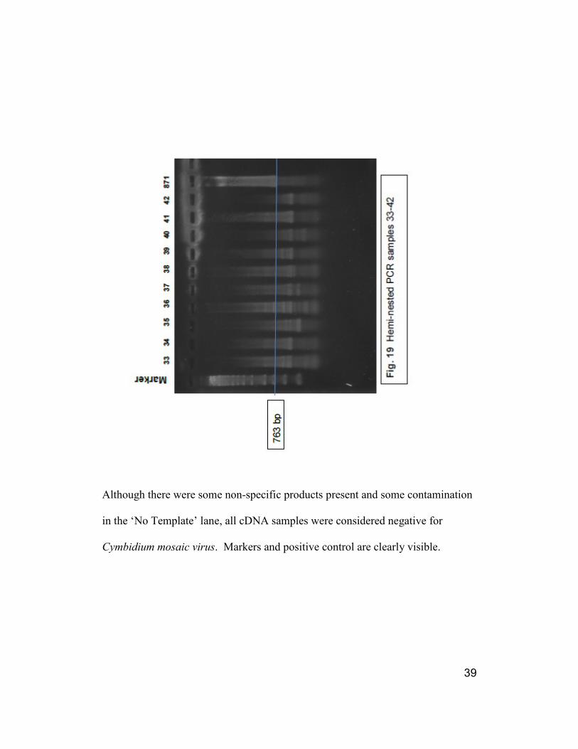
Figure 17. Initial PCR samples 33-42 ……………………………..………. 37
Figure 18. Hemi-nested PCR samples 20-32 …………………..…..……… 38
Figure 19. Hemi-nested PCR samples 33-42 ……………………..…...……39
Figure 20. Initial PCR samples 43-53 …………………………………....... 42
Figure 21. Initial PCR samples 54-58 …………………………………...… 43
Figure 22. Hemi-nested PCR samples 43-52 ……………………..……….. 44
Figure 23. Hemi-nested PCR samples 53-58 ……………………..………...45
Figure 24. Initial PCR samples 59-66 …………………………...………… 48
Figure 25. Initial PCR samples 67-73 ………………………………...…… 49
Figure 26. Hemi-nest PCR samples 59-66 ………………………….....…... 50
Figure 27. Hemi-nest PCR samples 67-73 …………………………….…... 51
Figure 28. Initial PCR samples 74-83 ………………………………...…… 54
Figure 29. Initial PCR samples 84-93 ………………………………..……. 55
Figure 30. Hemi-nested PCR samples 74-83 ……………………………… 56
Figure 31. Hemi-nested PCR samples 84-93 ……………………..…..…… 57
Figure 32. Hemi-nested PCR samples 74-83 with initial primers of
CYMTGB2/CymCoatR ……………………………………………. 59
Figure 33. Hemi-nested PCR samples 84-93 with initial primers of
CYMTGB2/CymCoatR ……………………………………………. 60
Figure 34. Nested PCR of selected samples with primers
CYMCoatF/CymCP-R2 …………………………………………… 62
Figure 35. Nested PCR of selected samples with primers
CYMF23/CymR25 ………………………………………………… 63
Figure 36. Comparison of selected samples and four different
primer sets ……………………………………………………..…. 66
1
Chapter 1: Literature Review
Introduction
Worldwide, the orchid industry has enjoyed an unparalleled economic upswing in
the past decade and a half. This economic boom has been marked in all phases of
the industry: hobby, cut flower and the pot plant markets (Floriculture Crops
2011, Kiang Ho 2010). With this remarkable growth, a new awareness of orchid
related pests and diseases has resulted in the need for improved standards and
disease-prevention protocols. Cymbidium mosaic virus is the most prevalent
orchid disease in all areas of the industry and in all countries where they are
produced. CymMV is transmitted primarily by cutting tools, hands, and
contaminated water sources (Wisler, personal correspondence August 12,
2009). Potex viruses are not normally known to be transmitted by insect vectors
and lack a specific gene product for vector interactions (Hammond personal
correspondence 2011). Cymbidium mosaic virus expression is observed in flower
distortion, necrotic spotting and reduced plant vigor (Inouye 2008). The concept
of a chewing insect route of transmission has been considered, but not
pursued. Periplaneta australasiae is a common greenhouse and conservatory pest
(Bell et al.1999) whose feeding damage has been suspected in the transmission of
orchid virus disease.

2
History of Cymbidium Mosaic Virus
CymMV was first described in 1951 by Dilworth D. Jensen who observed black
necrotic spotting on Cymbidium spp and named the virus Cymbidium Black
Streak virus. Dr. Dilworth continued to discover other orchid virus diseases while
at the College of Agriculture, University of California, Berkeley during the mid
1950’s until the end of his life in 1973. His work with A. H. Gold successfully
identified the Cymbidium mosaic virus particle via electron microscopy and
described it as sinuous rods (about 18nm X 475 nm) (Gold and Jensen 1951)
(Figure 1). He is credited as the pioneer of orchid virus research and was named a
Fulbright Research Scholar in 1959-60 where he was assigned to the University of
Utrecht in the Netherlands (Freitag, et al. 2011). He continued to research orchid
virus transmission while in the Netherlands and found that both private and public
collections and commercial producers there were observing similar symptoms as
in the United States. He is credited with identifying 30 possible orchid viruses
during his life. Although he is known for groundbreaking work in orchid virus
disease, Jensen was an entomologist and doggedly looked for an insect vector for
Cymbidium mosaic virus, but without success.
3
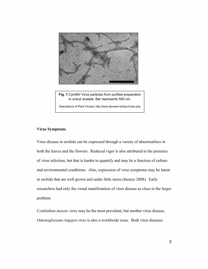
Virus Symptoms
Virus disease in orchids can be expressed through a variety of abnormalities in
both the leaves and the flowers. Reduced vigor is also attributed to the presence
of virus infection, but that is harder to quantify and may be a function of culture
and environmental conditions. Also, expression of virus symptoms may be latent
in orchids that are well grown and under little stress (Inouye 2008). Early
researchers had only the visual manifestation of virus disease as clues to the larger
problem.
Cymbidium mosaic virus may be the most prevalent, but another virus disease,
Odontoglossum ringspot virus is also a worldwide issue. Both virus diseases
Fig. 1 CymMV Virus particles from purified preparation
in uranyl acetate. Bar represents 500 nm.
Descriptions of Plant Viruses, http://www.dpvweb.net/dpv/index.php

4
typically have unique manifestations, but not consistently the same. Also an
individual plant or group of plants may harbor both diseases simultaneously.
CymMV was described originally as Cymbidium Black Streak virus and that is an
appropriate description of the leaf symptoms. Necrotic streaking or spotting is
typical and these lesions can be found on both flowers and leaves (Figure 2).
Odontoglossum ringspot virus can be visualized as a mottling of the color of the
leaf or as concentric necrotic rings (Figure 3).
Either virus can cause a condition known as ‘color break’ (Figure 4).
Fig. 2 Necrotic spotting on Phalaenopsis due to CymMV
Chin-An Chang
Fig. 3 Mottling due to infection
by ORSV Chin-An Chang

5
Determining CymMV Host Range
Researchers during the 1950’s were working primarily with bioassay techniques;
inoculating the sap from orchids with symptoms onto various plants, both orchid
and non-orchid in order to establish the host range. In 1952, Dr. Jensen was
successfully able to transmit this newly identified virus from Laelia anceps onto
Cymbidium sp. and from Cymbidium sp. to Cymbidium sp. (Jensen 1952).
When inoculated with the sap from diseased orchids, Datura stramonium (White
and Goodchild 1955), Cassia occidentalis (Corbett 1960), Tropaeolum majus,
Oryza sativa, Passiflora edulis, and Zinnia elegans (Murakishi 1958) all proved
to respond with the formation of leaf lesions. However, researchers were not able
to distinguish between Odontoglossum Ringspot virus and Cymbidium mosaic
Fig 4 ‘Color break’ abnormalities in
flower coloration due to virus
Chin-An Chang

6
virus based on symptoms alone. Frequently both virus diseases were present in
the same plant, adding to the confusion. Since each of these viruses has a different
alternate host range, some of the early host range studies show contradiction
among research groups.
Jensen seemed to have the best understanding of the two diseases and defined a
diagnostic host range for CymMV that included only Datura stramonium and
Cassia occidentalis (Jensen 1972). The hypersensitive response, leaf lesions or
chlorosis, in a host plant enables it to be utilized as a bioassay or indicator plant in
virus surveys. This localized reaction to CymMV puts Chenopodium
amaranticolor, C. quinoa, Tetragonia tetragonioides, Gomphrena globosa,
Datura stramonium, and Cassia occidentalis in that group of plants (Inouye
2008). The work of Inouye was further able to distinguish and separate the
diagnostic host range of Cymbidium mosaic virus and Odontoglossum ringspot
virus and determined that Cassia occidentalis was the most definitive test plant
for visualizing CymMV lesions alone.
Detection
Bioassay – the inoculation of a host plant with a dilution of the sap from a test
plant – is still widely employed by virologists as a quick and easy screening
method. The development of Enzyme-linked immunosorbent assay (ELISA) was
an advancement that made nursery wide screening easier and more accurate.
ELISA testing uses immunology to detect a reaction of a specific antibody or
antigen with the assistance of a color indicator.

7
However, ELISA testing requires laboratory equipment and is not available for
most orchid growers. For on-the-spot screening, immunoassay test strips
(ImmunoStrip, Agdia, Inc. 30380 County Road 6, Elkart, IN 46514) have been
developed that do not require laboratory equipment. ImmunoStrips are sensitive
for most CymMV isolates and are combined with ORSV detection. These are
simple enough for greenhouse managers or curators to use for accurate and rapid
on site testing.
RT-PCR (reverse transcription polymerase chain reaction) or one of the other
tests that detect the presence of the cDNA can be more sensitive, especially for a
low titer of virus particles. In RT-PCR, total RNA is extracted from the tissue
being tested and is transcribed into cDNA by a reverse transcriptase enzyme. The
viral cDNA is then amplified in the presence of template primers by DNA
polymerase and can be visualized by electrophoresis on an agarose gel. If an
exacting technique is employed in a clean laboratory, RT-PCR is a more reliable
method of virus detection.
Unfortunately no detection technique is fool proof. Both false positives and false
negatives are possible. Researchers have noted the time from virus inoculation to
expression of symptoms can be from seven months, as in the case of Potyvirus in
Vanilla in Tahiti (Wisler, personal communication 2009), to 30 months as
reported in experiments with Sophrolaeliocattleya hybrids in Venezuela
(Izaguirre-Mayoral et al., 1993). CymMV does associate with the vascular tissue
and can move more rapidly than ORSV, which moves from cell to cell (Borth, et

8
al., 2006) but testing a newly acquired orchid can lead to false negatives if
inoculation occurred at the time of division. The conscientious researcher or
grower will employ more than one type of test to confirm the presence of virus
infection and will repeat testing at regular intervals.
Worldwide Presence of Cymbidium Mosaic Virus
The relative incidence of CymMV in orchid crops has been studied by many
researchers. A 1992 survey of approximately 3,600 orchid plants in Hawaii found
that Cymbidium mosaic virus was found in 61% of the plants tested. ELISA
testing was the protocol used. At that time most commercial Dendrobium hybrids
(cut flower industry) were seed grown and the incidence of CymMV was 4% in
plants less than three years old. However, cloned Dendrobiums showed an
incidence of 45% (Hu 1993).
The Singapore Botanic Garden collection was tested between the years of 1988
to1991. 54.6% of the orchids tested were positive for Cymbidium mosaic virus.
Most disturbing was that 50.5% of the in-house tissue cultured plants were
infected with CymMV (Wong et al., 1994).
In 2003; bioassay, electron microscopy, ELISA, and RT-PCR testing was
performed on a group of various orchid genera that appeared symptomatic in
India This exhaustive protocol was employed as up to that time, it was believed
that CymMV was not found in that country. The orchids proved to be positive for

9
virus and through the above testing protocols the particles were identified as
Cymbidium mosaic virus (Sherpa et al., 2003).
The cut flower industry accounts for a significant segment of the Thai economy.
For example, 2004 saw $56 million (US) in cut flower exports alone. ELISA
testing was used to survey 280 vegetatively propagated Dendrobium plants and of
those plants 64.5% were positive for CymMV. Similar to the Hawaii survey, in
vitro cultured seedling plants showed no incidence of virus infection (Khentry et
al., 2006).
In all of the above surveys the incidence of Cymbidium mosaic virus in cultivated
orchids is significant. CymMV infection occurs worldwide in all genera, species
and hybrids of orchids (Brunt, et al 1996). There are many countries where
orchids are part of their world-based economy. The debilitating effects of the
virus on an orchid crop reduce the potential for economic gain. Unfortunately in
many cases where the orchid producer is growing for a throw-away pot plant
market, as long as the plant is marketable, they are not deeply concerned about the
presence of virus disease.
It is obvious that an industry wide renovation in attitude, cultivation techniques
and parent plant virus screening is needed. With deeply ingrained protocols,
minimum wage employees, and the expense of routine testing, it may be a long
time before the incidence of CymMV will be reduced.

10
Description
Cymbidium mosaic virus is classified as a member of the family:
Alphaflexiviridae, the genus: Potexvirus. It has a positive sense, single stranded
RNA gnome. Positive sense RNA is similar enough to mRNA that it can be
immediately translated by the host plant and is therefore immediately infective
(Hull 1970). Virions are filamentous and not enveloped. The particle is flexuous,
with a clear modal length of 480 nm X 13 nm wide. Axial canal is obscure. Basic
helix is obvious with the pitch of basic helix 2.8 nm (Büchen-Osmond 2011)
(Figure 5).
Genome
Cymbidium mosaic virus is approximately 6227 nucleotides in length not
including the polyadenylated tail at the 3’ end. The 5’ end is capped. Like other
potexviruses, it contains five open reading frames (ORF’s): an RNA-dependent
RNA polymerase (RdRp), three triple gene block (TGB1, TGB2, & TGB3), and a
coat protein (Wong et al., 1997). Movement between cells and through the plant
Fig. 5 Potex virus. Viral Zone

11
Fig 6 Schematic representation of the genome CymMV, genome organization with
scale. Open boxes represent the coding regions for the RNA-dependent RNA
polymerase (RdRp), 160 KDa, 26 KDa/13KDa/10 KDa triple gene block (TGB) and
24 KDa coat protein (CP). The 5’ and 3’ non-coding regions are represented as a
single line. The (A)n represents the poly (A) tail. Numerals represent nucleotide
positions. (Wong et al., 1997)
host is facilitated cooperatively by the triple-gene-block proteins and the coat
proteins (Lu et al., 2009). (Figure 6)
The virion RNA is infectious and serves as both the genome and viral messenger
RNA. RNA-dependent RNA polymerase (RdRp) is translated directly from the
genomic RNA. The other ORFS are transcribed presumably as monocistronic
(translates only a single protein) subgenomic mRNAs (sgRNAs) (ViralZone
2011). Although there are a high number of isolates, the coat proteins and RdRp
regions seem to be highly conserved (Moles et al., 2007).

12
Developing Resistance
CymMV has been observed worldwide in private and public collections, but with
even greater economic significance in the cut flower industry as described
previously. In traditional breeding programs flower count, size and color have
been the ultimate goals. Breeding or screening for CymMV resistance in
commercial orchid lines has not been a common research objective.
However, in 1988, Kuehnle, found that Dendrobiums that were susceptible to
CymMV when bred to another susceptible Dendrobium produced susceptible
offspring and resistant cultivars when crossed to another resistant cultivar
produced resistant offspring. She determined that susceptibility was the dominant
characteristic in cross breeding of types and that expression of floral necrosis was
genetically controlled.
Researchers are actively working to develop CymMV resistant strains of
Dendrobium varieties via genetic modification. In one case, a mutant movement
protein gene, mut11 was inserted via biolistics into two different Dendrobium
hybrids and the plants were repeatedly challenged by inoculations of a 1:1000
dilution of CymMV. Though the sampling was small, with only 24 original
plants, 9 of the transgenic plants remained CymMV free after 12 months
(Obsuwan, et al 2009).
In Taiwan, another research group isolated CymMV and the cDNA of the CP
gene was then synthesized and sequenced. Through particle bombardment, the

13
synthesized gene was transformed into very young Dendrobium plants. The
presence of the gene was confirmed by PCR, Southern, Northern, and Western
blot techniques. When these plants were challenged with CymMV they exhibited
considerably milder symptoms (Chang et al., 2005).
Another research group in Taiwan worked with Phalaenopsis hybrids. Resistance
in those plants was achieved by insertion of a CymMV coat protein and a nos
terminator placed downstream of a maize ubiquitin promoter. Those plants
exhibited improved resistance to CmyMV upon virus challenge (Liao, et al 2004).
Periplaneta australasiae, The Australian Cockroach
Stejskal, et al. (2004) describe the Australian cockroach as a rapidly spreading
pest moving from its native tropics into the temperate zone. It is thought to have
originated in North Africa despite its common name. The pest species of
cockroaches are believed to have dispersed with early human exploration (Kunkle
2007).
This cockroach infests not only food storage areas, but greenhouses and
conservatories as well. Unlike some of its more well-known relatives, it can feed
on tender plant material (Figure 7). Locally it can be found in damaging numbers
at the Smithsonian complexes (Tom Mirenda, personal communication 2010) and
the United States Botanic Garden. In greenhouses and conservatories its numbers
can build rapidly especially when the targeted sprays of a strict program of
integrated pest management are observed (Bell et al., 1999).
14
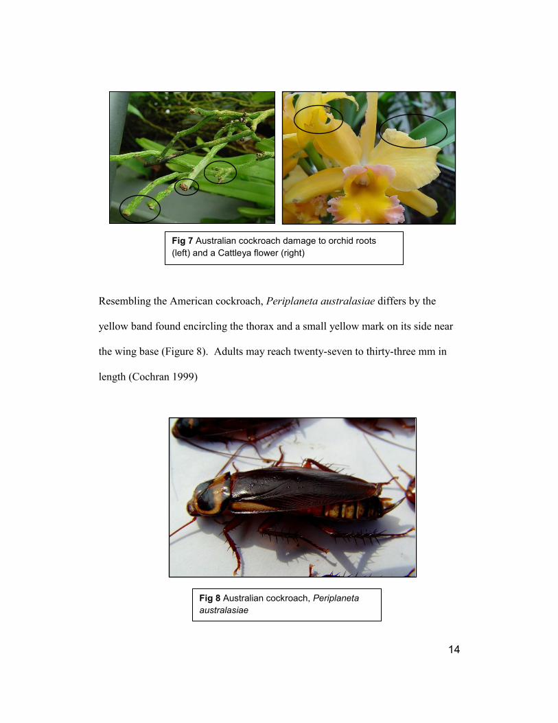
Resembling the American cockroach, Periplaneta australasiae differs by the
yellow band found encircling the thorax and a small yellow mark on its side near
the wing base (Figure 8). Adults may reach twenty-seven to thirty-three mm in
length (Cochran 1999)
Fig 7 Australian cockroach damage to orchid roots
(left) and a Cattleya flower (right)
Fig 8 Australian cockroach, Periplaneta
australasiae

15
Biology and Life Cycle
Adult Australian cockroaches are believed to live for 6 – 8 months and maturation
occurs at about five months of age. Their life cycle is one of gradual
metamorphosis: egg, nymph, and adult. Nymphs undergo nine to twelve molting
cycles before reaching maturity (Cochran 1999).
The females produce an egg case called an ootheca which can contain sixteen to
twenty-four eggs. Hatch rate is influenced by temperature and humidity. Females
may produce twenty to thirty egg cases in their life time. (Ramel 2001)
Australian cockroaches are in the order Blattodea (Cockroaches) and family,
Blattidae.
Chewing Insects as Vectors
Researchers have reported some virus vectors in the orders Orthoptera
(Grasshoppers) and Dermaptera (earwigs). More significant vectors are found in
the order Coleoptera (Beetles), but no work has been reported on insects found in
the order Blattaria (Cockroaches) as virus vectors in plants (Hull 1970)

16
Chapter 2: Materials and Methods
The cockroach colony and all experimental plants were housed in the University
of Maryland research greenhouses. A southeast facing, 69.7 square meters (750
sq. ft.) section was chosen for its appropriate light levels for maximum orchid
growth and health. Natural day length was allowed, supplemental lighting was
used only to maintain a set point of 30 Klux during cloudy weather.
Temperatures were set at 24oC day/18
oC night.
Periplaneta australasiae, Australia Cockroach
The Australia cockroach colony was initiated with a purchase of 40 mature, male
and female cockroaches (PNE, Inc., 169 Elsa Jane Lane, Pittsboro, NC 27312-
5167). The cockroaches were housed in containers that were modified water proof
document storage boxes. Water and dry dog chow were supplied ad libitum. The
cockroaches were allowed to breed freely and were moved to other containers as
the colony grew.
The cockroaches were subjected to a period of five weeks without food before
their introduction to orchid plant tissue. Periplaneta australasiae will resist a
change in diet up to the point of starvation (Barry Pawson, personal
communication 2009). The period of deprivation enabled the cockroaches to
accept the new food source rapidly.

17
Orchids
Three groups of orchids were used during the course of this experiment. Orchids
of known virus infection made up the first group. These were plants of various
genera that were donated from the United States Botanic Garden (United States
Botanic Garden, 100 Maryland Avenue, SW, Washington, DC 20001). Testing to
confirm virus infection was performed by a commercial laboratory (Agdia, Inc.,
30380 County Road 6, Elkart, IN 46514) using enzyme linked immunosorbent
assay. These plants all tested positive for Cymbidium mosaic virus and some
were positive for Odontoglossum ringspot virus as well.
The second group of orchids were Oncidium cultivars and hybrids donated from
private collections and through the generosity of a local commercial orchid
grower (Orchid Enterprise, Inc., 6 Perch Place, Alexandria, VA 22309). Surveys
were conducted via bioassay and ImmunoStrip (Agdia, Inc., 30380 County Road
6, Elkart, IN 46514) testing to ascertain virus infection.
To minimize the possibility of using test plants that had already been exposed to
orchid viruses, newly de-flasked Odontocidium Catatante 'Pacific Sunspots',
AM/AOS were purchased for the project (Carmela Orchids, P.O. Box 277.
Hakalau, HI 96710) and comprise the third group of orchids. These plants were
subsequently tested and found negative for virus infection by ImmunoStrip and
polymerase chain reaction assay.

18
Experimental Units
The experimental unit was a clear fronted, screened enclosure (Rearing and
Observation Cage, BioQuip Products, 2321 Gladwick Street, Rancho Dominguez,
CA 90220) that successfully provided both insect containment and sufficient light
for plant health and maintenance.
Thirty experimental units were set up on two greenhouse benches. The cages
were protected from extreme light by draping with pieces of standard greenhouse
shade cloth (60%) to keep the Australian cockroaches in a more comfortable
environment. The cages were numbered and randomized on the benches.
In each enclosure elevated pierced flooring was provided by the insertion of a 12”
X 12” piece of plastic egg crating. This elevation would prevent water
contamination between plants. A 4 ½” standard, square plastic pot was used as an
insect hide and water for the cockroaches was provided by a plastic petri dish
fitted with an acrylic sponge soaked with water.
The cockroaches were distributed in groups of either ten or twenty individuals per
experimental unit, ranging in size from 1.2 cm nymphs to mature adults. Gender
ratio was not considered significant.
The Treatment
After a period of food deprivation of approximately five weeks duration, pieces of
orchid leaf (approximately 2.5 cm X 2.5 cm) or an orchid flower were inserted
into a slit in the sponge of the cage water source. Twenty five of the randomized

19
cages were supplied with leaf tissue from known virus infected orchids. To act as
controls, five of the randomized cages were supplied with tissue from plants that
had been repeatedly tested as virus free. The tissue samples were changed out
after consumption or one week’s time. To accustom the cockroaches to feeding
on orchids, they were fed for a period of three weeks on orchid leaf tissue before
orchid seedling test plants were placed in the cages.
Introduction of Seedling Test Plants
Five orchid seedlings were placed in each cage. The plants were numbered by
cage and sequenced, 1 – 5. The cockroaches were allowed free access to feed.
The orchid seedlings were examined for feeding damage several times per week
and were removed as soon as damage occurred. A second period of deprivation
was initiated in late March due to a lag in feeding activity. All food was removed
for a period of approximately one month after which exposure to both infected
plants and test plants was resumed. The inoculated seedlings were placed on a
greenhouse bench to allow possible virus replication.
Time Interval for Virus Replication
Tissue samples were harvested a varying intervals to allow for virus replication
within the inoculated tissue. This time period (Time B) was initially set at greater
than 21 days. A longer period of time would have the advantage of a higher
number of virus particles able to be detected. Samples were harvested at 56, 62,
65, 69, 71, 89, and 90 days post inoculation.

20
Analysis Protocol for Virus Detection
The sample tissue from inoculated and control plants was ground in a mesh
extraction bag (Agdia, Inc. 30380 County Road 6, Elkart, IN 46514) containing
1.5 ml RLT, an RNeasy lysis buffer (QIAGEN Inc., 27220 Turnberry Lane,
Valencia, CA 91355). RNA extraction was then performed by standard procedure
using an RNeasy Plant Mini Kit (QIAGEN Inc.). For RNA extraction from
ImmunoStrip, the preserved strips were soaked for 5 minutes in an 11:2 solution
of RLT buffer then soaked for an additional 5 minutes in ethyl alcohol. The
solutions were combined and 700 µl were then placed in the pink columns from
the RNeasy Plant Mini Kit. Standard procedure was then followed.
Conversion from RNA to cDNA was performed on 5µl of RNA extract with the
addition of 15 µl of a master mix containing: 1 μl Moloney Murine Leukemia
Virus Reverse Transcriptase, 4 μl M-MLV Reverse Transcriptase 5X Reaction
Buffer, 4 μl Deoxynucleotide Triphosphates 2.5mM, 5 μl, Primer NSNC-odT (5′
ATCCATGGCATGCATCGATTTTTTTTTTTTTTTV 3′, where V = A, G, or C),
and 1 μl RNAsin (all reagents except NSNC-odt: Promega, 2800 Woods Hollow
Road, Madison, WI 53711-5399. NSNC-odT designed by John Hammond and
Michael Reinsel (USDA-ARS, USNA, FNPRU) and produced by Invitrogen, Life
Technologies Corp., 3175 Staley Road, Grand Island, NY 14072). The samples
were processed in a thermo cycler (Applied Biosystems, GeneAmp, PCR System
2700, Life Technologies Corporation, 5791 Van Allen Way, Carlsbad, CA 92008)
using the following program: 42oC for 60 minutes, 95
oC 5 minutes and 4
oC to

21
hold until the cDNA was either sampled for the PCR step, or stored frozen for
later use.
Initial testing of CymMV-infected and control plants was performed by PCR
using one of several combinations of primers designed by Michael Reinsel
(USDA-ARS, USNA, FNPRU) based on an alignment of multiple CymMV
sequences available in GenBank, or on the ‘tag’ portion of cDNA primer NSNC-
odT. These primers were: CymTGB2 (‘Forward’, 5′
TGCAATACATATCACCACCCCTGA 3′); CymCoatF (‘Forward’, 5′
TGGCGAGGGTTAAGTTACCA 3′); CymCoatR (‘Reverse’, 5′
TGCCAGTAGTGGAAACAAACTT 3′); and BNSNC (‘Reverse’, 5’
TTTATCGGATCCATGGCATGCATCG 3′) (Fig. 9). Each of these primer
combinations yielded a CymMV-specific product of sizes
(CymCoatF/CymCoatR, 763 bp; CymCoatF/BNSNC, 829 bp;
CymTGB2/CymCoatR, 881 bp; CymTGB2/BNSNC, 947 bp), with minor yields
of non-specific products. Although obvious CymMV-specific products were
obtained from systemically-infected positive control orchids, no products were
obtained in initial tests of plants exposed to cockroach feeding. Because the
Cymbidium mosaic virus was suspected to be in very small amounts in the sample
tissue, a hemi-nested PCR (Mullis and Faloona 1987) assay was then developed
and used as the protocol of choice to increase the sensitivity of detection.

22
Primer pairs were selected by running a temperature gradient PCR (Appendix B)
using samples of known positive, known negative as well as a plasmid positive
control. Calculated annealing temperatures were determined for all primer pair
combinations:
CymTGB2 (forward)/CymCoatR (reverse) 56o C
CymTGB2 (forward)/BNSNC (reverse) 63o C
CymCoatF (forward)/BNSNC (reverse) 58o C
CymCoatF (forward)/CymCoatR (reverse) 56o C
After analysis of the resulting cDNA product by gel electrophoresis, the primer
pair CymCoatF (forward)/BNSNC (reverse) was determined to be the most
advantageous for the initial PCR and CymCoatF (forward)/CymCoatR (reverse)
for the hemi-nest. Amplification was further maximized by increasing the cycles
from 35 to 40.
In the initial assay, sample plant tissue cDNA was subjected to PCR with
amplification targeted at the virus coat protein and 3′ non-coding region using
primers CymCoat F and BNSNC. That PCR product was then subjected to the
hemi-nested reaction with amplification targeted to a narrower area of virus coat
protein using primers CymCoat F and CymCoat R. The master mix used for all
PCR assays was as follows: 0.2 μl GoTaq, 4 μl 5X Green GoTaq Reaction Buffer,
2 μl Deoxynucleotide Triphosphates 2.5mM (all reagents supplied by Promega), 1
μl forward primer, 1 μl reverse primer, and 10.8 μl dH2O per sample. To 19 μl of

23
the master mix, 1 μl of sample cDNA or diluted (1:100) first PCR product was
added. The samples were processed in a thermo cycler using the following
protocol: 1 cycle 94o C for 3 minutes, 40 cycles: 94
o C 30 seconds, 63
o C 30
seconds, 72o C 90 seconds and then 1 cycle 72
o C for 7 minutes.
The PCR products from both steps of the hemi-nested protocol were separately
examined by Agarose gel electrophoresis. A standard 1% Agarose gel was
formed by the formula: 0.6g Agarose (Separation > 500bp, Genetic Performance
Certified, USB Corp, Cleveland, OH) dissolved in 60 ml tris-borate-EDTA buffer
0.5X. A 1 kb DNA Ladder (Promega) was used as a standard for the
electrophoresis product. All PCR assays included known positive and negative
control samples.
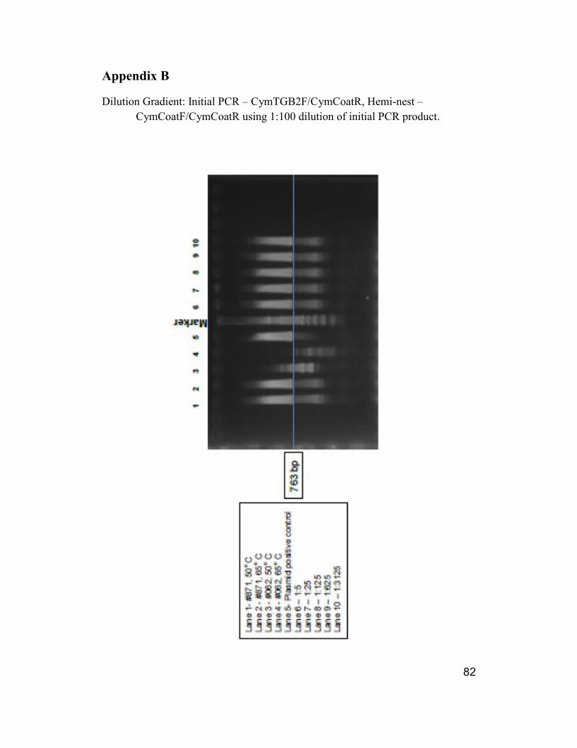
Since the amount of virus present in a sample could also affect the presence of the
final PCR product, a dilution gradient was run. Dilutions of a known positive
sample, a known negative sample, and a positive plasmid control were made at
the ratios of 1:5, 1:25, 1:125, 1:625, and 1:3125. Serial dilutions were made from
an initial 1:100 dilution of the first PCR product. The above hemi-nested protocol
was performed and the PCR products were visualized on gel electrophoresis.
Clear CymMV product bands were observed for all dilutions. (Appendix C)
A third set of primers were designed to be employed in future work with
Cymbidium mosaic virus. These primers were: one forward primer, CymF23
24
(‘Forward’, 5′ GTGGTGTGGAATCTGATGCTGGC 3′) and two reverse primers,
CymCP-R2 (‘Reverse’, 5′ GCAATGTTGGTGATGAGGTTGCCGG 3′) and
CymR25 (‘Reverse’, 5′ CTTGGTGACCTCGGCAATGTTGG 3′). (Figure 9). An
annealing temperature gradient was run on various combinations of existing and
new forward and reverse primers. (Appendix D). Selected cDNA samples were
run to test two of the new primer combinations.
A chi square test was applied to feeding interval data. (Appendix F)

25

26
Chapter 3: Results
Australian Cockroach
Feeding damage was observed within ten days after the introduction of orchid leaf
pieces. The damage resembled that of beetles (Fulton, et al. 1987) (Figure 10)
Damaged plants were removed as soon as observed and placed on the greenhouse
bench. Groups of samples were taken for RNA extraction at 56, 62, 65, 69, 71,
89, and 90 days post inoculation. Most samples consisted of the chewing damage
site and the surrounding leaf tissue; however there were two cases of pseudobulb
damage and that tissue was tested as well.
Fig 10 Feeding damage on leaf piece (left) and on test plant (right)

27
The Test Plants
Two different time intervals are noted. The time between the exposure of the
cockroaches to the infected plant material and the time of their access to feed on
test plants is one critical period, labeled Time A (Appendix E). The other critical
period is the time the virus has to replicate in the damaged test plant tissue,
labeled Time B. For Time B, plants were grouped by number of days post
feeding damage and labeled A through G. Damaged plant tissue was harvested
according to this second time period and polymerase chain reaction analysis was
performed.
Figure 11 shows the comparison between plants positive for Cymbidium mosaic
virus and the time interval between exposure of the cockroaches to infected
material and test plant feeding damage (Time A).
Number of Plants
Interval (days) Positive Negative
6-10 14 19
11-15 0 28
16-20 0 9
21-25 0 12
Nineteen plants in the interval between 6 and 10 days were found to be negative
for presence of CymMV. Fourteen plants were found positive for presence of
Fig 11. Incidence of positive and negative plants compared to
time interval between cockroach exposure to infected material
and feeding damage

28
CymMV after feeding damage by Australian Cockroaches in the same interval.
The results of the treatments are significantly significant at p <.0001. Forty-nine
plants from longer time intervals between exposure to infected leaf and observed
feeding damage were found to be negative for the presence to Cymbidium mosaic
virus.
Polymerase Chain Reaction Analysis
Test Groups A, B & C
Samples 1 – 19 (Test Group A, B, & C) were run via PCR analysis as described
above using CymTGB2 (forward) and CymCoatR (reverse). These samples were
taken from plants 62, 69 and 71 days post inoculation. Three samples were from
negative control cages.
Included in the initial polymerase reaction and the subsequent hemi- nested
procedure were the following samples:
Sample number Plant number dpi (days post inoculation)
1 1-2 62
2 1-3 62
3 1-5 71
4 5-3 62
5 8-1 69

29
6 8-4 62
7 8-5 69
8 11-4 69
9 12-3 62
10 13-3 62
11 16-3 62
12 16-4 62
13 17-2 69
14 18-1 69 Negative control
15 18-2 69 Negative control
16 18-3 69 Negative control
17 21-5 69
18 29-2 71
19 29-5 71
RNA extractions were made on 5/17/12, reverse transcription on 5/21/12, and the
initial PCR was run on 6/19/12 with the primers CymCoatF/BNSNC as described.

30
Two DNA ladders (100 kb and 1 kb) were loaded on this gel for comparison of
product size. In subsequent reactions only the 1 kb ladder was used as the
anticipated product for CymMV would be found at 763 kb. A plasmid CymMV
was used as the template for the positive control.

31
The initial PCR products were then diluted 1:100 with distilled water and a hemi-
nested PCR analysis was run on 6/21/12. A dilution of the initial PCR products
was made in an attempt to reduce non-specific product. The hemi-nested PCR
was run with primers CymCoatF/CymCoatR.

32
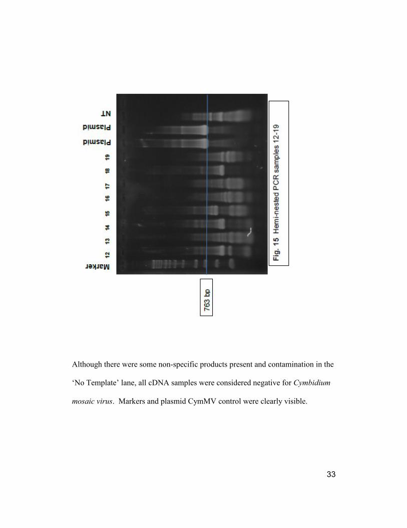
33
Although there were some non-specific products present and contamination in the
‘No Template’ lane, all cDNA samples were considered negative for Cymbidium
mosaic virus. Markers and plasmid CymMV control were clearly visible.

34
Test Group D
Samples 20 – 42 (Test Group D) were run using the above described initial then
hemi-nested protocol. These samples included chewing damage sites harvested
65 and 67 dpi. Also in this group were non-cockroach-exposed negative controls
(negative control A & B). In addition, there were three samples that had been
mechanically inoculated (Onc A, B, & C) and harvested 96 dpi. Four samples
were extractions from previous testing with ImmunoStrips and two samples were
from negative control cages.
Included in the initial polymerase reaction and hemi- nest procedures were the
following samples:
Sample number Plant number dpi (Days Post Inoculation)
20 1-4 65
21 5-1 65
22 8-2 65
23 16-1 65
24 18-4 65 Negative control
25 23-4 67 Negative control
26 25-2 65

35
27 25-3 65
28 25-5 67
29 26-4 65
30 28-5 67
31 29-4 65
32 30-4 67
33 Onc A 96
34 Onc B 96
35 Onc C 96
36 Negative Control A
37 Negative Control B
38 3-1 from ImmunoStrip
39 14-5 from ImmunoStrip
40 19-3 from ImmunoStrip
41 20-4 from ImmunoStrip
42 27-1 from ImmunoStrip
36
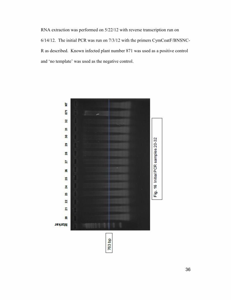
RNA extraction was performed on 5/22/12 with reverse transcription run on
6/14/12. The initial PCR was run on 7/3/12 with the primers CymCoatF/BNSNC-
R as described. Known infected plant number 871 was used as a positive control
and ‘no template’ was used as the negative control.

37

38
To better amplify the anticipated product, a hemi-nested PCR analysis was run
using primers CymCoatF/CymCoatR.
39
Although there were some non-specific products present and some contamination
in the ‘No Template’ lane, all cDNA samples were considered negative for
Cymbidium mosaic virus. Markers and positive control are clearly visible.

40
Test Group E
Samples 43 – 58 (Test Group E) were run using the above described initial PCR
analysis followed by a hemi-nested PCR. These samples were harvested 89 dpi.
Three of the samples were from negative control cages. Included in the initial
polymerase reaction and hemi- nest procedures were the following samples:
Sample number Plant number dpi (Days Post Inoculation)
43 4-2 89
44 5-5 89
45 7-3 89
46 9-4 89 Negative control
47 9-5 89 Negative control
48 10-3 89
49 11-1 89
50 12-4 89
51 15-5 89
52 21-3 89
53 21-1 89
54 24-3 89
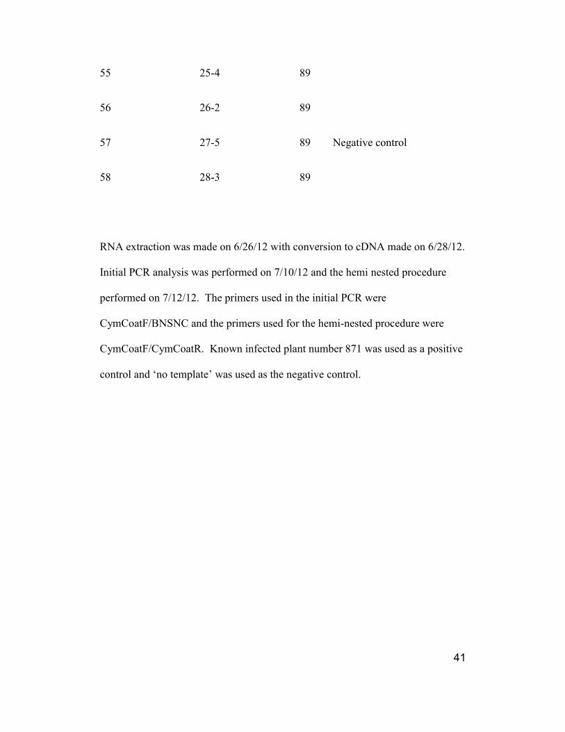
41
55 25-4 89
56 26-2 89
57 27-5 89 Negative control
58 28-3 89
RNA extraction was made on 6/26/12 with conversion to cDNA made on 6/28/12.
Initial PCR analysis was performed on 7/10/12 and the hemi nested procedure
performed on 7/12/12. The primers used in the initial PCR were
CymCoatF/BNSNC and the primers used for the hemi-nested procedure were
CymCoatF/CymCoatR. Known infected plant number 871 was used as a positive
control and ‘no template’ was used as the negative control.

42

43

44

45
All samples were negative for CymMV-specific product after gel electrophoresis,
though there was nonspecific product present and some contamination in the ‘No
Template’ lane. Ladder and positive control are visible.

46
Test Group F
Samples 59-73 (Test Group F) were run using the above described protocol.
These samples were 90 dpi. Four samples originated from negative control cages.
The leaves from the manual inoculation were tested again. Included in the initial
polymerase reaction and hemi- nest procedures were the following samples:
Sample number Plant number dpi (Days Post Inoculation)
59 4-3 90
60 5-4 90
61 9-1 90 Negative Control
62 9-2 90 Negative Control
63 10-1 90
64 10-2 90
65 13-4 90
66 13-5 90
67 14-1 90 Negative Control
68 20-4 90
69 26-1 90
70 27-2 90 Negative Control

47
71 Onc A 145
72 Onc B 145
73 Onc C 145
RNA extraction was performed on 7/11/12 with reverse transcriptase to cDNA on
7/12/12. The initial PCR using primers CymCoatF/BNSNC was performed on
7/23/12. The hemi-nest PCR with primers CymCoatF/CymCoatR was run on the
same day. The positive control was cDNA from a known infected plant, number
871 and ‘no template’ was used as a negative control.

48

49

50

51

52
All samples were negative for CymMV-specific product after gel electrophoresis,
though there was nonspecific product present and some contamination in the ‘No
Template’ lane. Ladder and positive control are visible
Test Group G
Samples 74-93 (Test Group G) were run using the above described hemi-nested
protocol. These samples were 56 dpi. There were two samples from negative
control cages. Included in the initial polymerase reaction and hemi- nest
procedures were the following samples:
Sample number Plant number dpi (Days Post Inoculation)
74 1-1 56
75 2-4 56
76 4-1 56
77 4-5 56
78 7-1 56
79 7-5 56
80 8-3 56
81 11-2 56
82 13-1 56

53
83 14-5 56 Negative Control
84 15-1 56
85 15-4 56
86 21-2 56
87 22-5 56
88 24-2 56
89 24-4 56
90 27-1 56 Negative Control
91 28-1 56
92 28-2 56
93 30-5 56
The RNA extraction was performed on 7/25/12 with conversion to cDNA on
7/26/12. The initial PCR was made on 7/26/12 using primers
CymCoatF/BNSNC. A 1:100 dilution of the initial PCR product was made with
distilled water. The hemi-nested PC was run on 7/27/12 using primers
CymCoatF/CymCoatR.

54

55

56

57
The initial PCR was unremarkable showing no bands indicating the presence of
Cymbidium mosaic virus in any of the samples. However the gels from the hemi-

58
nested PCR demonstrate bands at approximately the 763 kb position. Lanes 74,
75, 77, 78, 81, 82, 83, 84, 85, 87, 88, and 89 indicate the potential for CymMV
product. Unfortunately, the ‘No Template’ lane indicates contamination and that
contamination may have effect on the bands at the 763 kb position.
The PCR analysis was re-run on 8/6/12 from the cDNA with a different set of
initial primers, CYMTGB2/CymCoatR and then the hemi-nest with
CymCoatF/CymCoatR. A fresh dilution of the CymCoatF primer eliminated the
persistent contamination issue. Controls were run with cDNA from a plant
known to be free of CymMV, number 062 and a known positive, number 871 and
were run with the primers CYMTGB2/CymCoatR

59

60

61
This change in primer pairs resulted in a number of possible CymMV bands.
Noted were samples number 74, 75, 77, 79, 80, 84, 85, and 91. There was some
bleeding of the positive control into the ‘No Template’ lane when the gel was
loaded and some primer-dimers are evident as non-specific product.
Selected Samples
Three new primers were designed and tested on a selection of previously tested
cDNA samples listed below:
Sample number plant number tested & date
75 2-4 POS 7/27 & 8/6
76 4-1 NEG 7/27 & 8/6
78 7-1 POS 7/27 & NEG 8/6
82 13-1 POS 7/27 & NEG 8/6
83 14-5 POS 7/27 & NEG 8/6
85 15-4 POS 7/27 & 8/6
86 21-2 NEG 7/27 & 8/6
88 24-2 POS 7/27 & NEG 8/6
90 27-1 NEG 7/27 & 8/6
91 28-1 NEG 7/27 & POS 8/6

62
The 1:100 diluted products originated from the PCR analyzed on 8/2 and used
primers CYMTGB2/CymCoatR. The sample #871 was used as a positive control
and ‘No Template’ was used as the negative control.

63

64

65
On the gel for the reaction using primers CymCoatF/CymCP-R2 bands are seen at
the 640 bp region as expected. Samples 75, 76, 78, 83, 85, 88 and 91 appear to be
positive for CymMV. The positive control #871 and the maker are clearly visible.
The ‘No Template’ lane shows left over primers. There are considerable non-
specific products present in some other lanes.
On the gel using primers CymF23/CymR25 bands are seen at the 258 bp region.
The PCR product was anticipated to be visible in this area. Samples 75, 78, 82,
83, 85, 88, 90, and 91 appear to be positive for presence of Cymbidium mosaic
virus. There is considerable primer material that was not used and there are bands
in several lanes above the virus product.

66
Sample Plant Primers Primers Primers Primers
A B C D
75 2-4 Pos Pos Pos Pos
76 4-1 Neg Neg Pos Neg
78 7-1 Pos Neg Pos Pos
82 13-1 Pos Neg Neg Pos
83 14-5 Pos Neg Pos Pos
85 15-4 Pos Pos Pos Pos
86 21-2 Neg Neg Neg Neg
88 24-2 Pos Neg Pos Pos
90 27-1 Neg Neg Neg Pos
91 28-1 Neg Pos Pos Pos
A - CymCoatF/BNSNC, CymCoatF/CymCoatR
B - CYMTGB2/CymCoatR, CymCoatF/CymCoatR
C - CYMTGB2/CymCoatR,CymCoaF/CymCP-R2
D - CYMTGB2/CymCoatR, CymF23/CymR25
Fig. 36. Comparison of selected samples and four different
primer sets

67
Chapter 4: Conclusion
Research frequently does not go as expected and this study is certainly an
example of that. What started as a relatively simple plant science experiment has
morphed into a study of molecular biology techniques. It has moved so far from
its original intent, that I am hard pressed to state that the hypothesis is strongly
supported. Can Australian cockroach transfer Cymbidium mosaic virus? Yes, it
seems that they can, but at such a low level that it is hard to prove unequivocally.
Many factors conspire to negate that proof. First is the previously established
slow rate of movement of CymMV in orchids (Appendix A, conclusion). Any
study that seeks to clearly detect CymMV infection in orchid test subjects will
have to be of several years duration just to allow the virus to move into new
growth tissue.
Second is the seemingly very small amount of virus particles that the cockroaches
move as they feed from infected plant to uninfected plant. Both of these factors
conspire to make detection difficult. Hence the transformation of this plant
science study into one of molecular biology.
There are some other aspects of the possible transfer of virus particles from infect
plant to uninfected plants by cockroach feeding that can be addressed as well.

68
First, we know that the phenomenon of Australian Cockroach feeding damage on
orchids and other plants in conservatories and greenhouses has been well
documented (Bell et al.1999). Virus transfer could be hindered by the
cockroaches having been taken out of their natural or adapted habitat and placed
in confined and rather sterile cages. However, as demonstrated in this study, with
only a little manipulation they readily took to the foods that were offered and
adapted well enough to breed freely. The test plant chewing damage observed
under these controlled circumstances appeared to be equivalent to the damage that
has been seen in conservatories.
The low number of positive transmissions may also be a result of the type of
feeding damage. Successful virus transmission depends on wounded, but living
cells that will allow the movement and replication of the virus particles. The
cockroach damage in many instances appears as a shredding of the leaf area,
resulting in a wide band of dead plant cells. This band of dead cells could be
limiting virus transmission.
Cockroach feeding behavior may have reduced virus transmission as well. It is
suspected that they engage in a fast and gorge type of behavior. If a cockroach
satisfies its hunger on an infected plant and then goes for longer than seven days
before feeding again, there could be a reduction in virus particle viability by the
time it eats another plant and potentially transmits the virus. As stated in
ICTVBdB Index of Viruses (http://ictvdb.bio-mirror.cn/Ictv/index.htm), CymMV

69
virions are capable of infection only within a period of seven days at room
temperature. That stated period of time was supported by the observations made
during this study. The number of positive transmissions of CymMV occurred
within 6 – 10 days of exposure. Overall efficiency of transmission would be
affected by the short period of virion viability.
Other aspects of cockroach behavior could affect efficiency of virus transmission
as well. Mutual grooming behavior has been observed in cockroaches. This
could work to either spread the virus particles from one cockroach to another or to
effectively clean plant material residues and virus particles from the cockroaches’
mandibles. Follow-the-leader type of behavior when confronted with a new food
source would also affect virus spread through feeding. Possible interaction
between the virus particles and the insect saliva should also be considered.
Further study of cockroach behavior would shed light on the efficiency of
transmission from feeding damage.
The orchid plant has its own defenses to prevent virus transmission. The orchid
collection that was maintained for this study was well supported and well grown.
Though the plants were donated and were not in good health when received, they
quickly put on vigorous new growth and bloomed frequently. In most instances it
was impossible to determine by sight that a plant was virus infected. A healthy
orchid leaf has a very thick, protective cuticle and that cuticle could also decrease

70
the efficiency of virus transmission. Orchids plants that were manually
inoculated, even though they were pre-treated with a detergent, failed to be
infected. The cuticle barrier would also deter transmission through casual
wounding by insects.
The resistance pathway in orchids to CymMV has not been established. Neither
has innate resistance been cataloged in either species, intraspecies or intergeneric
hybrids. As Cymbidium mosaic virus is not found in wild populations of orchids,
natural resistance needs to be considered. Orchid growers have noted that a well
grown orchid plant may be positive for virus infection and yet not express
symptoms. Is that lack of symptom expression part of the natural resistance of the
orchid to virus infection? Research on the resistance pathways would potentially
be able to answer that question.
One factor that favors cockroach virus transmission is the high number of the
insects present in a typical conservatory and their frequently unlimited access to
susceptible plant material. That scenario can be compared to aphid transmission
of Lettuce mosaic virus (Broadbent et al., 1951) in field grown lettuce crops. This
was a case of a very small percent of seed born virus resulting in wide spread crop
loss when the crop was fed on by aphids. Researchers found that the most
effective method of virus control was reduction of virus infected seeds. The
incidence of LMV in lettuce seed was brought down to 0 in 30,000 seeds and only
then could a crop be protected. A large population of virus vectors, in the above

71
case, aphids, when applying constant feeding pressure had resulted in significant
transmission.
This study reinforced the concept that the period of time between cockroach
exposure to infected material and timely access to uninfected plants was a
significant factor. The other aspect that was considered was the time allowed for
the virus to replicate in the orchid tissue. This time period did not seem to
influence the number of infected plants. This aspect needs to be further studied
and a longer period of time allowed between cockroach feeding and sample
harvest. Perhaps less sensitive means of detection would then be effective if a
greater time period and therefore a higher titer of virus particles present had been
allowed.
The success of this study revolved around the sensitivity of detection and a very
large effort was made in improving those methods. The standard polymerase
chain reaction protocol was further enhanced with the use of a hemi-nest and
nested technique. Although at the initiation of this study, three specific primers
were available, when greater sensitivity was required, three more primers were
designed.
The most sensitive primer pair, CymF23/CymR25 has the optimum characteristics
of equivalent length, Tm and GC content. This primer pair yielded the greatest
number of positive samples and enhanced very low virus titer. Faint bands on a

72
gel can be the visual indication of a low virus titer. By comparing the gel
products of less sensitive primer pairs to the more sensitive pair it can be seen that
the additional amplification by the more sensitive primers resulted in stronger
bands of greater width. A good example of that more efficient amplification is
the comparison of samples #82 and #90 in the gel from the primer combination of
CymCoatF/Cym CP-R2 and the gel from primer pair CymF23/CymR25. The
improved efficiency of primer pairs by complimentary chemistry and cycling
conditions is demonstrated by the work of Arif, et al. 2012.
Effective limitation of the spread of Cymbidium mosaic virus depends on
controlling the Australian cockroach. Allowing high numbers of Australian
cockroaches has serious impact on conservatory collections or greenhouse crops.
Though the trend towards an integrated pest management system with targeted
pesticide applications is a laudable effort, pest control personnel need to be also
monitoring and controlling what up until now was considered only a nuisance
pest.
As public awareness grows concerning the large number of Cymbidium mosaic
virus infected orchids entering the market place, improved and reliable testing
methods need to be developed and used by orchid growers and breeders.
Cymbidium mosaic virus has been established as a world-wide occurring pathogen
that is capable of great economic impact in the orchid industry. Its control will

73
need a multidisciplinary approach involving consumer awareness, improved
asepsis in plant handling, grower compliance, improved methods of testing at the
greenhouse level, and the introduction of resistant plants.

74
Appendix A
Transmission of Virus in Orchids Through the Feeding Damage
of Australian Cockroach, Periplaneta australasiae
Carol Allen
Plant Science and Landscape Architecture
University of Maryland
College Park, MD 20742-4452 USA
Keywords: Australian cockroach, Cymbidium mosaic virus, CymMV,
Odontoglossum ring spot virus, ORSV
Abstract
The project goal was to demonstrate the possibility of orchid virus
transmission by a chewing insect, the Australian cockroach (Periplaneta
australasiae) under controlled conditions. The experiments were housed in
aluminum frame screen cages to contain the cockroaches. Two plants were
placed in each cage: an orchid that tested positive for orchid virus and a
young clone of Oncidium Sweet Sugar ‘Kalender’ that tested as virus free.
Australian cockroaches were introduced into four of the cages. Two cages
were used as controls containing the above plant material, but no
cockroaches. Approximately one third of the Australian cockroaches used
were “wild” caught in a nearby conservatory and the rest were purchased
from a commercial supplier. The cockroaches were communally housed for a
period of one week. We assumed that any of the wild-caught cockroaches
that carried an orchid virus would distribute the virus particles by mutual
grooming. The Australian cockroaches were housed with the plant material
until sufficient feeding damage was observed. At that time, the orchid virus
testing was repeated on the Oncidium Sweet Sugar ‘Kalender’. Samples of
new growth tissue were initially tested at a commercial laboratory and were
subjected to an orchid virus screen that identifies nine viral agents known
specifically to orchids. Test results were negative for presence of virus. Four
weeks after feeding damage was observed, testing was repeated. Tissue from
the feeding sites was tested for presence of CymMV and ORSV with Agdia’s
immunoStrip kits. Two sites were faintly positive for CymMV. Testing was
repeated 18 weeks later with Agdia’s immunoStrip kits and the same sites
showed strong response for both CymMV and ORSV.

75
INTRODUCTION
The Australian cockroach (Periplaneta australasiae) is a persistent pest in
many North American conservatories and botanic gardens. The tender shoots and
root tips of orchids (Fig. 1) are some of its preferred foods. The possibility of
virus transfer is a common topic for debate among curators and conservatory
gardeners. To date, viral transmission from cockroaches to orchids has not been
demonstrated under controlled conditions.
MATERIALS AND METHODS
Housing and Environment
Two controlled environment chambers were employed with a temperature
of 24 oC night and 29
oC day, with relative humidity levels between 65 and 85%.
One chamber was used to house the virus-infected plants until exposed to the
Australian cockroaches. The second and larger unit was used for the experiment
cages and the unexposed plants.
Aluminum frame, screen cages (46 cm × 46 cm × 76 cm) were used to
contain the cockroaches with the plants. Each cage contained: a virus-infected
orchid and a virus-free Oncidium Sweet Sugar ‘Kalender’. An average of 10
cockroaches was introduced into four of the cages and two cages were used as
controls. Care was taken so that the plants did not touch, and strict asepsis was
observed in handling the plant material during the experiment to prevent casual
contamination.
Plant Material
The following mature orchid plants were obtained from a local
conservatory: Bifrenaria harrisoniae ‘Ruth’ AM/AOS 01-0893, Calanthe Baron
Schroder 02-0206A, Laeliocattleya Irene Finney 98-2954C, Oncidium unknown,
Vuylstekeara Linda Isler ‘Red’ 04-0356C, Oncidium unknown BG 21794, Brassia
Starex 02-0507, Calanthe William Murray 01-0916A, Miltassia Charles M. Fitch
‘Izumi’ AM/AOS, and Odontocidium Big Mac 020171A. These plants were
suspected of possible virus infection because of their age and provenance.
Samples were sent to a commercial laboratory for testing of nine possible orchid
virus pathogens: cucumber mosaic virus, Cymbidium mosaic virus, Cymbidium
ringspot virus, Impatiens necrotic spot virus, Odontoglossum ringspot virus
(ORSV), Potyvirus group, tobacco mosaic virus, tomato ringspot virus, and
tomato spotted wilt virus. All plants were positive for either Cymbidium mosaic
virus (CymMV), Odontoglossum ringspot virus, or both.

76
Twelve young plants of the clone Oncidium Sweet Sugar ‘Kalendar’ were
purchased from a commercial supplier in Hawaii. Samples of each were sent to a
commercial laboratory and also subjected to virus screening. All results were
negative.
Cockroaches
Australian cockroaches were obtained from two sources. Approximately
18 adult and late instar juveniles were captured from a nearby conservatory, and
40 adult cockroaches were purchased commercially. The cockroaches were
allowed one week to socialize in a common cage. We assumed that if any of the
“wild caught” insects were contaminated with virus particles; the cockroach’s
behavior of mutual grooming would distribute the pathogen.
RESULTS
Five weeks after the Australian cockroaches were introduced into the
experimental cages, evidence of feeding on roots and flowers was observed.
New growth samples of the Oncidium Sweet Sugar ‘Kalander’ were taken one
month after feeding damage was observed and sent to a commercial laboratory for
virus testing. All samples returned negative. Samples were taken again four
weeks later, but this time from the actual feeding sites. Agdia ImmunoStrip test
kits were used in-house for CymMV and ORSV. Of seven samples, two tested
positive for presence of CymMV (sample No. 2 and 3) (Fig. 2).
The plants were tested again 18 weeks later and one site (sample 2a)
showed a strong response to both CymMV and ORSV (Fig. 3). The site that had
previously tested positive for CymMV (sample No. 3) had been consumed by the
cockroaches.
DISCUSSION
The initial results indicate possible virus pathogen transmission by
Australian cockroach through feeding damage. Periodic sampling will track the
distribution of virus infection through the test plants of Oncidium Sweet Sugar
‘Kalander’ over time. There are limitations to conducting a one-year test as
orchid virus movement through the plant can take a considerable amount of time.
CymMV associates with the vascular tissue and can move more rapidly than
ORSV, which moves from cell to cell (Borth, et al., 2006). This could account for
the initial detection of CymMV at the two-month interval and not ORSV. We
postulate that future sampling of new growth will not only test positive for
CymMV, but also ORSV and necrotic spotting will be observed.

77
CONCLUSION
Trace-back to the source of virus infection in a large orchid collection can
be a daunting, if not impossible task. Sources of virus transmission can be a
poorly disinfected tool, water dripping from an overhead contaminated orchid, or
a worker’s fingertips. (Wisler 2009) The time from virus inoculation to
expression of symptoms can be from seven months, as in the case of Potyvirus in
Vanilla in Tahiti (Wisler, 2009), to 30 months as reported in experiments with
Sophrolaeliocattleya hybrids in Venezuela (Izaguirre-Mayoral, 1993). This time
lag easily allows for multiple source inoculation and inability to pin-point the
exact contaminating source. Large, conservatory-sized plants can be 50 years old
or more and multiple owners, conservatories, and opportunities for virus infection
are possible.
The most common orchid viruses, CymMV and ORSV, are found in a
high number of plants in older collections. (Wisler, 2009) The potential for virus
transmission by Australian cockroach emphasizes the need for the systematic
testing and removal of virus-infected plants if a virus-free collection is to be
maintained.
ACKNOWLEDGMENTS
This project was supported by the University of Maryland College of
Agriculture and Natural Resources, Department of Plant Science and Landscape
Architecture student research program and the Francis R. Gouin Undergraduate
Research Award.
Literature Cited
Borth, W.B., Barry, K., Obsuwan, K., Xu, M.Q., Liu, R.W., Kuehnle, A.R. and
Hu, J.S. 2006. Movement of cymbidium mosaic virus and transgenic
resistance in Dendrobium orchids. Acta Hort. 722:137-146.
http://www.actahort.org/books/722/722_17.htm
Izaguirre-Mayoral, M.L., de Uzcategui, R.C. and Carballo, O. 1993. Crassulacean
acid metabolism in two species of orchids infected by tobacco mosaic
virus-orchid strain and/or Cymbidium mosaic virus. J. Phytopathology
137:272-282.

78
Lamborn, J.S.. 2009. Account Manager, Testing Services and Ornamental
Diagnostics. Agdia, Inc. Elkhart, IN. Personal correspondence. August 19,
2009.
Wisler, G.C. 2009. National Program Leader, Horticulture and Sugar. Agriculture
Research Service. Beltsville, MD. Personal correspondence. August 12,
2009.

79
Figures
Fig. 1.
Australian cockroach damage on orchid roots (top) feeding damage on bromeliad
leaves (middle) and feeding damage on Cattleya flower (bottom).

80
Fig. 2. Agdia ImmunoStrip tests. Upper line indicates a positive control;
lower line indicates a positive response to CymMV. Note the faint lower line on
test strips 2 and 3.

81
Fig. 3. Agdia ImmunoStrip tests. Test was taken 22 weeks after feeding damage.
Upper line indicates a positive control; lower lines indicate a positive
response to ORSV and CymMV. Note the faint lower lines on test strip
2a.
82
Appendix B
Dilution Gradient: Initial PCR – CymTGB2F/CymCoatR, Hemi-nest –
CymCoatF/CymCoatR using 1:100 dilution of initial PCR product.

83
Dilution Gradient: Initial PCR – CymCoatF/BNSNC, Hemi-nest –
CymCoatF/CymCoatR using 1:100 dilution of initial PCR product.

84
Appendix C
Annealing Temperature Gradient
A calculated annealing temperature gradient was run on all combinations of four
primers, BNSNC, CymTGB2-F, CymCoat-F, CymCoat-R.
Primer TM at 50mM NaCl
BNSNC 25nt 72.09o C 48 % GC
CymTGB2 24nt 65.46o C 45.8% GC
CymCoat-F 20nt 60.40o C 50% GC
CymCoat-R 22nt 58.81o C 40.9% GC
Primer
TM
degrees
C
Calculated
Annealing
Temperature
CymTGB2-F 65.46 56
o C
CymCoat-R 58.81
CymTGB2 65.46 63
o C
BNSNC 72.09
CymCoat-F 60.49 58
o C
BNSNC 72.09
CymCoat-F 60.49 56
o C
CymCoat-R 58.81
Figure 1. Calculated annealing
temperatures for primer pair
combinations

85
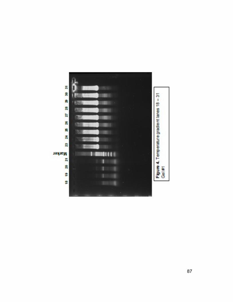
The four primer pairs above were analyzed by PCR at eight annealing
temperatures using a known negative sample, #062 and a known positive sample,
#871. A plasmid CymMV was used as a positive control and ‘No Template’ was
used as a negative control.
Lane Primer pair sample
temperature
degrees C
1 CymTGB2/BNSNC Plasmid 50
2 871 65
3 871 63.9
4 871 62.1
5 871 59.4
6 871 55.9
7 871 53.4
8 871 51.4
9 871 50
10 Marker
11 062 65
12 062 63.9
13 062 62.1
14 062 59.4
18 062 55.9
19 062 53.4
20 062 51.4
21 062 50
22 Marker 50
23 CymTGB2/CymCoatR 871 65
24 871 63.9
25 871 62.1
26 871 59.4
27 871 55.9
28 871 53.4
29 871 51.4
30 871 50
31 Plasmid 50
Figure 2. Legend for gel #1

86
87

88
Lane Primer pair sample
temperature
degres C
1 CymTGB2/CymCoatR 062 65
2 062 63.9
3 062 62.1
4 062 59.4
5 062 55.9
6 062 53.4
7 062 51.4
8 062 50
9 Marker 50
10 CymCoatF/BNSNC 871 65
11 871 63.9
12 871 62.1
13 871 59.4
14 Plasmid 50
18 871 55.9
19 871 53.4
20 871 51.4
21 871 50
22 Marker 50
23 062 65
24 062 63.9
25 062 62.1
26 062 59.4
27 062 55.9
28 062 53.4
29 062 51.4
30 062 50
Figure 3. Legend for gel #2

89

90
91
Lane Primer pair sample
temperature
degres C
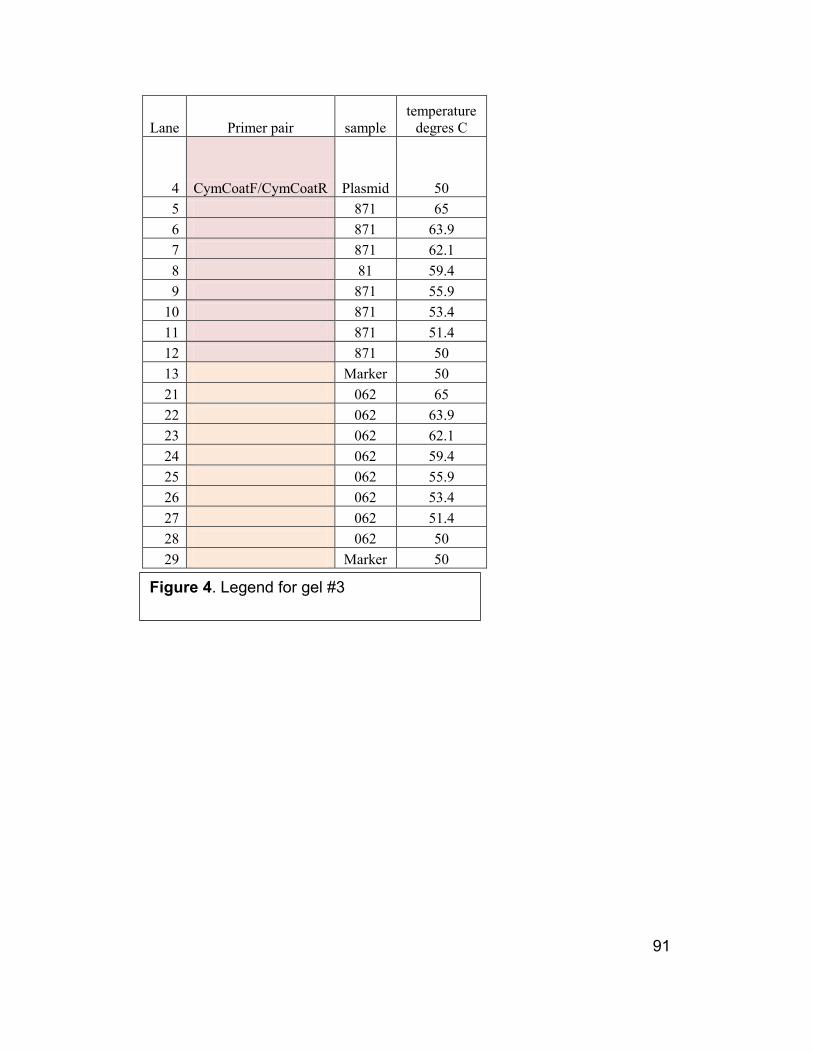
4 CymCoatF/CymCoatR Plasmid 50
5 871 65
6 871 63.9
7 871 62.1
8 81 59.4
9 871 55.9
10 871 53.4
11 871 51.4
12 871 50
13 Marker 50
21 062 65
22 062 63.9
23 062 62.1
24 062 59.4
25 062 55.9
26 062 53.4
27 062 51.4
28 062 50
29 Marker 50
Figure 4. Legend for gel #3

92

93

94
Appendix D
New Primer Annealing Temperature Gradient
The primer combination CymCoatF/CymCP-R2 gave strong bands in the range of
640 bp and when compared to the other combinations, less non-specific product.
Bands were strong at all annealing temperatures.
The primer combination CymF23/CymR25 gave strong bands in the range of 258
bp. In this trail there seemed to be little or no non-specific product. The bands
were strong at all annealing temperatures. There was, however some product in
the ‘No Template’ lane.
Gro
up
Lane
# Template
Forw
ard Reverse
Annealin
g
Temperat
ure
Produ
ct
length
A 1 cDNA #871, 1:100 dil
Cym
Coat
F
CymCP
-R2 50 (50)
640
bp
2 cDNA #871, 1:100 dil
Cym
Coat
F
CymCP
-R2 55 (55.0)
3 cDNA #871, 1:100 dil
Cym
Coat
F
CymCP
-R2 60 (60.6)
4 cDNA #871, 1:100 dil
Cym
Coat
F
CymCP
-R2 65 (65.2)
5 NT
Cym
Coat
F
CymCP
-R2 50 (50)
B 6 cDNA #871, 1:100 dil
Cym
Coat
F
CymR2
5 50 (50)
653
bp
7 cDNA #871, 1:100 dil
Cym
Coat
F
CymR2
5 55 (55.0)
8 cDNA #871, 1:100 dil
Cym
Coat
F
CymR2
5 60 (60.6)
9 cDNA #871, 1:100 dil
Cym
Coat
F
CymR2
5 65 (65.2)
10 NT
Cym
Coat
F
CymR2
5 50 (50)
C 11 cDNA #871, 1:100 dil Cym CymR2 50 (50) 258

95
F23 5 bp
12 cDNA #871, 1:100 dil
Cym
F23
CymR2
5 55 (55.0)
13 cDNA #871, 1:100 dil
Cym
F23
CymR2
5 60 (60.6)
14 cDNA #871, 1:100 dil
Cym
F23
CymR2
5 65 (65.2)
15 NT
Cym
F23
CymR2
5 50 (50)
D 16 cDNA #871, 1:100 dil
Cym
F23
CymCP
-R2 50 (50)
245
bp
17 cDNA #871, 1:100 dil
Cym
F23
CymCP
-R2 55 (55.0)
18 cDNA #871, 1:100 dil
Cym
F23
CymCP
-R2 60 (60.6)
19 cDNA #871, 1:100 dil
Cym
F23
CymCP
-R2 65 (65.2)
20 NT
Cym
F23
CymCP
-R2 50 (50)
E 21 cDNA #871, 1:100 dil
Cym
F23
CymCo
atR 50 (50)
368
bp
22 cDNA #871, 1:100 dil
Cym
F23
CymCo
atR 55 (55.0)
23 cDNA #871, 1:100 dil
Cym
F23
CymCo
atR 60 (60.6)
24 cDNA #871, 1:100 dil
Cym
F23
CymCo
atR 65 (65.2)
25 NT
CymF2
3
CymCo
atR 50 (50)

96

97

98
Appendix E
Inoculation interval (Time A)
X indicates no sample
Plant
No.
Inoculation
date interval CymMV
1-1 5/20 10 POS
1-2 3/1 15 Neg
1-3 3/1 15 Neg
1-4 3/1 17 Neg
1-5 3/1 6 Neg
2-1 x x
2-2 x x
2-3 5/30 7
2-4 5/20 10 POS
2-5 x x
3-1 3/1 6 Neg
3-2 5/30 7
3-3 5/30 14
3-4 5/30 14
3-5 x x
4-1 5/20 10 POS
4-2 3/18 11 Neg
4-3 3/18 25 Neg
4-4 5/30 7
4-5 5/20 10 POS
5-1 3/1 17 Neg
5-2 5/30 7
5-3 3/1 15 Neg
5-4 3/18 25 Neg
5-5 3/18 11 Neg
6-1 5/30 14
6-2 5/30 7
6-3 x x
6-4 5/30 14
6-5 x x
7-1 5/20 10 POS

99
7-2 x x
7-3 3/18 11 Neg
7-4 5/30 24
7-5 5/30 10 Neg
8-1 3/1 8 Neg
8-2 3/1 17 Neg
8-3 5/20 10 POS
8-4 3/1 15 Neg
8-5 3/1 8 Neg
9-1 3/18 25 Neg
9-2 3/18 25 Neg
9-3 x x
9-4 3/18 11 Neg
9-5 3/18 11 Neg
10-1 3/18 25 Neg
10-2 3/18 25 Neg
10-3 3/18 11 Neg
10-4 x x
10-5 5/30 7
11-1 3/18 11 Neg
11-2 5/20 10 POS
11-3 5/30 7
11-4 3/1 8 Neg
11-5 5/30 14
12-1 5/30 7
12-2 5/30 7
12-3 3/1 15 Neg
12-4 3/18 11 Neg
12-5 5/30 7
13-1 5/20 10 POS
13-2 5/30 7
13-3 3/1 15 Neg
13-4 3/18 25 Neg
13-5 3/18 25 Neg
14-1 3/18 25 Neg
14-2 x x
14-3 3/1 8 Neg
14-4 5/30 24
14-5 5/20 10 POS
15-1 5/20 10 POS
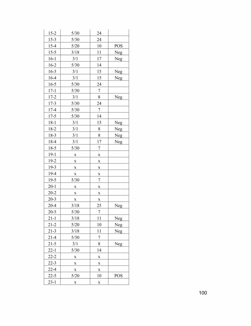
100
15-2 5/30 24
15-3 5/30 24
15-4 5/20 10 POS
15-5 3/18 11 Neg
16-1 3/1 17 Neg
16-2 5/30 14
16-3 3/1 15 Neg
16-4 3/1 15 Neg
16-5 5/30 24
17-1 5/30 7
17-2 3/1 8 Neg
17-3 5/30 24
17-4 5/30 7
17-5 5/30 14
18-1 3/1 15 Neg
18-2 3/1 8 Neg
18-3 3/1 8 Neg
18-4 3/1 17 Neg
18-5 5/30 7
19-1 x x
19-2 x x
19-3 x x
19-4 x x
19-5 5/30 7
20-1 x x
20-2 x x
20-3 x x
20-4 3/18 25 Neg
20-5 5/30 7
21-1 3/18 11 Neg
21-2 5/20 10 Neg
21-3 3/18 11 Neg
21-4 5/30 7
21-5 3/1 8 Neg
22-1 5/30 14
22-2 x x
22-3 x x
22-4 x x
22-5 5/20 10 POS
23-1 x x

101
23-2 5/30 7
23-3 x x
23-4 3/1 15 Neg
23-5 5/30 24
24-1 5/30 14
24-2 5/20 10 POS
24-3 3/18 11 Neg
24-4 5/20 10 POS
24-5 x x
25-1 5/30 7
25-2 3/1 17 Neg
25-3 3/1 17 Neg
25-4 3/18 11 Neg
25-5 3/1 6 Neg
26-1 3/18 25 Neg
26-2 3/18 11 Neg
26-3 5/30 14
26-4 3/1 17 Neg
26-5 5/30 14
27-1 5/20 10 Neg
27-2 3/18 25 Neg
27-3 x x
27-4 x x
27-5 3/18 11 Neg
28-1 5/20 10 Neg
28-2 5/20 10 Neg
28-3 3/18 11 N
28-4 5/30 7
28-5 3/1 15 Neg
29-1 5/30 7
29-2 3/1 6 Neg
29-3 5/30 7
29-4 3/1 17 Neg
29-5 3/1 6 Neg
30-1 5/30 14
30-2 5/30 7
30-3 5/30 24
30-4 3/1 15 Neg
30-5 5/20 10 Neg
102
Appendix F
Chi square statistical analysis
Interval Status A
A P 14
A N 19
B P 0
B N 28
C P 0
C N 9
D P 0
D N 12
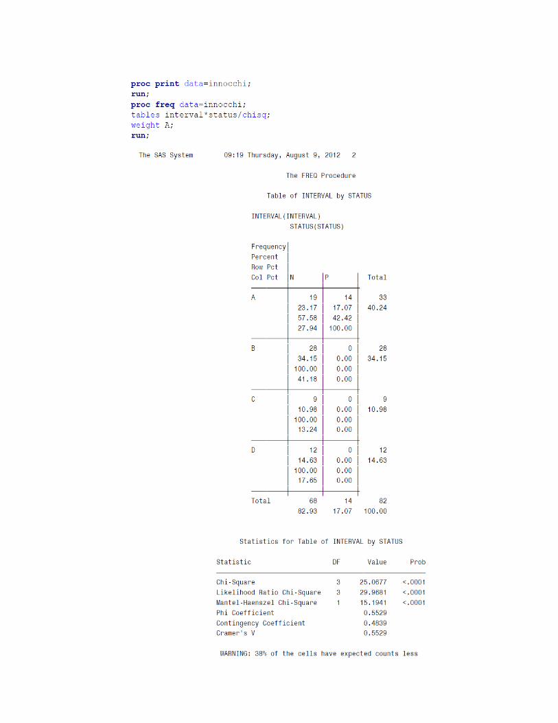
103

104

105
Bibliography
Arif, M., Fletcher, J., Ochoa Corona, F. M. Application of primer and probe
modifications in detection, biosecurity, and microbial forensics. Phytopathology
102, number 7 (Supplement 4) S4.6. July 2012
Bell, H. A., Cooke, D., Wildey, K.B., Baker, L.F., Mosson, J., and Short, J. Long-
Term Management of a Population of Australian Cockroaches (Periplaneta
australasiae) in a Tropical Plant House in the United Kingdom using the Juvenile
Hormone Analogue (S)-Hydroprene. Proceedings of the 3rd
International
Conference on Urban Pests. 1999.
Borth, W.B., Barry, K., Obsuwan, K., Xu, M.Q., Liu, R.W., Kuehnle, A.R. and
Hu, J.S. 2006. Movement of cymbidium mosaic virus and transgenic resistance in
Dendrobium orchids. Acta Hort. 722:137-146.
http://www.actahort.org/books/722/722_17.htm
Broadbent, L. Tinsley, T. W., Buddin, W., and Roberts, E. T. The spread of
lettuce mosaic in the field. Annals of Applied Biology 1951 38, 689-706
Brunt, A.A., Crabtree, K., Dallwitz, M.J., Gibbs, A.J., Watson, L. and Zurcher,
E.J. (eds.) (1996 onwards). ‘Plant Viruses Online: Descriptions and Lists from
the VIDE Database. Version: 20th
August 1996.' URL:
http://biology.anu.edu.au/Groups/MES/vide/. Accessed May 19, 2011
Büchen-Osmond, Cornelia. Cymbidium mosaic virus. ICTVdB - The Universal
Virus Database.
http://www.ictvdb.rothamsted.ac.uk/ICTVdB/00.056.0.01.007.htm. Accessed
May 19, 2011
Chang, Ching-An. Orchid virus diseases in Taiwan and their control strategies.
International Commercial Orchid Growers Organization.
http://www.icogo.org/images/Dr._Ching-An_Chang_-_Orchid_Viruses.pdf
Accessed 5/21/11
Chang, C., Chen, Y. C., Hsu, Y. H., Wu, J. T., Hu. C. C., Chang, W. C., Lin, N.
S., Transgenic resistance to Cymbidium mosaic virus in Dendrobium expressing
the viral capsid protein gene. Transgenic Research 2005 Feb; 14 (1): 41-46.
Cochran, Donald G. Cockroaches, their biology, distribution, and control. World
Health Organization, Communicable Diseases Prevention and Control, WHO
Pesticide Evaluation Scheme (WHOPES). 1999

106
Corbett, M. K. Purification by density gradient centrifugation, electron
microscopy, and properties of Cymbidium mosaic virus. Phytopathology 50: 346-
351. 1960
Floriculture Crops 2011 Summary (May 2012) USDA, National Agricultural
Statistics Service.
http://usda01.library.cornell.edu/usda/current/FlorCrop/FlorCrop-05-31-2012.pdf.
Accessed July 2012
Freitag, J. H., Frazier, N. W., and Sylvester, E. S. Dilworth D. Jensen. Essig
Museum of Entomology Collections. 2011.
http://content.cdlib.org/view?docId=hb9k4009c7&doc.view=content&chunk.id=d
iv00027&toc.depth=1&&anchor.id=0. Accessed May 20, 2011.
Fulton, J. P., Gergerich, R. C., and Scott, H. A. Beetle transmission of plant
viruses. Annual Review of Phytopathology. 25 (111-123) 1987
Gold, A. H. & Jensen, D. D. An electron microscope study of Cymbidium mosaic
virus. American Journal of Botany 38: 577-578., 1951
Hu, J. S. Ferreira, S., Wang, M., Xu, M. Q. Detection of Cymbidium Mosaic
Virus, Odontoglossum Ringspot virus, Tomato Spotted Wilt Virus, and
Potyviruses Infecting Orchids in Hawaii., American Phytopathological Society
DOI:10.1094/PD-77-0464. 1993
Hull, Roger. Matthew’s Plant Virology, Fourth Edition. Academic Press, London,
UK. 1970
Inouye, Narinobu. Viruses of Orchids, Symptoms, Diagnosis, Spread, and
Control. Blue Bird Publishers, The Netherlands. 2008
Izaguirre-Mayoral, M.L., de Uzcategui, R.C. and Carballo, O. 1993. Crassulacean
acid metabolism in two species of orchids infected by tobacco mosaic virus-
orchid strain and/or Cymbidium mosaic virus. J. Phytopathology 137:272-282.
Jensen, D. D. Virus Diseases of Orchids. California Agriculture. 6: 7, 15, 16.
January and February 1952.
Jensen, D. D. Virus diseases of orchids in The Netherlands., Netherlands Journal
of Plant Pathology. 76 (1970) 135-139.
Khentry, Y., Paradornuwat, A., Tantiwiwat, S., Phansiri, S., Thaveechai, N.
Incidence of Cymbidium mosaic virus and Odontoglossum ringspot virus in
Dendrobium spp. in Thailand. Crop Protection 25 (2006) 926-932

107
Kiang Ho, Wong. Orchid Industry in Malaysia and Global Market. Commercial
Orchid Grower Association of Malaysia. 2010.
http://sdvi.fama.net.my/maha/seminar/upload_module/paper_MAHA2010/Sesi02
_Kertas05.pdf. Accessed July 2012
Kim, J. D., Koo, Y. B., Chang, M. U. Genome characterization of a Korean
isolate of Cymbidium mosaic virus. Molecules and Cells. 1997 8 (2) 181-188
Kuehnle, A. R., Breeding and biotechnology of orchids with special emphasis
Dendrobiums. USDA Research, Education, & Economics Information System.
http://www.reeis.usda.gov/web/crisprojectpages/0014694-breeding-and-
biotechnology-of-orchids-with-special-emphasis-on-dendrobiums.html. Accessed
7/22/12
Kunkle, Joseph G. The Cockroach Homepage. Biology Department, University of
Massachusetts. 2007. http://www.bio.umass.edu/biology/kunkel/cockroach.html.
Accessed 8/9/12
Liao, L. J., Pan, I. C., Chan, Y. L, Hsu, Y. H., Chen, W. H., Chan, M. T.,
Transgene silencing in Phalaenopsis expressing the coat protein of Cymbidium
mosaic virus is a manifestation of RNA-mediated resistance. Molecular Breeding
2004 13(3) 229-242
Lu, Hsiang-Chia, Chen, Cheng-En, Tsai, Meng-Hsiun, Wang, Hsiang-iu, Su,
Hong-Ji, Yeh, Hsin-Hung. Cymbidium mosaic potexvirus isolate-dependent host
movement systems reveal two movement control determinants and the coat
protein is dominant. Virology 388 (2009) 147-159
Moles, M., Delatte, H., Farreyrol, K., Grisoni, M. Evidence that Cymbidium
mosaic virus (CymMV) isolates divide into two subgroups based on nucleotide
diversity of coat protein and replicase genes. Archives of Virology, 152:4 (705-
715) 2007
Mullis, K. B. and Faloona, F. Specific synthesis of DNA in vitro via a
polymerase-catalyzed chain reaction. Methods Enzymole, 155 (335-350) 1987
Murakishi, H. H., Host range, symptomalology, physical properties, and cross-
protection studies of orchid virus isolates. Phytopathology 48: 132-137. 1958
Namba, R. and Ishii, M. Failure of Aphids to transmit the Odontoglossum
Ringspot and Cymbidium mosaic viruses to orchid plantlets derived from
meristem cultures. Phytopathology 61:582-583. 1971

108
Obsuwn, K., Hieber, D. A., Mudalige-Jayawickrama, R. G., Kuehnle, A. R. A
dysfunctional CymMV movement protein confers resistance to CymMV in
Dendrobium Orchid., Acta Horticulturae 2009 No. 836 pp. 149-154
Potexvirus. ViralZone. http://www.expasy.ch/viralzone/all_by_species/272.html.
Accessed 5/20/11
Ramel, Gordon. The Blattodea or Cockroaches. 2001.
http://www.earthlife.net/insects/blatodea.html. Accessed 8/9/12
Sherpa, A. R., Hallan, V., Pathak, P., Zaidi, A. A. Complete nucleotide sequence
analysis of Cymbisium mosaic virus Indian isolate: further evidence for natural
recombination among Potexviruses. Journal of Bioscience 32 (4) June 2007, 663-
669
Sherpa, A. R., Hallan, V., Ram, R., Vij, S. P., Pathak, P., Garg, I. D., Zaidi, A. A.
The first report of Cymbidium mosaic virus (CymMV) in orchids from India.
BSPP Web, New Disease Reports (2003) 7, 10.
http://www.ndrs.org.uk/article.php?id=007010. Accessed 5/20/11
Stejskal, V., Lukas, J., and Aulicky, R. Temperature-dependent development and
mortality of Australian Cockroach, Periplaneta australasias (Fabricius) Plant
Protection Science 40(1) 11-15. 2004
ViralZone www.expasy.org/viralzone, Swiss Institute of Bioinformatics.
Accessed July 2012
White, N. H. and Goodchild, D. J. Mosiac or black streak disease of Cymbidium
and other orchid hybrids. Journal Australian Institute of Agricultural Science 21:
36-38. 1955
Wong, S. M., Chng, C. G., Lee, Y. H., Tan, K., Zettler, F. W. Incidence of
Cymbidium mosaic and Odontoglossum ringspot viruses and their significance in
orchid cultivation in Singapore. Crop Protection, 13:235-239. 1994
Wong, S. M., Mahtani, P.H., Lee, K. C., Yu, H. H., Tan, Y., Neo, K. K., Chan,
Y., Wu, M., and Cheng, C. G. Cymbidium mosaic potexvirus RNA: complete
nucleotide sequence and phylogenetic analysis. Archives of Virology, 142: 383-
391. 1997


![PRODUCTIVITY AND CYTOGENETIC STABILITY OF PROTOCORM … · 2014-12-17 · PRODUCTIVITY AND CYTOGENETIC STABILITY OF CYMBIDIUM PLBs 96 [GOGOI & al. 2012], hybrid Cymbidium Twilight](https://img.dokumen.tips/doc/110x75/5f23e5e348b1d45ec30cac2a/productivity-and-cytogenetic-stability-of-protocorm-2014-12-17-productivity-and.jpg)


![Desenfrascado De Cymbidium[1]](https://img.dokumen.tips/doc/110x75/5561d478d8b42aa83f8b55be/desenfrascado-de-cymbidium1.jpg)