Embed Size (px)
Citation preview
The Egyptian Journal of Hospital Medicine (April 2012) Vol., 47: 321 – 333
321
Avoiding mastoid cavity Problems: Mastoid obliteration using Bioactive glass®
** Said Shokry, MD; *Al`Sayed Hossieni Al`Sayed, MD; *Mohammed Fatehy Zidan, MD;
**Adel Ahmed Hafez,MD;**Hanafi Mahmoud Abdulsalam,MD
Abstract Background and objective: The aim of this study was to evaluate bioactive glass as an ideal material for the
purpose of mastoid cavity elimination after mastoid surgery to avoid mastoid cavity problems.
Materials and methods: In 20 patients diagnosed as cholesteatoma or chronic unsafe ear, we used different surgical
techniques according to pathology and situation during surgical exploration, basically adhering to standard
principles of eradicating disease in chronic unsafe ear. After performing the canal wall down (CWD) or the canal
wall up (CWU) technique, mastoidectomy was followed by obliteration of mastoid cavity by particulate form
Bioglass®. Cases were divided according to operative procedures, type of reconstruction and material used into 3
groups A- Canal wall up mastoidectomy followed by obliteration of mastoid cavity by particulate form Bioglass®.
B- Canal wall down mastoidectomy followed by reconstruction of posterior meatal wall and obliteration of mastoid
cavity by particulate form Bioglass®. C- Canal wall down mastoidectomy followed by reconstruction of posterior
meatal wall by conchal cartilage and obliteration of mastoid cavity by Bioglass®.
Results: Bioactiveglass paste is very effective for mastoid obliteration in the three groups with good integration to
the surrounding tissues either connective tissue, bone, meninges or lateral dural sinus without any adverse reaction
on the dura even with contact to Bioglass®. Infection was seen in 2 cases (10%), however was readily controlled by
topical application of antibiotics daily for one week. In both cases no extrusion of the material occurred.
Conclusion: The successful formation of bone with elimination of mastoid cavity problems proved that using
Bioglass is appropriate for performing clinical mastoid obliteration.
Introduction
The ideal surgical procedure for cholesteatoma
should satisfy the following conditions: first, the creation of a dry safe ear; second, the prevention
of recurrence; and the reestablishment of a well-
aerated middle ear with a properly functioning sound-conducting mechanism. The choice of
surgical technique is mainly based on the
propagation of the disease, Eustachian tube
function, and the status of other middle ear structures.
Over the past years, the gold standard for
management of cholesteatoma has been the canal wall down technique (CWD). Removal of
the posterior canal wall allows exposure of the
entire epitympanum and middle ear and ensures
complete disease eradication. However, this
technique has several problems such as the
accumulation of debris, requiring periodic cleaning and water restriction; dizziness; and
difficulty with fitting in a hearing aid (Birzgalis
et al, 1994). With the canal wall up technique (CWU) these problems can be avoided, although
the rate of residual and recurrent disease tends to
be higher than when using the CWD (Meuser,
1985).
Various factors can contribute to a problematic
cavity, namely a large cavity, high facial ridge, narrow meatus, dependent mastoid tip, residual
disease and an open middle ear space. Each of
these problems is amenable to surgical correction.
*ENT Depart, Faculty of Medicine, Al`Azhar University Cairo
** Hearing and Speech Institute _Egypt

Avoiding mastoid cavity Problems….
322
However many techniques have been used since the beginning of last century for mastoid
obliteration to reduce the cavity size. Various
autologous materials have been used, such as
muscle, fat, cartilage, musculoperiosteal flaps, bone chip and bone pate. There are technical
problems with each, most commonly the
variable resorption that occurs postoperatively leading to an unpredictable final cavity size.
There is also the theoretical risk of reimplanting
cholesteatoma if bone pate is used for obliteration.
Different biomaterials have been tried on the
basis that they should be non-resorbable, non-reactive and integrate. Carbonated calcium
phosphate (CCP) bone cements have many
features useful in otologic surgery. These cements harden within minutes in a moist
environment, are non-toxic and non-exothermic,
and, like hydroxyapatite, have the potential for osseointegration and remodeling.
This study aimed to assess the long-term
effectiveness of Bony glass®, Bioglass® "45S5", as a suitable biomaterial for mastoid
obliteration. Bioglass® "45S5" is a bioactive
glass ceramics which is composed of 45% silicone dioxide, 24.5% calcium oxide, 24.5%
sodium dioxide and 6% phosphorous pentoxide
(Lossdorfer et al 2004).
The aim of using of such materials for the graft
is to promote adequate bone regeneration at the
defective site by acting as a scaffold for osseous growth. Bony glass® resorbs and regenerates
bone in 3 to 6 months depending on the site of
implantation, the size of the bony defect and the age of the patient. Many tests showed that
Bioactive glass® was neither carcinogenic nor
toxic to any of the tissues or systems with which
it was in contact (Wilson et al, 1981).
PATIENTS AND METHODS This study is a prospective study at a tertiary
care hospitals looking at the use of Bioglass®
for the obliteration of mastoid cavities in 20
patients. We conducted on 20 patients with chronic suppurative otitis media (7 with
granulation tissue and 13 with cholesteatoma).
They were suffering from chronic discharging ear. All cases were subjected to mastoid surgery
with complete eradication of the disease. The
patients presented to outpatient clinic of Hearing and Speech Institute and Al`Zahra University
Hospital Oto-Rhino-Laryngology department.
The age ranged from 10 to 50 years. They had
no previous ear surgeries. Cases in which complete eradication of disease was not certain
(e.g. extensive granulation on top of
cholesteatoma) were not subjected to obliteration and accordingly excluded from the
study.
operative -Pre All patients were subjected to
assessment:
Clinical evaluation: includes detailed history of
the disease (onset, duration, course, frequency of exacerbation, development of any complications
and history of previous surgery of the ear);
Examination of nose and throat; Otoscopic and microscopic examination ; Audiologic
assessment ; Plain x-ray on both mastoids;
Culture and sensitivity for ear discharge;
Systemic examination to assess surgical fitness and finally Preparation for mastoid operation.
Laboratory investigations: Includes complete
blood picture; Bleeding and clotting times; Random blood sugar; Urea and creatinine levels;
SGOT and SGPT levels. ECG and Plain chest
x-ray.
Operative procedures
Cases were operated upon during the period
from April 2006 to April 2007; with Bioglass® used in particulate form which presented in vials
containing 1gm of sterilized Bioglass®.

Mohammed Zidan et al
323
According to the operative procedures, cases were classified into three groups:-
Group A: Six cases with canal wall up mastoidectomy followed by obliteration of mastoid cavity by
particulate form Bioglass®. 1. Using a retroauricular incision, a canal wall-up mastoidectomy was performed; then a large graft 3X3
cm from temporalis fascia was taken. Large Palva flap anteriorly based to provide a good cover of the
filling material in the cavity. Using a large cutting burr, the bone above and behind the external auditory meatus (Mc Ewen's triangle) was gradually excavated to open the antrum, the wall of the
cavity was gradually widened until the lateral sinus plate, dural plate, and the dome of the lateral
semicircular canal and short process of the incus were identified. Working from the antrum; all air
cell were exenterated until the white bony plates over the middle and posterior cranial fossae were exposed. Any diseased tissues were removed. During the whole process of excavation, the bony
posterior meatal wall was thinned, but kept intact (Canal wall up mastoidectomy).
2. The annulus was elevated and any diseased tissue in the middle ear was removed. Gelfoam was applied to the middle ear, then the remanent of the drum was grafted with temporalis
fascia and gelfoam was applied above the graft in the ext canal. A small piece of gelfoam enough to
close the aditus ad antrum prevents particulate Bioglass® from escaping to the middle ear.
3. We prepared the filling by mixing the particulate form Bioglass® with venous blood. The mastoid cavity has been filled with the mixture. The particulate form Bioglass® was covered by a large piece
of gelfoam before closure of the wound to prevent incorporation of particles of Bioglass® inbetween
the edges of the wound. Post auricular incision was closed in layers with interrupted sutures. Group B: Nine cases with canal wall down mastoidectomy followed by reconstruction of posterior
meatal wall and obliteration of mastoid cavity by particulate form Bioglass®
1. Using a retroauricular incision, a canal wall-down mastoidectomy was performed. The skin of the external meatus was preserved as much as possible.
2. Combined reconstruction of the posterior meatal wall and obliteration of mastoid cavity by particulate
form Bioglass® was done in one step as follows:-
*A foil template which gives accurate measurement and degree of curvature of the posterior meatal wall has been used to get the template. This foil template has been used to keep the particulate Bioglass® in
the desired place. Apiece of cotton soaked in saline was applied in the middle ear and external canal to
maintain the position of the foil template. *The prepared mixture of Bioglass® particles and blood was used to fill the mastoid cavity until the
posterior meatal wall has been reconstructed. The piece of cotton in the middle ear was then removed.
3. Gelfoam was applied to the middle ear .Then a temporalis graft was applied below the drum remnant with extension to cover the Bioglass®. Gelfoam was applied in sufficient amount above the graft in
the renewed ext. canal to support the Bioglass® particulates in place then the foil template was
removed.
Group C: Five cases with canal wall down mastoidectomy. This is followed by reconstruction of posterior meatal wall by conchal cartilage and obliteration of mastoid cavity by Bioglass®.
1. Using a retroauricular incision, a canal wall-down mastoidectomy was performed. The skin of the
external meatus was preserved as much as possible, just like in group B. Then reconstruction of the posterior meatal wall by conchal cartilage was done by Conchal cartilage was adjusted, by scalpel
knife and trimmed to fit the wall defect and slipped between two grooves done by fine diamond burr
at the site of the superior and inferior buttresses.
2. Gel foam was applied to the middle ear, and then a temporalis graft was applied below the drum remnant. Gelfoam used again to cover the graft.
3. The prepared Bioglass®-blood mixture was used for obliteration of the mastoid cavity. The Operative
steps in group C are illustrated in the following figures (1-6).
Follow up -Regular follow up every two weeks during the first two months postoperative then every month during
the next six months has been done.
Avoiding mastoid cavity Problems….
324
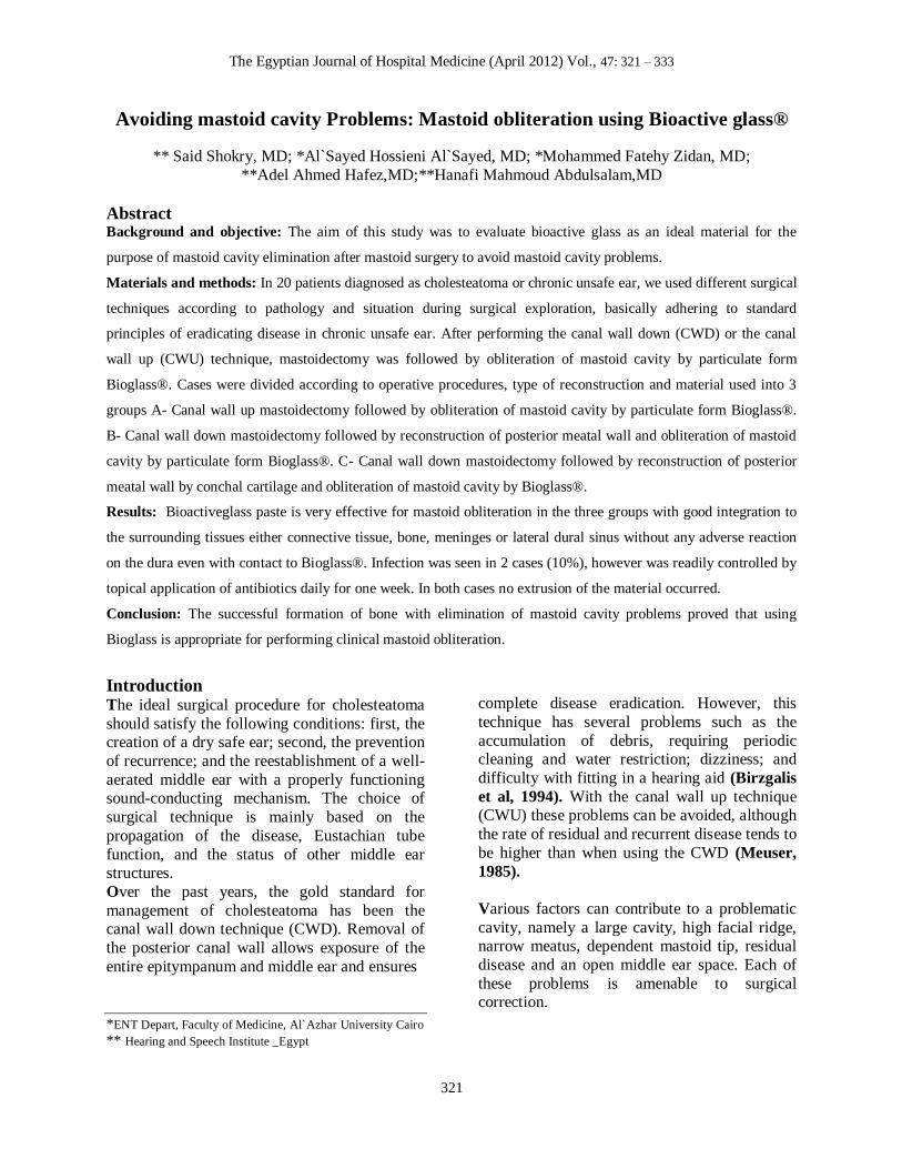
Figure 1. Mastoid cavity after completion of CWD mastoidectomy
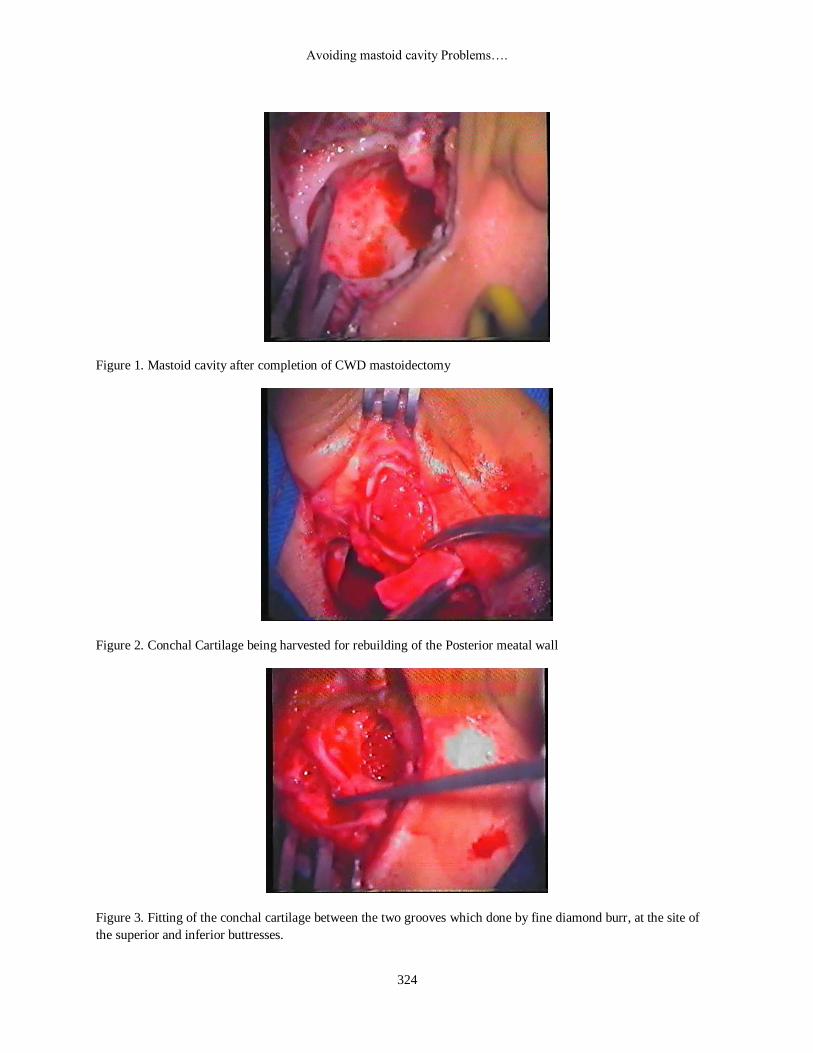
Figure 2. Conchal Cartilage being harvested for rebuilding of the Posterior meatal wall
Figure 3. Fitting of the conchal cartilage between the two grooves which done by fine diamond burr, at the site of
the superior and inferior buttresses.
Mohammed Zidan et al
325
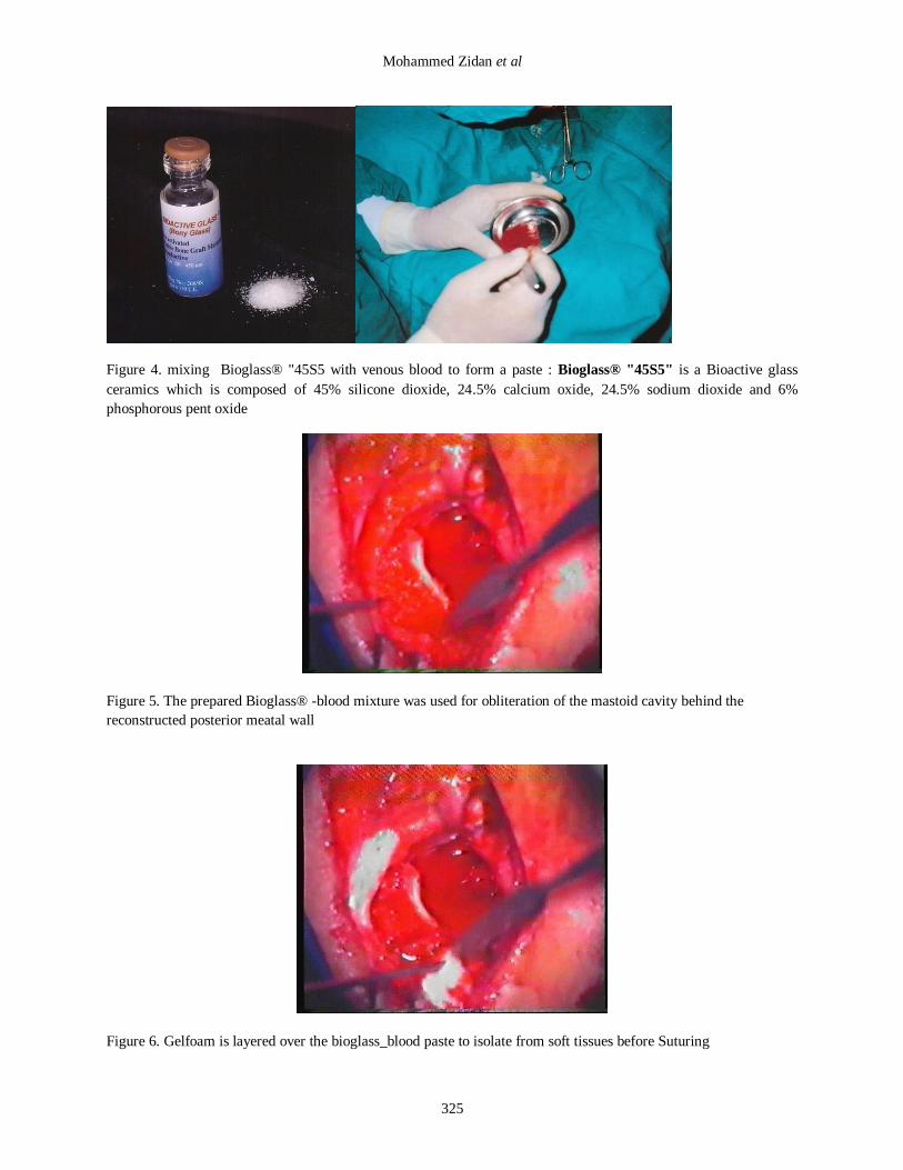
Figure 4. mixing Bioglass® "45S5 with venous blood to form a paste : Bioglass® "45S5" is a Bioactive glass
ceramics which is composed of 45% silicone dioxide, 24.5% calcium oxide, 24.5% sodium dioxide and 6%
phosphorous pent oxide
Figure 5. The prepared Bioglass® -blood mixture was used for obliteration of the mastoid cavity behind the
reconstructed posterior meatal wall
Figure 6. Gelfoam is layered over the bioglass_blood paste to isolate from soft tissues before Suturing
Avoiding mastoid cavity Problems….
326
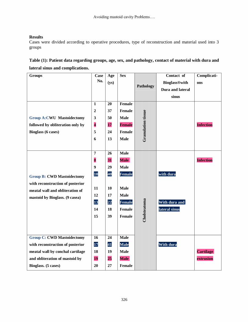
Results Cases were divided according to operative procedures, type of reconstruction and material used into 3
groups
Table (1): Patient data regarding groups, age, sex, and pathology, contact of material with dura and
lateral sinus and complications.
Complicati-
ons
Contact of
Bioglass®with
Dura and lateral
sinus
Pathology
Sex Age
(ys)
Case
No.
Groups
Infection
Gra
nu
lati
on
tis
sue
Female
Female
Male
Female
Female
Male
20
37
50
17
24
13
1
2
3
4
5
6
Group A:CWU Mastoidectomy
followed by obliteration only by
Bioglass (6 cases)
Infection
with dura
With dura and
lateral sinus
Ch
ols
tea
tom
a
Male
Male
Male
Female
Male
Male
Female
Female
Female
26
31
29
40
10
17
22
18
39
7
8
9
10
11
12
13
14
15
Group B: CWD Mastoidectomy
with reconstruction of posterior
meatal wall and obliteration of
mastoid by Bioglass. (9 casea)
Cartilage
extrusion
With dura
Male
Male
Male
Male
Female
24
41
19
25
27
16
17
18
19
20
Group C: CWD Mastoidectomy
with reconstruction of posterior
meatal wall by conchal cartilage
and obliteration of mastoid by
Bioglass. (5 cases)

Mohammed Zidan et al
327
Table (2) distribution of studied group as regards pathology.
Total Group C Group B Group A Groups
% No % No % No % No
35% 7 _ _ 11.11% 1 100% 6 Granulation tissue
65% 13 100% 5 88.88% 8 _ _ Cholesteatoma
100% 20 100% 5 100% 9 100% 6 Total
The pathology detected during operation, 7 cases
(35%) with extensive granulation tissue {all
cases of group A and 1 case in group B} were found, whereas 13 cases (65%) with
cholesteatoma {8 cases of group B and all cases
of group C} were found and none of group A were cholesteatoma (Graph1).
All cases of cholesteatoma have undergone
canal wall down procedure for complete
eradication of the disease and all cases of granulation tissue have undergone canal wall up
procedure without fear of incomplete eradication
of the disease except in one case transformed to CWD.
Bioglass® was in contact with dura in 2 cases
(10%) {Cases No 10 from group B and case No
17 from group C (Table1)}, and in contact to both dura and lateral sinus in another single case
(5%){Case No 13 from group B (Table1)}
without any adverse reaction on the dura. We found out that there was no adverse reaction on
the dura due to contact with Bioglass®
Graph (1)-Distribution of studied groups as
regards pathology
Table (3): Postoperative problems in different mastoid obliteration groups
Total Group C Group B Group A Groups
% No % No % No % No
10% 2 _ _ 11.11% 1 16.67 1 Infection
5% 1 20% 1 _ _ _ _ Extrusion of reconstructed cartilage of PMW
15% 3 20% 1 11.11% 1 16.67 1 Total

Avoiding mastoid cavity Problems….
328
Two complications were encountered infection
and cartilage extrusion. Infection occurred in two cases (one in group A No. 4 and the other in
group B No. 8 (Table 3). This was discovered
one week postoperative during removal of the
pack and was controlled by topical antibiotics daily for one week. In both cases no extrusion of
the material occurred. Extrusion of the conchal
cartilage used in reconstruction of posterior meatal wall occurred in 1 case in group C one
month post-operative (case No 18 – Table 1).
Discussion
Canal wall up (CWU) techniques preserve the
anatomy of the posterior canal wall, eliminating the need for periodic bowl cleaning and avoiding
the risk of recurrent bowl infection. However the
recidivism rate may be as high as 36% in adults and 67% in children after CWU procedures
(Shohet JA& De Jong 2002). Surgical
intervention is “closed” when afterwards there is no persistence of any communication between
the external meatus, which remains more or less
intact, and the antroattical cavities, which are
trepanned (trephined) during the operation (Jansen, 1958). Nevertheless, the debate is still
on due to new evidence, better imaging, high-
tech endoscopes and intraoperative use of facial nerve monitoring.
However, Canal wall-down mastoidectomy is
the most widely used surgical method worldwide. It is supposed to be easier, of shorter
duration, necessitates less surgical experience
than the CWU procedures, and has low recurrence and residual rate. CWD procedure is
claimed to be the unique solution for
cholesteatomas in an only – hearing ear and when there is a labyrinthine fistula
(Hulka&McElveen 1998). However, in a
situation such as the only hearing ear surgery,
Tu (2005) did not adopt CWD approach, except for special considerations.
Some surgeons propose a different procedure, the canal wall window (CWW) technique, which
involves slitting the posterior canal wall. They
claim that it provides good hearing results, especially in a young population who will bear
the surgical outcome for many decades
(Godinho et.al 2005).
Hopkin’s wide-angle endoscopes are
intraoperatively useful for checking areas that
cannot be visualized with the microscope. Therefore, the use of endoscopes is supposed to
reduce the residual cholesteatoma rate (Phelan
et. al 2008).This leads Nikolopoulos and
Gerbesiotis (2009) to revive and advocate the
well-known approach of inside to outside chase
of cholesteatoma sac, to find the mouth of the cholesteatoma sac, to follow it until the end, to
totally remove it creating a small mastoid cavity
after performing tympanoplasty. The procedure
is progressive, anterior-to posterior dissection, exposing the cholesteatoma, thus creating
atticotomy, atticoantrostomy and
mastoidectomy. Following the cholesteatoma and removing as much bone as needed allows
creating the smaller mastoid cavity possible.
Ideally, the middle ear cleft should be left as an air containing cavity not open to the EAM.
Where there is an extensive mastoid
involvement, the posterior meatal wall is totally removed and the facial ridge is taken down to
the floor of the external auditory meatus (EAM);
thus the mastoid cavity does not form an independent sump. HERE, bioactive glass can
be invited to obliterate the resulting mastoid
cavity after atticotomy, atticoantrostomy and
mastoidectomy tailored surgery to avoid mastoid cavity problems.
The disadvantages of biological grafts, gave attention to the use of synthetic materials. The
latter should have a high degree of
biocompatibility, shouldn't be extruded or resorbed, easily measured, contoured and should
provide predictable and consistent sound
transmission (El-seifi and Fouad 1998).
Bioglass® "45S5" is a bioactive glass ceramics which is composed of 45% silicone dioxide,
24.5% calcium oxide, 24.5% sodium dioxide
and 6% phosphorous pentoxide (Lossdorfer et
al 2004).
We therefore consider the bioactive glass to be a promising material as a bone substitute. In
addition, using autologous cartilage to

Mohammed Zidan et al
329
reconstruct the external auditory meatus is
advantageous in some cases of Canal down (CWD) mastoidectomy to aid fashioning
posterior meatal wall. In this study, the results
were satisfactory. Moreover, there were no
complications, such as hearing loss, vestibular dysfunction, cholesteatoma, or uncontrolled
proliferation of granulation. As preliminary
clinical report, our results indicate that Bioglass covered with gelfoam is likely to be useful for
mastoid obliteration. However, we need to have
a longer follow up to report more solid conclusion. We need further prospective case
control study.
However, Obliteration may solve the problems of the CWD presented due to no posterior canal
wall; with Bioglass obliteration, we expect much
improvement in auditory rehabilitation in patients who have mixed hearing loss due to
cholesteatoma and high frequency loss because
of the changing of external auditory canal resonance after surgery (Gantz et al 2005).
Bioactive glass obliteration has various
advantages in operations involving cholesteatoma. In addition to cholesteatoma,
chronic otitis media with poor Eustachian tube
function, adhesive otitis media, and a sclerotic mastoid cavity favorable to obliteration have
also become indications for using this technique.
Controversial Issues of this subject are still rising up.
One of the contraindications of reconstruction of
the posterior meatal wall and/ or obliteration of mastoid cavity is the incomplete removal of the
disease (Blak, 1995). So cases, in which
complete eradication of the disease was not certain, were excluded from this study.
Different synthetic materials have been used in
obliteration and reconstruction such as silicone, proplast, ionomer cement ceravital and hydroxyl
apatite. These materials did not fulfill optimal
criteriae and showed many disadvantages such as considerable foreign body reaction with
silicone (Rosenblut et al., 1994) and dehiscence
problems with proplast (Shea et al., 1984); Infection and encephalopathy with ionomer
cement (Renard et al 1994) and absorption and
lysis with ceravital (Reck, et al., 1988; El-Seifi
and Fouad, 1998).
Reported complications after obliteration with
synthetic material include: infection, extrusion,
resorption, myringitis, granulation tissue formation, recurrence of discharge, retraction
pocket formation, recurrence of cholesteatoma,
defect in external canal reepithelialisation, canal dehiscence and post-auricular fistula (Black,
1995). Our study showed infection in 2 cases
and extrusion of the cartilage in 1 case while myringitis, and canal dehiscence or granular
extrusion was not encountered.
Hydroxylapatite is the material most widely utilized as it has the best results among all
synthetic materials regarding to its bioactivity
and composition which resemble bone tissue (De Groot et al, 1988 & Ricci; 1992).
However, a comparative study of particulate
Bioglass® to hydroxylapatite as a bone graft substitute in animal models concluded that the
Bioglass® was superior to hydroxylapatite
because the latter showed encapsulation by
fibrous connective tissue, while Bioglass® showed true integration of the new bone without
any encapsulation. In Oonishi study
hydroxylapatite disappeared faster than Bioglass®, so that the empty spaces were not
completely filled with new bone formation.
Moreover, the speed of bone growth around the
Bioglass® was much faster and bone formation was much denser and more mature than with
hydroxylapatite (Oonishi et al., 1997).
Oonishi Radio- Histological Animal Studies on
guinea pig confirmed by (Jang, et al., 2007)
showed hyperintense areas of new bone formation in CT scans. Then Histological
evidence of new bone formation was observed in
the implant specimens that included: active
osteoblasts, osteocytes, chondrocytes and osteoid tissue. There was a definite bond
between the implant and the bone interface at
the areas of new bone formation. No inflammatory or foreign body reactions, caused
by the Bioactive glass® ceramic particle
implantation, were observed in the surrounding tissue.

Avoiding mastoid cavity Problems….
330
In our study all 6 cases with obliteration of the
cavity after canal wall up mastoidectomy showed neither retraction pocket formation nor
recurrent infection. Palva and Virtanent 1981;
Vartianen and Harma, 1987 concluded in their
comperative studies between obliterated and non-obliterated cavities after canal wall up
mastoidectomy that the obliterated cavities were
superior regarding to control of infection, hearing improvement and protection against the
formation of future retraction pocket in ears with
poor tubal function.
Bellantone et al., (2000); Jones et al (2006);
Lepparanta et al., (2007); Munukka et al.,
(2007); Waltimo et al, (2007) referred to the antibacterial effect of Bioactive glass® which
seems to be true since the 2 cases with infection
in our study showed no granular extrusion and complete bone formation occurred 6 months
later despite post-operative infection.
Drawbacks of reconstruction of posterior meatal
wall by conchal cartilage were the instability,
time consuming and subjecting the facial nerve
to surgical trauma during buttresses grooving as reported by Black (1995); also Dornhoffer and
Simmons, 2003 reported inadequate cartilage,
excessive curvature, the somewhat tedious process of cutting the cartilage making a good fit
impossible. For avoidance of these drawbacks in
the cases which were obliterated and
reconstructed, the use of particulate Bioglass® was preferred. This technique is faster, easier,
makes the particles adherent to each other like
one mass so extrusion became difficult and buttresses grooving with their complications
were avoided. This was achieved in all cases
which were reconstructed and obliterated by particulate form Bioglass®.
Black (1995) showed that the direct contact of
external auditory canal skin with hydroxylapatite used in reconstruction of posterior meatal wall is
followed by the formation of granulation tissue.
We inserted gelfoam between the particles of Bioglass® and the post-auricular incision before
closure of the wound thus, no granulation or
myringitis was formed in the external ear and the post-auricular incision showed good healing
in all cases.
In general, our results are comparable to previous results of Bioglass® application in
different sites of human body (Schepers et al.,
1993; shapoff, 1997; Stanley et al., 1997; Della
santina et., al 2006; Tuusa et al., 2007). In our study material probably results in new bone
formation in all cases with, no complications
were detected in cases where it became in contact with dura and lateral sinus and no
extrusion to particles of Bioglass® in all cases
even with cases in which infection was occurred.
Conclusion
Our study showed that CWR and mastoid
obliteration using Bioactive glass® is a technique that facilitates exposure of the middle
ear and ensure complete removal of
cholesteatoma. Reconstruction of the posterior canal wall with conchal cartilage or Bioactive
glass® recreates the normal external canal
anatomy and allow for elimination of the mastoid bowl. It is suitable for most patients
with chronic otitis media with cholesteatoma,
including adults and children.
Our study showed also that the Bioglass® material is bioactive, biocompatible, provides
favorable healing, resist infection, easily
prepared and placed, and probably provides new bone formation without any encapsulation. All
these findings demonstrate that Bioglass® is a
highly suitable synthetic material for
reconstruction and/or obliteration in temporal bone surgery.
References
1. Bellantone M, Coleman NJ and Hench,
LL (2000): Bacteriostatic action of a novel
four-component bioactive glass.J. Biomed Mater Res. Sep 5; 51 (3):484-490.
2. Birzgalis AR, Farrington WT, O’Keefe L.
(1994): Reconstruction of discharging
mastoid cavities using the temporalis
myofacial flap. Clin Otolaryngol Allied Sci;
19:70 –2.
3. Black B (1995): Mastoidectomy
elimination. Laryngoscope (Supple76);
105:1-30

Mohammed Zidan et al
331
4. De Groot K (1988): Degradable ceramics.
In: Biocompatibility of clinical implants
materials, Vol. I edited by D. F. Williams
Boca Raton, Fla: Press. Pp (19):9-224.
5. Della Santina CC and Lee SC (2006):
Ceravital reconstruction of canal wall down
mastoidectomy: long-term results, Arch
Otolaryngol. Head Neck Surg. 132 (June 6)
617–623.
6. Dornoffer, J and Simmons, O (2003):
Canal wall reconstruction with mimx
hydroxylapatite cement: Results in an
animal modile and case study. Laryngoscope
113: 2123-2128.
7. El-Seifi A and Fouad B (1998): Long-term
fate of plastipore in the middle ear. ORL 60:
198-201.
8. Gantz BJ, Wilkinson EP, Hansen MR
(2005): Canal wall reconstruction
tympanomastoidectomy with mastoid
obliteration, Laryngoscope 115 (October 10)
1734–1740.
9. Godinho RA, Kamil SH, Lubianca JN, Keogh IJ, Eavey RD (2005): Pediatric
cholesteatoma: canal wall window
alternative to canal wall down
mastoidectomy, Otol. Neurotol. 26 (May 3)
466–471.
10. Hulka GF, McElveen JT (1998) : A
randomized, blinded study of canal wall up
versus canal wall down mastoidectomy
determining the differences in viewing
middle ear anatomy and pathology, Am. J.
Otol. 19 (September 5) 574– 578.
11. Jang CH, Cho YB and Bae CS (2007): Evaluation of bioactive glass for mastoid
obliteration: A guinea pig model. In vivo.
Jul-Aug; 21(4):651-5.
12. Jansen C (1958): Uber radical operation
and tympanoplastic sits. Ber. forbid.
Arztekamm, Februau 18: 1958. Quoted
from Protmann, M. (1968): Open or closed
technique in surgery of the middle ear. Ann.
Otol. Rhinol. Laryngol. 77:927-937.
13. Jones JR, Ehrenfried LM,
Saravanapavan P, Hench LL (2006):
Controlling ion release from bioactive glass
foam scaffolds with antibacterial properties.
J Mater sci Mater Med Nov; 17(11):989-96.
Epub 2006 Nov 22.
14. Lepparanta O, Vaahtio M, Peltola T,
Zhang D, Hupa L, Hupa M, Ylanen H,
Salonen JI, Viljanen MK, Eerola E
(2007): Antibacterial effect of bioactive
glass on clinically important anaerobic
bacteria on vitro. J Mater Sci Mater Med. Jul
10; (Epub a head of print).
15. Lossdorfer S, Schwartz, Lohmann CH,
Greenspan DC, Ranly DM, Boyan BD
(2004): Osteoblast response to bioactive
glass in vitro correlates with inorganic
phosphate content.Biomaterials. Jun; 25(13)2547-55.
16. Munukka E, Lepparanta O, Orkeamaki
M, Vaahtio M, Peltila T, Zhang D, Hupa
L, Ylanen H, Salonen JI, Viljanen MK ,
Eerola E (2007) : Bactericidal effect of
bioactive glasses on clinically important
aerobic bacteria. J Mater Sci Mater Med.
Jun 14; (Epub a head of print)
17. Nikolopoulos TP, Gerbesiotis P (2009): International Journal of Pediatric
Otorhinolaryngology 73 :1222–1227
18. Oonishi H; Kushitani S, Yasukawa E,
Iwaki, H, Hench, LL, Wilson J, Tsuji E
and Sugihara T (1997): Particulate bioglass
compared with hydroxyapatite as bone graft
substitute. Clinical Orthopaedics and
Related Reasearch.334:316-325.
19. Palva T and Virtanent H (1981): Ear
surgery and mastoid air cell system. Arch. Otolaryngol. 107:71-73.
20. Phelan E, Harney M and Burns H (2008): Intraoperative findings in revision canal wall
down mastoidectomy, Ir. Med. J. 101
(January 1 14.
21. Reck R, Storkel S and Meyer A (1988):
Bioactive glass ceramics in middle ear
surgery. Annals of the New York Academy
of Sciences, 523,100-106.
22. Renard JL, Felten D and Bequet D
(1994): Post otoneuro surgery aluminium
encephalopathy. Lancet, ii, 63-64.

Avoiding mastoid cavity Problems….
332
23. Ricci JL, Blumenthal NC, Spivak JM,
and Alexander H (1992): Evaluation of a
low calcium phosphate particulate implants
material: Physical chemical properties and
in vivo bone response, J Oral Maxillofac surg 50, 969-978.
24. Rosenblunt B, Ahlvin RC, and Carr CD
(1966): Silicone implants in the mastoid
portion of the temporal bone. Ann Otol
Rhino Laryngol, 75: 889-892.
25. Shapoff CA, Alexander, DC and Clark
AE (1997): Clinical use of a bioactive glass
particulate in the treatment of human
osseous defects. Compandium. 18:352-363
26. Schepers EJG, Ducheyne P, Barbier L
and Schepers S (1993): Bioactive glass
particles of narrow size range: A new
material for the repair of bone defects, Int.
Dentistry, 2, 3:151-156.
27. Shohet JA, de Jong AL. (2002): The
management of pediatric cholesteatoma.
Otolaryngol Clin North Am 35:841-851
28. Stanley HR, Clark AE, Hench LL and Bete JJ (1997): Using 45S5 Bioglass cones
as endosseous ridge maintenance implants to
prevent alveolar ridge resorption: A 5- years
evaluation, The International Journal of Oral
and Maxillofacial Implants, 12:95-101.
29. Tu TY (2005): Cholesteatoma Surgery in
Pneumatized and Non-pneumatized
Temporal Bones J Chin Med Assoc •
October 2005 • Vol 68 • No 10
30. Tussa SM, Peltola MJ, Tirri T, Lassila
LV and Vallittu PK (2007): Frontal bone
defect repair with experimental glass-
reinforced composite with bioactive glass
granule coating. J. Biomed Mater Res B
Appl Biomater. Jul; 82(1):149-55.
31. Vartianen E and Harm R (1987): Mastoid
obliteration in intact canal wall. Clin.
Otolaryngol, 12: 327-329.
32. Waltimo T, Brunner TJ, Vollenweider M,
Stark WJ and Zehnder M (2007):
Antimicrobial effect of nanometric bioactive
glass 45S5. J Dent Res. Aug; 86(8):754-7
33. Wilson J, Clark AE and Hall M et al.
(1993): Tissue response to bioglass
endosseous ridge maintenance implants. J.
Oral Implantol. (19):295.
Correspondence: Al`Sayed Hossieni Al`Sayed, MD, Cairo, Egypt
Mobile: 01001640941
Financial Disclosure: None.

Mohammed Zidan et al
333
الملخص العربى
حنفى محمود عبد السالم.د , عادل أحمد حافظ.د , محمد فتحى زيدان.د , سعيد شكري محمد.د , السيد الحسينى السيد.د
تتعدد األساليب الجراحيه لعالج حاالت التهابات األذن الوسطى المزمنه والمصحوبه بتآكل عظام األذن الوسطى والنتوء الحلمى
هدافا ثالث أساسيه وهى التنظيف التام لفراغات األذن الوسطى والجيوب أ ويجب أن تحقق أيا من هذه الجراحات(. يستياتوماالكول)
مع المحافظه قدر االمكان على امكانية مرور الصوت عبر نظم ,وذوتهويه متناسبه الهوائيه المصابه لتحقيق تجويف جاف تماما
تمثلها قناة األذن الخارجيه ثم طبلة األذن والعظيمات السمعيه التى تحتويها األذن الوسطى على التى و التوصيل الهوائيه والعظميه
.التوالى
وأحيانا تجرى . Radical Mastoidectomyستئصال الجذرى ويجب عالج كافة حاالت الكوليستياتوما جراحيا وذلك بعملية اإل
يضمن حفظ التركيب التشريحى لألذن وما تحتويه من تركيبات modified Radical Mastoidectomyالجراحه بأسلوب حفاظى
وقدر ما وجدر مفصله قدر المستطاع وذلك طبقا المتداد األنسجه المرضيه وتوغلها فى منظومة التجاويف الهوائيه لألذن الوسطى
وث مضاعفات دانه من ناحية عدم حسالمة المريض وأم أساسا الجراحه تسببه من تآكل وانهدام للصفه التشريحيه لألذن بحيث نضمن
جراؤه بشروط تمليها حالة األنسجه المرضيه إوهذا النوع من الجراحات التحفظيه يمكن . أو انتشار للمرض والمحافظه على حياته
سنوات لضمان مكانية المتابعه على فترات متقاربه ولعدة إطبيعة المريض وثقافته وضافه الى باإل,ومدى انتشارها وتغلغلها كما أسلفنا
.عدم معاودة المرض والتوثق من تمام شفائه
زالة جدار األذن إستئصال للماستويد مع عملية اإل_ 1ليب الجراحيه المتبعه فى ثالث رئيسيه وهى اوبناء على هذا فقد تمثلت األس
لضمان استئصال كامل لألنسجه نظور العام لألذن بكامل تجاويفه وتكهفاته أمام الجراح مالخارجيه الخلفى وذلك لضمان فتح ال
Canal up) عملية االستئصال للماستويد مع حفظ جدار األذن الخارجيه الخلفى _ 2 (Canal down Technique)المرضيه
Technique) 3 _لبوابة أونواة توغل الكوليستياتوما من خالل زاوية األذن الخارجيه ستئصال للماستويد بالتتبع العكسى عملية اإل
وهى الطريقه التى تفصل Portman Technique))وهى الطريقة التى وصفها العالم الطبيب بورتمان وعرفت باسم ا الخلفيهالعلي
كما .داخل تجويفات األذن والنتوء الحلمى cholesteatoma sac)) المتقرناالستئصال الجراحى على قدر امتداد وتوغل كيس البشره
الشامله التى أحدثتها المناظير الجراحيه الحديثه حيث مكنتنا من الدخول الى الزوايا والتكهفات العميقه أننا ال نستطيع أن نغفل الثوره
لضمان الرؤيه ودون هدم جدرالمنظومه الطبيعيه للخاليا الهوائيه والجدر الفاصله لألذن دون اللجوء الى استئصال جذرى لألنسجه
.الكوليستياتوما داخلهاقه المختفيه والتى يحتمل تغلغل للزوايا واألماكن العمي الشامله والواضحه
شوء تجويف ناتج نمن يا كان األسلوب الجراحى المتبع فان نقطة بحثنا كانت فى كيفية عالج اآلثار الناتجه عن جراحات الماستويدأو
يه للنتوء الحلمى والمبطنه بالغشاء المخاطى المتواصل لمنظومة األذن زالة الفواصل التى تشكل المنظومه الطبيعيه للخاليا الهوائعن إ
جراحات الماستويد ؛ وبصفه خاصه جراحات استئصال ينتج عن عادة ما و .والذى تتم تهويته عن طريق قناة استاكيوس الوسطى
عن طريق قناة استاكيوس وكذلك فان ذنألتجويف اليتناسب فى حجمه وعمقه مع قدرة نظام التهويه الطبيعى ل الماستويدالجذريه؛
هى الغشاء المخاطى لألذن الوسطى وينتج عن هذا التى نسيج حبيبى مغاير للبطانه الطبيعيه من البطانه األوليه لهذا التجويف تتكون
نظام التنظيف الذاتى ذن الـتخلص منها الفتقارها للغشاء المخاطى الطبيعى وانهدام ألفرازات التى ال تستطيع االنسيج كميات من اإل
وذلك من قبل جراح األذن بانتظام الدائمهوهنا يحتاج المريض بعد عملية الماستويد الى الرعايه الطبيه .للبشره بقناة األذن الخارجيه
تخدام أسلوب وهنا يجب علينا اس.مكاناتهم مع كل المرضى باختالف ثقافاتهم وإالمتراكمه وقد اليتناسب هذا فرازاتإل بالتنظيف وشفط ا
استخدمنا طريقه سهله نسبيا ومأمونه لطمر التجويف الحلمى الناتج عن عملية تنظيف ومن هنا أسهل للتعامل مع كافة المرضى
فى التخلص من التجويف الناتج عن تحويل الماستويد الى جيب هوائى ممتازه ةباستخدام مادة البيوجالس والتى أثبتت فعالي الماستويد
تجعلها مادة نموذجيه لهذا وقد أظهرت مادة البيوجالس العديد من المميزات التى.المتواصل العنقودى من نظام الخاليا الهوائيهكبير بدال
ثم ومن بصوره حيويه مع األنسجه العظميه المحيطه فتتآلف بل تندمج معها وتتماسك ايجابيا و منها أنها مادة نشطه تتفاعل الغرض
مع االضمحالل التدريجى النسيج العظمى مكان العظم المزال شبكة وتبدأ فى اعادة بناء (osteoblasts) عظامبناء التغزوها خاليا
كذلك تتميز .لتهابات الميكروبيه حولها كما أنها ال تتفاعل سلبيا مع األنسجه المحيطه وال تسبب أى نوع من الحساسيه أو اإل, للبيوجالس
. سجة الجسم المحيطهنمع أ ةمتجانسة كمعجون لملء فراغ الماستويد مشكلة كتلة واحدستخدامها مادة البيوجالس بسهولة تحضيرها وا
سجة العظام المحيطه مما نواإللتحام مع أ لملء فراغ النتوء الحلمىبديلة تخدام البيوجالس كمادة مثالية سوالخالصه هو أننا نوصى با
الحاجه لمتابعة مريض الكوليستياتوما لسنوات دون أى آثار جانبيه ودون مليهيمكننا من تجنب مشاكل النتوء الحلمى الناتج بعد الع
.الماستويد بعد عمليات ةدائم بصفةأو طويله