Embed Size (px)
Citation preview
ABDORlINOPELVIC FASCIAE
MARK A. HAYES Departments of Anatomy and Surgery, Universi ty of Michigan Medical School,
Ann Arbor
TWENTY-FOUR FIGURES
INTRODUCTION
In this study, an attempt is made to correlate fascia1 con- figurations in the abdomen and pelris of the adult as they are interrelated one to the other. The method of approach is developmental, following the progressive alterations in the transition from a simple primitive state to the complex adult pattern. The alteration exhibits a logical sequence toward predictable adult fasciae. Minor vagaries and discrepancies occur but the process is remarkable in the precision that is exhibited.
Within the abdominopclvic cavity, three basic embryonic tiwies are concerned in the evolution of adult fasciae: the young mesenchymal tissue intimately associated with the de- veloping musculature of the parietes ; the loose mesenchymal tissue ubiquitously distributed between the developing in- trinsic fascia of the muscles and the maturing celomic epi- thelium; and the celoniic epithelium itself. The growth and modification of these three developing tissues produce the ' This paper represents an ahridgnient of a thesis presented in partial fulfill-
ment of the requirements for the degree of Doctor of Philosophy. The author wishey to express his gratitude to Dr. Brndlcy M. Patten, Professor of Anatom? and Chxiriixin of the Department, f o r his generosity in making available the research series of human enibryologieal material and for hi3 aid in interpreting the developmental aspects of the problem ; to Dr. Russell T. Woodburne, Pro- fessor of *4natomy, for his invaluable advke aild assistance n itli the gross ana- tomical s t u d k : and to Dr. Frederick ,4. Coller, Profwsor of Surgery and C1iairm:rn of the Dep:irtment, for generous finaiicinl assistance in siipport of this study.
119
120 MARK A. HAYES
conipletely formed fasciae, and their developmental history is basic to the adult disposition of fasciae.
The aIterations occurring in the loose mesenchymal tissue destined to become the adult extraperitoneal coiincctive tissue were first noted by His (1874) who developed elaborate me- chanical theories as to differential growth patterns and the part they play in the apparent migrations of organs. He re- garded primary growth as a relational change in an organ; secondary growth was defined as enlargement and matura- tion in situ. Additional factors in the structural alterations of embryonic connective tissue were described by Grossfeld ( ’31) who described the alterations in tissue growth caused by the character of the medium, and by Bauman (’45) who emphasized the importance of stress and strain.
Accepting these factors as operative in development, it may be presumed that, during the apparent migration of or- gans due to differential and intrinsic growth, characteristic and predictable changes must occur in the spongy loose mesen- chymal tissue surrounding them. As development continues, the primitive cellular tissue gradually assumes the adult morphological pattern. Apparent migration produces a linear orientation of the young connective tissue fibers, which are further compressed in their linear orientation by secondary growth. Such mechanisms provide a basis for the concept of “migration fascia.” It should be stressed, however, that sharp lines of demarcation of this type of fascia are not de- monstrable. By the very nature of their source and mode of production, these connective tissue coverings will be continu- ous everywhere with, and blend into, the general extraperi- toneal connective tissue.
Alterations involving the celomic epithelium produce a tis- sue modification which provides the starting point for a second type of fascia in the abdominopelvic cavity. The intimate ap- position of two mesothelially covered surfaces is followed by the disappearance of all the mesothelial cells, leaving only the young connective tissue of the developing tunica propria of the peritoneum. Two such naked connective tissue layers

ABDOMINOPELVIC FASCIAE 121
thus brought into contact fuse intimately, producing a single inseparable layer. In this study such a fascia is designated as a “fusion fascia.” That this process operates in develop- ment was recognized half a century ago by Cuneo and Veau (1899). Augier ( ’21) has pointed out, however, that in the period in which these changes occur the connective tissue of the embryonic peritoneum has not yet acquired its character- istic adult fibrous structure.
Fusion fasciae differ materially from migration fasciae. I n a fusion fascia, the layer is a thin, membranous one with def- inite limits, whose distribution is not uniform from specimen to specimen. In contrast to a migration fascia it has only one major criterion to satisfy: it must be continuous a t least at one edge with the tunica propria of two peritoneal surfaces.
MATERIAL AND METHODS
This study is based on an examination of a closely graded series of human embryos, fetuses, newborn infants, and adults.
Table 1 summarizes the embryological material studied and the method of its use. All the embryonic material was sec- tioned serially. Whenever possible specimens in the same general age range were studied in three planes of section in order to correlate better the findings. The embryos indi- cated by an asterisk were used for microdissection studies. Those designated by a cross were prepared in the following way. The entire specimen was frozen, sectioned a t a thick- ness of 1 to 2mm in the indicated plane, and then dissected under a wide-field binocular microscope. The dissection hav- ing been completed, the sections were dehydrated, cleared in xylol and benzyl benzoate, and then embedded in discs of ethyl methacrylate.
Several term fetuses and newborn infants were dissected to develop as complete as possible a series of specimens. The age range for preparations of this type spanned the period from the 7th fetal month through newborn infants and speci- mens from early postnatal life. Comparisons were made between the anatomical findings in these specimens and ana-

122 MARK A. HAYES
tomical studies in the adult which were carried out over a period of two years in the gross anatomical laboratories of the University of Michigan Medical School. The adult studies also included special regional dissections and aiialyses of transverse sections.
TABLE 1
STAIBING 3LETHOD
I'IIAN E OF
SBCTIOX
COLLECTION C-R NUMBER LENGTH AGE
E H 242 E H 216 EH 15h
EH 4114 E H 301d EH 370a EH 269% YII 148 EH 228c ETI 300j EH 26
EI-I 296g EH 181n EH 1 2 1 EII 3648 EH 439 E H 321 EH 440 EH 31
EH 441
mrri
23 27 30
31 48 53 67 80 8% 8 9 90
104 105 111 115 180 180 180 180
2 i S
ireuAs
T + 8-
8
8 + 1 0 10 11 12
12 + lntc 13
13
14 14
14-15 14-15
20 20 20
21.5
28 +
sagittal frontal sagittal
sagittal sagi tta 1 frontal sagittal
frontal frontal sagittal
sagittal sagittal
frontal frontal sagittal transverse sagittal
trans~-erse
* i
c 1
Masson Mass011 Hematoxylin and
Masson Masson Masson Masson
Masso11 Mamon Hematoxylin and
hlasvon Masson
Masson
Congo red
* l
Congo red
Y Y
+' +' + * Heniatoxylin and Congo red +=
* Used f o r dissection. + See method of haildling in text.
"Ages f rom table in Patten ('46).
Migrat ion fascia
A. PerirePza,l fasrin. The migration of the kidney is a mcll documented embryological phenomenon (Pohlman, '04 ; Jack- son, '09; Kclly and Burnam, '22). The juxtaposition of the kidney to the suprarciial gland is established at 25 miri (8th weck) (Ivanow, '27).
ARDOMINOPELVIC FASCIAE 123
Tobin ('44) did not find the differentiation of perirenal coiinective tissue until 145 mm; but at 23 mm the kidneys and suprarenal glands are dorsal to the primitive celomic epi- theliuni, embedded in loose mesenchymal tissue which also includes the aorta, subcardinal veins, reiial vessels, and ure- ters. This layer is continuous caudally into the pelvis and aiid cephalad onto the diaphragm and is the precursor of the general extraperitoneal connective tissue.
Figure 14, a photomicrograph of a 67-mm embryo (11th week), indicates the presence of a very definite perirenal layer of connective tissue. This envelope is several laminae in thick- ness and encloses the suprarenal gland and kidney in one compartment. Continuous with this comnioii outer investment is a thin but definite partition between the two organs. The coiiimoii outer investment crosses the midline only in the re- gion of the renal pcdicle where it is in intimate relation with the loose connective tissue surrounding the great vessels. It continues caudally around the ureter. The more ventrally located fusion fascia of the colon, which develops later and complicates the relations in this region, has not appeared as
Secondary growth in these structures accentuates the pres- ence of the perirenal fascia. In sagittal sections of one embryo and dissections of two others of 180 mm (20th week), the fas- cia1 layers appear more definite and where they enclose the great prevertebral vessels, they show one lamina ventral and one dorsal to them. The two laminae are now intimately asso- ciated with the adventitia of the great vessels and no open communic~ation exists from side to side. The ventral perirenal fascia is much thinner than the dorsal, accounting f o r the con- tentions by Lusclika (1863), Zuckerkandl ( I 883) , and Raumann ('45) that iio ventral fascia exists.
Further increase in absolute size of the kidney accounts for progressive compacting of the accumulated perirenal connec- tive tissue layers. Thus practically the adult configuration is demonstrable in dissect,ed transverse sections of a 279-mm fetus (fig. 13).
yet.
124 MARK A. HAYES
The concept of a prerenal and retrorenal layer of the peri- renal fascia was originally stated by Gerota (1895) and sup- ported by others (Sappey, 1879; Charpy, 1890; Disse, 1896; Augier, '21). Another group of observers (Lewis, '04; Southam, '23; Rouviitre, '24; .and Gray, '36) held views ap- proaching Gerota's except they believed there was no closure of the perirenal compartment caudally until the two layers \velar lost in the adipose tissue of the iliac fossa. Callander ('39) has described the perirenal laminae as resulting from a splitting of the transversalis fascia. Congdon and Edson ('41) and Congdon et al. ( '42), describing the eonal fascia, ap- parently have combined extraperitoneal connective tissue with the lamella resulting from peritoneal fusions. Such a dual origin of perirerial fascia was also suggested by Fredet ( 'O4a, b) . The conclusioiis of Vecchi ( '10) that the perirenal fascia is a closed envelope are completely supported by the results of the present study.
B. PPrisuprnre.rza1 fascia. A direct parallel to the form a t' ion of the perirenal fascia exists in the establishment of fasciae covering- the suprarenal gland. Bartlakowski ( '24-'25) and Ivanow ( '27) have described the migration of the suprarenal gland to its junction with the kidney and the adult position and interrelations of the two organs. The present investigation shows the suprarenal gland to be included within the duplica- tion of extraperitoiieal connective tissue around the kidney but not in immediate contact with the kidney. It is common knowledge that the gland is surrounded by, and separated from the kidney by, a continuous layer of adipose tissue (Knight, 1893 ; Billington, ' lo ; Vecchi, 'lo ; Augier, '21 ; Rou- viitre, '24; and Rleicher, '31). The concentration of connective tissue between the kidney and suprarenal gland is illustrated in figure 17 from an 84-mm embryo (l2tli x7eek). In summary. the perirenal fascia produces a closed sac which is partitioned into two individual spaces by the inter-suprarenal-renal fascia.
C. Perirectal fcrscia. In the young embryo the rectum is ensheathed by a thick layer of splanchnic mesoderm. Disse (1889) described the descent of the pelvic organs in the proc-
ABDOMINOPELVIC FASCIAE 125
ess of growth. This descent provides an orienting force to the loose mesenchymal tissue surrounding the various organs which migrate into the pelvis.
Figure 20, a photomicrograph of a 53-mm embryo (10th week), shows the predescent configuration of the rectum and pelvic colon and its covering of loose mesenchymal tissue. Figure 21, a similar figure of an 89-mm embryo (late in 13th week), illustrates the greater depth of the true pelvis at this age. The orientation and definite ensheathing character of the perireetal. connective fascia is here apparent and its char- acter as a migration fascia is becoming evident.
Luschka (1863) presented the first detailed description of the perirectal visceral fascia and its continuities with endo- pelvic fascia. Waldeyer (1899), in his excellent text on pelvic anatomy, described in detail the fasciae of the pelvis, in- cluding a sheath existing between rectal musculature and the discrete fascia recti (probably fusion fascia). This loose connective tissue, often containing fat, carries the branches of the superior hemorrhoidal vessels and is the perirectal migration fascia of this account. D. [Jirnbilicnl vesical fascia. The development and descent
of the bladder, with the formation and disappearance of its temporary mesentery, have been described by Cuneo and Veau (1899), Felix ('12) and Regq ('30). Prior to the for- mation of its mesentery, the bladder and umbilical arteries are surrounded by a generous envelope of loose mesenchymal tissue. The developing mesentery enfolds the urachus and umbilical arteries together with the mesenchymal tissue sur- rounding them and persists until about the 7th fetal month. Figure 15, a photomicrograph of a parasagittal section of a 67-mm specimen (11th week), shows the lateral peritoneal cul- de-sac of the mesentery and the concentration of developing connective tissue around the umbilical arteries and the hlad- der.
Though the trcnd of development is evident a t this age, the final position of the bladder and the configuration of its fascia is established in the postnatal period. The findings of this
126 MARK A. H A Y E S
study for older fetal specimens and f o r newborn infants fully agree with the observations of Disse (1889). The connective tissue surrounding the bladder and associated structures is continuous with the extraperitoneal connective tissue. Only because of the differential growth migration of, and the sec- ondary growth in, the visceral structures involved does it assume a more sheath-like character.
The umbilical vesical fascia was described in the adult by Hammond, Yglesias and Davis ( '41) but the exact limits and eontinuities of the layer were not given by them. Waldeyer (1899) suggested the presence of such a fascia1 sheet but did not describe its origin o r its complete extent. Other in- vestigators, in referring to the space of Retaius, have sug- gested its existence (Hyrtl, 1858; Charpy, 1892; Hinman, '37).
Fusion fasciae
The alterations which the gastro-intestinal tract undergoes in company with its supporting and related mesenteries in the course of development have been described by many authors (Braune, 1877 ; His, 1878, 1880-1885 ; Hoffmann and Rauber, 1886; Henke, 1891; Mall, 1891, 1897, 1898; Sernoff, 1894 ; Weinberg, 1896 ; Jackson, '05, '09 ; Frazer and Robbins, '13; Pernkopf, '22-'28; and Dott, '23).
A. T h e veuitral mese&ery a d i ts der iva t ives . The primary ventral mesentery Undergoes less drastic changes than other peritoneal segments because above the umbilicus it persists as a complete mesentery with essentially its original' attach- ments. With growth of the abdominal walls and with the expansion of the liver, the mesenteric support of the umbilical vein is tightened and flattened against the ventral portion of the abdominal wall. Subsequently, there is direct apposi- sition of the visceral' and parietal layers of peritoneum of the cul-de-sacs lateral to the peritoneum applied to the um- bilical vein. With the further reduction in caliber of the um- bilical vein by its obliteration, there is an additional impetus toward fusion of the opposing peritoneal surfaces. The lat-

ABDOMINOPELVIC FASCIAE 127
era1 limits of the fascia coiiceriied with the adult ligamentum teres a re determined by the width of the parietal attach- ment of the falciform ligament after fusion is completed.
A second modification of the ventral mesentery is in the region of the gallbladder. The mesentery of the gallbladder is recognizable a t 7 weeks (Bloom, '26; Boyden, '26). With increasing growth of both the liver and the gallbladder, their adjoining peritoneal surfaces are compressed together with resulting fusion. I n the adult the gallbladder's position varies from complete inclusion within the liver substance to free suspension on a wel,l-defined mesentery (Kehr, '13 ; Schacli- ner, '16; Walter and Nieman, '31 ; Gross, '36; Walters, Snell and Clagett, '47).
The third structural modification of the ventral mesentery concerns the peritoneal ligaments on the cephalic surface of the liver. As the liver expands, i ts original cephalic peritoneal covering is forced against the parietal peritoneum of the diaphragm. Centrally, there is the area in which the liver retains i ts primitive relation to the embryonic septum trans- versum. This becomes the iionfascial area of the liver while, completing the bare area of the liver, is the surrounding re- gion of the fused hepatic and diaphragmatic peritoneal sur- faces.
B. Dorsal mesogastriuin and its derivatives. There are many alterations in the basic plan of the dorsal mesogastrium. Examination of a frontal section from a 28-mm (8 week) embryo reveals the peritoneal relations of the structures in- volved, their essentials rotational shifts completed, but no fusion of peritoneum begun. This condition persists until about the third month of fetal life (Poirier and Charpy, 1898). At this age, the apposed mesogastrium and parietal peri- toneum first begin to fuse, as illustrated in the region of the spleen and in the region between the dorsal surface of the pancreas and the peritoneal covering of the left kidney (fig. 23). As stated by Poirier and Charpy, the lamina of tlie dor- sal mesogastriuin directly dorsal to the bursa omentalis fuses first, and the process then extends progressively to the left

128 MARK A. HAYES
until there remains little of the original dorsal mesogastrium on the left side. I n the adult, there remain only the gastro- phrenic ligament, the gastrosplenic ligament and the latter's continuations, the lienophrenic and lienorenal ligaments. The body and tail of the pancreas develop in the dorsal meso- gastrium and they, too, are involved in this fusion, though incompletely. The fusion extends to the left as fa r as the adult pancreatico-lienal ligament which is the peritonealized tip of the tail of the pancreas and the splenic vessels (Rou- viare, '24). The caudal limit of this obliterative process is marked by the fusion of the peritoneum covering the original right side of the pancreas to the dorsal body mall. Caudally, from this line, the dorsal mcsogastrium forms the greater omentum ventral, to the duodenum. The completeness of ob- literation of the omental cavity itself determines the length of the gastrocolic omentum and the depth of the inferior re- cess of the lesser peritoneal cavity (Broman, '04).
C. Mesoduodeiaiim a i d its dericatives. The serous surface of the mesoduodenum that is lost in the fusion process is its original right side which overlay the duodenal loop and the head of the pancreas. Figure 18, a photomicrograph of a 1 O4-mni embryo (14th week), illustrates the fusion process in the retropancreatic area. Figure 13 illustrates the condition of the duodenal mesentery a t the 28th fetal week. The only portion of the mesoduodenum concerned in the fusion is the part lying to the right of the midline. That par t of the pri- mary dorsal mesentery which suspends the 3rd and 4th parts of the duodenum is absorbed directly into the body wall by differential growth (Patten, '46).
The earliest description of the retropancreatic fusion fas- cia was by Treitz (1853) and his name is frequently employed in referring to it. That such a layer exists has been confirmed by other observers (Toldt, 1879 ; Broman, 1896 ; Rouvihe, '24). Broman (1896) explains Toldt 's confusing remark that a similar layer is often present ventral to the pancreas by stating that the prcpancreatic laper is found only when the

ABDOMINOPELVIC FASCIAE 129
transverse mesocolon inserts well cephalically on the pan- creas and then is found only caudal to that line of insertion. D. T h e mesentery . The duodenocolic isthmus in young
enibryos of 10 to 15 mm is an extremely narrow space (Dott, '23). I f this area were to grow comparably to the body wall area which it encompasses, it would delineate the area rep- resented in the adult by the distance between the duodeno- jejunal flexure and the attachment a t the inidline of the mesentery of the splenic flexure of the colon, outlining au intestinal segment supplied completely by the superior mesen- teric artery. Correlation of the two facts of arterial supply within a true mesentery and the disproportionate qromth of the mesentery, makes it evident that the mesentery in its adult form has escaped any fusion.
E. Mesen te ry of the large gut. Two segments of gut to- gether with their associated mesenteries are concerned. These segments a re divided by a convenient reference point, the "colic angle" (Frazer and Robbiiis, '15). The segment prox- imal to this point is actively engaged in the process of re- duction of the physiologic uinbilical hernia of development (Mall, 1897 ; Jackson, '09 ; Frazer and Robbins, '15 ; Dott, ' 2 3 ) . The distal segment is shifted from its midline position, passively, with loss of the left peritoneal covering by the loops of small bowel crowding back into the abdominal cavity. This forces the caudal segment to the left and dorsad. The proximal limit of the large gut comes to lie cephalic and ventral to the loops of small gut.
Very early evidence of fusion of the mesentery of the left colon to the parietal peritoneum is seen in figure 19, a photo- micrograph of a frontal section from an 84-mm embryo (12th week). It shows fusion of the colic mesenteries commencing a t about 12 weeks of age whicli is in good agreement with the observations of Ancel arid Cavaillmon ( '07) and Dott ( ' 2 3 ) .
Figure 24, froni a 104-nim ( l l w e e k ) embryo, shows the descending colon with its fusion process ventral to the left kidney and extending over the parietal wall. The fuqion of the transverse mesocolon to the ventral surface of the pan-
130 MARK A. HAYES
creas is shown in figure 18. It was named by Rouvi6r.e ( '24) the submescolic prepancreatic fusion fascia of Fredet. It was described earlier but not named by Treitz (1853) and by Toldt (1879).
F. Mesorectum. Participating in the formation of the fas- cia1 covering of the rectum is the rectovesical fascia o r Denon- villier's fascia. This layer, as its name implies, is related to both rectum and bladder. Denonvillier (1836, 1837), who first described the fascia1 layer, did not discuss its development. Cuneo and Veau (1899), describing the formation of the um- bilical prevesical layer of fascia, stated that DenonviIIier 's fascia had a similar development. According to them there are at first two peritoneal pouches dorsal to the bladder, one extending to the pelvic floor dorsal to the prostate and semi- nal vesicles, and one dipping between the seminal vesicles and bladder. Their study of sagittal sectioiis of progressively older embryos showed the gradual replacement of these two pouches by single fibrous connective tissue layers (also Rou- viGre, '24). Tobin and Benjamin ('45) in their excellent re- study of Denonvillier 's fascia noted that the pelvic cul-de-sac extends laterally around the rectum.
The peritoneal relations of the mesorectum are illustrated in figure 16 from a 27-mm (%week) embryo. At this early stage of pelvic development it is possible to visualize the pel- vis as a flattened, inverted, empty cone lined by peritoneum. There are two structures bulging into this cavity : the dorsally located rectum and the ventrally located urogenital sinus each suspended from the midline by its mesentery. The deep apex of the cone lies between the two at the level of the de- veloping pelvic diaphragm. The conical effect is accentuated by the peritoneal reflections onto the lcvator ani muscles. A kinetic process involving descent of the pelvic floor and sec- ondary growth of the viscera occurs, resulting in not only the apposition of the peritoneum of the pouch between the two visceral systems in a frontal plane, but also the circumferential
ABDOMINOPELVIC FASCIAE 131
apposition of visceral to parietal peritoneum in all areas of con tact.
The fusion involving the perirectal peritoneum progresses in a caudal to cephalic direction. Figure 22, a photomicro- graph of a frontal section of a 53-mm (10-week) embryo, il- lustrates both the peritoneal layers and the product of their fusion laterally.
The concept of the rectovesical fascia as a simple V-shaped wedge frontally disposed in the pelvis is no longer tenable. The dorsal and ventral limbs extending from the lateral ce- phalic apices must be included as integral parts of this fascia extending ventrolaterally around the bladder and continuing with the dorsal extensions of the umbilical prevesical fascia and dorsolaterally with the lateral parts of the perirectal fu- sion fascia.
G. Meso-urachus and 9nesocyst. At the 45-mm stage the allantoic stalk is suspended freely in the peritoneal cavity, at- tached ventrally in the midline by a primitive mesentery, which also surrounds the umbilical arteries (Cuneo and Veau, 1899; Felix, ' la) . Rouvicre ('24) noted that the primitive ventral mesentery also invested the bladder as the mesocyst. A more recent study of this subject by Hammond, Yglesias and Davis ('41) did not describe the caudal limits and the lateral connections of the fascia1 layer resulting from fusions in this region.
A photomicrograph (fig. 15) of a parasagittal section of a 67-mm embryo (11th-week) shows that the peritoneal pouches extend caudally on the ventral surface of the bladder. I n dissections of fetuses at 20 and 28 weeks, and of newborn infants, the fused elements of the original lateral peritoneal pouches are demonstrable as definite membranous sheets in- dicating that the descent of the pelvis cannot be the primary factor in the production of the fascia ventral to the bladder. It is evident, nevertheless, that the ultimate caudal limit of this lamella mill depend on the extent of the bladder's descent in postnatal life.
132 MARK A. HAYES
Secondary growth contributes the dorsolateral extensions and continuity with the rectovesical fascia. A definite fascia results from this lateral vesical fusion, the paravesical fusion fascia.
Transversalis fascia
The traiisversalis fascia has been the subject of a variety of interpretations in standard texts of anatomy (Piersol, '30; Davis, '34 ; Callander, '39 ; Norris, '42 ; Grant, '44 ; Gray, '48). That the transversalis fascia has an extent greater than that of the transversus abdominis muscle is one point on which there is unanimity of opinion. Nearly all anatomists maintain that the transversalis fascia forms the dorsal layer of the rectus abdominis sheath, at least caudal to the linea semicircularis ; some extend it as the fascia of the quadratus lumborum and of the diaphragm. As a matter of fact, the layer itself has an even greater extent. Many conflicting de- scriptions of the parietal fascia are in anatomical literature (Cooper, 1807, 1844 ; Hesselbach, 1808 ; Scarpa, 1809 ; Bogros, 1823 ; Bassini, 1890 ; Halsted, 1893 ; Gerota, 1895 ; Vecchi, '10 ; RouviBre, '24; Anson and McVay, '38; JlcVay and Anson, '40 ; Zieman, '42 ; Tobin, '44).
Embryologically, the most cephalic attachment of the leva- tor ani muscle is a t the superior pubic ramus. It is only with later differential growth and fusion to the obturator fascia that the origin of the levator ani muscle moves caudad to its arcuate line. In fetuses of 30 and 28 weeks, it is easy to free the levator ani muscles all the way to the pubic ramus. It seems justifiable to describe the fascia on the inner face of of the rectus abdominis and transversus muscles as continuous with the fascia of the cephalic surface of the levator ani mus- cle as do Martin ( '47) and Power ( '48). These considera- tions suggest the concept of a fascial layer everywhere lining the abdominopelvic cavity, since this fascia develops as the innermost layer of the musculoskeletal wall of the cavity. All visceral structures penetating the body wall must per- forate this fascial layer, arid in the process of differential

ABDOMIXOPELVIC FASCIAE 133
growth perforating structures may carry an investment of this layer a variable distance with them. An adequate de- scriptive term f o r such a fascial 1,ayer would be parietal fasc ia or fascia parietalis, greater specificity being allowed by the application of adjectives referring to the abdomen or pelvis ; such a general term is in line with the idea of Waldeyer (1899) in suggesting the term “fascia endo-abdominalis. ”
Thus far the modifications suggested are based on observ- able facts. In addition, Grant (’44) states that the psoas mus- cle is a part of the iliacus that has migrated above the iliac crest, comparable to certain muscles of the pectoral girdle. The pectoral girdle has been applied to the trunk for greater motion; the pelvic girdle has been assimilated into the trunk wall for greater stability. From this point it is theorized on a phylogenetic basis that the introduction of pelvic girdle structures into the body wall requires that the fascial cover- ings of these structures accompany them.
Dissections of fetuses at 20 and 28 weeks of age show the parietal fascia overlying the fascial covering of the iliacus and psoas muscles. It is also evident at 84mm (13th week) as illustrated in figure 19. This layer is thinner in character but definitely separated from the aponeurotic fascia of the ilio- psoas muscle by a thin layer of developing adipose tissue. Ex- tending the investigation to adult dissections, it is found that as the parietal fascia passes from the ventral surface of the quadratus lumborum into the iliac fossa, it fuses to the inner labrum of the iliac crest adjacent to the origin of the iliac fascia. The attachment to the iliac crest extends ventrally along the inguinal ligament (fig. 11). There the parietal fas- cia fuses with the ligament as far medial as the vascular com- partment. The internal abdominal ring is formed by the parietal fascia as it funnels out through the inguinal canal and continues over the cord as the internal spermatic fascia.
At the vascular compartment the parietal fascia passes caudally to begin its pelvic continuity. At the emergence of the femoral vessels (fig. 11) there is a prolongation of the parietal fascia forming a complete femoral sheath dorsal and
134 MARK A. H A Y E S
ventral to the vessels, which to this point are embedded in extraperitoneal connective tissue (also Luschka, 1863). The ventral rim of the abdominal orifice of the femoral canal is moderately thickened parietal fascia, the deep crural arch. Medial to the femoral vein the curve of the parietal fascia from abdominal lining to attachment on the vessels as femoral sheath produces the femoral canal. Dorsal to the vessels and to the insertion of the conjoined tendon, the parietal fascia attaches to the superior pubic ramus and to the pubic crest and then continues into the pelvis.
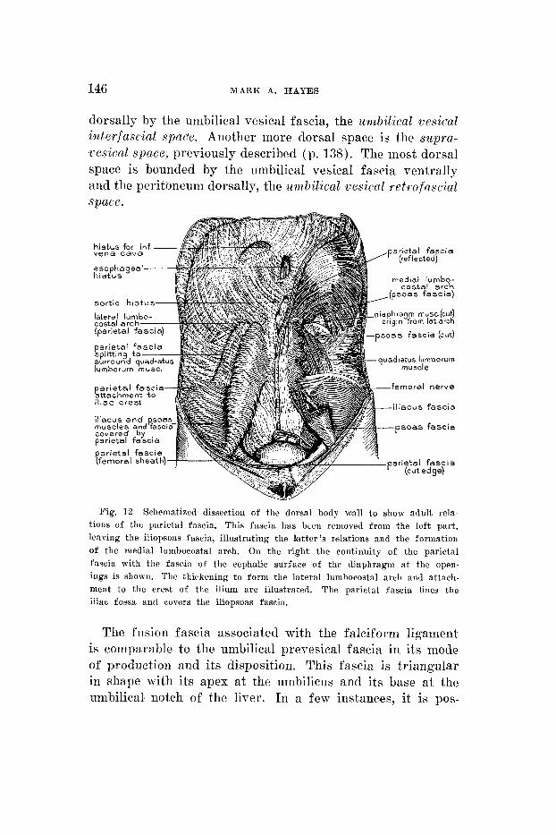
Cephalic to the iliac crest and dorsomedial from its lining position on the transversus abdominis muscle, the parietal fascia extends onto the quadratus lumborum muscle (fig. 12). At the lateral edge of this muscle, the parietal fascia splits into two laminae to enclose partially the quadratus muscle. This line of separation often is fused to the tendon of union of the two layers of lumbodorsal fascia; in many instances it remains free. The dorsal lamina attaches to the tips of the transverse processes of the lumbar vertebrae. The ventral lamina passes ventrally to the muscle to attach to the sides of the lumbar vertebral bodies. It is then reflected ventral to the psoas muscle, with the fascia of which it may fuse more or less completely, to pass ventral to the vertebral bodies and dorsal to all the prevertebral visceral structures, continuing into its counterpart of the opposite side.
A thickened band of parietal fascia over the quadratus lum- borum muscle forms the lateral lumbocostal arch serving as a part of the origin of the diaphragm. A thickening of the psoas fascia forms the medial arch and is covered by fused parietal fascia (fig. 12) . From the inner surface of the psoas, quadratus, and transversus muscles the parietal fascia con- tinues onto the caudal surface of the diaphragm. This fascia follows the structures traversing the diaphragm and at these points the cephalic and caudal fasciae of the diaphragm merge.
Progressing ventrally from the diaphragm, the parietal fascia is the innermost covering of the dorsal layer of the rectus sheath. It is most evident caudal to the semicircular

ABDOMINOPELVIC FAWIAE 135
folds of Douglas, where it persists as the only fascial cover- ing of the dorsal surface of the rectus abdominis muscle. At the pubis the rectus muscle inserts onto the ventral surface of the symphysis, and the parietal fascia passes to cross the dorsal surface of the pubis, blending with the periosteum. This produces the triangular suprapubic space (fig. 11).
At all points along the line marking the entrance to the true pelvis, the parietal abdominal fascia continues in the same anatomical plane as the parietal pelvic fascia, cover- ing the pubes and the obturator internus fascia caudally to the line of origin of the levator ani muscle, thence onto the cephalic surface of that muscle (fig. 11). In the midline ven- trally, at the interlevator cleft, the parietal fascia fuses with the superior fascial layer of the urogenital diaphragm.
At the site of exit of the nerves and blood vessels from the pelvis a funneling evagination of the parietal fascia compar- able to the femoral sheath is produced.
Fcrscial rclcrtions of the adult
Three basic subdivisions of the lining layers of the ah- dominopelvic cavity emerge from the foregoing detailed con- siderations. These consist of parietal fascia (outermost layer), extraperitoneal connective tissue (intermediate layer), and the peritoneum (the innermost layer).
The parietal fascia has been discussed, leaving the special- izations of the other two layers to be considered.
A. Migrat ion fasciae. The perirenal fascia is an enveloping sheath of fibroelastic connective tissue which closely follows the contours of the kidney and suprarenal gland, separated from these organs by the perirenal adipose tissue (fig. 3). There is a complete though thin and delicate septum between the suprarenal gland and the kidney creating an individual conipartment for each (fig. 4) . The ventral lamina of the perirenal fascia is somewhat thinner than the dorsal and both lalniiiae merge at the margins of the kidney where the fascia loses its individuality in continuity with the extra-
136 MARK A. H A Y E S
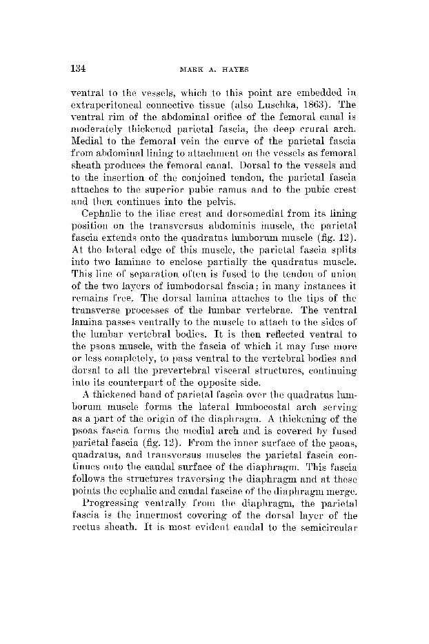
Fig. 1 Schematic view of the ventral abdominal wail viewed from within. On ttic right nearly a11 the umbilical vesical fascia has been i-cniov~l, revealing tlie umbilical prevesical fascia. On the left, only a part of the umbilical resic:tl fascia has been removed to show its distribution tnreioping the bladder, the d nctus deferens, and the seminal vesicles.
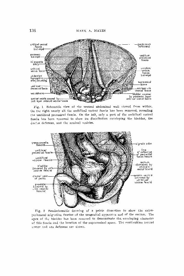
Fig. 2 Semiselrematic draming of a pelvic di~section to show the extra- peiitoiical migration fasciae of the urogenital apparatus and of the rectum. The apex of the bladder has been renioved to demonstrate tlie. enveloping character of this fascia and the location of the supravesical space. The continuities around ureter and vas deferens a r r s h o ~ n .
ABDOMINOPELVIC FASCIAE 137
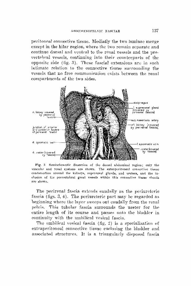
peritoneal connective tissue. Medially the two laminae merge except in the hilar region, where the two remain separate and continue dorsal and ventral to the renal vessels and the pre- vertebral vessels, continuing into their counterparts of the opposite side (fig. 3 ) . These fascia1 extensions are in such intimate relation to the connective tissue surrounding the vessels that no free communication exists between the renal compartments of the two sides.
rt kidney covered by perirenal
f a s c i a sup. mesenteric artery
junction of anterior and posterior layers of perirensl fascia
r t spermatic vein
r t ureter (covered by fascia)
Fig. 3 Semischematic dissection of the dorsal abdominal region; only the vascular and renal systems are shown. The extraperitoneal connective tissue condensation around the kidneys, suprarenal glands, and ureters, and the in- clusion of the prevertebral great vessels within this connective tissue sheath are sliown.
The perirenal fascia extends caudally as the periureteric fascia (figs. 3, 4) . The periureteric part may be regarded as beginning where the layer sweeps out caudally from the renal pclvis. This tubular fascia surrounds the ureter for the entire length of its course and passes onto the bladder in continuity with the unibilical vesical fascia.
The umbilical vesical fascia (fig. 2) is a specialization of extraperitoneal connective tissue enclosing the bladder and associated structures. It is a triangularly disposed fascia

138 MARK A. HAYES
(fig. l), the apex of which may reach to the umbilicus and encloses the urachus and both obliterated hypogastric ar- teries. Caudally the fascia ensheathes the bladder, seminal vesicles, arid prostate gland (figs. 1 and 2). Local' condensa- tions of this fascia form the lateral true ligaments of the bladder and the puboprostatic ligaments. At the apex of the
Fig. 4 Schematized drawing to demonstrate the septum dividing the renal compartment from the suprarenal, and the septum's continuity with the peri- renal fascia. The left kidney and supaarenal gland have been removed, making it possible to show the perirenal fascia as a continuous envelope. The caudal extension of the periureteric fascia is indicated on the left as cut.
bladder, the umbilical vesical fascia can be opened, demon- strating a conical potential space, the base of which is blad- der musculmature, the supravesical space (fig. 2 ) . An ex- tension of fascia is continued along the ductus deferens (fig. 1) to blend with the extraperitoneal connective tissue common to the components of the spermatic cord.
The perirectal migration fascia is a simple tubular fascia which extends from the line of peritoneal reflection of the rectosigmoid to terminate at the exit of the rectum through the pelvic diaphragm (fig. 2) . It lies just peripheral to the
ABDOAMINOPELVIC FASCIAE 139
tunica muscularis of the rectum and in it course the vessels, nerves, and lymphatics of the rectum.
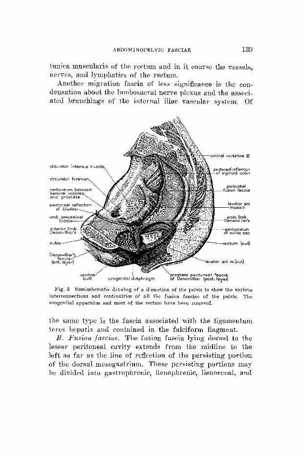
Another migration fascia of l'ess significance is the con- densation about the lnmbosacral nerve plexus and the associ- ated branchings of the internal iliac vascular system. Of
obturator internus muscle
obturator foremen,
pubis-
sacral vertebra IX
Fig. 5 Semischematic drawing of a dissection of the pelvis t o show the various The interconnections and continuities of all the fusion fasciae of the pelvis.
urogenital apparatus and most of the rectum have been removed.
the same type is the fascia associated with the ligamentum teres hepatis and contained in the falciform liagment.
B. Fusion fasciae. The fusion fascia lying dorsal to the lesser peritoneal cavity extends from the midline to the lseft as far as the line of reflection of the persisting portion of the dorsal mesogastrium. These persisting portions may be divided into gastrophrenic, lienophrenic, lienorenal, and

140 MABIZ A. HAYES
pancreaticolicnal ligaments (fig. 8). The cephalic limit of the fascia is the hare area of the esophagus. Its caudal limit is the caudal border of that par t of the pancreas lying to the left of the midline. The right border of this fascia1 plane is the midline as far caudal as the beginning of the retro- peritoneal portion of the first part of the duodenum. Below
‘left ureter in
Fig. 6 Semischematic drawing of a dissection t o show the relation of the retropancroatie fusion fascia lving ventral to the perirenal extraperitoneal migra- tion fascia. The rephalie extension of the retropaiicreatie layer into tlic dorsal niesogastriuni fusion lager is shown in part.
this level the fusion fascia extends to the right of the mid- line as far as the parietal reflection of the visceral peritoneum from the first and second parts, and a portion of the third part of the duodenum. The perirenal fascia is dorsal to this layer (fig. 6) . This fusion layer covers the hilar region and the medial border of tile right kidney and the most medial part of the right suprarenal gland; it crosses the middle third of the left kidney and overlies most of the medial aspect of the ce- phalic pole of the left kidney and most of the left suprarenal

A B D O M I N O P E L V I C FASCIAE 141
gland. The main vascular channels such as the left gastric ar- tery arid the splenic vessels course in the tissue between this fusion fascia and the retained peritoneum of the lesser peri- toneal cavity, the duodenum, and the pancreas (figs. 6, 8).
The fusion fasciae of the colon have a more extensive distribution (fig. 7 ) . The fascia resulting from the fusion
Fig. 7 St.iniscliem:rtic draiving of a dissection showing the fusion fasciae associated with the colon. The stomach, jejunum, ileum, and entire colon have been removed. The mesentery and the persisting peritoneum of the colon have all been removed, showing only the fibrous layer which remains after fusion.
of the right side of the ascending colon extends from the root of the mesentery to the right as f a r as the line of re- flec.tion of the peritoneurn from the colon to the parietal wall, laterally. Its caudal limit is determined by the degree of fusion that has occurred between the cecum and the peri- toiieum of the doi-sal body wall. Similarly, the lateral ce- plialic limit of this layer will depend on the amount of
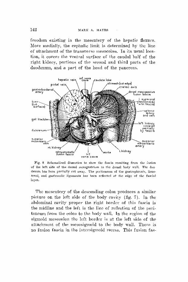
142 MARK A. HAYES
freedom existing in the mesentery of the hepatic flexure. More medially, the cephalic limit is determined by the line of attachment of the transverse mesocolon. I n its usual loca- tion, it coveix tlie ventral surface of the caudal half of the right kidney, portions of the second and third parts of the duodeniim, and a par t of the head of the pancreas.
retrqpancreatio ' I \aor ta ' I. kidney fusion fascia i nf.
vena c a v a
Fig. 8 Schematized dissection to show thc fascia resulting from the fusion of the left side of the dorsal mesogastrium to the dorsal body wall. The duo- denum has been partially cut away. The peritone'uni of the gastrophrenic, lieno- renal, and gastrocolic ligaments has been reflected a t the edge of the fascia1 layer.
The mesentery of the descending colon produces a similar picture on the left side of the body cavity (fig. 7). In the abdominal cavity proper the right border of this fascia is the midline and the left is the line of reflection of the peri- toneum from tlie coloii to the body wall. In the region of the sigmoid mesocolon the left border is a t the left side of the attachment of the rnesosigmoid to tlie body wall. There is no fusion fascia in tlie intersigmoid recess. This fusion fas-
ABDOMINOPELVIC FASCIAE 143
cia covers the ventral surface of the most caudal edge of the body of the pancreas and the caudal third of the left kidney.
At the entrance to the pelvis the mesentery of the large bowel retains its midline suspension. At the level of the third sacral vertebra the rectosigmoid segment begins to lose its complete peritoneal investment. The crescentic lines of reflection of peritoneum from gut t o parietes indicate the cephalic limit of the perirectal fusion fascia (fig. 5). From this peritoneal reflection the fascia extends caudally as a tubular structure encircling the rectum almost to the site of its passage through the pelvic diaphragm and external to the perirectal migration fascia. The ventral segment of this circular fusion fascial sheet forms the rectovesical septum or fascia of Deiionvillier and dorsally attaches at the mid- line, the original root of its mesentery.
The umbilical prevesical fascia is a triangularly shaped fascial sheet with its apex at the umbilicus (fig. 1). The dor- sal surface of this fascia is free; the ventral surface has a sagittal attachment at the midline, extending caudally from the umbilicus and indicating the line of attachment of the former meso-urachus and mesocyst. I ts lateral limits are the lateral umbilical ligaments or the parietal attachments of the mesenteries of these structures. The caudal’ limit of this fas- cia is quite variable. The usual location is dorsal to the pubis at the level of the puboprostatic ligaments. The um- bilical prevesical fascia is ventral t o the umbilical vesical fascia (fig. 1). The ceplialic limits of the dorsal limbs of this fascia are the obliterated hypogastric arteries and their continuations, the superior vesical arteries. The dorsal ex- tensions passing laterally to the bladder are in direct con- tinuity with the rectovesical fascia.
These facts provide material for simplification of the con- flicting views concerning the “space of Retzius ” (1 858). , I h y observers have given different boundaries for this space (Hyrtl, 1858 ; Charpy, 1892 ; Waldeyer, 1899 ; Rouvihre, ’24 ; Hinman, ’37 ; Callander, ’39) necessitating re-evaluation of the anatomy concerned. In view of the several fasciae and
144 MARK A. H A Y E S
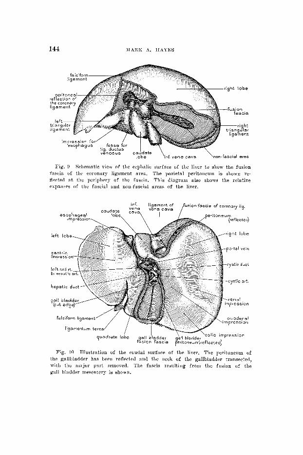
non-fascial area
Fig. 9 Schematic view of the cephalic surface of the liver t o show the fusion fascia of the coronary ligament area. The parietal peritoneum is shown re- flected a t the periphery of the fascia. This diagram also shows the relative cspaims of the fascia1 and non-fascia1 areas of the liver.
Fig. 10 Illustration of the caudal surface of the liver. The peritoneum of the gallbladder has been reflected and the neck of the gallbladder transected, with the major part removed. The fascia resulting from the fusion of the gall bladder mesentery is shown.
ABDOMINOPELVIC FASCIAE 145
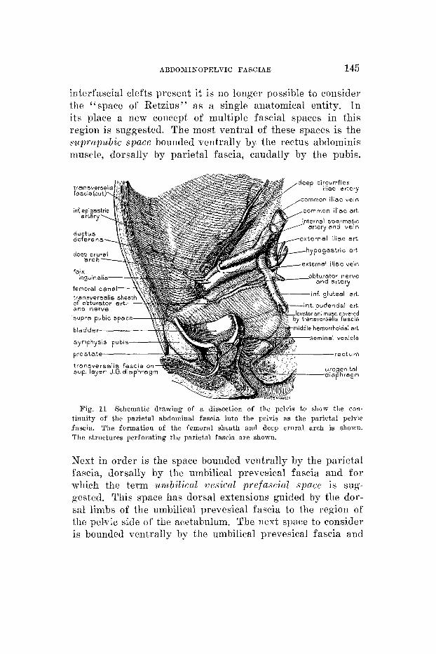
interfascial clefts present it is no longer possible to consider the “space of Retzius” as a single anatomical, entity. In its place a new concept of multiple fascia1 spaces in this region is suggested. The most ventral of these spaces is the suprapubic space bounded ventrally by the rectus abdominis muscle, dorsally by parietal fascia, caudally by the pubis.
omrnon iliac vein
ommon iliac art
xternel iheo art
transversa IS s of obturator e and nerve
an1 mmc covered sversalis fascla
hemorrhoidal art
eminal veslcle
Fig. 11 Schematic drawing of a dissection of the pelvis to ?how the con- tinuity of the parietal abdominal fascia into the pelvis as the parietal pelvic fascia. The formation of the femoral sheath and decp crural arch is shown. The structures perforating the parietal fascia are shown.
Next in order is the space bounded ventrally by the parietal fascia, dorsally by the umbilical prevesical fascia and fo r which the term umbilical uesical prefascial space is sug- gested. This space has dorsal extensions guided by the dor- sal limbs of the umbilical prevesical fascia to the region of tlio pelvic side of the acetabulum. The next space to consider is bounded ventrally by the umbilical prevesical fascia and
146 MARK A. HAYES
dorsally by the umbilical vesical fascia, the umbilical vesical interfascial spare. Another more dorsal space is the siipra- vesical space, previously described (p. 138). The most dorsal space is bounded by the umbilical vesical fascia ventraIly and the peritoneum dorsally, the zmbil ical cesical retrofascial space.
Fig. 1 2 Schematized dissection of the dorsal body wall to shoir adult rela- tions of the parietal fascia. This fascia has bceri removed from the left part, leaving the iliopsoas fascia, illustrating the latter’s relations and the formation of the medial lumbocostal arch. On the right the continuity of the parietal fascia with tlie fascia of the cephalic surface of the diaphragm a t the open- ings is shown. The thickening t o form the lateral lumbocostal arch and attach- ment to the crest of the ilium are illustrated. The parietal fascia lines the iliac fossa and covers the iliopsoas fascia.
The fusion fascia associated with the falciform ligament is comparable to the umbilical prevesical fascia in its mode of production and its disposition. This fascia is triangular in shape with its apex at the umbilicus and its base at tlie umbilicals notch of the liver. In a few instances, it is pos-

ABDOMINOPELVIC FASCIAE 147
sible to identify a continuity of it with the fusion fascia of the bare area of the liver. The lateral limits of this fascia are determined by the width of the base of the falciform liga- ment.
The “bare area” of the liver needs redefinition in that it has two components, the fascial area and the no%-fascia1 area. There is a fusion fascia bordering the entire periphery of the non-fascia1 area, o r the primitive attachment of the liver to the septum transversum (fig. 9) , and extending
PANCPKAS
to the line
R€i7?OP,,NCR€ATIC FUSlON FASCIA
P€,e/TONEUM
NSV€RS€ COLON
FUSION FA5
L RENAL W I N
QUADGATUS LUM BORUM MUSCLI
SPL/TT/NC 0 PARIETAL FA
Fig. 13 Transverse section through the abdominal cavity of a human fetus of 279 mm (28 + weeks). The level is at the top of the seeond lumbar vertebra. Extraperitoneal connective tissue fasciae and fusion fasciae at this level a re well shown. Schematic line drawing of a specimen prepared by dissection and embedding in ethyl methacrvlate. ( X 1.5, University of Michigan Coll., EII 441.)
of reflection of the coronary lmigament onto the diaphragm. Great variations in extents may be noted here; generally, the bare area is largely fascial.
Finally, the fusion fascia of the gallbladder fossa may be considered. Tliis fascia (fig. 10) closely conforms to the hepatic contour of the fossa, and is intimately associated with the underlying liver tissue and is bound to it, by a fibrous attachment, the original mesenteric attachment. The fascia is continuous at the periphery with the line of reflection of the peritoneum from the liver to the gallbladder.
148 MARK A. HAYES
SUMMARY AND CONCLUSIOSS
An investigation of adult abdominopelvic fasciae has been made, employing a developmental approach. The studies in- volved the sequential development of visceral changes as observed in a closely graded series of human embryos, fe- tuses, newborn infants, and adults.
Examination of the sequence of developmental events and the evaluation of each iiitermedjate stage in snch a sequence unfolds arid integrates a story that is essentially predictable in its final pattern.
Two processes of development produce different types of fasciae in the adult. Organ migrations produce reqional spe- cializations of the adult extraperitoneal connective tissue, designated naigrntiogz fasciac.
The fasciae resulting from fusions of primitive mesen- teries are termed fusion fasciae.
The third type of fascia is intrinsic to the structures in the wall of the developing abdominopelvic cavity. This fas- cia is the parietal fascia.
With an understanding of the embryological changes that occur it is possible to predict and confirm the presence of the abdoniinopelvic fasciae ; their presence and modifications are presented as the logical end results of normal develop- ment.
LITERATURE CITED
ANCEL, P., AKD P. CAVAILLON 1907 Sur les niBsoc6lons ascendant et descendant et leur mode de formation rhcz l’homme. Bihliog. Anat., Suppl., C.R. Assn. anat. , Lille, 1-11.
ANSON, B. J., AND C. B. MvlcVAY 1938 Inguiiial hernia; the anatomy of the region. Surg. G p . O k . , 66: 186-191.
AUGIFE, A. 1921 Apparei l TJrinaire - Les rciiis et leurs canaux excreteurs. Ext ra i t dii Trait6 d’Anntomie Hninnine. TV, F3. 3‘ ed.
BARTL~KOIVSKI, J. 1924-25 Ueber die Lage der Nebennieren zu den Nieren. Anat. Snz., 59: 508-511.
EASSINI, E. 1890 TTeher die Ee1i:rndlnng des Leistenbruehes. Arch. klin. Chir., 40: 429-476.
BATJIANN, J. A. 1945 D6reloppemeiit et anatoniie de la loge rBnale cllez I’homme. Act. Anat., 1 : 115-165.
REGG, R. C. 1930 The urachus; its anatomy, histology, a d derelopment. J. Anat.. 6 4 : 170-183.

AUDOMISOPELVIC FASCIAE 149
BILLINGTON, W. 1910 Movable Kidney. London. Cited by A. H. Southam. 1923 The fixation of the kidney. Quart. J. Med., 16: 283-308.
BLEICIIER, M. 1931 L’enveloppe fibro-adipeuse dca glands surr6u:ilea. C.R. Assn. Anat., 26th Meeting, Varsovie, pp. 54-57.
BLOOM, R. 1926 The embryogenesis of liunian bile capillaries and durts. Am. J. Anat., 56: 451465.
WROS, A. J. 1823 Essai sur l’anatoniie chirurgical de la r6gion iliaque ct description d’un nouveau prored6 pour faire la ligature des artBres Cpigastrique et iliaque externe. ThBse, no. 253, Paris.
EOYDEN, E. A. 1926 The accessory gallbladdcr. An embryological and coni- parative study of aberrant biliary vrsicles ocourring in man and the domestic niarnmnls. Am. J. Anat., 38: 177-231.
GRAUNE, W. 1877 Notiz iiher die Ringform dcs Duodenum. Arch. Anat. Physiol., Anat. Abt., 468473.
BROMAN, I. 1896 I n von Bardelehen, K., Handbuch der Anatomie des Men- schen. Bd. 6, Teil 2. Fischer, Jena.
Die Entwicklungygescliirhte dr r Bursa Omentalis und ahnliche Rcceszbildungcn bei den Wirbeltieren. Hirschwald, Weisbaden.
CALLANDLR, C. L. 1939 Surgical Anatomy. 2nd ed. Saunders, Pliiladelphia. CHARPY, A. 1890 Orgenes g6nito-urinaires. Masson, Toulouse.
La gaiue des muscles droits e t la cavit6 prC\&icale. Rtudes d’anatomie appliquCe. BailliBre, Paris.
CONGDQN, E. D., AND J. N. EDSON The cone of renal fascia in the adult white male. Anat. Rec., 80: 289-313.
CONOWN, E. D., R. BLLJMBEZW AND W. HENRY 1942 Fasciae of f l u i o n and elements of the fused entcric mesenteries in the human adult. Am. J. Anat., 70: 251-279.
COOPER, SIR A. P. 1807 The anatomy and surgical treatment of crural and urnliilical hernias. T. Cox, London. 1844 The anatomy and surgical treatment of abdomiual hernia.
2nd ed. rev. by C. Aston Key, Lea and Blanchard, Philadelphia. CUNEO, B., A N D V. VEAIJ 1899 De la signification morphologique des apo-
neuroses pCrivCsicales. J. Anat., Paris, 95 : 235-245. DAVIS, G. G. 1934 Applied Anatomy. 9th ed. rev. by G. P. Muller. Lippin-
cott, Philadelphia. DENONVILLIE, C. P. D. Bulletin no. 10 (3” serie) Bull. Soc. Anat., Paris,
1 2 : 105-107. 1837 Propositions et observations d ’anatomie, de pliysiologie, et
de psthologie. These Ecole Med., Paris, no. 285. DISSE, I. J. 1889 Bcitrage zur Kenntnis der Spaltraiime dcs Menschen. Arch.
Anat. Physiol., Anat. Abt. Suppl. Ed., 222-238. ___- 189G In von Bardelebm, K., Handbuch dcr Anatomie dcs Mcn-
schcn, Bd. 7, Teil 1, Fisclier, Jena. DOT-r, N. M. 1923 Anomalies of intestin:il rotation : their embryology and
surgical aspects: with reports of 5 cases. Brit. J. Surg., 11: 231-286. FELIX, W. 1912 Chap. X I S . In Keibel and Mall, Human Embryology. Lip-
pincott, Philadelphia.
1904
1892
1941
1836
150 MARK A. HAYES
FRAzFa, J. E., AND R. H. ROBBINS On the factors concerned in causing rotation of the intestine in man. J. Anat. Physiol., 50: 75-110.
FREDET, P. NOte sur la formation des capsules du rein chez l’homme. J. Anat. Physiol., 40: 599-609.
- -~ 1904b Documents sur la formation des capsules du rein chez l’embryon humain. Bull. Mbm. Soc. Anat. Paris, 79: 285288.
GEROTA, D. 1895 Beitrage zur Kenntnis des Befestigungsipparatus der Niere. Arch. Anat. Entwick., Anat. Abt., 265-285.
GRANT, J. C. B. 1944 A Method of Anatomy. 3rd ed. Williams and Wilkins, Baltimore.
GRAY, H. 1948 Anatomy of the Human Body. 25th ed. rev. by C. M. Goss. Lea and Febiger, Philadelphia.
GROSS, R. Congenital anomalies of the gall bladder; review of 148 cases, with a report of a double gall bladder. Arch. Surg., 32: 131-162.
G R O S S F ~ D , H. Production in vitro d’un epithelium aux depens des fibro- blastes du coeur d’embryon de poule. C. R. Soc. Biol., 108: 747- 750.
HALSTED. ’CV. S. 1893 The radical cure of inguinal hernia in the male. Ann. Surg., 17: 542-556.
HAMMOND, G., L. YGLESIAS AND J. E. DAVIS 1941 The urachus, its anatomy and associated fasciae. Anat. Rec., 80: 271-287.
HENKE, W. Der Raum der Bauchhole des Menschen, und die Vertheilung der Eingeweide im demselben. Arch. Anat. Entwick., 89-107.
HESSELBACH, F. C. 1808 Anatomische-cliirurgische Abhandlung iiber den Ur- sprung der Leistenbriiche. Cohn, Wurzburg.
HINMAN, F. 1937 The principles and practice of urology. Saunders, Phila- delphia.
HIS, W. 1874 Unsere Korperform. Vogel, Leipzig.
1915
1904a
19116
1931
1891
1878 Uber praparate zum situs viscerum mit besonderen Bemer. kungen iiber die Form und Lage der Lcber, des Pankreas, der Nieren und Nebennieren, sowie der weiblichen Beckenorgane. Arch. Anat. Physiol., Anat. Abt.
1880-85 Anatomic menschlicher Embryonen. Vogel, Leipzig. ROFFFMANN, C. E. E., AND A. RAUBER Lehrbuch der Anatomie deu Menschen.
Besold, Berlin. HYRTL, J. Notiz iiber das Cavum praeperitoneale Retzii i n der vorderen
Bauchwand des Menschen. Sitzungsber. K. Acad. Wiss., Wien, 29 : 259- 264.
Ueber die Lagebeziehung der Nieren und Nebennieren beim Menschen. Anat. Anz., 64: 163-173.
On the topography of the pancreas in the human fetus. Anat. Anz., 3 7 : 488-510.
1909 On the developmental topography of the thornric and ab- dominal viscera. Anat. Rec., 3: 361-396.
KEHR, H. 1913 Ruckblick auf 2000 Operationen an dem Gallenwegen, ejne Gegeniiberstelluug der Erfolge des ersten uiid zweiten Tausend. Ver- haudl. deutsch. Gesellsch. Chir., 43: 273-288.
1886
1858
IVANOW, G.
JACKSON, C. M.
1927
1905
A B D O M I N O P E L V I C FASCIAE 151
KELLY, H. A., AND C. F. BURNAM Diseases of the Kidneys, Ureters, and Bladder. D. Appleton and Company, New York.
KNIGHT 1893 Movable kidney and intermittent hydronephrosis. London. Cited by A. H. Southam, 1923, The fixation of the kidney. Quart. J. Med.,
LEWIS, D. D. 1904 The present conreption of the perirenal fascia and its role in the fixation of the kidney. J. Am. Med. Assn., 43: 701-703.
LUSCHKA, H. 1863 Die Anatomie des Menschen. Lauppschen, Tubiugen. MALL, F. P. 1891 A human embryo 26 days old. J. Morph., 5: 8-10. _____ 1897 Ueber die Entwicklung des menschlichen Darmes und seiner
Lage beim Erwnchsenen. Arch. Anat. Physiol., Anat. Abt., Suppl., 403434.
____ 1898 Development of the human intestine and its position in the adult. Johns Hopkins Hosp. Bull., 9: 197.
MARTIN, C. P. 1947 On the embryology of the levator ani muscle. Abstract. Anat. Rec., 97: 39.
MORRIS 1946 Human Anatomy. 10th ed. rev. by J. P. Schaeffer. Blakiston, Philadelphia. See. V by J. C. B. Grant.
McVau, C. B., AND B. J. ANSON 1940 Aponeurotic and fascia1 continuities i n the abdomen, pelvis and thigh. Anat Rec., 76: 213-232.
PATTBN, B. M. 1946 Human Embryology. Blakiston, Philadelphia. PERNKOPF, E. 1922-28 Die Entwicklung der Form des Magendarmkanales beim
Menschen. Teil I. Zeitschr. Anat. Entwickl., 64: 96-275; Teil 11. Ibid., 79: 1-144; Teil 111. Ibid., 77: 1-143; Teil IV. Ibid., 8 5 :
PIERSOL, G. A. 1930 Human Anatomy. 9th ed. rev. by G. C. Huber. Lippin-
POHLMAN, -4. G. 1904 Concerning the embryology of kidney anomalies. Am.
I’OIRFR ET CHARPY 1898 Trait6 d’Anatomie Humaine. L. Bataille, Paris. POWEX, R. M. €I.
Am. J. Obs. Gyn., 55: 367-381. RETZIUS, A.
of Douglas. Edinburgh Med. J., 3: 865-867. ROUVI~RE, H. 1924 Anatomie Humaine. Masson, Paris. SAPPEY, M. P. 0. Trait6 d’Anatomie d6scriptive; avec figures intercal6es
dans le texte. 3rd ed. rev. Delahaye et Lecrosnier, Paris. SCARPA, A. 1809 Sull’ernie memoire anatomico-chirurgische. Reale Stomperia,
Milano. SCHACHNER, A. 1916 Anomalies of the gall bladder and bile passages with
the report of a double gall bladder and a floating gall bladder. Ann. Surg., 64: 419-433.
Zur ISPnntnis der Lage und Form des Mesenterialen Teiles des Dunndarmes und seines Gekroses. Internat. Monatschr. Anat. Physiol., 11: 437466.
SOUTHAM, A. H. 1923 The fixation of the kidney. Quart. J. Med., 16: 283- 308.
1922
16: 283-308.
1-130.
cott, Philadelphia.
Med., 7: 987-990.
1948 Embryological development of the levator an i muscle.
Some remarks on tlie proper design of tlie semilunar lines 1858
1879
SERNOFF, D. 1894
152 MARK A. HAYES
TOBIX’, C. 3:. 1944 The reiial fascia and i ts relations t o the transversalis fas- cia. Anat. Rec., 89: 295-311.
TOBIN, c. E., AND J. A. BENJAXIN 1945 Anatomical and surgical restudy of Deiionvillier’s fascia. Surg. Gyn. O h . , 80: 373-388.
TOLDTT, C. 1879 Ban und VVachstumsveranderungcn der mensclilichen Darm- kanales. Dcnkschrift. K. Aknd. Wiss. Abt. 11, 4 : 1-57.
TREITZ, W. 1853 Uber einem neuen Muskel am Duodenum des Menschen, iiher clnstielie Sehnen, und einige andere anatomisrlie Verhaltnisse. Viertel- jalir pralct. Heilkunde., 37: 113-144.
VECCHI, A. 1910 Osservazioni sul rompartmento della fascia renale. Anat. Anz., 36: 149-186.
WAJ~DEYER, W. 1899 Das Becken. Cohen, Bonn. WALT^, 0. M., AND A. NIEMAN 1931 Case report of an intrnliepatic gall blad-
der in an adult with a review of the literature. Ill. 35. J., 60: 478- 480.
WALTERS, W., A. M. SNELL AND I). T. CLAGLTI’ Scetion in Lewis, System of Surgery, 7 : 1-145, Chap. 2.
WEINBERG, R. 1896 Topographie der Mesenterien und dcr Windungen des Jejuno-ileum beim neugeborenen Menschen. Internat. Monatschr. Bnat. Physiol., 13: 66-89.
ZIEMAN, S. A. 1942 Importancs and distribution of the transversalis fascia from the viewpoint of the surgeon. Arch. Surg., 4.5: 926-934.
Z U C K ~ L K A N D L , E. 1883 Beitrage zur Anatomie des menschliclien Korpers : I. Uber den Fixationsapparat dcr h-ieren. Wein. Med. Jahrbucher, 23 : 59-67.
1947
PLATES
PLATE 1
EXPLANATION OF FIGDRES
14 Parasagittal section through the left kidney of a 6i inm (11-week) liuinau embryo, showing the perirenal inesenrhymal condensation (perirenal coiinective tissue). The condensation also includes the suprarenal gland with a septum between the two structures. The thin parietal fascia is shown. (Photomicrograph, X 14, from Unirersity of Michigan Coll., KH 2 6 % ~ )
Parasagittal section through the pelvic region of a human embryo of 67 min (11 weeks) to show fascia1 relations. The lateral paravesical peritoneal relations, perivesical mesenchymal ticusue, as w ~ l l as the parietal fascia are shown. (Photo- micrograph, x 9, from University of Michigan Coll., EH 269a.)
16 Frontal section through the abdominal and pelvic cavities of a human einbryo of 27 inin (8 weeks) to show the mesenchymal concentrations around the kidney and ureter. The lateral peritoneal relations of the rectosignloid are shown and incipient fusion is apparent. (Photomicrograpli, X 1.5, froiii University of Michigan Coll., EH 216.)
1 5
154
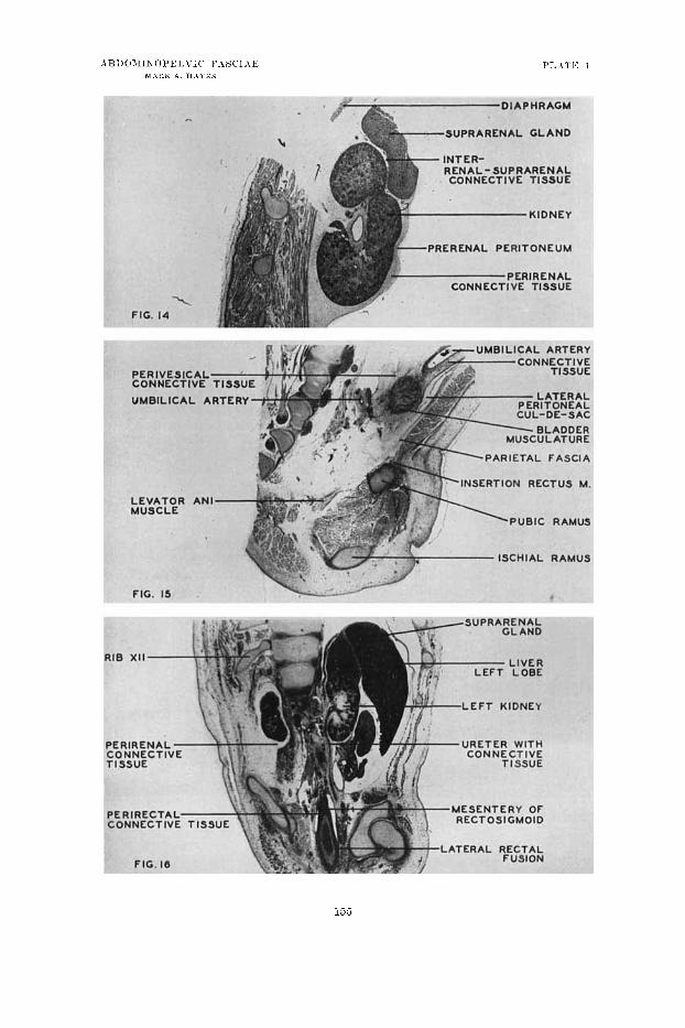

PLATE 2
EXPLANATION O F FIGURES
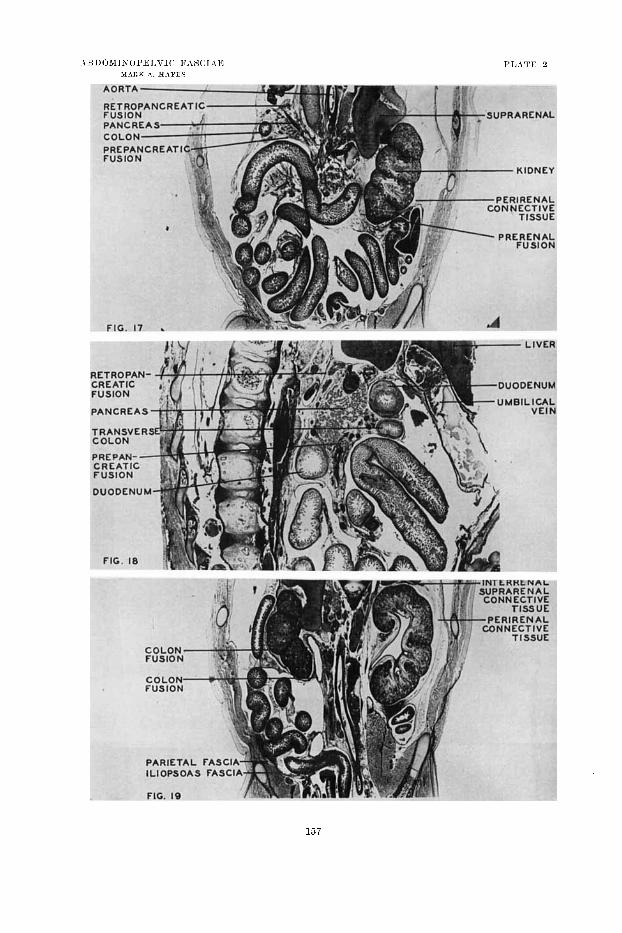
1 7 Froiital section through tlie abdominal cavity of a h in i i~n e i i i l q o of 81 111111
(12 + wceks) to show relations of the various parts of the duodciiuiii and asso- cia.tctl fusion arcas. The l o f t prerenal fusion :ind the free duodenal segiiieats are s11o~v.n. The fusion of the transreme niesorolon ventral t o the duodeiiuni
is appears. The retroyailcrwtic fusion of the dorsal mesognstriuin is well illustrated. (Photoinicrogral)li, x 8.5, f rom TJnirersity of Michignu Coll., EII 228c.)
Parasagittal section through tlic head of the pancreas of a human em7riryo (J€ 10-1 niin (14 wccks) t o show peritoneal fusion in the regioli of the pancreas, duodenum, aiitl transverse colon. (Photoniicrograph, X 5.5, from University of hIicliigan Cvoll., EII ZJGg.)
19 Frontal section of the abdominal cavity of a 1iuin;rii embryo of 84 imii
(12 + weeks) to sliow migration and fusion fasckc. The colon fusion 011 the right aiid tlic perireiml fascia on the left arc well shown. This view dcnionstrates the relations of parietal sud iliopsoas fasciae :it their attachinenis t o the crest of tlie iliuni. (Photon~ici.ogra~~li, x 11.5, from University of Micliigan Coll., EH 288c.)
18
136
137
PLAT?: 3
EXF’L.\K.\TIOK OF FIUI-RES
2U l’roiita I section of the pcli-ic repioii of a liuniaii embryo of 33 iiiiii (10 weeks) to sliow f:iscial mid ~ieritoiieal rc~latioiis. The rectosigiiioid 1iieseiiter:- persists. Earl:- Iicrirectal fascia is apparent. Tlie parietal fascia exteiids up to the pelvic brim oii the right, and its relation to the oliturator fascia is illustrated. (IJliotoiiiicroginph. X 10, froiu Uiiiiwsity of 1\ficliigaii C01l.~ EH 3SOn.)
Frontal section of tlie dorsal region of the pelvic cavi tv of a liumaii eiii- hryo of 89 iriiii (late 13th week) ivliicli s l i o n d perirtctal fusion fascia iii t.he ppoccss of‘ foriiintioii a t the deptli of the lateral cul-de-sac. The Icvntor ani muscle esteiitls uearl- to the peli-ic 1,riiii. Tlie br,anclies of the superior Ireiiior- rlioidal artery are included iii the perirectal inigratioii fascia. (l’liotoiiiicrogrnpli, X 12, froni l’iiivtrsit>- of Mic1lig:in Cull., EH 300.j.)
22 Froiital wetioil of thc 13elvic region of :I human embryo of 53iiiiii (10
n-eeks) to show fascia1 relations in the dorsal part of the pelris. The lateral rectal enl-de-sac with attaching fusion fasei:ie 2 x 1 ~ shoivii. The parietal fascia is distiiict froiii the ohturator f:rs&. (Pli~~totiiicrogl.nI,h, X 8.5, f r o m TTiiiwr- sity of hficliignu Coll., EH 3iOa. I
2 1
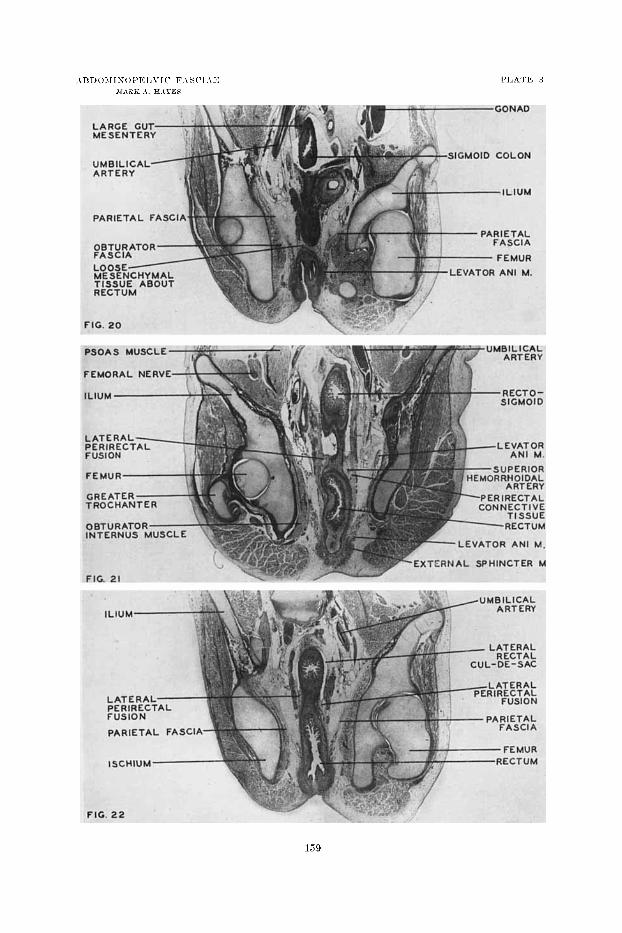
159
PLATE 4
2 3 Parasagitt;rl sectioii througli tlie lef t kidney area of a Iiuiiiaii embryo of 104 iiiiii (14 iweks). ('olon fusion fascia reiitral t o tlic kidney is shown with its continuity into the attacliineiit of the transverse mcsocolon caudal to the body nt' tlic pancreas. Fusion of the dorsal inesog:rstrium (retropaiicreatic) is well rstablished. The gastrocolie ligaiiieiit is iiot yet formed. (Photoiiiicrogr:iph, x 6, from Vniwrsity of ?vIichigan Coll., EH 29Gg.)
Palasagittal section through tile left kidney area of a huimii embryo of 104 1 i m (14 weclrs) to show pei.iieiia1 : i d iiiterreiial-siiprareiial niigration fascia. Prereiial coloil fusion fascia and retropaiicreatic fusion ventral t o the left supra- ~ e n n l gland are also iwll shown. (Photomicrograph, X 6, froin Cnivcrsity of Michigaii Coll., 1311 296g.)
24
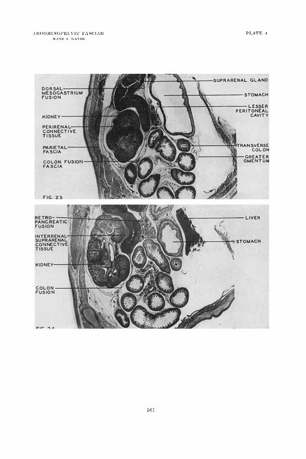
PLATE 4