Embed Size (px)
Citation preview

John C. KelleherM.D., F.A.C.S.
Abdominal Pedicle FlapsTo The
Hand And Forearm
Global-HELP Publications

Chapter Eight: TECHNICAL REQUIREMENTS FOR FORMATION OF A TUBED PEDICLE FLAP
Creating a tube pedicle is advantageous in many situations, therefore principles of tubeformation are important.
Donor Site: Usually the abdomen or groin are the best donor sites. There are considerations:
a. Scars from previous surgery or trauma b. The amount of skin required should be planned (see chapter 5). c. Blood supply must be considered. A major vessel should be incorporated into a tube pediclewhen possible (superior epigastric, superficial inferior epigastric, superficial circumflex iliac) . d. Avoid hairy areas when possible and consider moving the flap laterally if possible.
Layout of the Plan for the Tube Pedicle: It is well to layout this plan in the office or thehospital well before the time of the actual performance of the surgery. This allows the surgeonto think about the area of skin requirements and the previous scars that might interfere withthe blood supply. Also other factors such as the presence of abdominal hair or the thickness ofthe abdominal fat can be considered. Usually one can grasp the roll of skin of the abdomenbetween the thumb and fingers to get an idea of how much skin needs to be carried betweenthe parallel incisions in order for the tube to be closed with limited tension. This also gives aprediction of the ability to close the donor site. If the donor site area cannot be closed it canbe covered with a split thickness skin graft (see drawing 8.1). Parallel lines are drawn on the skin of the abdomen of the appropriate length.
Parallel incisions are then made down to the deep fascia (see drawing 8.2). The surroundingskin is undermined and closed in layers with absorbable sutures and then interrupted 4-0 nylonsutures in the skin. Using traction sutures of nylon at the distal ends of the skin to be tubed,the seam of the tube is closed with interrupted 4-0 nylon sutures.

next

Chapter Eight: TECHNICAL REQUIREMENTS FOR FORMATION OF A TUBED PEDICLE FLAP
If the tension with the closure of the skin edges is judged to be too excessive then it should beopened. Using sharp tissue scissors, some fat is removed until the skin can be closed withouttension. Lot Howard described an alternate method of forming a tubed pedicle that gives alittle more laxity to the skin and does not have opposing seams in the tissue closure (seedrawing 8.3).
Another method is to make use of staggered parallel incisions which provide a wider pedicle ofcirculatory inflow at each end and make for easier donor site closure (see drawing 8.4). Drains are usually not required. Dressings consist of only a single dry gauze 4x4 between the tubeand the donor site which is changed frequently. Of course a tie over dressing would also beused in the event of a split thickness skin graft covering the donor site. Alternate sutures areremoved at two weeks and the remaining sutures are removed at three weeks.

next

Chapter Eight: TECHNICAL REQUIREMENTS FOR FORMATION OF A TUBED PEDICLE FLAP
EXAMPLES OF HAND RECONSTRUCTION USING TUBED FLAPS - Case No. 1:
A 26-year-old man caught his right dominant hand in a paint roller and was taken to a localhospital for care. At the time of the injury, the little finger was amputated at the metacarpalbase and 3 weeks later the thumb was amputated at the mid proximal phalanx. A meshed skingraft was applied. A month later a poorly planned "Alpha type" flap was applied to the palmaraspect of the hand. Note the inadequate size, straight line scars and biscuit shape of that flap(see photos 8.1A, 8.1B). These photos were taken 7 months after injury and this surgicaltreatment prior to his transfer here.
Photo 8.1A
Photo 8.1B
Our examination revealed heavy scarring and limited motion. Action of the extensor pollicislongus could not be demonstrated because of scarring in the area. It was decided to make useof a tubed flap because of the need for cover on both sides of the hand and wrist (see photos 8.1C, 8.1D).

Photo 8.1C Photo 8.1D
A superior "pancake" of tissue with W darts is used on the border to replace the scar on theulnar side of the hand (see photo 8.1E).
Photo 8.1E
Although not depicted in photographs, this tube was then transferred after several "delays" tocover the Palm of the hand and thumb as seen in the final photos (see photos 8.1F, 8.1G).
Photo 8.1F

Photo 8.1G
next

Chapter Eight: TECHNICAL REQUIREMENTS FOR FORMATION OF A TUBED PEDICLE FLAP
Case No. 2:
A 35-year-old man caught his left hand in a punch press suffering loss of all parts of the thumb throughthe distal end of the metacarpal along with avulsion of the skin of the thumb-index web space. There wasalso a partial amputation of the tips of the index and long fingers. (see photo 8.2A). His surgical treatment involved 3 operations.
Photo 8.2A
Stage I. A patterned abdominal tubed flap was raised in the left lower quadrant based on the superficialinferior epigastric vessels. This type of skin coverage was selected to effectively cover the web space and tohave additional skin and fat for future stages in the thumb reconstruction. (see drawing 8.5 and photo8.2B).
Photo 8.2B
The donor site was closed primarily. The proximal portion of the flap was tubed as described above and thedistal portion of the flap to be applied to the open web space was thinned of excess fat just deep to thesubdermal plexus.
Stage II. Three weeks later the tube flap was detached with closure of the abdominal wound and closure ofthe distal stump of the tube flap. (see photos 8.2C and 8.2D). There was some delayed wound healing anda small foreign body that had to be removed in the healing phase.

Photo 8.2C Photo 8.2D
Stage III. Nineteen weeks following the injury a transposition of the left index finger to the thumbmetacarpal was performed. (see photos 8.2E and 8.2F). These photos were taken 18 years later.
Photo 8.2E Photo 8.2F
next

Chapter Eight: TECHNICAL REQUIREMENTS FOR FORMATION OF A TUBED PEDICLE FLAP
EXAMPLES OF HAND RECONSTRUCTION USING TUBED FLAPS
Case No. 3:
A 37-year-old man had a punch press injury suffering avulsion of the distal portion of thethumb and fractures of the proximal phalanx with amputation of the index finger through themid proximal phalanx. There was also an injury to the distal phalanx of the long finger (see photos 8.3A, 8.3B).
Photo 8.3A Photo 8.3B
Stage I. The injuries to the fingers were closed primarily and this required removal of theremaining proximal phalanx of the index finger. The tube flap was selected to cover the skinloss of the proximal phalanx of the thumb (see photo 8.3C). The tube was raised from the right lower quadrant of the abdomen based on the superficial inferior epigastric vessels. The distalend of the tube had multiple W' s or darts to break up the straight line circular scar around thebase of the thumb.
Photo 8.3C
Stage II. One month later the tube was divided and closed. There was some delay in healingwhich is not uncommon and his appearance is noted five weeks later (see photo 8.3D).

Photo 8.3D
Stage III. It was felt that the patient would benefit from more length and better sensation inthe thumb. We elected to do this by using a bone graft of the index metacarpal and byresurfacing the reconstruction with a neurovascular island flap based on the index finger. Thebone graft consisted of the distal 1/2- 2/3 of the index metacarpal turned backwards andinserted ("Dunce hat") over the remaining proximal phalanx of the thumb (see photos 8.3E and8.3F).
Photo 8.3E Photo 8.3F
next

Chapter Eight: TECHNICAL REQUIREMENTS FOR FORMATION OF A TUBED PEDICLE FLAP
EXAMPLES OF HAND RECONSTRUCTION USING TUBED FLAPS
Case No. 4:
A 26-year-old man suffered loss of his entire thumb including the metacarpal, the index finger at the MPjoint and the long finger at the PIP joint. This was his dominant right hand. His initial repair was doneelsewhere and consisted of a split thickness skin graft on the radial side of the hand and closure of theamputation of the long finger. The base of the thumb metacarpal was present, the abductor insertion waspresent and there was still some thenar muscle mass present (see photos 8.4A, 8.4B).
Photo 8.4A Photo 8.4B
My operative plan was to provide the skin coverage necessary for the reconstruction with a tube flap placedinitially on the dorsum of the hand in the area of the bases of the index and long metacarpals. The base orabdominal end of the tube will be delayed once then transferred to replace the skin graft and scar at thebase of the thenar eminence. The tube flap is used for deferred coverage of both sides of the reconstructedthumb index web space. At the time of the bony reconstruction, this tube will be opened in the center toallow access for the thumb reconstruction. The tube should not be opened and defatted until the deep boneand tendon work is completed otherwise the tissue will dry out and cause problems with circulation.
Stage I. A tube flap was raised from the right lower quadrant measuring 9 cm in width by 18 cm in length.The superior end of the flap was defatted and inserted into a "Y" shaped incision on the dorsal aspect of thehand at the base of the ring and long finger metacarpals (see photos 8.4C, 8.4D). A heavy Kirschner wire was driven across the ulna into the radius to hold the hand in full pronation and provide a point of traction atthe wrist.
Photo 8.4C Photo 8.4D
Stage II Three weeks later a primary delay (see photo 8.4E).
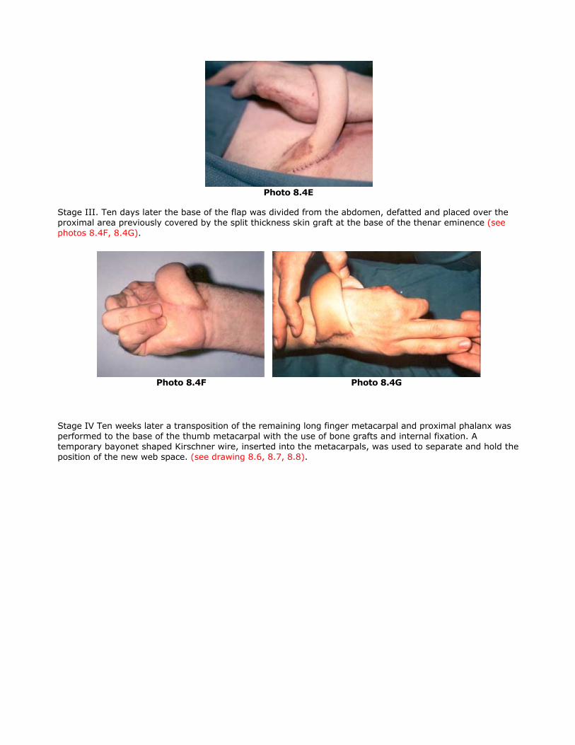
Photo 8.4E
Stage III. Ten days later the base of the flap was divided from the abdomen, defatted and placed over theproximal area previously covered by the split thickness skin graft at the base of the thenar eminence (seephotos 8.4F, 8.4G).
Photo 8.4F Photo 8.4G
Stage IV Ten weeks later a transposition of the remaining long finger metacarpal and proximal phalanx wasperformed to the base of the thumb metacarpal with the use of bone grafts and internal fixation. Atemporary bayonet shaped Kirschner wire, inserted into the metacarpals, was used to separate and hold theposition of the new web space. (see drawing 8.6, 8.7, 8.8).


The tube flap was then transected horizontally at its midpoint. The tube on the volar side was opened,defatted and trimmed to fit the skin defect on the volar side of the thenar eminence. The tube on the dorsalside was opened, defatted and thinned to cover the open dorsal and ulnar side of the web space (see photos 8.4H, 8.4I).
Photo 8.4H Photo 8.4I
His final function was excellent and photos are shown taken six years post injury with excellent grasp andstrong pinch between the reconstructed thumb and the remaining digits (see photos 8.4J, 8.4K, 8.4L).
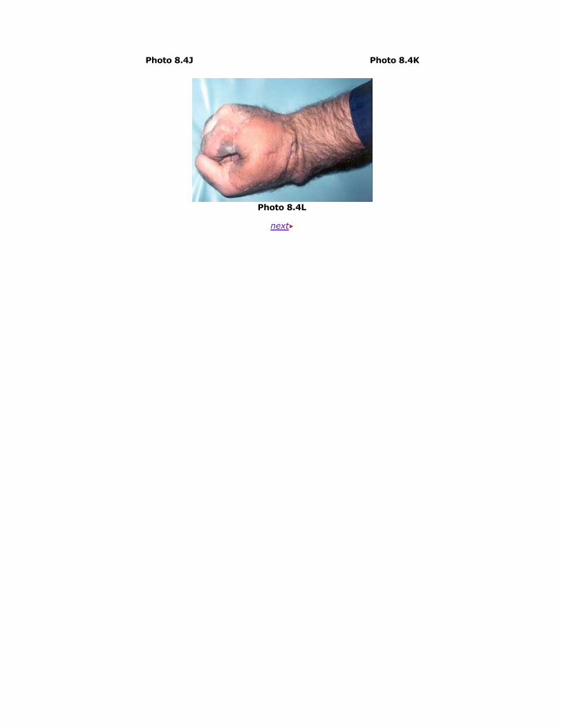
Photo 8.4J Photo 8.4K
Photo 8.4L
next

Chapter Eight: TECHNICAL REQUIREMENTS FOR FORMATION OF A TUBED PEDICLE FLAP
EXAMPLES OF HAND RECONSTRUCTION USING TUBED FLAPS
Case No. 5:
A 40-year-old man suffered a crush/avulsion injury of the left-hand with loss of the phalangesIII, IV, and V and near complete degloving on both sides of the hand (see photos 8.5A, 8.5B).
Photo 8.5A Photo 8.5B
Stage I. The wound was extensively debrided and W's were incorporated along the skin line ofattachment for the flap. The W's were not as large as I would use today. A large patterned flapwas planned on paper and placed over the left lower quadrant measuring 19 cm in width andextending from the midline of the abdomen and the left hip area incorporating the superficialinferior epigastric vessels. The proximal portion of the flap was tubed and the distal portion tobe applied to the hand was thinned of fat. The tube was rotated 90 ° so that the palmar skinwould be taken from the lateral flank which was free of hair. The donor site was closed byadvancement and with a split thickness skin graft. Because of the concavity of the palmarsurface, a suction drain was used to obliterate dead space. A spreader was used to maintain thethumb index web space. The Kirschner wire was used through the thumb as a point ofsuspension to keep the hand away from the abdomen and keep the flap in good position (seephotos 8.5C, 8.5D).
Photo 8.5C Photo 8.5D

Stage II. Three weeks later a primary delay was performed close to where the flap joined thehand.
Stage III. Four weeks after attaching the flap, the pedicle was divided. The remaining tube wasopened to replace the split thickness skin graft previously used to close the donor site. Thepostoperative result is shown eight years later (see photo 8.5E). This case illustrates theusefulness of the tubed abdominal flap to cover both sides of the hand.
Photo 8.5E
next