Embed Size (px)
Citation preview

A Technique for Microscopical Soil
Examinations
Skip Palenik
Microtrace LLC
Elgin, IL USA
www.microtracescientific.com


Edmond Locard


Preliminary Separation
Color (dry)
SievingLow PowerMicroscopy
Seeds, Leaf FragmentsPaint, Glass, etc.
Sonication in Water
Sinks Suspended
Sinks in < 10 min.Clean Sand and Silt
Fraction 1 Fraction 2
Does not settle in10 min.
Fraction 3

Color

Fraction 1
Sand and Silt
Sieve>90 < 180
FloatsHeavy Mineral
Separation< 2.89
Sinks
Light MineralsMount in 1.540
Identify andQuantitate byPolarized Light
Microscopy
Heavy MineralsMount in 1.660


Heavy Mineral Separation
Bromoform= 2.84
Cleaned Silt> 90 < 180 m

Mineral Separation in Heavy Liquids

Freezing Heavy Minerals in Tip of Tube with LN2

Isolation ofMineral Separates
LN2
FrozenBromoform
1 2
3
4
Oven Dry

Heavies Frozen in Bottom of Microcentrifuge Tube
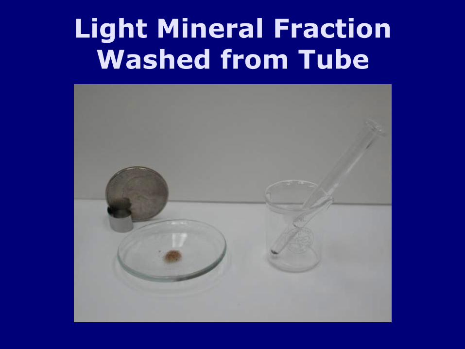
Light Mineral Fraction Washed from Tube

Light and Heavy Fractions Ready for Mounting

PLM Study of Density Fractions

Light Mineral Fraction in 1.540 Cargille Liquid

Plagioclase FeldsparsCrossed Polars

Rock Fragments

Heavy Mineral SuitePlane Polarized Light

Mineral Suite Varies by Provenance of Source Rocks

Refractive Index and Dispersion Colors
Top. Apatite grain in 1.660 refractive
index oil. Plane polarized light.
Bottom. Crossed polars.

Birefringence
Top. Kyanite in
1.660 index of refraction oil.
Bottom. Crossed polars

Pleochroism
Top. Glaucophane
1.660 refractive index oil. N-S polarizer.
Bottom. E-W polarizer.

Pleochroism
Top. Euhedral tourmaline in 1.660 index of refraction oil. N-S polars
Bottom. E-W polars.

Pleochroism
Top. Subhedral tourmaline in 1.660 refractive index oil.
Bottom. E-W polars.

Mineral Varieties
Hypersthene from Mount St. Helens
eruption collected from Yakima, WA days after the event. 1.660 index of refraction oil. Top. N-S polars. Bottom. E-W polars.

Mineral Varieties
Hypersthene from Africa. 1.660 refractive index oil.
Top. N-S polars.
Bottom. E-W polars.

Mineral Varieties
Hypersthene from Martinique. 1.660 refractive index oil.
Top. E-W polars.
Bottom. N-S polars.

Coarse Mineral Fractions
Examination for identification and surface texture.
Stereomicroscopy
Scanning Electron Microscopy
Cathodoluminescence

Light Microscopy
Monahan Sand Dunes in West Texas. Mounted in 1.660 refractive index oil for contrast.

Stains and Reagents
Sahara sand stained with methylene blue to show distribution of amorphous silica (silicic acid) on grain surfaces.

SEM of Quartz Grain Surfaces
Rounded quartz grain from Monahan Dunes in Texas showing surface coating.

Indicators on Quartz Grain Surfaces

Diatoms on Marine Quartz Grain Surface

Detail of Diatom on Quartz Grain Surface

“Silica Flowers” Deposited on Quartz Grain Surface

Etching and Dissolution of Silica on Quartz Surface

Deep Etching on Quartz Grain Surface

Fresh Quartz Grain from Glacier in Canada

Cathodoluminescence

Calcite Wollastonite
Zircon Willemite

Fraction 2
Check for Diatoms or Plant Opal
Absent
Heat with HFin Plastic Tube
1/2
Present
Split Sediment
Acetolysis inGlass Tube
30% H2O2
+ heat
Density Separation< 2.3
Wash, Mount in Glycerine - Water

Acetolysis in 1.5 mL Glass Microcentrifuge Tube

Fraction 2After Acetolysis

Fraction 2Identification of Isolates
Light Microscopy
Pollen and spores
Resistant plant tissue
Tire rubber
Combustion soot
Diatoms and plant opal

Pollen Fraction after Acetolysisand Staining

Study of Internal Structure at High Magnification

Examination of Exine Sculpturing

Details of surface sculpturing can be enhanced by scanning electron microscopy
Scanning Electron Microscopy Light Microscopy
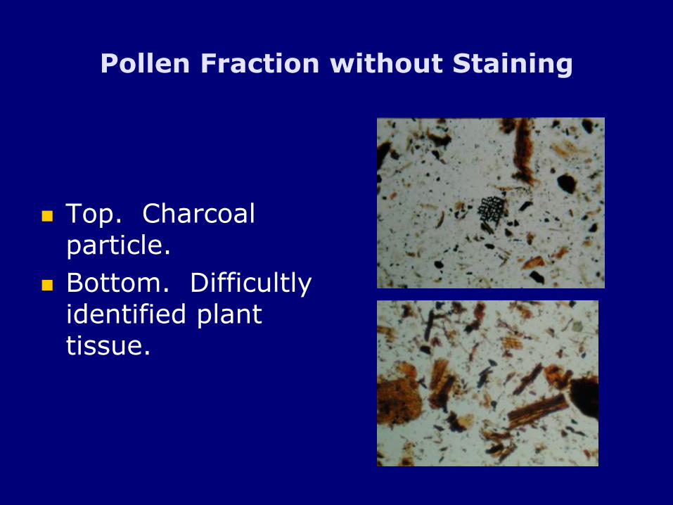
Pollen Fraction without Staining
Top. Charcoal particle.
Bottom. Difficultly identified plant tissue.

Pollen Fraction without Staining

Opal Phytoliths
Distinctive morphology.
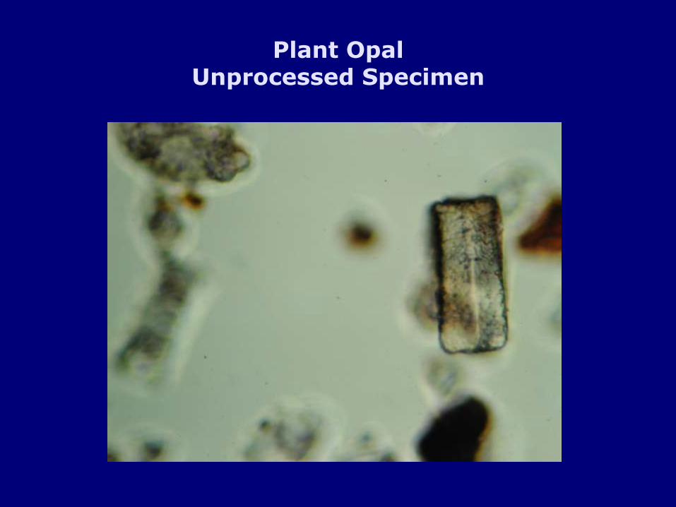
Plant OpalUnprocessed Specimen

Fraction 2 After Treatment withHot Concentrated Hydrogen Peroxide
Opal Phytolith Rich Specimen

Fraction 2 After Treatment withHot Concentrated Hydrogen Peroxide
Diatom Rich Specimen

Fraction 3(clays)
Centrifuge and Filtration
XRD Microscopy Thermal Analysis
• Staining Tests• Phase Contrast –Oil Immersion onOriented Platelets
FTIR


Sm
ectite
Illite
Illite
Illite
Kaolinite
Illite