Embed Size (px)
Citation preview

_____________________________Cristian Comes et al 97
orIgInal arTIClES
A study on the ergonomicAl working modAlities using the dentAl operAting microscope (dom). pArt ii: ergonomic design elements oF the operAting microscopes
Cristian Comes1, Anca Valceanu2, Darian Rusu3, Andreea Didilescu4, Alexandru Bucur5, Mirella Anghel6, Veronica Argesanu7, Stefan-Ioan Stratul3
reZumAtMicroscopia dentară, cu întregul echipament, accesorii şi metode de lucru aduse în panoplia terapeutică a medicinei dentare, nu face nici o excepţie de la imperativele ergonomice ale stomatologiei. Deşi relativ recent utilizată în terapia dentară modernă, microscopia dentară este intens folosită în specialităţi dentare ca endodonţia, microchirurgia parodontală, stomatologia estetică etc. Ca rezultat, activitatea îndelungată la microscopul dentar are ca o consecinţă solicitarea corpului, pe de o parte. Pe de altă parte, microscopul dentar – ca parte integrată a echipamentului terapeutic şi diagnostic din cabinetele dentare – este considerat ca făcând parte din circuitele operatorii. Ambele motive impun stabilirea unor criterii de activitate ergonomică, care devine obiectul unei analize ştiinţifice.Cuvinte cheie: microscop operator dentar (DOM), ergonomie
ABstrActThe dental microscopy, with its whole equipment, accessories and working methods brought in the therapeutic panoply of the dental medicine, does not make any exception from the ergonomic imperatives of the dental profession. Although relatively recent introduced in the modern dental therapy, the dental microscope is used intensely in dental specialties such as endodontics, periodontal microsurgery, dental esthetics, etc. As a result, working for a long time with the dental microscope has as a consequence solicitations of the body, on one hand. On the other hand, the dental microscope – as an integrated part of the therapeutic and diagnostic equipment in the dental office – is considered to be part of the operating circuits. Both reasons imposed the establishment of ergonomic working criteria, which became the object of a scientific analysis. Key-words: dental operating microscope (DOM), ergonomics
Received for publication: Jun. 05, 2009. Revised: Dec. 03, 2009.
1Department of Dental Ergonomics and Oral diagnosis, Faculty of Dental Medicine, Carol Davila University of Medicine and Pharmacy, Bucharest, Romania2Department of Esthetic Dentistry, 3Department of Periodontology, Faculty of Dental Medicine, Victor Babes University of Medicine and Pharmacy, Timisoara, Romania4Department of Anatomy and Embriology, Faculty of Dental Medicine, Carol Davila University of Medicine and Pharmacy, Bucharest, Romania5Department of Oral and Maxillofacial Surgery, Carol Davila University of Medicine and Pharmacy, Bucharest, Romania6Department of Oral diagnosis and Dental Ergonomics, Faculty of Dental Medicine, Victor Babes University of Medicine and Pharmacy, Timisoara, Romania7Department of Mecatronics, Faculty of Mechanics, Traian Vuia Polytechnical University, Timisoara, Romania
Correspondence to:Dr. Cristian Comes, E-mail: [email protected]
introduction
ergonomic features of current types of modTo obtain a good working position with the dental
operating microscope (DOM), a kinematical chain must be created in the chosen working standard, so that the microscope should not interfere with the procedures carried out. On this purpose, conceptual systems to carry out the operating microscope and maneuvering systems that should be found in all movements of the dentist have been analyzed.
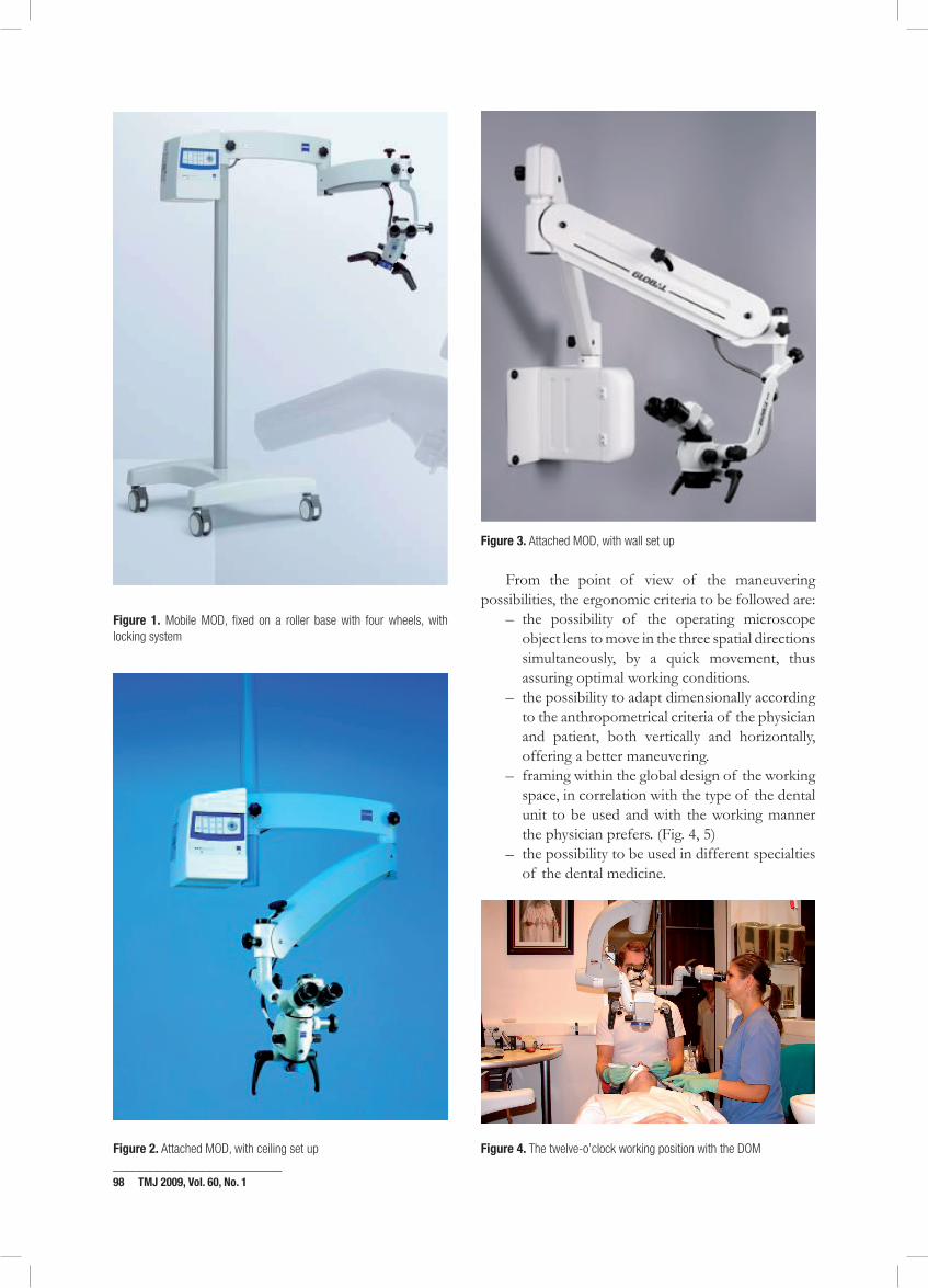
Several types of operating microscopes can be found currently on the market, as derived from the conceptual models: mobile (fixed on a roller base) or attached – either by the ceiling or against a lateral wall of the room or even by the dental unit or a mobile cart attached to the unit.1 (Fig. 1, 2, 3)
_____________________________98 TMJ 2009, Vol. 60, No. 1
Figure 1. Mobile MOD, fixed on a roller base with four wheels, with locking system
Figure 2. Attached MOD, with ceiling set up
Figure 3. Attached MOD, with wall set up
From the point of view of the maneuvering possibilities, the ergonomic criteria to be followed are:
– the possibility of the operating microscope object lens to move in the three spatial directions simultaneously, by a quick movement, thus assuring optimal working conditions.
– the possibility to adapt dimensionally according to the anthropometrical criteria of the physician and patient, both vertically and horizontally, offering a better maneuvering.
– framing within the global design of the working space, in correlation with the type of the dental unit to be used and with the working manner the physician prefers. (Fig. 4, 5)
– the possibility to be used in different specialties of the dental medicine.
Figure 4. The twelve-o’clock working position with the DOM
_____________________________Cristian Comes et al 99
Figure 5. The nine-o’clock working position with the DOM
By attaching the Centrocolumn to the equipment (the dental unit), the vertical bar of support and/or the equipment base is eliminated, assuring an increased comfort. We can notice that the majority of the attached systems are adjustable by a horizontal rotation, with a high degree of rotating liberty of the supporting microscope arms. (Fig. 6) The vertical adjustment is made by rotation, as compared with the articulation of the following arm. These assure the possibility of the dentist’s access to different heights, during the operating acts.1
Figure 6. Attachment of the operating microscope on the dental unit arm for space economy (Centrocolumn Kavo platform for the Zeiss microscopes).
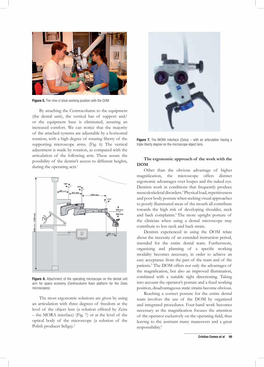
The most ergonomic solutions are given by using an articulation with three degrees of freedom at the level of the object lens (a solution offered by Zeiss – the MORA interface) (Fig. 7) or at the level of the optical body of the microscope (a solution of the Polish producer Seliga).2
Figure 7. The MORA interface (Zeiss) – with an articulation having a triple liberty degree on the microscope object lens.
the ergonomic approach of the work with the dom
Other than the obvious advantage of higher magnification, the microscope offers distinct ergonomic advantages over loupes and the naked eye. Dentists work in conditions that frequently produce musculoskeletal disorders.3 Physical load, repetitiveness and poor body posture when seeking visual approaches to poorly illuminated areas of the mouth all contribute towards the high risk of developing shoulder, neck and back complaints.4 The more upright posture of the clinician when using a dental microscope may contribute to less neck and back strain.
Dentists experienced in using the DOM relate about the necessity of an extended instruction period, intended for the entire dental team. Furthermore, organizing and planning of a specific working modality becomes necessary, in order to achieve an easy acceptance from the part of the team and of the patients.5 The DOM offers not only the advantages of the magnification, but also an improved illumination, combined with a suitable sight directioning. Taking into account the operator’s posture and a fixed working position, disadvantageous static strains become obvious.
Reaching a correct posture for the entire dental team involves the use of the DOM by organized and integrated procedures. Four-hand work becomes necessary as the magnification focuses the attention of the operator exclusively on the operating field, thus leaving to the assistant many maneuvers and a great responsibility.6

_____________________________100 TMJ 2009, Vol. 60, No. 1
The treatment enhanced by DOM means reduced, even minimal movements of the patient and of the instruments. The optimal position of the target structure of the oral cavity is in the center of the visual field of the DOM.
The study of the working conditions with the DOM involves usually the creation of a working environment based on the human-centered ergonomics, usually a dental office provided with a modern dental unit and fully digitalized.7 The literature contains very few such analyses. In a study from 2004, the most comfortable positioning for a dental operating microscope during non-surgical endodontics was investigated.8 The time taken to obtain magnified images and angles of binoculars, microscope body and the mirror to the floor surface were recorded, for operators distributed in three groups according to heights. (Fig. 8) Results indicated that shorter operators had to adopt a strained position for mandibular observation, but became more comfortable when using angled optics or short objective lenses.
Figure 8. The lines used to measure the instrument angles (Kinomoto et al., 2004)
Imagining such studies allows the dentist (in all dental specialties) to simultaneously benefit of the advantages offered by every ISO-4 standard and to eliminate their individual disadvantages. The result is an individualized microscope-enhanced working
modality in every dental specialty. The study uses usually a dummy placed both in horizontal and half-sitting positions and requires simulations of the positioning of the head as follows: vertically in a neutral position (Fig. 9), then by modifying the position of the head by adjusting the head-rest, then by rotating movements of approx.45 degrees to the left and to the right and by extension-flexion movements, in order to access various segments of the dental arches.
Figure 9. The DOM placed in neutral position above the patient’s head
particular requirements for the ergonomic work with the dom
The DOM essentially benefits of large adjustment possibilities, which makes its use extremely advantageous from ergonomic point of view. An improper adjustment of the DOM leads to unfavorable postures and to a negative experience in its use. While the positioning of the DOM (on the floor, on the wall or on the ceiling) depends on the spatial conditions offered by the dental practice, the accessories provided with certain types of DOM can largely enhance its clinical benefits: for instance, microscopes provided with zoom offer more comfort than microscopes provided only with magnification steps. Modern microscopes can be thus positioned with one hand only, while some commands (e.g. the capture of the images, the freezing of the captured images on digital recording units etc.) have been already transferred to foot switches. Future developments of DOMs consider even voice commands for some adjustments of the position.
The lack of vibrations of the DOM is certainly an ergonomic feature resulting both from its construction and from its location/mounting in the dental office. All supporting component parts of the DOM are designed to ensure a stable – but easily adjustable – of the optical part, which can be also moved easily in all three dimensions.

_____________________________Cristian Comes et al 101
One of the features the DOM unfortunately cannot provide is the direct visibility, which is limited by the anatomical conditions of the mouth. Direct visibility is only possible for the anterior segments of the arches, with the patient placed in extreme horizontal or sitting positions. For all other segments of the dental arches, the use of non-refractive mirrors provides indirect visibility.
A minimal distance of 18 cm must exist between the target structure and the optical body of the microscope. Furthermore, the DOM offers coaxial illumination, a suitable, constant and adjustable intensity of the light. For special situations, light filters are provided: green for surgical procedures (it removes the confounding red reflections of the blood) and yellow/orange (it prevents the premature setting of composite resins).
Particular ergonomic indications should be considered when starting the work with a DOM:
1. Position the DOM right above the patient’s face
2. Place the working area in the center of the illuminated field
3. Incline the binocular to the microscope body to an angle of approx.135°
4. Adjust to a small focal length 5. Select a small magnification in the beginning 6. Increase the magnification as required by the
clinical procedure, by maintaining the focus on the target anatomical structure
7. When adjusting the position of the body, follow the steps:
1. Position of the body – upright 2. Sagittal position of the head – the cervical
spine continues the thoracic spine 3. The lateral position of the head – the
bipupilar line perfectly horizontal.
Acknowledgement
Research carried out within the research grant MICRODENT (funded by the National State Authority for Research, contract CNMP 41-034/2007).
reFerences
1. http://www.meditec.zeiss.de/C125679E00510B81/ContentsFrame/53925FEF1BD8FD1EC125726400283349
2. www.seliga.pl 3. Bernard PB. Musculoskeletal Disorders (MSDs) and Workplace
Factors. A critical Review of Epidemiologic Evidence for Work – Related MSD of Neck, Upper Extremity, and Lower Back – NIOSH, Cincinnati, 1997,11.
4. Valachi B, Valachi K. Mechanisms leading to musculoskeletal disorders in dentistry – JADA 2003:;123(10):1344-50.
5. Finkbeiner BL. Four Handed Dentistry. A Handbook of Clinical Application and Ergonomic Concepts. Prentice Hall, New Jersey, 2001,65.
6. Argesanu V. Ergonomia Echipamentelor si a Departamentelor Medicale. Ed. Eurostampa, Timisoara, 2004,15.
7. Comes C, Valceanu A, Rusu D, et al. A study on the ergonomical working modalities using the dental operating microscope (DOM). Part I: Ergonomic principles in dental medicine. Timisoara Medical Journal 2008;58(3-4):218-23.
8. Kinomoto Y, Takeshige F, Hayashi M, Ebisu S. Optimal positioning for a dental operating microscope during non-surgical endodontics. J Endod 2004;30(12):860-2.