Embed Size (px)
Citation preview

_____________________________102 TMJ 2010, Vol. 60, No. 1
orIgInal arTIClES
A study on the ergonomicAl working modAlities using the dentAl operAting microscope (dom). pArt iii: ergonomicAl FeAtures oF contemporAry top dentAl microscopes commented
Andreea Didilescu1, Cristian Comes2, Darian Rusu3, Mihai Bucur4, Mirella Anghel5, Veronica Argesanu6, Stefan-Ioan Stratul3
reZumAtActivitatea clinică îndelungată asistată de microscopul operator dentar (MOD) are ca o consecință solicitarea corpului, atât posturală, cât și a segmentelor sale particulare implicate în actul terapeutic. Ca urmare, MOD au beneficiat de-a lungul timpului de numeroase optimizări de natură ergonomică. Firmele producătoare manifestă astăzi o preocupare permanentă pentru introducerea și adaptarea de caracteristici ergonomice. Liderii de piață cu cele mai marcante preocupări în asigurarea ergonomiei MOD oferă astăzi caracteristici precum: oculare înclinabile până la 180 grade, controlul continuu al luminozității, lentile oculare cu distanță interpupilară variabilă și adaptabile la purtătorii de ochelari, mânere ergonomice cu priză sporită, adaptabile, demontabile și sterilizabile, mânere ergonomice cu butoane de funcții interschimbabile și individualizate, lentilă obiectiv manevrabilă atât de dreptaci cât și de stângaci, interfața Mora, sistemul de focalizare automata VarioskopTM, sistemul de suspensie magnetică al corpului microscopului etc. Articolul enumeră, comentează și compară câteva din caracteristicile ergonomice ale principalilor producători de MOD contemporani.Cuvinte-cheie: microscopul operator dentar, caracteristici ergonomice
ABstrActThe prolongated clinical activity assisted by the dental operating microscope (DOM) has as a consequence solicitations of the body, generally postural as on its particular segments involved in the clinical activities. Subsequently, DOMs have been designed and continuously optimized with ergonomical features. Manufacturers are today constantly preoccupied to introduce and adapt ergonomical design elements. The market leaders in dental microscopy offer currently ergonomical features as: the 180° tiltable tube with angled optics, the continuous brightness control, the widefield eyepieces with adjustment for glass-wearers and variable interpupilar distance, the ergonomically designed grips, the focusing objective lens adjustable for left-handed and right-handed, the MORA interface, the motorized focusing system VarioskopTM, the Free Float Magnetic System etc. The article comments and compares some ergonomical features of the contemporary major producers of DOMs.Key-words: dental operating microscope, ergonomic features
Received for publication: Jun. 07, 2009. Revised: Dec. 02, 2009.
1Department of Anatomy and Embriology, 2Department of Dental Ergonomics and Oral diagnosis, 3Department of Periodontology, 4Department of Oral Implantology,5Department of Oral diagnosis and Dental Ergonomics, Faculty of Dental Medicine, Victor Babes University of Medicine and Pharmacy, Timisoara, Romania6Department of Mecatronics, Faculty of Mechanics, Traian Vuia Polytechnical University, Timisoara, Romania
Correspondence to:Dr.Cristian Comes, Department of Anatomy and Embriology, Carol Davila BucurestiE-mail: [email protected]
introduction
The use of dental microscope does not change the basic ergonomic principles associated with gaining visual and working access familiar to non-microscope operators. In that, gaining visual access as well as working access to the operating field are totally dependent on the limitations imposed by the posture of the operator.1 The prolongated clinical activity assisted by the dental operating microscope (DOM) has as a consequence solicitations of the body, generally postural as on its particular segments involved in the clinical activities.2, 3 Beyond application of the basic ergonomical principles and the ergonomical integration of the DOM in the contemporary dental practice stay the numerous technical ergonomic details that DOM producers have already implemented on their products.4
Among contemporary DOM producers, Zeiss (Carl Zeiss Meditec AG, Jena Germany) counts among

_____________________________Andreea Didilescu et al 103
the most dedicated to ergonomy. Therefore, the ergonomical features of two of the most appreciated Zeiss dental microscopes (OPMI Pico and ProErgo) are listed and commented below and compared with similar features of other two DOM brands.5
Below some of the ergonomic features of the Zeiss OPMI Pico dental microscope:
1. The 180° tiltable tube, the two-joints angled optics and tube dovetail – enables an upright position of the cervical spine, despite the anterior-posterior inclination of the microscope body, while reaching difficult-to-reach working areas. However, the newly-gained flexibility of the DOM’s ocular tube should not replace the correct positioning of the patient’s head. (Fig.1)
Figure 1. The tiltable tube with angled optics of the Pico OPMI dental microscope
2. The continuous brightness control through a rotating button placed on the first vertical joint of the DOM – allows the adjustment of the light intensity for work in profound sites, like root canals, thus avoiding the eye strain. Working in the root canal depth may necessitate increasing of the light brightness during a limited period of time. (Fig.2)
3. The widefield eyepieces – allows simultaneously the adjustment for glass-wearers and variable interpupilar distance. (Fig.3)
4. The ergonomically designed grips – allow a confortable and secure handling of the
microscope, as they can be rotated at various angles; furthermore, they can be removed and sterilized. However, the repeated excessive rotational adjustment of the grips should be avoided, as the grips are relatively fragile. (Fig.4)
Figure 2. The brightness control button on the upper joint of the Pico OPMI
Figure 3. The widefield eyepieces of the Pico OPMI. Note the upper rotating button, which regulates the interpupillar distance
5. The focusing objective lens – can be adjusted for both left-hand and right-hand users by mean of a switch centrally placed, that can be turned in either direction. With the time, this type of fine adjustment is avoided by the correct initial placing of the patient’s head. (Fig.5)

_____________________________104 TMJ 2010, Vol. 60, No. 1
Figure 4. The ergonomical rotating, detacheable and sterilizable grips of Pico OPMI.
Figure 5. The focusing objective lens of Pico OPMI. Note the central switch, which allows turning in either direction.
6. Altough not mentioned by the producer among the ergonomic design elements, the universal adapter with the observation terminal (the so-called „spy”) allows the simultaneous observation by a second person (i.e. the dental assistant), therefore enhancing the ergonomical workflow. Unfortunately, adding this equipment (most of the times long, with the adapter and the digital camera) results in an overall bulky microscope body, more prone to injuries and collisions with the own parts of the microscope when moved away. Lately, this costly and heavy optional equipment has been replaced increasingly with two flat screens placed each on the walls of the dental office, across the operator and across the assistant.
Figure 6. Thanks to the MORA interface, the Pico OPMI can be tilted laterally, while the eyepieces remain in horizontal position.
7. The MORA interface. Designed by Dr.Assad Mora, the MORA interface raises the mobility of microscopes in this class to a new level.
_____________________________Andreea Didilescu et al 105
Basically, the MORA interface adds a horizontal antero-posterior joint between the oculars and the microscope body, which allows the vertical tilting of the latter, by keeping the first (and the eyepieces) in horizontal position. Thus, dentists continue looking straight into the microscope, while sitting in an upright position in each phase of the treatment. While sitting in the 12 o’clock position, the MORA interface also enables the DOM-using dentist: to swing the DOM body in a panning motion to the left and to the right sides of the mouth (25° in each direction); to reduce the effort that goes into the rotation movement; to sit in an upright position with an upright neck; to equally extend the arms around the patient’s head and to work more comfortably.1 The MORA interface is currently used in all clinical applications of the DOM.6- 8 (Fig.6)
The ProErgo dental microscope from Zeiss, the top of its class, features the following ergonomical characteristics:
1. The motorized focusing system VarioskopTM
- focuses the image automatically by button push, and not by combining the movement of the microscope body with the fine manual adjustment. Once focused, the image can be zoomed in with another button push, avoiding the usual mechanical magnification steps. Despite its obvious ergonomical advantages, the VarioskopTM system may sometimes automatically focus on irrelevant details (i.e. the coronal margin of the access cavity in endodontically treated teeth), making the work relatively difficult. With the time, the operator gains the ability to precisely focus only on the relevant details.
2. The Free Float Magnetic System – probably one of the most appreciated ergonomical features of this microscope, the Free Float Magnetic System unlocks the magnetic brakes of the system. Without the brakes, the heavy microscope body could simply fall on the patient’s face, hurting him. As the release buttons are placed on the solid handgrips of the microscope body, they can only be pushed while sustaining the latter with little effort, thus preventing accidents. Once released the button, the brakes intervene automatically, blocking the microscope in the required position. The system is also useful for the lateral displacements of the microscope body. (Fig. 7)
Figure 7. The main joint of the ProErgo microscope, hosting the Free Float Magnetic System. Note the multidirectional freedom degree of the joint.
3. The ergonomically designed handgrips with function keys concentrate multiple controls, like the zoom, the focus, and the light switch. The functions of the keys can be configurate to operate the brightness, the SpeedFokus system or to freeze the image. The grips feature nutshape joints for an ergonomic use. (Fig. 8)
Figure 8. The handgrip of ProErgo microscope, with the function keys. Standard designation of the function keys include: blue button left: light switch, blue button right: the SpeedFokus system, black switches: the zoom, yellow buttons: the brightness regulators. Note the palmprint internal surface of the grip.

_____________________________106 TMJ 2010, Vol. 60, No. 1
4. The ZoomLink focus and the ZoomLink illumination correlate the magnification with the focusing speed and the illumination intensity. This correlation became necessary because of the continuous adjustment of the magnification, especially in deep root canals. (Fig.9)
Figure 9. The ZoomLink coaxial illumination highlighted by a steamy environment below the objective lens.
5. The digital control panel and console – allows the personalization of the default settings as light, zoom or magnification. The settings can be defined for various users of the microscope and can be stored. The advantage is the placement of the digital display on the microscope arm; however, the control panel may be sometimes difficult to reach because of the space limitations of the dental office and cannot be seen from the operator’s position. (Fig. 10)
6. The optional foot control panel allows the transfer of some controls of the microscope from the hand to the foot. Of special importance is the image freeze, which can be done by the operator, without necessitating verbal orders and the coordination with the dental assistant. (Fig. 11)
7. The optional angled optics and the tube dovetail enables, as to the OPMI Pico model, an upright position of the cervical spine, despite the inclination of the microscope body, while reaching difficult-to-reach working areas. (Fig. 12)
Figure 10. The digital control panel of the ProErgo microscope
Figure 11. The foot control of ProErgo allows here the adjustment of the focus and of the zoom. Note the tiltable pads, which allow four simultaneous controls with a single foot movement.
In what concerns the standard ergonomic features of another US top brand of dental microscopes (Global from Global SurgicalTM, USA - the models G6, G3), they are pretty much the same as for the top Zeiss models: inclinable binocular, adjustable (helicoidal) eyecups, adjustable maneuvering handles and manual fine focus. 9 Additional accessories with ergonomic role count among them binocular extenders, an extension arm, and a binocular rotating ring with locking thumb, which rotate the binocular -/+ 25 degrees and allows for better operator positioning. (Fig.13, 14)

_____________________________Andreea Didilescu et al 107
Figure 12. The angled optics of the ProErgo dental microscope. Note the additional optical terminal (universal adapter) for the capture of the images, either classic or digital
Figure 13. The inclinable binocular of G6 DOM from Globalsurgical USA (www.globalsurgical.com)
Figure 14. The extension arm of G6. Note the slim design of the extension, which reduces the overall weight of the DOM, making it more maneuverable (www.globalsurgical.com)
To compare, the german Kaps microscopes (Karl Kaps GmbH & Co.KG, Asslar/Wetzlar, Germany) display the following ergonomic features, as stated by the producer: the rotating binocular plate (allows an upright ergonomical position, even with the DOM body laterally tilted; however, the ergonomic effect doesn’t meet the versatility of the MORA interface from Zeiss); the ergonomical detacheable and sterilizable handgrips (however, slimmer and therefore providing a less firmer grip than the similar parts from Pico); the inclinable 0-60° binocular head; a counterbalanced pantographic arm.10 (Fig. 15)
Figure 15. The inclinable binocular head of Kaps DOM (www.kaps-optik.de)
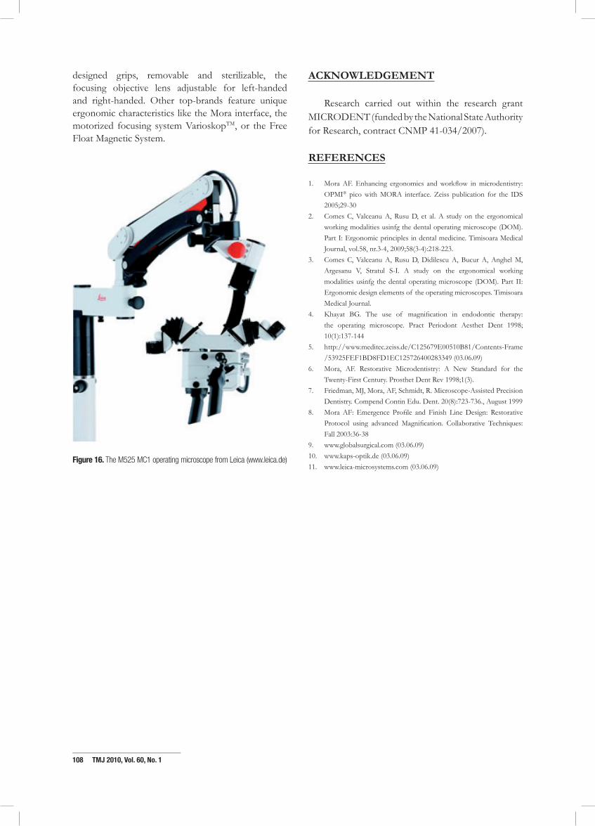
The DOMs from the Leica family (Leica Microsystems GmbH, Wetzlar, Germany) also display a couple of ergonomical features of clinical importance.11 The options designed for a comfortable observation are grouped in a package called ErgonOptic™, which include the variable binocular tube; the optics are ergonomically maneuvered by electromagnetic brakes; the coupling of the brightness control with the working distance for a better adjustment of the eyes of the observer at small distances (the AutoIris™). (Fig. 16)
To conclude, the market leaders in dental microscopy currently share common ergonomical features as: the 180° tiltable tube with angled optics, the continuous brightness control (either manual or electronic), the widefield eyepieces with adjustment for glass-wearers and variable interpupilar distance, the ergonomically
_____________________________108 TMJ 2010, Vol. 60, No. 1
designed grips, removable and sterilizable, the focusing objective lens adjustable for left-handed and right-handed. Other top-brands feature unique ergonomic characteristics like the Mora interface, the motorized focusing system VarioskopTM, or the Free Float Magnetic System.
Figure 16. The M525 MC1 operating microscope from Leica (www.leica.de)
Acknowledgement
Research carried out within the research grant MICRODENT (funded by the National State Authority for Research, contract CNMP 41-034/2007).
reFerences
1. Mora AF. Enhancing ergonomics and workflow in microdentistry: OPMI® pico with MORA interface. Zeiss publication for the IDS 2005;29-30
2. Comes C, Valceanu A, Rusu D, et al. A study on the ergonomical working modalities usinfg the dental operating microscope (DOM). Part I: Ergonomic principles in dental medicine. Timisoara Medical Journal, vol.58, nr.3-4, 2009;58(3-4):218-223.
3. Comes C, Valceanu A, Rusu D, Didilescu A, Bucur A, Anghel M, Argesanu V, Stratul S-I. A study on the ergonomical working modalities usinfg the dental operating microscope (DOM). Part II: Ergonomic design elements of the operating microscopes. Timisoara Medical Journal.
4. Khayat BG. The use of magnification in endodontic therapy: the operating microscope. Pract Periodont Aesthet Dent 1998; 10(1):137-144
5. http://www.meditec.zeiss.de/C125679E00510B81/Contents-Frame/53925FEF1BD8FD1EC125726400283349 (03.06.09)
6. Mora, AF. Restorative Microdentistry: A New Standard for the Twenty-First Century. Prosthet Dent Rev 1998;1(3).
7. Friedman, MJ, Mora, AF, Schmidt, R. Microscope-Assisted Precision Dentistry. Compend Contin Edu. Dent. 20(8):723-736., August 1999
8. Mora AF: Emergence Profile and Finish Line Design: Restorative Protocol using advanced Magnification. Collaborative Techniques: Fall 2003:36-38
9. www.globalsurgical.com (03.06.09)10. www.kaps-optik.de (03.06.09)11. www.leica-microsystems.com (03.06.09)