Embed Size (px)
Citation preview
at SciVerse ScienceDirect
Biomaterials 33 (2012) 2282e2288
Contents lists available
Biomaterials
journal homepage: www.elsevier .com/locate/biomateria ls
A pyrene-imidazolium derivative that selectively Recognizes G-Quadruplex DNA
Ha Na Kim a,1, Eun-Hae Lee b,1, Zhaochao Xu a,c,1, Hee-Eun Kim b, Hee-Seung Lee d, Joon-Hwa Lee b,**,Juyoung Yoon a,e,*
aDepartment of Chemistry and Nano Science, Ewha Womans University, Seoul 120-750, Republic of KoreabDepartment of Chemistry and RINS, Gyeongsang National University, Jinju, Gyeongnam 660-701, Republic of KoreacDalian Institute of Chemical Physics, Chinese Academy of Sciences, Dalian 116023, ChinadMolecular-Level Interface Research Center, Department of Chemistry, KAIST, Daejeon 305-701, Republic of KoreaeDepartment of Bioinspired Science, Ewha Womans University, Seoul 120-750, Republic of Korea
a r t i c l e i n f o
Article history:Received 11 October 2011Accepted 25 November 2011Available online 21 December 2011
Keywords:G-quadruplexNMRFluorescent sensorImidazoliumPyreneExcimer emission
* Corresponding author. Fax: þ82 2 3277 2384.** Corresponding author. Department of ChemisNational University, Jinju, Gyeongnam 660-701, Repub1489.
E-mail addresses: [email protected] (J.-H. Lee), j1 contributed equally to this work.
0142-9612/$ e see front matter � 2011 Elsevier Ltd.doi:10.1016/j.biomaterials.2011.11.073
a b s t r a c t
G-quadruplexes, formed of four stranded guanine bases stabilized by monovalent cations, serveimportant role in cancer cell growth and control gene expression in telomere. Since there are varioustypes of quadruplex structures, rapid and simple screening methods with high selectivity, sensitivity andnontoxicity are required for understanding about the biological roles of quadruplex DNA as well as indesigning therapeutics. Herein, we report a pyrene-imidazolium derivative, JY-1, which can with highselectivity recognize G-quadruplex using fluorescence and NMR spectroscopy. This is the first examplebased on the imidazolium derivative, which can detect the G-quadruplex directly to utilize the excimer/monomer emission change in pyrene fluorophore. The selectivity of strong binding to a specific sequencecan allow for quadruplex sensing and the detection method presented here is very simple, using fluo-rescence and NMR study. Also, the groove binding characteristic of JY-1 to the G-quadruplex hasa relatively low nonspecific toxicity and the structure-specific differences in fluorescent characterbetween DNA duplex and G-quadruplex may offer more discovery and application in biological study.
� 2011 Elsevier Ltd. All rights reserved.
1. Introduction
For many years, the development of DNA sensing and recogni-tion technology has attracted a great deal of attention since variousdiseases and genetic disorders have been found to be directlyrelated to changes in DNA [1e4]. Among the various types ofsecondary structures in human DNA, such as B-DNA, Z-DNA, hairpins, etc [5]., guanine rich sequences like, G-quadruplexes serve animportant role in cancer cell growth and control gene expression intelomeres [6]. G-quadruplexes are composed of four strandedguanine bases stabilized by monovalent cations such as Naþ or Kþ
between tetrad guanine bases [7]. Recent studies show that thisunique structure can be formed not only in the telomeric region ofthe genome, but also among other guanine rich sequences havingthe potential to form quadruplex elsewhere in the human genome[8,9]. Efficient detection of G-quadruplex in addition to stabiliza-tion of the G-quadruplex structure is very helpful in designing
try and RINS, Gyeongsanglic of Korea. Fax: þ82 55 772
[email protected] (J. Yoon).
All rights reserved.
cancer drugs and in diagnosis of various diseases [10,11]. Detectionmethod taking advantage of fluorescence is so much simple andsensitive [12e15] that there have been reports on fluorophorelabelled G-quadruplex oligonucleotide [16e18] or label free probesystems for sensing G-quardruplex [19e23]. However, thesestudies mainly set out to probe potassium ion stabilizing in G-quartet structure, or detect the conformational change of the G-quartet. Some studies were performed using a labelled oligonu-cleotide system, which is expensive and laborious. In addition, theintercalative methods for detection of the quadruplex can becytotoxic with relatively low selectivity and can induce the desta-bilization of the quadruplex within telomeres, which is critical fortelomerase activity inhibition [6]. Since there are various types ofquadruplex structures, rapid and simple screening methods withhigh selectivity, sensitivity and nontoxicity are required forunderstanding the biological roles of quadruplex DNA as well as fordesigning therapeutics [24,25].
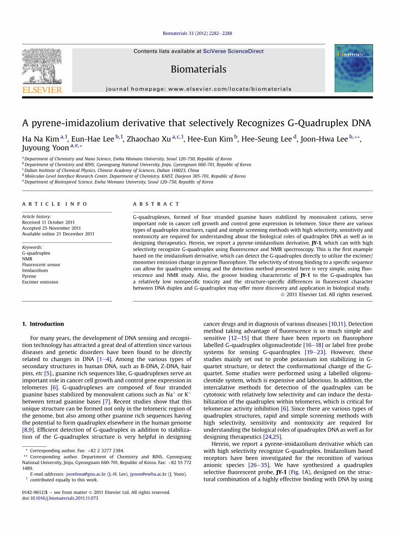
Herein, we report a pyrene-imidazolium derivative which canwith high selectivity recognize G-quadruplex. Imidazolium basedreceptors have been investigated for the reconition of variousanionic species [26e35]. We have synthesized a quadruplexselective fluorescent probe, JY-1 (Fig. 1A), designed on the struc-tural combination of a highly effective binding with DNA by using
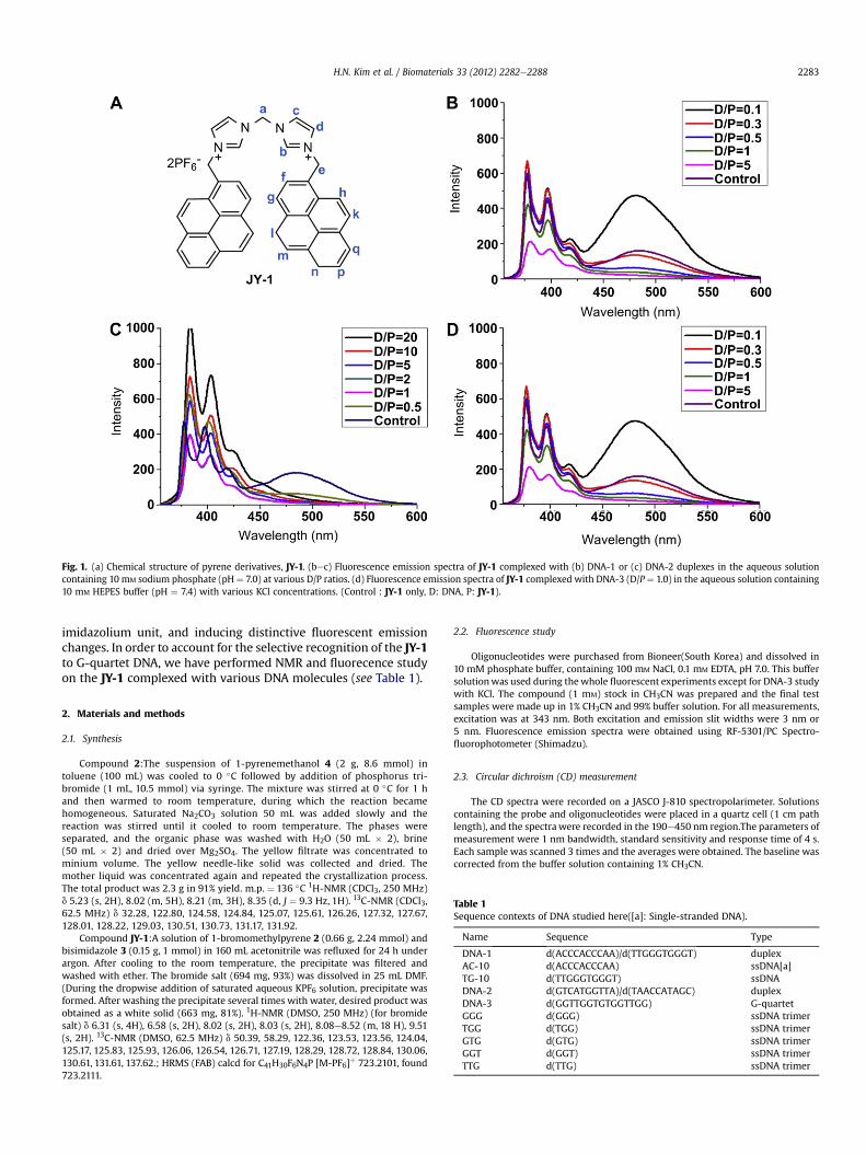
Fig. 1. (a) Chemical structure of pyrene derivatives, JY-1. (bec) Fluorescence emission spectra of JY-1 complexed with (b) DNA-1 or (c) DNA-2 duplexes in the aqueous solutioncontaining 10 mM sodium phosphate (pH ¼ 7.0) at various D/P ratios. (d) Fluorescence emission spectra of JY-1 complexed with DNA-3 (D/P ¼ 1.0) in the aqueous solution containing10 mM HEPES buffer (pH ¼ 7.4) with various KCl concentrations. (Control : JY-1 only, D: DNA, P: JY-1).
H.N. Kim et al. / Biomaterials 33 (2012) 2282e2288 2283
imidazolium unit, and inducing distinctive fluorescent emissionchanges. In order to account for the selective recognition of the JY-1to G-quartet DNA, we have performed NMR and fluorecence studyon the JY-1 complexed with various DNA molecules (see Table 1).
Table 1Sequence contexts of DNA studied here([a]: Single-stranded DNA).
Name Sequence Type
DNA-1 d(ACCCACCCAA)/d(TTGGGTGGGT) duplexAC-10 d(ACCCACCCAA) ssDNA[a]TG-10 d(TTGGGTGGGT) ssDNADNA-2 d(GTCATGGTTA)/d(TAACCATAGC) duplexDNA-3 d(GGTTGGTGTGGTTGG) G-quartetGGG d(GGG) ssDNA trimerTGG d(TGG) ssDNA trimerGTG d(GTG) ssDNA trimerGGT d(GGT) ssDNA trimerTTG d(TTG) ssDNA trimer
2. Materials and methods
2.1. Synthesis
Compound 2:The suspension of 1-pyrenemethanol 4 (2 g, 8.6 mmol) intoluene (100 mL) was cooled to 0 �C followed by addition of phosphorus tri-bromide (1 mL, 10.5 mmol) via syringe. The mixture was stirred at 0 �C for 1 hand then warmed to room temperature, during which the reaction becamehomogeneous. Saturated Na2CO3 solution 50 mL was added slowly and thereaction was stirred until it cooled to room temperature. The phases wereseparated, and the organic phase was washed with H2O (50 mL � 2), brine(50 mL � 2) and dried over Mg2SO4. The yellow filtrate was concentrated tominium volume. The yellow needle-like solid was collected and dried. Themother liquid was concentrated again and repeated the crystallization process.The total product was 2.3 g in 91% yield. m.p. ¼ 136 �C 1H-NMR (CDCl3, 250 MHz)d 5.23 (s, 2H), 8.02 (m, 5H), 8.21 (m, 3H), 8.35 (d, J ¼ 9.3 Hz, 1H). 13C-NMR (CDCl3,62.5 MHz) d 32.28, 122.80, 124.58, 124.84, 125.07, 125.61, 126.26, 127.32, 127.67,128.01, 128.22, 129.03, 130.51, 130.73, 131.17, 131.92.
Compound JY-1:A solution of 1-bromomethylpyrene 2 (0.66 g, 2.24 mmol) andbisimidazole 3 (0.15 g, 1 mmol) in 160 mL acetonitrile was refluxed for 24 h underargon. After cooling to the room temperature, the precipitate was filtered andwashed with ether. The bromide salt (694 mg, 93%) was dissolved in 25 mL DMF.(During the dropwise addition of saturated aqueous KPF6 solution, precipitate wasformed. After washing the precipitate several times with water, desired product wasobtained as a white solid (663 mg, 81%). 1H-NMR (DMSO, 250 MHz) (for bromidesalt) d 6.31 (s, 4H), 6.58 (s, 2H), 8.02 (s, 2H), 8.03 (s, 2H), 8.08e8.52 (m, 18 H), 9.51(s, 2H). 13C-NMR (DMSO, 62.5 MHz) d 50.39, 58.29, 122.36, 123.53, 123.56, 124.04,125.17, 125.83, 125.93, 126.06, 126.54, 126.71, 127.19, 128.29, 128.72, 128.84, 130.06,130.61, 131.61, 137.62.; HRMS (FAB) calcd for C41H30F6N4P [M-PF6]þ 723.2101, found723.2111.
2.2. Fluorescence study
Oligonucleotides were purchased from Bioneer(South Korea) and dissolved in10 mM phosphate buffer, containing 100 mM NaCl, 0.1 mM EDTA, pH 7.0. This buffersolutionwas used during thewhole fluorescent experiments except for DNA-3 studywith KCl. The compound (1 mM) stock in CH3CN was prepared and the final testsamples were made up in 1% CH3CN and 99% buffer solution. For all measurements,excitation was at 343 nm. Both excitation and emission slit widths were 3 nm or5 nm. Fluorescence emission spectra were obtained using RF-5301/PC Spectro-fluorophotometer (Shimadzu).
2.3. Circular dichroism (CD) measurement
The CD spectra were recorded on a JASCO J-810 spectropolarimeter. Solutionscontaining the probe and oligonucleotides were placed in a quartz cell (1 cm pathlength), and the spectrawere recorded in the 190e450 nm region.The parameters ofmeasurement were 1 nm bandwidth, standard sensitivity and response time of 4 s.Each sample was scanned 3 times and the averages were obtained. The baseline wascorrected from the buffer solution containing 1% CH3CN.
H.N. Kim et al. / Biomaterials 33 (2012) 2282e22882284
2.4. NMR experiment
All DNA and JY-1 samples were dissolved in 60% H2O/10% D2O/30% CD3CN buffercontaining 5 mM sodium phosphate (pH 8.0) and 50 mM NaCl. All NMR experimentswere performed on a Varian Inova 600 MHz spectrometer (KAIST, Daejeon) usinga HCN triple-resonance probe. One dimensional (1D) NMR datawere processedwitheither the program VNMR J (Varian, Palo Alto) or FELIX2004 (Accelrys, San Diego),whereas 2D data were processed with the program NMRPIPE and analyzed with theprogram Sparky.
3. Results and discussion
3.1. Design, synthesis, and fluorescence study of compound JY-1
For the synthesis of JY-1 (Fig. 1A), 1-bromomethylpyrene (2) wasprepared by treating 1-pyrenemethanol (4) with phosphorous tri-bromide in toluene (see Scheme 1). 1-bromomethylpyrene wasthen reacted with bisimidazole (3) followed by anion-exchangewith KPF6, yielding 81% JY-1 (see Scheme 1). Fig. 1 shows thefluorescence emission spectra of JY-1 with various DNA in aqueoussolution. In the absence of DNA, JY-1 shows the distinctive spectrawith the monomer emission and excimer emission at 375 and482 nm, respectively. There were no significant changes in theemission spectra of JY-1 when 10 equivalents of various phosphateand nucleotide anions were added (Supporting informationS-Figure 1). Surprisingly, JY-1 displayed fluorescent enhancementof the excimer peak (482 nm) upon the addition of DNA-1 duplexeven at the DNA/JY-1molar ratio (D/P ratio) ¼ 0.1 in 10 mM sodiumphosphate buffer (Fig. 1B). However, when DNA-2, random DNAdecamer duplex, was added, no significant enhancement of theexcimer fluorescence peak at 482 nm was observed even with theD/P ratio of 20 (Fig. 1C). On the other hand, the intensity ofmonomer peak increased upon interaction with DNA-2, as the D/Pratio increased (Fig. 1C). These data indicate that JY-1 binds withDNA-1 through unusual molecular interaction unlike DNA-2.
3.2. NMR studies for prove the interaction between JY-1 and DNAcomplexes
To understand this unusual interaction, we performed 1H-NMRexperiments on the JY-1/DNA-1 complex. The assignment for theproton resonances of the JY-1 were made by the analysis of NOESYspectra at 25 �C (see Fig. 2). Fig. 2A shows the titration of the DNA-1duplex with JY-1, where the imino proton resonances extensivelybroadened as JY-1 was added and then completely disappeared atthe D/P ratio ¼ 1.0. 1H-NMR spectra of JY-1 were also dramaticallychanged by addition of the DNA-1 duplex, indicating specificinteraction between DNA-1 and JY-1 (Fig. 2B). These results clearlydemonstrated that the double helix of DNA-1 was destabilizedupon interaction with JY-1 and subsequently melted out to singlestrands. To further clarify this interaction, the titrations of JY-1witheach strand of DNA-1 (TG-10 and AC-10) were monitored by 1DNMR spectra (Fig. 2C and D). Interestingly, like DNA-1, the TG-10strand caused a significant change in the NMR spectra of JY-1
Scheme 1. Synth
(Fig. 2B). Even though the D/P ratio is only 0.1, the NMR spectra ofthese complexes were completely different from that of free JY-1(Fig. 2D). For example, at D/P ¼ 0.1, the peaks a and e are dis-appeared upon binding to the TG-10 strand and new four reso-nances observed at that region (Fig. 2C). In the free JY-1, eachmethylene moiety which connect between two imidazolium rings(peak a) or between imidazolium and pyren rings (peak e) showsonly one signal in the proton NMR spectrum. However, in the caseof the JY-1/TG-10 complex, the methylene protons positioned ata and e exhibit more than two resonances because the pyren andimidazolium rings cannot rotate freely any more. In the case of AC-10, no clear change in the NMR spectra of JY-1was observed, as theD/P ratio increased up to 1.0 (Fig. 2D). The slight upfield shifts ofsome resonances of JY-1, such as a resonance at 6.3 ppm (peak e),imply a nonspecific interaction between the imidazolium cation ofJY-1 and the phosphate anion of AC-10 (Fig. 2C). From these results,the molecular interaction between JY-1 and DNA-1 duplex can besummarized as follows: i) JY-1 destabilizes the double helix of DNA-1 and then separates it into two single strands, TG-10 and AC-10; ii)JY-1 selectively binds to the single-stranded TG-10 and exhibitsa unique conformation in the complex; iii) JY-1 in this uniqueconformation can emit the excimer fluorescence.
3.3. NMR study of compound JY-1 for selective GGG trimerdetection
The single-stranded TG-10 has a G-rich sequence and can formG-quartet DNA helix under KCl buffer conditions. To address thecorrelation between the G-quartet DNA structure and the excimerfluorescence of JY-1, the fluorescence study on the TG-rich 15mer,DNA-3, which is known as a thrombin-binding aptamer and canform the G-quartet structure with Kþ ion [12], was performed.Here, the HEPES buffer (pH 7.4) was used instead of 10 mM sodiumphosphate buffer containing 100 mM NaCl to exclude the Naþ ioneffect because some metal cations including Kþ, Naþ, or Pb2þ canstabilize the G-quartet structure [36,37]. Surprisingly, JY-1 showedsignificant excimer fluorescence as the KCl concentration increased(Fig. 1D). From these results, we can suggest the hypothesis that theexcimer fluorescence is a unique characteristic of JY-1 complexedwith G-quartet DNA.
In order to verify this hypothesis, we performed the fluores-cence and NMR experiments on the JY-1 complexed with five DNAtrimers (TTG, TGG, GGG, GGT, and GTG), which are designed fromcutting the TG-10 into trimers and GG dimer (Fig. 3). This approachprovides the information for the minimum sequence requirementfor DNA binding of the JY-1molecule. Only GGG trimer induced thelarge enhancement of excimer emission from JY-1, whereas othertrimers and GG dimer did not induce the excimer emission(Fig. 3A). These results indicate that there is a unique interactionbetween JY-1 and GGG trimer sequence. To prove the interaction,the NMR study of JY-1 with GGG and TTG trimers was also per-formed and then compared with each other (Fig. 3C and D). Fig. 3Cshows the titration of the GGG trimer to JY-1 dissolved in the NMR
esis of JY-1.

Fig. 2. (A) 1D 1H-NMR spectra of free DNA-1 (top) and DNA-1�JY-1 complexes in 60% H2O/10% D2O/30% CD3CN buffer containing 5 mM sodium phosphate (pH 8.0) and 50 mM NaClat 25 �C. The JY-1/DNA-1 ratios (inverse D/P ratios) are shown on the left of each spectrum. 1D 1H-NMR spectra of JY-1 in 70% D2O/30% CD3CN buffer containing 5 mM sodiumphosphate (pH 8.0) and 50 mM NaCl at 25 �C upon titration with (B) DNA-1 duplex, (C) TG-10, or (D) TG-10. 1D 1H-NMR spectra of free DNA-1, TG-10 and AC-10 are shown on the topof the spectra.
H.N. Kim et al. / Biomaterials 33 (2012) 2282e2288 2285
buffer (60% H2O/10% D2O/30% CD3CN, 5 mM sodium phosphate(pH ¼ 8.0) and 50 mM NaCl). Like TG-10, at D/P ¼ 1.5, the peaksa and e are disappeared upon binding to the GGG trimer and newresonances are observed at that region (Fig. 3C). This data indicatesthat the JY-1 binds to the GGG trimer with similar binding mode toTG-10 strand. Thus, NMR structural study of the JY-1/GGG complexcan provide structural information about the general interactionbetween JY-1 and G-rich DNA strand. Surprisingly, six sharp peaksat 10e11.5 ppm, corresponding to guanine imino protons of G-quartet structure, appeared as the amount of GGG increased(Fig. 3C). Under this condition, free GGG showed no imino protonresonances (Fig. 3B). These results indicate that, under our exper-imental condition, free GGG did not form G-quartet structure butJY-1 interacts with unstructured GGG and induces the G-quartet
Fig. 3. (A) Fluorescence emission spectra of JY-1 complexed with DNA trimers (TTG, TGGphosphate (pH ¼ 7.0) at D/P ¼ 1.0. (B) 1H-NMR spectrum of free GGG in 60% H2O/10% D2O/31D 1H-NMR spectra of JY-1 in 60% H2O/10% D2O/30% CD3CN buffer containing 5 mM sodium ptrimers. The D/P ratios are shown on the left of spectra. The part of spectrum inwhich iminoFig. 3C.
structure of the GGG trimer, which shows some imino resonancesat 10e11.5 ppm. When TTG was added to JY-1, this type of iminoproton resonance was not observed, indicating that JY-1 could notinduce the G-quartet structure in the case of the TTG trimer(Fig. 3D).
3.4. Structural binding model studies
In order to indentify the suitable geometry of the inducedG-quatet structure, the NOESY experiments on the JY-1�GGGcomplex were performed. Six imino peaks of this complex indicatethat four molecules of GGG form two-fold symmetric G-quartetstructure. The non-exchangeable base (H8) and sugar (H10, H20, H200)protons of GGG in the complex were assigned according to their
, GGG, GGT, or GTG) or GG dimer in the aqueous solution containing 10 mM sodium0% CD3CN buffer containing 5 mM sodium phosphate (pH 8.0) and 50 mM NaCl at 25 �Chosphate (pH 8.0) and 50 mM NaCl at 25 �C upon titration with the (C) GGG or (D) TTGproton region are expaned to emphasize the six imino proton peaks are added on top of
Fig. 4. (A) Schematic structure of tetrameric parallel G-quartet of GGG. (B) Expanded regions (imino-to-base: upper; imino-to-amino: lower) of 2DWatergate NOESY spectra of theJY-1�GGG complex (D/P ¼ 1.0) in 60% H2O/10% D2O/30% CD3CN buffer containing 5 mM sodium phosphate (pH 8.0) and 50 mM NaCl at 10 �C. Sequential imino-to-imnioconnectivities are represented with red lines. (C) Specific imino-H8 connectivity pattern around a G-tetrad (G,3G6,G3,G6) indicated with magenta lines. (For interpretation ofthe references to colour in this figure legend, the reader is referred to the web version of this article.)
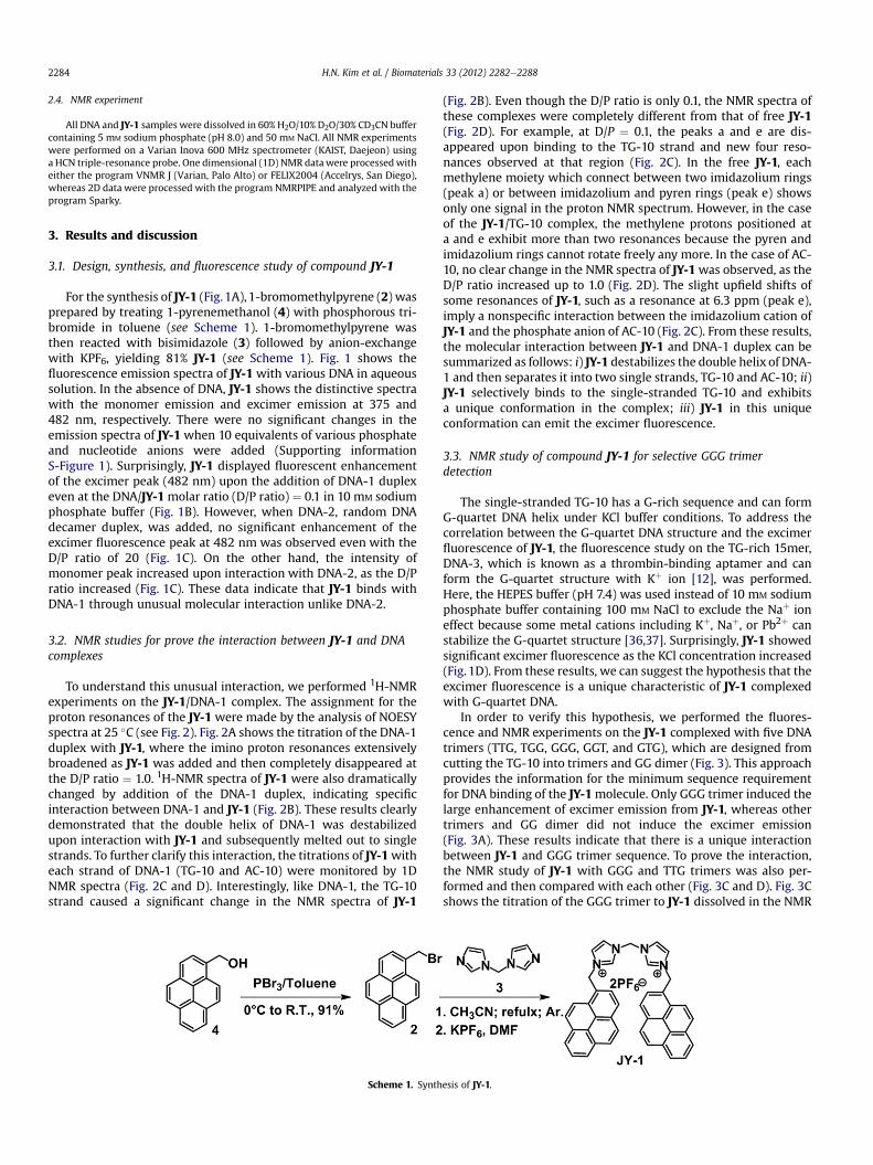
Fig. 5. Representative model for the JY-1 complexed with (A) parallel G-quartet of GGG and (B) B-form DNA. The JY-1 atoms are green. Positive charged nitrogen atoms of JY-1 areblue. The sugar and base atoms of DNA are yellow and white, respectively. Phosphate backbone anions are red. (For interpretation of the references to colour in this figure legend,the reader is referred to the web version of this article.)
H.N. Kim et al. / Biomaterials 33 (2012) 2282e22882286
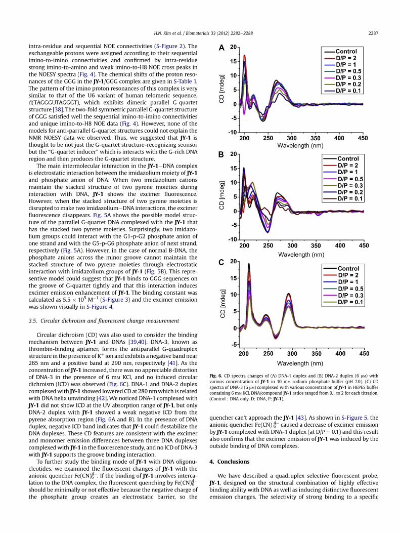
Fig. 6. CD spectra changes of (A) DNA-1 duplex and (B) DNA-2 duplex (6 mM) withvarious concentration of JY-1 in 10 mM sodium phosphate buffer (pH 7.0). (C) CDspectra of DNA-3 (6 mM) complexed with various concentration of JY-1 in HEPES buffercontaining 6 mM KCl. DNA/compound JY-1 ratios ranged from 0.1 to 2 for each titration.(Control : DNA only, D: DNA, P: JY-1).
H.N. Kim et al. / Biomaterials 33 (2012) 2282e2288 2287
intra-residue and sequential NOE connectivities (S-Figure 2). Theexchangeable protons were assigned according to their sequentialimino-to-imino connectivities and confirmed by intra-residuestrong imino-to-amino and weak imino-to-H8 NOE cross peaks inthe NOESY spectra (Fig. 4). The chemical shifts of the proton reso-nances of the GGG in the JY-1/GGG complex are given in S-Table 1.The pattern of the imino proton resonances of this complex is verysimilar to that of the U6 variant of human telomeric sequence,d(TAGGGUTAGGGT), which exhibits dimeric parallel G-quartetstructure [38]. The two-fold symmetric parrallel G-quartet structureof GGG satisfied well the sequential imino-to-imino connectivitiesand unique imino-to-H8 NOE data (Fig. 4). However, none of themodels for anti-parrallel G-quartet structures could not explain theNMR NOESY data we observed. Thus, we suggested that JY-1 isthought to be not just the G-quartet structure-recognizing seonsorbut the “G-quartet inducer”which is interacts with the G-rich DNAregion and then produces the G-quartet structure.
The main intermolecular interaction in the JY-1�DNA complexis electrostatic interaction between the imidazoliummoiety of JY-1and phosphate anion of DNA. When two imidazolium cationsmaintain the stacked structure of two pyrene moieties duringinteraction with DNA, JY-1 shows the excimer fluorescence.However, when the stacked structure of two pyrene moieties isdisrupted tomake two imidazolium�DNA interactions, the excimerfluorescence disappears. Fig. 5A shows the possible model struc-ture of the parrallel G-quartet DNA complexed with the JY-1 thathas the stacked two pyrene moieties. Surprisingly, two imidazo-lium groups could interact with the G1-p-G2 phosphate anion ofone strand and with the G5-p-G6 phosphate anion of next strand,respectively (Fig. 5A). However, in the case of normal B-DNA, thephosphate anions across the minor groove cannot maintain thestacked structure of two pyrene moieties through electrostaticinteraction with imidazolium groups of JY-1 (Fig. 5B). This repre-sentive model could suggest that JY-1 binds to GGG sequences onthe groove of G-quartet tightly and that this interaction inducesexcimer emission enhancement of JY-1. The binding constant wascalculated as 5.5 � 105 M�1 (S-Figure 3) and the excimer emissionwas shown visually in S-Figure 4.
3.5. Circular dichroism and fluorescent change measurement
Circular dichroism (CD) was also used to consider the bindingmechanism between JY-1 and DNAs [39,40]. DNA-3, known asthrombin-binding aptamer, forms the antiparallel G-quadruplexstructure in the presence of Kþ ion and exhibits a negative bandnear265 nm and a positive band at 290 nm, respectively [41]. As theconcentration of JY-1 increased, therewas no appreciable distortionof DNA-3 in the presence of 6 mM KCl, and no induced circulardichroism (ICD) was observed (Fig. 6C). DNA-1 and DNA-2 duplexcomplexedwith JY-1 showed loweredCD at 280 nmwhich is relatedwith DNA helix unwinding [42].We noticed DNA-1 complexedwithJY-1 did not show ICD at the UV absorption range of JY-1, but onlyDNA-2 duplex with JY-1 showed a weak negative ICD from thepyrene absorption region (Fig. 6A and B). In the presence of DNAduplex, negative ICD band indicates that JY-1 could destabilize theDNA duplexes. These CD features are consistent with the excimerand monomer emission differences between three DNA duplexescomplexedwith JY-1 in the fluorescence study, and no ICD of DNA-3with JY-1 supports the groove binding interaction.
To further study the binding mode of JY-1 with DNA oligonu-cleotides, we examined the fluorescent changes of JY-1 with theanionic quencher Fe(CN)64�. If the binding of JY-1 involves interca-lation to the DNA complex, the fluorescent quenching by Fe(CN)64�
should be minimally or not effective because the negative charge ofthe phosphate group creates an electrostatic barrier, so the
quencher can’t approach the JY-1 [43]. As shown in S-Figure 5, theanionic quencher Fe(CN) 6
4� caused a decrease of excimer emissionby JY-1 complexed with DNA-1 duplex (at D/P¼ 0.1) and this resultalso confirms that the excimer emission of JY-1was induced by theoutside binding of DNA complexes.
4. Conclusions
We have described a quadruplex selective fluorescent probe,JY-1, designed on the structural combination of highly effectivebinding ability with DNA as well as inducing distinctive fluorescentemission changes. The selectivity of strong binding to a specific
H.N. Kim et al. / Biomaterials 33 (2012) 2282e22882288
sequence can allow for quadruplex sensing and the detectionmethod presented here is very simple, using fluorescence and NMRstudy. We found that JY-1 is thought to be the 0 0G-quartet inducer0 0
which is interacts with the G-rich DNA region and then producesthe G-quartet structure, contrast to other sensors recognizing theG-quartet DNA structure. Also, the groove binding characteristic ofJY-1 to the G-quadruplex has a relatively low nonspecific toxicityand the structure-specific differences in fluorescent characterbetween DNA duplex and G-quadruplex may offer more discoveryand application in biological study.
Acknowledgments
We thank Dr. Jae-Sun Shin and Prof. Byong-Seok Choi for sup-porting NMR experiments. This work was supported by the NRFGrants (2010-0014199, NRF-C1ABA001-2010-0020480 to J.-H.L.;2011-0020450, R31-2008-000-10010-0 (WCU) to J.Y.) funded bythe Korean Government (MEST). J.-H.L. also thanks to the supportby a grant from the Next-Generation BioGreen 21 Program (SSAC,grant #: PJ008109), Rural Development Administration, Republic ofKorea. J.Y. also thanks to the support by the Ewha Global Top5 Grant2011 of Ewha Womans University.
Appendix. Supplementary material
Supplementary data associated with this article can be found, inthe online version, at doi:10.1016/j.biomaterials.2011.11.073
References
[1] Kumara GS, Dasa S, Bhadraa K, Maiti M. Protonated forms of poly[d(G-C)] andpoly(dG).poly(dC) and their interaction with berberine. Bioorg Med Chem2003;11(23):4861e70.
[2] Feng X, Liu L, Wang S, Zhu D. Water-soluble fluorescent conjugated polymersand their interactions with biomacromolecules for sensitive biosensors. ChemSoc Rev 2010;39(7):2411e9.
[3] Liu J, Cao Z, Lu Y. Functional nucleic acid sensors. Chem Rev 2009;109(5):1948e98.
[4] Tsui C, Coleman LE, Griffith JL, Bennett EA, Goodson SG, Scott JD, et al. Singlenucleotide polymorphisms (SNPs) that map to gaps in the human SNP map.Nucleic Acids Res 2003;31(16):4910e6.
[5] Gomez-Marquez J. DNA G-quadruplex: structure, function and human disease.Febs J 2010;277(17):3451.
[6] Cuesta J, Read MA, Neidle S. The design of G-quadruplex ligands as telomeraseinhibitors. Mini Rev Med Chem 2003;3(1):11e21.
[7] Manet I, Manoli F, Zambelli B, Andreano G, Masi A, Cellai L, et al. Affinityof the anthracycline antitumor drugs Doxorubicin and Sabarubicin forhuman telomeric G-quadruplex structures. Phys Chem Chem Phys 2011;13(2):540e51.
[8] Davis JT. G-quartets 40 years later: from 5’-GMP to molecular biology andsupramolecular chemistry. Angew Chem Int Edit 2004;43(6):668e98.
[9] White EW, Tanious F, Ismail MA, Reszka AP, Neidle S, Boykin DW, et al.Structure-specific recognition of quadruplex DNA by organic cations: Influ-ence of shape, substituents and charge. Biophys Chem 2007;126(1e3):140e53.
[10] Song G, Ren J. Recognition and regulation of unique nucleic acid structures bysmall molecules. Chem Commun 2010;46:7283e94.
[11] He F, Tang Y, Yu M, Feng F, An L, Sun H, et al. Quadruplex-to-duplex transitionof G-rich oligonucleotides probed by cationic water-soluble conjugatedpolyelectrolytes. J Am Chem Soc 2006;128(21):6764e5.
[12] Peng X, Du J, Fan J, Wang J, Wu Y, Zhao J, et al. A selective fluorescent sensorfor imaging Cd2þ in living cells. J Am Chem Soc 2007;129(6):1500e1.
[13] Zhou Y, Xu Z, Yoon J. Fluorescent and colorimetric chemosensors for detectionof nucleotides, FAD and NADH: highlighted research during 2004-2010. ChemSoc Rev 2011;40(5):2222e35.
[14] Kim HN, Guo Z, Zhu W, Yoon J, Tian H. Recent progress on polymer-basedfluorescent and colorimetric chemosensors. Chem Soc Rev 2011;40(1):79e93.
[15] Zhang JF, Zhou Y, Yoon J, Kim JS. Recent progress in fluorescent and colori-metric chemosensors for detection of precious metal ions (silver, gold andplatinum ions). Chem Soc Rev 2011;40(7):3416e29.
[16] Nagatoishi S, Nojima T, Juskowiak B, Takenaka S. A pyrene-labeled G-quad-ruplex oligonucleotide as a fluorescent probe for potassium ion detection inbiological applications. Angew Chem Int Ed 2005;44(32):5067e70.
[17] Seo YJ, Lee IJ, Yi JW, Kim BH. Probing the stable G-quadruplex transition usingquencher-free end-stacking ethynyl pyrene-adenosine. Chem Commun;2007:2817e9.
[18] Wu Z-S, Hu P, Zhou H, Shen G, Yu R. Fluorescent oligonucleotide probe basedon G-quadruplex scaffold for signal-on ultrasensitive protein assay. Bioma-terials 2010;31(7):1918e24.
[19] Allain C, Monchaud D, Teulade-Fichou MP. FRET templated by G-quadruplexDNA: a specific ternary interaction using an original pair of donor/acceptorpartners. J Am Chem Soc 2006;128(36):11890e3.
[20] Yang P, De Cian A, Teulade-Fichou MP, Mergny JL, Monchaud D. Engineeringbisquinolinium/thiazole orange conjugates for fluorescent sensing ofG-quadruplex DNA. Angew Chem Int Ed 2009;48(12):2188e91.
[21] Li T, Wang E, Dong S. Parallel G-quadruplex-specific fluorescent probe formonitoring DNA structural changes and label-free detection of potassium ion.Anal Chem 2010;82(18):7576e80.
[22] He F, Tang Y, Wang S, Li Y, Zhu D. Fluorescent amplifying recognition for DNAG-quadruplex folding with a cationic conjugated polymer: A platform forhomogeneous potassium detection. J Am Chem Soc 2005;127(35):12343e6.
[23] Zhu J, Li T, Zhang L, Dong S, Wang E. G-quadruplex DNAzyme based molecularcatalytic beacon for label-free colorimetric logic gates. Biomaterials 2011;32(30):7318e24.
[24] Paramasivan S, Bolton PH. Mix and measure fluorescence screening forselective quadruplex binders. Nucleic Acids Res 2008;36(17):e106.
[25] Ou TM, Lu YJ, Tan JH, Huang ZS, Wong KY, Gu LQ. G-quadruplexes: targets inanticancer drug design. ChemMedChem. 2008;3(5):690e713.
[26] Xu Z, Kim SK, Yoon J. Revisit to imidazolium receptors for the recognition ofanions: highlighted research during 2006-2009. Chem Soc Rev 2010;39(5):1457e66.
[27] Amendola V, Boiocchi M, Colasson B, Fabbrizzi L, Rodriguez Douton M-J,Ugozzoli F. A metal-based trisimidazolium cage that provides six C-Hhydrogen-bond-donor fragments and includes anions. Angew Chem Int Ed2006;45(41):6920e4.
[28] Lu Q-S, Dong L, Zhang J, Li J, Jiang L, Huang Y, et al. Imidazolium-functionalizedBINOL as a multifunctional receptor for chromogenic and chiral anionrecognition. Org Lett 2009;11(3):669e72.
[29] Kumar S, Luxami V, Kumar A. Chromofluorescent probes for selective detec-tion of fluoride and acetate ions. Org Lett 2008;10(24):5549e52.
[30] Amendola V, Boiocchi M, Colasson B, Fabbrizzi L, Monzani E, Douton-Rodriguez MJ, et al. Redox active cage for the electrochemical sensing ofanions. Inorg Chem 2008;47(11):4808e16.
[31] Chen X, Kang S, Kim MJ, Kim J, Kim YS, Kim H, et al. Thin-film formation ofimidazolium-based conjugated polydiacetylenes and their application forsensing anionic surfactants. Angew Chem Int Ed 2010;49(8):1422e5.
[32] Xu Z, Singh NJ, Kim SK, Spring DR, Kim KS, Yoon J. Induction-driven stabili-zation of the anion-pi interaction in electron-rich aromatics as the key tofluoride inclusion in imidazolium-cage receptors. Chem-Eur J 2011;17(4):1163e70.
[33] Chellappan K, Singh NJ, Hwang I-C, Lee JW, Kim KS. A calix[4]imidazolium[2]pyridine as an anion receptor. Angew Chem Int Ed 2005;44(19):2899e903.
[34] Kim HN, Lim J, Lee HN, Ryu JW, Kim MJ, Lee J, et al. Unique x-ray sheetstructure of 1,8-bis(imidazolium) anthracene and its application as a fluores-cent probe for DNA and DNase. Org Lett 2011;13(6):1314e7.
[35] Kim HN, Moon JH, Kim SK, Kwon JY, Jang YJ, Lee JY, et al. Fluorescent sensingof triphosphate nucleotides via anthracene derivatives. J Org Chem 2011;76(10):3805e11.
[36] Fletcher TMSD, Salazar M, Hurley LH. Effect of DNA secondary structure onhuman telomerase activity. Biochemistry 1998;37(16):5536e41.
[37] Kan ZY, Yao Y, Wang P, Li XH, Hao YH, Tan Z. Molecular crowding inducestelomere G-quadruplex formation under salt-deficient conditions andenhances its competition with duplex formation. Angew Chem Int Ed 2006;45(10):1629e32.
[38] Phan AT, Patel DJ. Two-repeat human telomeric d(TAGGGTTAGGGT) sequenceforms interconverting parallel and antiparallel G-quadruplexes in solution:distinct topologies, thermodynamic properties, and folding/unfoldingkinetics. J Am Chem Soc 2003 Dec 10;125(49):15021e7.
[39] Lyng R, Hard T, Norden B. Induced CD of DNA intercalators: electric dipoleallowed transitions. Biopolymers 1987;26(8):1327e45.
[40] Kim HK, Kim JM, Kim SK, Rodger A, Norden B. Interactions of intercalative andminor groove binding ligands with triplex poly(dA).[poly(dT)]2 and withduplex poly(dA).poly(dT) and poly[d(A-T)]2 studied by CD, LD, and normalabsorption. Biochemistry 1996;35(4):1187e94.
[41] Monchaud D, Yang P, Lacroix L, Teulade-Fichou MP, Mergny JL. A metal-mediated conformational switch controls G-quadruplex binding affinity.Angew Chem Int Ed 2008;47(26):4858e61.
[42] Duff MR, Mudhivarthi VK, Kumar CV. Rational design of anthracene-basedDNA binders. J Phys Chem B 2009;113(6):1710e21.
[43] Scaria PV, Shafer RH. Binding of ethidium bromide to a DNA triple helix.Evidence for intercalation. J Biol Chem 1991;266(9):5417e23.


![Benzo[a]pyrene (BaP)](https://img.dokumen.tips/doc/110x75/56815173550346895dbfa88c/benzoapyrene-bap-56a2c44d6ca0b.jpg)
![Biotransformation of benzo[a]pyrene - Analysis, metabolism](https://img.dokumen.tips/doc/110x75/61a84ea8bf373a5a8e635299/biotransformation-of-benzoapyrene-analysis-metabolism-.jpg)


