Embed Size (px)
Citation preview

A PRELIMINARY BIOCLIMATIC APPROACH TO PREDICTING POTENTIAL DISTRIBUTION OF PHELLINUS NOXIUS AND GEOGRAPHICAL AREAS AT RISK FROM INVASION Ned B. Klopfenstein1,Eric W. I. Pitman1,John W. Hanna1,Phil G. Cannon2,Jane E. Stewart!,Norio Sahashi'1, Yuko Ota4
, Tsutomu Hattorz-4, Mitsuteru Akiba4, Louise Shuey5, Robert L. Schlub6
, Fred Brooks7
, Ndeme Atibalentia8, Alvin M.C. Tang'10
, Regent Y. C. Lam9,1°, Mike W. K. Leung, L. M. Chu10,H. S. Kwan10,Mohd Farid bin Ahmad11
, Su See Lee11,Hsin-Han Lee12,Jyh-Nong Tsai13, Yu
Ching Huang12, Chia-Lin Chung12,Ruey-Fen Liou12
, and Mee-Sook Kim14
INTRODUCTION
Phellinus noxius, the cause of brown root-rot disease, is an invasive pathogen that was first described by Corner in Singapore (Corner 1932). It has a wide host range of primarily woody plants representing over 200 species from diverse families (Ann et al . 2002). This pathogen is also widespread, and has been reported to occur in many tropical/subtropical areas of Asia, Australia, Central America, Africa, and Oceania, where it can be quite destructive . Phellinus noxius appears to attack hosts regardless of health condition, and it can survive in organic matter long after host death. Early symptoms of brown root-rot disease are similar to other root diseases, including leaf chlorosis, wilt, and branch dieback; however,
In: Ramsey , A. & P. Palacios (Comps). Proceedings of the 63rd Annual Western International Forest Disease Work Conference; 2015 Sept. 21-15; Newport, OR. 1USDA Forest Service, Rocky Mountain Research Station, Moscow, Idaho. 2USDA Forest Service, Forest Health Protection (FHP), Region 5, Vallejo, CALIFORNIA . 3Department of Bioagricultural Sciences and Pest Management, Colorado State University , Fort Collins, Colorado. 4Forestry and Forest Products Research Institute, Tsukuba, lbaraki , Japan. 5School of Biological Sciences , The University of Queensland, Brisbane, Australia. 6University of Guam, ANR/CES/CNAS, University Station, Mangilao, Guam. 7Department of Plant and Environmental Protection Sciences , University of HawaiiManoa , Honolulu, HI. 8 American Samoa Community CollegeCNR, Division of Community and Natural Resources , Pago Pago, AS. 9Muni Arborist Limited, Kowloo, Hong Kong SAR, China. toschool of Life Sciences, The Chinese University of Hong Kong , Shatin, Hong Kong SAR, China. 11 Forest Research Institute Malaysia, Selango, Malaysia. 12Department of Plant Pathology and Microbiology, National Taiwan University, Taipei City, Taiwan. 13Plant Pathology Division , Taiwan Agricultural Research Institute , Taichung City , Taiwan. 14Department of Forestry, Environment and Systems, Kookmin University, Seoul , Korea.
137
mortality can occur relatively quickly after infection (Sashashi et al. 2012). A prominent sign of brown root-rot disease is a dark brown or blackish mycelial crust covering the stem base and root collar (Figure IA). The associated wood decay typically displays honeycomb-shaped zone lines of reddish-brown to black (Figure lB). The pathogen is often spread via root-to-root contact, but dispersal via basidiospores is also possible .
The general objective of this study is to predict suitable climate space for P. noxius, based on documented occurrences of DNA-sequence verified samples. By comparing bioclimatic variables associated with the precise locations of verified P. no xi us , bioclimatic variables that influence pathogen occurrence can be identified and geographic locations that have suitable climate space for the pathogen can be predicted. Such information can predict potential distribution of P.
noxius, and predict areas at risk from this invasive pathogen.
MATERIALS AND METHODS
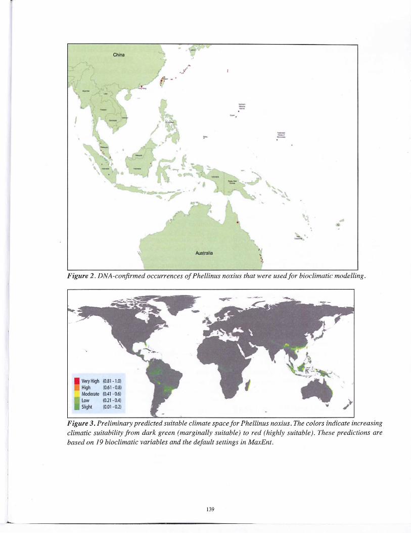
A Maxent (Maximum Entropy) Species Distribution Model (version 3 .3 .3k) was used, because of its applications designed for presenceonly data (Phillips et al. 2005) . For this model , 19 bioclimatic variables (e.g., annual mean temperature, annual precipitation, mean temperature of coldest quarter, prec1p1tation in warmest quarter, etc.) were used from WorldClim (worldclim.org). Sequencing of the ITS rDNA and translation elongation factor 1 a gene confirmed the identity of ca. 100 isolates (Figure 2) used for
This file was created by scanning the printed publication.Errors identified by the software have been corrected;
however, some errors may remain.

pathogen locations (latitude and longitude) to create SWD (Samples With Data ) files that served as a basis for the Maxent bioclimatic model (Auto Features) using WorldClim climate surfaces. The regularization multiplier was kept at default, response curves and jackknifes were added to measure variable importance, and default settings were used for all experimental options. The final output, which is an average of all replicates under set parameters, displays current areas with suitable climate space for P. noxius . Minimal evidence of outliers was found, and cross-validation was used to verify data results. A total to 10 replicates were run with data split into training and test groups to assure accuracy and provide a statistical analysis. Quantum GIS (QGIS) was used to create the final outputs of the maps (Figure 3).
RESULTS AND DISCUSSION
The preliminary prediction maps show potential distribution (areas with suitable climate space) of P. no xi us and areas that are at risk from this invasive pathogen, based on known locations of ca. 100 confirmed isolates from southeastern Asia, Australia, and Pacific islands (Figure 3) . Please note that P. noxius isolates from Central America, Caribbean, South America, and Africa were not included in this study. Continued DNA-based characterization will help determine if isolates
belong to different species or genetic groups . Until more is known, caution is warranted for geographic areas that have suitable climate space for P. noxius, especially where this invasive pathogen does not currently occur (e.g., movement of plant material, infected wood, and infested soil should be restricted from areas were P. noxius occurs).
REFERENCES
Ann, P.J., Chang, T.T., and Ko, W.H. 2002. Phellinus noxius brown root rot of fruit and ornamental trees in Taiwan. Plant Disease 86:820-826.
Corner, E.J.H. 1932. The identification of the brown root fungus . Gardens' Bulletin. Straits Settlements 5:317-350.
Phillips, SJ., Anderson, R.P., and Schapire, R .E. 2006. Maximum entropy modeling of species geographic distributions . Ecological Modelling 190:231-259
Sahashi, N., Akiba, M., Ota, Y., and Kanzaki, N. 2012. Brown root rot of trees caused by Phellinus noxius in the Ryukyu Islands, subtropical areas of Japan. Forest Pathology 42:353-361.
Figure 1. Phellinus noxius infection on a dying flame tree (Delonix regia) during the dry season (A) and associated wood decay ( B).
138
L
China
I
•' -. ,,~ .
! r -
. -.. ;"
Australia
---. --·
'J .
•
~ . . ,
Figure 2 . DNA-confirmed occurrences of Phellinus noxius that were used for bioclimatic modelling.
Very High High Moderate Low Slight
(0.81 · l.O) (0.61 · 0.8) (0.41 ·0.6) (0.21 -0.4) (0.01 · 0.2)
- ---
Figure 3. Preliminary predicted suitable climate space for Phellinus noxius. The colors indicate increasing climatic suitability from dark green (marginally suitable) to red (highly suitable). These predictions are based on 19 bioclimatic variables and the default settings in MaxEnt.
139


![Bioclimatic Buildings [ ] - RIO 12 · Bioclimatic Buildings Roberto Lamberts Federal University of Santa Catarina ... NBR 15220-3 CLIMATE + MAN + HABITAT. 15 Bioclimatic architecture](https://img.dokumen.tips/doc/110x75/5c4466cd93f3c34c3c35c1b5/bioclimatic-buildings-rio-12-bioclimatic-buildings-roberto-lamberts-federal.jpg)