Embed Size (px)
Citation preview

A novel CBCT-based method for derivation of CTV-PTV margins forprostate and pelvic lymph nodes treated with stereotactic ablativeradiotherapyLyons, C. A., King, R. B., Osman, S. O. S., McMahon, S. J., O'Sullivan, J. M., Hounsell, A. R., ... McGarry, C. K.(2017). A novel CBCT-based method for derivation of CTV-PTV margins for prostate and pelvic lymph nodestreated with stereotactic ablative radiotherapy. Radiation oncology (London, England), 12(1), 124. DOI:10.1186/s13014-017-0859-z
Published in:Radiation oncology (London, England)
Document Version:Publisher's PDF, also known as Version of record
Queen's University Belfast - Research Portal:Link to publication record in Queen's University Belfast Research Portal
Publisher rightsCopyright 2017 the authors.This is an open access Creative Commons Attribution-NonCommercial License (https://creativecommons.org/licenses/by-nc/4.0/), whichpermits use, distribution and reproduction for non-commercial purposes, provided the author and source are cited.
General rightsCopyright for the publications made accessible via the Queen's University Belfast Research Portal is retained by the author(s) and / or othercopyright owners and it is a condition of accessing these publications that users recognise and abide by the legal requirements associatedwith these rights.
Take down policyThe Research Portal is Queen's institutional repository that provides access to Queen's research output. Every effort has been made toensure that content in the Research Portal does not infringe any person's rights, or applicable UK laws. If you discover content in theResearch Portal that you believe breaches copyright or violates any law, please contact [email protected].
Download date:03. Jun. 2018

RESEARCH Open Access
A novel CBCT-based method for derivationof CTV-PTV margins for prostate and pelviclymph nodes treated with stereotacticablative radiotherapyCiara A. Lyons1,2, Raymond B. King1,3*, Sarah O.S. Osman1,3, Stephen J. McMahon1, Joe M. O’Sullivan1,2,Alan R. Hounsell1,3, Suneil Jain1,2 and Conor K. McGarry1,3
Abstract
Background: Traditional CTV-PTV margin recipes are not generally applicable in the situation of stereotacticablative radiotherapy (SABR) treatments of multiple target volumes with a single isocentre. In this work, we presenta novel geometric method of margin derivation based on CBCT-derived anatomical data.
Methods: Twenty patients with high-risk localized prostate cancer were selected for retrospective review. Individualvolumes of interest (prostate, prostate and seminal vesicles and pelvic lymph nodes) were delineated on five representativeCBCTs and registered to the planning CT using two registration protocols: bone match or prostate-based soft tissue match.Margins were incrementally expanded around composite CTV structures until 95% overlap was achieved.
Results: CTV-PTV margins of 5.2, 6.5 and 7.6 mm were required for prostate, prostate and seminal vesicles and pelviclymph nodes respectively using a prostate matching protocol. For the prostate and seminal vesicle structures, marginscalculated using our method displayed good agreement with a conventional margin recipe (within ±1.0 mm).
Conclusions: We have presented an alternative method of CTV-PTV margin derivation that is applicable to SABRtreatments with more than one isocentric target. These results have informed an institutional trial of prostate and pelvicnodal SABR in men with high-risk localized prostate cancer.
Keywords: SABR, Stereotactic radiotherapy, Margin derivation, Prostate cancer, Elective nodal irradiation, Multiple isocentrictargets
IntroductionStereotactic ablative radiotherapy (SABR) is increasinglyused for the treatment of prostate cancer (PC), which issensitive to larger fraction size due to a low α/β ratio[1–4]. Improved accuracy in treatment delivery, particu-larly since the widespread adoption of cone-beam CT(CBCT), has enabled reductions in CTV-PTV margins,facilitating dose escalation while also reducing the risk of
toxicity [5–7]. Geometric accuracy is particularly importantin the setting of SABR. Due to the high fractionation dose,steep dose gradients and smaller margins, a geographicmiss in a single fraction could lead to considerable targetunder-dosing and an increased risk of toxicity [1, 8, 9].To date, the majority of prostate SABR evidence has
been for low- to medium-risk groups, where generallythe prostate alone is treated [2–4]. There is a relativepaucity of data regarding the use of SABR in men withhigh-risk prostate cancer, who potentially have the mostto gain from dose escalation [10]. For this patient cohortelective pelvic nodal irradiation (ENI) is delivered inmany centres, for patients treated with conventionallyfractionated radiotherapy [11]. Results from a phase I/II5-fraction SABR trial, where 25Gy was delivered to
* Correspondence: [email protected] A. Lyons and Raymond B. King are joint first authors.Suneil Jain and Conor K. McGarry are joint senior authors.1Centre for Cancer Research and Cell Biology, Queen’s University Belfast,Belfast BT7 1NN, UK3Radiotherapy Physics, Northern Ireland Cancer Centre, Belfast City Hospital,Belfast, UKFull list of author information is available at the end of the article
© The Author(s). 2017 Open Access This article is distributed under the terms of the Creative Commons Attribution 4.0International License (http://creativecommons.org/licenses/by/4.0/), which permits unrestricted use, distribution, andreproduction in any medium, provided you give appropriate credit to the original author(s) and the source, provide a link tothe Creative Commons license, and indicate if changes were made. The Creative Commons Public Domain Dedication waiver(http://creativecommons.org/publicdomain/zero/1.0/) applies to the data made available in this article, unless otherwise stated.
Lyons et al. Radiation Oncology (2017) 12:124 DOI 10.1186/s13014-017-0859-z

pelvic node CTVs simultaneously with a 40Gy prostateCTV prescription dose, indicated that the treatment waswell tolerated in the acute setting with further follow-updata expected in the future [12].Our centre is also currently recruiting to a high-risk
prostate SABR trial with an ENI arm, where 50% ofpatients are prescribed 25 Gy to a pelvic node PTV,delivered simultaneously with 36.25Gy to the prostatePTV over 5 fractions [13]. However, the use of conven-tional margin recipes in the situation of SABR for elect-ive pelvic nodal irradiation is potentially suboptimal, asthey rely on a number of assumptions that are not metin this scenario. A fundamental assumption that is un-realistic in this case is that the target’s geometry is typic-ally modelled as a rigid sphere. Not only is the shape ofthe pelvic nodal CTV structure overtly complex, but it isalso subject to considerable daily variation that isdependent on bladder filling as well as other parameters[14–17].Conventional CTV-PTV margin calculations are also
not easily applicable to irradiation of multiple targets viaa single treatment isocentre. For example, the prostateand lymph node CTV structures are known to move in-dependently relative to each other, with displacements ofup to 6 mm reported [14]. Additional considerationmust therefore be given when employing an image guid-ance regime that only matches to the primary target site(e.g. the prostate).This paper describes a composite volume approach
that allows derivation of margins for two or more separ-ate CTVs treated using a single isocentre; in this case,the prostate (PO), prostate and seminal vesicles (PSV)and pelvic lymph nodes (LN). This composite volumemethod was used to calculate individual margins foreach structure, with the PO and PSV margins subse-quently compared to PO/PSV margins derived using acommonly employed conventional statistical method[18].
MethodsPatients, treatment planning and deliveryTwenty patients, previously treated with conventionallyfractionated radical radiotherapy to the prostate and pel-vis, were selected for this retrospective review. Each pa-tient had planning CT (pCT) images acquired using ahelical CT-simulator (512 × 512 field of view, 1 mmaxial pixel resolution, 2.5 mm slice width). All patientswere instructed to empty their bladder and to drink500 mL of water and had a micro-enema (Micralax®) ad-ministered prior to their pCT and each treatment ses-sion. The pCT images were imported into Eclipse™v13.5.35 (Varian Medical Systems, Palo Alto, CA) forcontouring target and organ at risk (OAR) volumes.
Target volumes of interest (PO, PSV and LN) were indi-vidually delineated following a previously described pel-vis IMRT protocol [19].All treatments were delivered using a Varian True-
Beam™ linac (Varian Medical Systems) with kV-basedCBCT on-board imaging. Patient set-up and CBCT veri-fication imaging were carried out as per our institutionalprotocol: images were acquired following set-up for thefirst three fractions of treatment and on a weekly basissubsequent to this. For treatment delivery, online regis-tration of CBCT images to the pCT was performedusing the patient’s bony anatomy as a surrogate for thePTV; however, these registrations were not used in thisinvestigation.
Image registrationAdditional offline registrations of the CBCT images tothe pCT were performed independently to the onlineregistration by a single clinical oncologist (CL), using theregistration workspace within Eclipse™. Only transla-tional shifts were considered in all cases. Two imagematching protocols were studied – bone (bony pelvis)and soft tissue (ST, prostate-based), resulting in two sep-arate datasets for analysis. Every match pair was per-formed in the same order: in all cases, an automatedpelvic bone match was carried out first, this wasfollowed by a separate automated ST match using theprostate as the common reference, with the resultingregistration manually adjusted where required.
ContouringFive CBCT image sets were selected for each patient (toreflect commonly used SABR fractionation schedules)[12, 20]. To generate an appropriate representation ofthe variation in individual patient anatomy throughouttheir entire treatment, the first and last CBCT imagesets were selected for each patient, with the remaining 3CBCT image sets evenly sampled across the patient’streatment schedule. CBCT image sets with optimumimage quality were selected for analysis to ensure accur-acy in the soft-tissue structures delineated and imagesets of insufficient image quality were excluded from thestudy. Following registration of the CBCT images to thepCT, structures of interest were contoured manually inEclipse™ by a single clinical oncologist (CL) and peer-reviewed by a second clinical oncologist (SJ).
Composite volume generation and overlap analysisVolumes contoured on each CBCT were transferred to asingle pCT structure set. Two datasets per patient werecreated, which accounted for translational shifts deter-mined from each registration method (i.e., bone and STmatches). Fig. 1a shows an example of LN axial contoursfrom five different CBCTs (cyan) overlaid on the
Lyons et al. Radiation Oncology (2017) 12:124 Page 2 of 8

corresponding pCT axial slice; in this example, a STmatch to the prostate was used to register each of theCBCT images to the pCT.The image-registration and contouring tools available
within Eclipse™ were used to combine the CBCT con-tours and to compare the resulting composite structure(blue) to the CTV structures originally delineated oneach patient’s pCT (red). CBCT contours that weretransferred using the same registration method (bone orST) were combined to create two separate composite
structures. A 3D rendering comparing the original pCTLN CTV structure (red) to the ST-matched compositestructure (blue) is also illustrated in Fig. 1b, with thepCT PSV structure (pink) included for reference. Thevolume of each composite structure was recorded andused for subsequent analysis.A uniform margin was incrementally increased around
the pCT CTV structure (0 to 12 mm; 1 mm increments)to generate a series of PTVs for these structures. Foreach margin increment, the volume of the overlappingregion between the generated PTV and each of the com-posite structures (bone or ST match) was determined.Fig. 1c shows an example of this analysis on the sameaxial slice shown in Fig. 1a. In this example, a 3 mmmargin has been uniformly extended around the CTV togenerate the PTV. Overlapping regions between thePTV and the composite volume are indicated by thegreen-shaded areas, while the purple-shaded areas indi-cate non-overlapping regions. The overlapping volumewas expressed as a percentage of the composite struc-ture for each PTV margin increment. Additional illustra-tive examples of this technique are provided inAdditional file 1.
Margin calculationTo correlate with other margin derivation methods [18],a percentage overlap of 95% was selected as the desiredthreshold criterion. The margins required to achieve95% overlap for each individual patient and registrationoption were determined through linear interpolation ofthe relevant increments.A population margin was then determined for the 20
patients sampled. Again, as with other techniques, thepopulation margin was defined as the margin requiredto achieve the desired 95% overlap in 90% of the patientpopulation. For a normal distribution, this can simply bedetermined from the mean ( x ) and standard deviation(s) of the sampled group, using the formula:
Margin ¼ x þ 1:28s ð1ÞPO and PSV margins were then calculated using a
commonly used conventional statistical method [18] andcompared to those calculated using our composite vol-ume technique. Systematic (Σ) and random (σ) errors,obtained from analysis of a recent audit of our institu-tional set-up protocol, were employed to determine mar-gins for the two set-up protocols, using the followingmargin recipe [18]:
Margin ¼ 2:5Σþ 0:7σ ð2ÞIt is worth noting that this conventional recipe assumes
that treatments consist of a very large number of treat-ment sessions, each delivering a very small dose fraction,
Fig. 1 a Composite volume generation for soft tissue-registered LNCTV contours. b 3D rendering of the original pCT LN CTV and thesoft tissue composite LN structure. c Overlap analysis for a uniform3 mm margin
Lyons et al. Radiation Oncology (2017) 12:124 Page 3 of 8

effectively assuming an infinite number of treatment ses-sions to simplify the mathematical method [18, 21]. Theseassumptions are clearly not met with SABR treatmentsand alternative methods have been proposed to addressthis limitation [21, 22]. However, margin calculationsperformed using an adapted version of this conventionalrecipe (VH1 described in [22]) with our derived systematicand random errors agreed well (≤0.3 mm deviation) withthe conventional recipe and therefore only the conven-tional margins are compared with our composite volumemethod.Additional data are supplied in the Additional file 2.
Statistical methodsMATLAB v8.2.0 (MathWorks, Natick, MA) was used toperform a two-sided Wilcoxon signed-rank test to assessthe significance in differences between the percentageoverlap distributions obtained for each margin incre-ment for the two image-registration matching scenarios(where p < 0.05 was considered statistically significant).For both image-matching protocols, a Shapiro-Wilk
normality test was performed using SPSS v22.0.0 (IBM,Armonk, NY), to determine whether the individual mar-gins required to achieve 95% overlap for the 20 patientswere normally distributed.
ResultsA total of 120 CT images were individually contoured(20 pCTs and 100 CBCTs). For each target structure(PO, PSV and LN), a minimum of 9 different CTV-PTVmargin sizes were analyzed to determine the percentageoverlap of the PTV structure with the CBCT compositestructures produced for either a bone or ST match.The results of the percentage overlap analysis for the
three target structures for all 20 patients analyzed aredisplayed as box-whisker plots in Fig. 2. Differences be-tween the median values of the population overlap dis-tributions for the two registration options (bone or ST)were statistically significant (p < 0.05) for all structuresand margin sizes investigated.Table 1 reports the mean and standard deviation of
the 95% overlap margin for each of the structures andregistration options, as well as additional results ob-tained from the composite volume analysis. The ratio ofthe composite to pCT volume was 1.40 and 1.33, 1.57and 1.44 and 1.22 and 1.33 respectively for bone and STmatches for the PO, PSV and LN structures. As an ex-ample, employing a CTV-PTV margin of 5 mm, andusing a prostate-based ST match, the average percentageoverlap of the composite CBCT volume with the pCTvolume was 98.0%, 96.6% and 93.6% for the PO, PSVand LN structures respectively.Table 2 reports the population CTV-PTV margins for
the composite volume and conventional statistical
techniques. The composite volume analysis indicatedPO population margins of 8.3 and 5.2 mm (s = 2.83 and1.87 mm) for bone and ST matches respectively. For thePSV structures, calculated population margins were 9.9and 6.5 mm for the bone and ST matches.The bone-matching protocol required smaller margins
around the LN structures, indicating that a margin of5.9 mm was required to achieve 95% overlap in 90% ofthe patients. A margin of 7.6 mm was calculated for theLN CTV structure when a prostate-based ST match wasperformed. Margins calculated using the composite vol-ume method for the PO and PSV structures showedgood agreement with the results of the conventional
Fig. 2 Box-whisker plots of the percentage overlap distributions forthe (a) prostate (PO) structure, (b) prostate and seminal vesicles(PSV) structure, and (c) pelvic lymph node (LN) CTV. Differencesbetween the two image-matching protocols were significant for alltarget structures and margin sizes (p < 0.05). The whiskers indicatethe last percentage overlap value within 1.5× the interquartile rangeof its nearest quartile. Individual data points (+/○) represent patientoutliers with percentage overlap values outside of this range
Lyons et al. Radiation Oncology (2017) 12:124 Page 4 of 8
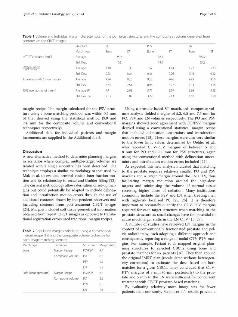
margin recipe. The margin calculated for the PSV struc-ture using a bone-matching protocol was within 0.5 mmof that derived using the statistical method (9.9 and9.4 mm for the composite volume and conventionaltechniques respectively).Additional data for individual patients and margin
increments are supplied in the Additional file 3.
DiscussionA new alternative method to determine planning marginsin scenarios where complex multiple-target volumes aretreated with a single isocentre has been described. Thetechnique employs a similar methodology to that used byMak et al. to evaluate seminal vesicle inter-fraction mo-tion and its relationship to rectal and bladder filling [23].The current methodology allows derivation of set-up mar-gins but could potentially be adapted to include delinea-tion and intrafraction motion errors through the use ofadditional contours drawn by independent observers andincluding contours from post-treatment CBCT images[24]. Margins included soft tissue geometrical informationobtained from repeat CBCT images as opposed to transla-tional registration errors used traditional margin recipes.
Using a prostate-based ST match, this composite vol-ume analysis yielded margins of 5.2, 6.5 and 7.6 mm forPO, PSV and LN volumes respectively. The PO and PSVmargins showed good agreement with PO/PSV marginsderived using a conventional statistical margin recipethat included delineation uncertainty and intrafractionmotion errors [18]. These margins were also very similarto the lower limit values determined by Oehler et al.,who reported CTV-PTV margins of between 5 and8 mm for PO and 6-11 mm for PSV structures, againusing the conventional method with delineation uncer-tainty and intrafraction motion errors included [24].As expected, this new analysis indicated that matching
to the prostate requires relatively smaller PO and PSVmargins and a larger margin around the LN CTV, thusfacilitating margin reduction around the high-dosetargets and minimising the volume of normal tissuereceiving higher doses of radiation. Many institutionscommonly include the PSV and LN when treating menwith high-risk localized PC [25, 26]. It is thereforeimportant to accurately quantify the CTV-PTV marginsrequired for each target structure when matching to theprostate structure as small changes have the potential tocause much larger shifts in the LN CTV [15, 27].A number of studies have reviewed LN margins in the
context of conventionally fractionated prostate and pel-vic radiotherapy, each adopting a different approach andconsequently reporting a range of nodal CTV-PTV mar-gins. For example, Ferjani et al. mapped original plan-ning structures to selected CBCTs using bone andprostate matches for six patients [16]. They then appliedthe original IMRT plan (recalculated without heterogen-eity correction) to estimate the dose based on bothmatches for a given CBCT. They concluded that CTV-PTV margins of 8 mm (6 mm posteriorly) to the pros-tate and 5 mm to the LN were sufficient for concurrenttreatment with CBCT prostate-based matching.By evaluating relatively more image sets for fewer
patients than our study, Ferjani et al.’s results provide a
Table 1 Volume and individual margin characteristics for the pCT target structures and the composite structures generated fromcontours on the CBCT images
Structure PO PSV LN
Match type Bone ST Bone ST Bone ST
pCT CTV volume (cm3) Average 25.9 36.1 360.2
Std. Dev. 10.5 14.1 55.2Composite volume
pCT volume Average 1.40 1.33 1.57 1.44 1.22 1.33
Std. Dev. 0.23 0.24 0.36 0.26 0.16 0.22
% overlap with 5 mm margin Average 93.4 98.0 90.5 96.6 95.9 93.6
Std. Dev. 6.64 2.51 8.96 3.72 1.34 3.15
95% overlap margin (mm) Average (x) 4.71 2.83 5.71 3.79 3.93 5.55
Std. Dev. (s) 2.83 1.87 3.29 2.13 1.50 1.59
Table 2 Population margins calculated using a conventionalmargin recipe [18] and the composite volume technique foreach image-matching scenario
Match type Technique Structure Margin (mm)
Bone Margin Recipe PO/PSV 9.4
Composite volume PO 8.3
PSV 9.9
LN 5.9
Soft Tissue (prostate) Margin Recipe PO/PSV 6.7
Composite volume PO 5.2
PSV 6.5
LN 7.6
Lyons et al. Radiation Oncology (2017) 12:124 Page 5 of 8

better indication of intra-patient variability. However, byincluding a larger sample size (n = 20) and manually de-lineated individualized CTVs for each CBCT, our studyprovides a strong indication of inter-patient variabilityand a more accurate representation of the true treat-ment anatomy. Additionally, given the uncertainties in-herent in CBCT-based dose modeling [28], we elected topursue a purely anatomy-based approach.The 5 mm LN margin recommended by Ferjani et al.
is substantially smaller than the ≥13 mm “vascularspace” margin recommended by Wang et al., which wasalso based on a prostate-matching regime [14]. Wang etal.’s margin was derived by mapping three separateIMRT plans (with varying CTV-PTV margin) onto serialCBCTs for eight patients. The dose computed on eachCBCT was subsequently mapped back to the originalpCT and summed to generate DVHs for each structureof interest which were analyzed to determine theoptimum margin.Hinton et al. employed another technique which used
measured couch shifts to derive nodal CTV-PTV marginsof 9 mm in the anterior-posterior direction and 7 mm lat-erally [15]. These margins are similar (<1.4 mm difference)to those calculated in our study which considered an iso-tropic margin expansion in all three Cartesian planes asanalysis of each patient’s composite volume indicatedcomparable structure motion in all three directions.With regard to SABR, in a single study, Kishan et al.
used fiducial-based CBCT matching to evaluate 12 pa-tients [17]. Selected CBCTs were registered to the pCT,allowing transfer of dose distributions and the originalpCT contours. They found that standard LN margins of4-5 mm were acceptable, under the conditions that thesuperior displacement of the prostate was kept to≤5 mm and the relative change in bladder height was<18%. This margin is considerably smaller than the7.6 mm calculated from our analysis, which avoidedCBCT dose calculation uncertainty and used delineatedstructures based on actual CBCT anatomy.The authors acknowledge that there are some limitations
to the current study. Firstly, post-treatment CBCT, pitch /roll and rotational corrections and real-time tracking of theprostate were not incorporated, reflecting current clinicalpractice in our and many other institutions. However, avail-able data indicate that these are largely accounted for byconventional margin expansion in SABR [29–31] and futureextensions to the technique are planned to confirm this.Secondly, the ST resolution with CBCT is poorer than withconventional CT, particularly with regard to delineation ofthe prostate-rectum interface [32–34]. This issue, inconjunction with potential errors with image-matching, isan inherent feature of this type of study [35, 36]. In thisinvestigation, CBCTs of insufficient image quality were notincluded in the analysis and a single experienced uro-
oncologist (CL) contoured and matched all CTs following awell-defined protocol [19], and a second clinical oncologist(SJ) peer-reviewed the resulting structures and registrations.While contouring of serial CBCTs is currently a time-intensive process that is not routinely implementable intoclinical workflow, rapid advances in auto-contouring algo-rithms may facilitate wider adoption of this method. Finally,due to the inherent difficulties in performing accurate dosecalculations using CBCT [37], this study only evaluated geo-graphic changes and did not include a dosimetric analysis.Particular strengths of this study include the sample size
and number of CBCTs evaluated, the use of individuallycontoured LN CTVs for pCTs and CBCTs, and the inde-pendence of the composite volume CTV-PTV marginderivation method from the assumptions of conventionalmargin recipes. In many previous studies, couch shifts[38], representative slices [15] or the original nodal CTValone [39] were chosen to represent serial nodal CBCTCTVs. In addition, the superior-inferior CTV-PTV marginwas not accounted for in some cases [15]. In this study,the entirety of each volume was considered in all threeplanes. Individual protocol-based CTVs were generatedfor all 100 CBCTs in a more accurate reflection of the trueanatomical situation during treatment. This is particularlyimportant in the case of the LN CTV, due to the greaterimpact of variability of the size and position of the OARs.The results of this analysis will be utilized in an institu-tional trial of prostate and pelvic nodal SABR in men withhigh-risk localized PC [13]. However, this method couldalso be applied to other sites where multiple target vol-umes are treated with a single isocentre.
ConclusionsCurrent methods of CTV-PTV margin calculation forconventional radiotherapy may not be sufficient for thederivation of margins in the setting of SABR and/ormultiple isocentric CTVs. We have presented a novelmethod of CTV-PTV margin derivation that is applic-able to the single isocentre treatment of more than onetarget volume and/or SABR. When applied to prostateand seminal vesicle target structures, which are a goodfacsimile for the target geometries assumed in conven-tional margin recipes, this method yielded comparableresults to conventional methods. Margins calculatedfrom this analysis have been used to inform an institu-tional prostate and pelvic nodal SABR trial.
Additional files
Additional file 1: Appendix A: PTV Expansion Percentage OverlapExample. (DOCX 1032 kb)
Additional file 2: Appendix B: Derivation of Population Margins.(DOCX 75 kb)
Lyons et al. Radiation Oncology (2017) 12:124 Page 6 of 8

Additional file 3: Appendix C: Margin expansion results for individualpatients. (XLSX 35 kb)
AbbreviationsCBCT: Cone-beam CT; CT: Computed tomography; CTV: Clinical targetvolume; DVH: Dose-volume histogram; kV: Kilovoltage; LN: Lymph nodes;OAR: Organs at risk; PC: Prostate cancer; pCT: Planning CT; PO: Prostate;PSV: Prostate and seminal vesicles; PTV: Planning target volume;SABR: Stereotactic ablative radiotherapy; ST: Soft tissue
AcknowledgementsStudy was carried out using equipment kindly donated by the Friends of theCancer Centre, registered with The Charity Commission for Northern Ireland(NIC101345).
FundingThis work was supported by the Simms family and through grants fromProstate Cancer UK, CRUK, the Movember foundation (grant number:CEO13_2–004 (FASTMAN Centre)) and the R & D division of the PublicHealth Agency (grant number: COM/4965/14).
Availability of data and materialsAll relevant data generated or analyzed during this study are included in thispublished article [and its additional files]. Additional data are available fromthe corresponding author on reasonable request.
Authors’ contributionsConception and design: CL, RK, SJ, CMG, AH, JOS. Provision of studymaterials or patients: JOS, SJ. Collection and assembly of data: CL, RK. Dataanalysis and interpretation: RK, CL, SO, SMM, SJ, CMG. Manuscript writing: CL,RK, SJ, CMG. Final approval of manuscript: All authors read and approved thefinal manuscript.
Ethics approval and consent to participateApproval for performing this planning study was obtained from theHospital’s New Technologies Steering Group.
Consent for publicationNot applicable.
Competing interestsThe authors declare that they have no competing interests.
Publisher’s NoteSpringer Nature remains neutral with regard to jurisdictional claims inpublished maps and institutional affiliations.
Author details1Centre for Cancer Research and Cell Biology, Queen’s University Belfast,Belfast BT7 1NN, UK. 2Clinical Oncology, Northern Ireland Cancer Centre,Belfast City Hospital, Belfast, UK. 3Radiotherapy Physics, Northern IrelandCancer Centre, Belfast City Hospital, Belfast, UK.
Received: 24 March 2017 Accepted: 21 July 2017
References1. Kirkbride P, Cooper T. Stereotactic Body Radiotherapy. Guidelines for
Commissioners, Providers and Clinicians: a National Report. Clin Oncol. 2011;23(3);163-4.
2. Fowler JF, Toma-Dasu I, Dasu A. Is the α/β ratio for prostate tumours reallylow and does it vary with the level of risk at diagnosis? Anticancer Res.2013;33(3):1009–11.
3. Vogelius I, Bentzen SM. Meta-analysis of the α/β-ratio for prostate cancer inthe presence of an overall time factor: bad news, good news or no news?Int J Radiat Oncol. 2011;81(2):S404.
4. Miralbell R, S a R, Zubizarreta E, Hendry JH. Dose-fractionation sensitivity ofprostate cancer deduced from radiotherapy outcomes of 5,969 patients inseven international institutional datasets: α/β = 1.4 (0.9-2.2) Gy. Int J RadiatOncol Biol Phys. 2012;82(1):e17–24.
5. Zelefsky MJ, Levin EJ, Hunt M, Yamada Y, Shippy AM, Jackson A, et al.Incidence of late rectal and urinary toxicities after three-dimensionalconformal radiotherapy and intensity-modulated radiotherapy for localizedprostate cancer. Int J Radiat Oncol Biol Phys. 2008;70(4):1124–9.
6. Engels B, Soete G, Verellen D, Storme G. Conformal arc radiotherapy forprostate cancer: increased biochemical failure in patients with distendedrectum on the planning computed tomogram despite image guidance byimplanted markers. Int J Radiat Oncol Biol Phys. 2009;74(2):388–91.
7. Engels B, Soete G, Gevaert T, Storme G, Michielsen D, De Ridder M. Impactof planning target volume margins and rectal distension on biochemicalfailure in image-guided radiotherapy of prostate cancer. Radiother Oncol.2014;111(1):106–9.
8. Morris DE, Emami B, Mauch PM, Konski AA, Tao ML, Ng AK, et al. Evidence-based review of three-dimensional conformal radiotherapy for localizedprostate cancer: an ASTRO outcomes initiative. Int J Radiat Oncol Biol Phys.2005;62(1):3–19.
9. Consortium US. Stereotactic ablative body radiation therapy (SABR): aresource. Version 5.0, January 2015. 2015. Available from: http://www.sabr.org.uk/consortium/.
10. Dearnaley DP, Jovic G, Syndikus I, Khoo V, Cowan RA, Graham JD, et al.Escalated-dose versus control-dose conformal radiotherapy for prostatecancer: long-term results from the MRC RT01 randomised controlled trial.Lancet Oncol. 2007;8(6):475–87.
11. Lawton CA, DeSilvio M, Roach M, Uhl V, Kirsch R, Seider M, et al. An Updateof the Phase III Trial Comparing Whole Pelvic to Prostate Only Radiotherapyand Neoadjuvant Total Androgen Suppression: Updated Analysis of RTOG94–13, With Emphassis on Unexpected Hormone/Radiation Interactions. IntJ Radiat Oncol Biol Phys. 2007;69(3):646–55.
12. Musunuru HB, Davidson MT, D’Alimonte L, Ho L, Cheung P, Vesprini D, et al.Phase 1-2 study of stereotactic ablative radiation therapy including regionallymph node irradiation for patients with high-risk prostate cancer (SATURN).Int J Radiat Oncol Biol Phys. 2015;93(3):E222.
13. Health Research Authority. SPORT High-Risk Trial. 2017. http://www.hra.nhs.uk/news/research-summaries/sport-high-risk-trial/. Accessed 26 June 2017.
14. Wang Z, Wang K, Lerma FA, Liu B, Amin P, Yi B, et al. Planning marginsto CTV for image-guided whole pelvis prostate cancer intensity-modulated radiotherapy. Int J Med Physics, Clinical Eng Radiat Oncol.2012;1(2):23–31.
15. Hinton BK, Fiveash JB, Wu X, Dobelbower MC, Kim RY, Jacob R. Optimalplanning target volume margins for elective pelvic lymphatic radiotherapyin high-risk prostate cancer patients. ISRN Oncol. 2013;2013:941269.
16. Ferjani S, Huang G, Shang Q, Stephans KL, Zhong Y, Qi P, et al. Alignmentfocus of daily image guidance for concurrent treatment of prostate andpelvic lymph nodes. Int J Radiat Oncol Biol Phys. 2013;87(2):383–9.
17. Kishan AU, Lamb JM, Jani SS, Kang JJ, Steinberg ML, King CR. Pelvic nodaldosing with registration to the prostate: implications for high-risk prostatecancer patients receiving stereotactic body radiation therapy. Int J RadiatOncol. 2015;91(4):832–9.
18. van Herk M, Remeijer P, Rasch C, Lebesque JV. The probability of correcttarget dosage: dose-population histograms for deriving treatment marginsin radiotherapy. Int J Radiat Oncol Biol Phys. 2000;47(4):1121–35.
19. Harris V, Staffurth J, Naismith O, Esmail A, Gulliford S, Khoo V, et al.Consensus Guidelines and Contouring Atlas for Pelvic Node Delineation inProstate and Pelvic Node Intensity-modulated Radiotherapy. Int J RadiatOncol. 2015;92(4):874–83.
20. Henderson DR, Tree AC, van As NJ. Stereotactic body radiotherapy forprostate cancer. Clin Oncol (R Coll Radiol). 2015;27(5):270–9.
21. Herschtal A, Foroudi F, Silva L, Gill S, Kron T. Calculating geometric marginsfor hypofractionated radioatherapy. Phys Med Biol. 2013;58:319–33.
22. Gordon JJ, Siebers JV. Convolution method and CTV-to-PTV margins forfinite fractions and small systematic errors. Phys Med Biol. 2007;52:1967–90.
23. Mak D, Gill S, Paul R, Stillie A, Haworth A, Kron T, et al. Seminal vesicleinterfraction displacement and margins in image guided radiotherapy forprostate cancer. Radiat Oncol. 2012;7(1):139.
24. Oehler C, Lang S, Dimmerling P, Bolesch C, Kloeck S, Tini A, et al. PTVmargin definition in hypofractionated IGRT of localized prostate cancerusing cone beam CT and orthogonal image pairs with fiducial markers.Radiat Oncol. BioMed Central. 2014;9(1):229.
25. Morikawa LK, Roach M. Pelvic nodal radiotherapy in patients withunfavorable intermediate and high-risk prostate cancer: evidence, rationale,and future directions. Int J Radiat Oncol Biol Phys. 2011;80(1):6–16.
Lyons et al. Radiation Oncology (2017) 12:124 Page 7 of 8

26. Mohler JL, Kantoff PW, Armstrong AJ, Bahnson RR, Cohen M, D'Amico AV, etal. NCCN clinical practice guidelines in oncology: prostate cancer, version 2.2014. J Natl Compr Canc Netw. 2014;12:686–718.
27. van Herk M. Errors and margins in radiotherapy. Semin Radiat Oncol. 2004;14(1):52–64.
28. Bissonnette J-P, Balter PA, Dong L, Langen KM, Lovelock DM, Miften M, et al.Quality assurance for image-guided radiation therapy utilizing CT-basedtechnologies: a report of the AAPM TG-179. Med phys. Am AssociationPhysi Med. 2012;39(4):1946–63.
29. Quon H, Loblaw DA, Cheung PC, Holden L, Tang C, Pang G, et al. Intra-fraction motion during extreme hypofractionated radiotherapy of theprostate using pre- and post-treatment imaging. Clin Oncol (R Coll Radiol).2012;24(9):640–5.
30. Gladwish A, Pang G, Cheung P, D’Alimonte L, Deabreu A, Loblaw A.Prostatic displacement during extreme hypofractionated radiotherapy usingvolumetric modulated arc therapy (VMAT). Radiat Oncol BioMed Central.2014;9(1):262.
31. Snir JA, Battista JJ, Bauman G, Yartsev S. Evaluation of inter-fraction prostatemotion using kilovoltage cone beam computed tomography duringradiotherapy. Clin Oncol (R Coll Radiol). 2011;23(9):625–31.
32. White EA, Brock KK, Jaffray DA, Catton CN. Inter-observer variability ofprostate delineation on cone beam computerised tomography images. ClinOncol (R Coll Radiol) R Coll Radiol. 2009;21(1):32–8.
33. Choi HJ, Kim YS, Lee SH, Lee YS, Park G, Jung JH, et al. Inter- and intra-observer variability in contouring of the prostate gland on planningcomputed tomography and cone beam computed tomography. ActaOncol. 2011;50(4):539–46.
34. Lütgendorf-Caucig C, Fotina I, Stock M, Pötter R, Goldner G, Georg D.Feasibility of CBCT-based target and normal structure delineation inprostate cancer radiotherapy: multi-observer and image multi-modalitystudy. Radiother Oncol. 2011;98(2):154–61.
35. Gao Z, Wilkins D, Eapen L, Morash C, Wassef Y, Gerig L. A study of prostatedelineation referenced against a gold standard created from the visiblehuman data. Radiother Oncol. 2007;85(2):239–46.
36. Fotina I, Lütgendorf-Caucig C, Stock M, Pötter R, Georg D. Critical discussionof evaluation parameters for inter-observer variability in target definition forradiation therapy. Strahlenther Onkol. 2012;188(2):160–7.
37. Hatton J, Mccurdy B, Greer PB. Cone beam computerized tomography: theeffect of calibration of the Hounsfield unit number to electron density ondose calculation accuracy for adaptive radiation therapy. Phys Med Biol.2009;54:329–46.
38. Rossi PJ, Schreibmann E, Jani AB, Master VA, Johnstone PAS. Boost first,eliminate systematic error, and individualize CTV to PTV margin whentreating lymph nodes in high-risk prostate cancer. Radiother Oncol. 2009;90(3):353–8.
39. Chung HT, Xia P, Chan LW, Park-Somers E, Roach M. Does image-guidedradiotherapy improve toxicity profile in whole pelvic-treated high-riskprostate cancer? Comparison between IG-IMRT and IMRT. Int J Radiat OncolBiol Phys. 2009;73(1):53–60.
• We accept pre-submission inquiries
• Our selector tool helps you to find the most relevant journal
• We provide round the clock customer support
• Convenient online submission
• Thorough peer review
• Inclusion in PubMed and all major indexing services
• Maximum visibility for your research
Submit your manuscript atwww.biomedcentral.com/submit
Submit your next manuscript to BioMed Central and we will help you at every step:
Lyons et al. Radiation Oncology (2017) 12:124 Page 8 of 8