Embed Size (px)
Citation preview
THE JOURNAL OF Browom~~ CHEM~W~Y Vol. 251, No. 23, Issue of December 10, pp. X90-7698, 1976
Printed in U.S.A.
A Nonelectrogenic H+ Pump in Plasma Membranes of Hog Stomach*
(Received for publication, October 28, 1975, and in revised form, April 12, 1976)
GEORGE SACHS,* HSUAN HUNG CHANG, EDD RABON, ROBERT SCHACKMAN, MIGUEL LEWIN, AND GAETANO SACCOMANI
From the Laboratory of Membrane Biology, University of Alabama Medical Center, Birmingham, Alabama 35294
Differential and density gradient centrifugation were used to prepare a vesicular membrane fraction from hog gastric mucosa enriched 17-fold with respect to cation-activated ATPase and 5’- AMPase. Fractionation of the gradient material by free flow electrophoresis resulted in a fraction 35-fold enriched in cation-activated ATPase and essentially free of 5’-AMPase and Mg2+ATPase. The addition of ATP to either fraction resulted in H+ uptake and Rb+ efflux. The ionophoric and osmotic sensitivity showed that these ion movements were due to transport rather than binding. The cation selectivity sequences, substrate specificities and action of inhibitors indicated that the transport was a function of K+ATPase activity. The characteristics of the ATP-dependent enhancement of SCN- uptake and S-anilinonapthalene-I-sulfonate fluorescence in the presence of valinomycin and the action of ionophores and lipid-permeable ions suggested that the energy dependent K+:H+ exchange was effectively nonelectrogenic. Thus these vesicles contain a nonelectrogenic (H+ + K+)-ATPase, hence acid secretion by the stomach is probably due to an ATP-dependent H+ + K+ exchange.
Hydrogen ion transport systems have been demonstrated in several microorganisms (1) in organelles such as Escherichia coli membrane vesicles (21, mitochondria (3), and chloroplasts (4). Hydrogen ion transport has also been shown in artificial vesicles containing oxidation-reduction enzymes (5) or mito- chondrial ATPase with F, (6). In all these cases the pump appears to be primarily electrogenic (7) and the hydrogen ion gradient to be of major importance in energy transduction and solute transport (8).
The gastric proton pump is the longest studied, the most potent and, so far, the most elusive. Work on the intact amphibian mucosa has suggested that this proton pump, like all the others, is electrogenic (9). In addition, studies of the intact tissue have not been decisive in terms of an ATP or oxidation-reduction-driven proton pump in the stomach (10). Studies of a microsomal fraction have shown the presence of an ATP-dependent H+ uptake (11).
In the present study we describe the preparation of mem- brane fraction from the stomach that carries out an ATP- dependent electroneutral H+:K+ exchange. It would appear therefore, that the gastric proton pump contains an ATP- driven component which is nonelectrogenic.
MATERIALS AND METHODS
Vesicle Preparation -Stomachs were obtained from freshly slaughtered hogs. The fundic mucosal surfaces were flooded with saturated NaCl and wiped dry to remove mucus cells. The gastric
* This work is supported by National Institutes of Health Grant AM 15878 and National Science Foundation Grant GB31075.
$ To whom correspondence should be addressed.
mucosa was then scraped from underlying connective tissue and homogenized in a 10% suspension (w/w) in 0.25 M sucrose, using a Teflon-glass homogenizer. The microsomal fraction was obtained by centrifugating the post-20,000 x g supernatant at 100,000 x g for 60 min. The pellet was resuspended in 0.25 M sucrose and was separated on a step gradient consisting of a layer of 7% (w/w) Ficoll in 0.25 M sucrose on top of a 30% (w/w) sucrose layer in a Beckman Z60 rotor by centrifuging for 2 h at 59,000 rpm. The membranes (GI) banding at the top of the gradient were more active than the second membrane fraction (GII) in the transport studies. The lighter fraction was used in the studies unless otherwise indicated. As described in detail elsewhere (12), the lighter fraction was shown to have undetectable levels of mitochondrial markers such as succinic dehydrogenase, monamine oxidase, or cytochrome oxidase and to be Ill-fold enriched with respect to the total homogenate in terms of K+ATPase and 5’- AMPase activity. This fraction was further separated by free flow electrophoresis on a Hannig FF5 free flow machine (Biomedical Instruments, New York) using 8 mM Tris’ base, 8 mM acetic acid, and 250 mM sucrose, adjusted with 2 N NaOH at pH 7.4, in the presence of 0.1 mM MgATP as curtain buffer with a voltage gradient of 100 V/cm and a flow rate of 180 ml/h at 7.5”. This divides the gradient preparation into K+ATPase (FI) and 5’-AMPase (FII) enriched fractions.
Sodium dodecyl sulfate gel electrophoresis was carried out as previously described (13).
Enzyme Assay - ATPase activity was measured in a medium con- taining approximately 10 pg of protein, 2 mM MgCl,, 2 rnM ATP in 40 mM Tris/acetate buffer with or without 20 mM KC1 or other salts at pH 7.4 in a final volume of 1 ml. In some assays 0.25 M sucrose was present in addition to the other constituents. In some experiments
1 The abbreviations used are: Tris, tris(hydroxymethyl)-methyla- mine; pNPP, p-nitrophenyl phosphate; ANS, 8-anilinonaphthalene- 1-sulfonate; CCCP, m-chloro(carbony1 cyanide)phenylhydrazone; DCCD, N,iV’-dicyclohexylcarboniimide; pCMBS, p-chloromercuri- benzenesulfonate.
7690
by guest on February 23, 2019http://w
ww
.jbc.org/D
ownloaded from
H+ Transport by Gastric Membranes 7691
ATP hydrolysis was measured during transport studies after 10-s incubation but at a ZO-fold greater protein concentration. Ionophores were added in 10 ul of methanol with appropriate methanol controls. Incubation was carried out for usually 15 min at 37”. Phosphate release was measured by the method of Yoda and Hokin (14). Protein was measured according to Lowry et al. (151.
p-Nitrophenyl phosphate hydrolysis was measured as previously described (16) in a medium containing 10 pg of protein, 6 mM MgCl,, 6 mMp-nitrophenyl phosphate (pNPP) and 40 mM Tris/acetate buffer (pH 7.5) with or without 20 m&r KCl. 5’-AMPase was measured as previously described (16).
H+ Transport -Proton uptake was measured using a pH electrode placed in a magnetically stirred vessel at room temperature. Mem- branes (about 1 mg of protein) suspended in 1 ml of 0.25 M sucrose were added to 6 ml of a medium containing 5 mM glycylglycine buffer (pH 6.11), 150 rnM salt (usually KCl), and 2 m&r MgCl,. After a 3-min preincubation, ATP or other substrates were then added to give final concentrations between 1.5 x 1O-1 and 3 x 10m4 M. In some experi- ments the nature and concentration of the alkali metal chloride was varied both in the external medium and the intravesicular water. Preincubation of the vesicles in the alkali metal salt and adding these vesicles to a medium containing the identical salt concentra- tion gave conditions of zero cation gradient with varying cation concentration. Adding the preincubated vesicles in 1 ml to 5 ml of medium containing choline chloride in place of the metal chloride gave a constant outward gradient with varying internal cation con- centration. Preincubation of the vesicles for 48 h at 4” as above in choline chloride and adding to varying concentrations of cation gave varying external cation concentrations and varying initial inward cation gradients. This procedure allowed the effects of varying cation concentration and varying cation gradient to be assessed.
In some experiments samples of the medium were taken for phos- phate analysis 10 s after ATP addition and the quantity of H+ transported at this time measured. Ionophores such as valinomycin (1.5 x 1Om6 M), nigericin (1 pglml), and tetrachlorsalicylanilide (9 X
lo-’ M) (final concentrations) were added in 10 ~1 of methanol. Kinetic analyses were performed using the initial 6 s of measure- ment (initial rate).
The measurement of pH change was carried out using a Radiome- ter pHM 64 pH meter coupled to a servorecorder with a REA 112 amplifier. One centimeter chart width corresponded to a change of ,008 pH unit. The H+ concentration change was measured in each experiment by back titration with 10m3 M HCl which allowed for buffering artifacts.
86Rb+ Transport-For uptake experiments a 200-/J vesicle suspen- sion (2.5 to 4 mglml) was mixed with an equal volume of a solution containing 150 rnM RbCl with 10 pCi/ml of X6Rb+, 10 m&r glycylgly- tine (pH 6.121, and 4 rnM MgCl,. At various times 20 ~1 of the incubation solution were transferred to 1 ml of a solution at O-5” containing 150 mM choline chloride, 5 rnM glycylglycine, and 2 mM M&l2 at pH 6.12. This was blended on a Vortex mixer and filtered on a Millipore type HA filter (0.45 p pore size) and washed four times with 2 ml of the ice-cold choline chloride solution. All samples were run in duplicate. Adding 1% Triton X-100 to the stop solution re- duced trapped radioactivity to 5% of the radioactivity obtained with- out detergent and treating the vesicles with distilled water prior to the experiment reduced radioactivity to less than 15% of control treated in the usual way.
EfIlux experiments were carried out from equilibrated vesicles. Forty-eight hours of incubation at 4” was required for equilibration. After incubation in the uptake medium 2 mM ATP was added di- rectly to the solution so that efflux in the presence of ATP was measured in the absence of any initial gradient of Rb+ or 86Rb+. Efllux of x6Rb+ under these conditions wor?ld be against a concentra- tion gradient. Sampling was carried out as above. The amount of @RB+ on the filter was expressed as nanomoles of mg protein-‘.
36CZ- Transport-Movement of Cl ’ across the vesicle membrane was studied essentially in the same way as Rb+ movement. Uptake studies were performed as above, except that the solutions contained 150 mM KC1 with 10 @Zi/ml of 36C11 and choline or lithium sulfate solutions were used as stop solutions. Experiments were also carried out using equilibrated vesicles as for Rb+, with again the use of SO,*- instead of Cl- as the anion in the washing solutions to reduce anion exchange efflux.
Uptake of SWN-Thiocyanate was used as a lipid-permeable anion (17), since dipicrylamine inhibited ATPase and tetraphenyl- borate precipitates K+. Uptake of SCN- was studied by adding 200 ~1 of vesicles as in the cation uptake studies to 200 /ill of a solution
containing 150 mM KCl, 1.82 mM NaS”C!N (4 &I), 80 mM Trisl acetate buffer at pH 6.1, and 2 mM MgCl,. In addition one set of tubes contained 10e6 M valinomycin in 4 ~1 of methanol After sampling for 15 min, 2 mM ATP was added to half of both sets of tubes and sampling continued for another 15 min. The 20-~1 samples were added to 1 ml of ice-cold 120 mM L&SO, solution, filtered, and washed once with 4 ml of the Li,SO, solution. The filters were dried and counted as before.
Fluorescence of 8.Anilinonapthalene-1 -sulfonate -Fluorescence of ANS was measured at 27” in an Aminco Bowman spectrofluorimeter with an excitation wavelength of 375 nm and emission wavelength of 480 nm. Standard conditions were the addition of 100 ~1 of the membrane suspension (5 mg of protein/ml) to a quartz cuvette con- taining 2 ml of 150 mM KC1 and 40 mM Tris/acetate buffer (pH 6.11. ANS was added in 10 ~1 of methanol usually at 2.5 x 10m6 M, followed by valinomycin 5 x 1Om6 M also in methanol, and then MgATP, usually at 5 x 10m5 M. Other additions such as nigericin (1 yglml) or m-chlorotcarbonvl cvanidelahenvlhvdrazone (CCCP) (10m5 M) were in 10 ~1 of methanol. AT@ analogs were added at the same final concentration as ATP, and dicyclohexvlcarbodiimide was added at 10m3 M, final concentration. - -
Chemicals-ATP and its analogs were purchased from Sigma Chemicals. radioactive substances from ICN. and ANS from East- man Kodak. Ionophores were purchased commercially or obtained as gifts from Dr. H. A. Lardy. Hog stomachs were donated by Lumber- jack Meats, Inc.
RESULTS
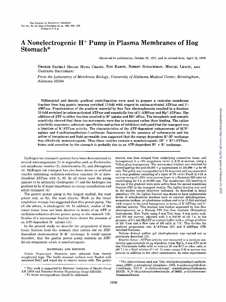
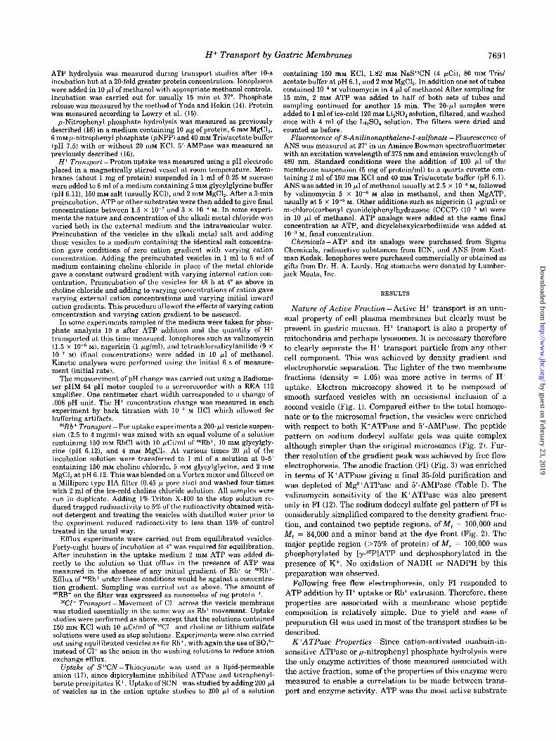
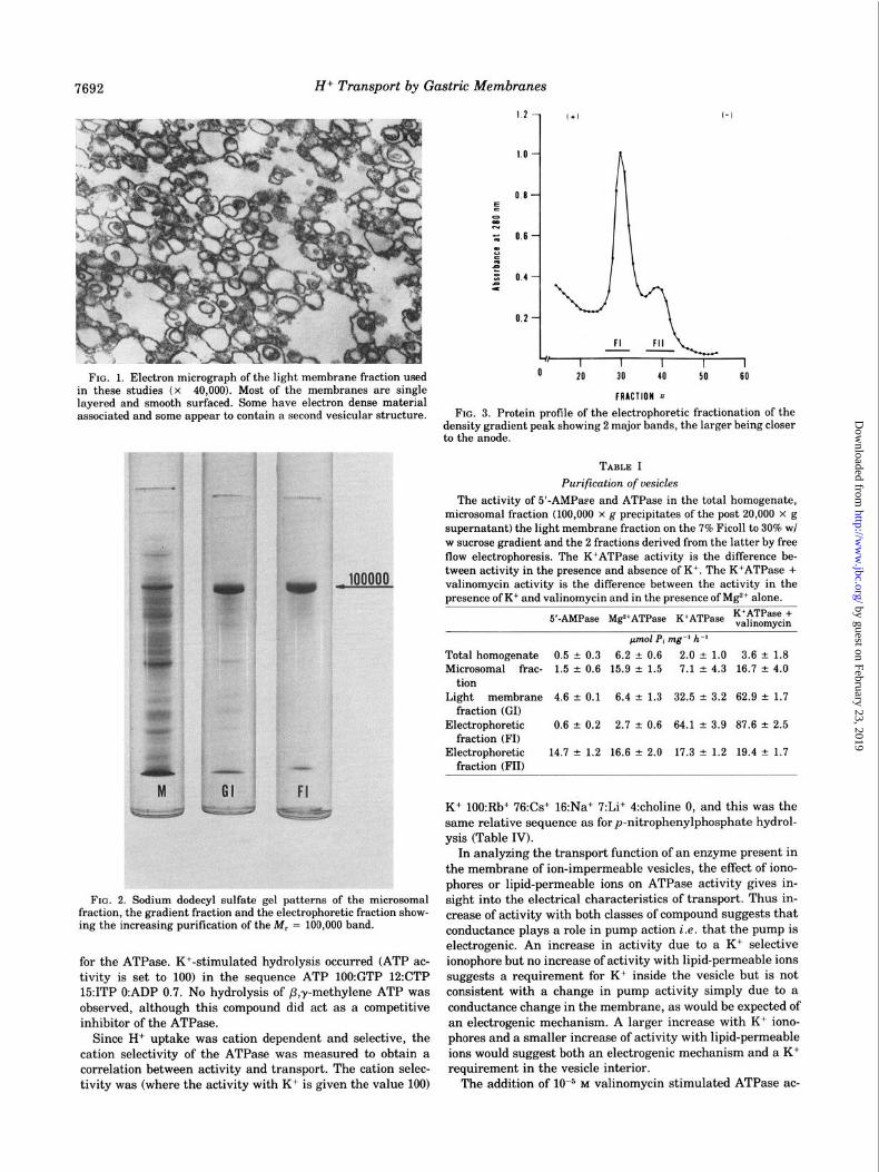
Nature of Active Fraction-Active H+ transport is an unu- sual property of cell plasma membranes but clearly must be present in gastric mucosa. H+ transport is also a property of mitochondria and perhaps lysosomes. It is necessary therefore to clearly separate the H+ transport particle from any other cell component. This was achieved by density gradient and electrophoretic separation. The lighter of the two membrane fractions (density = 1.05) was more active in terms of H+ uptake. Electron microscopy showed it to be composed of smooth surfaced vesicles with an occasional inclusion of a second vesicle (Fig. 1). Compared either to the total homoge- nate or to the microsomal fraction, the vesicles were enriched with respect to both K’ATPase and 5’-AMPase. The peptide pattern on sodium dodecyl sulfate gels was quite complex although simpler than the original microsomes (Fig. 2). Fur- ther resolution of the gradient peak was achieved by free flow electrophoresis. The anodic fraction (FI) (Fig. 3) was enriched in terms of K+ATPase giving a final 35fold purification and was depleted of Mg2+ATPase and 5’-AMPase (Table I). The valinomycin sensitivity of the K+ATPase was also present only in FI (12). The sodium dodecyl sulfate gel pattern of FI is considerably simplified compared to the density gradient frac- tion, and contained two peptide regions, of M, = 100,000 and M, = 84,000 and a minor band at the dye front (Fig. 2). The major peptide region (>75% of protein) of M, = 100,000 was phosphorylated by [y-“2PlATP and dephosphorylated in the presence of K+. No oxidation of NADH or NADPH by this preparation was observed.
Following free flow electrophoresis, only FI responded to ATP addition by H+ uptake or Rb+ extrusion. Therefore, these properties are associated with a membrane whose peptide composition is relatively simple. Due to yield and ease of preparation GI was used in most of the transport studies to be described.
K+ATPase Properties -Since cation-activated ouabain-in- sensitive ATPase or p-nitrophenyl phosphate hydrolysis were the only enzyme activities of those measured associated with the active fraction, some of the properties of this enzyme were measured to enable a correlation to be made between trans- port and enzyme activity. ATP was the most active substrate
by guest on February 23, 2019http://w
ww
.jbc.org/D
ownloaded from
7692 Hi Transport by Gastric Membranes
FIG. 1. Electron micrograph of the light membrane fraction used in these studies (x 40,000). Most of the membranes are single layered and smooth surfaced. Some have electron dense material associated and some appear to contain a second vesicular structure.
\
I, I I I I 0
I 20 30 40 50 60
FRACTION 11
FIG. 3. Protein profile of the electrophoretic fractionation of the density gradient peak showing 2 major bands, the larger being closer to the anode.
TABLE I
Purification of vesicles
The activity of 5’-AMPaee and ATPase in the total homogenate, microsomal fraction (100,000 x g precipitates of the post 20,000 x g supernatant) the light membrane fraction on the 7% Ficoll to 30% wl w sucrose gradient and the 2 fractions derived from the latter by free flow electrophoresis. The K+ATPase activity is the difference be- tween activity in the presence and absence of K+. The K+ATPase + valinomycin activity is the difference between the activity in the nresence of K+ and valinomvcin and in the nresence of Ma*+ alone.
FIG. 2. Sodium dodecyl sulfate gel patterns of the microsomal fraction, the gradient fraction and the electrophoretic fraction show- ing the increasing purification of the M, = 100,000 band.
for the ATPase. K+-stimulated hydrolysis occurred (ATP ac- tivity is set to 100) in the sequence ATP 1OO:GTP 12:CTP 15:ITP O:ADP 0.7. No hydrolysis of P,y-methylene ATP was observed, although this compound did act as a competitive inhibitor of the ATPase.
Since H+ uptake was cation dependent and selective, the cation selectivity of the ATPase was measured to obtain a correlation between activity and transport. The cation selec- tivity was (where the activity with K+ is given the value 100)
pm01 P, nag-’ h-’
Total homogenate 0.5 of: 0.3 6.2 -t- 0.6 2.0 f 1.0 3.6 * 1.8 Microsomal frac- 1.5 of 0.6 15.9 f 1.5 7.1 LI, 4.3 16.7 ? 4.0
tion Light membrane 4.6 -C 0.1 6.4 rt 1.3 32.5 + 3.2 62.9 ? 1.7
fraction (GI) Electrophoretic 0.6 + 0.2 2.7 k 0.6 64.1 + 3.9 87.6 ” 2.5
fraction (FI) Electrophoretic 14.7 rt 1.2 16.6 + 2.0 17.3 * 1.2 19.4 rt 1.7
fraction (FII)
K+ lOO:Rb+ 76:Cs+ 16:Na+ 7:Li+ 4:choline 0, and this was the same relative sequence as for p-nitrophenylphosphate hydrol- ysis (Table IV).
In analyzing the transport function of an enzyme present in the membrane of ion-impermeable vesicles, the effect of iono- phores or lipid-permeable ions on ATPase activity gives in- sight into the electrical characteristics of transport. Thus in- crease of activity with both classes of compound suggests that conductance plays a role in pump action i.e. that the pump is electrogenic. An increase in activity due to a K+ selective ionophore but no increase of activity with lipid-permeable ions suggests a requirement for K+ inside the vesicle but is not consistent with a change in pump activity simply due to a conductance change in the membrane, as would be expected of an electrogenic mechanism. A larger increase with K+ iono- phores and a smaller increase of activity with lipid-permeable ions would suggest both an electrogenic mechanism and a K+ requirement in the vesicle interior.
The addition of low5 M valinomycin stimulated ATPase ac-
by guest on February 23, 2019http://w
ww
.jbc.org/D
ownloaded from
Hi Transport by Gastric Membranes 7693
tivity by 78% in the density gradient fraction and by 40% in the free flow FI fraction (Table I), but had no effect on p- nitrophenyl phosphate hydrolysis. Protonophores such as CCCP or 3,3’,4,5’-tetrachlorosalicylanilide at 10m5 M did not affect ATPase activity and had no additional effect on the valinomycin stimulation. Lipid-permeable ions such as tri- phenylmethyl phosphonium or dimethyldibenzyl ammonium did not increase ATPase activity at 10eR M even in the presence of SCN-.
Dipicrylamine, Zn 2+ F-, pCMBS, and DCCD all inhibited , the K+ATPase activity (Table II), but SCN- and dimethyldi- benzyl ammonium were without effect on enzyme activity.
H+ Up&&e-The presence of an ATPase in these vesicles and the absence of any detectable oxidation-reduction activity such as oxidation of NADH or NADPH suggested that ATP was probably the required substrate for transport.
The addition of ATP did indeed produce a rapid but tran- sient alkalinization of the medium (Fig. 4). The reversal of the alkalinization was due to the consumption of the added ATP (1.7 x 1OW M) followed by leaks of the accumulated H+ back into the medium. The dissipation of the gradient was acceler- ated by ionophores such as valinomycin or nigericin but only transiently accelerated by protonophores such as tetrachloro- salicylanilide.
The effect of ionophores is strong evidence for the presence of a gradient of H+ across the vesicle membrane. This was further established by the effect of varying the vesicle volume which would be expected to affect uptake but not binding. Fig. 5 shows that the H+ uptake (maximum uptake) was a function of the medium osmolarity. This also allowed assessment of the amount of H+ bound due to ATP addition. Extrapolation to infinite osmolarity showed that binding was quantitatively small in relation to transport of H+.
To relate uptake to ATPase activity, various experiments were performed. Uptake of H+ occurred only with ATP and not with GTP, CTP, ITP, TI’P, or ADP. P,-y-Methylene ATP was also inactive and p-nitrophenyl phosphate up to 10 mM had no effect on H+ distribution. ATPase inhibitors such as Zn2+, F-, DCCD, and pCMBS inhibited Hi uptake at 1 mM. Alkali cation was required for H+ uptake and different rates of H+ uptake were obtained with different cations. The sequence of effectiveness found (with K+ set to 100) was K+ lOO:Rb+ 60:Cs+ 45:Na+ 0.24:Li+ O.l:choline 0 (Table IV). All these data were consistent with the hypothesis that ATPase activity was re- quired for the H+ uptake.
Measurement of the H+ uptake and ATP hydrolysis over the initial 10 s following addition of the vesicles to the uptake medium allowed calculation of the ratio of moles of H+ trans- port to moles of ATP hydrolyzed. Considering only the K+
TABLE II
Effect of inhibitors on K+ATPase
The action of various inhibitors on K+ATPase activity. ATPase was assayed as described in the text.
Inhibition %
pCMBS (W5 M) 12 DCCD (W3 M) 82 F- (IO-* M) 80 Zn*+ (W3 M) 90 Dimethyldibenzyl ammonium (10m3 M) 0 SCN- (1O-2 M) 0 Dipicrylamine (10m3 M) 100
activated component of the ATPase, the value for this ratio was 4.1 -C 0.2 (n = 10). Using a value of 2 ~1 mg-’ for vesicular volume, the minimal pH inside the vesicles based on the quantity of H+ disappearing was 1.7. The actual value is probably higher due to the buffer present as well as due to the intrinsic buffering capacity of the inside surface of the vesi- cles.
The electrical characterisitcs of H+ transport were investi- gated using ionophores and lipid-permeable ions. The finding that the H+ gradient could dissipate spontaneously and that this dissipation could be accelerated by valinomycin in the presence of an inward K+ gradient suggested the presence of an H+ conductance in the vesicle membrane (Fig. 4). The relative ineffectiveness of a protonophore such as tetrachloro- salicylanilide, in the absence of an added K+ conductance argues for a low inherent K+ conductance or indeed anion
40
30
20 + I
b z VI
; IO
0
-10
-20
4L 1
I \
I L
I 2 3 4 5 6
time (mid
\
FIG. 4. H+ uptake by the vesicles. Curve A, vesicles were added to a solution of 150 mM KC1 with additions as in text followed by 1.7 x 10m5 M ATP. At times indicated tetrachlorsalicylanilide (TCS) and valinomycin (VAL) were added. Curve B, valinomycin was added immediately after the addition of the vesicles from the same prepa- ration, then ATP at 1.7 x 10m5 M was added.
30, ,90
20 -
0.002 0.003 0.004 0.005
m osm-1
FIG. 5. The effect of vesicular volume on H+ and Rb+ uptake. The vesicles were added to a solution containing 90 mM KC1 with appro- priate addition of mannitol to vary the osmolarity and other addi- tions as detailed in text. ATP (1.7 x 10m5 M) was added, and the H+ uptake measured (0-O). ssRb+ uptake was measured by adding the vesicles to a medium containing 75 mM RbCl with mannitol added to vary the osmolarity and other additions as detailed in the text (X-XI.
by guest on February 23, 2019http://w
ww
.jbc.org/D
ownloaded from
7694 H+ Transport by Gastric Membranes
conductance. This finding also suggests that K+ conductance or penetration may be rate limiting for H+ uptake. This was confirmed by the finding that valinomycin added before ATP increased the uptake rate of H+ (Fig. 4). This effect is not due to a nonspecific conductance increase in the membrane result- ing in shunting of a potential due to electrogenic H+ transport. This is shown by the finding that a lipid-permeable cation such as dimethyldibenzyl ammonium did not influence H+ uptake at concentrations as high as IO-:’ M. SCN- inhibited H+ uptake by 40% at 10m2 M but this appeared to be due to a more rapid dissipation of the gradient since the efflux of H+ was accelerated. Other lipid-permeable ions such as TPMP+ or dipicrylamine inhibited H+ uptake but also inhibited ATPase activity by the same amount.
The effect of valinomycin is therefore due to the specific effect of increasing the K+ penetration into the vesicle. This is substantiated by the finding that preincubation of the vesicles in KC1 prior to addition of ATP has a similar accelerating effect on H+ uptake. In fact, the K+ gradient is an important factor in determining the rate and magnitude of H+ uptake (Table III). The smallest apparent K,, for K+ (32 mM) is ob- tained in the absence of an initial K+ gradient and is of the same order of magnitude as the value of 20 mM found for the ATP at pH 6.1 under the same conditions. An outward K+ gradient considerably increases the rate and magnitude of the H+ uptake also suggesting that the cation gradient is related to the H+ ion gradient developed in the presence of ATP.
Coupling of movement of cation to movement of H+ can be obtained also in the absence of ATP. For example, adding KC1 preincubated vesicles to choline chloride produced no change in medium pH. This is expected from the finding of a low K+ conductance in the vesicles. The addition of valinomycin, or more effectively valinomycin and tetrachlorosalicylanilide but not tetrachlorosalicylanilide alone induces an H+ uptake. Con- versely, adding valinomycin to the KC1 medium before vesicle addition results in an acidification of the medium (Fig, 4) when vesicles are added due to the inward K+ gradient. The overshoot obtained with valinomycin addition following ATP and tetrachlorosalicylanilide is also explained by electrical coupling of the inward K+ movement to H+ efIlux. Since tetrachlorosalicylanilide alone does not induce H+ movement in response to a K+ gradient this confirms what was suggested above, namely, that the intrinsic H+ conductance of the vesi- cles is larger than the intrinsic K+ conductance.
These data then suggest that H+ uptake is a function of the ATPase, is probably nonelectrogenic, and may well be coupled to an eMux of cation, rather than associated with an influx of anion.
Rb+ Transport -To assess whether cation efflux or anion influx accompanied H+ uptake, Rb+ movement across the vesicle membrane was measured. Isotope methods had to be used since the cation selective electrodes available were not sensitive enough for these measurements.
The passive uptake of Rb+ was temperature dependent and also osmotically sensitive (Fig. 5). The t,,z of uptake was 12 h at 4”, 40 min at 22”, and 10 min at 37”. Incubation for prolonged periods of time at 22” and short periods of time at 37” damaged the vesicles as assessed by the maximal H+ gradient obtained. ATP transiently reduced influx presumably due to accelera- tion of efflux. To study this further the vesicles were therefore preloaded with 86Rb+ before ATP addition for the efllux stud- ies.
Incubation at 4” resulted in equilibration in terms c&trapped radioactivity by 48 h. The quantity trapped allowed calcula-
tion of the intravesicular volume with the value found being 2.0 t 0.3 p.1 mg-i (n = 23).
Although efflux could be measured by diluting the vesicles into nonradioactive medium, the conditions of zero gradient were more appropriate for determining the effect of ATP on Rb+ efllux. Thus if there is a decreased trapped radioactivity found with ATP addition directly to the preincubation medium followed by re-equilibration to the previous value after the ATP is hydrolyzed, this can be taken as evidence for active cation extrusion.
As shown in Fig. 6, the addition of ATP to the Rb+-loaded vesicles did indeed result in rapid cation extrusion followed by a slow re-uptake. The re-uptake rate had a similar t,,z to uptake found in the experiments where the vesicles were added to the radioactive medium.
The relationship of this phenomenon to ATPase activity was established by the inhibition of Rb+ efflux by 1 mM concentra- tions of F-, Zn2+, DCCD, and pCMBS.
If Rb+ efllux were due to movement of Rb+ in response to a potential generated by an electrogenic H+ pump, then the distribution ratio of Rb+ between vesicle and medium would allow calculation of the potential. The vesicles lose 44 ? 7% (n = 16) of their radioactivity with ATP addition. This would correspond to a potential of 18 mV vesicle interior positive. However, this interpretation is in conflict with the data previ- ously presented on ATPase activity and H+ uptake which were
TABLE III
Effect of K+ gradient and concentration on H+ uptake
The preincubation conditions were as detailed in the text and the ionic concentrations were varied from 1 to 150 rn~, mannitol being used to maintain isotonicity. The initial gradient was kept constant under two conditions (i.e. outward and zero) and only the i ic concentrations were varied allowing calculation of apparent KA f m a best fit Lineweaver Burk plot.
[K+l,./W+l,,, Initial velocity V mar G nmol H+ mg-’ min-’ nmol H+ mg-’ mic’ mM -
0.006” 130 216 62 1.0 163 218 32 6.0 563 1.370 182
a With the inward gradient, the gradient varied due to the condi- tions used. The value of 0.006 was the gradient under standard conditions, since a value of 1 rn~ K+ was found in the vesicle preparation before addition of K+. The data above are obtained following a 3-min preincubation, hence the magnitude of the inward gradient is only an estimate. All the experiments were carried out on the same preparation.
P ; I’ z ,’
150
:
I’ i//r - i iL
FIG. 6. The eMux of Rb+. Vesicles were pre-equilibrated for 48 h as detailed in the text and 2 rnM ATP added to the suspension at zero time on the graph. It can be seen that there is a transient rapid efRux of Rb+ to about 50% of the measured equilibrium value (horizontal line) and with consumption of the ATP there is re-equilibration of the Rb+.
by guest on February 23, 2019http://w
ww
.jbc.org/D
ownloaded from
Hi Transport by Gastric Membranes 7695
interpreted as excluding a significant K+ conductance. If indeed movement of Rb+ were due to a potential, then
adding lipid-permeable cations would reduce Rb+ movement since the cation would effmx instead of the Rb+. Adding a Rb+ ionophore such as valinomycin would accelerate Rb+ move- ment since the Rb+ conductance of the membrane would be increased. In fact neither dimethyldibenzyl ammonium, a lipid-permeable cation (even in the presence of SCN- to in- crease its lipid solubility), nor valinomycin had any effect on Rb+ efIlux. Valinomycin did increase the re-uptake rate of Rb+.
Accordingly, it is unlikely that cation efIIux is coupled to a potential but more likely that cation efflux is directly related to ATPase activity.
Measurement of the ratio between moles of Rb+ transported and ATP hydrolyzed gave a value of 3.5 2 0.4 (n = 4) using only the cation-stimulated component of the ATPase activity.
Cl- Transport -Although from the studies on cation eMux it would seem unlikely that Cl- uptake accompanied H+ up- take, the efIIux studies were performed on equilibrated vesi- cles. It was possible that under conditions of an inward K+ gradient, Cll uptake occurred with addition of ATP.
Cl- uptake had similar characteristics to passive uptake of Rb+. The t,,2 of Cll at room temperature was 36 min as compared to the t,,* of 40 min for Rb+. The equilibrium uptake volume was also 2.0 ~1 mg-’ of protein. The addition of ATP, however, was without any effect on the uptake rate. Moreover, there was no effect of ATP on the Cl- distribution after the vesicles had equilibrated with :Wl in contrast to the data found with Rb+. The addition of valinomycin had no effect on Cll uptake in the presence of a K+ gradient or following ATP addition showing that these vesicles had minimal Cl- conduct- ance. This had already been concluded from the effect of ionophores on the H+ gradient.
Thus data on H+, Rb+, and Cll movement across the vesicle membrane argue against the development of a potential dur- ing transport.
Uptake of SWN--TO determine whether the above indi- rect arguments excluding the development of a potential dif- ference were correct, the effect of ATP on the distribution of a lipid-permeable anion, SCN-, was determined.
Thiocyanate is taken up in a time-dependent fashion (Fig. 7) into an osmotically sensitive space. The tliz of 2 min is very much less than the tliz for Cl- or Rb+ confirming the SCN- enters vesicles by an additional path presumably due to its lipid permeability. The apparent uptake of SCN- in the pres- ence of valinomycin is increased. This increased uptake re- quires the presence of K+. Presumably this-is due to increased binding of SCN- in the hydrophobic phase due to the forma- tion of the lipid-soluble valinomycin K+ SCN- complex (Fig. 7) since preincubation of vesicles in KC1 does not inhibit the valinomycin enhancement. This valinomycin increment is not osmotically sensitive confirming that this effect is on binding.
The addition of ATP has no effect on the distribution of SCN- between vesicles and medium in the absence of valino- mycin. This suggests that no potential develops under these conditions. From Rb+ efflux studies a K+ gradient would de- velop with the addition of ATP and this K+ gradient would result in a potential difference only in the presence of valino- mycin. The enhanced uptake of SCN- obtained by ATP addi- tion in the presence of valinomycin is consistent with the development of a potential in the presence of valinomycin. This increased uptake is osmotically sensitive.
Fluorescence of ANS -Radioactive isotope distribution may
have too long a lag phase to detect a transient potential. For example, an electrogenic H+ transport would result in devel- opment of an interior positive potential. This potential could result in the development of a K+ conductance and current which would then shunt the potential. Hence, a fluorescent probe of potential, ANS, was used on the assumption that optical detection would have a more rapid response time than trapping of radioactivity.
The sequential addition of membranes ANS and ATP re- sults in the fluorescence changes shown in Fig. 8. The addition of ANS results in rapid increase in fluorescence with a t,,z too rapid to measure with our instrumentation. This rapid fluo- rescence increase is followed by a second slow rise to a plateau after 30 min. The addition of ATP at any time under these conditions results in no change in fluorescence. Accordingly, if ANS is acting as a probe of potential, no potential change occurs.
KCI +w ’
MCI + VA1
KU * ATP
.ATP
I I I I I I 5 10 15 20 15 30
Tlme
FIG. 7. The uptake of SWN- with time when added to vesicles alone (x-x) or vesicles in the presence of valinomycin (0-O). ATP was added to both sets of tubes at the time indicated, but enhanced SCN- uptake only in the presence of valinomycin (O-O).
' - 70
0 a VAL
L 0 I 2 3 4 5 6 0
TIM (min)
FIG. 8. The ANS fluorescence events of the gastric vesicles. The initial addition is that of the membranes to the cuvette, followed by ANS. It can be seen that there is an initial rapid rise with ANS addition followed by a slow increase of fluorescence (X-X) which eventually reaches the valinomycin level (not shown). The addition of valinomycin (VAZ,) before the plateau results in a further rapid increase in fluorescence, and the subsequent addition of ATP (1.7 x 10m5 M) results in a transient fluorescent enhancement, which can be repeated with a second addition of ATP. Also shown is the effect of nigericin (iVIG) on the ATP induced fluorescence, giving immediate inhibition of the fluorescence and inhibition of the effect of subse- quent ATP addition.
by guest on February 23, 2019http://w
ww
.jbc.org/D
ownloaded from

7696 H+ Transport by Gastric Membranes
Quite different data are obtained in the presence of valino- mycin. The addition of valinomycin after the initial fluores- cence increment produces a rapid rise in fluorescence to the same level as occurs without valinomycin after 30 min. This increase in fluorescence requires the presence of K+ and the magnitude is a function of the concentration of K+, but not the K+ gradient. Hence this fluorescence change is due to the increased binding of ANS in the membrane because of the increased solubility of the ANS . valinomycin . K+ complex in the hydrophobic phase of the membrane. The presence of a positive interior potential as would occur with an i&ard K+ gradient in the presence of valinomycin is therefore insuffi- cient to produce a large enough change in fluorescence to obscure the fluorescence increase due to increased penetration of ANS. It should also be noted that an inward K+ gradient in the presence of valinomycin would produce an alkalinization of the vesicle interior.
Addition of ATP in the presence of valinomycin produces a transient increase in fluorescence with precisely the same time course as the H+ uptake (Fig. 9). This is due to a combina- tion of a potential and a H+ gradient. From the Rb+ efflux data and the SCN- uptake, a cation gradient in the presence of valinomycin results in the development of a potential across the vesicle membrane. The requirement for valinomycin to see an ANS fluorescence response suggests that a change of poten- tial is necessary for the effect. Fig. 10 shows that the initial rate of change of fluorescence is a linear function of the log of the K+ concentration (r = 0.992). This would be expected of a Nernst diffusion potential due to a K+ gradient provided a fixed internal [K+] was reached with the addition of ATP. If a potential difference were the only factor involved in the ANS response in the presence of valinomycin then dissipation of the H’ gradient by nigericin which is a neutral exchange iono- phore would not be expected to abolish or completely inhibit development of the fluorescence. In fact, both nigericin and carbonyl cyanide p-trifluoromethoxyphenylhydrazone inhibit or abolish the response (Fig. 8). Moreover, if ANS was respon- sive only to a potential in this system, then valinomycin alone
+
Z
TG
ii c
0 I 2 3 4 5 I
TIM (mm)
FIG. 9. The time course of ANS fluorescence and H+ uptake was measured on the same preparation under identical conditions. 100 ~1 of vesicles (c 50 pg of protein) were added to 2 ml of a medium containing 150 mM KC1 and 5 mM glycylglycine (pH 6.1) followed by 2.5 x 1O-6 M ANS and 5 x 10m6 M valinomycin (VAL). ATP (5 x 10m5 M) was then added and the pH change and fluorescence change monitored as detailed in the text. It can be seen that the time course of the change in fluorescence has virtually an identical time course to the H+ uptake showing the close relationship of the two effects of ATP addition.
should enhance the fluorescence of ANS as a function of the K+ gradient, not the K+ concentration. ANS appears capable of rapidly responding to a potential difference and an H+ gradient in these vesicles. The lack of response in the absence of valino- mycin is therefore reasonable evidence for the absence of a potential during transport by the gastric vesicles. The cation selectivity sequence is shown in Table IV which also shows the effectiveness of the cations in acid secretion.
Effect of Cl- Removal -One of the more significant ap- proaches to the problem of electrogenic transport by the intact amphibian stomach has been the removal of Cll and the substitution of SOd2- in the bathing solutions. Under these conditions, there is a linear relationship between development of a potential difference and acid rate (9). Accordingly, the effect of Cl- removal and SOd2- substitution was determined on the ATPase, H+ uptake, cation eElux, and SCN- and ANS- related properties of the vesicles.
If in the absence of a Cl- conductance, the electrogenicity of H+ uptake was unmasked due to the slow diffusion rate of Sod’- as opposed to Cl-, then effects of potential on the various parameters should be detectable. For example, ATPase activ- ity should decrease unless the potential is shunted by some other permeable ion. H+ uptake should decrease (if H+ move- ment is the electrogenic component) and Rb+ efflux increase (if Rb+ movement is compensating for a potential generated by the H+ pump). Changes in ANS fluorescence and SCN- distri- bution should occur in the absence of valinomycin. In fact ATPase activity was not affected by Cll removal and S042m substitution. H+ uptake and Rb+ efflux were each reduced by 40% and the valinomycin requirement for detection of a poten- tial difference was still present. Moreover, the magnitude of
i Ib lb0 '
FIG. 10. The relationship of the initial rate of change of ANS fluorescence with the addition of ATP to the log of the K+ concentra- tion. The rate of change of fluorescence (arbitrary units in the first 6 s) is plotted against the log of the K+ concentration present in the medium to which the vesicles are added. The concentrations of ANS, valinomycin, and ATP added to the vesicles were 2.5 x 10m6 M, 5 x 10e6 M, and 5 x 10m5 M, respectively, and the concentration of KC1 was varied between 5 and 150 mM.
TABLE IV
Cation selectivity of vesicle function and H+ secretion
This shows the relative effectiveness of the alkali metal cations and choline in stimulating ATPase andpNPP hydrolysis, H+ uptake, and ATP-enhanced ANS fluorescence in gastric membrane vesicles with the effectiveness of K+ set to 100. These values are compared to the effectiveness of these cations and choline in supporting H+ secre- tion by the intact frog mucosa mounted in an Ussing chamber.
ATPaselpNPP hydrolysis H+ ANS H+ Secretion
K+ 100 100 100 100 Rb+ 76 60 90 90
Csf 16 4.5 30 3.4
Na+ 7 .24 1 0
Li+ 4 .l 1 0 Choline+ 0 0 0 0
by guest on February 23, 2019http://w
ww
.jbc.org/D
ownloaded from

Hi Transport by Gastric Membranes 7697
the potential difference was reduced by 40% as would be expected from the diminished cation efIlux. Accordingly, Cl- removal and SOd2- substitution results in reduction of trans- port activity of the ATPase but does not result in electrogenic transport by the ATPase.
DISCUSSION
To date, two types of transport ATPase have been described. One type is exemplified by mitochondrial ATPase and cata- lyzes an electrogenic H+ translocation. Examples of the other type are (Na+ + K+)-ATPase and Ca2+-ATPase which catalyze cation translocation that is nonelectrogenic or partially elec- trogenic. In this work we have described a translocation of H+ by a plasma membrane ATPase from gastric mucosa that ap- pears to be nonelectrogenic and to have a mechanism similar to that of Ca2+ or (Na+ + K+)-ATPase.
This statement can be justified by comparing the character- istics of H+ translocation by mitochondrial ATPase as typical of one class of ATPase on one hand and by the gastric (H+ + K+)-ATPase on the other.
Historically one of the key features of coupling between respiration and phosphorylation was the absence of an identi- fiable covalently linked phosphorylated intermediate (8). It is now clear that the coupling between respiration and phospho- rylation depends on the generation of an electromotive force and an H+ gradient across the mitochondrial membrane (8). In contrast, plasma membrane ATPases (18) and the gastric (H+ + K+)-ATPase (16, 19) form isolatable phosphorylated in- termediates.
The transport of the hydrogen ion by mitochondrial ATPase is electrogenic. Membranes containing this type of ATPase develop both a potential and an H+ gradient (20, 21) and the ATPase is reversible with respect to both parameters. Accord- ingly, alteration of membrane conductance by ionophores or protonophores increases ATPase activity and abolishes the potential induced by the ATPase. The change of potential can be shown directly by measuring the distribution of lipid- permeable ions or by measuring changes in ANS fluorescence (20, 22).
In contrast gastric (H+ + K+)-ATPase activity in the vesi- cles shows no stimulation by increments in membrane con- ductance such as induced by protonophores or lipid-permeable ions.
Measurement of the moles of H+ transported per high en- ergy phosphate bond has given data between 2 and 4 (8,23,24) for the electrogenic type of H+ ATPase. Our measurements using the initial rate gave values close to 4, and it would appear that 2 Ca2+ are transported per ATP hydrolyzed by the Ca2+ ATPase, i.e. 4 charges per ATP. From our data however, it is not possible to conclude that the ratio is fixed and the value of 4 may apply only under the specific conditions of study.
There have been suggestions that an H+:K+ exchange may occur in mitochondrial phosphorylation (3) but in reconsti- tuted vesicles (21) no specific cation requirement for H+ gra- dient formation by the mitochondrial ATPase has been estab- lished.
It is evident from the data presented that gastric vesicle H+ transport is cation-requiring, similar to Na+ transport by (Na+ + K+)-ATPase (18).
Valinomycin and other ionophores are capable of uncou- pling mitochondrial respiration, or stimulating mitochondrial ATPase activity in coupled particles. In a sense similar data are found for gastric vesicles (Table I, and Refs. 19 and 25) in that ATPase activity is stimulated by K+ ionophores. This
stimulation is reduced by K+ preincubation, K+ is required in the interior of the vesicles for H+ uptake and K+ dephosphory- lates the protein (16,19). Stimulation by NH,+ is not increased by gramicidin2 which increases membrane conductance to NH,+. This would be expected if NH,+ was required in the vesicle interior and the permeability of NH, was already very high. Since NH, movement is nonconducting, gramicidin would stimulate NH,+-dependent ATPase activity if a poten- tial developed which would be reduced by the shunt conduct- ance provided by gramicidin. The effect of K+ ionophores is therefore not due to the provision of a shunt conductance for an H+ or K+ battery but is due to an increase of intravesicular K+ required for H+:K+ exchange and ATPase activity.
Concomitant with the H+ uptake there is cation eIIlux. Lipid-permeable ions do not substitute for the alkali metal cation nor do they inhibit cation eMux or Hi uptake (in the absence of ATPase inhibition). This is in contrast to their inhibitory action on oxidation-reduction-dependent transport by bacterial vesicles (2). Thus, in contrast to the electrogenic H+ transport systems cation transport is a specific accompani- ment of H+ transport by gastric (H+ + K+)-ATPase.
There are also considerable differences in the subunit com- plexity of the mitochondrial ATPase and the gastric ATPase transport system. The F,ATPase of mitochondria is a complex multisubunit structure (26) and on its own does not act as an H+ translocator. Other factors such as F,, (27) are required before H+ transport across a membrane can occur. The interac- tion between the F,ATPase and the membrane H+ conducting proteins is, however, specific in that DCCD or oligomycin block ATPase activity only of the intact complex (28). This is assumed to be due to inhibition of H+ translocation through F,, since F,ATPase by itself is not affected by these inhibitors. This ATPase inhibition cannot be overcome by protonophores (i.e. uncouplers) hence the H+ ion exchange between F,ATPase and the F,-transporting subunit is direct and provi- sion of alternate H+ pathways is not sufficient to bypass the blockade of H+ movement through F,,.
The gastric H+-transporting system is considerably less complicated in subunit composition (Fig. 2). DCCD inhibits the ATPase activity of these gastric particles as well as H+ transport and also blocks formation of the phosphorylated intermediate2 Both the H+ and K+ component of transport are specifically required in this ATPase and as for mitochondrial ATPase transport, protonophores do not provide effective al- ternate transport pathways.
It is possible by various means, including ANS fluorescence (22), to show the presence of potentials and changes of poten- tial in untreated mitochondria or submitochondrial particles. Changes in ANS fluorescence in submitochondrial particles show an increase in fluorescence with an increase of positive potential in the interior concomitant with an increase of H+ concentration (22). This is similar to the data obtained with ANS in the gastric system (Fig. 7), which also correlated with the data on SCN- distribution (Fig. 8). The interpretation of a change of potential and H+ distribution accounting for these findings is consistent therefore with data on mitochondria. However, for this potential to occur in the gastric vesicles valinomycin was required. This showed that the methods used were sufficiently sensitive to detect a potential but that the unmodified gastric ATPase particle did not develop a potential during transport activity. Ca’+ATPase particles also show changes in ANS fluorescence with ATP addition (29) which are interpreted as being due to increased Ca2+ binding.
2 Unpublished observations.
by guest on February 23, 2019http://w
ww
.jbc.org/D
ownloaded from

7698 H+ Transport by Gastric Membranes
From these considerations, although gastric ATPase trans- located H+ as do the electrogenic H+ATPases of mitochondria, chloroplasts, and bacteria, the gastric ATPase at least at this stage of analysis is similar to the plasma membrane type of ATPases as exemplified by the (Na+ + K+)- and Ca2+ATPases (30).
It should be possible to correlate the properties of the vesi- cles described above and the properties of the H+ secretory process of the intact gastric mucosa. For example, acid secre- tion should be K+-requiring and ATP-dependent. The electri- cal conductance of the luminal (acid secretory) membrane to K+ or Cl- should be low and the process of H+ secretion should be nonelectrogenic.
Most of the data on the electrical characteristics of acid secretion by the intact mucosa have been obtained on the amphibian stomach mounted in an Ussing chamber (9). K+ is required for the process (31) and addition of mucosal K+ more rapidly restores acid secretion after K+ has been removed from both sides of the tissue. Analysis of tissue K+ content after K+ removal from the bathing solutions showed that H+ secretion stopped when only a small amount of tissue K+ had been lost (32). The concept of a small K+ compartment involved in acid secretion was confirmed in experiments on intact dog (33). Flux studies show that the K+ flux from serosa to mucosa is only a fraction (c 10%) of the H+ flux (34). This, however, would be compatible with a small K+ compartment that did not readily exchange with the bulk of tissue K+. Measurement of change of transmucosal potential or change of potential across the luminal membrane (35) in response to changes in K+ and Cl- concentrations in luminal bathing solutions showed that the electrical conductance of the luminal mem- brane to K+ or Cll was indeed low. Thus, the data from the vesicles are quite compatible with the data summarized above for the intact mucosa.
The role of ATP in acid secretion by the stomach has been difficult to establish by direct approaches (10). Measurement of metabolites in the resting and secreting dog stomach (36) gave data which were difficult to interpret as being due to stimulation of a simple ATPase mechanism. However, the large quantities of mitochondria in the parietal cell (c 40% of cell volume) may maintain steady state ATP levels in spite of large changes in work load.
Perhaps the major problem in accepting the vesicles as isolated as totally representative of the secretory apparatus of the intact stomach is the question of electrogenicity. It seems well established that in Cl- free, S0.,2--containing solutions, H+ secretion by amphibian mucosa is electrogenic (9). More- over under these SO,+ conditions a K+ conductance is present on the luminal surface of the tissue (35). In the vesicles, although SOb2- substitution reduced both H+ and Rb+ flux by 40% this effect was not overcome by ionophores or lipid-perme- able ions, suggesting that the effect was not due to unmasking of the electrogenicity of the H+ pump. Moreover, S0,,2- re- duced the valinomycin-dependent ANS effect, but did not remove the valinomycin requirement, hence no evidence for a potential in the vesicles in S0,2- media was observed.
Perhaps the effect of Cl- removal and SOa2- substitution in the intact mucosa is not due to unmasking of an electrogenic H+ pump, but due to development of a K+ conductance on the luminal surface which can be shown to be present in SOd2- solutions (35). The potential across this surface will then de- pend on the log of the concentration ratio of K+ cell to lumen. In turn the luminal concentration of K+ will depend on the activity of the (H+ + K+)-ATPase i.e. on acid rate. The lower
the activity, the lower the potential, as is found (9). A model for acid secretion by the intact mucosa then re-
quires a functional (H+ + K+)-ATPase, and the presence of luminal K+. This K+ diffuses into the lumen accompanying the active Cll transport present in the tissue, and is ex- changed for H+, resulting in the net secretion of HCl.
Acknowledgments -Thanks are due to the excellent techni- cal assistance of B. Stewart, D. Shaw, and D. Dailey.
1.
2. 3.
4.
5.
6.
7. 8. 9.
10. 11.
12.
13.
14.
15.
16.
17
18. 19
20. 21.
22.
23.
24. 25.
26. 27.
28. 29.
30.
31.
32.
33.
34.
35.
36.
REFERENCES
Malonev. P. D.. Kashket. E. R.. and Wilson. T. H. (1974) Proc. Natl.“/&ad. &. U. S. k. 71, 3896-3900
Kaback, H. R. (1974) Science 186, 882-892 Azzone, G. F., Massari, S., Colonna, R., Dell Antone, P., and
Frigert, L. (1974) Ann. N.Y. Acad. Sci. 227, 337-347 Jagendorf, A. T., and Uribe, E. (1966) Proc. Natl. Acad. Sci.
U. S. A. 55, 170-177 Hinkle, P. C., Kim, J. J., and Racker, E. (1972) J. Biol. Chem.
247, 1338-1339 Kagawa, Y., Kaudrach, A., and Racker, E. (1973)5. Biol. Chem.
248, 676-684 Harold. F. M. (1974) Ann. N.Y. Acad. Sci. 227, 297-331 Mitchell, P. (1966) Biol. Rev. 41, 445502 Rehm, W. S. (1965) Fed. Proc. 24, 1387-1395 Hersey, S. J. (1974) Biochim. Biophys. Acta 344, 157-203 Lee, J., Simpson, G., and Scholes, P. (1974) B&hem. Biophys.
Res. Commun. 60, 825-832 Saccomani, G., Stewart, H. B., Shaw, D., Lewin, M., and Sachs,
G. (1976) Biochim. Biophys. Acta, in press Suennev. J. G.. Saccomani. G.. Soitzer, H. L., Tomana, M., and
‘Sachs,‘G. (1974) Arch. B;ochem. Biobhys. 161, 456-471 Yoda. A.. and Hokin. L. E. (1970) Biochem. Biophrs. Res. Com-
mu;. 46, 880-886 Lowry, 0. H., Rosebrough, N. J., Farr, A. L., and Randall, R. J.
(1951) J. Biol. Chem. 193, 265-275 Saccomani, G., Shah, G., Spenney, J. G., and Sachs, G. (1975) J.
Bid. Chem. 250, 4802-4809 Scarborough, G. A. (1976) Proc. N&Z. Acad. Sci. U. S. A. 73,
1485-1488 Skou, J. C. (1965) Physiol. Rev. 45, 596-617 Forte, J. G., Ganser, A. L., and Tanisawa, A. S. (1974) Ann.
N.Y. Acad. Sci. 242, 255-267 Kagawa, Y., and Racker, E. (1971) J. Biol. Chem. 246,5477-5487 Skulachev. V. P. (1976) in Energy Transducing Mechanisms
(Racker,’ E., ed) pp. 31-74, Butterworths, London Azzi. A. P.. Gherardini, P., and Santato, M. (1971) J. Biol.
Chem. 246, 2035-2042 Brand, H. P., Reynafarje, B., and Lehinger, A. L. (1976) Proc.
Natl. Acad. Sci. U. S. A. 73, 437-441 Witt, T. H. (1974) Ann. N.Y. &ad. Sci. 227, 203-206 Sachs. G.. Rabon, E., Saccomani, G., and Sarau, H. M. (1976)
Ann. N:Y. Acad. Sci. 264, 456-475 Senior, A. E. (1973) Biochim. Biophys. Acta 301, 249-277 Mitchell, P., and Moyle, J. (1974) in Membrane ATPase and
Transport Processes (Bronk, J. R., ed) pp. 91-112, Biochemical Society, London
Tzagoloff, A., and Meagher, P. (1971) J. Biol. Chem. 247,594-603 Vanderkooi. J.. and Martonosi, A. (1971) Arch. Biochem. Bio-
phys 144; 87198 Martonosi, A. (19721 in Metabolic Transport (Hokin, L. E., ed)
pp. 317-350, Academic Press, New York Harris, J. B., and Edelman, I. S. (1964)Am. J. Physiol. 206,769-
782 Davies, T. L., Rutledge, J. R., Keasce, D. C., Bajandas, F. J.,
and Rehm. W. S. (1965) Am. J. Phrsiol. 209, 146-152 Hirschowitz, B. I., and Sachs, G. (i967) Am. J. Physiol. 213,
1401-1405 Sachs. G.. Collier. R. H.. Pacifico. A.. Shoemaker, R. L., and
Hirschowitz, B. I. (1969) Biochim. Blophys. Acta i73, 509-517 Sachs, G., Shoemaker, R. L., Blum, A. L., Helander, H. F., and
Makhlouf, G. M. (1971) in Electrophysiology ofEpithelia1 Cells (Giebisch, G., ed) pp. 257-279, Schattauer, Stuttgart
Sarau, H. M., Foley, J., Moonsammy, G., Wiebelhaus, V. D., and Sachs, G. (1975) J. Biol. Chem. 250, 8321-8329
by guest on February 23, 2019http://w
ww
.jbc.org/D
ownloaded from

G Sachs, H H Chang, E Rabon, R Schackman, M Lewin and G SaccomaniA nonelectrogenic H+ pump in plasma membranes of hog stomach.
1976, 251:7690-7698.J. Biol. Chem.
http://www.jbc.org/content/251/23/7690Access the most updated version of this article at
Alerts:
When a correction for this article is posted•
When this article is cited•
to choose from all of JBC's e-mail alertsClick here
http://www.jbc.org/content/251/23/7690.full.html#ref-list-1
This article cites 0 references, 0 of which can be accessed free at
by guest on February 23, 2019http://w
ww
.jbc.org/D
ownloaded from


![Plasma Membranes [Read-Only]](https://img.dokumen.tips/doc/110x75/62375f419f3c9d188e64b806/plasma-membranes-read-only.jpg)