Embed Size (px)
Citation preview
5 | STRUCTURE ANDFUNCTION OFPLASMAMEMBRANES
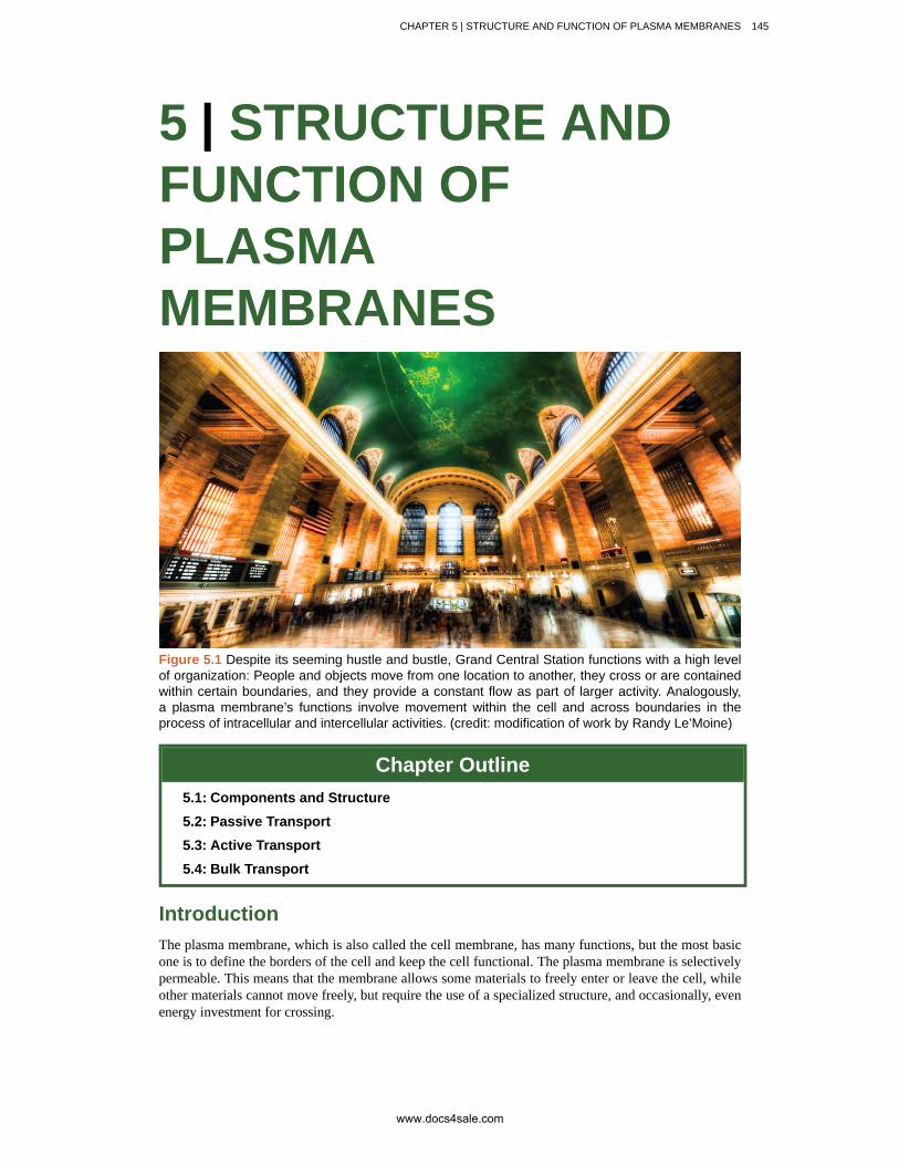
Figure 5.1 Despite its seeming hustle and bustle, Grand Central Station functions with a high levelof organization: People and objects move from one location to another, they cross or are containedwithin certain boundaries, and they provide a constant flow as part of larger activity. Analogously,a plasma membrane’s functions involve movement within the cell and across boundaries in theprocess of intracellular and intercellular activities. (credit: modification of work by Randy Le’Moine)
Chapter Outline5.1: Components and Structure
5.2: Passive Transport
5.3: Active Transport
5.4: Bulk Transport
IntroductionThe plasma membrane, which is also called the cell membrane, has many functions, but the most basicone is to define the borders of the cell and keep the cell functional. The plasma membrane is selectivelypermeable. This means that the membrane allows some materials to freely enter or leave the cell, whileother materials cannot move freely, but require the use of a specialized structure, and occasionally, evenenergy investment for crossing.
CHAPTER 5 | STRUCTURE AND FUNCTION OF PLASMA MEMBRANES 145
www.docs4sale.com
5.1 | Components and Structure
By the end of this section, you will be able to:
• Understand the fluid mosaic model of cell membranes
• Describe the functions of phospholipids, proteins, and carbohydrates in membranes
• Discuss membrane fluidity
A cell’s plasma membrane defines the cell, outlines its borders, and determines the nature of itsinteraction with its environment (see Table 5.1 for a summary). Cells exclude some substances, take inothers, and excrete still others, all in controlled quantities. The plasma membrane must be very flexibleto allow certain cells, such as red blood cells and white blood cells, to change shape as they pass throughnarrow capillaries. These are the more obvious functions of a plasma membrane. In addition, the surfaceof the plasma membrane carries markers that allow cells to recognize one another, which is vital fortissue and organ formation during early development, and which later plays a role in the “self” versus“non-self” distinction of the immune response.
Among the most sophisticated functions of the plasma membrane is the ability to transmit signalsby means of complex, integral proteins known as receptors. These proteins act both as receivers ofextracellular inputs and as activators of intracellular processes. These membrane receptors provideextracellular attachment sites for effectors like hormones and growth factors, and they activateintracellular response cascades when their effectors are bound. Occasionally, receptors are hijacked byviruses (HIV, human immunodeficiency virus, is one example) that use them to gain entry into cells, andat times, the genes encoding receptors become mutated, causing the process of signal transduction tomalfunction with disastrous consequences.
Fluid Mosaic ModelThe existence of the plasma membrane was identified in the 1890s, and its chemical components wereidentified in 1915. The principal components identified at that time were lipids and proteins. The firstwidely accepted model of the plasma membrane’s structure was proposed in 1935 by Hugh Davson andJames Danielli; it was based on the “railroad track” appearance of the plasma membrane in early electronmicrographs. They theorized that the structure of the plasma membrane resembles a sandwich, withprotein being analogous to the bread, and lipids being analogous to the filling. In the 1950s, advancesin microscopy, notably transmission electron microscopy (TEM), allowed researchers to see that thecore of the plasma membrane consisted of a double, rather than a single, layer. A new model that betterexplains both the microscopic observations and the function of that plasma membrane was proposed byS.J. Singer and Garth L. Nicolson in 1972.
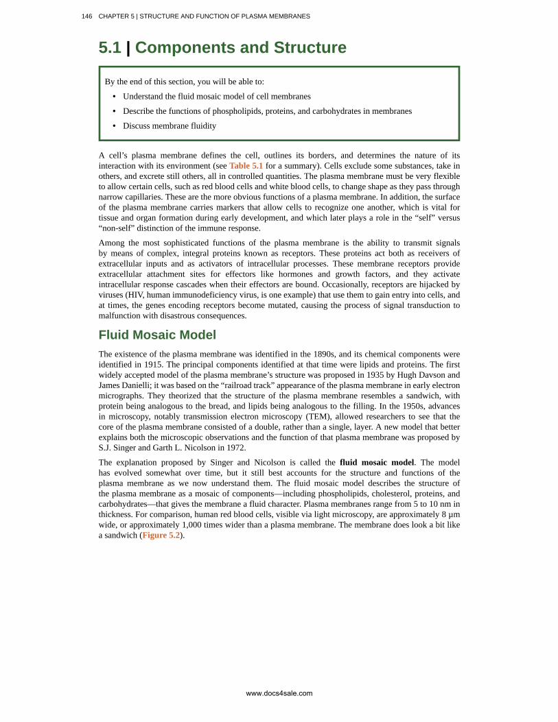
The explanation proposed by Singer and Nicolson is called the fluid mosaic model. The modelhas evolved somewhat over time, but it still best accounts for the structure and functions of theplasma membrane as we now understand them. The fluid mosaic model describes the structure ofthe plasma membrane as a mosaic of components—including phospholipids, cholesterol, proteins, andcarbohydrates—that gives the membrane a fluid character. Plasma membranes range from 5 to 10 nm inthickness. For comparison, human red blood cells, visible via light microscopy, are approximately 8 µmwide, or approximately 1,000 times wider than a plasma membrane. The membrane does look a bit likea sandwich (Figure 5.2).
146 CHAPTER 5 | STRUCTURE AND FUNCTION OF PLASMA MEMBRANES
www.docs4sale.com
Figure 5.2 The fluid mosaic model of the plasma membrane describes the plasma membraneas a fluid combination of phospholipids, cholesterol, and proteins. Carbohydrates attached tolipids (glycolipids) and to proteins (glycoproteins) extend from the outward-facing surface of themembrane.
The principal components of a plasma membrane are lipids (phospholipids and cholesterol), proteins,and carbohydrates attached to some of the lipids and some of the proteins. A phospholipid is a moleculeconsisting of glycerol, two fatty acids, and a phosphate-linked head group. Cholesterol, another lipidcomposed of four fused carbon rings, is found alongside the phospholipids in the core of the membrane.The proportions of proteins, lipids, and carbohydrates in the plasma membrane vary with cell type, butfor a typical human cell, protein accounts for about 50 percent of the composition by mass, lipids (ofall types) account for about 40 percent of the composition by mass, with the remaining 10 percent ofthe composition by mass being carbohydrates. However, the concentration of proteins and lipids varieswith different cell membranes. For example, myelin, an outgrowth of the membrane of specialized cellsthat insulates the axons of the peripheral nerves, contains only 18 percent protein and 76 percent lipid.The mitochondrial inner membrane contains 76 percent protein and only 24 percent lipid. The plasmamembrane of human red blood cells is 30 percent lipid. Carbohydrates are present only on the exteriorsurface of the plasma membrane and are attached to proteins, forming glycoproteins, or attached tolipids, forming glycolipids.
Phospholipids
The main fabric of the membrane is composed of amphiphilic, phospholipid molecules. The hydrophilicor “water-loving” areas of these molecules (which look like a collection of balls in an artist’s renditionof the model) (Figure 5.2) are in contact with the aqueous fluid both inside and outside the cell.Hydrophobic, or water-hating molecules, tend to be non-polar. They interact with other non-polarmolecules in chemical reactions, but generally do not interact with polar molecules. When placed inwater, hydrophobic molecules tend to form a ball or cluster. The hydrophilic regions of the phospholipidstend to form hydrogen bonds with water and other polar molecules on both the exterior and interior ofthe cell. Thus, the membrane surfaces that face the interior and exterior of the cell are hydrophilic. Incontrast, the interior of the cell membrane is hydrophobic and will not interact with water. Therefore,phospholipids form an excellent two-layercell membrane that separates fluid within the cell from thefluid outside of the cell.
A phospholipid molecule (Figure 5.3) consists of a three-carbon glycerol backbone with two fatty acidmolecules attached to carbons 1 and 2, and a phosphate-containing group attached to the third carbon.This arrangement gives the overall molecule an area described as its head (the phosphate-containinggroup), which has a polar character or negative charge, and an area called the tail (the fatty acids),which has no charge. The head can form hydrogen bonds, but the tail cannot. A molecule with thisarrangement of a positively or negatively charged area and an uncharged, or non-polar, area is referredto as amphiphilic or “dual-loving.”
CHAPTER 5 | STRUCTURE AND FUNCTION OF PLASMA MEMBRANES 147
www.docs4sale.com

Figure 5.3 This phospholipid molecule is composed of a hydrophilic head and two hydrophobictails. The hydrophilic head group consists of a phosphate-containing group attached to a glycerolmolecule. The hydrophobic tails, each containing either a saturated or an unsaturated fatty acid, arelong hydrocarbon chains.
This characteristic is vital to the structure of a plasma membrane because, in water, phospholipids tendto become arranged with their hydrophobic tails facing each other and their hydrophilic heads facingout. In this way, they form a lipid bilayer—a barrier composed of a double layer of phospholipids thatseparates the water and other materials on one side of the barrier from the water and other materials onthe other side. In fact, phospholipids heated in an aqueous solution tend to spontaneously form smallspheres or droplets (called micelles or liposomes), with their hydrophilic heads forming the exterior andtheir hydrophobic tails on the inside (Figure 5.4).
148 CHAPTER 5 | STRUCTURE AND FUNCTION OF PLASMA MEMBRANES
www.docs4sale.com

Figure 5.4 In an aqueous solution, phospholipids tend to arrange themselves with their polar headsfacing outward and their hydrophobic tails facing inward. (credit: modification of work by MarianaRuiz Villareal)
Proteins
Proteins make up the second major component of plasma membranes. Integral proteins (somespecialized types are called integrins) are, as their name suggests, integrated completely into themembrane structure, and their hydrophobic membrane-spanning regions interact with the hydrophobicregion of the the phospholipid bilayer (Figure 5.2). Single-pass integral membrane proteins usually havea hydrophobic transmembrane segment that consists of 20–25 amino acids. Some span only part of themembrane—associating with a single layer—while others stretch from one side of the membrane to theother, and are exposed on either side. Some complex proteins are composed of up to 12 segments ofa single protein, which are extensively folded and embedded in the membrane (Figure 5.5). This typeof protein has a hydrophilic region or regions, and one or several mildly hydrophobic regions. Thisarrangement of regions of the protein tends to orient the protein alongside the phospholipids, with thehydrophobic region of the protein adjacent to the tails of the phospholipids and the hydrophilic regionor regions of the protein protruding from the membrane and in contact with the cytosol or extracellularfluid.
CHAPTER 5 | STRUCTURE AND FUNCTION OF PLASMA MEMBRANES 149
www.docs4sale.com

Figure 5.5 Integral membranes proteins may have one or more alpha-helices that span themembrane (examples 1 and 2), or they may have beta-sheets that span the membrane (example 3).(credit: “Foobar”/Wikimedia Commons)
Peripheral proteins are found on the exterior and interior surfaces of membranes, attached either tointegral proteins or to phospholipids. Peripheral proteins, along with integral proteins, may serve asenzymes, as structural attachments for the fibers of the cytoskeleton, or as part of the cell’s recognitionsites. These are sometimes referred to as “cell-specific” proteins. The body recognizes its own proteinsand attacks foreign proteins associated with invasive pathogens.
Carbohydrates
Carbohydrates are the third major component of plasma membranes. They are always found on theexterior surface of cells and are bound either to proteins (forming glycoproteins) or to lipids (formingglycolipids) (Figure 5.2). These carbohydrate chains may consist of 2–60 monosaccharide units and canbe either straight or branched. Along with peripheral proteins, carbohydrates form specialized sites onthe cell surface that allow cells to recognize each other. These sites have unique patterns that allow thecell to be recognized, much the way that the facial features unique to each person allow him or her tobe recognized. This recognition function is very important to cells, as it allows the immune system todifferentiate between body cells (called “self”) and foreign cells or tissues (called “non-self”). Similartypes of glycoproteins and glycolipids are found on the surfaces of viruses and may change frequently,preventing immune cells from recognizing and attacking them.
These carbohydrates on the exterior surface of the cell—the carbohydrate components of bothglycoproteins and glycolipids—are collectively referred to as the glycocalyx (meaning “sugar coating”).The glycocalyx is highly hydrophilic and attracts large amounts of water to the surface of the cell. Thisaids in the interaction of the cell with its watery environment and in the cell’s ability to obtain substancesdissolved in the water. As discussed above, the glycocalyx is also important for cell identification, self/non-self determination, and embryonic development, and is used in cell-cell attachments to form tissues.
150 CHAPTER 5 | STRUCTURE AND FUNCTION OF PLASMA MEMBRANES
www.docs4sale.com

How Viruses Infect Specific OrgansGlycoprotein and glycolipid patterns on the surfaces of cells give many viruses anopportunity for infection. HIV and hepatitis viruses infect only specific organs or cells in thehuman body. HIV is able to penetrate the plasma membranes of a subtype of lymphocytescalled T-helper cells, as well as some monocytes and central nervous system cells. Thehepatitis virus attacks liver cells.
These viruses are able to invade these cells, because the cells have binding sites ontheir surfaces that are specific to and compatible with certain viruses (Figure 5.6). Otherrecognition sites on the virus’s surface interact with the human immune system, promptingthe body to produce antibodies. Antibodies are made in response to the antigens orproteins associated with invasive pathogens, or in response to foreign cells, such asmight occur with an organ transplant. These same sites serve as places for antibodies toattach and either destroy or inhibit the activity of the virus. Unfortunately, these recognitionsites on HIV change at a rapid rate because of mutations, making the production of aneffective vaccine against the virus very difficult, as the virus evolves and adapts. A personinfected with HIV will quickly develop different populations, or variants, of the virus thatare distinguished by differences in these recognition sites. This rapid change of surfacemarkers decreases the effectiveness of the person’s immune system in attacking the virus,because the antibodies will not recognize the new variations of the surface patterns. In thecase of HIV, the problem is compounded by the fact that the virus specifically infects anddestroys cells involved in the immune response, further incapacitating the host.
Figure 5.6 HIV binds to the CD4 receptor, a glycoprotein on the surfaces of T cells. (credit:modification of work by NIH, NIAID)
Membrane FluidityThe mosaic characteristic of the membrane, described in the fluid mosaic model, helps to illustrate itsnature. The integral proteins and lipids exist in the membrane as separate but loosely attached molecules.These resemble the separate, multicolored tiles of a mosaic picture, and they float, moving somewhatwith respect to one another. The membrane is not like a balloon, however, that can expand and contract;
CHAPTER 5 | STRUCTURE AND FUNCTION OF PLASMA MEMBRANES 151
www.docs4sale.com


![Plasma membranes [2015]](https://img.dokumen.tips/doc/110x75/55d39deebb61ebe7268b4821/plasma-membranes-2015.jpg)


![Plasma Membranes [Read-Only]](https://img.dokumen.tips/doc/110x75/62375f419f3c9d188e64b806/plasma-membranes-read-only.jpg)


