Embed Size (px)
Citation preview

A DEVICE FOR IMPOSING UNIFORM, CYCLIC STRAIN
TO CELLS GROWING ON IMPLANT ALLOYS
By
Larry Chad Winter
A Thesis Submitted to the Faculty of Mississippi State University
in Partial Fulfillment of the Requirements for the Degree of Master of Science
in Biomedical Engineering in the Department of Agricultural and Biological Engineering
Mississippi State University
August 2002

A DEVICE FOR IMPOSING UNIFORM, CYCLIC STRAIN
TO CELLS GROWING ON IMPLANT ALLOYS
By:
Larry Chad Winter
Approved: ____________________ ____________________ Joel D. Bumgardner Jerome A. Gilbert Associate Professor of Graduate Coordinator of the Agricultural and Biological Engineering Department of Agricultural (Director of Thesis) and Biological Engineering ____________________ ____________________ Steven H. Elder Dwayne A. Wise Associate Professor of Professor of Agricultural and Biological Engineering Biological Sciences (Committee Member) (Committee Member) ____________________ ____________________ David B. Smith A. Wayne Bennett Associate Professor of Dean of College of Engineering Agricultural and Biological Engineering (Committee Member)
Name: Larry Chad Winter Date of Degree: August 3, 2002 Institution: Mississippi State University Major Field: Biomedical Engineering Major Professor: Joel D. Bumgardner Title of Study: A DEVICE FOR IMPOSING UNIFORM, CYCLIC STRAIN TO CELLS GROWING ON IMPLANT ALLOYS Pages in Study: 74 Candidate for Degree of Master of Science
Since bone tissues grow in intimate contact with implant surfaces in vivo, there is a
need to investigate how bone cells respond to mechanical loads adjacent to implants under
well characterized loading conditions that stimulate the bone-implant surface. Thus, the
objective of this study was to demonstrate an effective means for applying known, uniform,
cyclic strain to cells growing on implant materials in vitro.
A cell culture strain plate device was developed based on the application of the four-
point bending principle. The device uses a small electric motor to drive belts attached to
shafts, which turn a set of cams. The cams are attached to pins that connect to a titanium
plate, which rests over arched supports. When deflected and depending on which set of
cams are used, strains generated range from around 200 to 1000 µstrain. UMR-106
osteoblast-like cells were cultured on the titanium plate, and the plate was deflected at three
strain magnitudes at 1.5 Hz for durations of 4 and 24 hours. Strain gages recorded average
maximum strain levels of 182 ± 3, 366 ± 9, and 984 ± 7µstrain. The strain device, with

attached cells, was tested in an amiable bioenvironment. Results from strain gages indicated
a uniform strain field existed within the center region of the plate and culture area. Cells in
the test plates stained viable, exhibited similar morphology to controls, and were assayed for
alkaline phosphatase (ALP) activity, total protein production, and calcium deposition.
Results also indicated that stretched cells exhibited increases in proliferation, as well as
changes in ALP activity vs. unstrained controls. Thus, the device was successful in
distinguishing differences in cell response to mechanical perturbations and may be used to
investigate how cells respond to strains at implant-bone interfaces.
Keywords - Osteoblast, Mechanical loading, Cell Culture, Implants, Strain

ACKNOWLEDGEMENTS First, I would like to express my sincere gratitude to Dr. Joel Bumgardner for his
inspiration, wisdom, and friendship throughout my graduate studies.
I would also like to thank the committee members: Dr. Jerry Gilbert, Dr. Steve
Elder, Dr. David Smith, and Dr. Dwayne Wise for their valuable time, assistance, and
expertise. I also want to recognize Dr. Filip To of the Agricultural and Biological
Engineering Department and Bill Monroe and Richard Kuliniski of the Electron
Microscopy Center.
A special thanks also goes to my family who have been so influential and
supportive in my pursuit of this degree.
This list would not be complete without showing my appreciation to Suzanne
Hutchinson Parker, Dana Lynn Nettles, and Hsin-Yi Lin for putting up with me in the lab
and for making my stay there an enjoyable one.
ii

TABLE OF CONTENTS
Page
ACKNOWLEDGEMENTS........................................................................................... ii LIST OF TABLES......................................................................................................... v LIST OF FIGURES ....................................................................................................... vi SYMBOL DEFINITIONS............................................................................................. vii CHAPTER I. INTRODUCTION.............................................................................................. 1
Background........................................................................................................ 1 Bone at the macrostructure-level ........................................................... 1 Bone at the microstructure-level............................................................ 3
Mechanical loading of bone............................................................................... 5 Implant Considerations ...................................................................................... 9 Cellular Mechanics ............................................................................................ 15
Early Devices ......................................................................................... 15 Requirements for a New System............................................................ 18
Goals/Specific Aims .......................................................................................... 19 Hypothesis ......................................................................................................... 20
II. A DEVICE FOR IMPOSING KNOWN, CYCLIC STRAIN TO CELLS
GROWING ON IMPLANT ALLOYS.................................................. 21
Introduction........................................................................................................ 21 Materials and Methods....................................................................................... 27
Strain Device.......................................................................................... 27 Strain Plate Characterization ................................................................. 28
Strain Magnitudes ...................................................................... 28 Strain Uniformity ....................................................................... 29
iii

CHAPTER Page
Cell Culture............................................................................................ 30 Statistics ............................................................................................................. 32
Results................................................................................................................ 32 Strain Magnitudes .................................................................................. 32 Strain Uniformity ................................................................................... 33 Cell Culture............................................................................................ 33 Discussion.......................................................................................................... 34 Conclusion ......................................................................................................... 36 III. STRETCH-MEDIATED RESPONSES OF CELLS CULTURED ON IMPLANT ALLOYS............................................................................. 45
Introduction........................................................................................................ 45 Materials and Methods....................................................................................... 49
Cell Strain Plate ..................................................................................... 49 Cell Culturing System............................................................................ 50
Cell Strain Design.................................................................................. 50 Cell Activity........................................................................................... 51
Total Protein Production........................................................... 51 Alkaline Phosphatase Activity.................................................. 51 Calcium Deposition .................................................................. 52
Statistics ............................................................................................................. 52 Results................................................................................................................ 52
Total Protein .......................................................................................... 52 Alkaline Phosphatase ............................................................................. 53 Calcium.................................................................................................. 54
Discussion.......................................................................................................... 54 Conclusion ......................................................................................................... 58 IV. CONCLUSION................................................................................................. 64 REFERENCES .............................................................................................................. 69
iv

LIST OF TABLES
TABLE Page 3.1. Percent differences in total protein content ........................................................ 62 3.2. Percent differences in ALP activity .................................................................... 63
v

LIST OF FIGURES
FIGURE Page 2.1. Shear (v) and moment (M) diagram.................................................................... 37 2.2. Strain plate device image and schematic ............................................................ 38 2.3. Strain gage diagram and mean peak magnitudes................................................ 39 2.4. Typical strain profile of maximum deflection .................................................... 40 2.5. Tension and compression profiles ...................................................................... 41 2.6. Strain Profile sampled at 5 Hz ............................................................................ 42 2.7. Confocal microscopy images.............................................................................. 43 2.8. Neutral red results ............................................................................................... 44 3.1. Total protein results ............................................................................................ 59 3.2. ALP activity results ........................................................................................... 60 3.3. Calcium results ................................................................................................... 61
vi

SYMBOL DEFINITIONS
1. IMZ – A type of dental implant
2. Hz – Hertz
3. µ – Micro
4. N – Newton
5. ε – Strain
6. β – Beta
7. mM – Milli molar
8. M – Molar
9. nm - Nanometer
vii

CHAPTER I
INTRODUCTION
Background
Metal implants are used to replace/augment function of bony tissues. The
response of human bone to stress is very important to the success of implant materials.
For implants to be successful, a well-developed bone matrix in close apposition to the
implant must be achieved. The development of bone around an implant is due in large
part to the mechanical environment. Thus, understanding the composition and structure
of bone is necessary to properly understand how and why bone reacts to implants. The
interaction of the mechanical and structural properties of bone, a heterogeneous,
anisotropic material, is quite complex.
Bone at Macro-Structure Level
At the macroscopic level, bone can be characterized as one of two types: cortical
or cancellous (Martin and Burr, 1989; Wainwright et al., 1976). Cortical bone is compact
and dense and can typically be found in the shafts of long bones. It is composed of
densely packed, longitudinal, layered (lamellar) columns called osteons, the center of
which contains a Haversian canal carrying blood vessels. Each osteon (also called a
Haversian system) is formed by one Haversian canal and the bone lamellae that surround
it. Throughout the lamellae, bone cells (osteocytes) may be found within small spaces,
called lacunae. The osteocytes are connected by tiny canals called canaliculi, through
1

2 which oxygen and carbon dioxide, nutrients, and wastes pass to and from the blood vessel of
the closest Haversian canals. Interstitial bone, which can be defined as the remnants of
former Haversian systems that have been partially destroyed as the bone grew, fills in the
irregular spaces between the lamellar columns.
Cancellous bone consists of bony trabeculae, thin plates, or spicules and
predominates in the pelvic region, vertebrae, and at the ends of long bones (Martin and Burr,
1989; Wainwright et al., 1976). Blood vessels occupy the spaces between the trabeculae.
Cancellous bone, when treated as a tissue, has a much lower Young’s Modulus than does
cortical bone.
Both cortical and cancellous bone undergo similar sequences of remodeling. Bone
remodeling is the life-long process by which mature bone is renewed through the continual
processes of bone resorption and bone formation. Disturbances in bone remodeling can lead
to detrimental alteration in bone architecture, such as the extreme consequence called
osteopenia. The removal of important structural elements (number of trabeculae, trabeculae
plate thickness, connectivity, etc.) without proper replacement can lead to mechanical
incompetence and failure (Wainwright et al., 1976).
Bone remodeling is initiated by osteoclastic precursors that eventually become multi-
nucleated osteoclasts. These osteoclasts destroy bone matrix (resorption). Once resorption
has ceased, osteoblast precursors invade the bone. These preosteoblasts differentiate into
osteoblasts and form new matrix (bone formation). The initiation of bone formation in the
resorption cavity following resorption is called coupling. This coupling process ensures that
the amount of bone lost via resorption is replaced during formation. While coupling denotes

3 a temporal sequence of events, an imbalance may occur. For example, a resorption cavity
may be incompletely refilled thus leading to osteoporosis. Conversely, excess bone
formation can occur and cause an imbalance, as well. The final differentiated units that
maintain bone matrix in the lacunae are called osteocytes (Marks and Popoff, 1988; Cohen et
al., 1992).
Bone at Micro-Structure Level
Bone is composed of 30% organic compounds, of which 90 to 95% is collagen, the
rest being non-collagenous proteins. The remaining 70% of bone is made up of the
inorganic mineral hydroxyapatite, which includes calcium phosphate, calcium carbonate,
calcium fluoride, calcium hydroxide and citrate. Microstructurally, bone can be
characterized into three basic types: woven, primary, and secondary bone (Martin and Burr,
1989).
Woven bone is laid down rather quickly, that is, collagen and other materials are
secreted and form the bone matrix. Consequently, it is not surprising that this type of bone is
found in fetuses, rapidly growing bone, and fractured bone. It is highly mineralized with
fibers randomly oriented. Woven-fibered bone is much less dense than other types of bone
due to its loose packing of collagen fibers and high porosity. A very distinguishing
characteristic of woven bone is that it may be deposited de novo, or deposited without any
previous hard tissue substrate. Woven bone’s primary function is mechanical in nature, but
it also serves functions in skeletal repair and defense. A large cell/bone volume ratio
underlies woven bone’s ability to proliferate rapidly in response to bone trauma (Martin and
Burr, 1989).

4
Primary bone can be divided into three sub-categories: primary lamellar, plexiform,
and primary osteons. It is laid out much more slowly than secondary and woven bone,
orienting its fibers to the principal strains in the bone. Also, it is mineralized to a lesser
extent than woven bone (Martin and Burr, 1989).
Primary lamellar bone is arranged in circular rings around the endosteal and
periosteal circumference of whole bone. Cancellous bone is primary lamellar bone that can
be found at the ends of long shafts. It has a very large surface area that is in intimate contact
with marrow and vascular tissues, which makes it ideal for calcium ion exchange. Primary
lamellar bone’s mechanical strength is generally considered excellent. At areas of large
surface area, it may be that hematopoiesis and increased mineralization may influence the
quality of the primary bone. Thus, it is not only mechanical functions of the skeleton that
primary lamellar bone satisfies, but also calcium homeostatic and hematopoietic functions
(Martin and Burr, 1989).
Plexiform bone serves as an intermediary between woven bone and primary bone,
that is, it is laid down rather quickly like woven bone, but it has greater mechanical
properties like primary lamellar bone. This feature is important in rapidly growing animals
(cows, elephants, dinosaurs, etc.) in which the failure of the skeletal system to keep up with
the rapid growth of other structures would be catastrophic. Plexiform bone forms by buds
from the subperiosteal or subendosteal surfaces growing perpendicularly for a short distance
and then reorienting to grow parallel to the bone. As these buds grow, they unite with other
growing buds and form a bridge of bone that is separated from the original surface of the
bone by a vascular space. This space is eventually filled with bone, and it appears as a brick

5 wall with new bone acting as the bricks and an interlamellar cement substance serving as the
mortar. This process increases mechanical strength, because the parallel buds provide 2 to 3
times more surface area than exists in primary bone formation. Also, since plexiform bone is
most often seen on the periosteal surface of the long bone diaphysis, a small amount of new
bone adds a large amount to the structural rigidity of the bone (Martin and Burr, 1989).
Primary osteons are composed of concentric lamellae surrounding individual vascular
channels, along with associated bone cells and a central vascular channel. One of the main
distinctions between primary osteons and secondary bone is that primary osteons do not have
cement lines, because they are not products of bone remodeling. They also have smaller
vascular channels and fewer lamellae, thus the strength of primary osteons is increased as
compared to secondary bone. Primary osteons are found within well-organized primary
lamellar bone and may serve as storage space for exchangeable calcium ions (Martin and
Burr, 1989).
In addition to woven and primary bone there is secondary bone. Secondary bone is
the product of the resorption of previously existing bone and the deposition of new bone in
its place. This is important because control of primary bone apposition to periosteal or
endosteal bone surfaces is different from that of replacing pre-existing bone by secondary
bone (Martin and Burr, 1989).
Mechanical Loading of Bone
According to Wolff’s Law, “form follows function”, or in more applicable words,
“living bone changes its internal architecture in response to the mechanical forces exerted
upon it” (Dunne and Maxian, 1994). Bone remodeling can be observed through tissue
6 response to applied forces. Rubin et al. (1996) observed differentiation between axial and
torsional loading in bone remodeling in the turkey ulna. They applied 5000 cycles per day of
axial loading sufficient to cause 1000 µstrain normal to the long axis of the bone and 5000
cycles per day of torsional loading sufficient to cause 1000 µstrain of shear strain. Axial
loading increased the number of intra cortical pores by a factor of seven, and it increased the
area lost because of porosis as compared to controls. Conversely, torsional loading resulted
in no significant changes compared to controls. Rubin et al. (1996) demonstrated that not
only did strain influence bone remodeling, but also specific parameters within the strain
environment have distinct strategic roles in determining bone’s architecture.
Mosley and Lanyon (1998) observed that the rate of change of strain to which bone is
subjected is an important determinant to bone’s subsequent functionally adaptive modeling
response. Dynamic axial loading was applied to the ulnae of male rats in-vivo at 1200
loading cycles per day on days 4¯8 and 11¯15. A standard high dwell time of 50 milliseconds
was used, during which time the load was held constant. The left ulna in three groups of rats
was loaded cyclically between 1 and 20 N using a trapezoidal waveform to produce
dynamic, longitudinal compression of 4000 µstrain at the medial midshaft. Three strain rates
were used: low (±0.018 per sec); moderate (±0.030 per sec); and high (±0.100 per sec). Their
results indicated that the loaded ulnae were significantly shorter (2.7% to 5.6%) than their
contralateral controls. Effects were noticed most at lower strain rates, associated with an
increased load-bearing time. Each loaded group displayed similar patterns of adaptive
modeling along the bone shaft, with reduced rates of periosteal expansion proximally, and
increases in periosteal new bone production distally.

7
Similarly, Barbier and Schepers (1997) investigated the influence of axial and
nonaxial loading on bone remodeling around dental implants. They applied “primarily” axial
and “primarily” nonaxial loading conditions by placing a bilaterally supported fixed partial
prosthesis and a cantilever fixed partial prosthesis on two IMZ (33.5 mm long and 3.3 mm
wide) implants in the mandibles of beagle dogs. Axial loading was achieved by changing the
occlusal contacts toward the middle of the conventional prosthesis, and nonaxial loading was
accomplished by allowing occlusal contacts only on the mesial extension of the cantilever
prosthesis. Barbier and Scheper’s experimental model involved: the initial tooth extraction,
implant placement 12 weeks later, a second operation eleven weeks later, and finally,
loading beginning one week later for a duration of seven weeks. Quantitative and qualitative
analyses revealed different remodeling tendencies between the loading conditions. Axial
loading induced a more uniform, histologically quiescent remodeling response that gradually
decreased from the coronal aspect to the apex of the implant, whereas nonaxial loading
elicited a more dynamic remodeling of the surrounding cortical and especially trabecular
bone tissue. Barbier and Schepers concluded that nonaxial loading induced a higher cellular
response with particularly stronger trabecular bone anchorage, but the involvement of
osteoclasts and inflammatory cells at certain sites suggested that nonaxial loading should be
avoided whenever possible. Axial loading, however, seemed to be well-accepted by the
mandible.
Soballe et al. (1992) investigated the influence of micromovements on bony ingrowth
into titanium alloy (Ti) and hydroxyapatite (HA)-coated implants. A loaded unstable device
producing movements of 500 microns during each gait cycle was developed and implanted

8 into the weight-bearing regions of all four femoral condyles in each of seven mature dogs.
Mechanically stable implants were also implanted and served as controls. Histological
analysis after 4 weeks of implantation showed a fibrous tissue membrane surrounding both
Ti and HA-coated implants subjected to micromovements, whereas variable amounts of bony
ingrowth were seen in mechanically stable implants. The pushout test showed that the shear
strength of unstable Ti and HA implants was significantly reduced as compared with the
corresponding mechanically stable implants. However, shear strength values of unstable
HA-coated implants were significantly greater than those of unstable Ti implants and
comparable to those of stable Ti implants. The greatest shear strength was obtained with
stable HA-coated implants, which was threefold stronger than the stable Ti implants.
Quantitative determination of bony ingrowth agreed with the results of the mechanical test
except for the stronger anchorage of unstable HA implants as compared with unstable Ti
implants, where no difference in bony ingrowth was found. Unstable HA-coated implants
were surrounded by a fibrous membrane containing islands of fibrocartilage with higher
collagen concentration, whereas fibrous connective tissue with lower collagen concentration
was predominant around unstable Ti implants. Soballe et al. concluded that micromovements
between bone and implant inhibited bony ingrowth and led to the development of a fibrous
membrane.
9
These studies showed that bone is not only responsive to loading, but bone responds
differently to different types of loading (axial or torsional). Also, specific parameters within
the loading regimen, such as: magnitude, frequency, and duration, elicit different bone
responses. The complexity of the loading environment is increased even more with the
implantation of orthopaedic devices.
Implant Considerations
The addition of implant devices within bone tissues in orthopedic and dental
applications creates additional complexities concerning local loading, healing, and
remodeling. Differences in the modulus of implants and bone affects load transmission to
adjacent tissues; the presence of the device itself disrupts normal bone organization,
blood/fluid flow, and types and numbers of cells present; and corrosion may influence
cellular activity and matrix development. Placement of implants (biomaterials, medical
devices, or prostheses) creates injury to tissues which in turn initiates the cellular cascade of
wound healing described as acute inflammation, chronic inflammation, granulation tissue,
foreign body reaction, and fibrous encapsulation. This cellular cascade of wound healing
leads to two responses; 1) the foreign body reaction with macrophages and foreign body
giant cells at the tissue/implant interface and 2) the fibrous capsule formation or fibrous
encapsulation which surrounds or encapsulates the device and the interfacial foreign body
reaction. Thus, wound healing, in part, determines the biocompatibility of the implant and
may impact the long-term success or failure of an implant. Of course, bone wound healing
can result in very little to essentially no fibrous encapsulation, which is generally desired in
orthopaedic/dental applications.

10
Following injury, a series of healing events ensue. Injury serves as a releasing
stimulus for various growth factors, as well as various cell types, such as mesenchymes.
After the release of these growth factors, new local connective tissue, capillaries, and
supportive tissue form. This is termed the granulation stage. Following the granulation
stage is the callus phase of bone healing. Here, a balance exists among several tissues that
proliferate due to the bone injury. New bone cells, supportive tissues, capillaries, and local
connective tissues are all regulated and maintained by chemical signals called mediators.
Surgical procedures are often necessary in response to serious bone injury, and incorporation
of artificial implants may be required. When implants are necessary, secure rigid fixation of
the implant in the bone becomes very important.
Alberktsson and Zarb (1991) defined osseointegration as “the process whereby
clinically asymptotic rigid fixation of alloplastic materials is achieved, and maintained, in
bone during functional loading”. This concept is based on the idea of a stable bone
anchorage of an implant in contrast to soft-tissue anchorage. Implants anchored to soft tissue
have been known to function poorly over extended periods of time. Thus, for proper bone
formation around the implant, or osseointegration, extracellular organic matrix (osteoid)
must be produced, followed by mineralization of the matrix to form bone, and finally, bone
remodeling by resorption and reformation. Mineralization occurs when bone marrow
stromal cells, or osteoprogenitor cells, differentiate into pre-osteoblastic and then finally
mature into osteoblastic cells lining the endosteal surfaces of bone. Osteoblasts then direct
the deposition of bone matrix and its calcification.

11
Osteoclasts, which are responsible for bone resorption, differentiate from early bone
marrow precursors of the granulocyte macrophage family that differentiate into mononuclear
precursor cells (pre-osteoclasts) and that form mature osteoclasts under the influence of
several differentiating factors including interleukin-1, tumor necrosis factor (TNF),
parathyroid hormone (PTH), and 1,25 (OH)2 Vitamin D. The multinucleated, mature
osteoclast has a characteristic ruffled border that overlies the endosteal surface forming a
bone-resorbing compartment. Osteoclast attachment is dependent on specific cell-surface
integrin receptors that bind to specific matrix protein sequences. The osteoclast synthesizes
lysosomal enzymes including tartrate-resistant acid phosphatase (TRAP), and collagenases
that are secreted via the ruffled border into this extracellular space. Osteoclasts synthesize
carbonic anhydrase and H+ exchangers (Na-K-ATPase, HCO3/Cl, and Na/H exchangers) that
facilitate the secretion of acid across the ruffled border into the sealed zone thus inducing
resorption of the underlying bone matrix.
The osteocyte is the final differentiation stage for the osteoblast. During the process
of bone formation, osteoblasts on the endosteal surface are incorporated into bone matrix
where they become osteocytes. Mature osteocytes are stellate-shaped cells enclosed in the
lacunar-canalicular system of mineralized bone matrix. The dendritic processes of
individual osteocytes are in contact with other cells though the canalicular system, thus
providing a syncitial meshwork of cells that communicate with each other through cell
membrane gap junctions.
Additional factors that affect osseointegration include 1) biocompatibility of the
implant material, 2) design and surface characteristics, 3) the state of the host bed, 5) the
12 surgical technique, and 5) loading conditions. All of these factors need to be simultaneously
controlled in order to achieve successful bone anchorage (Hobkirk and Watson, 1995).
Implant biocompatibility has been most satisfied by metals, such as: commercially
pure titanium and Ti6Al4V. Their high level of acceptance can be attributed to each metal’s
adherent, self-repairing oxide layer that provides excellent resistance to corrosion. Other
metal alloys such as stainless steels and cobalt-chromium-molybdenum alloys provide a
lesser degree of biocompatibility, while metals such as, copper and silver provide poor
attachment substrates and cause extreme local and systemic effects (Hobkirk and Watson,
1995).
Implant design and surface are also very important factors in successful implant
attachment. For instance, surface irregularities greatly influence and increase cellular
adhesion to the implant. In dental applications, threaded implant designs seem to have better
long-term suitability than smooth cylindrical implants. Polished surfaces simply will not
support proper cellular adhesion. While the exact surface topography has not been clearly
quantified, it is known that some level of small surface irregularities is beneficial (Hatano et
al., 1999). On the other hand, if irregularities are too large, then problems such as increased
metallic ion leakage may occur (Hobkirk and Watson, 1995).
The state of the host bed (implant site) is very important as well. A strong, healthy
site is desired. Detrimental factors such as irradiation, osteoporosis, and resorption can all
have negative effects on the successful integration of the implant and in bone (Hobkirk and
Watson, 1995).

13
Surgical technique factors such as temperature during drilling of bone, placement and
orientation of implant in prepared site, and surgeon experience also affect implant success.
For instance, temperature at the implant site caused by friction due to drilling should be
limited. Also, handling and placement of the implant should be exercised with great care
(Hobkirk and Watson, 1995).
Loading conditions influence successful osseointegration. For instance, in dental
applications, once an implant has been put in place, almost immediately, soft tissue begins to
form around the wound area. If excessive premature loading occurs, the implant will secure
itself to the soft tissue, and hence result in poor long-term function. However, if the
implant/bone area is stabilized for a substantial time period, it will allow for further
anchorage into the bone (hard tissue), and osseointegration will be successful (Hobkirk and
Watson, 1995).
Most of the early literature investigating osseointegration indicated that premature
loading results in poor implant stability (Cameron et al, 1972; Brunski et al., 1979). More
recent studies, however, have introduced early loading benefits to overall implant success
(Deporter et al., 1990; Piatelli et al. 1993). The concept of threshold micromovement was
introduced by Cameron et al. in 1973. They asserted that there are two different types of
movements experienced at the implant/tissue interface: macromovements and
micromovements. As expected, macromovements prevented bone ingrowth into porous
implants and resulted in fibrous tissue encapsulation; on the other hand, micromovements
did not prevent porous ingrowth nor did it result in a fibrous tissue encapsulation. The
14 critical threshold was later clarified for implants with a bioinert surface to be in the range of
50 and 100 microns (Pilliar et al., 1995; Brunski, 1993).
The different types of micro-motion and related strains experienced by the cell at the
tissue/implant surface induce different responses. These deformations may damage cells and
disrupt tissue/implant surface responses. Therefore, to improve long-term implant fixation,
an understanding of cell/tissue responses while in contact with the metal implant should be
studied. A model for simulating the cell/implant interface may be achieved in an in-vitro
cell culture environment and hence, contribute to the fundamental understanding of rigid
implant fixation for orthopaedic and dental devices.
Strain magnitude, cycle number, frequency, loading rate, and duty cycles are all
mediators of the biologic response (Stanford and Brand, 1999). Several studies have focused
on an individual loading parameter as the critical factor in the adaptive response. However,
the general consensus is that bone adaptation depends on the integration of all of the
mechanical loading factors. Stanford and Brand (1994) summarized that tissues “temporally
process” mechanical signals in four ways. First, bone elicits a “trigger response” growth
behavior. Then, modeling and remodeling responses only occur within a certain range of
strain magnitudes. Next, bones exhibit a refractory period, and finally, mineralized bone
matrix has a mechanical and biologic memory for previous stimuli. Stanford and Brand
concluded that the relationship between the mechanical environment and tissue adaptation
involves a “temporal processing” of mechanical signals as a means to evaluate a combination
of the signal parameters that would be more predictive of tissue responses than any one
parameter alone.

15
Cellular Mechanics
Studying bone cell interactions with orthopaedic and dental implant materials should
encompass an accurate simulation of the in-vivo environment. The method should consider
the implant materials, strain ranges experienced by cells growing adjacent to implant
surfaces, and appropriate environmental conditions. For orthopaedic and dental implants,
these factors may involve fibroblast or osteoblast/osteoclast cells for culture, titanium as the
implant material, a cyclic strain application, and an incubator to simulate the in-vivo
environment.
Early Devices
There have been numerous studies involving the application of strain to cells via
orthodontic screws, motor-driven systems, weight-bearing systems, and various types of
vacuum operated systems (Anderson and Norton, 1991; Brown, 2000; Hasegawa et al.,
1985; Meazzini et al., 1998; Neidlinger-Wilke et al., 1995; Williams et al., 1991; Winston et
al., 1989). These studies have incorporated various substrates, including: petri dishes,
silicon elastin membranes, rubber surfaces, pellethane, glass plates, elastomeric diaphragms,
and various metals. These researched methods also varied on which type of cell/tissue types
they used, including aortic cells, endothelial cells, bone cells, and tendons, as well as what
responses they looked for, such as: proliferation, morphology, DNA synthesis, protein
response, etc.
Banes et al. (1985) used a vacuum unit to apply an average maximum compressive
strain of 1300 µstrain to flexor hallucis longus tendon chick cells growing on a plastic Petri

16 dish. Their cell culture study utilized 25 seconds of 1000 µstrain compression followed by 5
minutes relaxation over 1300 cycles and evaluated changes in protein synthesis. Their
results indicated no significant change in the synthesis of actin; however, tubulin synthesis
decreased from 12.7 ± 0.45% to 8.53 ± 0.182%. Banes et al. demonstrated that the vacuum
system did apply strain to cells adhered to the plastic dish, and synthesis was altered. Their
results suggested that cyclic compression of the cells affected cytoskeletal integrity, due to
the reduction of tubulin. They also suggested that cellular motility was not affected, since
actin levels did not drop. A problem with the vacuum system and its substrate was that the
plastic Petri dish would crack after 3500 cycles as well as the low limit of maximum stress.
Brighton et al. (1991) subjected isolated bone cells from the calvaria of newborn rats
to cyclic biaxial mechanical strain across pellethane membranes. They used fields of 200,
400, and 1000 µstrain at a frequency of 1 Hz for periods ranging from 15 minutes to 72
hours. Their findings indicated that the initial responses of bone cells subjected to 400
µstrain were decreased synthesis of macromolecules such as collagen and proteoglycan,
decreased alkaline phosphatase activity, and increased cell proliferation. A significant
finding of this study was that these effects could be seen following only 15 minutes of
applied strain. These results suggest that at 400 µstrain, cells do proliferate, but cellular
integrity may be compromised. The mechanism for cell deformation was a motor-driven
pump which increased hydrostatic pressure resulting in a biaxial, convexed deflection of
pellethane membranes.
A popular commercial device is the Flexercell® Strain Unit by FLEXCELL. This
product consists of culture plates with flexible bottoms that are stretched via a vacuum unit.

17 Jordan et al. (1988) used FLEXCELL and applied cyclic tension to osteoblast-like cells of 4-
week old chick calvaria at 3 cycles/minute of up to 240,000 µstrain elongation for periods
ranging from 0.5 hrs. to 9 days. Their results showed that cells stretched and aligned
perpendicularly to the applied strain field within hours after initiation of tension. They also
found that actin polymerized and certain areas of the rubber substrate were wrinkled due to
specific cytoskeletal attachment features. The wrinkled areas suggested that the cells were
exerting a force on their substrate. These findings further suggested that cells do align
according to principle strain axes and that cellular attachment is affected by strain (Jordan et
al., 1988).
Soutedeh et al. (1997) designed a system that applied equi-biaxial stress to bovine
aortic endothelial cells from 0 to 550,000 µstrain at frequencies ranging from 0 to 2 Hz on a
silicon elastic membrane. The apparatus that applied the stress consisted of a small motor
that rotates a connecting shaft to the flexible membrane. The results showed that the device
did apply uniform, homogenous, and equi-biaxial strain, either dynamic or static, on the
elastic silicon membrane. Also, the device provided wide ranges of stretch magnitude and
frequency. Their cell culture results showed that cell morphology was unaffected. Soutedeh’s
device proved to be a good device for applying well-characterized strain regimens to various
types of cells.
Rutherford (1998) designed an in-vitro cellular/biomaterial interface strain simulator
based on the application of the four-point bending principle. The device consisted of a small
electric motor that rotated two shafts via belts. Each shaft was connected to a cam, which, in
turn was connected to pins that attached to each end of a titanium plate sitting on two arched

18 bars. Thus, when the motor was turned on, the system caused deflection of the plate causing
a bending strain on the plate. The cams had threaded holes at 8 different radii, and by
varying the attachment radii, it was possible to achieve different deflections of the plate and
hence different strain magnitudes. Strain measurements were calculated for the plate using
simple beam theory. To verify that homogenous strain fields were achieved, a photo elastic
film was applied to the strain plate. The device was tested with the film and viewed with a
polarizer at the maximum cam setting. Rutherford’s system was quite unique in that it
utilized an actual implant material for the cell substrate. There were problems with
Rutherford’s design when strain was induced for durations of 24 hours or longer. The
cam/screw system would wear, and the screws would slip out of the cams, resulting in the
plate detaching. Although the device generated cyclic strain with variable frequency, precise
strain profiles were not determined. Additionally, the device was never used for an in-vitro
experiment involving cultured cells.
Requirements for a New System
All of the aforementioned strain devices were successful with respect to the
experiments in which they were designed. Many were capable of producing uniform, cyclic,
repeatable strain profiles. Many were applicable in measuring biochemical process of cells.
However, the substrates used to support the cultured cells are not representative of implant
alloy surfaces. Also, cells attach differently to different surfaces, and surfaces affect cell
behavior (Schwartz et al., 1999; Kudelska-Mazur, 1999; Naiji and Harmand, 1990; Naiji and
Harmand, 1991; Puleo et al., 1991; Schmidt, 2001). Labat et al. (2000) investigated human
osteoblasts’ responses to mechanical strain while growing on bioceramics common in

19 current dental and orthopaedic materials. They applied 400 µstrain over 6 hours at a
frequency of 0.5 Hz to osteoblasts growing on disks coated with alumina, hydroxyapatite
(HA), or a duplex system composed of hydroxyapatite-covered alumina. The effects of
strains on short-term cell viability, cell growth, alkaline phosphatase (ALP) activity, and
collagen biosynthesis were assessed. They noticed that mechanical strains resulted in a
decrease in DNA corresponding to decrease in cell number. ALP activity was unchanged for
cells strained on alumina and decreased for cells grown on HA and the duplex material.
Collagen and total protein synthesis was decreased for cells grown on HA and the duplex
material and increased for cells grown on alumina. Thus, their results indicated that
mechanical loading affects bone cell growth and matrix development, and these effects are
further modified based on the substrate on which the cells were grown.
The cell culture strain plate used in this study was a modified design of Rutherford
(1998). The same motor-driven system was used. Changes, however, were made in the cam
system. The new cams were designed to accommodate longer periods of continuous strain
application with limited wear. Also, strain gages and a data acquisition system was used to
more precisely quantify the strain profiles.
Goals/Specific Aims
The objectives of this project are to: 1) Redesign the cell culture stain plate device, 2)
Characterize the strain magnitudes, cyclic profile, and frequencies of the device at various
cell substrate deflections, 3) Subject osteoblast-like cells to pre-determined strain
magnitudes and durations and record viability and morphology of strained cells to unstrained
controls, 4) Quantify levels of calcium and protein produced by cells as compared to

20 controls, and 5) Measure alkaline phosphatase activity of strained cells and compare to
controls.
Hypotheses
Specific hypotheses for this investigation are:
1) Osteoblasts will attach and grow on a titanium substrate without being detrimentally
affected by changes in the magnitude and duration of strain generated at the cell-substrate
interface.
2) Increased strain magnitude will induce perpendicular alignment of attached cells to the
axial line of strain.
3) Biochemical processes of the cells, specifically, alkaline phosphatase activity, total
protein production, and calcium deposition will increase at increased strain magnitudes.

CHAPTER II
A DEVICE FOR IMPOSING KNOWN, CYCLIC STRAIN
TO CELLS GROWING ON IMPLANT ALLOYS
Introduction
Many types of cells, especially bone cells, are subjected to and respond to
mechanical loading in vivo. Evidence of bone remodeling in response to loading has
been studied since Julius Wolff’s work was published in the late 19th century (Wolff,
1892). Rubin et al. (1996) observed differentiation between axial and torsional loading in
bone remodeling in the turkey ulna. They applied 5000 cycles per day of axial loading
sufficient to cause 1000 µstrain normal to the long axis of the bone and 5000 cycles per
day of torsional loading sufficient to cause 1000 µstrain of shear strain. Axial loading
increased the number of intra cortical pores by a factor of seven, and it increased the area
lost because of porosis as compared to controls. Conversely, torsional loading resulted in
no significant changes compared to controls. Rubin et al. (1996) demonstrated that not
only did strain influence bone remodeling, but also specific parameters within the strain
environment have distinct strategic roles in determining bone’s architecture.
Other investigators similarly have reported that that the types, amounts, and
method of strain applied to bone affect bone architecture. For instance, Mosley and
Lanyon (1998) observed that the rate of change of strain to which bone is subjected is an
important determinant to bone’s subsequent functionally adaptive modeling response.
21

22
Dynamic axial loading was applied to the ulnae of male rats in-vivo at 1200 loading
cycles per day on days 4¯8 and 11¯15. A standard high dwell time of 50 milliseconds was
used, during which time the load was held constant. The left ulna in three groups of rats
was loaded cyclically between 1 and 20 N using a trapezoidal waveform to produce
dynamic, longitudinal compression of -4000 ustrain at the medial midshaft. Three strain
rates were used: low (±0.018 per sec); moderate (±0.030 per sec); and high (±0.100 per
sec). Their results indicated that the loaded ulnae were 2.7% to 5.6% significantly shorter
than their contralateral controls. Effects were noticed most at lower strain rates,
associated with an increased load-bearing time. Each loaded group displayed similar
patterns of adaptive modeling along the bone shaft, with reduced rates of periosteal
expansion proximally, and increases in periosteal new bone production distally.
Barbier and Schepers (1997) investigated the influence of axial and nonaxial
loading on bone remodeling around oral implants. They applied “primarily” axial and
“primarily” nonaxial loading conditions by placing a bilaterally supported fixed partial
prosthesis and a cantilever fixed partial prosthesis on two IMZ (33.5mm long and 3.3mm
wide) implants in the mandibles of beagle dogs. Axial loading was achieved by changing
the occlusal contacts toward the middle of the conventional prosthesis, and nonaxial
loading was accomplished by allowing occlusal contacts only on the mesial extension of
the cantilever prosthesis. Barbier and Scheper’s experimental model involved: the initial
tooth extraction, implant placement 12 weeks later, a second operation eleven weeks
later, and finally, loading beginning one week later for a duration of seven weeks.
Quantitative and qualitative analyses revealed different remodeling tendencies between
the loading conditions. Axial loading induced a more uniform, histologically quiescent

23
remodeling response that gradually decreased from the coronal aspect to the apex of the
implant, whereas nonaxial loading elicited a more dynamic remodeling of the
surrounding cortical and especially trabecular bone tissue. Barbier and Schepers
concluded that nonaxial loading induced a higher cellular response with particularly
stronger trabecular bone anchorage, but the involvement of osteoclasts and inflammatory
cells at certain sites suggested that nonaxial loading should be avoided whenever
possible. Axial loading, however, seemed to be well-accepted by the mandible.
O’Mahoney et al. (2000) evaluated the simulated effects of axial and off-axial
vertical loads on stress gradients at the implant/bone interface of a single-unit
osseointegrated root-form endosseous dental implant. A two-dimensional finite element
model was generated. A 490 N load was applied at 0, 2, 4, and 6 mm from the vertical
axis of the implant. Off-axis loading resulted in greatly increased compressive stresses
within the crestal cortical bone on the side to which the load was applied and similarly
increased tensile stresses on the side opposite the load. These stresses increased
considerably with each 1 mm increase off axis of the applied load. O’Mahoney et al.
suggested that off-axis loading of single-unit implant restorations provides a significant
contribution to increased stresses at the implant/cortical bone interface and that the
distance off axis at which the load is applied is also significant.
Therefore, in addition to factors such as implant surface characteristics, patient
health, and surgical procedure, the loading environment also plays a major role in
determining overall implant success.
Numerous in-vitro studies examining the application of strain to cells have
included a variety of strain mechanisms such as: orthodontic screw devices, motor-

24
driven, weight-bearing, fluid flow and different types of vacuum operated systems
(Anderson and Norton, 1991; Banes, 1985; Brown, 2000; Hasegawa et al., 1985; Leung
et al., 1977; Meazzini et al., 1998; Murray and Rushton, 1990; Neidlinger-Wilke, 1994;
Neidlinger-Wilke et al., 1995; Sotoudeh et al, 1997; Williams et al., 1991; Winston et al.,
1989; Brown, 2000). These studies have incorporated different substrates to strain cells,
including: petri dishes, elastomeric diaphragms, rubber surfaces, polyurethane, glass
plates, and different types of silicon elastic membranes. A variety of cell/tissue types,
including aortic, endothelial, bone, and tendon cells have been subjected to mechanical
strain, and different end point cell responses, such as: proliferation, morphology, DNA
synthesis, protein production, etc measured. For example, Neidlinger-Wilke et al. (1994)
designed a device to stimulate cell cultures by uniform and cyclic biaxial strain of the cell
culture surface. Subconfluent cell cultures were grown in rectangular silicone dishes and
were cyclically stretched in the longitudinal direction by an electric motor that controlled
magnitude and frequency. Strain magnitudes of 10,000; 24,000; 53,000; and 88,000
µstrain were applied to human osteoblasts for 15 minutes per day on 3 consecutive days.
Results indicated similar morphology, alkaline phophatase (ALP) activity, and total
protein content of stretched and nonstretched cells. Proliferation of human osteoblasts
was increased significantly (16.4-100%) by 10,000 µstrain, although higher strain
magnitudes had lesser (nonsignificant) effects or decreased the mitotic activity of the
cells.
Similarly, Murray and Rushton (1990) investigated mechanisms of strain-induced
bone remodeling by subjecting mouse calvarial cells to primarily uni-axial, cyclic strains
in vitro using computer-controlled stretching of a plastic substrate on which the cells

25
were cultured. They applied 3,000; 7,000; 10,000; or 28,000 µstrain at 1 Hz for durations
ranging from 20 seconds to 10 minutes. They observed that prostaglandin E2 (PGE2)
released by the cells was dependent on strain magnitude but independent of cycle time.
When bone cells were subjected to strain, they released more PGE2 and the amount of
PGE2 released depended on the magnitude of the applied strain. For strain cycles of 10
minutes or less, PGE2 release was independent of strain duration. Five hours after
straining, PGE2 release ceased and returned to control levels. Murray and Rushton’s data
indicated that mechanical strain might affect the release of cell extracellular
chemiokines/cytokines that influence bone remodeling. These and other studies have
clearly demonstrated that bone cells are responsive to mechanical loading regimens
(Kaspar et al., 2000; Brighton et al.; 1991; Brighton et al., 1992; Neidlinger-Wilke et al.,
1994; Neidlinger-Wilke et al., 1995).
However, cells adhere differently to different materials and material surfaces and
these differences affect cell activities and function (Schwartz et al., 1999; Kudelska-
Mazur, 1999; Naiji and Harmand, 1990; Naiji and Harmand, 1991; Puleo et al., 1991;
Schmidt, 2001). For example, Puleo et al. (1991) noticed that neonatal rat calvarial
osteoblasts exhibited significantly lower and slower attachment and proliferation while
growing on 316L stainless steel, titanium alloys, and Co-Cr alloys versus apatite-like
materials, such as, poly-methyl methacrylate (PMMA), hydroxyapatite (HA), and
borosilicate glass. Similarly, Schmidt et al. (2001) observed a significantly lower ALP
activity for osteoblasts grown on Ti-6Al-7Nb versus 316 stainless steel and cp Ti.
Labat et al. (2000) investigated human osteoblasts’ responses to mechanical strain
while growing on bioceramics common in current dental and orthopaedic materials.

26
They applied 400 µstrain over 6 hours at a frequency of 0.5 Hz to osteoblasts growing on
disks coated with alumina, hydroxyapatite (HA), or a duplex system composed of
hydroxyapatite-covered alumina. The effects of strains on short-term cell viability, cell
growth, alkaline phosphatase (ALP) activity, and collagen biosynthesis were assessed.
They noticed that mechanical strains resulted in a decrease in DNA corresponding to a
decrease in cell number. ALP activity was unchanged for cells strained on alumina and
decreased for cells grown on HA and the duplex material. Collagen and total protein
synthesis were decreased for cells grown on HA and the duplex material and increased
for cells grown on alumina. Thus, their results indicated that mechanical loading affects
bone cell growth and matrix development and these effects are further modified based on
the substrate on which the cells were grown
Since orthopaedic and dental implant devices are not composed of elastomeric
materials and cells behave differently on different substrates and surfaces, the responses
of cells growing on elastomeric substrates may not be the same as that of cells growing
on and adjacent to an implant alloy. Hence data from in vitro studies using plastic
substrates would not provide clinically relevant information regarding mechanical
loading of cells on osseointegration of implant devices. Therefore, it is necessary to
investigate the responses of bone cells to strains while growing on implant alloys in vitro.
The objective of this study was to develop a new mechanical device for imposing known,
cyclic strain to cells growing on implant alloys and to demonstrate its utility in straining
bone cells.

27
Materials and Methods Strain Device
In the design and construction of the cell culture strain plate device the four-point
bending principle was utilized, because in theory a uniform surface strain field may be
generated across the inner span of the material (Figure 2.1). This area may be used to
grow and subject cells to known strain magnitudes.
The base of the device (Figure 2.2) consisted of an aluminum alloy plate (36.8 cm
x 41.9 cm) with two 40.6 cm tall arched aluminum alloy bars spaced 40.6cm apart.
Aluminum was chosen because it is sturdy and was readily available. The dimensions
were chosen to provide an appropriate amount of surface area to culture cells in the
middle span of the plate and for the whole device to fit into an incubator. A medical-
grade-4 commercially pure titanium plate (38.1cm x 15.2 cm x 1 mm) was chosen as the
cell substrate because it is readily available in sheet form and is a common alloy used in
implant devices. The titanium plate rests on the arched bars and is connected to a loading
pin on each end. Each pin is screwed into a cam so that as the cam turns, the pins pull
down the edges of the plate. By varying the location of the connection of the pins to each
set of cams, it was possible to achieve different deflections of the plate to generate
different levels of strain. Deflection magnitudes (3.2, 9.5, and 19 mm) were calculated to
provide approximately 200, 400, and 1000 µstrain. The strain levels (200-1000 µstrain)
were chosen based on single stance hip-implant walking calculations (Rutherford, 1998).
Additionally, the strain range was below the limit of permanent deformation of titanium
(2000 µstrain). Each cam is mounted on the end of a shaft that is turned via belts from a

28
variable speed electrical motor (AC / DC Gear motor, Model 2Z803D, Dayton Electric
Mfg. Co., Niles, IL).
Cells were grown directly on the titanium surface using a special ring system to
contain cells and growth media. First, the titanium plate was wet ground to a smooth
surface with 1500 grit SiC paper, degreased in acetone, and thoroughly rinsed with
distilled water. Three rings were then attached to the plate using a silicone adhesive
(100% Silicone Sealant, DAP Inc., Baltimore, MD) to create three separate culture wells
with 8.9 cm2 growth areas on the titanium. The rings were made from polystyrene tissue
culture petri dishes from which the bottoms had been removed. The adhesive was
allowed to cure at ambient conditions for a minimum of 24 hours. Prior to cell culture
experiments, the plate was placed into a laminar flow cell culture hood, rinsed with 70%
ethanol, and exposed to UV light for a minimum of twenty-four hours to sterilize the cell
wells. A second titanium plate was prepared in the exact same manner to be used as
nonstrained controls.
Strain Plate Characterization
Standardization of the device involved three phases: 1) Determination of the
strain levels and loading profiles, 2) Characterization of the strain field within the area of
cell attachment and growth, and 3) Cellular viability assessments.
Strain Magnitudes
To determine the magnitudes and loading profile of strain generated during cyclic
deflection, several steps were taken. First, two strain gages were mounted on the plate to
record the strain, one centered on the upper surface and the other on the lower surface, in

29
the region where cells would be cultured. These gages were used to determine the strain
magnitudes generated during deflection and to identify loading profiles. Strain gages
(CEA-06-250UN-350, Measurements Group Inc., Raleigh, NC) were connected to a
digital strain gage indicator (Model 3800, Measurements Group, Raleigh, NC), and the
strain was converted to a voltage via a quarter-bridge amplifier. The analog signal output
from the strain gage indicator was sampled using LabView software at 250 Hz via a 12-
bit A/D converter (PCI-6024E data acquisition card, National Instruments, Austin, TX) in
a microcomputer.
The plate was cyclically deflected by 3.2, 9.5, and 19 mm to determine the
magnitude of strain generated at 1.5 Hz for 24 hours in a cell culture incubator at 37°C,
5% CO2, and 100% humidity. Strain gage data was collected for 2 minutes at 2, 4, 8, and
24 hours from both top and bottom gages over the entire 24-hour period and the tests
were repeated three times for each deflection level. Additionally, strain data was
collected at the maximum deflection level at 5 Hz to determine if strain magnitudes and
loading profiles changed.
Strain Uniformity
To characterize strain levels across the region of cell attachment, two separate
tests were performed. The first test involved mounting six strain gages on the bottom of
the plate across the area where the cells would be cultured (Figure 2.3). The gages were
mounted on the bottom of the plate to compare strain gage data to data collected during
experiments with cells. Thus the gages did not interfere with the culturing of cells in the
growth wells. The plate was cyclically deflected at 1.5 Hz at each deflection magnitude
for 24 hours in a cell culture incubator at 37ºC, 5% CO2, and 100% humidity. Strain data

30
were collected for 2 minutes at 2, 4, 8, and 24 hours. Ten peak values were selected at
each test interval from each test deflection and were used to determine average maximum
strain values at each location.
In a second series of tests, two strain gages were mounted centrally on top of the
plate; one parallel to the plate long axis and the other perpendicular to the long axis. A
large three element 45o single plane rosette (CEA-XX-250UR-350, Measurements
Group, Raleigh, NC) was also mounted centrally on the top of the plate. An additional
two gages were mounted in the exact same arrangement on the bottom of the plate. The
plate was maximally deflected and µstrain data were collected from each gage. These
data were used to determine the principle strain and principle strain directions.
Cell Culture
For our study, rat osteogenic sarcoma UMR-106 cells were used because they
were relatively inexpensive, readily available, and bone-derived. They were maintained
in Dulbeco’s Modified Eagles’s Medium (DMEM; Gibco BRL, Grand Island, NY) with 4
mM L-glutamine adjusted to contain 1.5 g/L sodium bicarbonate, 4.5 g/L glucose and 1.0
mM sodium pyruvate and supplemented to contain 10% fetal bovine serum and 1%
antibiotics-antimycotics (Gibco, Gathersburg, MD). Cells were seeded into the wells of
the strain plate device at 105 cells/cm2, and the unit was placed in an incubator. In the
same incubator, control experiments were performed on an unstrained titanium plate
identically prepared with the same types of growth wells. Control cells with the same
growth area as the strain plate were seeded in conventional tissue culture plastic petri
dishes. Controls were seeded at the same level, 105 cells/cm2. Experiments and controls
were run in triplicate.

31
The cells were allowed to attach and grow on the Ti and control wells for 72
hours before applying strain to the plate. The cells were then subjected to the minimum
(3.2 mm; 182 ± 3 µstrain), medium (9.5 mm; 366 ± 9 µstrain), and maximum (19 mm;
984 ± 7 µstrain) deflection levels @ 1.5 Hz for 4 hours and 20 hours rest or 24 hours with
no rest. Strain gage data were collected throughout the experiment from a single gage
mounted at the center on the bottom of the plate. These data were collected to compare
and verify that the cells were experiencing the same strain magnitudes from experiment
to experiment.
Viability of the cells was determined using the Neutral Red Viability assay. The
neutral red (NR) procedure is a cell survival/viability assay, based on the ability of viable
cells to incorporate and bind neutral red, a supravital dye (Bumgardner and Lucas, 1995).
NR is a weak cationic dye that readily penetrates cell membranes by non-ionic diffusion,
accumulating intracellularly in lysosomes, where it binds with anionic sites in the
lysosomal matrix. Briefly, at the end of the test, the viability of strained and control cells
was assessed by first removing the cell growth media. Then, the cells were treated with
NR dye (50 µg/ml) in PBS. Following a three-hour incubation period, the dye was
removed and the cells were washed three times with formaldehyde-CaCl2 solution. The
incorporated dye was then extracted from the cells using acidified ethanol. The
absorbance of the NR-containing cell extract was measured at 540 nm using a µQuant
Universal Microplate Spectrophotometer (Bio-Tek Instruments, Inc., Winooski, VT).
Rhodamine phalloidin (Molecular Probes Inc., Eugene, OR) was also used to stain
the cells for qualitative information. Rhodamine phalloidin is a high-affinity probe for F-
actin that is made from a mushroom toxin conjugated to the orange-fluorescent dye,

32
tetramethylrhodamine (TRITC). Used at nanomolar concentrations, phallotoxins are
convenient probes for labeling, identifying and quantitating F-actin in formaldehyde-
fixed and permeabilized tissue sections, cell cultures or cell-free experiments. Briefly,
after strain testing, media was removed from the culture wells, and cells were rinsed
twice with PBS. Next, cells were fixed for 10 minutes in 3.7% formaldehyde in PBS at
room temperature. Cells were again rinsed twice with PBS and then stained with the
given concentration of phalloidin stain. Culture wells were then carefully removed from
the titanium substrate, and a cover slip was placed over the cells. Cells were viewed via
confocal microscopy within 2-3 hours.
Statistics
Each experiment was performed independently for a minimum of three trials. A
one-way ANOVA was used to determine statistical differences between controls and
strained groups. Differences were considered statistically significant at p < 0.05.
Results
Strain Magnitudes
Figure 2.4 represents a typical strain profile for the titanium plate when deflected
by 19 mm. The graph clearly demonstrates that deflection of the plate followed a smooth
sinusoidal waveform. Similar waveform patterns were obtained when the plate was
deflected at 3.2 and 9.5 mm. The loading pattern at each deflection magnitude remained
sinusoidal over 24 hours with the waveform consistently cycling from 0 to peak
magnitude values. The peak strain magnitude generated over the 24-hour cyclic test was
984 ± 7 µstrain when the plate was maximally deflected. Deflections of 3.2 and 9.5 mm

33
generated average peak strain magnitudes of 182 ± 3 and 366 ± 9 µstrain, respectively
during the 24-hour tests. Data from the strain gage on the bottom of the plate exhibited
compressive strain values equal in magnitude to tensile strains measured on top of the
plate. The loading waveform of the strain gage on the bottom of the plate was also
identical except in sign, to that obtained from the top gage (Figure 2.5). Figure 2.6
represents a graph of strain data taken at the maximum deflection level with a frequency
of 5 Hz.
Strain Uniformity
Variation in the strain field where the cells would be cultured is shown in Figure
2.3. The three circles on each graph represent strain magnitudes in the cell culture areas.
Statistical analyses indicated that at each deflection, peak strain magnitudes were highly
reproducible and consistent.
Readings from the gages mounted parallel and perpendicular to the plate long axis
and from the rosette were used to determine the principal axial strains. The long axis and
the transverse axes corresponded to the principal axes. The strains determined from a
static deformation test were εx = 1160 µstrain and εy = -60 µstrain, where x corresponds
to the long axis direction and y to the transverse direction.
Cell Culture
Qualitative analysis of confocal microscopy images of cells stained with
Rhodamine phalloidin showed that cells remained attached and viable on controls and on
the titanium plate after undergoing loading for 4 or 24 hours at the maximum, medium,
and minimum deflection levels; 984 ± 7, 366 ± 9, and 182 ± 3 µstrain, respectively

34
(Figure 2.7). Cells’ actin filaments stained successfully with the rhodamine phalloidin
stain, and a network of filaments was evident in control cells and strained cells. No
particular orientation tendencies were observed in response to the loading regimen, and
no discernable differences were observed between control and experimental groups.
Neutral Red assays were performed only for cells stretched at the maximum
deflection level for 24 hours (Figure 2.8). High absorbance values, indicating high cell
viability were observed for all test and control cells. There were no significant differences
in the uptake of neutral red dye between test or control cells.
Discussion
This study evaluated a new cell culture device that accurately delivers
reproducible, cyclic strains of predetermined, constant peak magnitude to cultured cells
on a metal substrate. This device may be used to detect differences in how cells respond
to different mechanical conditions that may exist adjacent to implant alloys (Figure 2.2).
Strain gage data analyses clearly demonstrated negligible discrepancies in strain between
cell culture growth wells, from test to test, and over time. The long and the transverse
axes of the plate corresponded to the principal axes and that the cells experienced a
predominate strain along the long axis (εx = 1160 µstrain) of the plate which was an order
of magnitude larger than the compressive strain (εy = -60 µstrain) in the transverse
direction. Variability in the strain data may be accounted for by slight deviations of the
pins connecting the substrate to the cams, subtle variations in thickness of the plate and
height of arched bars, plate positioning on the arched supports, and very slight
differences in the axial alignment of strain gages. Nevertheless, Figure 2.3 shows the
strain magnitudes for each strain level within the areas of cell culture were consistent and

35
repeatable. Hence, this device was successful in generating known cyclic strain in a
consistent and repeatable manner.
The frequency of strain application was also controlled without affecting strain
magnitudes. The device fit into a cell culture incubator and was able to support cell
culture growth (Figures 2.7 and 2.8). The device may be easily adapted to allow different
types of metallic substrates to be used by simply substituting different alloy substrates for
the Ti substrate as used in this study. This device would then allow for the in vitro
evaluation of the effects of strain on cells growing on different implant alloys since alloys
and surfaces will also affect cell behavior (Schmidt et al., 2001; Kudelska-Mazur et al.,
1999; Puleo et al., 1991; Schwartz et al., 1999; Labat et al., 2000)
Actual strain values varied slightly from calculated deflection magnitudes due to
minor imperfections in design such as machining the cams, pins, and pin attachments and
positioning the plate on the arched supports. Additionally, it is realized that over time the
plate may develop some permanent deformation due to repetitive loading even though
strain values are well under the 0.2% (2,000 µstrain) elastic limit. Therefore, it will be
necessary to maintain an individual strain gage mounted and centered on the bottom of
the plate to monitor the strain being applied to the cells during each test. To date, no
changes in the strain magnitudes applied to the cells have been noted and hence the plate
has not been replaced.
Another slight imperfection is the frequency. Although frequency can be adjusted
within a wide range (0.5 to > 10 Hz) and monitored, an electric motor with an increased
torque may be better for controlling frequencies at less than 1 Hz. However, for this
experiment, frequency within a range of 1.5 – 2 Hz was acceptable.

36
While we consider this device as an appropriate in vitro simulator of mechanical
strain to cells growing on implant materials, it is understood that there are many complex
in vivo conditions that are not represented, ie. fluid shear stress, adjacent cell attachments,
random magnitude and frequency changes, etc. However, in order to better understand in
vivo conditions, it is important to perform in vitro studies on mechanical regulation of
cellular responses and processes to different strain levels. Controlling magnitude,
frequency, and duration, as well as utilizing typical implant substrates provides a
beneficial means of investigating possible mechanisms and regulatory responses of
cell/implant interactions in vivo. This strategy is important since it provides the means to
measure and assess biomechanical interactions between cells and materials on a local
level that can not be determined by other means. By improving the understanding of
osteoblast responses triggered by mechanical stimuli, positive steps in understanding
bone remodeling, healing, and osseointegration of implant devices may be gained.
Conclusion
A novel cell culture strain plate device was shown to apply uniform, cyclic strain
to cells growing on implant alloys. The strain environment experienced by the cells was
accurately quantified and reproducible. Additionally, cells remained viable throughout
the testing procedure. Thus, the device proved successful in applying a quantifiable
strain environment for which cellular process may be evaluated while the cells are
attached to implant alloy surfaces.

37
v
M
Figure 2.1. Shear (v) and moment (M) diagram. This diagram of the 4-point bend principle illustrates the uniform strain field (a) between inner supports.

38
m
t c
p s
s p
t
m
c
Figure 2.2. Strain plate device image and schematic. This is an image of the strain plate device placed in an incubator along with a schematic of the device. The device operates based on 4-point bending principle to apply strain to cells. The electric motor(m) shown in the back drives two belts to turn the shafts to which cams are attached. As the cams rotate, the ends of the titanium plate (t) deflect due to attachment via pins(p). Deflection of the plate causes deformation (strain) of the plate between the two inner supports. Cell attachment and growth is performed in the growth wells (c) in the center of the plate between the two inner supports (s).

39
Minimum
0
100
200
1 2 3 4 5 6Gage Number
Mic
rost
rain
Medium
0
150
300
450
1 2 3 4 5 6Gage Number
Mic
rost
rain
Maximum
0
150
300
450
600
750
900
1050
1 2 3 4 5 6Gage Number
Mic
rost
rain
titanium plate
4-point bend supports
6
1 2 3 4 5
Figure 2.3. Strain gage diagram and mean peak magnitudes. This is a diagram of strain gage placement on the titanium plate along with strain profiles at the minimum, medium, and maximum deflection levels cycled at 1.5 Hz. Asterisks (*) represent significant differences (p<0.05) in mean peak strain magnitudes. Circles in each graph designate the areas where cells are cultured.

40
Mic
rost
rain
Time (seconds)
Figure 2.4. Typical strain profile of maximum deflection. This is a representative strain profile at the maximum deflection (19mm) level. Profile is typical of all three deflection levels. The profile demonstrates the sinusoidal waveform and consistency of peak strain magnitudes that cells strained by the device experience.

41
Mic
rost
rain
23 21 22 19 20
Time (seconds) 1000
500
500
0 0
1000
Figure 2.5. Tension and compression profiles. This shows results from strain gages mounted on top (tension, positive) and bottom (compression, negative) of the plate during cyclic loading demonstrating that strains were equal in magnitude.

42
Mic
rost
rain
Figure 2.6. Strain profile sampled at 5 Hdeflection level cycled at 5 Hz demonstelectric motor.
Time (seconds)
z. This strain profile was taken at the maximum rating wide range of frequencies capable of the
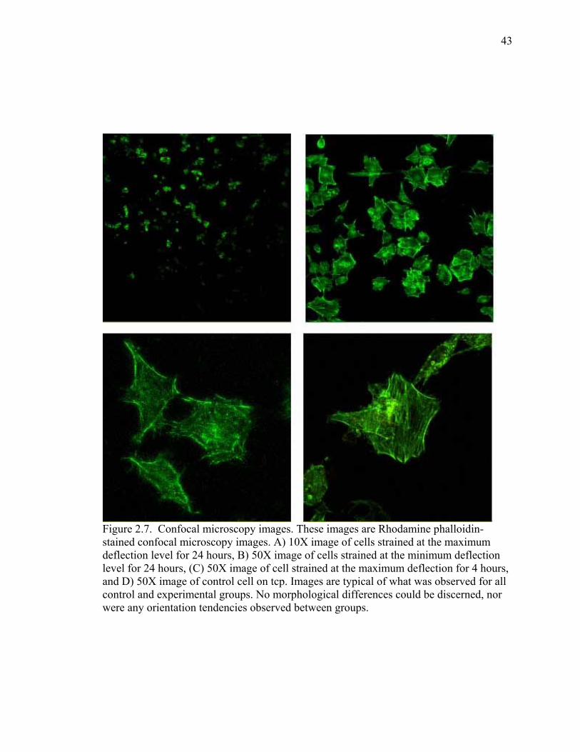
43
Figure 2.7. Confocal microscopy images. These images are Rhodamine phalloidin-stained confocal microscopy images. A) 10X image of cells strained at the maximum deflection level for 24 hours, B) 50X image of cells strained at the minimum deflection level for 24 hours, (C) 50X image of cell strained at the maximum deflection for 4 hours, and D) 50X image of control cell on tcp. Images are typical of what was observed for all control and experimental groups. No morphological differences could be discerned, nor were any orientation tendencies observed between groups.

44
Figure 2.8. Neutral Red results. No significant differences were observed in cell viability between strained cells or controls at p<0.05. n=9 for each group.

CHAPTER III
STRAIN-MEDIATED RESPONSES OF CELLS CULTURED
ON IMPLANT QUALITY TITANIUM
Introduction
Since the 19th century (Wolff, 1892), issues of load-induced bone modeling and
remodeling have been investigated. The importance of this phenomenon has become
critical given that more than $2 billion are spent yearly in this country on hip and knee
implants, plates and pins for broken bones, dental implants, and other reconstructions
(Berkeley, 2000). An estimated 11 million persons in the United States have at least one
such medical implant, and this number is growing as the population receiving implants
increases (Berkeley, 2000). For instance, according to the World Health Organization,
the number of hip fractures worldwide could rise from 1.7 million in 1990 to 6.3 million
by 2050. The average yearly number of dental implants surgically placed by dentists
nearly tripled from 17.7 million in 1986 to 51.5 million implants in 1997 according to a
random sample survey conducted by the American Dental Association.
Overloading at the implant/tissue interface is often the cause of implant failure.
Soballe et al. (1992 and 1993) compared cylinders coated with a porous titanium plasma-
sprayed layer implanted in canine femurs in a controlled micromotion study in dog
femurs. Micromotion of 150 microns at the interface was induced in test implants and
45

46
led to fibrous tissue interposition, while stable implants, controls, were well anchored in
bone. When implants were further immobilized for 12 weeks after 4 weeks of
mobilization, the low differentiated fibrous tissue was replaced by direct bone apposition,
confirming that micromotion of implants leads to fibrous tissue encapsulation instead of
the osseointegration. Similarly, Brunski (1991) inserted titanium implants bilaterally
between two natural teeth in the mandible of eight dogs. A blade was connected to the
adjacent natural teeth in a bridge and put immediately into function for 2 months to one
year. Contralateral blades were kept out of function and shielded from direct loading.
The immediately loaded implants displayed a thick fibrous tissue encapsulation that led
to implant failure while the nonloaded implants osseointegrated successfully. Brunski
demonstrated that premature loading led to fibrous tissue encapsulation of the implant
which resulted in implant failure. Thus, a better understanding of stain-induced
responses of bone/implant interactions may lead to improvements in the long term
success of implants. Prevention and treatment of healthy and unhealthy bone may also be
improved.
Bone adaptation to its mechanical environment is dependent on local bone cells’
modeling and remodeling responses. These responses to mechanical strain have been
shown in numerous studies (Brighton et al., 1991; Brighton et al., 1992; Duncan and
Turner, 1995; Glantschnig et al., 1996; Hasegawa et al, 1985; Neidlinger-Wilke, 1994;
Owan et al., 1997; Roelofsen et al., 1995). In general, these studies indicate that
responses of bone cells loaded within certain magnitudes (≤ 1000 µstrain) may result in
increased proliferation, yielding, increased matrix production, and increased matrix
synthesis.

47
Brighton et al. (1991) applied cyclic biaxial mechanical strain to bone cells from
the calvaria of newborn rats cultured on polyurethane. Their findings indicated that bone
cells subjected to mechanical perturbations of 400 µstrain for 48 hours at 1 Hz exhibited
decreased synthesis of macromolecules such as collagen and proteoglycan, decreased
alkaline phosphatase activity, and increased cell proliferation as compared to unstrained
controls. A significant finding of this study was that prostaglandin E2 content was
significantly increased after 5, 15, and 30 minutes of mechanical stimulation, whereas net
cyclic AMP (cAMP) content did not change significantly. These results suggested that
cellular events such as increased proliferation and decreased macromolecular synthesis
that occurred secondary to mechanical strain are mediated, in part, by prostaglandin E2.
In a follow-up study, Brighton et al. (1992) investigated the role of the inositol
phosphate pathway as a mechanism for the transduction of mechanical strain for inducing
cellular proliferation. Biaxial mechanical strain (1,700 µstrain) was applied at 1 Hz for
periods from 20 seconds to 60 minutes to newborn rat calvarial bone cells grown on
polyurethane. Results showed a 16% increase in DNA synthesis as compared to controls.
Also, activation of the inositol phosphate pathway occurred only 20 seconds after the
onset of mechanical strain as opposed to previous studies’ reports of 30 minutes.
Brighton et al. concluded that the inositol pathway was activated in bone cells by
mechanical strain and therefore may be the primary signalling mechanism for
mechanically inducing cell proliferation.
Kaspar et al. strained human-cancellous bone-derived osteoblasts cultured on
silicone at 1000 µstrain for 30 minutes per day for two days at a frequency of 1 Hz. Their
data revealed an increased proliferation (10-48%) of cells and release of carboxyterminal

48
collagen type I propeptide (7-49%). However, cellular alkaline phosphatase activity and
osteocalcin release were significantly reduced by 9-25 and 5-32%, respectively.
Labat et al. (2000) investigated human osteoblasts’ responses to mechanical strain
while growing on bioceramics common in current dental and orthopaedic materials.
They applied 400 µstrain over 6 hours at a frequency of 0.5 Hz to osteoblasts growing on
disks coated with alumina, hydroxyapatite (HA), or a duplex system composed of
hydroxyapatite-covered alumina. The effects of strains on short-term cell viability, cell
growth, alkaline phosphatase (ALP) activity, and collagen biosynthesis were assessed.
They noticed that mechanical strains resulted in a decrease in DNA corresponding to cell
number. ALP activity was unchanged for cells strained on alumina and decreased for
cells grown on HA and the duplex material. Collagen and total protein synthesis was
decreased for cells grown on HA and the duplex material and increased for cells grown
on alumina. Thus, their results indicated that mechanical loading affects bone cell growth
and matrix development and these effects are further modified based on the substrate on
which the cells were grown.
While these studies confirmed that osteoblast-like cells are affected by local
mechanical conditions and implant surfaces; it remains unclear what mechanical
conditions stimulate bone cells to develop an osseous interface with actual dental and
orthopaedic metallic implants. Given that, worldwide, there are an estimated 850,000 hip
- 570,000 knee (Lemons et al., 1997)- and 2.5 million dental implants (American Dental
Association) placed every year, including revisions, the importance of understanding the
strain-mediated response of cells cultured on implant alloys is clear. To better understand
tissue-implant interactions under in vivo conditions, it is necessary to investigate bone-

49
like cells’ responses to mechanical loading while growing on actual implant materials in
vitro. Therefore, the objective of this study was to investigate responses of bone-like
cells cultured on an orthopaedic material under mechanical loading conditions.
Materials and Methods
Cell Strain Plate
The cell substrate consists of a medical-grade-2 titanium alloy plate (38.1cm x
15.2 cm x 1 mm) (Fig. 2.2). This alloy was chosen as the cell substrate because it is
readily available in sheet form and is a common alloy used in implant devices.
Cells were cultured on the titanium substrate using a special ring system to
contain cells and growth media. First, the titanium plate was wet ground to a smooth
surface with 1500 grit SiC paper, degreased in acetone, and thoroughly rinsed with
distilled water. Three individual polystyrene wells were then attached to the plate using a
silicone adhesive (100% Silicone Sealant, DAP Inc., Baltimore, MD) to create three
separate culture wells, each with a 8.9 cm2 growth area on the titanium. The polystyrene
wells were simply tissue culture petri dishes from which the bottoms had been removed.
The adhesive was allowed to cure at ambient conditions for a minimum of 24 hours. Prior
to cell culture experiments, the plate was placed into a laminar flow cell culture hood,
rinsed with 70% ethanol, and exposed to UV light for a minimum of twenty-four hours to
sterilize the cell wells. A second titanium plate was prepared in the exact same manner to
be used as non-mechanically strained controls.

50
Cell Culturing System
For our study, rat osteogenic sarcoma UMR-106 cells were used because they
were relatively inexpensive, readily available, and bone-derived. They were maintained
in Dulbeco’s Modified Eagles’s Medium (DMEM; Gibco BRL, Grand Island, NY) with 4
mM L-glutamine adjusted to contain 1.5 g/L sodium bicarbonate, 4.5 g/L glucose and 1.0
mM sodium pyruvate and supplemented to contain 10% fetal bovine serum and 1%
antibiotics-antimycotics. Additionally, 50 mM ascorbic acid, 10 mM Na-β-
glycerophosphate was added to the culture media to induce mineralization (Chak et
al.,1995). Cells were seeded into the wells of the strain plate device at 105 cells/cm2, and
the unit was placed in an incubator. In the same incubator, control experiments were
performed on an unstrained titanium plate identically prepared. As additional controls,
cells were seeded in conventional tissue culture plastic petri dishes with the same growth
area as used for the Ti plates. Experiments and controls were run in triplicate. Controls
were seeded at the same level, 105 cells/cm2.
Cell Strain Design
The cells were allowed to attach and grow on the Ti and control wells for 72
hours before applying strain to the plate. The test Ti plate with cells was attached via pins
to the electric motor-cam system of the strain plate device. The cells were then subjected
to 182 ± 3 µstrain, 366 ± 9 µstrain, or 984 ± 7 µstrain at 1.5 Hz for 4 hours and 20 hours
rest or 24 hours with no rest.

51
Cell Activity
Total Protein Production
Total protein production was used as an indirect method of determining cell
proliferation (Bradford, 1976). First, old media was removed. Cells were then rinsed
with sterile Ca-Mg free PBS. Next, 1ml of 0.3 M NaOH in dH2O to was added to each
well. Cells were then incubated for 30 minutes. In a 96-well plate, 10 µl stock/cell
solution was combined with 200 µl Bradford’s solution and incubated for 30 minutes at
room temperature. Absorbance was read at 595 nm using a µQuant Universal Microplate
Spectrophotometer (Bio-Tek Instruments, Inc., Winooski, VT). Absorbance values were
interpolated using the standard curve and converted. Results were expressed in ng / ml.
Alkaline Phosphatase Activity
Alkaline phosphatase (ALP) was used as a general indicator of osteoblast cell
activity. ALP activity was measured using a quantitative, colorimetric assay kit (Sigma
Diagnostics). Once the cells completed the appropriate strain regimen, media was
removed, cells were rinsed once with sterile Ca+-Mg2+ free PBS, and 1 ml of lysing
solution (20 mM Tris, 0.1% Triton X100, 1mM MgCl2, and 0.1 mM ZnCl2) was added to
each well for 30 minutes. Next, to a 96-well plate, 100 µl of sample and 100 µl of
substrate solution (5mM paranitrophenylphosphate: Sigma) were added. Following 15
minutes of incubation, the reaction was stopped by addition of 0.3M NaOH, and the plate
was read at 410 nm in a 96-well plate reader (Bio-Tek, USA). The intensity of color
generated was proportional to alkaline phosphatase activity and was converted to Sigma
units using a standard curve. One Sigma unit is equivalent to one µmol of p-nitrophenol
per minute. ALP values were then normalized to total protein measurements.

52
Calcium Deposition Alizarin red-S (AR-S) is a dye that binds selectively to calcium salts and is widely
used for calcium mineral histochemistry. At the end of each strain regimen, cultures
were briefly rinsed with PBS followed by fixation (ice cold 70% ethanol, 1 h). Cultures
were rinsed with dH2O and stained for 10 min with 40 mM AR-S (Keller et al, 1997).
Cultures were then rinsed with dH2O followed by a 15 min wash with PBS to reduce non-
specific AR-S stain. Extracts of these AR-S/cell solutions were aliquoted into 96-well
plates, and absorbance was read at 562 nm using a µQuant Universal Microplate
Spectrophotometer (Bio-Tek Instruments, Inc., Winooski, VT).
Statistics
Each experiment was performed independently for a minimum of three trials. A
2-way ANOVA was used to determine statistical differences of cellular responses
between controls and groups strained at the three different magnitudes for 4 or 24 hours.
Tukeys’s test was used to determine where post-ANOVA differences might exist, and
differences were considered statistically significant at p<0.05.
Results
Total Protein
Statistical analyses revealed a significant interaction between strain magnitude
and strain duration on the total amount of protein in the cultures (Figure 3.1 and Table
3.1). Cell cultures strained at the 984±7 µstrain for 24 hours exhibited the most protein
content (295 ± 34 ng/ml), whereas cultures strained at 182±2 µstrain for 4 hours exhibited
the least amount of protein (195 ± 18.3 ng/ml).

53
Results from the 2-way ANOVA (p<0.05) indicated a significant increase (9%) in
total protein content for cells strained for 24 hours (252 ng/ml) vs. 4 hours (226 ng/ml).
Results also showed that cells strained at the maximum deflection level (984±7 µstrain)
exhibited 21% and 24% more protein than cells strained at the minimum (182±2 µstrain)
and medium (366 ± 9 µstrain) deflection levels, respectively (Figure 3.1 and Table 3.1).
Cells strained at the maximum deflection level (984±7 µstrain) also exhibited 16% (vs.
tcp) and 20% (vs. Ti) more protein than unstrained controls (Figure 3.1 and Table 3.1).
Alkaline Phosphatase
For ALP activity of the cells, there was also an interaction between strain
magnitude and strain duration (Figure 3.2 and Table 3.2). Cells strained at 984±7 µstrain
for 24 hours exhibited the least amount of ALP activity (0.27 ± 0.15 µmol ml hr-1 ng-1),
whereas cells strained at 366 ± 9 µstrain for 24 hours exhibited the greatest ALP activity
(1.38 ± 0.19 µmol ml hr-1 ng-1).
Results from the 2-way ANOVA (p < 0.05) indicated a significant increase (20%)
in ALP activity for cells strained for 4 hours (1.14 µmol/hr/ng/ml) vs. 24 hours (0.9 µmol
ml hr-1 ng-1). Results also showed that cells strained at the maximum deflection level
(984 ± 7 µstrain) exhibited 45% and 49% less ALP activity than cells strained at the
minimum (182 ± 2 µstrain) and medium (366 ± 9 µstrain) deflection levels, respectively.
No significant difference was observed between cells strained at the minimum (182 ± 2
µstrain) and medium (366 ± 9 µstrain) and deflection levels. Cells strained at the
maximum deflection level (984 ± 7 µstrain) also exhibited 37% (tcp) and 42% (Ti) less
ALP activity than unstrained controls (Figure 3.2 and Table 3.2).

54
Calcium
No significant differences were observed in calcium absorbencies between
experimental cells and/or control cells (Figure 3.3).
Discussion
This study presented findings on osteoblast-like cells’ responses to a well-
quantified strain environment while growing on an actual orthopaedic/dental implant
material, titanium. Results of this study demonstrated that the magnitude and duration of
applied mechanical strain affected cellular ALP activity and total protein content.
Related studies have reported a wide range of results on the response of
osteoblastic cells to mechanically applied strain. (Brighton et al., 1991; Brighton et al.,
1992, Hasegawa et al., 1985; Neidlinger-Wilke et al., 1994). Significant increases in
DNA content, as an indicator of cell proliferation, and a decrease in protein synthesis, as
well as activity of ALP were reported for rat calvarial cells subjected to 400 µstrain at 1
Hz for periods ranging from 15 minutes to 72 hours but no significant effects at 1000
µstrain (Brighton et al., 1991). On the other hand, Neidlinger-Wilke et al (1994)
observed significant increases (16.4-100%) in the proliferation of human osteoblasts
cultured on silicone substrates subjected to 1000 µstrain at 1 Hz applied for 15 minutes
per day on 3 consecutive days as compared to cells strained at lower levels and
unstrained controls. ALP activity showed no significant changes in response to the strain
regimen. Kaspar et al. (2000) reported increased cell proliferation and collagen
production but decreased ALP activity of human bone cells on silicone dishes in response
to 1000 µstrain (30 min at 1 Hz each day for 2 days) as compared to unstrained controls.
However, Labat et al. (2000) observed decreased proliferation of cells cultured on

55
bioceramics subjected to only 400 µstrain vs. unstrained controls and that changes in
ALP activity and protein production were dependent on both the bioceramic substrate and
strain applied. While direct comparisons between these studies is difficult due to
differences in cell types, mechanical conditions and substrates used, they do strongly
suggest that there may be some critical measure of mechanical strain important to bone
cell proliferation and matrix development. Indeed, Brighton et al. (1992) showed that for
even very short applications of 1,700 µstrain mechanical strain, there was an increase in
intracellular signaling for cellular proliferation. The results of this study are in general
agreement with this hypothesis in that increases in total protein content of the cultures, as
a general indicator of cell number, were observed for cells subjected to 987±7µstrain as
compared to lower strain levels. An increase in cell numbers may be beneficial to the in-
vivo healing response at the bone/implant interface. Simply speaking, more cells may
yield greater quantities of cells mineralizing, which would yield increased mineralization
(and osseointegration) at the bone/implant interface.
ALP belongs to a class of cell surface enzymes which are covalently bound to
phosphatidylinositol (PI) phospholipid complexes in the plasma membrane and function
to dephosphorylate matrix proteins and regulate extracellular phosphate levels for matrix
mineralization (Davies, 2000). Increases in ALP activity, in-vivo, are observed as
mineralization commences, but when ALP activities are significantly reduced,
mineralization is impaired (Davies, 2000). In this study, increased ALP activity in cells
strained at 182 ± 3 µstrain and 366 ± 9 µstrain strongly suggests that the cells were being
induced to mineralize. Therefore, it may be inferred that strain magnitudes from

56
approximately 182-366 µstrain may “jump start” the onset of mineralization in this
osteoblast-like cell line.
ALP activity was suppressed by straining cells at the maximum deflection level
(984 ± 7 µstrain) for 24 hours vs. controls. Decreased ALP activity, in-vitro, is a common
and well-documented response of cells to mechanical strain in the range of 200-1000
µstrain depending on time of strain application, cell type and substrate (Stanford, 1995;
Labat et al, 2000; Kaspar et al., 2000; Peverali et al, 2001). This decrease in ALP
activity is usually coincident with increased cellular proliferation. From these data it may
be speculated that there may be mechanical strain environments that promote cell
proliferation but not extracellular matrix production and conversely, matrix production
but not cell proliferation. What is not clear from these studies is if increased cell
proliferation would eventually result in increased production of extracellular matrix and
subsequently improved tissue mineralization or if tissue mineralization is better than cell
proliferation.
Clinically, an early onset of mineralization at the bone/implant interface would be
beneficial to patients. Increased mineralization would give greater strength to the bone at
a faster rate, or in other words, early mineralization might increase the healing response
by decreasing the healing time.
Although the UMR-106 cells were cultured with ascorbic acid and β-
glycerophosphate to initiate mineralization, culture periods were probably too short for
cells to develop sufficient mineralized matrix. Hence no differences were observed in this
study regarding the deposition of Ca-P mineral. Primary osteoblast-like cells may have

57
provided more suitable results since they mineralize more “naturally”, in response to
proper signals and chemicals in-vivo.
The strategy of this experimental design is of great value since it provides the
means to measure and assess biomechanical interactions between cells and metallic
implant materials on a local level that can not be determined by other means. By
improving the understanding of osteoblast responses triggered by mechanical stimuli,
positive steps in understanding bone remodeling, healing and integration of implant
devices may be taken. Although direct clinical applications of stretch-mediated
responses of bone cells in-vitro are difficult to determine, models such as the device used
in this study have great merit. For instance, the consensus from the 1970’s was that
following all dental implant procedures, a period of three months was necessary to
prevent implant anchorage into fibrous tissue rather than bone (Szmukler-Moncler et al.,
1998). It has been shown that only excessive macro movements detrimentally affect the
successful integration of the implant/bone interface and that loading the implant within a
certain range (<150 microns ) may actually promote osseointegration. Additionally,
some studies have shown that loading bone/implant interfaces in-vivo may be applied
after only one week of implantation, which is obviously of great benefit to patients
(Soballe et al, 1992 and 1993).
Results of this study support a general stimulatory effect of the mechanical
loading regimens to the cells. Increases in ALP activity in cells strained at 182 ± 3 and
366 ± 9 µstrain suggest an enhancement of tissue mineralization while an increases in
total protein content in cells strained at 984 ± 7 µstrain indicate an increase in cell
proliferation which may lead to increased tissue matrix.

58
Conclusion
A novel cell culture strain plate device was successful in applying a quantified
strain environment to bone-like cells while growing on a metallic implant alloy, titanium.
Cells remained viable throughout experimental testing, and quantifiable measurements in
response to mechanical strain were made. Differences in proliferative and
mineralization-related activities of osteoblast-like cells attached to titanium were detected
in response to the applied mechanical strain Thus, the cell culture strain plate device was
successful in applying a quantifiable strain to cells attached and growing on implant alloy
surfaces.

59
0
50
100
150
200
250
300
350
tcp Ti min 24 min 4 med 24 med 4 max 24 max 4
Strain Regimen
ng /
ml
Figure 3.1. Total protein results. This graph represents the mean total protein production of strained cells and controls.

60
0.00
0.40
0.80
1.20
1.60
tcp Ti min 24 min 4 med 24 med 4 max 24 max 4
Strain Regimen
µmol
ml h
r-1 n
g -1
Figure 3.2. ALP activity results. This graph represents the mean ALP activity of strained cells and controls.

61
0.00
1.00
2.00
3.00
4.00
tcp Ti min 4 min 24 max 24 max 4 med 24 med 4
Strain Regimen
Abs
orba
nce
Figure 3.3. Calcium results. This graph represents the mean calcium absorbencies of strained cells and controls. No significant differences were observed at p<0.05.

62
strain time
substrate (hr) (ng/ml)
tcp 0 237±24 tcp
Ti 0 226±22 ------ Ti
Ti max 24 295±34 20% 23% max 24
Ti max 4 269±35 ------ 12% ------
max 4
Ti med 24 209±21 ------ ------ 29% 22%
med 24
Ti med 4 216±16 ------ ------ 27% 20% ------- med 4
Ti min 24 250±18 ------ ------ 15% ------ 16% ------- min 24
Ti min 4 195±18 18% ------ 24% 28% ------- ------- 22% min
4 Table 3.1. Percent differences in total protein content (ng/ml, average ± std dev, n=9) between strained and nonstrained cell groups according to Tukey’s test following 2-way Anova at p<0.05. Significant differences between groups are indicated by the percentage difference, and dashed lines indicate no differences between groups. UMR 106 osteoblastic cells subjected to 984±7 µstrain at 1.5 Hz for 24 hours exhibited the highest amount of protein of the test and control cultures and 20 – 23% more than non-strained controls. tcp = tissue culture plastic Petri dishes, Ti = titanium plate, min = 182±3 µstrain, med = 366±9 µstrain, and max = 984±7 µstrain, 0 = controls not subjected to any strain, 4 = cells strained cyclically at 1.5 Hz for 4 hours followed by 20 hours no strain, 24 = cells strained cyclically at 1.5 Hz for 24 hours continuously.

63
strain time
substrate (hr) (µmol ml hr-1 ng-1)
tcp 0 1.01±0.17
tcp
Ti 0 1.10±0.18 ------
Ti
Ti max 24 0.27±0.15 73% 75% max 24
Ti max 4 1.01±0.19 ------ ------ 73% max
4
Ti med 24 1.38±0.19 27% 22% 80% 27% med 24
Ti med 4 1.13±0.13 ------ ------ 76% ------ 18% med 4
Ti min 24 1.06±0.11 ------ ------ 75% ------ 23% --------- min 24
Ti min 4 1.27±0.18 20% ------ 79% 20% --------- --------- --------- min
4
Table 3.2. Percent differences in ALP activity (µmol ml hr-1 ng-1, average ± std dev, n=9) between strained and non strained cell groups according to Tukey’s test following 2-way Anova at p<0.05. Significant differences between groups are indicated by the percentage difference, and dashed lines indicate no differences between groups. UMR 106 osteoblastic cells subjected to 984±7 µstrain at 1.5 Hz for 24 hours exhibited the lowest amount of ALP activity of the test and control cultures. UMR 106 osteoblastic cells subjected to 366±9 µstrain at 1.5 Hz for 24 hours exhibited the highest ALP activity of the test and control cultures and 22-27% more than non-strained controls. tcp = tissue culture plastic Petri dishes, Ti = titanium plate, min = 182±3 µstrain, med = 366±9 µstrain, and max = 984±7 µstrain, 0 = controls not subjected to any strain, 4 = cells strained cyclically at 1.5 Hz for 4 hours followed by 20 hours no strain, 24 = cells strained cyclically at 1.5 Hz for 24 hours continuously.

CHAPTER IV
CONCLUSION
The role of mechanical strain on bone cells at the tissue-implant interface and
subsequent osseointegration has not been well established aside from overload
conditions. In vitro studies provide the means to explore and evaluate the effects of
mechanical strain on cells not possible by other means. To evaluate the effects of
mechanical strain on cells, a cell culture strain plate system was developed to apply strain
to cells growing on typical implant alloy surfaces. The objectives of this research were to
use the cell culture strain plate to evaluate hypotheses that 1) osteoblasts would attach
and grow on the titanium substrate without experiencing significant changes in viability
by changes in the magnitude and duration of strain generated at the cell-substrate
interface, 2) Increased strain magnitudes would induce perpendicular alignment of
attached cells to the axial line of strain, and 3) Biochemical processes of the cells,
specifically, alkaline phosphatase activity, calcium deposition, and total protein
production would increase at increased strain magnitudes.
This study evaluated a new cell culture device that accurately delivers
reproducible, known, cyclic strains to cultured cells on a metal substrate and has the
potential to detect differences in how cells respond to different mechanical conditions
that may exist adjacent to implant alloys. Strain gage analyses clearly demonstrated
64

65
negligible discrepancies in strain between cell culture growth wells, from test to test,
and over time. The long and the transverse axes of the plate corresponded to the
principal axes and that the cells experienced a predominate strain along the long axis
of the plate which was an order of magnitude larger than the compressive strain in the
transverse direction. Variability in the strain data may be accounted for by slight
deviations of the pins connecting the substrate to the cams, subtle variations in
thickness of the plate and height of arched bars, plate positioning on the arched
supports, and very slight differences in the axial alignment of strain gages.
Additionally, over time the plate may develop some permanent deformation due to
repetitive loading even though strain values are well under the 0.2% elastic limit.
Therefore, it is necessary to maintain an individual strain gage mounted and centered
on the bottom of the plate to monitor the strain being applied to the cells during each
test. Nevertheless, analysis of the data demonstrated that strain magnitudes for each
strain level within the areas of cell culture were consistent and repeatable. Hence, this
device was successful in generating known cyclic strain in a consistent and repeatable
manner.
The frequency of strain application was also controlled without affecting strain
magnitudes. Although frequency can be adjusted within a wide range (0.5 to > 10
Hz) and monitored, an electric motor with an increased torque may be better for
controlling frequencies at less than 1 Hz. However, for this experiment, frequency
within a range of 1.5 – 2 Hz was acceptable.
The device fit into a cell culture incubator and was able to support cell culture
growth. The device may be easily adapted to allow different types of metallic
substrates to be used by simply substituting different alloy substrates for the Ti

66
substrate as used in this study. This device would then allow for the in vitro
evaluation of the effects of strain on cells growing on different implant alloys.
While we consider this device as an appropriate in vitro simulator of
mechanical strain to cells growing on implant materials, it is understood that there are
many complex in vivo conditions that are not represented, ie. fluid shear stress,
adjacent cell attachments, random magnitude and frequency changes, etc. However,
in order to better understand in vivo conditions, it is important to perform in vitro
studies on mechanical regulation of cellular responses and processes to different strain
levels. Controlling magnitude, frequency, and duration, as well as utilizing typical
implant substrates provides a beneficial means of investigating possible mechanisms
and regulatory responses of cell/implant interactions in vivo.
Osteoblast-like cells’ responses to a well-quantified strain environment while
growing on an actual orthopaedic/dental implant material, titanium, were evaluated.
Qualitative and quantitative analyses showed high viability for cells cultured on
titanium and strained between 200 and 1000 µstrain for 4 or 24 hours vs. unstrained
controls. The results of this study indicated that mechanical strain applied to cells
cultured on titanium stimulated cellular proliferation, based on total protein assays, at
higher strain magnitudes (984 ± 7 µstrain), but not at the smaller strain magnitudes
evaluated (182 ± 3 µstrain or 366 ± 9 µstrain). ALP activity was significantly
different among groups of strained cells. Cells strained at 366 ± 9 µstrain for 24 hours
demonstrated the most ALP activity, whereas cells strained at 984 ± 7 µstrain for 24
hours showed the least amount of ALP activity. Both increased cell proliferation and
decreased ALP activity, in-vitro, as reported in this study are consistent with previous
reports on the response of cells to mechanical strain in the range of 200-1000 µstrain
(Stanford, 1995; Labat et al, 2000; Kaspar et al., 2000; Peverali et al, 2001).

67
However, due to differences in application of strain, cells, and cell substrate, these
changes in cell behavior can not be correlated to specific strain conditions. It does
appear though that mechanical strain affects cellular processes such as, proliferative
and matrix-related activities and that these activities may be differentially affected.
Increases in ALP activity, in-vivo, are observed as mineralization commences,
but when ALP activities are inhibited, mineralization is impaired. Therefore, it may
be inferred that the increased ALP activity in cells strained at 182 ± 3 µstrain and 366
± 9 µstrain may induced cells to develop mineralized extracellular matrix whereas at
increased strains, mineralization may be inhibited. Improvements in mineralization of
matrix next to an implant would improve osseointegration.
However, at the higher application of strain, (984 ± 7 µstrain) an increase in
total protein content, as an indicator of cell proliferation, was observed in cultures.
Increased cell proliferation may be beneficial to the in-vivo healing response at the
bone/implant interface. Simply speaking, more cells yield greater quantities of cells
mineralizing, which may yield increased mineralization (and osseointegration) at the
bone/implant interface.
Calcium deposition was non-significant between experimental and control
groups, which may have been due to the assays being performed too soon following
the strain regimen. Longer culture periods following straining may be required to
give the cells enough time to deposit calcium.
The strategy of this experimental design is of great value since it provides the
means to measure and assess biomechanical interactions between cells and metallic
implant materials on a local level that can not be determined by other means. By
improving the understanding of osteoblast responses triggered by mechanical stimuli,
positive steps in understanding bone remodeling, healing and integration of implant

68
devices may be taken. Although direct clinical applications of stretch-mediated
responses of bone cells in-vitro are difficult to determine, models such as the device
in this study have great merit.
The novel cell culture strain plate device was successful in applying a
quantified strain environment to bone-like cells while growing on a metallic implant
alloy, titanium. Cells remained viable throughout experimental testing, and
quantifiable measurements in response to mechanical strain were made. Proliferative
and mineralization-related activities of osteoblast-like cells attached to titanium were
altered in response to mechanical strain. Thus, the device proved successful in
applying a quantifiable strain environment for which cellular process may be
evaluated while the cells are attached to implant alloy surfaces.

REFERENCES
Albrektsson T, Zarb GA. Osseointegration-A Requiem for the Periodontal Ligament? An Editorial. Int & Periodont Restorative Dent 1991; 11:88-91. Anderson KL, Norton LA. A device for the application of known simulated orthodontic forces to human cells in vitro. J Biomech 1991; 24(7):649-54. Banes AJ, Gilbert J, Monbureau O, Taylor D. A new vacuum-operated stress-providing instrument that applies static or variable duration cyclic tension or compression to cells in vitro. J Cell Sci 1985; 75:35-42. Barbier L, Schepers E. Adaptive bone remodelling around oral implants under axial and nonaxial loading conditions in the dog mandible. Int J Oral Maxillofac Implants 1997; 12(2):215-23. Berkeley Lab Research Review. Winter 2000. Bioactive Glass Coating Improves Metal Bone Implants by: Lynn Yarris. Bradford MM. A rapid and sensitive method for the quantitation of microgram quantities of protein utilizing the principle of protein-dye binding. Anal Biochem. 1976 May; 72:248-54. Brighton CT, Gross SB, Leatherwood DF, Pollack SR, Strafford B, Williams JL. The proliferative and synthetic response of isolated calvarial bone cells of rats to cyclic biaxial mechanical strain. J Bone Joint Surg Am 1991; 73(3):320-31. Brighton CT, Sennett BJ, Farmer JC, Iannotti JP, Hansen CA, Williams JL, Williamson J. The inositol phosphate pathway as a mediator in the proliferative response of rat calvarial bone cells to cyclical biaxial mechanical strain. J Orthop Res 1992; 10(3):385-93. Brown TD. Techniques for mechanical stimulation of cells in vitro: a review. J Biomech 2000; 33(1):3-14. Brunski JB. Avoid pitfalls of overloading and micromotion of intraosseous implants. Dent Implantol 1993, update; 4:1-5. Brunski JB, Moccia AF Jr., Pollock SR, Korostoff E, Trachtenberg DI. The influence of function use of endosseous dental implants on the tissue implant interface: I. Histological Aspects. J Dent Res 1979; 58:1953-69.
69
70 Bumgardner JD, Lucas LC. Cellular response to metallic ions released from nickel chromium dental alloys. J Dent Res 1995; 74(8):1521-7. Buckley MJ, Banes AJ, Levin LG, Sumpio BE, Sato M, Jordan R, Gilbert J, Link GW, Tran Son Tay R. Osteoblasts increase their rate of division and align in response to cyclic, mechanical tension in vitro. Bone Miner 1988; 4(3):225-36. Cameron H, Pilliar RM, Macnab I. Porous surfaced vitallium staples. S Afr J Surf 1972; 10:63-70. Cameron H, Pilliar RM, Macnab I. The effect of movement on the bonding of porous metal to bone. J Biomed Mat Res 1973; 7:301-11. Chak CW, Lee KM, Leung KS, Fung KP. No change in bone-specific alkaline phosphatase activities in cultured rat osteoblastic cells under L-ascorbate and beta-glycerophosphate-induced mineralization. Cell Biol Int 1995; (12):979-85. Cohen IK, Diegelmann RF, Lindblad WJ. 1992. Wound Healing: Biochemical & Clinical Aspects. W.B. Saunders Co., Philadelphia, PA. Davies JE, ed. 2000. Bone Engineering. em squared, Toronto, Canada. Deporter DA, Watson PA, Pilliar RM, Chipman ML, Valiquette N. A histological comparison in the dog of porous-coated vs threaded dental implants. J Dent Res 1990; 69:1138-45. Duncan R, Turner C. Mechanotransduction and the functional response of bone to mechanical strain. Calcif Tissue Int 1995; 57:344¯58. Dunn MG, Maxian SH. 1994. Biomaterials used in orthopedic surgery. In: Greco RS, Implantation Biology: The Host Response and Biomedical Devices. CRC Press, Boca Raton, FL, pp. 229-42. Glantschnig H, Varga F, Rumpler M, Klaushofer K. Prostacyclin (PGI2): A potential mediator of c-fos expression induced by hydrostatic pressure in osteoblastic cells. Eur J Clin Invest 1996; 26:544¯48. Hasegawa S, Sato S, Saito S, Suzuki Y, Brunette DM. Mechanical stretching increases the number of cultured bone cells synthesizing DNA and alters their pattern of protein synthesis. J Calcif Tissue 1985; 37:431-36.

71 Hatano K, Inoue H, Kojo T, Matsunaga T, Tsujisawa T, Uchiyama C, Uchida Y. Effect of surface roughness on proliferation and alkaline phosphatase expression of rat calvarial cells cultured on polystyrene. Bone 1999; 25(4):439-45. Hobkirk JA, Watson RM. 1995. Dental and Maxill-Facial Implantology. Mosby-Wolfe, Barcelona, Spain. Jordan R, Buckley M, Hicks H, Transontay R, Banes AJ. Morphologic changes in osteoblast-like cells subjected to cells to cyclic tension. Ins. Assoc. Dent. Res 1988 ; (67:234). Kaspar D, Seidl W, Neidlinger-Wilke C, Ignatius A, Claes L. Dynamic cell stretching increases human osteoblast proliferation and CICP synthesis but decreases osteocalcin synthesis and alkaline phosphatase activity. J Biomech 2000; (33):45-51. Keller JC, Collins JG, Niederauer GG, McGee TD. In vitro attachment of osteoblast-like cells to osteoceramic materials. Dent Mater. 1997; 13(1):62-8. Kudelska-Mazur D, Lewandowska-Szumiel M, Komender J. Human osteoblast in contact with various biomaterials in vitro. Ann Transplant 1999; 4(3-4):98-100. Labat B, Chepda T, Frey J, Rieu J, Aurelle JL, Douet M, Alexandre C, Chamson A. Practice of a testing bench to study the effects of cyclic stretching on osteoblast-orthopaedic ceramic interactions. Biomat 2000; 21(12):1275-81. Lemons JE, Ratner BD, Scheon FJ. ed. 1997. Biomaterials Science. Harcourt Brace and Co. Leung DYM, Glagov S, Mathews MB. A new in vitro system for studying cell response to mechanical stimulation. Exp Cell Res 1977; 109:285-98. Marks Jr, SC, Popoff SN. Bone Cell Biology: The Regulation of Development, Structure, and Function in the Skeleton. Am J Anat 1988; 183:1-44. Martin RB, Burr DB. 1989. Structure, Function, and Adaptation of Compact Bone. Raven Press, NY, NY. McElfresh E, Petty W. 1991. History of Arthroplasty. Total Joint Replacement. W.B. Saunders Company, Philadelphia, PA, pp. 3-18. Meazzini MC, Toma CD, Schaffer JL, Gray ML, Gerstenfeld LC. Osteoblast cytoskeletal modulation in response to mechanical strain in vitro. J Orthop Res 1998; 16:170-80.

72 Mosley JR, Lanyon LE. Strain rate as a controlling influence on adaptive modeling in response to dynamic loading of the ulna in growing male rats. Bone 1998; 23(4):313-8. Murray DW, Rushton N. The effect of strain on bone cell prostaglandin E2 release: A new experimental method. Calcif Tissue Int 1990; 47:35¯9. Naji A, Harm MF. Cytocompatibility of two coating materials, amorphous alumina and silicon carbide, using human differentiated cell cultures. Biomat 1991; 12(7):690-4. Naji A, Harm MF. Study of the effect of the surface state on the cytocompatibility of a Co-Cr alloy using human osteoblasts and fibroblasts. J Biomed Mater Res 1990; (7):861-71. Neidlinger-Wilke C, Wilke HJ, Claes L. Cyclic stretching of human osteoblasts affects proliferation and metabolism: a new experimental method and its application. J Orthop Res 1994; (1):70-8. Neidlinger-Wilke C, Stalla I, Claes L, Brand R, Hoellen I, Rubenacker S, Arand M, Kinzl L. Human osteoblasts from younger normal and osteoporotic donors show differences in proliferation and TGFβ-release in response to cyclic strain. J Biomech 1995; 28:1411-8. O'Mahony A, Bowles Q, Woolsey G, Robinson SJ, Spencer P. Stress distribution in the single-unit osseointegrated dental implant: finite element analyses of axial and off-axial loading. Implant Dent 2000; 9(3):207-18. Owan I, Burr DB, Turner CH, Qiu J, Tu Y, Onyia JE, Duncan RL. Mechanotransduction in bone: Osteoblasts are more responsive to fluid forces than mechanical strain. Am J Physiol 1997; 273:C810¯5. Petty W. 1991. Total Hip Arthroplasty: Preoperative Evaluation. Total Joint Replacement. W.B. Saunders Company, Philadelphia, PA, pp. 3-18. Peverali FA, Basdra EK, Papavassiliou AG. Stretch-mediated activation of selective MAPK subtypes and potentiation of AP-1 binding in human osteoblastic cells. Mol Med 2001; (7):68-78. Piatelli A, Ruffieri A, Franchi M, Romasco N, Trisi PA. A histologic and histomophometric study of bone reactions to unloaded and loaded non-submerged single implants in monkeys : a pilot study. J Oral Implantol 1993; 19:314-20. Pilliar RM, Deporter D, Watson PA. Tissue-implant interface: micro-movements effects. In: Vincenzinzi P. Materials in Clinical Applications, Advances in Science and Technology, 12, Proceedings of the 8th CIMTEC World Ceramic Congress. Fanexa, Italy: Techna; 1995 569-79.

73 Puleo DA, Holleran LA, Doremus RH, Bizios R. Osteoblast responses to orthopedic implant materials in vitro. J Biomed Mater Res 1991; 25(6):711-23. Roelofsen J, Klein-Nulend J, Burger E. Mechanical stimulation by intermittent hydrostatic compression promotes bone-specific gene expression in vitro. J Biomech 1995; 28:1493¯1503. Rubin C, Gross T, Qin YX, Fritton S, Guilak F, Mcleod F. Differentiation of the bone-tissue remodeling response to axial and torsional loading in the turkey ulna. J Bone Joint Surg Am 1996; 78(10):1523-33. Rutherford DW. The In-vitro Cellular/Biomaterial Interface Strain Simulator. Senior Design Paper. Agricultural and Biological Engineering Department, Mississippi State Universtiy. 1998. Schmidt C, Ignatius AA, Claes LE. Proliferation and differentiation parameters of human osteoblasts on titanium and steel surfaces. J Biomed Mater Res 2001; 54(2):209-15. Schwartz Z, Lohmann CH, Oefinger J, Bonewald LF, Dean DD, Boyan BD. Implant surface characteristics modulate differentiation behavior of cells in the osteoblastic lineage. Adv Dent Res 1999; 13:38-48. Shin HY, Iverson RD, Blumenstock FA, Bizios R. 1994. Osteoblast responses to steady shear stress. In: Mow VC, Guilak F, Tran-Sun Tay R, Hochmuth RM. Cell Mechanics and Cellular Engineering. Springer-Verlag, NY, NY, Inc. 95-105. Soballe K, Hansen ES, Brockstedt-Rasmussen H, Bunger C. Tissue ingrowth into titanium and hydroxyapatite coated implnats during stable and unstable mechanical condtions. J Ortho Res 1992; 10:285-99. Soballe K, Hansen ES, Brockstedt-Rasmussen H, Bunger C. 1993. The effects of osteoporosis, bone deficiency, bone grafting and micromotion on fixation of porous-coated hydroxyapatie-coated implants. In: Geesink RGT, Manley MT. Hydoxyapatite Coatings in Orthopaedic Surfery. NY: Raven Press. 107-36. Sotoudeh M, Jalali S, Usami S, Shyy J, Chien S. A strain device imposing dynamic and uniform equi-biaxial strain to cultured cells. Ann Biomed Eng 1998; 26:181-9. Stanford CM, Brand RA. Toward an understanding of implant occlusion and strain adaptive modeling and remodeling. J Prosthet Dent 1999; 81:553-61.

74 Stanford CM, Morcuende JA, Brand RA. Proliferative and phenotypic responses of bone-like cells to mechanical deformation. J Orthop Res 1995; 13:664-70. Szmukler-Moncler S, Salama H, Reingewirtz Y, Dubruille JH. Timing of loading effect of micromotion on bone-dental implant interface: review of experimental literature. J Biomed Mater Res. 1998 43(2):192-203. Wainwright SA, Biggs WD, Currey JD, Gosline JM. 1976. Mechanical Design in Organisms. Princeton University Press, Princeton, NJ. Williams JL, Chen JH, Beloli DM. Strain fields on cell stressing devices employing clamped circular elastic diaphragms as substrates. J Biomech Eng 1991; 114:377-84. Winston FK, Macarak EJ, Gorfien SF, Thibault LE. A system to reproduce and quantify the biomechanical environment of the cell. J Appl Physiol 1989; 61:397-405. Wolff J. The law of bone remodeling. Maquet P, Furlong R, translators. Berlin: Springer-Verlag; 1986.