Embed Size (px)
Citation preview

Instructions for use
Title A Mathematical Study to Select Fractionation Regimen Based on Physical Dose Distribution and the Linear‒QuadraticModel
Author(s) Mizuta, Masahiro; Takao, Seishin; Date, Hiroyuki; Kishimoto, Naoki; Sutherland, Kenneth L.; Onimaru, Rikiya;Shirato, Hiroki
Citation International Journal of Radiation Oncology, Biology, Physics, 84(3): 829-833
Issue Date 2012-11
Doc URL http://hdl.handle.net/2115/51087
Type article (author version)
File Information Date_mathematical.pdf
Hokkaido University Collection of Scholarly and Academic Papers : HUSCAP

Journal name: International Journal of Radiation Oncology•Biology•Physics
Article type: Full length article
Title: A MATHEMATICAL STUDY TO SELECT FRACTIONATION REGIMEN BASED
ON PHYSICAL DOSE DISTRIBUTION AND THE LINEAR-QUADRATIC MODEL
Authors: Masahiro Mizuta, Ph.D.,* Seishin Takao, Ph.D.,
† Hiroyuki Date, Ph.D.,
‡ Naoki
Kishimoto, B.Sc.,§ Kenneth L. Sutherland, Ph.D.,
|| Rikiya Onimaru, M.D.,
¶ and Hiroki
Shirato, M.D.¶
*Laboratory of Advanced Data Science, Information Initiative Center,
†Faculty of Engineering,
‡Faculty of Health Sciences,
§Graduate School of Information Science and Technology,
||Department of Medical Physics, and
¶Department of Radiation Medicine, Graduate School of
Medicine, Hokkaido University
Corresponding Author: ‡Hiroyuki Date
Postal address: Kita-12, Nishi-5, Kita-ku, Sapporo 060-0812, JAPAN
Email address: [email protected]
Tel: +81-11-706-3423, Fax: +81-11-706-4916

Mizuta
2
Abstract
Purpose: Hypofractionated irradiation is often used in precise radiotherapy instead of
conventional multi-fractionated irradiation. We propose a novel mathematical method for
selecting a hypofractionated or multi-fractionated irradiation regime based on physical dose
distribution adding to biological consideration.
Methods and Materials: The linear quadratic (LQ) model was employed for the radiation
effects on tumor and normal tissues, especially OARs. Based on the assumption that the OAR
receives a fraction of the dose intended for the tumor, the minimization problem for the
damage effect on the OAR was treated under the constraint that the radiation effect on the
tumor is fixed.
Results: For an N-time fractionated irradiation regime, the constraint of tumor lethality was
described by an N-dimensional hypersphere. The total dose of the fractionated irradiations
was considered for minimizing the damage effect on the OAR under the hypersphere
condition. It was found that the advantage of hypofractionated or multi-fractionated
irradiation therapies depends on the magnitude of the ratio of parameters for the OAR and
the tumor in the LQ model and the ratio of the dose for the OAR and the tumor.
Conclusions: The present mathematical method shows that the multi-fractionated irradiation

Mizuta
3
with a constant dose is better if the ratio of for the OAR and the tumor is less than the
ratio of the dose for the OAR and the tumor, while hypofractionated irradiation is better
otherwise.
Key words: radiotherapy, radiobiology, dose fractionation, linear-quadratic model,
hypersphere
Mizuta
4
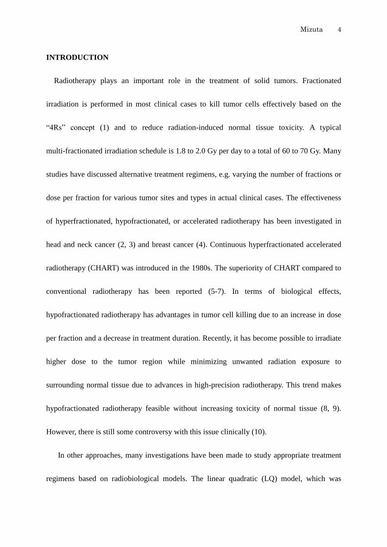
INTRODUCTION
Radiotherapy plays an important role in the treatment of solid tumors. Fractionated
irradiation is performed in most clinical cases to kill tumor cells effectively based on the
“4Rs” concept (1) and to reduce radiation-induced normal tissue toxicity. A typical
multi-fractionated irradiation schedule is 1.8 to 2.0 Gy per day to a total of 60 to 70 Gy. Many
studies have discussed alternative treatment regimens, e.g. varying the number of fractions or
dose per fraction for various tumor sites and types in actual clinical cases. The effectiveness
of hyperfractionated, hypofractionated, or accelerated radiotherapy has been investigated in
head and neck cancer (2, 3) and breast cancer (4). Continuous hyperfractionated accelerated
radiotherapy (CHART) was introduced in the 1980s. The superiority of CHART compared to
conventional radiotherapy has been reported (5-7). In terms of biological effects,
hypofractionated radiotherapy has advantages in tumor cell killing due to an increase in dose
per fraction and a decrease in treatment duration. Recently, it has become possible to irradiate
higher dose to the tumor region while minimizing unwanted radiation exposure to
surrounding normal tissue due to advances in high-precision radiotherapy. This trend makes
hypofractionated radiotherapy feasible without increasing toxicity of normal tissue (8, 9).
However, there is still some controversy with this issue clinically (10).
In other approaches, many investigations have been made to study appropriate treatment
regimens based on radiobiological models. The linear quadratic (LQ) model, which was

Mizuta
5
introduced in the 1960s and widely spread in the 1980s, is one of the most frequently used
models (11-15). Fowler et al. examined biological effective dose, tumor control, and late
effects for normal tissues in various treatment schedules using the LQ model (16). They also
investigated the robustness of the LQ model against parameter variations (17). Yang and Xing
analyzed optimal treatment strategies based on the LQ model considering tumor proliferation
(18). However, there seems to be no report discussing the influence of tumor size, location,
and distance to organs at risk (OARs) in determining treatment regimen. For example,
hypofractionated irradiation may be preferred to multi-fractionated irradiation for the tumor
located far from an OAR in terms of OAR sparing. Existing radiobiological models are
unable to deal with such situations.
In this study, we propose a novel mathematical approach to calculate the optimal dose
fractionation. The problem is defined as the minimization of the damage effect on normal
tissues subject to radiation effect on the tumor based on the LQ model. We approached this
problem mathematically as a constrained optimization problem and evaluated what treatment
schedule, i.e. hypofractionated or multi-fractionated irradiation, is appropriate.
METHODS AND MATERIALS
The basic assumption in this study relies on the LQ model for both tumors and normal
tissues, including organs at risk (OARs); that is, the formula 2)( dddE is employed

Mizuta
6
for the effect )(dE as a function of absorbed dose d where are parameters. We use the
notation for the tumor and for the OAR as the parameters, respectively.
Let us consider the damage effect on the OAR exposed to irradiation to yield a radiation
effect on the tumor to be (this is predefined based on the intent to treat radically or
palliatively, e.g. –ln0.01 or –ln0.05). Assuming that the tumor and OAR are exposed to the
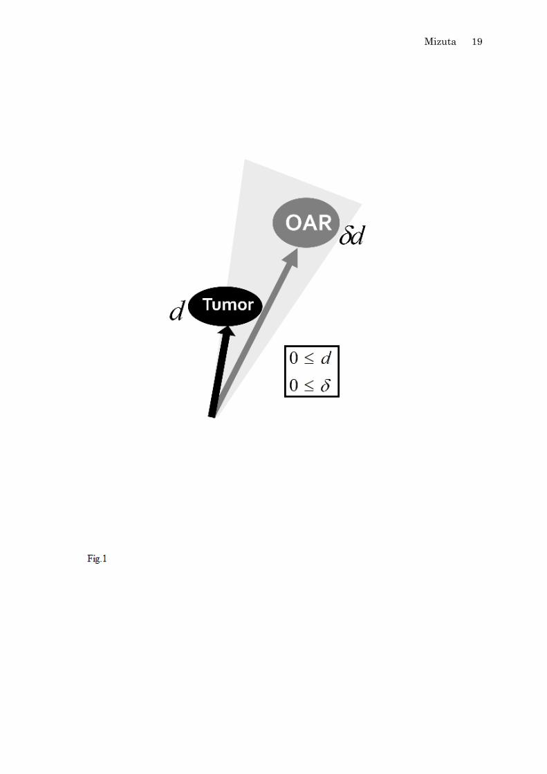
same irradiation field, it should be reasonable to consider that the dose for the OAR is
proportional to the dose for the tumor, i.e. the dose for the OAR is given by d, where the
dose for the tumor is d and the proportionality factor satisfies 0 (Fig.1).
For multi-fraction radiation therapy with N-fraction doses ( Nddd ,...,, 21 ), the radiation
effect on the tumor is represented by
N
i
ii dd1
2
11 )( and is fixed as E1, that is
N
i
ii ddE1
2
111 )( . (1)
Since the doses for the OAR are denoted as Nddd ,...,, 21 , the damage effect on the OAR
(E0) by N times exposure is given by
N
i
ii ddE1
2
000 ])()([ . (2)
Thus, the problem for the fractionation regimen can be handled mathematically as an
optimization problem,
Min ])()([1
2
00
N
i
ii dd
under the constraint of Eq.(1). If the damage effect on the OAR in formula (2) is smaller with
an increase in the number of fractions, multi-fractionated irradiation is better. If the damage

Mizuta
7
effect on the OAR in formula (2) is larger with an increase in the number of fractions,
hypofractionated irradiation is better.
RESULTS
From Eq.(1), we have the following equation,
N
i
i
N
i
i dEd1
11
11
2 1
. (3)
The formula (2) can be transformed as follows.
N
i
i
N
i
i
N
i
i
N
i
i
N
i
ii
dEd
dd
ddE
1
11
1
2
0
1
0
1
22
0
1
0
1
22
000
1
)(
1
1
2
0
101
01
1
01
1
1
2
0
11
12
00
Ed
Ed
N
i
i
N
i
i
1
1
2
0
111
00
1
01 EdN
i
i
. (4)
Then, this formula is interpreted as follows;
(a) if
1
1
0
0 , the lower
N
i
id1
is, the lower the damage effect on the OAR is.
(b) if
1
1
0
0 , the larger
N
i
id1
is, the lower the damage effect on the OAR is.

Mizuta
8
Next, we move to the problem of the minimization or maximization of
N
i
id1
. From Eq.
(1), we have
N
i
ii
Edd
1 1
1
1
12
2
1
1
1 1
1
2
1
1
1
12
22
N
Edd
N
i
ii
2
1
1
1 1
1
2
1
1
22
N
Ed
N
i
i. (5)
The set of fractionated doses ),,( 1 Ndd satisfying the above equation represents an
N-dimensional hypersphere )0,,0( 1 Ndd , where the center is
1
1
1
1
2,,
2
and
the radius is
2
1
1
1
1
2
N
E, as depicted in a two-dimensional plane in Fig.2.
In Fig.2, the circle represents the condition of Eq. (5) (for radiation effect on tumor, E1),
and the lines (with minus one inclination) correspond to the values of
N
i
id1
. Under the
reservation 0id ,
N
i
id1
is maximized when the line is tangent to the circle, while
N
i
id1
is
minimized when the line crosses the points that the circle intersects the axes. Therefore, we
can summarize the conditions as:
N
i
id1
is maximized when Ndd 1 , and
N
i
id1
is
minimized when one of Ndd ,, 1 is positive and the others are zero (i.e., single exposure).
Ultimately, the adjudication can be described as follows:
(i) if
1
1
0
0 , hypofractionated irradiation is better than multi-fractionated irradiation.
Mizuta
9
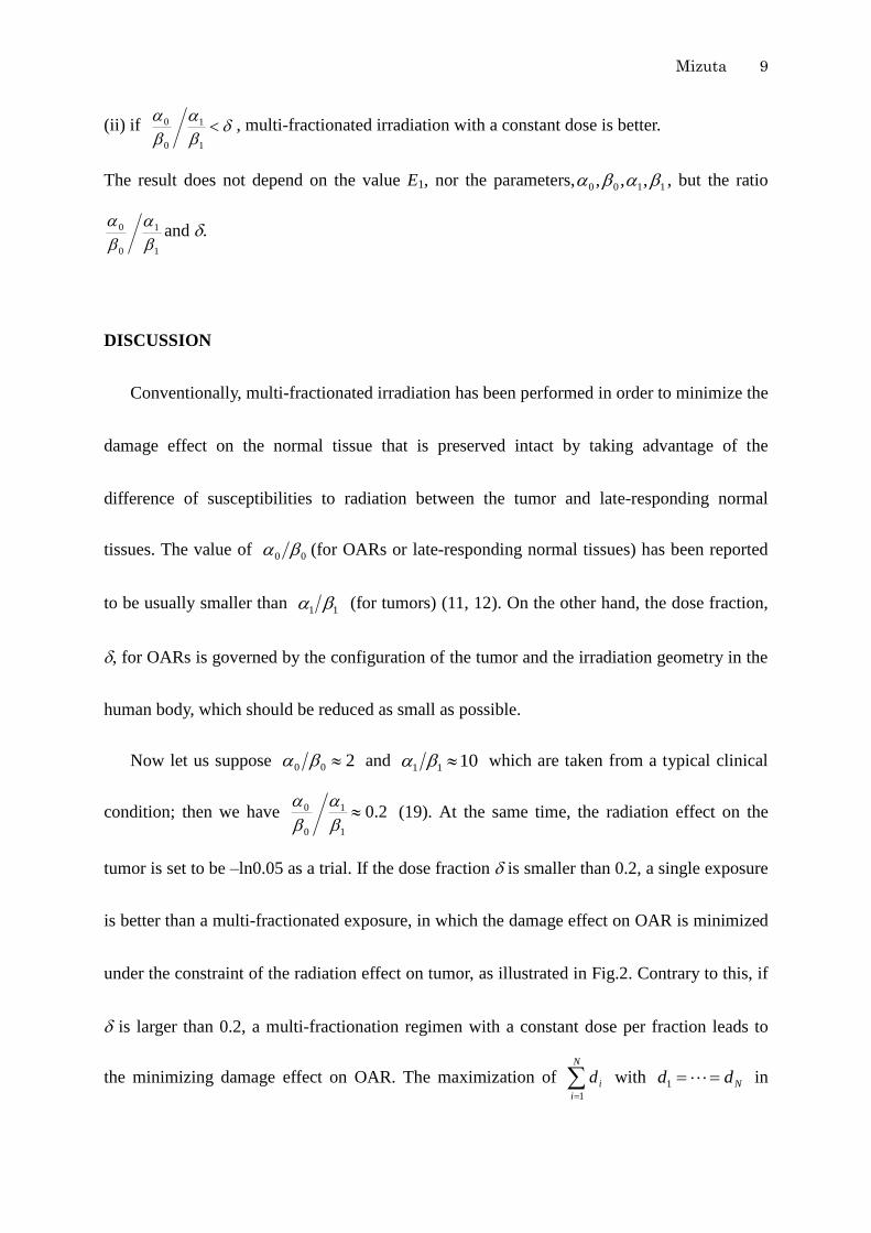
(ii) if
1
1
0
0 , multi-fractionated irradiation with a constant dose is better.
The result does not depend on the value E1, nor the parameters, 1100 ,,, , but the ratio
1
1
0
0
and .
DISCUSSION
Conventionally, multi-fractionated irradiation has been performed in order to minimize the
damage effect on the normal tissue that is preserved intact by taking advantage of the
difference of susceptibilities to radiation between the tumor and late-responding normal
tissues. The value of 00 (for OARs or late-responding normal tissues) has been reported
to be usually smaller than 11 (for tumors) (11, 12). On the other hand, the dose fraction,
, for OARs is governed by the configuration of the tumor and the irradiation geometry in the
human body, which should be reduced as small as possible.
Now let us suppose 200 and 1011 which are taken from a typical clinical
condition; then we have 2.01
1
0
0
(19). At the same time, the radiation effect on the
tumor is set to be –ln0.05 as a trial. If the dose fraction is smaller than 0.2, a single exposure
is better than a multi-fractionated exposure, in which the damage effect on OAR is minimized
under the constraint of the radiation effect on tumor, as illustrated in Fig.2. Contrary to this, if
is larger than 0.2, a multi-fractionation regimen with a constant dose per fraction leads to
the minimizing damage effect on OAR. The maximization of
N
i
id1
with Ndd 1 in

Mizuta
10
the latter case means that a multiple (N) exposure with a constant dose per fraction is
favorable for obtaining a low effect on OAR. For example, if 1011 is given by
05.01 and 005.0 1 , the relation of 05.0ln)(1
2
11
N
i
ii dd is satisfied by 25N
with 0.21 ddd N Gy, where 0.2d Gy is a typical dose in a multi-fractionation
regimen. The total accumulated dose is 50 Gy in this case with daily dose of 2.0 Gy, while
d=20 Gy is required for a single exposure (N=1) as an extreme example (as another example,
the total dose is 30 Gy for N=3) to achieve the same biological effect. Although the total dose
N
i
id1
in the multi-fraction irradiation is much larger than that of the hypofractionated
irradiation, the effect on the OAR can be smaller than the effect resulting from the
hypofractionated irradiation, when
1
1
0
0 . This condition is probable when 0.2 < for
an OAR near the tumor with 200 (e.g., from 04.00 and 02.0 0 (19)). It should
be emphasized that the decision whether to choose a hypofractionated irradiation or a
multi-fractionation in radiation therapy depends on the ratio 1
1
0
0
and the dose fraction
for the OAR ( ), while the total dose is determined by the values (1 and
1 ) and the dose
per fraction ( d ) to yield a certain degree of radiation effect on the tumor.
In figure 3, the damage effect on OAR versus the number of fractions is exemplified for
two cases, =0.1 and =0.8, respectively, keeping the radiation effect on the tumor to be
–ln0.05. In practice, hypofractionation of 3-5 times rather than single fractionation is often
used expecting to increase reoxygenation for hypoxic cells. Figure 3(a) suggests that

Mizuta
11
hypofractionated irradiation (e.g., 3-5 times) provides less damage effect on OAR than
multi-fractionated irradiation (30 times) when =0.1 (i.e., OAR receives 10% of tumor dose)
and 2.01
1
0
0
. Figure 3(b) suggests that multi-fractionated irradiation provides less
damage effect on OAR than hypofractionated irradiation when =0.8 (i.e., OAR receives 80%
of tumor dose) and 2.01
1
0
0
.
In actual situations, the value for a tumor or a specific organ (or tissue) may vary
depending on its volume due to the oxygen effect (20) and other factors such as the cell cycle.
Modification for treatment time in the LQ model alters the result to some extent. However, as
far as the simple assumption mentioned earlier holds, the present study can provide us with a
criterion for the validity of the hypofraction or the multi-fractionation regimen as Eqs.(1) and
(2).
The clinical feasibility of this model can be examined assuming two lung tumors with the
same volume of 2.0 cm but situated at different locations; for example peripheral lung tissue
and central lung. The organs at risk are normal lung tissue, spinal cord, brachial plexus,
pulmonary artery, heart, esophagus, and the proximal bronchial tree (21, 22). We can assume
that the complication probabilities of OARs other than the proximal bronchial tree are
negligible for both tumors whether a single or fractionated schedule is employed. 00 of
the proximal bronchial tree is very likely to be smaller than 11 of squamous cell
carcinoma so that we can assume 1
1
0
0
is smaller than 1.0. In the treatment of peripheral
Mizuta
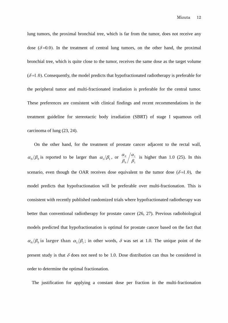
12
lung tumors, the proximal bronchial tree, which is far from the tumor, does not receive any
dose (. In the treatment of central lung tumors, on the other hand, the proximal
bronchial tree, which is quite close to the tumor, receives the same dose as the target volume
(. Consequently, the model predicts that hypofractionated radiotherapy is preferable for
the peripheral tumor and multi-fractionated irradiation is preferable for the central tumor.
These preferences are consistent with clinical findings and recent recommendations in the
treatment guideline for stereotactic body irradiation (SBRT) of stage I squamous cell
carcinoma of lung (23, 24).
On the other hand, for the treatment of prostate cancer adjacent to the rectal wall,
00 is reported to be larger than 11 , or
1
1
0
0
is higher than 1.0 (25). In this
scenario, even though the OAR receives dose equivalent to the tumor dose ( the
model predicts that hypofractionation will be preferable over multi-fractionation. This is
consistent with recently published randomized trials where hypofractionated radiotherapy was
better than conventional radiotherapy for prostate cancer (26, 27). Previous radiobiological
models predicted that hypofractionation is optimal for prostate cancer based on the fact that
00 is larger than 11 ; in other words, was set at 1.0. The unique point of the
present study is that does not need to be 1.0. Dose distribution can thus be considered in
order to determine the optimal fractionation.
The justification for applying a constant dose per fraction in the multi-fractionation

Mizuta
13
regimen is also presented from a mathematical point of view. The model for a single tumor as
the target and a single OAR is treated here based on the assumption that each organ is
irradiated uniformly. However, the method can be extended to the condition for a non-uniform
irradiation to OARs such as in intensity modulated radiation therapy.
The real interest of the present approach would be the determination of the optimum
solution for N in clinical practice. However, this requires a better modeling of cellular
dynamics following each fraction, incorporating the 4 Rs.
CONCLUSION
In this paper, we have discussed the validity of the multi-fractionation regimen in
radiotherapy, based on the LQ model for both tumors and normal tissues (OARs). The
problem of minimizing the radiation effect on OAR was solved under the constraint of
prescribed effect on the tumor, in which a multi-dimensional hypersphere representing the
constraint was taken into account. The result shows that a multi-fractionated irradiation with a
constant dose is better when the ratio of values for OAR and tumor is less than (ratio
of doses to the OAR and the tumor), while hypofractionation irradiation is appropriate when
the ratio is greater than .

Mizuta
14
REFERENCES
1. Withers HR. The four R's of radiotherapy. Adv Radiat Biol 1975; 5: 241-271
2. Bourhis J, Overgaard J, Audry H, et al. Hyperfractionated or accelerated radiotherapy in
head and neck cancer: a meta-analysis. The Lancet 2006; 368: 843-854
3. Monroe AT, Bhandare N, Morris CG, et al. Preventing radiation retinopathy with
hyperfractionation. Int J Radiat Oncol Biol Phys 2005; 61: 856-864
4. Whelan TJ, Pignol JP, Levine MN, et al. Long-term results of hypofractionated radiation
therapy for breast cancer. New Engl J Med 2010; 362: 513-520
5. Michele I. Saunders and S. Dische. Radiotherapy employing three fractions in each day
over a continuous period of 12 days. Brit J Radiol 1986; 59: 523-525
6. Dische S, and Saunders MI. The rationale for continuous, hyperfractionated, accelerated
radiotherapy (CHART). Int J Radiat Oncol Biol Phys 1990; 19: 1317-1320
7. M. Saunders, S. Dische, A. Barrett, et al. Continuous, hyperfractionated, accelerated
radiotherapy (CHART) versus conventional radiotherapy in non-small cell lung cancer:
mature data from the randomised multicentre trial. Radiother Oncol 1999; 52: 137-148
8. Hijal T, Hamad AA, Niazi T, et al. Hypofractionated radiotherapy and adjuvant
chemotherapy do not increase radiation-induced dermatitis in breast cancer patients. Curr
Oncol 2010; 17: 22-27
9. Vogelius IS, Westerly DC, Cannon GM, et al. Hypofractionation does not increase

Mizuta
15
radiation pneumonitis risk with modern conformal radiation delivery techniques, Acta
Oncol 2010; 49: 1052-1057
10. Ho KF, Fowler JF, Sykes AJ et al. IMRT dose fractionation for head and neck cancer:
variation in current approaches will make standardisation difficult. Acta Oncol 2009; 48:
431-439
11. Sachs RK, Hahnfeld P, and Brenner DJ. The link between low-LET dose-response
relations and the underlying kinetics of damage production/repair/misrepair. Int J Radiat
Biol 1997; 72: 351-374
12. Fowler JF. The linear-quadratic formula and progress in fractionated radiotherapy, The
Brit J Radiol 1989; 62: 679-694
13. Chadwick KH and Leenhouts HP. A molecular theory of cell survival. Phys Med Biol
1973; 18:78-87
14. Kellerer AM and Rossi HH. RBE and the primary mechanism of radiation action. Radiat
Res 1971; 47:15-34
15. Neary GJ. Chromosome aberrations and the theory of RBE. Int J Radiat Biol 1965;
9:477-502
16. Fowler JF, Tome WA, Fenwick JD, et al. A challenge to traditional radiation oncology, Int
J Radiat Oncol Biol Phys 2004; 60: 1241-1256
17. Fowler JF. Sensitivity Analysis of Parameters in Linear-Quadratic Radiobiologic

Mizuta
16
Modeling, Int J Radiat Oncol Biol Phys 2009; 73: 1532-1537
18. Yang Y, and Xing L. Optimization of radiotherapy dose-time fractionation with
consideration of tumor specific biology. Med Phys 2005; 32: 3666-3677
19. Thames HD, Bentzen SM, Turesson I, et al. Time-dose factors in radiotherapy: a review
of the human data. Radiother Oncol 1990; 19: 219-235
20. Carlson DJ, Stewart RD, and Semenenko VA. Effects of oxygen on intrinsic radiation
sensitivity: A test of the relationship between aerobic and hypoxic linear-quadratic (LQ)
model parameters. Med Phys 2006; 33: 3105-3115
21. Onimaru R, Shirato H, Shimizu S, et al. Tolerance of organs at risk in small-volume,
hypofractionated, image-guided radiotherapy for primary and metastatic lung cancers. Int
J Radiat Oncol Biol Phys 2003; 56 :126-135
22. Kong FM, Ritter T, Quint DJ, et al. Consideration of dose limits for organs at risk of
thoracic radiotherapy: atlas for lung, proximal bronchial tree, esophagus, spinal cord, ribs,
and brachial plexus. Int J Radiat Oncol Biol Phys 2010; In press
23. Timmerman R, McGarry R, Yiannoutsos C, et al. Excessive toxicity when treating central
tumors in a phase II study of stereotactic body radiation therapy for medically inoperable
early-stage lung cancer. J Clin Oncol 2006; 24: 4833-4839.
24. Nagata Y, Wulf J, Lax I, et al. Stereotactic radiotherapy of primary lung cancer and other
targets: results of consultant meeting of the International Atomic Energy Agency. Int J

Mizuta
17
Radiat Oncol Biol Phys 2011; 79: 660-669
25. Fowler JF, Ritter MA, Chappell RJ, et al. What hypofractionated protocols should be
tested for prostate cancer? Int J Radiat Oncol Biol Phys 2003; 56: 1093-1104
26. Yeoh EE, Botten RJ, Butters J, et al. Hypofractionated versus conventionally fractionated
radiotherapy for prostate carcinoma: final results of phase III randomized trial. Int J
Radiat Oncol Biol Phys 2010; In press
27. Arcangeli G, Fowler J, Gomellini S, et al. Acute and late toxicity in a randomized trial of
conventional versus hypofractionated three-dimensional conformal radiotherapy for
prostate cancer. Int J Radiat Oncol Biol Phys 2011; 79: 1013-1021

Mizuta
18
Figure Legends
Figure 1. Doses for tumor and OAR.
Figure 2. Maximum and minimum conditions of
N
i
id1
under the constraint for the radiation
effect on tumor.
Figure 3. Damage effect on OAR versus the number of fractionation (N) keeping the radiation
effect on tumor (E1) in Eq.(1) is set to be –ln0.05: (a) for =0.1 and (b) for =0.8. Here, the
dose per fraction (d) was assumed to be constant, and obtained from the constraint of
E1=–ln0.05. The damage effect on OAR (E0) was then determined by Eq.(2) with
04.00 , 02.0 0 and the value d for every fractionation number (N).
Mizuta
19

Mizuta
20

Mizuta
21

Mizuta
22