Embed Size (px)
Citation preview
A discontinuous DNA glycosylase domain in afamily of enzymes that excise 5-methylcytosineMarıa Isabel Ponferrada-Marın, Jara Teresa Parrilla-Doblas,
Teresa Roldan-Arjona and Rafael R. Ariza*
Department of Genetics, University of Cordoba, 14071-Cordoba, Spain
Received July 29, 2010; Revised September 30, 2010; Accepted October 4, 2010
ABSTRACT
DNA cytosine methylation (5-meC) is a widespreadepigenetic mark associated to gene silencing. Inplants, DEMETER-LIKE (DML) proteins typified byArabidopsis REPRESSOR OF SILENCING 1 (ROS1)initiate active DNA demethylation by catalyzing5-meC excision. DML proteins belong to theHhH-GPD superfamily, the largest and most func-tionally diverse group of DNA glycosylases, but themolecular properties that underlie their capacity tospecifically recognize and excise 5-meC are largelyunknown. We have found that sequence similarity toHhH-GPD enzymes in DML proteins is actuallydistributed over two non-contiguous segments con-nected by a predicted disordered region. We usedhomology-based modeling to locate candidateresidues important for ROS1 function in bothsegments, and tested our predictions bysite-specific mutagenesis. We found that aminoacids T606 and D611 are essential for ROS1 DNAglycosylase activity, whereas mutations in either oftwo aromatic residues (F589 and Y1028) reverse thecharacteristic ROS1 preference for 5-meC over T.We also found evidence suggesting that ROS1uses Q607 to flip out 5-meC, while the contiguousN608 residue contributes to sequence-context spe-cificity. In addition to providing novel insights intothe molecular basis of 5-meC excision, our resultsreveal that ROS1 and its DML homologs possess adiscontinuous catalytic domain that is unprecedent-ed among known DNA glycosylases.
INTRODUCTION
DNA methylation at carbon 5 of cytosine (5-meC) is areversible epigenetic mark for transcriptional genesilencing that plays critical roles in development and re-production of most eukaryotic species. Animal DNA
methylation is mostly confined to symmetrical CG se-quences, but plants also have significant levels ofmethylated cytosines in CHG and CHH sequences(where H is A, C or T) (1,2). DNA methylation patternsare subject to dynamic regulation involving both methy-lation and demethylation processes (3,4) and dysfunctionof methylation control is a key factor in several forms ofhuman disease, including cancer (5,6). Active DNAdemethylation in mammals occurs in early embryos andprimordial germ cells but its molecular mechanisms arepoorly understood (7). In plants, biochemical andgenetic analyses have identified a family of DNAglycosylases that remove 5-meC as a free base andinitiate a base excision repair demethylation pathway(8–12).Plant 5-meC DNA glycosylases are typified by
Arabidopsis ROS1 (REPRESSOR OF SILENCING 1)and DME (DEMETER) (8,9), which together withparalogs DML2 and DML3 (DEMETER-LIKEproteins 2 and 3) (13,14) are the founding members ofthe DML family. All four proteins remove 5-meC fromDNA and cleave the phosphodiester backbone by succes-sive b,d-elimination, leaving a gap that has to be furtherprocessed to generate a 30-OH terminus suitable for poly-merization and ligation (10,11,13,14). In vivo, ROS1,DML2 and DML3 are needed to counteract the robustRNA-dependent DNA methylation pathway at hundredsof discrete regions across the plant genome (13–15),whereas DME contributes to genome-wide demethylationduring endosperm development and is required for im-printing (11,16–18). Genes encoding putative DMLproteins are only found in plant genomes, includingmosses and unicellular green algae. Members of theDML family are large polypeptides containing a regionthat shows sequence similarity with members of thewell-known HhH-GPD superfamily of DNA repairglycosylases (19). In addition, they also share acarboxy-terminal domain of unknown function (10), anda short amino-terminal domain significantly rich in lysine(20) (Supplementary Figure S1).In an ongoing effort to elucidate the molecular basis of
active DNA demethylation, we have chosen ROS1 as an
*To whom correspondence should be addressed. Tel: +34 957 218 979; Fax: +34 957 212 072; Email: [email protected]
Published online 29 October 2010 Nucleic Acids Research, 2011, Vol. 39, No. 4 1473–1484doi:10.1093/nar/gkq982
� The Author(s) 2010. Published by Oxford University Press.This is an Open Access article distributed under the terms of the Creative Commons Attribution Non-Commercial License (http://creativecommons.org/licenses/by-nc/2.5), which permits unrestricted non-commercial use, distribution, and reproduction in any medium, provided the original work is properly cited.

archetypal 5-meC DNA glycosylase for detailed analysis.We have recently reported that the short lysine-richamino-terminal domain is not required for catalyticactivity, but mediates strong methylation-independentbinding to DNA, and allows efficient excision of 5-meCin long substrates (20). However, it remains unknown howthe enzymes of the DML family specifically recognize5-meC in DNA and distinguish it from unmethylated C.The fact that ROS1 activity is strongly inhibited by re-placement of the C5 methyl group by halogen derivatives,even if these substituents decrease the strength of thescissile C10-N glycosidic bond, suggests an importantrole for selective steric recognition of the target base atthe active site (21). After 5-meC excision, ROS1 remainsbound to its reaction product (20,21). This binding leadsto a highly distributive behavior of the enzyme on DNAsubstrates containing multiple 5-meC residues, and mayhelp to avoid generation of double-strand breaks duringprocessing of bimethylated CG dinucleotides or denselymethylated DNA regions (21).A comprehensive understanding of how plant 5-meC
DNA glycosylases specifically recognize and excise theirtarget base will require solving their crystal structure incomplex with DNA. Nevertheless, some useful informa-tion may still be obtained by combining structural infor-mation available from DNA glycosylases of theHhH-GPD superfamily and the analysis of amino acidsspecifically conserved in the DML group. All HhH-GPDDNA glycosylases share a common core structure thatconsists of two helical domains whose interface containsthe enzyme active site (22,23). One of these domainscontains the signature HhH-GPD motif (a helix–hairpin–helix and Gly/Pro rich loop followed by aconserved catalytic aspartate) (19), which interacts withthe DNA minor groove. An extensive body of evidencestrongly suggests that all DNA glycosylases, includingHhH-GPD enzymes, perform extrahelical base excisionthrough a reaction path that involves (i) DNA distortionand base flipping, which gives enzyme access for a nucleo-philic attack on the anomeric C10carbon, and (ii) insertionof the base lesion into a substrate recognition pocket(22,23). In this base-flipping mechanism, the propertiesof the active site pocket rather than the HhH-GPDmotif are a major component of the base specificity ofeach enzyme.In the present study, we performed multiple sequence
alignment and structural homology analysis to predict thelocation of several candidate ROS1 residues important forrecognition and/or catalysis that were functionally-testedby site-directed mutagenesis. In addition to providing in-structive clues on the molecular origins of 5-meC recogni-tion and excision, our results reveal that proteins of theDML family are endowed with a discontinuous DNAglycosylase domain.
MATERIALS AND METHODS
Homology-based modeling
A multiple sequence alignment of DML proteins andseveral members of the HhH-GPD superfamily was
performed using the program T-Coffee (24). The align-ment was viewed, adjusted and refined manually withJalview (25). A 3D model structure of the two alignedregions from Arabidopsis ROS1 (amino acids 567–625and 883–1062) was built using Swiss-Model (26) and the3D structure of Bacillus stearothermophilus EndonucleaseIII as a template [Protein Data Bank accession code: 1P59,(27)]. Nucleic acid coordinates extracted from 1P59 wereused to superimpose a DNA structure with a flipped-outabasic (AP) site analog onto the ROS1 model. The struc-tural figures were prepared with PyMOL (http://www.pymol.org). Protein structural disorder predictions wereperformed with VL3H [http://www.ist.temple.edu/disprot/Predictors.html; (28)].
DNA substrates
Oligonucleotides used as DNA substrates (SupplementaryTable S1) were synthesized by Operon and purified byPAGE before use. Double-stranded DNA substrateswere prepared by mixing a 5 mM solution of a50-fluorescein- or alexa-labeled oligonucleotide (upper-strand) with a 10 mM solution of an unlabeled oligomer(lower-strand), heating to 95�C for 5min and slowlycooling to room temperature. Annealing reactions forthe preparation of the 1-nt gapped duplex were carriedout at 95�C for 5min in the presence of a 2-fold molarexcess of both unlabeled 50-phosphorylated oligonucleo-tide (P30_51) and unlabeled oligonucleotide (CGR) withrespect to the 50-alexa-labeled 30-phosphorylated oligo-nucleotide (Al-28P), followed by cooling to roomtemperature.
Production of ROS1 variants derivatives
Site-directed mutagenesis was performed using theQuick-Change II XL kit (Stratagene). The mutationswere introduced into the expression vector pET28a(Novagen) containing the full-length wild-type (WT)ROS1 cDNA using specific oligonucleotides(Supplementary Table S2). Mutational changes were con-firmed by DNA sequencing and the constructs were usedto transform Escherichia coli BL21 (DE3) dcm� CodonPlus cells (Stratagene). WT and mutant versions were ex-pressed and purified as N-terminal His-tagged proteins, aspreviously described (21) (Supplementary Figure S2).Protein stability was measured by limited proteolysiswith thermolysin (29). WT and mutant proteins(160 mM) were preincubated (4 h) or not under DNAglycosylase assay conditions (see below) in the absenceof DNA substrate, and then digested for 5min with5 mg/ml thermolysin. Samples were analyzed by SDS/PAGE and relative band intensities were used toestimate the percentage of stable protein remaining afterpre-incubation (Supplementary Figure S2).
Enzyme activity assays
Double-stranded oligodeoxynucleotides (20 nM, unlessotherwise stated) were incubated at 30�C for the indicatedtimes in a reaction mixture containing 50mM Tris–HClpH 8.0, 1mM EDTA, 1mM DTT, 0.1mg/ml BSA, andthe indicated amounts of WT ROS1 or mutant variant in a
1474 Nucleic Acids Research, 2011, Vol. 39, No. 4

total volume of 50 ml. When reactions included AP endo-nuclease 1 (APE 1, 5U; New England BioLabs), EDTAwas omitted and 5mM mM MgCl2 was added. Reactionswere stopped by adding 20mM EDTA, 0.6% sodiumdodecyl sulphate and 0.5mg/ml proteinase K, and themixtures were incubated at 37�C for 30min. DNA wasextracted with phenol:chloroform:isoamyl alcohol(25:24:1) and ethanol precipitated at �20�C in thepresence of 0.3mM NaCl and 16 mg/ml glycogen.Samples were resuspended in 10 ml of 90% formamideand heated at 95�C for 5min. When measuring DNAglycosylase activity, samples were treated with NaOH100mM and immediately transferred to 90�C for 10min.After adding an equal volume of 90% formamide, sampleswere heated at 95�C for 5min. Reaction products wereseparated in a 12% denaturing polyacrylamide gel con-taining 7M urea. Fluorescein-labeled DNA was visualizedin a FLA-5100 imager and analyzed using Multigaugesoftware (Fujifilm).
When measuring AP lyase activity, a fluorescein-labeledoligonucleotide duplex containing U opposite G (200 nM)was incubated at 30�C for the indicated times in a reactionmixture containing 50mM Tris–HCl pH 8.0, 1mMEDTA, 1mM DTT, 0.1mg/ml BSA, 2.5U of E. coliUracil DNA glycosylase (New England BioLabs), andthe indicated amounts of WT ROS1 or mutant variantin a total volume of 5 ml. Reactions were stopped byadding 20mM EDTA, 0.6% sodium dodecyl sulphate,and 0.5mg/ml proteinase K. After adding 10 ml of 90%formamide, samples were heated at 95�C for 5min.Products were resolved and analyzed as described above.
Kinetic analysis
As we have shown previously (20,21) ROS1 does notexhibit significant turnover in vitro due to strongproduct binding, and therefore a simple Michaelis–Menten model is inadequate for a correct kineticanalysis of this enzyme. Accordingly, we have used a pre-viously described method (30) successfully employed tomeasure and compare single-turnover kinetics with differ-ent orthologs of thymine DNA glycosylase (TDG) (31).The standard reaction conditions were equimolar (20 nM)enzyme/substrate ratios and incubation at 30�C. Datawere fitted to the equation [Product]=Pmax[1 – exp
(–kt)]using non-linear regression analysis and the softwareSigmaplot. For each mutant enzyme and substrate, wedetermined the parameters Pmax (maximum substrate pro-cessing within an unlimited period of time), T50 (the timerequired to reach 50% of the product plateau level, Pmax),and the relative processing efficiency (Erel=Pmax/T50).Representative examples of 5-meC DNA glycosylaseassays and kinetic analysis are shown in SupplementaryFigure S3.
Electrophoretic mobility shift assay
Standard band-shift reactions were performed with theindicated amounts of protein and 100 nM fluorescein-and/or alexa-labeled duplex oligonucleotides. Bindingreactions were carried out at 25�C for 60min, unlessotherwise stated, in 10 nM Tris–HCl pH 8.0, 1mM
DTT, 10 mg/ml BSA, 1mM EDTA, in a final volume of10 ml. Complexes were electrophoresed through 0.2%agarose gels in 1� TAE. Electrophoresis was carried outin 1� TAE for 40min at 80V at room temperature.Fluorescein- and/or alexa-labeled DNA was visualized ina FLA-5100 imager and analyzed using Multigaugesoftware (Fujifilm).
RESULTS
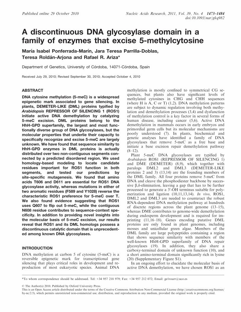
An unusual sequence insertion is present in the DNAglycosylase domain of DML proteins
To gain insight into residues that comprise the DNAglycosylase domain of DML proteins, we performed amultiple sequence alignment that included ArabidopsisROS1 and DME, Nicotiana tabacum ROS1, and severalHhH-GPD proteins (Figure 1). The alignment revealedthat sequence similarity to HhH-GPD enzymes in DMLproteins is actually distributed over two non-contiguoussegments. The first segment corresponds to a region thatin HhH-GPD members contains the base-flipping wedgeand its flanking alfa-helixes, as well as some of the residuesthat line the active site pocket (32). The second segmentincludes the HhH-GPD motif and its invariant aspartate,which is absolutely required for catalysis of 5-meCexcision by DML proteins (10,11,13,14). This region alsocontains a lysine residue that is only present in the subsetof HhH-GPD proteins with bifunctional DNAglycosylase/lyase activity (19) and a [4Fe–4S] clusterloop (FCL) motif that in some HHh-GPD proteins,such as E. coli Endo III and MutY, ligates a [4Fe–4S]cluster (19) (Figure 1B). The two separate segments withsequence similarity to HhH-GPD proteins are intercon-nected by a non-conserved linker region that is highlyvariable in sequence and length among members of theDML family (Figure 1B and Supplementary Figure S1).We applied a well-characterized disorder predictor [VL3H(28)] to analyze the location of ordered and disorderedregions in ROS1 (Supplementary Figure S4). The resultspredict a high disorder content within this linker region,thus suggesting that it is intrinsically unstructured undernative conditions.We used the crystal structure of Bacillus
stearothermophilus Endonuclease III [Protein Data Bankaccession code: 1P59, (27)] as a template to generate a 3Dmodel structure of the two ROS1 polypeptide segmentsthat show sequence similarity to HhH-GPD proteins(amino acids 567–625 and 883–1062) (‘Materials andMethods’ section). The model predicts a typicalHhH-GPD core structure with two alpha domains: asix-helix barrel domain (6a–6f), and a four-helix domainformed by one N-terminal (4a) and three C-terminal(4b–4d) helixes (Figure 1). The non-conserved linkerregion of 258 amino acids is inserted between helixes 6band 6c, which are part of the characteristically sequence-continuous six-helix barrel domain (Figure 1). Two otherHhH-GPD proteins (AlkA and Ogg1) contain at this samelocation position a much shorter insertion (13 and 11amino acids, respectively) (Figure 1). The model alsopredicts a second DML-specific insertion between helixes
Nucleic Acids Research, 2011, Vol. 39, No. 4 1475
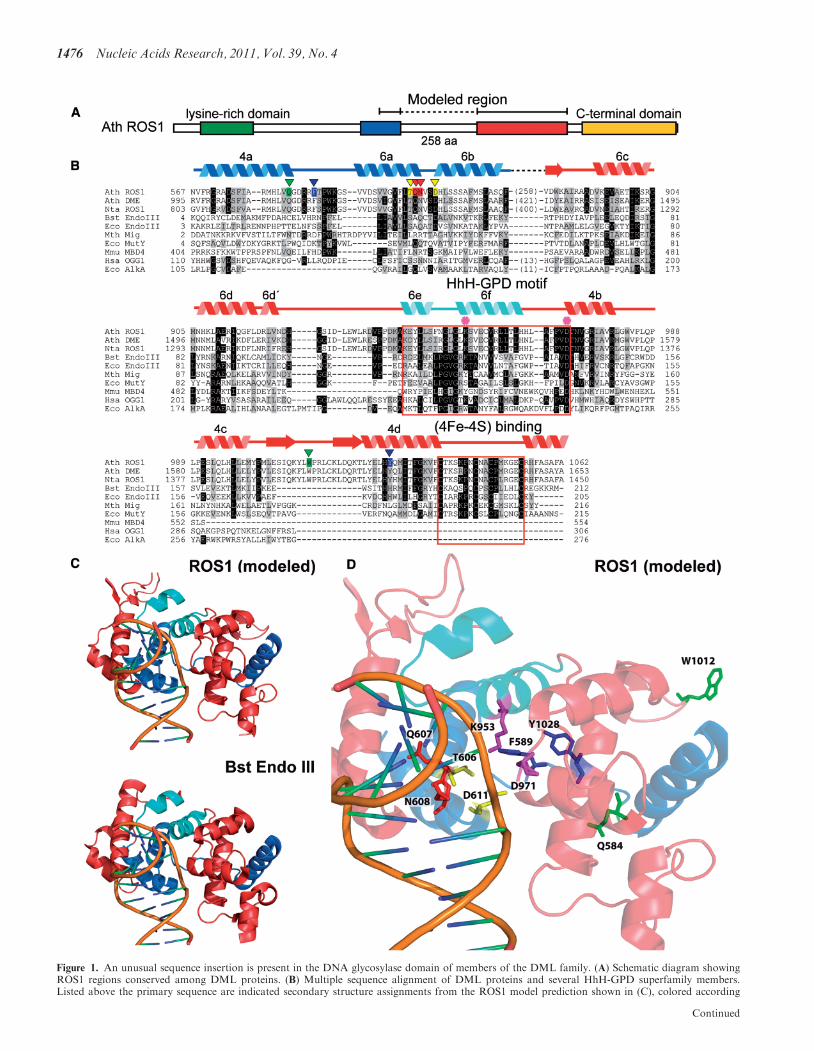
Figure 1. An unusual sequence insertion is present in the DNA glycosylase domain of members of the DML family. (A) Schematic diagram showingROS1 regions conserved among DML proteins. (B) Multiple sequence alignment of DML proteins and several HhH-GPD superfamily members.Listed above the primary sequence are indicated secondary structure assignments from the ROS1 model prediction shown in (C), colored according
Continued
1476 Nucleic Acids Research, 2011, Vol. 39, No. 4
4c and 4d. Thus, homology modeling predicts thatproteins of the DML family have a discontinuous DNAglycosylase domain structure interrupted by an unusuallylong insertion.
T606 and D611 are critical residues for ROS1 DNAglycosylase activity
In order to identify residues specifically involved in 5-meCrecognition and catalysis, we used this tentative model as aguide to design site-specific mutations of the ROS1 DNAglycosylase domain. Following a general approach thathas been well detailed elsewhere (33), we searched notonly for residues conserved among DML proteins andother HhH-GPD enzymes, but also for residues specific-ally conserved within the DML group. The former classmay be important for the general catalytic mechanism,whereas the latter may contribute to specific recognitionof 5-meC.
In E. coli Endonuclease III, amino acids S39 and D44are both required for catalytic activity (34), and their hom-ologous residues in ROS1 are T606 and D611, respectively(Figure 1B). The modeled structure of ROS1 predicts thatT606 and D611 are positioned at the mouth of the groovethat separates the six-helix barrel domain and thefour-helix domain (Figure 1D). In HhH-GPD DNAglycosylases this groove lies between the DNA basestack from which the lesion is extruded and the base rec-ognition pocket where it is inserted (35), and containsresidues suitably disposed to access the N-glycosyl bond
of the flipped base. To test the prediction that T606 andD611 have a role in catalysis, we mutated them to Leu(T606L) and Val (D611V), respectively.We examined the ability of WT and mutant proteins to
process a 51-mer duplex oligo substrate that containedeither 5-meC or T opposite G at position 29 in a CGcontext (Table 1). Consistent with our previouslyreported observations (10,21) we found that WT ROS1processed 5-meC with higher relative processing efficiencythan T (Table 1). We found that both the T606L andD611V mutations abolished the catalytic activity ofROS1 on both substrates (Table 1). For both ROS1variants, neither 5-meC:G nor T:G processing wasdetected even after prolonged incubation times (data notshown). The DNA-binding capacity of WT and mutantproteins was assessed by electrophoretic mobility shiftassay with different labeled substrates (Figure 2). As pre-viously reported, ROS1 bound with similar efficiency tomethylated and non-methylated DNA, and displayed ahigher affinity for the 1-nt gapped reaction product. TheT606L mutant enzyme exhibited a somewhat reducedbinding activity compared to that of WT ROS1, whereasthe D611V variant displayed higher binding capacity(Figure 2).Since ROS1 is a bifunctional enzyme, we asked whether
these mutant proteins lack DNA glycosylase activity, lyaseactivity or both. To differentiate 5-meC excision andstrand incision, we analyzed the reaction productsgenerated by different ROS1 variants with or without
Figure 1. Continuedto regions shown in (A). The helix–hairpin–helix of the HhH-GPD motif is shown in cyan. ROS1 amino acids mutated in this study are indicated byinverted triangles and highlighted in green (Q584 and W1012), blue (F589 and Y1028), yellow (T606 and D611) or red (Q607 and N608). The lysineresidue that is diagnostic of bifunctional glycosylase/lyase activity, and the conserved aspartic acid residue in the active site are indicated by asterisks.The HhH-GPD and the [4Fe–4S] cluster loop (FCL) motifs are boxed. Names of organisms are abbreviated as follows: Ath, Arabidopsis thaliana;Nta, Nicotiana tabacum; Bst, Bacillus stearothermophilus; Eco, Escherichia coli; Mth, Methanobacterium thermoautotrophicum; Mmu, Mus musculus;Hsa, Homo sapiens. Genbank accession numbers are: Ath ROS1: AAP37178; Ath DME: ABC61677; Nta ROS1: BAF52855; Bst EndoIII: 1P59;Eco EndoIII: P20625; Mth Mig: NP_039762; Eco MutY: NP_417436; Mmu MBD4: 1NGN; Hsa OGG1: O15527; Eco AlkA: P04395.(C) Ribbon diagrams of the structural model for the DNA glycosylase domain of ROS1 and the crystallographic Bst EndoIII structure used astemplate. Structural elements are colored as in (A). The duplex DNA is shown in orange. Nucleic acid coordinates extracted from the BstEndoIII-DNA trapped complex were used to superimpose a DNA structure with a flipped-out AP site analog onto the ROS1 model.
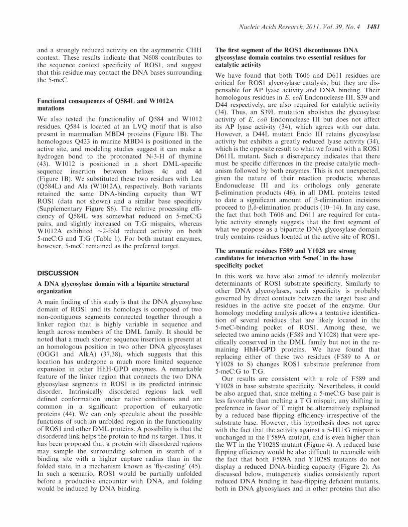
Table 1. Relative substrate processing efficiencies of WT and mutant variants of ROS1a
ROS1 variant 5-meC:G T:G
Pmax (nM) T50 (h) Erel Pmax (nM) T50 (h) Erel
WT 10.24±0.17 3.30 3.10±0.05 7.64±0.18 4.83 1.58±0.04Q584L 12.23±0.45 5.07 2.41±0.09 9.93±0.51 5.42 1.83±0.09F589A 1.84±0.11 5.10 0.36±0.02 3.22±0.21 4.76 0.68±0.04T606L n.d.b n.a.c n.a. n.d. n.a. n.a.Q607A 1.19±0.09 2.63 0.45±0.04 0.29±0.04 0.62 0.48±0.06N608A 13.64±0.35 4.73 2.88±0.07 12.07±0.33 9.38 1.29±0.04D611V n.d. n.a. n.a. n.d. n.a. n.a.W1012A 7.99±0.27 5.01 1.60±0.06 5.41±0.45 6.62 0.82±0.07Y1028S 7.76±0.22 5.11 1.50±0.04 10.87±0.62 5.66 1.92±0.11
aPurified proteins (20 nM) were incubated at 30�C with 51-mer double-stranded oligonucleotide substrates (20 nM) containing either a single5-meC:G pair or a T:G mispair. Reaction products were separated in a 12% denaturing polyacrylamide gel and quantified by fluorescencescanning. Shown are the plateau levels of substrate nicking (Pmax) and the time required for processing of 50% of Pmax (T50). Relative processingefficiency was calculated as Erel=Pmax/T50. Values are mean±SE from two independent experiments.bn.d., none detected.cn.a., not applicable.
Nucleic Acids Research, 2011, Vol. 39, No. 4 1477

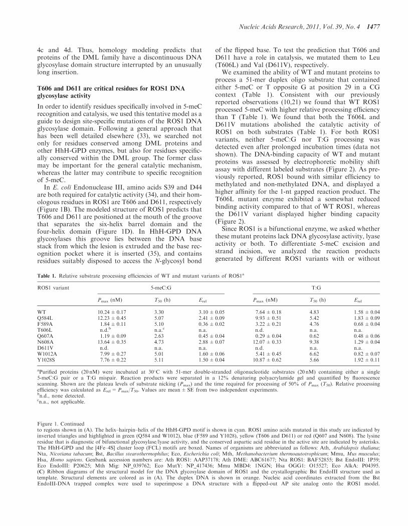
additional alkaline treatment with NaOH (Figure 3A).Incisions in the absence of NaOH reveal the combinedDNA glycosylase/AP lyase action of the enzyme,whereas the alkaline treatment cleaves all AP sitesgenerated by the enzyme and reflects DNA glycosylaseactivity. Consistent with our previously reported observa-tions (21), we found that the amount of incision productsgenerated by WT ROS1 was only slightly increased afterNaOH treatment, thus suggesting that glycosyl bondscission is usually coupled to the AP lyase step. Thesame pattern was observed for all ROS1 mutantenzymes except T606L and D611V, which did notgenerate detectable incision products either in theabsence or the presence of NaOH. The incapacity ofboth mutants to generate abasic sites was confirmed byperforming reactions in the presence of human AP endo-nuclease APE1 (Figure 3B). We next tested whetherT606L or D611V retained AP lyase activity by incubatingboth proteins with a 51-mer duplex oligo substrate thatcontained an AP site opposite G at position 29 in a CGcontext (Figure 3C). Although a significant level of spon-taneous AP incision is observed in the absence of enzyme,the amounts of enzyme-dependent strand incision after0.5, 2 and 24 h incubation were similar to those generatedby WT ROS1 (Figure 3C; a representative gel is shown inSupplementary Figure S5). We also found that the incisionproduct generated by both WT and mutant proteinsmigrates as a b-elimination product (SupplementaryFigure S5). Altogether, these results indicate that bothT606L and D611V mutants lack DNA glycosylaseactivity but retain AP lyase activity. In addition, theyprovide experimental evidence of the location of critical
catalytic residues in the first segment of the discontinuousROS1 DNA glycosylase domain.
F589A and Y1028S mutations change ROS1 preferencefor 5-meC over T
To investigate residues possibly contributing to 5-meCspecificity, we focused on two aromatic amino acids(F589 and Y1028) that are specifically conserved withinthe DML family (Figure 1B). Their positions correspondto residues that in other HhH-GPD enzymes interact withthe lesion base (27,32,36), and the modeled ROS1 struc-ture suggests that they are located in the base bindingpocket of the enzyme (Figure 1D). To test the predictionthat F589 and Y1028 are involved in the specific recogni-tion of 5-meC, we substituted them with Ala (F589A) andSer (Y1028S), respectively. Both F589A and Y1028S dis-played a somewhat higher non-specific DNA-bindingcapacity than the WT protein (Figure 2).
We found that mutation Y1028S produced a proteinwith a 2-fold reduced efficiency on 5-meC:G pairs,but with slightly increased activity on T:G mispairs(Table 1). On the other hand, the F589A mutationreduced enzyme efficiency on 5-meC:G �9-fold, butdecreased activity on T:G only 2.3-fold (Table 1). As aresult, both F589A and Y1028S exhibit a higher prefer-ence for T over 5-meC (Figure 4), which is just theopposite of the base specificity characteristic of WTROS1 and its homologs (10,14). We also tested theeffect of both mutations on excision of 5-HU and foundthat the mutant protein F589A exhibited a similar activityto that of WT ROS1, whereas Y1028S displayed �2-foldincreased efficiency (Figure 4). ROS1 activity is strongly
Figure 2. Binding of WT and mutant ROS1 proteins to substrate and product DNA. DNA-binding reactions were performed incubating increasingconcentrations of WT ROS1 or mutant variants with 100 nM of fluorescein-labeled 5-meC:G substrate (upper panel), alexa-labeled homoduplex(center panel) or alexa-labeled 1-nt-gapped duplex product (lower panel). After nondenaturing gel electrophoresis, the gel was scanned to detectfluorescein- or alexa-labeled DNA. Protein–DNA complexes were identified by their retarded mobility compared with that of free DNA, as indicated.The fraction of bound DNA is indicated below each lane. The asterisk depicts 50-end labeling of the upper strand. M: 5-meC.
1478 Nucleic Acids Research, 2011, Vol. 39, No. 4
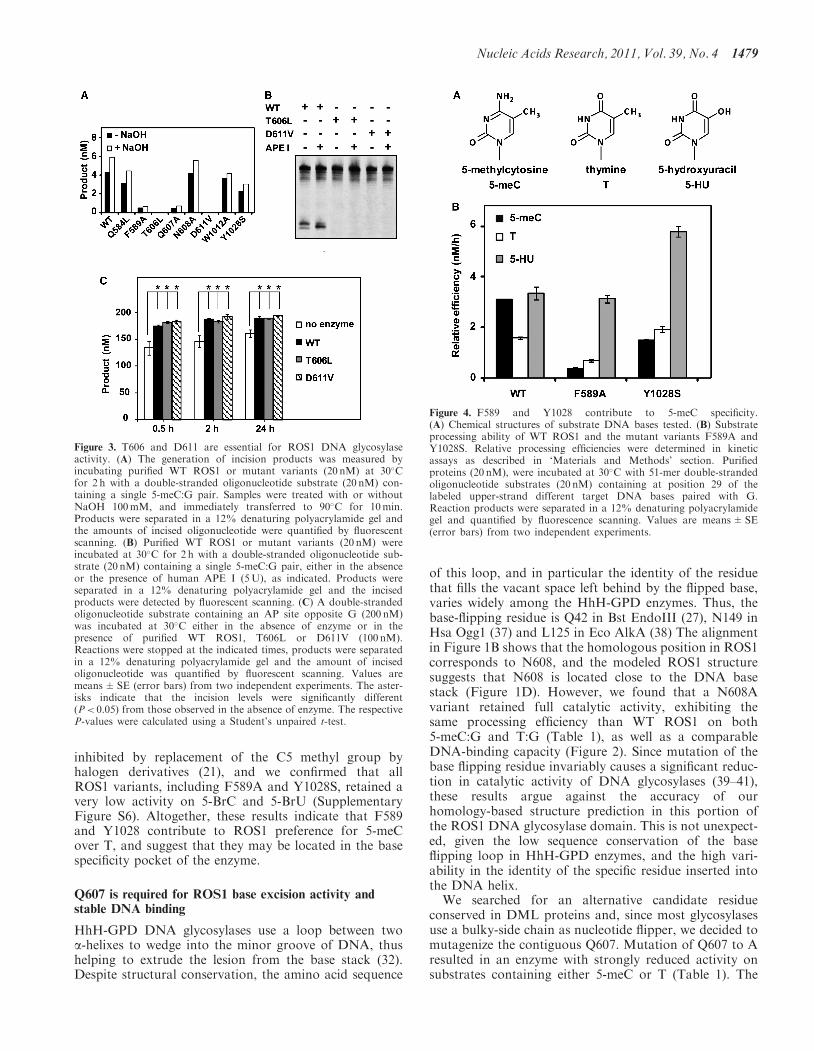
inhibited by replacement of the C5 methyl group byhalogen derivatives (21), and we confirmed that allROS1 variants, including F589A and Y1028S, retained avery low activity on 5-BrC and 5-BrU (SupplementaryFigure S6). Altogether, these results indicate that F589and Y1028 contribute to ROS1 preference for 5-meCover T, and suggest that they may be located in the basespecificity pocket of the enzyme.
Q607 is required for ROS1 base excision activity andstable DNA binding
HhH-GPD DNA glycosylases use a loop between twoa-helixes to wedge into the minor groove of DNA, thushelping to extrude the lesion from the base stack (32).Despite structural conservation, the amino acid sequence
of this loop, and in particular the identity of the residuethat fills the vacant space left behind by the flipped base,varies widely among the HhH-GPD enzymes. Thus, thebase-flipping residue is Q42 in Bst EndoIII (27), N149 inHsa Ogg1 (37) and L125 in Eco AlkA (38) The alignmentin Figure 1B shows that the homologous position in ROS1corresponds to N608, and the modeled ROS1 structuresuggests that N608 is located close to the DNA basestack (Figure 1D). However, we found that a N608Avariant retained full catalytic activity, exhibiting thesame processing efficiency than WT ROS1 on both5-meC:G and T:G (Table 1), as well as a comparableDNA-binding capacity (Figure 2). Since mutation of thebase flipping residue invariably causes a significant reduc-tion in catalytic activity of DNA glycosylases (39–41),these results argue against the accuracy of ourhomology-based structure prediction in this portion ofthe ROS1 DNA glycosylase domain. This is not unexpect-ed, given the low sequence conservation of the baseflipping loop in HhH-GPD enzymes, and the high vari-ability in the identity of the specific residue inserted intothe DNA helix.We searched for an alternative candidate residue
conserved in DML proteins and, since most glycosylasesuse a bulky-side chain as nucleotide flipper, we decided tomutagenize the contiguous Q607. Mutation of Q607 to Aresulted in an enzyme with strongly reduced activity onsubstrates containing either 5-meC or T (Table 1). The
Figure 3. T606 and D611 are essential for ROS1 DNA glycosylaseactivity. (A) The generation of incision products was measured byincubating purified WT ROS1 or mutant variants (20 nM) at 30�Cfor 2 h with a double-stranded oligonucleotide substrate (20 nM) con-taining a single 5-meC:G pair. Samples were treated with or withoutNaOH 100mM, and immediately transferred to 90�C for 10min.Products were separated in a 12% denaturing polyacrylamide gel andthe amounts of incised oligonucleotide were quantified by fluorescentscanning. (B) Purified WT ROS1 or mutant variants (20 nM) wereincubated at 30�C for 2 h with a double-stranded oligonucleotide sub-strate (20 nM) containing a single 5-meC:G pair, either in the absenceor the presence of human APE I (5U), as indicated. Products wereseparated in a 12% denaturing polyacrylamide gel and the incisedproducts were detected by fluorescent scanning. (C) A double-strandedoligonucleotide substrate containing an AP site opposite G (200 nM)was incubated at 30�C either in the absence of enzyme or in thepresence of purified WT ROS1, T606L or D611V (100 nM).Reactions were stopped at the indicated times, products were separatedin a 12% denaturing polyacrylamide gel and the amount of incisedoligonucleotide was quantified by fluorescent scanning. Values aremeans±SE (error bars) from two independent experiments. The aster-isks indicate that the incision levels were significantly different(P< 0.05) from those observed in the absence of enzyme. The respectiveP-values were calculated using a Student’s unpaired t-test.
Figure 4. F589 and Y1028 contribute to 5-meC specificity.(A) Chemical structures of substrate DNA bases tested. (B) Substrateprocessing ability of WT ROS1 and the mutant variants F589A andY1028S. Relative processing efficiencies were determined in kineticassays as described in ‘Materials and Methods’ section. Purifiedproteins (20 nM), were incubated at 30�C with 51-mer double-strandedoligonucleotide substrates (20 nM) containing at position 29 of thelabeled upper-strand different target DNA bases paired with G.Reaction products were separated in a 12% denaturing polyacrylamidegel and quantified by fluorescence scanning. Values are means±SE(error bars) from two independent experiments.
Nucleic Acids Research, 2011, Vol. 39, No. 4 1479

observed reduction in relative processing efficiency ishigher for the 5-meC:G pair (6.9-fold) than for the T:Gmispair (3.3-fold). Furthermore, and unlike all otherROS1 variants tested in this study, the Q607A mutantexhibited drastically reduced DNA binding to bothmethylated and non-methylated DNA, as well as to the1-nt gapped reaction product (Figure 2). We reasoned thatthe Q607A mutation might compromise the stability of theprotein–DNA complex. In order to investigate this possi-bility, we performed DNA-binding measurements at dif-ferent time points to analyze the ability of Q607A toremain bound to DNA (Figure 5 and SupplementaryFigure S7). We found that the mutant protein formed adetectable complex with either the methylated substrate orthe gapped reaction product, but rapidly dissociated fromboth DNA probes. Altogether, these results indicate thatQ607 is essential for base excision activity and stable DNAbinding, and strongly support the hypothesis that it is aDNA-intercalating residue.
N608 modulates 5-meC excision in different sequencecontexts
Since plant DNA methylation is deposited in differentsequence contexts, it is not surprising that DMLproteins exhibit the capacity to excise 5-meC at CG,CHG and CHH sequences (10–14). The context specificityof DML family members has not been exhaustivelycharacterized, but excision of 5-meC in vitro is apparentlymore efficient on those sequences more likely to bemethylated in vivo. For example, DME and ROS1remove 5-meC from a CHG context more efficientlywhen H=A than when H=C (10), in agreement with
the fact that CCG is the sequence showing the lowestmethylation level among CHG sites (42).
In order to determine whether any of the mutatedresidues contributes to sequence-context specificity, wetested all ROS1 variants for their relative capacity toexcise 5-meC from CG, CHG and CHH sequences(Figure 6A). We found that all mutants except N608Aexhibited a sequence-context specificity similar to that ofWT ROS1. Unlike the WT enzyme and the rest of ROS1variants, the N608A mutant showed a significantlyreduced activity on the asymmetric CHH context(Figure 6A). To confirm this result, we performed akinetic analysis to compare the relative processingefficiencies of WT ROS1 and N608A on 5-meC locatedin CG, CHG or CHH contexts (Figure 6B). We found thatboth enzymes displayed a similar efficiency on CG sites,and also showed a very low activity on the CHG contextwhen H=C. However, the N608A mutation caused ahigher efficiency than WT ROS1 on CAG sequences,
Figure 6. N608 contributes to sequence-context specificity. (A) PurifiedWT ROS1 or mutant variants (20 nM) were incubated at 30�C for 4 hwith 51-mer double-stranded oligonucleotide substrates (20 nM) con-taining at position 29 of the labeled upper-strand a 5-meC residue indifferent sequence contexts. Products were separated in a 12%denaturing polyacrylamide gel and the amount of incised oligonucleo-tide was quantified by fluorescent scanning. For ease of comparison,the incision values for each substrate are normalized to the totalincision detected in all four substrates for each individual enzyme.(B) Substrate processing ability of type ROS1 and the mutant variantN608 in different sequence contexts. Relative processing efficiencieswere determined in kinetic assays as described in ‘Materials andMethods’ section. Purified proteins (20 nM), were incubated at 30�Cwith 51-mer double-stranded oligonucleotide substrates (20 nM) con-taining at position 29 of the labeled upper-strand a 5-meC residue indifferent sequence contexts. Reaction products were separated in a 12%denaturing polyacrylamide gel and quantified by fluorescence scanning.Values are means±SE (error bars) from two independent experiments.
Figure 5. Q607 is required for stable ROS1 binding to substrate andproduct DNA. Purified WT ROS1 or mutant variant Q607A (100 nM)were incubated with a mixture of 100 nM fluorescein-labeled 5-meC:Gsubstrate and 100 nM alexa-labeled 1-nt gapped product, and the reac-tions were monitored for 60min. After non-denaturing gel electrophor-esis, the gel was scanned to detect fluorescein- (upper panel) oralexa-labeled (lower panel) DNA. Protein–DNA complexes wereidentified by their retarded mobility compared with that of freeDNA, as indicated. The asterisk depicts 50-end labeling of the upperstrand. M: 5-meC.
1480 Nucleic Acids Research, 2011, Vol. 39, No. 4
and a strongly reduced activity on the asymmetric CHHcontext. These results indicate that N608 contributes tothe sequence context specificity of ROS1, and suggestthat this residue may contact the DNA bases surroundingthe 5-meC.
Functional consequences of Q584L and W1012Amutations
We also tested the functionality of Q584 and W1012residues. Q584 is located at an LVQ motif that is alsopresent in mammalian MBD4 proteins (Figure 1B). Thehomologous Q423 in murine MBD4 is positioned in theactive site, and modeling studies suggest it can make ahydrogen bond to the protonated N-3-H of thymine(43). W1012 is positioned in a short DML-specificsequence insertion between helixes 4c and 4d(Figure 1B). We substituted these two residues with Leu(Q584L) and Ala (W1012A), respectively. Both variantsretained the same DNA-binding capacity than WTROS1 (data not shown) and a similar base specificity(Supplementary Figure S6). The relative processing effi-ciency of Q584L was somewhat reduced on 5-meC:Gpairs, and slightly increased on T:G mispairs, whereasW1012A exhibited �2-fold reduced activity on both5-meC:G and T:G (Table 1). For both mutant enzymes,however, 5-meC remained as the preferred target.
DISCUSSION
A DNA glycosylase domain with a bipartite structuralorganization
A main finding of this study is that the DNA glycosylasedomain of ROS1 and its homologs is composed of twonon-contiguous segments connected together through alinker region that is highly variable in sequence andlength across members of the DML family. It should benoted that a much shorter sequence insertion is present atan homologous position in two other DNA glycosylases(OGG1 and AlkA) (37,38), which suggests that thislocation has undergone a much more limited sequenceexpansion in other HhH-GPD enzymes. A remarkablefeature of the linker region that connects the two DNAglycosylase segments in ROS1 is its predicted intrinsicdisorder. Intrinsically disordered regions lack welldefined conformation under native conditions and arecommon in a significant proportion of eukaryoticproteins (44). We can only speculate about the possiblefunctions of such an unfolded region in the functionalityof ROS1 and other DML proteins. A possibility is that thedisordered link helps the protein to find its target. Thus, ithas been proposed that a protein with disordered regionsmay sample the surrounding solution in search of abinding site with a higher capture radius than in thefolded state, in a mechanism known as ‘fly-casting’ (45).In such a scenario, ROS1 would be partially unfoldedbefore a productive encounter with DNA, and foldingwould be induced by DNA binding.
The first segment of the ROS1 discontinuous DNAglycosylase domain contains two essential residues forcatalytic activity
We have found that both T606 and D611 residues arecritical for ROS1 glycosylase catalysis, but they are dis-pensable for AP lyase activity and DNA binding. Theirhomologous residues in E. coli Endonuclease III, S39 andD44 respectively, are also required for catalytic activity(34). Thus, an S39L mutation abolishes the glycosylaseactivity of E. coli Endonuclease III but does not affectits AP lyase activity (34), which agrees with our data.However, a D44L mutant Endo III retains glycosylaseactivity but exhibits a greatly reduced lyase activity (34),which is the opposite result to what we found with a ROS1D611L mutant. Such a discrepancy indicates that theremust be specific differences in the precise catalytic mech-anism followed by both enzymes. This is not unexpected,given the nature of their reaction products; whereasEndonuclease III and its orthologs only generateb-elimination products (46), in all DML proteins testedto date a significant amount of b-elimination incisionsproceed to b,d-elimination products (10–14). In any case,the fact that both T606 and D611 are required for cata-lytic activity strongly suggests that the first segment ofwhat we propose as a bipartite DNA glycosylase domaintruly contains residues located at the active site of ROS1.
The aromatic residues F589 and Y1028 are strongcandidates for interaction with 5-meC in the basespecificity pocket
In this work we have also aimed to identify moleculardeterminants of ROS1 substrate specificity. Similarly toother DNA glycosylases, such specificity is probablygoverned by direct contacts between the target base andresidues in the active site pocket of the enzyme. Ourhomology modeling analysis allows a tentative identifica-tion of several residues that are likely located in the5-meC-binding pocket of ROS1. Among these, weselected two amino acids (F589 and Y1028) that were spe-cifically conserved in the DML family but not in the re-maining HhH-GPD proteins. We have found thatreplacing either of these two residues (F589 to A orY1028 to S) changes ROS1 substrate preference from5-meC:G to T:G.Our results are consistent with a role of F589 and
Y1028 in base substrate specificity. Nevertheless, it couldbe also argued that, since melting a 5-meC:G base pair isless favorable than melting a T:G mispair, any shifting inpreference in favor of T might be alternatively explainedby a reduced base flipping efficiency irrespective of thesubstrate base. However, this hypothesis does not agreewith the fact that the activity against a 5-HU:G mispair isunchanged in the F589A mutant, and is even higher thanthe WT in the Y1028S mutant (Figure 4). A reduced baseflipping efficiency would be also difficult to reconcile withthe fact that both F589A and Y1028S mutants do notdisplay a reduced DNA-binding capacity (Figure 2). Asdiscussed below, mutagenesis studies consistently reportreduced DNA binding in base-flipping deficient mutants,both in DNA glycosylases and in other proteins that also
Nucleic Acids Research, 2011, Vol. 39, No. 4 1481

rotate bases. Therefore, our results suggest that F589 andY1028 play a role in base substrate specificity rather thanbase flipping.Obviously, in the absence of detailed structural infor-
mation it is not possible to identify the precise interactionsproviding the basis for specific base recognition However,it is conceivable that the aromatic side chains of F589 and/or Y1028 could help to stabilize the flipped-out 5-meCinto the substrate-binding pocket of ROS1 throughstacking interactions. Such stacking interactions havebeen suggested to be important in the recognition andbinding of alkylated base lesions by 3-methyladenineDNA glycosylase MagIII (47). Interestingly, stackinginteractions have also been reported as relevant forspecific recognition of 5-meC by UHRF1, a mammalianprotein that binds hemimethylated sites and is required formaintenance of DNA methylation (48–50). UHRF1 doesnot perform any catalytic reaction on 5-meC, but its SRAdomain flips the methylated base out of the DNA helixand places it in a tight binding pocket, stacked betweentwo aromatic residues (48–50).
N608 may contact bases adjacent to 5-meC
To our knowledge, no detailed information has beenreported about the sequence context preference of anyHhH-GPD DNA glycosylase. The only relevant dataavailable pertain to human TDG, a DNA glycosylase be-longing to another structural superfamily (51). TDGexcises T from T:G mismatches with a preference for aCG context, and the crystal structure of the enzyme hasrevealed that part of this sequence-context specificity isdue to contacts between base pairs neighboring the T:Gmismatch and amino acids adjacent to the enzymebase-flipping residue (52).ROS1 removes 5-meC from CG, CHG and CHH
contexts (10–14), although shows a strongly reducedactivity on a CHG sequence when H=C (10). Wetested all ROS1 variants for altered context specificityand found that all of them retained a very low preferencefor CCG sequences. However, the N608A mutant ex-hibited a higher activity than WT ROS1 on a CAGcontext and a marked lower efficiency on an asymmetricCHH context. Our homology analysis initially predictedthat N608 is the base-flipping residue of ROS1, but thishypothesis is unlikely since the N608A mutant retainsboth full catalytic activity and DNA-binding capacity.In fact, our results rather suggest that ROS1 uses theside chain of the contiguous amino acid (Q607) for baseflipping (see below). However, given that these tworesidues are adjacent on the primary ROS1 structure, itis very likely that N608 forms part of the wedge used byROS1 to contact the DNA minor groove. Altogether ourresults suggest that N608 is probably located in a positionsuitable disposed to make contacts with DNA bases sur-rounding the methylated cytosine.
Q607 is a putative base flipping residue required forstable DNA binding
Our results suggest that Q607 functions as a criticalanchor to stabilize the protein–DNA complex, and
support the hypothesis that ROS1 uses this residue toflip out 5-meC and compensates its extrusion by fillingin the vacant space in the DNA base stack. Base flippingis a widespread mechanism used to gain access to theDNA helix by those proteins that need to interact withthe bases rather than the phosphodiester backbone (53).Accumulating evidence point to the conclusion that suchan extrusion process also plays a key role in stabilizingprotein–DNA complexes. Thus, a consistent result repeat-edly observed with structurally different DNAglycosylases is that mutating their base-flipping residuestrongly reduces not only their base excision activity, butalso their DNA-binding capacity (39–41), just as weobserve with the ROS1 Q607A mutant. The relevance ofbase flipping for stable DNA binding has been also docu-mented for cytosine-5 DNA methyltransferases (54), andthere is evidence that also plays an important role inproteins that do not perform chemistry on bases. Thus,a mutation in the base-flipping residue of the SRA domainof UHRF1 results in a protein with significantly loweraffinity for DNA (50). Recently, it has been reportedthat base flipping is also essential for stable DNAbinding of MTERF1, a human mitochondrial transcrip-tional terminator (55).
Interestingly, we have found that the Q607A mutationis not only detrimental for stable ROS1 binding tomethylated substrates but also for non-specific bindingto unmethylated DNA. We have recently reported thatmethylation-independent DNA binding by ROS1 islargely mediated by a lysine-rich domain located at theamino terminus of the enzyme (20). The results reportedhere indicate that this domain is necessary but not suffi-cient for stable DNA binding. Furthermore, they stronglysuggest that base flipping is a feature of both specific andnon-specific DNA binding by ROS1, thus hinting at thepossibility that the enzyme performs extrahelical interro-gation of unmethylated base pairs.
It has been suggested that DNA glycosylases operatingon modifications causing little or no disturbance of theDNA helix must extrude every base they encounter torecognize their target (56). Recent views, mostly gainedthrough a combination of biophysical and structuralapproaches with the well-studied DNA glycosylasesUNG, hOGG1 and MutM (57–59), propose that interro-gation of normal bases is a transient phase of a generalmulti-step mechanism for base damage search and detec-tion (60). The process would initially involve DNA slidingby the DNA glycosylase in a conformation designed as the‘search complex’, followed by formation of a transient‘interrogation complex’ that would extrude normal anddamaged bases for inspection, and finally the conversionto a catalytically productive ‘excision complex’ upon en-countering the cognate base modification (60). UnlikeUNG, hOGG1 or MutM, the non-specific complexes ofROS1 with DNA do not dissociate rapidly and are fairlystable. The capacity to form stable non-specific complexesis also found in other DNA glycosylases such as TDG(51,61). It is therefore possible that each enzyme haveevolved a different balance among the relative magnitudesof the search, interrogation, and excision stages of baserepair.
1482 Nucleic Acids Research, 2011, Vol. 39, No. 4

SUPPLEMENTARY DATA
Supplementary Data are available at NAR Online.
ACKNOWLEDGEMENTS
The authors thank members of their laboratory for helpfuldiscussions and advice.
FUNDING
Spanish Ministry of Science and Innovation and theEuropean Regional Development Fund (grant numberBFU2007-60956/BMC); Junta de Andalucıa, Spain(grant number P07-CVI-02770); Junta de Andalucıa,PhD Fellowship (to M.I.P.-M.); Fellowship forInitiation in Research from the University of Cordoba(to J.T.P.-D.). Funding for open access charge: SpanishMinistry of Science and Innovation and the EuropeanRegional Development Fund (grant numberBFU2007-60956/BMC); Junta de Andalucıa, Spain(grant number P07-CVI-02770).
Conflict of interest statement. None declared.
REFERENCES
1. Zemach,A., McDaniel,I.E., Silva,P. and Zilberman,D. (2010)Genome-wide evolutionary analysis of eukaryotic DNAmethylation. Science, 328, 916–919.
2. Feng,S., Cokus,S.J., Zhang,X., Chen,P.Y., Bostick,M., Goll,M.G.,Hetzel,J., Jain,J., Strauss,S.H., Halpern,M.E. et al. (2010)Conservation and divergence of methylation patterning in plantsand animals. Proc. Natl Acad. Sci. USA, 107, 8689–8694.
3. Roldan-Arjona,T. and Ariza,R.R. (2009) DNA demethylation.In Grosjean,H. (ed.), DNA and RNA modification Enzymes:Comparative Structure, Mechanism, Functions, Cellular Interactionsand Evolution. Landes Bioscience, Austin, TX, pp. 149–161.
4. Zhu,J.K. (2009) Active DNA demethylation mediated by DNAglycosylases. Annu. Rev. Genet., 43, 143–166.
5. Robertson,K.D. (2005) DNA methylation and human disease.Nat. Rev. Genet., 6, 597–610.
6. Esteller,M. (2007) Cancer epigenomics: DNA methylomes andhistone-modification maps. Nat. Rev. Genet., 8, 286–298.
7. Reik,W. (2007) Stability and flexibility of epigenetic generegulation in mammalian development. Nature, 447, 425–432.
8. Gong,Z., Morales-Ruiz,T., Ariza,R.R., Roldan-Arjona,T.,David,L. and Zhu,J.K. (2002) ROS1, a repressor oftranscriptional gene silencing in Arabidopsis, encodes a DNAglycosylase/lyase. Cell, 111, 803–814.
9. Choi,Y., Gehring,M., Johnson,L., Hannon,M., Harada,J.J.,Goldberg,R.B., Jacobsen,S.E. and Fischer,R.L. (2002)DEMETER, a DNA glycosylase domain protein, is required forendosperm gene imprinting and seed viability in Arabidopsis.Cell, 110, 33–42.
10. Morales-Ruiz,T., Ortega-Galisteo,A.P., Ponferrada-Marin,M.I.,Martinez-Macias,M.I., Ariza,R.R. and Roldan-Arjona,T. (2006)DEMETER and REPRESSOR OF SILENCING 1 encode5-methylcytosine DNA glycosylases. Proc. Natl Acad. Sci. USA,103, 6853–6858.
11. Gehring,M., Huh,J.H., Hsieh,T.F., Penterman,J., Choi,Y.,Harada,J.J., Goldberg,R.B. and Fischer,R.L. (2006) DEMETERDNA glycosylase establishes MEDEA polycomb geneself-imprinting by allele-specific demethylation. Cell, 124, 495–506.
12. Agius,F., Kapoor,A. and Zhu,J.K. (2006) Role of theArabidopsis DNA glycosylase/lyase ROS1 in active DNAdemethylation. Proc. Natl Acad. Sci. USA, 103, 11796–11801.
13. Penterman,J., Zilberman,D., Huh,J.H., Ballinger,T., Henikoff,S.and Fischer,R.L. (2007) DNA demethylation in the Arabidopsisgenome. Proc. Natl Acad. Sci. USA, 104, 6752–6757.
14. Ortega-Galisteo,A.P., Morales-Ruiz,T., Ariza,R.R. andRoldan-Arjona,T. (2008) Arabidopsis DEMETER-LIKE proteinsDML2 and DML3 are required for appropriate distribution ofDNA methylation marks. Plant Mol. Biol., 67, 671–681.
15. Zhu,J., Kapoor,A., Sridhar,V.V., Agius,F. and Zhu,J.K. (2007)The DNA glycosylase/lyase ROS1 functions in pruning DNAmethylation patterns in Arabidopsis. Curr. Biol., 17, 54–59.
16. Hsieh,T.F., Ibarra,C.A., Silva,P., Zemach,A., Eshed-Williams,L.,Fischer,R.L. and Zilberman,D. (2009) Genome-widedemethylation of Arabidopsis endosperm. Science, 324,1451–1454.
17. Gehring,M., Bubb,K.L. and Henikoff,S. (2009) Extensivedemethylation of repetitive elements during seed developmentunderlies gene imprinting. Science, 324, 1447–1451.
18. Kinoshita,T., Miura,A., Choi,Y., Kinoshita,Y., Cao,X.,Jacobsen,S.E., Fischer,R.L. and Kakutani,T. (2004) One-waycontrol of FWA imprinting in Arabidopsis endosperm by DNAmethylation. Science, 303, 521–523.
19. Nash,H.M., Bruner,S.D., Scharer,O.D., Kawate,T., Addona,T.A.,Spooner,E., Lane,W.S. and Verdine,G.L. (1996) Cloning of ayeast 8-oxoguanine DNA glycosylase reveals the existence of abase-excision DNA-repair protein superfamily. Curr. Biol., 6,968–980.
20. Ponferrada-Marin,M.I., Martinez-Macias,M.I., Morales-Ruiz,T.,Roldan-Arjona,T. and Ariza,R.R. (2010) Methylation-independentDNA binding modulates specificity of repressor of silencing 1(ROS1) and facilitates demethylation in long substrates.J. Biol. Chem., 285, 23032–23039.
21. Ponferrada-Marin,M.I., Roldan-Arjona,T. and Ariza,R.R. (2009)ROS1 5-methylcytosine DNA glycosylase is a slow-turnovercatalyst that initiates DNA demethylation in a distributivefashion. Nucleic Acids Res., 37, 4264–4274.
22. Huffman,J.L., Sundheim,O. and Tainer,J.A. (2005) DNA basedamage recognition and removal: new twists and grooves.Mutat. Res., 577, 55–76.
23. Dalhus,B., Laerdahl,J.K., Backe,P.H. and Bjoras,M. (2009)DNA base repair–recognition and initiation of catalysis.FEMS Microbiol. Rev., 33, 1044–1078.
24. Poirot,O., O’Toole,E. and Notredame,C. (2003) Tcoffee@igs: aweb server for computing, evaluating and combining multiplesequence alignments. Nucleic Acids Res., 31, 3503–3506.
25. Clamp,M., Cuff,J., Searle,S.M. and Barton,G.J. (2004) TheJalview Java alignment editor. Bioinformatics, 20, 426–427.
26. Schwede,T., Kopp,J., Guex,N. and Peitsch,M.C. (2003)SWISS-MODEL: An automated protein homology-modelingserver. Nucleic Acids Res., 31, 3381–3385.
27. Fromme,J.C. and Verdine,G.L. (2003) Structure of a trappedendonuclease III-DNA covalent intermediate. EMBO J., 22,3461–3471.
28. Peng,K., Vucetic,S., Radivojac,P., Brown,C.J., Dunker,A.K. andObradovic,Z. (2005) Optimizing long intrinsic disorder predictorswith protein evolutionary information. J. Bioinform. Comput.Biol., 3, 35–60.
29. Park,C. and Marqusee,S. (2005) Pulse proteolysis: a simplemethod for quantitative determination of protein stability andligand binding. Nat. Methods, 2, 207–212.
30. Hardeland,U., Bentele,M., Jiricny,J. and Schar,P. (2000)Separating substrate recognition from base hydrolysis in humanthymine DNA glycosylase by mutational analysis. J. Biol. Chem.,275, 33449–33456.
31. Hardeland,U., Bentele,M., Jiricny,J. and Schar,P. (2003) Theversatile thymine DNA-glycosylase: a comparativecharacterization of the human, Drosophila and fission yeastorthologs. Nucleic Acids Res., 31, 2261–2271.
32. Mol,C.D., Arvai,A.S., Begley,T.J., Cunningham,R.P. andTainer,J.A. (2002) Structure and activity of a thermostablethymine-DNA glycosylase: evidence for base twisting to removemismatched normal DNA bases. J. Mol. Biol., 315, 373–384.
33. Zharkov,D.O. and Grollman,A.P. (2002) Combining structuraland bioinformatics methods for the analysis of functionally
Nucleic Acids Research, 2011, Vol. 39, No. 4 1483

important residues in DNA glycosylases. Free Radic. Biol. Med.,32, 1254–1263.
34. Watanabe,T., Blaisdell,J.O., Wallace,S.S. and Bond,J.P. (2005)Engineering functional changes in Escherichia coli endonucleaseIII based on phylogenetic and structural analyses. J. Biol. Chem.,280, 34378–34384.
35. Krokan,H.E., Standal,R. and Slupphaug,G. (1997) DNAglycosylases in the base excision repair of DNA. Biochem. J., 325,1–16.
36. Fondufe-Mittendorf,Y.N., Harer,C., Kramer,W. and Fritz,H.J.(2002) Two amino acid replacements change the substratepreference of DNA mismatch glycosylase Mig.MthI from T/G toA/G. Nucleic Acids Res., 30, 614–621.
37. Bruner,S.D., Norman,D.P. and Verdine,G.L. (2000) Structuralbasis for recognition and repair of the endogenous mutagen8-oxoguanine in DNA. Nature, 403, 859–866.
38. Hollis,T., Ichikawa,Y. and Ellenberger,T. (2000) DNA bendingand a flip-out mechanism for base excision by thehelix-hairpin-helix DNA glycosylase, Escherichia coli AlkA.EMBO J., 19, 758–766.
39. Vallur,A.C., Feller,J.A., Abner,C.W., Tran,R.K. and Bloom,L.B.(2002) Effects of hydrogen bonding within a damaged base pairon the activity of wild type and DNA-intercalating mutants ofhuman alkyladenine DNA glycosylase. J. Biol. Chem., 277,31673–31678.
40. Slupphaug,G., Mol,C.D., Kavli,B., Arvai,A.S., Krokan,H.E. andTainer,J.A. (1996) A nucleotide-flipping mechanism from thestructure of human uracil-DNA glycosylase bound to DNA.Nature, 384, 87–92.
41. Maiti,A., Morgan,M.T. and Drohat,A.C. (2009) Role of twostrictly conserved residues in nucleotide flipping and N-glycosylicbond cleavage by human thymine DNA glycosylase.J. Biol. Chem., 284, 36680–36688.
42. Cokus,S.J., Feng,S., Zhang,X., Chen,Z., Merriman,B.,Haudenschild,C.D., Pradhan,S., Nelson,S.F., Pellegrini,M. andJacobsen,S.E. (2008) Shotgun bisulphite sequencing of theArabidopsis genome reveals DNA methylation patterning.Nature, 452, 215–219.
43. Wu,P., Qiu,C., Sohail,A., Zhang,X., Bhagwat,A.S. and Cheng,X.(2003) Mismatch repair in methylated DNA. Structure andactivity of the mismatch-specific thymine glycosylase domain ofmethyl-CpG-binding protein MBD4. J. Biol. Chem., 278,5285–5291.
44. Dyson,H.J. and Wright,P.E. (2005) Intrinsically unstructuredproteins and their functions. Nat. Rev. Mol. Cell. Biol., 6,197–208.
45. Shoemaker,B.A., Portman,J.J. and Wolynes,P.G. (2000) Speedingmolecular recognition by using the folding funnel: the fly-castingmechanism. Proc. Natl Acad. Sci. USA, 97, 8868–8873.
46. Bailly,V. and Verly,W.G. (1987) Escherichia coli endonuclease IIIis not an endonuclease but a beta-elimination catalyst. Biochem.J., 242, 565–572.
47. Eichman,B.F., O’Rourke,E.J., Radicella,J.P. and Ellenberger,T.(2003) Crystal structures of 3-methyladenine DNA glycosylaseMagIII and the recognition of alkylated bases. EMBO J., 22,4898–4909.
48. Hashimoto,H., Horton,J.R., Zhang,X., Bostick,M., Jacobsen,S.E.and Cheng,X. (2008) The SRA domain of UHRF1 flips5-methylcytosine out of the DNA helix. Nature, 455, 826–829.
49. Arita,K., Ariyoshi,M., Tochio,H., Nakamura,Y. andShirakawa,M. (2008) Recognition of hemi-methylated DNA bythe SRA protein UHRF1 by a base-flipping mechanism. Nature,455, 818–821.
50. Avvakumov,G.V., Walker,J.R., Xue,S., Li,Y., Duan,S.,Bronner,C., Arrowsmith,C.H. and Dhe-Paganon,S. (2008)Structural basis for recognition of hemi-methylated DNA by theSRA domain of human UHRF1. Nature, 455, 822–825.
51. Cortazar,D., Kunz,C., Saito,Y., Steinacher,R. and Schar,P. (2006)The enigmatic thymine DNA glycosylase. DNA Repair, 6,489–504.
52. Maiti,A., Morgan,M.T., Pozharski,E. and Drohat,A.C. (2008)Crystal structure of human thymine DNA glycosylase boundto DNA elucidates sequence-specific mismatch recognition.Proc. Natl Acad. Sci. USA, 105, 8890–8895.
53. Roberts,R.J. and Cheng,X. (1998) Base flipping. Annu. Rev.Biochem., 67, 181–198.
54. Estabrook,R.A., Lipson,R., Hopkins,B. and Reich,N. (2004) Thecoupling of tight DNA binding and base flipping: identificationof a conserved structural motif in base flipping enzymes.J. Biol. Chem., 279, 31419–31428.
55. Yakubovskaya,E., Mejia,E., Byrnes,J., Hambardjieva,E. andGarcia-Diaz,M. (2010) Helix unwinding and base flipping enablehuman MTERF1 to terminate mitochondrial transcription. Cell,141, 982–993.
56. Verdine,G.L. and Bruner,S.D. (1997) How do DNA repairproteins locate damaged bases in the genome? Chem. Biol., 4,329–334.
57. Parker,J.B., Bianchet,M.A., Krosky,D.J., Friedman,J.I.,Amzel,L.M. and Stivers,J.T. (2007) Enzymatic capture of anextrahelical thymine in the search for uracil in DNA. Nature,449, 433–437.
58. Banerjee,A., Yang,W., Karplus,M. and Verdine,G.L. (2005)Structure of a repair enzyme interrogating undamaged DNAelucidates recognition of damaged DNA. Nature, 434, 612–618.
59. Banerjee,A., Santos,W.L. and Verdine,G.L. (2006) Structure of aDNA glycosylase searching for lesions. Science, 311, 1153–1157.
60. Friedman,J.I. and Stivers,J.T. (2010) Detection of damaged DNAbases by DNA glycosylase enzymes. Biochemistry, 49, 4957–4967.
61. Waters,T.R., Gallinari,P., Jiricny,J. and Swann,P.F. (1999)Human thymine DNA glycosylase binds to apurinic sites in DNAbut is displaced by human apurinic endonuclease 1. J. Biol.Chem., 274, 67–74.
1484 Nucleic Acids Research, 2011, Vol. 39, No. 4