Embed Size (px)
Citation preview

A Cure for the Code Blues?
Vasopressin, Steroid and Epinephrine Cocktail for Use in Advanced
Cardiac Life Support
Hannah Davis, Pharm.D.
PGY2 Emergency Medicine Pharmacy Resident
University Health System, San Antonio, TX
Division of Pharmacotherapy, The University of Texas at Austin College of Pharmacy
Pharmacotherapy Education and Research Center,
University of Texas Health Science Center at San Antonio
August 28, 2015
Learning Objectives
1. Define therapeutic endpoints for basic life support and advanced cardiac life support (ACLS)
2. Describe the current level of evidence for vasopressor agents recommended by national ACLS
guidelines
3. Identify complications following return of spontaneous circulation which may decrease survival
4. Analyze evidence for role of combination vasopressin, steroids and epinephrine in ACLS

H. Davis | Page 2
I. Cardiac arrest (CA)
Figure 1. Introduction to CA
A. Incidence
1,2
i. Out-of-hospital cardiac arrests (OHCA): 94 per year per 100,000 people
ii. In-hospital cardiac arrests (IHCA): 1-5 per 1,000 patient admissions
a. Bias due to lack of official reporting and documenting
B. Survival rates1-7
i. Favorable determinants
a. Younger age
b. Fewer comorbidities
c. Ventricular fibrillation (VFib) or pulseless ventricular tachycardia (VTach)
d. Witnessed arrest
e. Shorter time to chest compressions and defibrillation
f. Shorter duration of code
g. IHCA
h. Occurrence during the day on a weekday
ii. National OHCA survival to hospital discharge: 3-16%
iii. National IHCA survival to hospital discharge: 23%
a. VFib or VTach: 18-64%
b. Pulseless electrical activity (PEA) or asystole arrests: 1-14%
iv. 60% of patients do not survive to discharge after return of spontaneous circulation
(ROSC)
a. Most deaths following ROSC occur within 24 hours
v. Neurologic function
a. Severe cerebral disability or vegetative state in 25-50%
b. Post-cardiac arrest brain injury manifests as coma, seizures, myoclonus,
neurocognitive dysfunction and brain death
c. Contributory factors: microcirculatory failure, impaired cerebral
autoregulation, hypercarbia, hyper/hypoxia, pyrexia, hyperglycemia and
seizures
Medical emergency
Sudden cessation of effective
blood circulation
Prevents delivery of oxygen and
nutrients
Results in loss of consciousness
and death

H. Davis | Page 3
C. Pathophysiology of CA8-11
i. Lack of effective cardiac contraction with minimal cardiac output (CO)
ii. Presenting rhythms
a. Shockable
1. VTach – organized electrical activity of ventricles
2. VFib – disorganized electrical activity of ventricles
b. Non-shockable
1. PEA – additional rhythms with lack of sufficient ventricular
activity to generate pulse
2. Asystole – absence of detectable ventricular electrical activity
aa. End-stage rhythm following prolonged VFib or PEA
ab. Worst prognosis
iii. Etiology
a. Underlying cause
1. Non-reversible causes
2. Reversible causes, “H’s and T’s” (see Appendix A)
iv. Physiologic response
a. Activation of sympathetic nervous system
1. Increases plasma catecholamines
2. Releases vasopressin (VASO) and activates renin-angiotensin-
aldosterone system
b. Hypothalamic-pituitary-adrenal (HPA) axis response
Figure 2. HPA axis
www.paleomom.com
Posterior
pituitary
VASO

H. Davis | Page 4
II. Basic Life Support (BLS) and Advanced Cardiac Life Support (ACLS)
A. Guidelines8,12
i. American Heart Association (AHA) ACLS 2010
ii. European Resuscitation Council (ERC) 2010
Figure 3. Chest compressions8
A. Defibrillation8
i. VFib or VTach rhythms following every cycle of chest compressions
a. Biphasic preferred, initial 120-200 J
ii. Increases survival
B. Ventilation8
i. Bag-mask-valve with 100% FiO2
a. 2 breaths every 30 seconds without advanced airway
b. 1 breath every 6-8 seconds with advanced airway
ii. Minimize excessive ventilation
a. Increases intrathoracic pressure, decreases blood flow to vital organs
b. Other complications: gastric inflation, regurgitation, aspiration
C. Medications in ACLS8,12,14,15
i. Administered by intravenous (IV) or intraosseous (IO) route
ii. Increase cardiac and cerebral perfusion pressure
iii. Facilitate restoration and maintenance of a perfusing spontaneous rhythm
iv. Guideline recommended drug therapy (See Appendix B)
a. All rhythms
1. Epinephrine (EPI)
2. VASO
b. Refractory VFib/VTach
1. Amiodarone
aa. α, β, Na+
, K+
, Ca++
channel antagonist
ab. After 1st
defibrillation attempt, following 2 minutes of
chest compressions
ac. Increases ROSC and survival to hospital admission
•Compression= systole
•Recoil = diastole
Generates CO
•≥ 2 inches deep
•Good recoil
•≥ 100 per min
•1 cycle = 2 min
Push hard and fast
•Increases odds of defibrillation success
Improves survival
H. Davis | Page 5
v. Clinically evaluated endpoints
a. ROSC
b. Short term survival/survival to hospital admission
c. Survival to hospital discharge
d. Survival to hospital discharge with favorable neurologic recovery
1. Cerebral Performance Category (CPC) (See Appendix C)
e. Long term survival
vi. Evidence for medications
a. Increase ROSC and survival to hospital admission when compared to no
medications
b. Every 1 minute delay in vasopressor administration is a 4% decrease in
ROSC
c. None for long-term survival or favorable neurologic outcomes
vii. Consider reversible causes and treat underlying pathophysiology (See Appendix A)
II. EPI
A. Background13,16
i. Catecholamine hormone and neurotransmitter
ii. Chemical mediator conveying nerve impulses to organs (heart, lung, etc.)
iii. First used in 1906 to treat CA
iv. Integral part of CPR recommendations since 1974
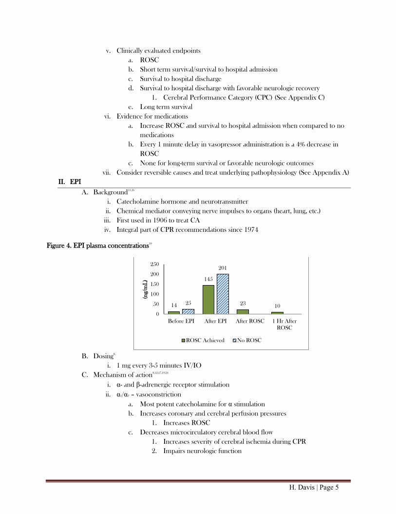
Figure 4. EPI plasma concentrations10
B. Dosing
8,
i. 1 mg every 3-5 minutes IV/IO
C. Mechanism of action8,12,17,19-24
i. α- and β-adrenergic receptor stimulation
ii. α1/α2 – vasoconstriction
a. Most potent catecholamine for α stimulation
b. Increases coronary and cerebral perfusion pressures
1. Increases ROSC
c. Decreases microcirculatory cerebral blood flow
1. Increases severity of cerebral ischemia during CPR
2. Impairs neurologic function
14
145
23 10
25
201
0
50
100
150
200
250
Before EPI After EPI After ROSC 1 Hr After
ROSC
(ng/
mL
)
ROSC Achieved No ROSC

H. Davis | Page 6
iii. β1 – inotropic, chronotropic
a. No evidence of benefit from β-stimulation
1. β-agonism has demonstrated harm over BLS alone
2. Decreases survival vs. phenylephrine or EPI + esmolol
3. Increases defibrillation attempts required to attain ROSC
b. Detrimental effects
1. Increases myocardial work, oxygen consumption and utilization of
scarce energy reserves
aa. Lactic acid production
ab. Ectopic ventricular arrhythmias
ac. Secondary VFib
2. Transient hypoxemia due to pulmonary arteriovenous shunting
3. Post-ROSC tachycardia and hypertension
aa. Precipitates recurrence of VFib
4. Post-cardiac arrest myocardial dysfunction
aa. Greater incidence of post-resuscitation shock
iv. β2 – vasodilation
a. Increases blood flow to skeletal muscles
b. Predominantly masked by α1 effects
D. Efficacy data14,25-28
Table 1. EPI vs. Placebo
Author (n) Patients Intervention Results
Woodhouse et al. 1995
339
OHCA/IHCA
Asystole or resistant VFib
Prospective RCT
HDE= 10 mg x 2 vs.
SDE= 1 mg x 2 or
placebo (not randomized) x 2
Followed by open label 1 mg EPI
No difference in:
Immediate survival
Hospital discharge
Herlitz et al. 1995 1203
OHCA
VFib
Retrospective
EPI after ≥3 rounds defibrillation vs.
no EPI regardless of defibrillation
attempts
↑ ROSC
↑ hospital admission
No difference in:
Hospital discharge
Ong et al. 2007 1296 OHCA
Prospective observational
Before vs. after protocol:
EPI 1 mg x 1 prior to transport with 1
mg dose x 1 in ED
No difference in:
ROSC
Hospital admission
Hospital discharge
Olsveengen et al. 2010 851 OHCA
Prospective RCT
ACLS with vs. without IV access and
standard drugs
↑ ROSC
↑ hospital admission
↑ CPR time
No difference in:
Hospital discharge
Favorable neurologic outcome
Jacobs et al. 2011 534 OHCA Prospective, DB, RCT
EPI 1 mg vs. placebo every 3 min
↑ ROSC
↑ hospital admission
No difference in:
Hospital discharge
*All differences demonstrated statistical significance
OHCA: out-of-hospital cardiac arrest; IHCA: in-hospital cardiac arrest; RCT: randomized controlled trial; HDE: high dose epinephrine; SDE:
standard dose epinephrine; EPI: epinephrine; VFib: ventricular fibrillation; ROSC: return of spontaneous circulation; ED: emergency
department; ACLS: advanced cardiac life support; IV: intravenous; CPR: cardiopulmonary resuscitation; DB: double blind

H. Davis | Page 7
E. Bottom line19,29
i. EPI increases likelihood of achieving ROSC and survival to hospital admission
ii. Effects on long-term survival remain uncertain
iii. No role for higher doses
a. No improvement in survival with EPI doses > 1 mg
iv. Dose-dependent adverse effects
a. Unfavorable neurologic outcomes
III. VASO
A. Background8,12
i. Endogenous peptide and antidiuretic hormone
ii. Regulates water retention and electrolyte homeostasis
iii. Increases peripheral vascular resistance
iv. Guideline recommendations
a. AHA: alternative to EPI since 2000
b. ERC: not enough evidence to recommend or refute use
Figure 5. VASO plasma concentrations10
B. Dosing
8
i. 40 units once IV/IO
a. May replace first or second dose of EPI
C. Mechanism of action12
i. Non-adrenergic peripheral vasoconstrictor
a. Stimulates smooth muscle V1 receptors
D. Physiologic effects31-35
i. Increases coronary perfusion pressure
ii. Causes cerebral vasodilation
iii. Minimal effect on pulmonary vasculature
iv. Longer t1/2 and duration of effect than EPI
a. Increases mean arterial pressure (MAP) post-resuscitation
v. Potentiates release of ACTH and cortisol
vi. Greater stability in acidic environment compared to catecholamines
193 177
117
24
70 58
0
50
100
150
200
250
Before EPI After EPI After ROSC 1 Hr After
ROSC
(pg/
mL
)
ROSC Achieved No ROSC
H. Davis | Page 8
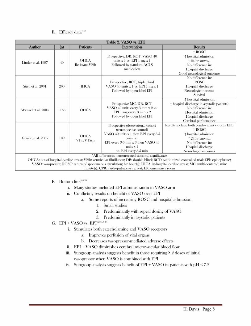
E. Efficacy data35-38
Table 2. VASO vs. EPI
Author (n) Patients Intervention Results
Linder et al. 1997 40 OHCA
Resistant VFib
Prospective, DB, RCT, VASO 40
units x 1 vs. EPI 1 mg x 1
Followed by standard ACLS
medication
↑ ROSC
↑ hospital admission
↑ 24 hr survival
No difference in:
Hospital discharge
Good neurological outcome
Stiell et al. 2001 200 IHCA
Prospective, RCT, triple blind
VASO 40 units x 1 vs. EPI 1 mg x 1
Followed by open label EPI
No difference in:
ROSC
Hospital discharge
Neurologic outcome
Survival
Wenzel et al. 2004 1186 OHCA
Prospective MC, DB, RCT
VASO 40 units every 3 min x 2 vs.
EPI 1 mg every 3 min x 2
Followed by open label EPI
(↑ hospital admission,
↑ hospital discharge in asystolic patients)
No difference in:
Hospital admission
Hospital discharge
Cerebral performance
Grmec et al. 2005 109 OHCA
VFib/VTach
Prospective observational cohort
(retrospective control)
VASO 40 units x 1 then EPI every 3-5
min vs.
EPI every 3-5 min x 3 then VASO 40
units x 1
vs. EPI every 3-5 min
Results include both combo arms vs. only EPI:
↑ ROSC
↑ hospital admission
↑ 24 hr survival
No difference in:
Hospital discharge
Neurologic outcomes
*All differences demonstrated statistical significance
OHCA: out-of-hospital cardiac arrest; VFib: ventricular fibrillation; DB: double blind; RCT: randomized controlled trial; EPI: epinephrine;
VASO: vasopressin; ROSC: return of spontaneous circulation; hr: hour(s); IHCA: in-hospital cardiac arrest; MC: multi-centered; min:
minute(s); CPR: cardiopulmonary arrest; ER: emergency room
F. Bottom line35,37,38
i. Many studies included EPI administration in VASO arm
ii. Conflicting results on benefit of VASO over EPI
a. Some reports of increasing ROSC and hospital admission
1. Small studies
2. Predominantly with repeat dosing of VASO
3. Predominantly in asystolic patients
G. EPI + VASO vs. EPI1,20,37,39,40
i. Stimulates both catecholamine and VASO receptors
a. Improves perfusion of vital organs
b. Decreases vasopressor-mediated adverse effects
ii. EPI + VASO diminishes cerebral microvascular blood flow
iii. Subgroup analysis suggests benefit in those requiring > 2 doses of initial
vasopressor when VASO is combined with EPI
iv. Subgroup analysis suggests benefit of EPI + VASO in patients with pH < 7.2

H. Davis | Page 9
H. Efficacy data1,32,39,41-44
Table 3. EPI +VASO vs. EPI
Author (n) Patients Intervention Results
Guyette et al. 2004 298 OHCA
Retrospective
VASO administration determined by
physician on scene
EPI 1 mg every 3-5 min + VASO 40 units x 1
vs. EPI 1 mg every 3-5 min
↑ ROSC
↑ ER arrival
Callaway et al. 2006 325 OHCA
Prospective, DB, RCT
VASO 40 units x 1 vs. placebo x 1
after ≥ 1 EPI
Followed by open label EPI
No difference in:
ROSC
Hospital arrival
Mally et al. 2006
598
OHCA
Resistant to
defibrillation
Prospective observational
VASO 40 units then EPI 1 mg every 3 min
vs. EPI 1 mg every 3 min
↑ petCO2; MAP
↑ ROSC
↑ 24 hr survival
↑ neurologic outcome upon
discharge
(↑ hospital discharge in asystole
subgroup)
No difference:
Hospital discharge
Gueugniaud et al. 2008 2894 OHCA
resistant VFib/VTach
Prospective DB, MC, RCT
VASO 40 units + EPI 1 mg
every 3 minutes x 2
Followed by open label EPI
vs. EPI 1 mg + placebo
every 3 minutes x 2
Followed by open label EPI
(↓ hospital discharge in PEA
subgroup analysis)
No difference in:
ROSC
Hospital admission
Hospital discharge
Neurologic recovery
1 year survival
Cody et al. 2010 191 OHCA
Retrospective cohort evaluating protocols
VASO 40 units + EPI 1 mg
Followed by EPI 1 mg every 3-5min
vs. EPI 1 mg every 3 min
No difference in:
Hospital admission
Hospital discharge
Ducros et al. 2010 44 OHCA
Prospective, DB, RCT
EPI 1 mg every + VASO 40 units every 5 min
vs. EPI 1 mg every 5 min
vs. EPI 1 mg + VASO 40 units + nitroglycerin
300 mcg every 5 min
No difference in:
Diastolic BP
ROSC
Ong et al. 2012 727 OHCA/IHCA
(ED patients)
Prospective MC, DB, parallel, RCT
VASO 40 units x 1 vs. EPI 1 mg x 1
Followed by open label EPI
↑ hospital admission (when
variables accounted for)
No difference in:
Hospital discharge
*All differences demonstrated statistical significance
OHCA: out-of-hospital cardiac arrest; EPI: epinephrine; mg: milligram; min: minute(s); VASO: vasopressin; ROSC: return of spontaneous
circulation; ER: emergency room; DB: double blind; RCT: randomized controlled trial; petCO2: end-tidal carbon dioxide; hr: hour; MC: multi-
centered; mcg: micrograms; BP: blood pressure; IHCA: in-hospital cardiac arrest
I. Bottom line40,45-48
i. Systematic reviews and meta-analyses failed to demonstrate benefit of VASO over
EPI or in combination with EPI
a. Secondary analysis suggests dose dependent increases in survival rates
when treated with VASO vs. EPI in asystolic patients
b. Largest OHCA study associated with mean time to medications > 20
minutes
ii. Meta-analysis 2014, reviewing 10 RCTs
a. No improvement in ROSC, survival to hospital admission or discharge

H. Davis | Page 10
b. IHCA subgroup analysis
1. Higher ROSC
2. Higher survival to hospital admission and discharge
3. Favorable neurologic outcomes
4. Non-traditional, repeated dosing of vasopressin
5. All vasopressin, steroid, epinephrine (VSE) data
IV. Complications after ROSC
A. Adrenal insufficiency (AI) of CA11,49-52
i. Inability to adequately increase cortisol secretion in response to ACTH during and
after ACLS
ii. Etiology
a. Adrenal gland ischemia
b. Inflammatory response to ischemia results in further damage
iii. Increases mortality
iv. Diminishes response to catecholamines
v. Leads to hemodynamic instability and circulatory collapse
B. Inflammatory mediated ischemic/reperfusion injury50,52-54
i. Activation of cytokines, endotoxins, and reactive species after CA
ii. Inflammatory response further damages ischemic organs
a. Compounds neurologic damage
b. Cytokines associated with mortality and unfavorable neurologic outcomes
iii. Normal physiologic response to cytokines is stimulation of cortisol release
a. Unable to adequately respond due to AI
C. Post-resuscitation shock6,51,55-60
i. Precise definitions of hemodynamic instability/shock are lacking
ii. Impaired contractile function and diastolic function that reverses several hours to
days after resuscitation
iii. Hyperdynamic phase, lasting minutes
a. Tachycardia and hypertension
iv. Post-resuscitation cardiovascular collapse phase
a. Cardiac index (CI) and filling pressures are low
1. Lactic acid produced
2. Myocardial stunning results in left ventricular dysfunction
3. Pro-inflammatory response resulting in loss of vascular tone
b. Occurs several hours after CA
c. Results in multi-system organ dysfunction which may progress to mortality
1. Decreases cerebral blood flow

H. Davis | Page 11
d. CI increases approximately 24 hours after arrest
1. Independent of filling pressures or inotropic agents
e. Vasodilation continues and requires vasoactive drugs
1. Recovery often seen within 3 days
v. Risk factors for development of post-resuscitation shock
a. Fifteen minute time interval between onset of arrest and ROSC
b. More frequent doses of EPI
c. Greater number of defibrillation attempts
vi. Treatment
a. Avoid hypotension
b. AHA recommends maintaining systolic blood pressure ≥ 90 mmHg and
MAP ≥ 65 mmHg
c. Other authors advocate for more aggressive goals
1. American Academy of Neurology and the Rocky Mountain
Critical Care Conference recommend MAP goal of 80-100
mmHg
aa. Based on expert opinion
ab. Theoretical improvement in neurologic outcomes
d. Hemodynamic instability was not associated with worse neurologic
outcomes when aggressively treated
1. MAP goal ≥ 75 mmHg
2. Volume expansion based on left ventricle end diastolic pressure
3. Vasoactive drugs: epinephrine or dobutamine with advanced
hemodynamic monitoring
V. Steroids
A. Background8,12
i. Decrease inflammatory response and regulate homeostasis
ii. No role for steroids in AHA or ERC guidelines
B. Cortisol plasma concentrations10,49,50,53
i. Normal range
a. Without stress: 5-20 mcg/dL
b. Under stress: 50-90 mcg/dL
ii. Low levels during or after CA indicate lack of appropriate response
a. Associated with early post-resuscitation mortality
iii. Higher cortisol levels have been associated with neurologically intact survival and
survival > 1 month
C. Mechanism of action11
i. Regulates gene expression
a. Decreases production of kinins, histamines, liposomal enzymes,
prostaglandins, leukotrienes
b. Inhibits cell migration to area of injury
c. Decreases vessel permeability

H. Davis | Page 12
D. Physiologic effects11,49,50,61,62
i. Homeostasis
a. Fluid and electrolyte balance
1. Decreases cell apoptosis and provides positive inotropy through
calcium homeostasis
b. Maintains integrity of membrane structures
ii. Anti-inflammatory
a. Decreases ischemia/reperfusion injury
iii. Vascular
a. Decreases permeability, increases SVR
b. Decreases production of vasodilators
c. Increases vascular reactivity to catecholamines and angiotensin II
iv. Cardiac
a. Increases coronary perfusion pressure
i. Increases production of coronary vasodilators
b. Increases contractility
v. Endocrine
a. Supplements cortisol not produced during AI
E. Detrimental effects62
i. Electrolyte disturbances, sodium or glucose
ii. Infections
iii. Negative regulation on HPA axis, may result in AI
F. Dosing5,52,61
i. ACLS
a. Hydrocortisone (HCT) 100 mg IV push once
b. Methylprednisolone 40 mg IV push once
ii. Stress dose steroids
a. HCT 300 mg IV per day
H. Davis | Page 13
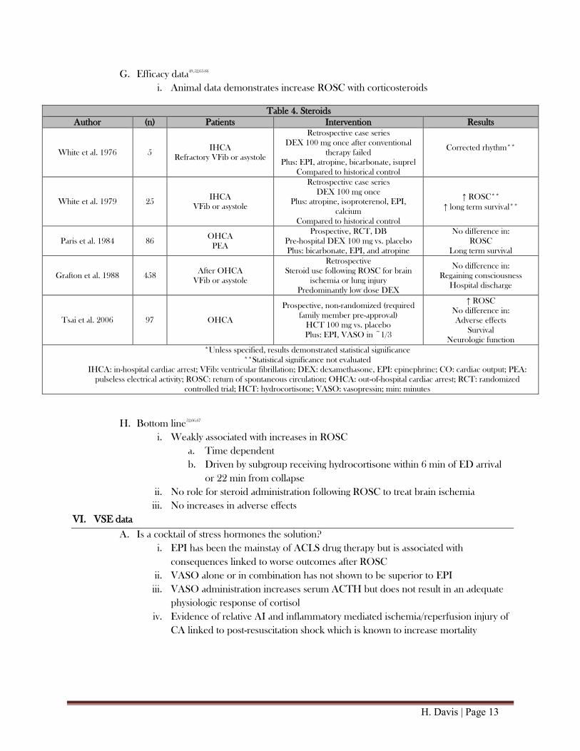
G. Efficacy data49,52,63-66
i. Animal data demonstrates increase ROSC with corticosteroids
Table 4. Steroids
Author (n) Patients Intervention Results
White et al. 1976 5 IHCA
Refractory VFib or asystole
Retrospective case series
DEX 100 mg once after conventional
therapy failed
Plus: EPI, atropine, bicarbonate, isuprel
Compared to historical control
Corrected rhythm**
White et al. 1979 25 IHCA
VFib or asystole
Retrospective case series
DEX 100 mg once
Plus: atropine, isoproterenol, EPI,
calcium
Compared to historical control
↑ ROSC**
↑ long term survival**
Paris et al. 1984 86 OHCA
PEA
Prospective, RCT, DB
Pre-hospital DEX 100 mg vs. placebo
Plus: bicarbonate, EPI, and atropine
No difference in:
ROSC
Long term survival
Grafton et al. 1988 458 After OHCA
VFib or asystole
Retrospective
Steroid use following ROSC for brain
ischemia or lung injury
Predominantly low dose DEX
No difference in:
Regaining consciousness
Hospital discharge
Tsai et al. 2006 97 OHCA
Prospective, non-randomized (required
family member pre-approval)
HCT 100 mg vs. placebo
Plus: EPI, VASO in ~1/3
↑ ROSC
No difference in:
Adverse effects
Survival
Neurologic function
*Unless specified, results demonstrated statistical significance
**Statistical significance not evaluated
IHCA: in-hospital cardiac arrest; VFib: ventricular fibrillation; DEX: dexamethasone, EPI: epinephrine; CO: cardiac output; PEA:
pulseless electrical activity; ROSC: return of spontaneous circulation; OHCA: out-of-hospital cardiac arrest; RCT: randomized
controlled trial; HCT: hydrocortisone; VASO: vasopressin; min: minutes
H. Bottom line52,66,67
i. Weakly associated with increases in ROSC
a. Time dependent
b. Driven by subgroup receiving hydrocortisone within 6 min of ED arrival
or 22 min from collapse
ii. No role for steroid administration following ROSC to treat brain ischemia
iii. No increases in adverse effects
VI. VSE data
A. Is a cocktail of stress hormones the solution?
i. EPI has been the mainstay of ACLS drug therapy but is associated with
consequences linked to worse outcomes after ROSC
ii. VASO alone or in combination has not shown to be superior to EPI
iii. VASO administration increases serum ACTH but does not result in an adequate
physiologic response of cortisol
iv. Evidence of relative AI and inflammatory mediated ischemia/reperfusion injury of
CA linked to post-resuscitation shock which is known to increase mortality

H. Davis | Page 14
B. Efficacy data61
Table 5. Mentzelopoulos S, Zakynthinos S, Tzoufi M, et al. Vasopressin, epinephrine, and corticosteroids for in-hospital cardiac arrest.
Arch Intern Med. 2009;169:15-24.
Objective Determine if VSE supplementation during and after resuscitation improves survival in refractory IHCA
Study Design
Prospective, single-center, double blind, placebo-controlled, parallel group, randomized controlled trial
Intervention:
Methylprednisolone 40 mg x 1
EPI 1 mg + VASO 20 units (per resuscitation cycle)
Following ROSC, HCT 300 mg/day continuous
infusion x 3-7 days, followed by taper, if post-
resuscitation shock present
Control:
Placebo x 1
EPI 1 mg + placebo (per resuscitation cycle)
Following ROSC, placebo x 3-7 days, followed by
taper, if post-resuscitation shock present
Post-resuscitation shock – sustained > 4 hours, MAP ≤ 70 mmHg despite appropriate fluid resuscitation, or doubling of peri-arrest
vasopressor requirements
Patients Inclusion: IHCA with PEA/asystole or VFib/VTach after 2 defibrillation attempts
Exclusion: pediatric, terminal illness, do not resuscitate order, arrest due to exsanguination, recent treatment with IV corticosteroids
Statistics
ITT analysis
Dichotomous variables: χ2
or Fisher exact test
Continuous variables: independent t test or Mann-Whitney exact test
Linear mixed-model analysis for post-resuscitation shock
Kaplan-Meier for survival
Multivariate analysis for independent predictors of death
Results
VSE:
n=100
↑ ROSC: 39/48 (81%) vs. 27/52 (52%) p=0.003
↑ survival to discharge: 9/48 (19%) vs. 2/52 (4%) p=0.02
Following ROSC: ↑ MAP, ↓ vasopressor requirements, ↓ cytokine levels, ↑ central venous oxygen saturation, ↓ in lactate,
↑ renal-failure free days
Stress dose steroids:
↑ hospital discharge 8/27 (30%) vs. 0/15 (0%) p=0.02
↑ organ failure free days
Authors’
Conclusions VSE during resuscitation and stress-dose HCT in post-resuscitation shock improves survival by a factor of 4.5 in refractory IHCA
Critique
Examined IHCA, did not exclude trauma patients
Examined physiologic endpoints potentially associated with improvements in survival
Predominantly asystole patients (75-80%)
More reversible causes of CA in study group
Use of multiple interventions; unclear if individual interventions are beneficial
Used a previously unstudied dose of VASO
Administered EPI and VASO every 2-3 minutes vs. 3-5 minutes
Feasibility of administering VASO and EPI at the same time when not prepared ahead of time by study personnel
C. Bottom line
i. Combination VSE demonstrates improvement in ROSC and survival to hospital
discharge compared to EPI alone
ii. Benefits in ROSC likely from EPI + VASO
iii. Benefits in survival likely from methylprednisolone
iv. Neurologically favorable survival not demonstrated in this study
H. Davis | Page 15
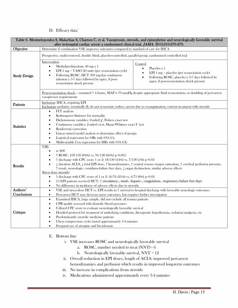
D. Efficacy data5
Table 6. Mentzelopoulos S, Malachias S, Charnos C, et al. Vasopressin, steroids, and epinephrine and neurologically favorable survival
after in-hospital cardiac arrest: a randomized clinical trial. JAMA. 2013;310:270-279.
Objective Determine if combination VSE improves outcomes compared to standard of care for IHCA
Study Design
Prospective, multi-centered, double blind, placebo-controlled, parallel-group, randomized controlled trial
Intervention
Methylprednisolone 40 mg x 1
EPI 1 mg + VASO 20 units (per resuscitation cycle)
Following ROSC, HCT 300 mg/day continuous
infusion x 3-7 days followed by taper, if post-
resuscitation shock present
Control
Placebo x 1
EPI 1 mg + placebo (per resuscitation cycle)
Following ROSC, placebo x 3-7 days followed by
taper, if post-resuscitation shock present
Post-resuscitation shock – sustained > 4 hours, MAP ≤ 70 mmHg despite appropriate fluid resuscitation, or doubling of peri-arrest
vasopressor requirements
Patients Inclusion: IHCA, requiring EPI
Exclusion: pediatric, terminally ill, do not resuscitate orders, arrests due to exsanguination, current treatment with steroids
Statistics
ITT analysis
Kolmogorov-Smirnov for normality
Dichotomous variables: 2-sided χ2
, Fishers exact test
Continuous variables: 2-sided t test, Mann-Whitney exact U test
Bonferroni correction
Linear mixed model analysis to determine effect of groups
Logistical regression for ORs with 95% Cis
Multivariable Cox regression for HRs with 95% CIs
Results
VSE:
n=268
↑ ROSC: 109/130 (84%) vs. 91/138 (66%) p=0.005
↑ discharge with CPC score 1 or 2: 18/130 (14%) vs. 7/138 (5%) p=0.02
↓ duration ACLS, ↓ total EPI dose, ↑ hemodynamics, ↑ central venous oxygen saturation, ↑ cerebral perfusion pressure,
↑ renal-, neurologic-, ventilator-failure free days, ↓ organ dysfunction, similar adverse effects
Stress dose steroids:
↑ discharge with CPC score of 1 or 2: 16/76 (21%) vs. 6/73 (8%) p=0.02
15 EPI patients received HCT, ↑ circulatory-, renal-, hepatic-, coagulation-, respiratory-failure free days
No difference in incidence of adverse effects due to steroids
Authors’
Conclusions
VSE and stress-dose HCT vs. EPI results in ↑ survival to hospital discharge with favorable neurologic outcomes
Post-arrest HCT may decrease poor outcomes, but requires further investigation
Critique
Examined IHCA, large sample, did not exclude all trauma patients
CPR quality assessed with diastolic blood pressures
Utilized CPC score to evaluate neurologically favorable survival
Detailed protocol for treatment of underlying conditions, therapeutic hypothermia, sedation/analgesia, etc
Predominantly asystolic medicine patients
Chest compression cycles lasted approximately 3-4 minutes
Frequent use of atropine and bicarbonate
E. Bottom line
i. VSE increases ROSC and neurologically favorable survival
a. ROSC, number needed to treat (NNT)= 6
b. Neurologically favorable survival, NNT = 12
ii. Overall reduction in EPI doses, length of ACLS, improved peri-arrest
hemodynamics and perfusion which results in improved long-term outcomes
iii. No increase in complications from steroids
iv. Medications administered approximately every 3-4 minutes

H. Davis | Page 16
VII. Summary of evidence
A. Majority of studies are in OHCA vs. IHCA of non-traumatic origin and are powered for
detecting differences in hospital admission
B. Many variables throughout studies over the years
i. Time to initiation of BLS/ACLS
ii. Variation in protocols of emergency responders
iii. Unknown quality of chest compressions and approach to ventilatory support
iv. Change in availability of AEDs and defibrillators
v. Change in standard medications and doses
C. Many of the principles of therapy are based on data from previously healthy animals
D. Prior to VSE, no ACLS medication or combination of medications has improved survival
to discharge or neurologically favorable survival
E. VSE demonstrated neurologically favorable survival
VIII. Conclusions
A. Addition of methylprednisolone complements EPI + VASO in improving ROSC rates
i. Improves hemodynamic stability
ii. Decreases ischemia/reperfusion injury
iii. Reduces end-organ damage
B. Improves neurologic outcomes and overall survival in IHCA requiring vasopressors
IX. Recommendations
A. IHCA
i. Administer methylprednisolone 40 mg IV x 1 during first compression cycle
requiring medications
ii. Alternate every compression cycle (2 minutes) between
a. EPI 1 mg IV every 4 minutes
b. VASO 20 units IV every 4 minutes
1. Maximum of 5 doses
iii. Continue standard use of other adjunctive treatments during ACLS when indicated
based on clinical scenario
B. If ROSC is obtained, observe for post-resuscitation shock
i. At minimum maintain a MAP of ≥ 65 mmHg consider goals of ≥ 75 mmHg
a. After appropriate volume resuscitation
b. After appropriate vasopressors are initiated
i. If norepinephrine requirements ≥ 0.2-0.5 mcg/kg/min, consider
additional stress dose steroids
ii. Avoid inotropes

H. Davis | Page 17
Appendices
Appendix A. “H’s & T’s” of Cardiac Arrest8
Cause Indications Treatment
Hypovolemia
Distributive shock
Anaphylactic shock
Hemorrhagic shock
Crystalloids
Crystalloids, EPI, H1RA, H2RA, steroids
Blood products
Hypoxia ↓ oxygen saturation, cyanosis, PEA Oxygen
Hydrogen ion (acidosis) ↓ pH, ↓ CO2 Sodium bicarbonate
Treat underlying condition
Hypokalemia ↓ K+ ↓ Mg
++
K+
, Mg++
replacement
Hyperkalemia
EKG progressing from peaked T waves to
absent P waves to prolonged PR and QRS
to sine-wave prior to loss of pulse
Shift K+
:
Calcium, insulin, dextrose
Sodium bicarbonate
Hypothermia Core temp ≤ 30°C Prevent additional heat loss
Rewarm
Tension pneumothorax
Tracheal deviation away from tension,
tachycardia, tachypnea, hypoxia resulting in
CA associated with PEA
Needle decompression initially
Chest tube for definitive management
Cardiac tamponade ↓ ventricular filling and cardiac output
causing hypotension and ultimately CA
Pericardiocentesis – needle aspiration
Pericardial window –hole in pericardium
Toxins Toxidrome presentations, patient history,
UDS or other laboratory indicators
CCB/β blockers: calcium, glucagon, high dose
insulin/D50, fat emulsions
Digoxin: antidigoxin Fab antibodies
Cyanide: hydroxocobalamine or sodium nitrite/sodium
thiosulfate
Local anesthetics: fat emulsion
Thrombosis (pulmonary)
Tachycardia, tachypnea prior to arrest, right
heart strain, PEA with rapid/narrow QRS,
evidence of DVT
Fibrinolytics and prolonged chest compressions
Thrombosis (cardiac) Chest pain, diaphoretic, EKG findings prior
to arrest, troponin elevation, cardiac history
Early coronary revascularization
Fibrinolytics as a less favorable alternative
Appendix B. Guideline Concordant ACLS Medication Dosing8
Medication Dose Indication
Epinephrine 1 mg every 3-5 minutes All rhythms
Vasopressin 40 units x 1
To replace the 1st
or 2nd
dose of epinephrine per
AHA guidelines
All rhythms
Amiodarone
1st
dose: 300 mg x 1
Refractory VFib or VTach After 3-5 minutes
2nd
dose: 150 mg x 1
After 3-5 minutes
Per ERC guidelines: 900 mg IV drip over 24 hours
All medications may be administered by IV or IO routes

H. Davis | Page 18
Appendix C. Cerebral-preformance Category5,61
References
1. Guyette F, Guimond G, Hostler D, et al. Vasopressin administered with epinephrine is associated with a
return of a pulse in out-of-hospital cardiac arrest. Resuscitation. 2004;63:277-82.
2. Sandroni C, Nolan J, Cavallaro F, et al. In-hospital cardiac arrest: incidence, prognosis, and possible
measures to improve survival. Intensive Care Med, 2007;33:237-45.
3. Moulaert V, Verbunt J, vanHeugten C, et al. Cognitive impairments in survivors of out-of-hospital cardiac
arrest: a systematic review. Resuscitation. 2009;80:297-305.
4. Stiell I, Wells G, Field B, et al. Advanced cardiac life support in out-of-hospital cardiac arrest. N Engl J Med.
2004;351(7):647-56.
5. Mentzelopoulos S, Malachias S, Chamos C, et al. Vasopressin, steroids, and epinephrine and neurologically
favorable survival after in-hospital cardiac arrest: a randomized clinical trial. JAMA. 2013;310(3):270-9.
6. Laurent I, Monchi M, Chiche J, et al. Reversible myocardial dysfunction in survivors of out-of-hospital
cardiac arrest. J Am Coll Cardiol. 2002;40(12):2110-16.
7. Ruiz-Bailen M, Aguayo de Hoyos E, Ruiz-Navarro S, et al. Reversible myocardial dysfunction after
cardiopulmonary resuscitation. Resuscitation. 2005;66:175-81.
8. Neumar R, Otto C, Link M, et al. Adult advanced cardiovascular life support: 2010 American Heart
Association guidelines for cardiopulmonary resuscitation and emergency cardiovascular care. Circulation.
2010;122:729-67.
9. Vanden Hoek T, Morrison L, Shuster M, et al. Cardiac arrest in special situations: 2010 American Heart
Association guidelines for cardiopulmonary resuscitation and emergency cardiovascular care. Circulation.
2010;122:829-61.
10. Linder K, Haak T, Keller A, et al. Release of endogenous vasopressors during and after cardiopulmonary
resuscitation. Heart. 1996;75:145-50.
11. Varvarousi G, Stefaniotou A, Varavaroussis D, et al. Glucocorticoids as an emergency pharmacologic agent
for cardiopulmonary resuscitation. Cardiovasc Drugs Ther. 2014;28:477-88.
12. Nolan J, Soar J, Zideman D, et al. European Resuscitation Council guidelines for resuscitation 2010.
Resuscitation. 2010;81:1219-444.
13. Sunde K, Steen P. The use of vasopressor agents during cardiopulmonary resuscitation. Crit Care Clin.
2012;28:189-98.
14. Olasveengen T, Sunde K, Brunborg C, et al. Intravenous drug administration during out-of-hospital cardiac
arrest. JAMA. 2009;302(20):2222-29.
15. Hubble M, Johnson C, Blackwelder J, et al. Probability of return of spontaneous circulation as a function of
timing of vasopressor administration in out-of-hospital cardiac arrest. Prehosp Emerg Car. 2015; 1-7.
16. Crile G, Dolley D. An experimental research into the resuscitation of dogs killed by anesthetics and asphyxia.
J Exp Med. 1906;8:713-25.
17. Epinephrine. Clinical Pharmacology. http://www.clinicalpharmacology-
ip.com/Forms/Monograph/monograph.aspx?cpnum=223&sec=monmech&t=0. July 14, 2015. Accessed:
August 25, 2015.
18. Ditchey R, Lindenfeld J. Failure of epinephrine to improve the balance between myocardial oxygen supply
and demand during closed-chest resuscitation in dogs. Circulation. 1988;78:382-9.
19. Dumas F, Bougouin W, Geri G, et al. Is epinephrine during cardiac arrest associated with worse outcomes in
resuscitated patients? J Am Coll Cardiol. 2014;64(22):2360-7.
CPC Indicates
1 Conscious with normal function or only slight disability
2 Conscious with moderate disability
3 Conscious with severe disability
4 Comatose or in a vegetative state
5 Brain-dead or dead
H. Davis | Page 19
20. Ristagno G, Tang W, Huang L, et al. Epinephrine reduces cerebral perfusion during cardiopulmonary
resuscitation. Crit Care Med. 2009;37(4)1408-15.
21. Niemann J, Haynes K, Garner D, et al. Postcountershock pulseless rhythms: response to CPR, artificial
cardiac pacing, and adrenergic agonists. Ann Emerg Med. 1986;15(2):112-20.
22. Tang W, Weil M, Sun S, et al. Epinephrine increases the severity of postresuscitation myocardial
dysfunction. Circulation. 1995; 92:3089-93.
23. Prengel A, Linder K, Ensinger H, et al. Plasma catecholamine concentrations after successful resuscitation in
patients. Crit Care Med. 1992;20:609-14.
24. Ristagno G, Sun S, Tang W, et al. Effects of epinephrine and vasopressin on cerebral microcirculatory flows
during and after cardiopulmonary resuscitation. Crit Care Med. 2007;35(9):2145-9.
25. Woodhouse S, Cox S, Boyd C, et al. High dose and standard dose adrenaline do not alter survival compared
with placebo, in cardiac arrest. Resuscitation. 1995;30:243-9.
26. Herlitz J, Ekstrom L, Wennerblom B, et al. Adrenaline in out-of-hospital ventricular fibrillation, does it make
any difference? Resuscitation. 1995;29:195-201.
27. Ong M, Tiah L, Leong B, et al. A randomized, double-blind, multi-center trial comparing vasopressin and
adrenaline in patients with cardiac arrest presenting to or in the emergency department. Resuscitation.
2012;83:953-60.
28. Jacobs I, Finn J, Jelinek G, et al. Effect of adrenaline on survival in out-of-hospital cardiac arrest: a
randomized double-blind placebo-controlled trial. Resuscitation. 2011;82:1138-43.
29. Vandycke C, Martens P. High dose versus standard dose epinephrine in cardiac arrest- a meta-analysis.
Resuscitation. 2000;45(3):161-6.
30. Holmberg M, Holmberg S, Herlitz J. Low chance of survival among patients requiring adrenaline or
intubation after out-of-hospital cardiac arrest in Sweden. Resuscitation. 2002;54(1):37-45.
31. Morris D, Dereczyk B, Gryzybowski M, et al. Vasopressin can increase coronary perfusion pressure during
human cardiopulmonary resuscitation. Acad Emerg Med. 1997;4(9):878-83.
32. Mally S, Jelatancev A, Grmec S. Effects of epinephrine and vasopressin on end-tidal carbon dioxide tension
and mean arterial blood pressure in out-of-hospital cardiopulmonary resuscitation: an observational study.
Crit Care Med. 2006;11:1-8.
33. Kornberer E, Prengel A, Krismer A, et al. Vasopressin-mediated adrenocorticotropin release increases
plasma cortisol concentrations during cardiopulmonary resuscitation. Crit Care Med. 2000;28(10):3517-21.
34. Larabee T, Liu K, Campbell J, et al. Vasopressors in cardiac arrest: a systematic review. Resuscitation.
2012;83:ndi932-9.
35. Linder K, Dirks B, Strohmenger H, et al. Randomized comparison of epinephrine and vasopressin in
patients with out-of-hospital ventricular fibrillation. Lancet. 1997;349:535-7.
36. Stiell I, Hebert P, Wells G, et al. Vasopressin versus epinephrine for in-hospital cardiac arrest: a randomized
controlled trial. Lancet. 2001;358:105-9.
37. Wenzel V, Krismer A, Arntz R, et al. A comparison of vasopressin and epinephrine for out-of-hospital
cardiopulmonary resuscitation. N Engl J Med. 2004;350(2):105-13.
38. Grmec S, Mally S. Vasopressin improves outcomes in out-of-hospital cardiopulmonary resuscitation of
ventricular fibrillation and pulseless ventricular tachycardia: a observational cohort study. Crit Care.
2006;10:1-7.
39. Gueugniaud P, David J, Chanzy E, et al. Vasopressin and epinephrine vs. epinephrine alone in
cardiopulmonary resuscitation. New Engl J Med. 2008;359(1):21-30.
40. Turner D, Attridge R, Hughes D. Vasopressin associated with an increase in return of spontaneous
circulation in acidotic cardiopulmonary arrest patients. Ann Pharmacother. 2014; 48(8):986-91.
41. Callaway C, Hostler D, Doshi A, et al. Usefulness of vasopressin administered with epinephrine during out-
of-hospital cardiac arrest. Am J Cardiol. 2006;98:1316-21.
42. Cody P, Lauderdale S, Hogan D, et al. Comparison of two protocols for pulseless cardiopulmonary arrest:
vasopressin combined with epinephrine versus epinephrine alone. Prehosp Emerg Car. 2009;25(5):419-23.
43. Ducros L, Vicaut E, Soleil C, et al. Effect of the addition of vasopressin or vasopressin plus nitroglycerin to
epinephrine on arterial blood pressure during cardiopulmonary resuscitation in humans. J Emerg Med.
2010;41(5):453-9.
44. Ong M, Tan E, Pen F, et al. Survival outcomes with the introduction of intravenous epinephrine in the
management of out-of-hospital cardiac arrest. Ann Emerg Med. 2007;50(6):635-42.
45. Biondi-Zoccai G, Abbae A, Parisi Q, et al. Vasopressin superior to adrenaline or placebo in the management
of cardiac arrest? A meta-analysis. Resuscitation. 2003;59:221-4.

H. Davis | Page 20
46. Aung K, Htay T. Vasopressin for cardiac arrest. Arch Intern Med. 2005;165:17-24.
47. Mentzelopoulos S, Zakynthinos S, Siempos I, et al. Vasopressin for cardiac arrest: meta-analysis of
randomized controlled trials. Resuscitation. 2012;83:32-9.
48. Layek A, Maitra S, Pal S, Bhattacharjee S, et al. Efficacy of vasopressin during cardio-pulmonary resuscitation
in adult patients: a meta-analysis. Resuscitation. 2014;85:855-63.
49. Smithline H, Rivers E, Appleton T, et al. Corticosteroid supplementation during cardiac arrest in rats.
Resuscitation. 1993;25:257-64.
50. Ito T, Saitoh D, Takasu A, et al. Serum cortisol as a predictive marker of the outcome in patients resuscitated
after cardiopulmonary arrest. Resuscitation. 2004;62:55-60.
51. Wijdicks E, Hijdra A, Young G, et al. Quality standards subcommittee of the American Academy of
Neurology. Practic parameter: prediction of outcome in comatose survivors after cardiopulmonary
resuscitation (an evidence-based review): report of the quality standards subcommittee of the American
Academy of Neurology. Neurology. 2006;67(2):203-10.
52. Tsai M, Huang C, Chang W, et al. The effect of hydrocortisone on the outcome of out-of-hospital cardiac
arrest patients: a pilot study. Am J Emerg Med. 2007;25:318-25.
53. Tavakoli N, Bidari A, Vahdati S. Serum cortisol levels as a predictor of neuroloigic survival in successfully
resuscitated victims of cardiopulmonary arrest. J Cardio Thor Res. 2012;4(4):107-11.
54. Adriel C, Adib-Conquy M, Laurent I, et al. Successful cardiopulmonary resuscitation after cardiac arrest as a
“sepsis-like” syndrome. Circulation. 2002;106:562-68.
55. Menegazzi J, Ramos R, Wang H, et al. Post-resuscitation hemodynamics and relationship to the duration of
ventricular fibrillation. Resuscitation. 2008;78:355-8.
56. Mayr V, Luckner G, Jochberger S, et al. Arginine vasopressin in advanced cardiovascular failure during the
post-resuscitation phase after cardiac arrest. Resuscitation. 2007;72:35-44.
57. Jones A, Shapiro N, Kilgannon J, et al. Goal-directed hemodynamic optimization in the post-cardiac arrest
syndrome: a systematic review. Resuscitation. 2008;77:2-29.
58. Bell D, Brindley P, Forrest D, et al. Management following resuscitation from cardiac arrest:
recommendations from the 2003 Rocky Mountain Critical Care Conference. Can J Anesth. 2005;52(3):309-
22.
59. Pene F, Hyvernat H, Mallet V, et al. Prognostic value of relative adrenal insufficiency after out-of-hospital
cardiac arrest. Intensive Care Med. 2005;31:627-33.
60. Rittenberger J, Doshi A, Reynolds J. Postcardiac arrest management. Emerg Med Clin N Am. 2015;33:691-
712.
61. Mentzelopoulos S, Zakynthinos S, Tzoufi M, et al. Vasopressin, epinephrine, and corticosteroids for in-
hospital cardiac arrest. Arch Intern Med. 2009;169:15-24.
62. Czock D, Keller F, Rasche F, et al. Pharmacokinetics and pharmacodynamics of systemically administered
glucocorticoids. Clin Pharmacokinet. 2005;44(1):61-98.
63. White B. Pulseless idioventricular rhythm during CPR: an indication for massive intravenous bolus
glucocorticoids. JACEP. 1976;5(6):449-54.
64. White B, Petinga T, Hoehner P, et al. Incidence, etiology, and outcome of pulseless idioventricular rhythm
treated with dexamethasone during advanced CPR. JACEP. 1979;8(5):188-93.
65. Paris P, Stweart R, Deggler F. Prehospital use of dexamethasone in pulseless idioventricular rhythm. Ann
Emerg Med. 1984;13(11):1008-10.
66. Grafton S, Longstreth W. Steroids after cardiac arrest: a retrospective study with concurrent nonrandomized
controls. Neurology. 1988;38:1315-6.
67. Jastermski M, Sutton-Tyrrell K, Vaagenes P, et al. Glucocorticoid treatment does not improve neurological
recovery following cardiac arrest. JAMA. 1989;262(24):3427-30.