Embed Size (px)
Citation preview

I
A COMPARATIVE STUDY OF CLINICAL EXAMINATION SCORES, SEMMES-
WEINSTEIN MONOFILAMENT EXAMINATION, VIBRATION PERCEPTION
THRESHOLD AND NERVE CONDUCTION STUDIES IN THE DIAGNOSIS OF
DIABETIC POLYNEUROPATHY
By
DR.CHETHANA .R, M.B.B.S
Dissertation Submitted to the Rajiv Gandhi University of Health Sciences,
Bangalore, Karnataka,
in partial fulfillment of the requirements for the degree of
M. D. (GENERAL MEDICINE)
Under the guidance of
DR. VEDAVATHI . R
PROFESSOR
DEPARTMENT OF GENERAL MEDICINE
KEMPEGOWDA INSTITUTE OF MEDICAL SCIENCES & RESEARCH CENTRE
BANGALORE.
2015-2018


VIII
LIST OF ABBREVIATIONS USED
1. 2-H PG: 2 HOUR PRANDIAL GLUCOSE
2. A1C: GLYCOSYLATED HEMOGLOBIN
3. CKD: CHRONIC KIDNEY DISEASE
4. DN: DIABETIC NEUROPATHY
5. DNE : DIABETIC NEUROPATHY EXAMINATION SCORE
6. DNS: DIABETIC NEUROPATHY SYMPTOM SCORE
7. DPN : DIABETIC POLYNEUROPATHY
8. DPNP: DIABETIC PERIPHERAL NEUROPATHIC PAIN
9. DSMN: DISTAL SENSORY MOTOR NEUROPATHY
10. DSN: DISTAL SENSORY NEUROPATHY
11. DSPN : DISTAL SYMMETRICAL POLYNEUROPATHY
12. ESRD: END-STAGE RENAL DISEASE
13. HTN R: HYPERTENSIVE RETINOPATHY
14. IDDM: INSULIN DEPENDENT DIABETES MELLITUS
15. IENF: INTRA EPIDERMAL NERVE FIBERS
16. IGT: IMPAIRED GLUCOSE TOLERANCE
17. MN: MOTOR NEUROPATHY
18. MODY: MATURITY-ONSET DIABETES OF THE YOUNG
19. NCS: NERVE CONDUCTION STUDIES
20. NCV: NERVE CONDUCTION VELOCITY
21. NPDR: NON PROLIFERTIVE DIABETIC RETINOPATHY

IX
22. OGTT: ORAL GLUCOSE TOLERANCE TEST
23. PDR: PROLIFERATIVE DIABETIC RETINOPATHY
24. QAFT: QUANTITATIVE AUTONOMIC FUNCTION TEST
25. SMBG: SELF-MONITORING OF BLOOD GLUCOSE
26. SWME: SEMMES-WEINSTEIN MONOFILAMENT EXAMINATION
27. VPT: VIBRATION PERCEPTION THRESHOLD

X
LIST OF TABLES
Sl NO NAME P.NO
1 CRITERIA FOR THE DIAGNOSIS OF DIABETES 12
2 TREATMENT GOALS FOR ADULTS WITH DIABETES 15
3 CLASSIFICATION FOR DIABETIC NEUROPATHIES 20
4 DISTRIBUTION OF CASES ACCORDING TO AGE 52
5 MEAN AGE OF CASES 52
6 DISTRIBUTION OF CASES ACCORDING TO SEX 52
7 ASSOCIATION OF AGE AND NEUROPATHY 53
8
DISTRIBUTION OF CASES ACCORDING
TO DURATION OF DM
54
9 MEAN DURATION OF DM OF CASES 55
10
COMPARISON OF MEAN VARIABLES BETWEEN
WITH/WITHOUT NEUROPATHY
56
11
DISTRIBUTION OF CASES ACCORDING TO DNE
56
12 ASSOCIATION OF DNE AND NEUROPATHY 57

XI
13 DISTRIBUTION OF CASES ACCORDING TO
MONOFILAMENT
58
14 ASSOCIATION OF MONOFILAMENT AND NEUROPATHY 58
15 DISTRIBUTION OF CASES ACCORDING TO VPT 59
16 ASSOCIATION OF VPT AND NEUROPATHY 60
17
18
SENSTIVITY, SPECIFICITY, PPV, NPV AND ACCURACY OF
DNE, VPT AND MONOFILAMENT
ASSOCIATION OF DAIBETIC FOOT AND NEUROPATHY
61
62
19 ASSOCIATION OF DIABETIC RETINOPATHY AND
NEUROPATHY
63

XII
LIST OF FIGURES
SL No NAME P.no
1 PATHOGENESIS OF TYPE 2 DIABETES MELLITUS 11
2 TREATMNT ALGORITHM OF TYPE 2 DM 16
3 MECHANISMS OF DIABETIC NEUROPATHY 24
4
CLINICAL PRESENTATIONS OF
DIABETIC NEUROPATHIES.
29
5 TREATMENT ALGORITHM FOR NEUROPATHIC PAIN 42
6 BIOTHESIOMETER 48
7 ASSOCIATION OF AGE AND SEX 53
8
DISTRIBUTION OF CASES ACCORDING TO
DURATION OF DM
55
9 ASSOCIATION OF DNE AND NEUROPATHY 57
10
ASSOCIATION OF MONOFILAMENT
AND NEUROPATHY
59
11 ASSOCIATION OF VPT AND NEUROPATHY 60

XIII
12
DISTRIBUTION OF CASES ACCORDING TO NCS
61

1
ABSTRACT
“A COMPARATIVE STUDY OF CLINICAL EXAMINATION
SCORES, SEMMES - WEINSTEIN MONOFILAMENT
EXAMINATION, VIBRATION PERCEPTION THRESHOLD AND
NERVE CONDUCTION STUDIES IN THE DIAGNOSIS OF
DIABETIC POLYNEUROPATHY”
BACKGROUND:
The number of patients with diabetes mellitus is increasing by epidemic proportions in
the world, particularly in India. India has 69.2 million people living with diabetes (8.7%)
as per the 2015 WHO data. Diabetic Polyneuropathy (DPN) is the most common type of
diabetic neuropathy and contributes to 50 to 70% of nontraumatic amputations. The
overall prevalence of neuropathy in the South Indian type 2 diabetic subjects is around
19.1% (1)
.
Up to 50% of diabetic peripheral neuropathies may be asymptomatic. If not recognized
and if preventive foot care is not implemented, patients are at risk for injuries to their
insensate feet(2)
.
OBJECTIVES:
1. To assess and compare clinical examination scores: Diabetic Neuropathy Examination
Score, Semmes -Weinstein monofilament examination, Vibration perception threshold
and Nerve conduction studies in the diagnosis of diabetic polyneuropathy.
2. To find out the optimal bedside method in the diagnosis of diabetic polyneuropathy.

2
METHODS :
100 patieints who fulfill the inclusion criteria were subjected to Diabetic Neuropathy
Symptom Score (DNS) and those subjects with positive DNS score were subjected to
Diabetic Neuropathy Examination Score(DNE) ,Semmes-Weinstein monofilament
examination(SWME), Vibration Perception Threshold(VPT) and Nerve conduction
studies(NCS). Mean and Standard Deviation for various characteristics were calculated.
Sensitivity and specificity for DNE, SWME, VPT were calculated taking NCS as gold
standard.
RESULTS:
In the present study, out of 100 study subjects 78 had diabetic neuropathy evidence
confirmed by NCS and 22 did not have neuropathy. The DNE Score gave a sensitivity of
85.9% and a specificity of 68.2% . The sensitivity and specificity of of SWME was 87.2
% and 63.6% respectively.VPT showed a sensitivity of 91% and specificity of 72.7% .
Diabetic sensory motor polyneuropathy is the most common type of neuropathy
accounting for about 71.7% of neuropathy cases.
CONCLUSION:
Simple neurological bedside clinical scores and tests are useful tools in diagnosing
diabetic peripheral neuropathy. They are effective screening tools in peripheral setting in
view of the ease of applicability and cost effectiveness.

3
KEY WORDS:
Diabetic neuropathy, Diabetic Neuropathy Symptom Score (DNS) , Diabetic Neuropathy
Examination Score (DNE), Semmes-Weinstein monofilament examination(SWME),
Vibration Perception Threshold(VPT), Nerve conduction studies(NCS).

4
“A COMPARATIVE STUDY OF CLINICAL EXAMINATION
SCORES, SEMMES - WEINSTEIN MONOFILAMENT
EXAMINATION, VIBRATION PERCEPTION THRESHOLD AND
NERVE CONDUCTION STUDIES IN THE DIAGNOSIS OF
DIABETIC POLYNEUROPATHY”
INTRODUCTION
Diabetes is an increasingly prevalent disorder with a range of systemic complications
.Diabetic neuropathy is one of the most common long-term complications of diabetes
affecting 50% of all diabetic people. (3)
This heterogeneous group of conditions affects
different parts of the nervous system and presents with diverse clinical manifestations.
The early recognition and appropriate management of neuropathy in the patient with
diabetes is very important.
Lower extremity disease, including peripheral neuropathy, foot ulceration, peripheral
arterial disease, or lower extremity amputation, is twice as common in diabetic persons
compared with non diabetic persons and it affects 30 per cent of diabetic persons who are
older than 40 yr. (3)
Up to 50% of diabetic peripheral neuropathies may be asymptomatic.
If not recognized patients are at risk of foot complications.
Screening and early identification of neuropathy offer a crucial opportunity for the patient
with diabetes to actively modulate the course of suboptimal glycaemic control to
currently recommended targets, and to implement improved foot care before the onset of
significant morbidity. (4)

5
We designed a comparative study to assess the effectiveness of various diagnostic
modalities like clinical scoring system Diabetic neuropathy examination score(DNE) ,
10-g Semmes-Weinstein Monofilament Examination (SWME) , Quantitative Sensory
Testing by Vibration Perception Threshold (VPT) and Nerve conduction studies(NCS) in
the diagnosis of diabetic polyneuropathy. The various subtypes of neuropathy was also
studied.
In the study by Mithili et al (4)
, 71 of 100 subjects had evidence of neuropathy confirmed
by Nerve Conduction Studies, while 29 did not have neuropathy. The DNE score gave a
sensitivity of 83% and a specificity of 79%. The sensitivity of SWME was 98.5% and
specificity was 55%. Vibration Perception Thresholds yielded a sensitivity of 86% and a
specificity of 76%.
In the study by Jayaprakash p et al(5)
, 1044 patients with diabetes mellitus were
studied.All subjects had a detailed clinical assessment including Diabetic Neuropathy
Symptom (DNS) score, Diabetic Neuropathy Examination (DNE) score, ankle reflex,
vibration sensation with a 128 Hz tuning fork, 10 g Semmes-Weinstein monofilament
and vibration perception threshold (VPT) and interpreted that simple bed side tests are
useful for assessing peripheral diabetic neuropathy, even in those subjects in whom foot
care practices are not followed.

6
In a study by Meijer JW et al (6)
, 73 diabetic subjects were studied and concluded that the
DNE score is a sensitive and well-validated hierarchical scoring system that is fast and
easy to perform in clinical practice.
In a study by Kumar S and Fernando et al (7)
, 182 subjects of diabetes were studied The
SWME was more sensitive (100%) but less specific (77.7%) in identifying patients who
had foot ulcers compared to biothesiometry which was less sensitive (78.6%) but more
specific (93.4%) and concluded that the filaments are therefore reliable and may be
superior to biothesiometry in screening for patients at risk of foot ulceration since
sensitivity is the more important parameter. In addition, they are inexpensive (12 pounds)
compared to the biothesiometer (400 pounds) and are simple and easy to use.
In a study by Olaleye D (8)
et al, 478 subjects were studied and the screening tests were
significantly and positively correlated with NCS.
In a study by Armstrong DG (9)
et al 115 subjects were studied and evaluated the
sensitivity and specificity of 2 commonly used neuropathy assessment tools (vibration
perception threshold testing and the Semmes-Weinstein 10-g monofilament wire system)
and suggested that testing instruments are sensitive in identifying patients at risk for
ulceration.

7
AIMS AND OBJECTIVES
The present study aims at evaluating patients by bedside clinical examination scores:
DNE, Semmes-Weinstein Monofilament Examination and Vibration perception threshold
which are cost effective.
OBJECTIVES:
1. To assess and compare clinical examination scores: DNE, Semmes-Weinstein
Monofilament Examination, Vibration perception threshold and Nerve conduction studies
in the diagnosis of diabetic polyneuropathy.
2. To find out the optimal bedside method in the diagnosis of diabetic polyneuropathy.
3. The prevalence of various subtypes of neuropathy depending on the distribution was
studied.

8
REVIEW OF LITERATURE
DIABETES MELLITUS
Diabetes mellitus (DM) refers to a group of common metabolic disorders that share the
phenotype of hyperglycemia .(10)
Several distinct types of DM are caused by a complex
interaction of genetics and environmental factors. Depending on the etiology of the DM,
factors contributing to hyperglycemia include reduced insulin secretion, decreased
glucose utilization, and increased glucose production.
The metabolic dysregulation associated with DM causes secondary pathophysiologic
changes in multiple organ systems that impose a tremendous burden on the individual
with diabetes and on the health care system. DM is the leading cause of end-stage renal
disease (ESRD), non-traumatic lower extremity amputations, and adult blindness. It also
predisposes to cardiovascular diseases.
CLASSIFICATION(11)
Diabetes can be classified into the following general categories:
1. Type 1 diabetes (due to autoimmune b(beta)-cell destruction, usually leading to
absolute insulin deficiency)
2. Type 2 diabetes (due to a progressive loss of b(beta)-cell insulin secretion frequently
on the background of insulin resistance)

9
3. Gestational diabetes mellitus (GDM) (diabetes diagnosed in the second or third
trimester of pregnancy that was not clearly overt diabetes prior to gestation)
4. Specific types of diabetes due to other causes, e.g., monogenic diabetes syndromes
(such as neonatal diabetes and maturity-onset diabetes of the young (MODY), diseases of
the exocrine pancreas (such as cystic fibrosis), and drug- or chemical-induced diabetes
(such as with glucocorticoid use, in the treatment of HIV/AIDS, or after organ
transplantation)
PATHOPHYSIOLOGY
TYPE 1 DM
Type 1 DM is the result of interactions of genetic, environmental, and immunologic
factors that ultimately lead to the destruction of the pancreatic beta cells and insulin
deficiency. Pathologically, the pancreatic islets are infiltrated with lymphocytes (a
process termed insulitis). After all beta cells are destroyed, the inflammatory process
abates, the islets become atrophic, and most immunologic markers disappear. The precise
mechanisms of beta cell death are not known but may involve formation of nitric oxide
metabolites, apoptosis, and direct CD8+ T cell cytotoxicity.
The islet destruction is mediated by T lymphocytes rather than islet autoantibodies, as
these antibodies do not generally react with the cell surface of islet cells and are not
capable of transferring DM to animals. Suppression of the autoimmune process at the

10
time of diagnosis of diabetes slows the decline in beta cell destruction, but the safety of
such interventions is unknown.
TYPE 2 DM
Type 2 DM is characterized by impaired insulin secretion, insulin resistance,
excessive hepatic glucose production, and abnormal fat metabolism. Obesity,
particularly visceral or central (as evidenced by the hip-waist ratio), is very common in
type 2 DM (80% or more are obese). In the early stages of the disorder, glucose tolerance
remains near-normal, despite insulin resistance, because the pancreatic beta cells
compensate by increasing insulin output.
As insulin resistance and compensatory hyperinsulinemia progress, the pancreatic islets
in certain individuals are unable to sustain the hyperinsulinemic state. IGT(Impaired
glucose tolerance), characterized by elevations in postprandial glucose, then develops. A
further decline in insulin secretion and an increase in hepatic glucose production lead to
overt diabetes with fasting hyperglycemia. Ultimately, beta cell failure ensues.

11
Figure:1, PATHOGENESIS OF TYPE 2 DIABETES MELLITUS

12
TABLE 1:CRITERIA FOR THE DIAGNOSIS OF DIABETES ( 11)
FPG 126 mg/dL (7.0 mmol/L).
Fasting is defined as no caloric intake for at least 8 h.* OR
2-h PG 200 mg/dL (11.1 mmol/L) during an OGTT.
The test should be performed as described by the WHO, using a glucose load containing
the equivalent of 75 g anhydrous glucose dissolved in water.* OR
A1C 6.5% (48 mmol/mol). The test should be performed in a laboratory using a method
that is NGSP certified and standardized to the DCCT assay.* OR
In a patient with classic symptoms of hyperglycemia or hyperglycemic crisis, a random
plasma glucose 200 mg/dL (11.1 mmol/L).
*In the absence of unequivocal hyperglycemia, results should be confirmed by repeat
testing.
MANAGEMENT OF TYPE 2 DM
Diabetes care:
A) Initial evaluation: a complete evalution must be performed to assess the complete
health status of the patient.
B) Lifestyle Management

13
C) Glycemic control: (12 , 13)
ASSESSMENT OF GLYCEMIC CONTROL: Patient self-monitoring of blood
glucose (SMBG) and A1C are available to health care providers and patients to
assess the effectiveness and safety of the management plan on glycemic control.
Glycemic goals in adults: lowering A1C to below or around 7% has been shown to
reduce microvascular complications and if implemented soon after diagnosis is associated
with long term reduction in macrovascular complications. HbA1c values above 8% (64
mmol/mol) are generally unacceptable by all guidelines. (13)
PHARMACOLOGIC APPROACHES TO GLYCEMIC TREATMENT
PHARMACOLOGIC THERAPY FOR TYPE 1 DIABETES
Insulin Therapy
Insulin is the mainstay of therapy for individuals with type 1 diabetes. Generally, the
starting insulin dose is based on weight, with doses ranging from 0.4 to 1.0 units/kg/ day
of total insulin with higher amounts required during puberty. . The American Diabetes
Association/JDRF Type 1 Diabetes Sourcebook notes 0.5 units/kg/day as a typical
starting dose in patients who are metabolically stable, with higher weight-based dosing
required immediately following presentation with ketoacidosis (14).

14
Pramlintide:
Pramlintide, an amylin analog, is an agent that delays gastric emptying, blunts pancreatic
secretion of glucagon, and enhances satiety. It is U.S. Food and Drug Administration
(FDA)–approved for use in adults with type 1 diabetes.
PANCREAS AND ISLET TRANSPLANTATION
Pancreas and islet transplantation have been shown to normalize glucose levels but
require lifelong immunosuppression to prevent graft rejection and recurrence of
autoimmune islet destruction. Given the potential adverse effects of immunosuppressive
therapy, pancreas transplantation should be reserved for patients with type 1 diabetes
undergoing simultaneous renal transplantation, following renal transplantation, or for
those with recurrent ketoacidosis or severe hypoglycemia despite intensive glycemic
management (15)
.
PHARMACOLOGIC THERAPY FOR TYPE 2 DIABETES
Initial Therapy :
Metformin monotherapy should be started at diagnosis of type 2 diabetes unless there are
contraindications. Metformin is effective and safe, is inexpensive, and may reduce risk of
cardiovascular events and death (16)
. Metformin may be safely used in patients with
estimated glomerular filtration rate (eGFR) as low as 30 mL/min/1.73 m2 (17)
Combination therapies:

15
A patient centered approach should be used to guide the choice of pharmacologic agents.
Considerations include efficacy, cost, potential side effects, effects on weight,
comorbidities, hypoglycemia risk and patient preferences.
TABLE 2:TREATMENT GOALS FOR ADULTS WITH DIABETES
INDEX GOAL
Glycemic control
HbA1C <7.0% (13)
Preprandial capillary plasma glucose 3.9–7.2 mmol/L (70–130 mg/dL)
Peak postprandial capillary plasma
glucose
<10.0 <1.7 mmol/L (<180 mg/dL)

16
Blood pressure <140/90mmHg (18,19)
Lipids Low-density lipoprotein <2.6 mmol/L (100 mg/dL)
High-density lipoproteinmmol/L >1 mmol/L (40 mg/dL) in men
>1.3 (50mg/dL) in women
Triglycerides <1.7 mmol/L (150 mg/dL)
FIGURE 2: TREATMNT ALGORITHM OF TYPE 2 DM International Diabetes
Federation (IDF)-treatment Algorithm (20)

17
COMPLICATIONS OF DIABETES MELLITUS
All forms of diabetes, both inherited and acquired, are characterized by hyperglycemia, a
relative or absolute lack of insulin, and the development of diabetes-specific
microvascular pathology in the retina, renal glomerulus, and peripheral nerve. Diabetes is
also associated with accelerated atherosclerotic macrovascular disease affecting arteries
that supply the heart, brain, and lower extremities. Pathologically, this condition
resembles macrovascular disease in nondiabetic patients, but it is more extensive and
progresses more rapidly.
Overall, life expectancy is about 7 to 10 years shorter than for people without diabetes
mellitus because of increased mortality from diabetic complications.
ACUTE COMPLICATIONS:
1. Diabetic keto acidosis
2. Hyperglycemic Hyperosmolar State
3. Hypoglycemia
4. Lactic acidosis
CHRONIC COMPLICATIONS:
MACROVASCULAR:
Coronary heart disease
Peripheral arterial disease
Cerebrovascular disease

18
MICRO VASCULAR:
NEPHROPATHY:
Diabetic kidney disease, or CKD attributed to diabetes, occurs in 20–40% of patients
with diabetes and is the leading cause of end-stage renal disease (ESRD). Diabetic kidney
disease typically develops after a diabetes duration of 10 years, or at least 5 years in type
1 diabetes, but may be present at diagnosis of type 2 diabetes.
Chronic kidney disease (CKD) is diagnosed by the presence of elevated urinary albumin
excretion (albuminuria), low estimated glomerular filtration rate (eGFR), or other
manifestations of kidney damage (21,22)
.Intensive glycemic control with the goal of
achieving near-normoglycemia has been shown in large prospective randomized studies
to delay the onset and progression of albuminuria and reduced eGFR in patients with type
1 diabetes and type 2 diabetes . (23,24)
ACE inhibitors or ARBs are the preferred first-line agent for blood pressure treatment
among patients with diabetes, hypertension, eGFR ,60 mL/min/1.73 m2 , and UACR 300
mg/g Cr because of their proven benefits for prevention of CKD progression and major
Cardiovascular events . (25)
In general, ACE inhibitors and ARBs are considered to have
similar benefits and risks. In the setting of lower levels of albuminuria (30–299 mg/g Cr),
(26) ACE inhibitor or ARB therapy has been demonstrated to reduce progression to more
advanced albuminuria (300 mg/g Cr) and cardiovascular events but not progression to
ESRD(27)

19
RETINOPATHY (28,29)
Diabetic retinopathy is a highly specific vascular complication of both type 1 and type 2
diabetes, with prevalence strongly related to both the duration of diabetes and the level of
glycemic control. Diabetic retinopathy is the most frequent cause of new cases of
blindness among adults aged 20–74 years in developed countries.
Screening Adults with type 1 diabetes should have an initial dilated and comprehensive
eye examination by an ophthalmologist or optometrist within 5 years after the onset of
diabetes. Patients with type 2 diabetes should have an initial dilated and comprehensive
eye examination by an ophthalmologist or optometrist at the time of the diabetes
diagnosis.
Promptly refer patients with any level of macular edema, severe nonproliferative diabetic
retinopathy (a precursor of proliferative diabetic retinopathy), or any proliferative
diabetic retinopathy to an ophthalmologist who is knowledgeable and experienced in the
management of diabetic retinopathy.
NEUROPATHY
DN(Diabetic Neuropathy) is not a single entity but a number of different syndromes,
ranging from subclinical to clinical manifestations depending on the classes of nerve
fibers involved. According to the San Antonio Convention , the main groups of
neurologic disturbance in diabetes mellitus include: subclinical neuropathy, determined
by abnormalities in electrodiagnostic and quantitative sensory testing, 2) diffuse clinical
20
neuropathy with distal symmetric sensorimotor and autonomic syndromes, and 3) focal
syndromes. (30)
Diabetic peripheral neuropathy (DPN) is a common complication estimated to affect 30–
50% of individuals with diabetes (31)
.The primary risk factor for DPN is hyperglycaemia
(32, 31) .Other independent risk factors include age, duration of disease, cigarette smoking,
hypertension, elevated triglycerides, higher BMI, alcohol consumption, and taller height
(32, 31) .Interestingly, between 25 and 62% of patients with idiopathic peripheral
neuropathy are reported to have prediabetes; among these 11–25% are thought to have
peripheral neuropathy, and 13–21% have neuropathic pain (33)
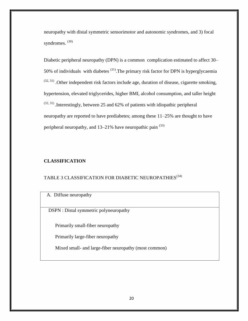
CLASSIFICATION
TABLE 3 CLASSIFICATION FOR DIABETIC NEUROPATHIES(34)
A. Diffuse neuropathy
DSPN : Distal symmetric polyneuropathy
Primarily small-fiber neuropathy
Primarily large-fiber neuropathy
Mixed small- and large-fiber neuropathy (most common)

21
Autonomic
Cardiovascular
Reduced HRV (Heart rate variability)
Resting tachycardia
Orthostatic hypotension
Sudden death (malignant arrhythmia)
Gastrointestinal
Diabetic gastroparesis (gastropathy)
Diabetic enteropathy (diarrhea)
Colonic hypomotility (constipation)
Urogenital
Diabetic cystopathy (neurogenic bladder)
Erectile dysfunction
Female sexual dysfunction
Sudomotor dysfunction
Distal hypohydrosis/anhidrosis,
Gustatory sweating

22
Hypoglycemia unawareness
Abnormal pupillary function
B. Mononeuropathy (mononeuritis multiplex) (atypical forms)
Isolated cranial or peripheral nerve (e.g., CN III, ulnar, median, femoral, peroneal)
Mononeuritis multiplex (if confluent may resemble polyneuropathy)
C. Radiculopathy or polyradiculopathy (atypical forms)
Radiculoplexus neuropathy (a.k.a. lumbosacral polyradiculopathy, proximal motor
amyotrophy) Thoracic radiculopathy
D. Nondiabetic neuropathies common in diabetes
Pressure palsies
Chronic inflammatory demyelinating polyneuropathy
Radiculoplexus neuropathy
Acute painful small-fiber neuropathies (treatment induced)
PATHOPHYSIOLOGIC MECHANISMS INVOLVED IN THE DEVELOPMENT OF
DIABETIC PERIPHERAL NEUROPATHY
1. ADVANCED GLYCATION END PRODUCTS (AGES) (35)

23
Formed during the Maillard reaction, advanced glycation end products (AGEs) act on
specific receptors (RAGEs), inducing monocytes and endothelial cells to increase the
production of cytokines and adhesion molecules. AGEs can form cross-links in matrix
structural proteins induce mutagenesis of bacterias, physiologically increase in number as
patient ages, and in pathologic states like diabetes and renal failure.
2. PROTEIN KINASE C (PKC) (36)
A family of 12 isoenzymes, PKC is activated by phosphorylation and is involved in
intracellular signaling and binding to the second messenger diacylglycerol. Conditions
with increased levels of glucose have found increased levels of PKC and diacyclglycerol
in retinal, aortic, and renal tissue but decreased in neural tissues. However, PKC
inhibitors studies suggest improvement in Na+-K+ ATPase activity, which contributes to
the diminished NCV in diabetes.
3. OXIDATIVE STRESS (37)
Radicals are generated from glucose metabolism to produce ATP. Radicals, such as
superoxide anion, are capable of profound tissue damage as well as diacyclglycerol
synthesis, which activates PKC.
4. POLYOL PATHWAY (38)
This is generally a physiologic catabolic pathway supplied by intracellular glucose. It
becomes pathologic when excessive glucose increases the reduction and regeneration of

24
glutathione requiring NADPH. This results in an imbalance NADPH/NAD ratio that
causes depletion of glutathione, increases AGEs, and activates PKC and diacylglycerol.
The first redox reaction of this pathway produces sorbitol. Accumulation of this
compound was once thought to be destructive to nerves; however, a study was conducted
showing insignificant levels of sorbitol in diabetic patients.
FIGURE 3:Mechanisms of diabetic neuropathy. Factors linked to type 1 diabetes
(yellow), type 2 diabetes (blue), and both (green) cause DNA damage, endoplasmic
reticulum stress, mitochondrial dysfunction, cellular injury, and irreversible damage. The
relative importance of the pathways in this network will vary with cell type, disease
profile, and time. ER, endoplasmic reticulum; FFA, free fatty acids; PI3-K,

25
phosphatidylinositol-3 kinase; RNS, reactive nitrogen species; ROS, reactive oxygen
species.
CLINICAL PRESENTATION
The spectrum of clinical neuropathic syndromes described in patients with diabetes
mellitus includes dysfunction of almost every segment of the somatic peripheral and
autonomic nervous system (39)
. Each syndrome can be distinguished by its
pathophysiologic, therapeutic, and prognostic features.
FOCAL NEUROPATHIES
Mononeuritis and Entrapment Syndromes
Mononeuropathies occur primarily in the older population, their onset is generally acute,
associated with pain, and their course is self-limiting, resolving within 6-8 weeks. These
are due to vascular obstruction after which adjacent neuronal fascicles take over the
function of those infarcted by the clot.(40)
Mononeuropathies must be distinguished from
entrapment syndromes that start slowly, progress and persist without intervention.
Common entrapment sites in diabetic patients involve median, ulnar, radial, femoral,
lateral cutaneous nerves of the thigh, peroneal, and medial and lateral plantar nerve.
Carpal tunnel syndrome occurs twice as frequently in a people with diabetes compared
with a normal healthy population, and its increased prevalence in diabetes may be related

26
to repeated undetected trauma, metabolic changes, or accumulation of fluid or edema
within the confined space of the carpal tunnel.
DIFFUSE NEUROPATHIES
Proximal motor neuropathies (Diabetic amyotrophy, femoral neuropathy)
Proximal motor neuropathy can be clinically identified based on recognition of these
common features:
1) primarily affects the elderly,
2) gradual or abrupt onset,
3) begins with pain in the thighs and hips or buttocks
4) followed by significant weakness of the proximal muscles of the lower limbs with
inability to rise from the sitting position (positive Gower's maneuver).
5) begins unilaterally and spreads bilaterally,
6) coexists with distal symmetric polyneuropathy, and
7) spontaneous muscle fasciculation, or provoked by percussion.
The condition is now recognized as being secondary to a variety of causes unrelated to
diabetes, but which have a greater frequency in patients with diabetes than the general
population. It includes patients with chronic inflammatory demyelinating polyneuropathy
(CIDP), monoclonal gammopathy, circulating GM1 antibodies and antibodies to neuronal
cells and inflammatory vasculitis (41,42)
.It was formerly thought to resolve spontaneously
in 1.5 to 2 years, but now, if found to be immune-mediated, can resolve within days on
immunotherapy.

27
The condition is readily recognizable clinically with prevailing weakness of the
iliopsoas, obturator, and adductor muscles, together with relative preservation of the
gluteus maximus and minimus and hamstrings (43)
. Those people affected have great
difficulty rising out of chairs unaided and often use their arms to assist themselves. Heel
or toe standing is surprisingly good. In the classic form of diabetic amyotrophy, axonal
loss is the predominant process and the condition coexists with DSPN. (44)
DISTAL SYMMETRIC POLYNEUROPATHY (DSPN)
Distal symmetric polyneuropathy (DSPN) is the most common and widely recognized
form of DN. The onset is usually insidious but occasionally is acute, following stress or
initiation of therapy for diabetes. DSPN may be either sensory or motor, and involve
small fibers, large fibers, or both .
Small nerve fiber dysfunction usually occurs early and often is present without objective
signs or electrophysiologic evidence of nerve damage . It is manifested early with
symptoms of pain and hyperalgesia in the lower limbs, followed by a loss of thermal
sensitivity and reduced light touch and pinprick sensation. (39)
There is now evidence that DSPN may be accompanied by loss of cutaneous nerve fibers
that stain positive for the neuronal antigen PGP9.5 as well as impaired neurovascular
blood flow. (45,46)
Clinical manifestations of small fiber neuropathies (47)

28
Symptoms are prominent. Pain is of the C-fiber type. It is burning and superficial
and associated with allodynia i.e. interpretation of all stimuli as painful (e.g.
touch)
Late in the condition there is hypoalgesia
Defective warm thermal sensation
Defective autonomic function with decreased sweating, dry skin, impaired
vasomotor function and blood flow and a cold foot.
There is remarkable intactness of reflexes, motor strength
Electrophysiologically silent
Loss of cutaneous nerve fibers using PGP 9.5 staining
Diagnosed clinically by reduced sensitivity to 10g Semmes Weinstein
monofilament and pricking sensation using the Waardenberg wheel or similar
instrument
Abnormalities in thresholds for warm thermal perception, neurovascular function,
pain, quantitative sudorimetry and quantitative autonomic function tests.
Risk is foot ulceration and subsequent gangrene (there are 65,000 amputations in
the US each year, 1 every 2 minutes, 50% are preventable)

29
FIGURE 4:CLINICAL PRESENTATIONS OF DIABETIC NEUROPATHIES. (48)
ACUTE PAINFUL NEUROPATHY(49-52)
Some patients develop a predominantly small-fiber neuropathy, which is
manifested by pain and paresthesias early in the course of diabetes. It may be
associated with the onset of insulin therapy and has been termed insulin neuritis.
By definition, it has been present for less than 6 months.
Symptoms often are exacerbated at night and are manifested in the feet more than
the hands. Spontaneous episodes of pain can be severely disabling. The pain
varies in intensity and character. Pain syndromes lasting longer than 6 to 12
months are classified as chronic.

30
Neuropathy may be associated with profound weight loss and severe depression
that has been termed diabetic neuropathic cachexia. This syndrome occurs
predominantly in male patients and can occur at any time in the course of T1DM
or T2DM. It is self-limited and invariably responds to simple symptomatic
treatment.
CHRONIC PAINFUL NEUROPATHY
Chronic painful neuropathy is another variety of painful polyneuropathy. Onset is
later, often years into the course of the diabetes; pain persists for longer than 6
months and becomes debilitating. This condition can result in tolerance to
narcotics and analgesics, finally resulting in addiction. It is extremely resistant to
all forms of intervention and is most frustrating to both patient and physician.
LARGE FIBER NEUROPATHIES
Large fiber neuropathies may involve sensory and/or motor nerves. These tend to
be the neuropathies of signs rather than symptoms. Large fibers subserve motor
function, vibration perception, position sense, and cold thermal perception. Unlike
the small nerve fibers these are the myelinated, rapidly conducting fibers that
begin in the toes and have their first synapse in the medulla oblongata. They tend
to be affected first because of their length and the tendency in diabetes for nerves
to "die back".
Clinical presentation of large fiber neuropathies

31
Impaired vibration perception (often the first objective evidence) and position
sense.
Depressed tendon reflexes.
A delta type deep-seated gnawing, dull, like a toothache in the bones of the feet,
or even crushing or cramp-like pain.
Sensory ataxia (waddling like a duck).
Wasting of small muscles of feet with hammertoes .Shortening of the Achilles
tendon with pes equinus.
Increased blood flow (hot foot).
Most patients with DSPN, however, have a "mixed" variety of neuropathy with both
large and small nerve fiber damages. In the case of DSPN, a "glove and stocking"
distribution of sensory loss is almost universal.
DIABETIC AUTONOMIC NEUROPATHIES (53,54)
Autonomic neuropathies affect the autonomic neurons (parasympathetic, sympathetic, or
both) and are associated with a variety of site-specific symptoms. The symptoms and
signs of autonomic dysfunction should be elicited carefully during the medical history
and physical examination.
Major clinical manifestations of diabetic autonomic neuropathy include hypoglycemia
unawareness, resting tachycardia, orthostatic hypotension, gastroparesis, constipation,
diarrhea, fecal incontinence, erectile dysfunction, neurogenic bladder, and sudomotor
dysfunction with either increased or decreased sweating. Although Cardiac autonomic
neuropathy is the most studied and clinically relevant of the diabetic autonomic

32
neuropathies, gastrointestinal, genitourinary, and sudomotor dysfunction should be
considered in the optimal care of patients with diabetes.
DIAGNOSIS OF PERIPHERAL NEUROPATHY
The diagnosis of diabetic neuropathy rests heavily on a careful history, for which a
number of questionnaires have been developed . The initial neurologic evaluation should
be directed toward detecting the specific part of the nervous system affected by diabetes.
Bedside neurologic examination is quick and easy but provides nominal or ordinal
measures and contains substantial interindividual and intraindividual variation.
The 2010 Toronto conference on diabetic neuropathy and the 2009 conference of the
American Academy of Neurology recommended that at least one parameter from each of
the following five categories be measured to classify diabetic neuropathy: symptom
profiles, neurologic examination, QST, nerve conduction study, and autonomic function
testing.
A number of simple symptom screening questionnaires are available to record symptom
quality and severity. Diabetic Neuropathy Symptom (DNS )Score adopted from the
Neuropathy Symptom Score (NSS) of Dyck. (55)
Diabetic neuropathy examination
score: DNE score, which is a modification of the Neuropathy Disability Score of Dyck.
The DNE score consists of eight items, two testing muscle strength, one a tendon reflex,
and five sensations. The maximum score is 16. A score of >3 points is considered
abnormal. (56)

33
The Michigan Neuropathy Screening Instrument (MNSI) is a 15-item questionnaire that
can be administered to patients as a screening tool for neuropathy. (57)
Other similar
symptom-scoring systems have also been described, such as the nerve impairment score
of the lower limbs (NIS-LL). (58)
PERIPHERAL TESTING DEVICES
A number of relatively inexpensive devices allow suitable assessment of somatosensory
function, including vibration, thermal, light touch, and pain perception. (59)
The most widely used device in clinical practice is the Semmes-Weinstein monofilament.
(60-62)
The filament assesses pressure perception when gentle pressure is applied to the handle
sufficient to buckle the nylon filament. Although filaments of many different sizes are
available, the one that exerts 10 g of pressure is most commonly used to assess pressure
sensation in the diabetic foot. It is also referred to as the 5.07 monofilament because,
during calibration, the filaments are calibrated to exert a force measured in grams that is
10 times the log of the force exerted at the tip: hence, 5.07 exerts 10 g of force. A number
of cross sectional studies have assessed the sensitivity of the 10-g monofilament to
identify feet at risk for ulceration. Sensitivities vary from 86% to 100%, although there is
no consensus as to how many sites should be tested. (63,64)

34
VIBRATION PERCEPTION THRESHOLD TESTING BY BIOTHESIOMETER
The biothesiometer (or neurothesiometer) is a simple handheld device that gives
semiquantitative assessment of vibration perception threshold (VPT). VPT is a sensitive
measure of peripheral neuropathy (65)
.
NERVE CONDUCTION STUDIES (66)
Whole-nerve electrophysiologic procedures (e.g., NCV, F waves, sensory amplitudes,
motor amplitudes) have emerged as important methods of tracing the onset and
progression of peripheral neuropathy. An appropriate battery of electrophysiologic tests
supports the measurement of the speed of sensory and motor conduction, the amplitude of
the propagating neural signal, the density and synchrony of muscle fibers activated by
maximal nerve stimulation, and the integrity of neuromuscular transmission.
These are objective, parametric, noninvasive, and highly reliable measures. However,
standard procedures, such as maximal NCV(nerve conduction velocity), reflect only a
limited aspect of neural activity, and then only in a small subset of the large-diameter and
heavily myelinated axons. Even in large-diameter fibers, NCV is insensitive to many
pathologic changes known to be associated with peripheral neuropathy.
However, a key role for electrophysiologic assessment is to rule out other causes of
neuropathy or to identify neuropathies superimposed on peripheral neuropathy. Unilateral
conditions, such as entrapments, are far more common in patients with diabetes than in

35
healthy subjects. Nerve Conduction Velocity is gradually diminished by peripheral
neuropathy, with estimates of loss of approximately 0.5 m/sec per year.
CARDIOVASCULAR TESTING DEVICES (67)
QAFT consists of a series of simple, noninvasive tests for detecting cardiovascular
autonomic neuropathy. These tests are based on detection of heart rate and BP responses
to a series of maneuvers. Specific tests are used in evaluating disordered regulation of
gastrointestinal, genitourinary, and sudomotor function and peripheral skin blood flow
induced by autonomic diabetic neuropathy.
EMERGING MARKERS OF DPN: FOCUS ON SMALL FIBERS
Contact heat-evoked potentials (CHEP)
It can be elicited by non-noxious heat or painful stimuli from the toe to the dorsum of the
back with measurement of negative and positive amplitudes and latencies by recording
impulses in the central nervous system. This promises to provide both temporal and
spatial resolution of measures of nociception and has the unique ability to measure
conduction in C and Aδ fibers, which are normally below the resolution of standard
methods. CHEP coupled with functional MRI may lead to an improved understanding of
nociceptive pathways and enhance the generation of new therapies directed at the
pathways involved. (68)

36
Nerve biopsy
Nerve biopsy detects unmyelinated fiber damage while myelinated nerve fiber
morphology is still normal in patients with early DPN . However, nerve biopsy is an
invasive and highly specialized procedure that requires electron-microscopy and cannot
be advocated for routine use. (69)
Skin biopsy
Skin punch biopsy, a minimally invasive procedure, allows morphometric quantification
of intra epidermal nerve fibers (IENF) most commonly expressed as the number of IENF
per length of section (IENF/mm). Intra- and inter-observer variability for the assessment
of IENF density is good, declines with age, is lower in males than in females, and is not
influenced by weight or height. The blister technique is a less invasive procedure that
assesses innervation of the epidermis alone and shows good agreement with punch
biopsy. (70)
IENF density is lower in diabetic patients with painful compared with painless
early neuropathy. (71)
Corneal confocal microscopy (72)
Corneal confocal microscopy is a noninvasive technique that can detect small sensory
corneal nerve fiber loss in diabetic neuropathy , idiopathic small fibre neuropathy and
Fabry disease. Corneal nerve fiber damage correlates with IENF loss and the severity of
neuropathy in diabetic patients was more prominent in painful neuropathy. Its
quantification may be a surrogate marker of diabetic neuropathy.

37
Nerve axon reflex/flare response (73)
Stimulation of C-nociceptive fibers by acetylcholine iontophoresis induces vasodilation,
which can be quantitatively measured and serve as a measure of small fiber function.
This technique correlates with other measurements of small fiber function and may be
considered for the diagnosis of SFN in diabetic patients. The laser Doppler imaging flare
test evaluates 44°C heat-induced vasodilation and is reduced in subjects with type 2
diabetic patients with and without neuropathy .Further studies are required to validate
these tests as diagnostic tools or as outcome measures in clinical trials.
MANAGEMENT OF DN
PREVENTIVE PAIN MANAGEMENT
Controlling hyperglycemia not only helps prevent the development of DPNP (Diabetic
Peripheral Neuropathic Pain), but it also delays its progression . Observational studies
suggest that good glucose control and avoidance of extreme blood glucose fluctuations
improve neuropathic symptoms. In fact, the United Kingdom Prospective Diabetes Study
(UKPDS) showed that progression of disease depends on management of glucose in both
type I and type II diabetes. (74)
LIFESTYLE MODIFICATIONS
The initial approach to glucose control is with lifestyle modifications. One lifestyle
modification is weight loss. In the geriatric population, a 7% reduction in weight and 150
minutes of moderate exercise weekly is recommended. Others, such as improvement of

38
lipid and blood pressure indexes, smoking cessation, and alcohol drinking reduction are
recommended even if there are no definitively positive preventive studies.
PHARMACOLOGIC INTERVENTION
PAIN MANAGEMENT OPTIONS FOR DPNP
There are numerous agents, both non pharmacologic and pharmacologic, available for
DPNP and each has some risk of adverse side effects. Careful selection of the appropriate
agent depends upon a thorough understanding of the patient‘s history, such as past
medical history and current medications taken, and the findings from the physical
examination.
According Argoff et al, the first-line or first-tier medications for DPNP include
duloxetine, pregabalin, oxycodone CR, and the class of tricyclic antidepressants. Only
duloxetine and pregabalin are FDA approved for the treatment of DPNP. (75)
Tricyclic antidepressants (TCAs).
Tricyclic antidepressants (TCAs) inhibit reuptake of serotonin and/or norepinephrine in
the presynaptic membrane, and modulate sodium channels and NMDA receptors.TCAs
include amitriptyline, imipramine, desipramine, nortriptyline, clomipramine, and other
agents. Amitriptyline has been the most studied of the TCAs and has been found to be
effective for pain, independent of its effect on depression.
Pregabalin.
Pregabalin is a selective high affinity ligand for alpha2-delta protein subunit of voltage-
gated calcium channels, which play a role in the development of pathologic changes

39
believed to be associated with neuropathic pain in humans. These agents reduce calcium
influx and, subsequently, the release of several neurotransmitters associated with
analgesia such as glutamate, substance P, and calcitonin gene-related peptide (CGRP). (76)
Duloxetine. (77)
Duloxetine, serotonin-norepinephrine reuptake inhibitor, has shown promise as an
effective initial agent for DPNP. The trials used to investigate its potential to relieve
DPNP took into consideration its effect as an anti-depressant by excluding candidates
with any history suggestive of depression.
At a dose of 120 mg/d duloxetine has shown improvement in shooting, stabbing, hot-
burning, and splitting pain sensations and at 60mg/d patients it has shown improvement
in stabbing and sharp sensations.
Oxycodone CR. (78)
Oxycodone CR is an opioid agonist designed to slowly release oxycodone over many
hours. Just like all of the other opioids analgesics, oxycodone has historically been
considered after other pharmacologic agents have failed for the treatment of DPNP.
Gabapentin. (79)
Gabapentin is an alpha2-delta ligand anticonvulsant much like pregabalin. Its effect on
pain is related to its interaction with the L-amino acid transporter, alteration of the release
of GABA, high affinity for alpha2-delta subunit of voltage-activated calcium channels,
inhibition of voltage-activated sodium channels, and alteration of monoamine
neurotransmitter release, and blood serotonin levels.It is renally excreted and crosses the
blood brain barrier. In trials comparing its effect with TCAs such as amitriptyline, the
pain relief was equal, but the onset was more rapid. It also markedly improves sleep.

40
Tramadol. Tramadol is a ―central acting analgesic with unique properties as a weak
inhibitor of norepinephrine and serotonin and low affinity binding to mu receptors.
Several trials have found it to provide pain relief.
Lamotrigine (80)
Lamotrigine is an anticonvulsant believed to inhibit glutamate release by stabilizing
neural membranes through blocking of the sodium channels. It has been found to produce
pain relief in both AIDS-related neuropathy and DPN. However, it is associated with
adverse cutaneous effects which may limit its use in the general population.
Venlafaxine
Venlafaxine is a serotonin-norepinephrine reuptake inhibitor. Two trials have been
conducted suggesting its effectiveness in DPNP. It has an advantage of once daily dosing
with the ER formulation.
ADJUNCT MANAGEMENT AND TREATMENT OF COMPLICATIONS
It is important to improve strength and balance in patients with large-fiber neuropathy.
Patients can benefit from high-intensity strength training by increasing muscle strength
and improving coordination and balance, thereby reducing fall and fracture risks. (81)
Low-impact activities that emphasize muscle strength and coordination and challenge the
vestibular system, such as Pilates, yoga, and tai chi, can also be particularly helpful. In
addition, options
to prevent and correct foot deformities are available, including orthotics, surgery, and
reconstruction.

41
Basic management of small-fiber neuropathies by the patient should be encouraged.
These include foot protection and ulcer prevention by wearing padded socks; daily foot
inspection using a mirror to examine the soles of the feet; selection of proper footwear;
scrutiny of shoes for the presence of foreign objects that lodge themselves in closed
shoes; and avoidance of sun-heated surfaces, hot bathwater, and sleeping with feet in
front of a fireplace or heater. Patient education should reinforce these strategies and
should also discourage soaking of the feet in water.
STIMULATION. (82)
Transcutaneous nerve stimulation (electrotherapy) occasionally is helpful and certainly
represents one of the more benign therapies for painful neuropathy. Care should be taken
to move the electrodes around to identify sensitive areas and obtain maximum relief.
Static magnetic field therapy has been reported to be of benefit, but it is difficult to blind
such studies. Similarly, the use of infrared light has reportedly had benefit, but this
remains to be proved.

42
FIGURE 5:Treatment algorithm for neuropathic pain after exclusion of nondiabetic
etiologies and stabilization of glycemic control. IVIg, intravenous immune globulin;
SNRI, serotonin-norepinephrine reuptake inhibitors; TCA, tricyclic antidepressants. (83)

43
METHODOLOGY
SOURCE OF DATA:
Patients of type 2 diabetes mellitus both outpatient and inpatient in the Department of
General Medicine, Kempegowda Institute of Medical Sciences, Bangalore during study
period from December 2015 to September 2017 were taken for study considering the
inclusion and exclusion criteria.
SAMPLE SIZE:
Total: 100 cases.
TYPE OF STUDY: Comparative study.
DURATION OF STUDY: 22 months
METHODS OF COLLECTION OF DATA:
Informed consent was taken from all the Patients/ care takers of the patients enrolled for
the study. The patients data was collected in a well designed proforma.
1. The patients dermographics, clinical history regarding the type, duration and treatment
of diabetes, history of foot ulcer were recorded.
2.Those subjects who fulfill the inclusion criteria were subjected to:
Diabetic Neuropathy Symptom Score(DNS)
3.Those subjects with positive DNS score were subjected to
-Diabetic Neuropathy Examination Score(DNE)
-Semmes-Weinstein monofilament examination(SWME)

44
-Vibration Perception Threshold(VPT)
-Nerve Conduction Studies(NCS)
4.Investigations: routine haematological and biochemical tests, LFT,RFT,
FBS,PPBS,HbA1C
INCLUSION CRITERIA :
1.Patients with DIABETES MELLITUS according to ADA criteria with positive
Diabetic Neuropathy Symptom score
2.Patients willing to participate in the study.
EXCLUSION CRITERIA:
Patients with
1.Liver disease, chronic alcoholics
2.Renal diseases
3.Anaemia
4.History of toxic exposure, drugs that can cause peripheral neuropathy
5. Patients with other known causes of peripheral neuropathy like inflammatory diseases,
monoclonal gammopathy.
DIABETIC NEUROPATHY SYMPTOM SCORE
All subjects were questioned regarding the presence or otherwise of symptoms, either
positive or negative suggesting the presence of neuropathy. The questionnaire was the

45
Diabetic Neuropathy Symptom DNS Score (3)
adopted from the Neuropathy Symptom
Score (NSS) of Dyck (84)
.
Diabetic neuropathy symptom Score: The questions should be answered ‗yes‘ (positive: 1
point) if a symptom occurred more times a week during the last 2 weeks or ‗no‘
(negative: No point) if it did not.
1. Symptoms of unsteadiness in walking?
2. Do you have a burning, aching pain or tenderness of your legs or feet?
3. Do you have pricking sensations at your legs and feet?
4. Do you have places of numbness on your legs or feet?
Maximum score: 4 points; 0 points- PNP absent; 1-4 points - PNP present
(PNP = Polyneuropathy)
Diabetic neuropathy examination score:
A thorough neurological examination was carried out and the neurological signs were
scored following a DNE score, which is a modification of the Neuropathy Disability
Score of Dyck.(84,4)
The DNE score consists of eight items, two testing muscle strength,
one a tendon reflex, and five sensations. The maximum score is 16. A score of >3 points
is considered abnormal.
Muscle strength
1. Quadriceps femoris: Extension of the knee

46
2. Tibialis Anterior: Dorsiflexion of the foot
Reflex
3. Ankle reflex
Sensation: Index finger
4. Sensitivity to pinpricks
Sensation: Big toe
5. Sensitivity to pinpricks
6. Sensitivity to touch
7. Vibration perception
8. Sensitivity to joint position
Only the right leg and foot are tested.
If right leg is amputated, then left leg is tested.
Scoring from 0 to 2
0 = Normal
1 = Mild/moderate deficit
Muscle strength: MRC scale 3-4

47
Reflex: Decreased but present
Sensation: Decreased but present
2 = severely disturbed/absent
Muscle strength: MRC scale 0-2
Reflex: Absent
Sensation: Absent
Maximum score: 16 points
A score of > 3 indicates presence of polyneuropathy.
SEMMES-WEINSTEIN MONOFILAMENT EXAMINATION
Light touch/pressure perception was assessed using a 10 g monofilament. These were
applied on both feet on the plantar surface of the hallux and centrally at the heel. The end
of the filament was pressed on the plantar surface of the hallux and centrally at the heel
with enough pressure to cause the monofilament to buckle. This was done six times at
each point and the participant was blinded to the application of the monofilament during
testing.(4)
A ‗yes-no‘ method was used, meaning that the patient says yes each time he/she
senses the application of a monofilament. The ability to correctly sense the monofilament
in six trials on both locations was defined as normal, whereas the inability to sense the
monofilament correctly in one or more trials was defined as disturbed.

48
VIBRATION PERCEPTION THRESHOLD
VPT was tested using a hand-held biothesiometer (Vibrocheck, Proactive Health Inc.).
After explaining the procedure, the button is applied to various parts of both the feet with
the patient relaxed, in the supine position in a quiet room. The vibration is increased
gradually from the minimum voltage and the transition from no vibration to the onset of
perceiving vibration is taken as VPT. The Yes/No method is used. The VPT is tested on
six areas on the plantar aspect of both feet- the hallux, the first metatarsal head, the third
metatarsal head, the fifth metatarsal head, the instep and the heel. An average of all the
areas tested is taken as the VPT of the subject. The voltage is gradually increased until
the patient senses the vibration by the Yes or No. The VPT is measured in volts. In the
present study, a voltage of more than 15 was taken as presence of neuropathy.
FIGURE 6: BIOTHESIOMETER

49
NERVE CONDUCTION STUDIES
All patients underwent conventional sensory and motor nerve conduction studies. The
nerves tested were median, ulnar, common peroneal, tibial and sural nerves. The
parameters recorded included distal latencies, amplitudes of compound motor action
potentials (CMAP), duration of CMAP, F wave latencies and conduction velocities in
motor nerves. In sensory nerves, latencies and amplitudes of the sensory nerve action
potentials and their conduction velocities were documented.
The presence or absence of neuropathy in these subjects was defined as follows:
Parameter No neuropathy Neuropathy
DNE 3 or less 4 or more
SWME Normal Disturbed
VPT 15 volts or less >15 volts
STATISTICAL ANALYSIS
All characteristics were summarized descriptively. For continuous variables, the
summary statistics of mean, standard deviation (SD) were used. For categorical data, the
number and percentage were used in the data summaries.

50
Chi-square (χ2)/ Freeman-Halton Fisher exact test was employed to determine the
significance of differences between groups for categorical data. The difference of the
means of analysis variables between two independent groups was tested by unpaired t
test. If the p-value was < 0.05, then the results were considered to be statistically
significant otherwise it was considered as not statistically significant.
Data were analyzed using SPSS software v.23.0. and Microsoft office.

51
SAMPLE SIZE ESTIMATION
Sample size calculation
With 95% confidence level, margin of error of ±8% and anticipated prevalence of
neuropathy as 19.1%, the minimum sample size is 93 (≈100).
By using the formula:
n = z2p(1-p)
d2
where
Z= z statistic at 5% level of significance
d is margin of error
p is anticipated prevalence rate

52
RESULTS
TABLE 4: DISTRIBUTION OF CASES ACCORDING TO AGE
AGE (Yrs) N %
31-40 5 5
41-50 18 18
51-60 31 31
61-70 32 32
71-80 14 14
Total 100 100
TABLE 5: MEAN AGE OF CASES
Variables Range Mean SD
AGE 35-80 59.4 10.7
TABLE:6 DISTRIBUTION OF CASES ACCORDING TO SEX
SEX N %
Male 46 46
Female 54 54
Total 100 100

53
FIGURE 7: ASSOCIATION OF AGE AND SEX
In the present study, out of 100 study subjects, 46 (46.0%) were males and 54 (54.0%)
were females. Majority i.e., 63 (63.0%) of the study subjects were in the age group of 51-
70 years The mean age was 59.4 + 10.7 years with a range from 35 to 80 years.
TABLE: 7 ASSOCIATION OF AGE AND NEUROPATHY
AGE
(Yrs)
Neuropathy No Neuropathy
N % N %
31-40 5 6.4 0 0.0
41-50 14 17.9 4 18.2
51-60 20 25.6 11 50.0
61-70 25 32.1 7 31.8
71-80 14 17.9 0 0.0
0.0
5.0
10.0
15.0
20.0
25.0
30.0
35.0
40.0
45.0
31-40 41-50 51-60 61-70 71-80
PER
CEN
TAG
E AGE DISTRIBUTION
Male
Female

54
Total 78 100.0 22 100.0
Majority of the patients with neuropathy were in the age group between 51 to 70. Out of
63 study subjects in this age group 45(71.4%) had neuropathy.
TABLE: 8 DISTRIBUTION OF CASES ACCORDING TO DURATION OF DM
DURATION OF
DM (Yrs)
N
NEUROPATHY
NO
NEUROPATHY
≤5 16 5 11
6-9 41 34 7
≥10 43 39 4
Total 100 78 22

55
Figure 8:DISTRIBUTION OF CASES ACCORDING TO DURATION OF DM
Neuropathy is more common in those with long duration of DM. 5 Subjects with duration
of diabetes ≤5 years had diabetic neuropathy. Out of 43 subjects with duration of
diabetes ≥10 years, 39 subjects had neuropathy .
TABLE 9: MEAN DURATION OF DM OF CASES
Variables Range Mean SD
DURATION OF
DM 3-30 9.5 5.0
The mean duration of Diabetes mellitus among the study subjects was 9.5 + 5 with a
range from 3 to 30.Majority i.e., 43(43%) of the study subjects had >/= 10 years
duration.
5
34
39
11
7
4
0
5
10
15
20
25
30
35
40
45
≤5 years 6 to 9 years ≥10 years
neuropathy
no neuropathy

56
TABLE:10 COMPARISON OF MEAN VARIABLES BETWEEN WITH AND
WITHOUT NEUROPATHY CASES
Variables
Neuropathy No Neuropathy
p value
Mean SD Mean SD
DURATION OF
DM(Yrs) 10.2 4.6 7.1 5.4 0.009
HbAIC 8.5 1.1 7.3 0.8 <0.001
Mean HbA1C ( 8.5 +/- 1.1) was high in patients with DN in the present study with a
significant p value.
TABLE 11: DISTRIBUTION OF CASES ACCORDING TO DNE
DNE N %
NEGATIVE 26 26
POSITIVE 74 74
Total 100 100

57
TABLE:12 ASSOCIATION OF DNE AND NEUROPATHY
DNE
Neuropathy No Neuropathy
p value
N % N %
NEGATIVE 11 14.1 15 68.2
<0.001* POSITIVE 67 85.9 7 31.8
Total 78 100.0 22 100.0
Note: *means significant at 5% level of significance (p<0.05)
FIGURE:9 ASSOCIATION OF DNE AND NEUROPATHY
Out of 100 study subjects DNE score was positive in 74(74%). The DNE was positive in
7(31.8%) study subjects without neuropathy and 68 (85.9%)subjects with neuropathy
had positive DNES score.
14.1
85.9
68.2 31.8
0.0
10.0
20.0
30.0
40.0
50.0
60.0
70.0
80.0
90.0
100.0
NEGATIVE POSITIVE
Pe
rce
nat
ge
DNE
Neuropathy
No Neuropathy

58
TABLE 13: DISTRIBUTION OF CASES ACCORDING TO MONOFILAMENT
(SWME)
MONOFILAMENT N %
IMPAIRED 76 76
NORMAL 24 24
Total 100 100
Out of 100 study subjects the SWME was impaired in 76 subjects(76%)
TABLE 14: ASSOCIATION OF MONOFILAMENT AND NEUROPATHY
MONOFILAMENT
Neuropathy No Neuropathy
p value
N % N %
IMPAIRED 68 87.2 8 36.4
<0.001* NORMAL 10 12.8 14 63.6
Total 78 100.0 22 100.0
Note: *means significant at 5% level of significance (p<0.05)
In the present study 68(87.2%) out of 78 patients with neuropathy had impaired
monofilament examination and 8 (36.4%)patients without neuropathy had impaired
monofilament examination.

59
FIGURE 10:ASSOCIATION OF MONOFILAMENT AND NEUROPATHY
TABLE 15: DISTRIBUTION OF CASES ACCORDING TO VPT
VPT N %
NEGATIVE 23 23
POSITIVE 77 77
Total 100 100
87.2
12.8
36.4
63.6
0.0
10.0
20.0
30.0
40.0
50.0
60.0
70.0
80.0
90.0
100.0
IMPAIRED NORMAL
Pe
rce
nat
ge
MONOFILAMENT
Neuropathy
No Neuropathy

60
TABLE 16: ASSOCIATION OF VPT AND NEUROPATHY
VPT
Neuropathy No Neuropathy
p value
N % N %
NEGATIVE 7 9.0 16 72.7
<0.001* POSITIVE 71 91.0 6 27.3
Total 78 100.0 22 100.0
Note: *means significant at 5% level of significance (p<0.05)
FIGURE 11: ASSOCIATION OF VPT AND NEUROPATHY
Out of 100 study subjects the VPT was impaired in 76 subjects(76%). 71(91%) out of 78
subjects with neuropathy had impaired VPT and 7(9%) subjects with neuropathy had
normal VPT.
9.0
91.0
72.7
27.3
0.0
10.0
20.0
30.0
40.0
50.0
60.0
70.0
80.0
90.0
100.0
NEGATIVE POSITIVE
Pe
rce
nat
ge
VPT
Neuropathy
No Neuropathy

61
TABLE 17: SENSTIVITY, SPECIFICITY, PPV, NPV AND ACCURACY OF DNE,
VPT AND MONOFILAMENT
DNE VPT MONOFILAMENT
Sensitivity 85.9% 91.0% 87.2%
Specificity 68.2% 72.7% 63.6%
PPV 90.5% 92.2% 89.5%
NPV 57.7% 69.6% 58.3%
Accuracy 82.0% 87.0% 82.0%
The sensitivity and specificity of DNE score in the study subjects was 85.9% and 68.2%
respectively. The sensitivity and specificity of SWME in the study subjects was 87.2%
and 63.6% respectively . VPT had the highest sensitivity and specificity of 91% and
72.7% respectively.
FIGURE 12: DISTRIBUTION OF CASES ACCORDING TO NCS
2
55
1
17
2 1
22
0
10
20
30
40
50
60
PER
CEN
TAG
E
NCS

62
Out of 100 study subjects ,nerve conduction study was normal in 22(22.0%). 78(78%)
subjects had evidence of neuropathy in NCS. DISTAL SENSORY MOTOR
NEUROPATHY(DSMN) is the commonest type which was present in 56 subjets,
followed by DISTAL SENSORY NEUROPATHY(DSN). MOTOR NEUROPATHY
(MN) and ENTRAPMET SYNDROMES(CTS) were rare. One patient had BELLS
PALSY.
TABLE 18: ASSOCIATION OF DAIBETIC FOOT AND NEUROPATHY
DAIBETIC
FOOT
Neuropathy
N %
NO 64 82.1
YES 14 17.9
Total 78 100.0
In the present study 14(17.9%) patients with neuropathy had diabetic foot.

63
TABLE 19: ASSOCIATION OF DIABETIC RETINOPATHY AND
NEUROPATHY
DIABETIC
RETINOPATHY
Neuropathy No Neuropathy
N % N %
NORMAL 15 19.2 13 59.1
NPDR 59 75.6 9 40.9
NPDR+HTN R 3 3.8 0 0.0
PDR 1 1.3 0 0.0
Total 78 100.0 22 100.0
63(80.7%) out of 78 subjects with neuropathy had evidence of diabetic retinopathy. The
commonest being non proliferative diabetic retinopathy(NPDR).

64
DISCUSSION
We have comapared our study with the following studies :
MYTHILI et al (4)
, ASHOK et al (1)
, BOULTON et al(85)
and JAYAPRAKASH et al(5).
In the present study, the prevalence of neuropathy in Type 2 diabetes was 78%
taking nerve conduction studies as gold standard. Studies on prevalence of
neuropathy in Type 2 diabetes had widely differing results, varying from 15 to
50%. The wide variability was attributed to differences in sample size, diagnostic
methods and criteria adopted for diagnosis.
STUDY PREVALENCE
PRESENT STUDY 78%
MYTHILI et al 71%
JAYAPRAKASH et al 34.9%
ASHOK et al 19.1%
In a study conducted by Mythili et al the prevalence of neuropathy in Type 2
diabetes, was 71% taking nerve conduction studies as gold standard.
Jayaprakash et al showed a prevalence of about 34.9%. In a South Indian study by
Ashok et al, the prevalence was about 19.1%.
The higher prevalence of neuropathy in the present study may be due to the
selection of patients with positive diabetic neuropathy symptom score who were

65
symptomatic and the diagnosis of neuropathy was established by nerve
conduction study which is a more sensitive method.
In the present study, duration of diabetes had a significant impact on prevalence
of neuropathy. The prevalence was 31.2% in those with duration less than 5 years
to 90.69% in those with duration more than 10 years.
Mythili et al the prevalence was 50% in those with duration less than 5 years to
92.8% in those with duration more than 10 years.
STUDY DURATION OF DM
<5YR
>10YR
PRESENT STUDY 31.2% 90.69%
MITHILI et al 50% 92.8%
Mean HbA1c was higher in those with neuropathy in the present study which was
8.5 +/- 1.1.
The present study uses the Symptom Score (DNS), and Examination Score
(DNE), which were designed by Meijer.
STUDY SENSITIVITY(DNE) SPECIFICITY
PRESENT STUDY 85.9% 68.2%
MITHILI et al 83% 79%
JAYAPRAKASH et al 68.6% 74%

66
The sensitivity and specificity of the DNE score was high in this study, sensitivity
being 85.9% and specificity of 68.2% .
In Mithili et al the sensitivity and specificity of the DNE score being 83% and
specificity 79 %respectively.
In Jayaprakash et al the sensitivity and specificity of the DNE score was 68.6 %
and specificity 74 %respectively.
STUDY SENSITIVITY(SWME) SPECIFICITY
PRESENT STUDY 87.2% 63.6%
MITHILI et al 98.5% 55%
JAYAPRAKASH et al 62.8% 92.9%
A number of cross-sectional studies have assessed the sensitivity of the 10-g
monofilament to identify feet at risk of ulceration. Sensitivity varied from 86 to
100%.The present study showed sensitivity of 87.2 %. The specificity of
monofilament in diagnosis of polyneuropathy varied from 45-60% in earlier
studies. The specificity of monofilament in diagnosis of polyneuropathy varied
from 45-60% in earlier studies .The present study showed a specificity of 63.6%.
Mithili et al study showed sensitivity of 98.5% and a specificity of 55%.
Jayaprakash et al study showed sensitivity of 62.8% and a specificity of 92.9%.

67
STUDY SENSITIVITY(VPT) SPECIFICITY
PRESENT STUDY 91% 72.7%
MITHILI et al 86% 76%
BOULTON et al 92% 81%
Boulton and colleagues documented that vibration thresholds provided a strong
indication of ―risk‖ for future ulceration across a wide range of ages and durations
of diabetes. In the present study, VPT remained highly sensitive (0.92) and
specific (0.81) Determination of VPT has the advantage of being a simple.
Present study: VPT sensitivity 91% and specificity 72.7%
Mithili et al: VPT sensitivity 86% and specificity 76%
Various subtypes of neuropathy was assessed in the present study by electro
diagnosis. Distal sensorimotor neuropathy was found to be the commonest,
followed by pure distal sensory neuropathy. Motor neuropathy was rare 3 of the
78 neuropathic subjects. Bells palsy was present in one subject. The prevalence of
the types of neuropathy in this study is similar to previous data by Mithili et al,
distal sensorimotor neuropathy was found to be the commonest, followed by pure
sensory neuropathy. Motor neuropathy was seen rarely, in 3 of the 71 neuropathic
subjects studied.

68
CONCLUSION
Among the three parameters tested, Vibration Perception Threshold is the most
specific and sensitive test. The results of Vibration Perception Threshold are
comparable to Nerve Conduction Studies in diagnosing DPN. Monofilament
examination, though quite sensitive, was less specific in diagnosing DPN. The
specificity and sensitivity of DNE is similar to monofilament.
Duration of diabetes was positively correlated with neuropathy.
Distal Sensorimotor Neuropathy and distal sensory neuropathy were the common
electrophysiological demonstrable types of neuropathy.
Majority of neuropathy cases were associated with diabetic retinopathy.
Simple cost effective Diabetic neuropathy examination score, Semmes-Weinstein
monofilament test is comparable to the Vibration perception threshold and Nerve
conduction studies in the diagnosis of diabetic neuropathy. They are an effective
screening tools in the diagnosis of diabetic peripheral neuropathy.

69
SUMMARY
The objective of the study was to assess and compare clinical examination scores:
DNE, Semmes-Weinstein Monofilament Examination, Vibration perception
threshold and Nerve conduction studies in the diagnosis of diabetic
polyneuropathy.
It was a Comparative study done in KIMS hospital. 100 patients aged above 18
years with the clinical diagnosis of DIABETIS MELLITUS were included in the
study done between DEC 2015-SEPT 2017.
Mean age in the study was 59.4 + 10.7. Of the 100 patients, 46% were male and
54% were female patients.
Out of 100 study subjects 78 showed the evidence of diabetic neuropathy in
Nerve conduction study. Vibration perception threshold showed a sensitivity and
specificity of 91% and 72.7% respectively. The DNE score and SWME were
equally good with sensitivity and specificity of DNE 85.9% and 68.2%
respectively.The sensitivity and specificity of SWME was 87.2% and 63.6%
respectively.
Distal Sensory motor neuropathy was the most common type of neuropathy.
17.9% of neuropathy subjects had evidence of diabetic foot.
80.7% of neuropathy patients had evidence of diabetic retinopathy.

70
BIBLIOGRAPHY
1. Ashok S, Ramu M, Deepa R, Mohan V. Prevalence of neuropathy in type 2
diabetic patients attending a diabetes centre in South India. Japi. 2002 Apr
1;50:546-0.
2. Pop-Busui R, Boulton AJ, Feldman EL, Bril V, Freeman R, Malik RA, Sosenko
JM, Ziegler D. Diabetic neuropathy: a position statement by the American
Diabetes Association. Diabetes Care. 2017 Jan 1;40(1):136-54.
3. Singh N, Armstrong DG, Lipsky BA. Preventing foot ulcers in patients with
diabetes. Jama. 2005 Jan 12;293(2):217-28.
4. Mythili A, Kumar KD, Subrahmanyam KA, Venkateswarlu K, Butchi RG. A
Comparative study of examination scores and quantitative sensory testing in
diagnosis of diabetic polyneuropathy. International journal of diabetes in
developing countries. 2010 Jan;30(1):43.
5. Jayaprakash P, Bhansali A, Bhansali S, Dutta P, Anantharaman R,
Shanmugasundar G, Ravikiran M. Validation of bedside methods in evaluation of
diabetic peripheral neuropathy. The Indian journal of medical research. 2011
Jun;133(6):645.
6. Meijer JW, van Sonderen E, Blaauwwiekel EE, Smit AJ, Groothoff JW, Eisma
WH, Links TP. Diabetic neuropathy examination: a hierarchical scoring system to
diagnose distal polyneuropathy in diabetes. Diabetes Care. 2000 Jun 1;23(6):750-
3.

71
7. Kumar S, Fernando DJ, Veves A, Knowles EA, Young MJ, Boulton AJ. Semmes-
Weinstein monofilaments: a simple, effective and inexpensive screening device
for identifying diabetic patients at risk of foot ulceration. Diabetes research and
clinical practice. 1991 Jan 1;13(1-2):63-7.
8. Olaleye D, Perkins BA, Bril V. Evaluation of three screening tests and a risk
assessment model for diagnosing peripheral neuropathy in the diabetes clinic.
Diabetes research and clinical practice. 2001 Nov 30;54(2):115-28.
9. Armstrong DG, Lavery LA, Vela SA, Quebedeaux TL, Fleischli JG. Choosing a
practical screening instrument to identify patients at risk for diabetic foot
ulceration. Archives of internal medicine. 1998 Feb 9;158(3):289-92.
10. Harrison's Principles of Internal Medicine, Eugene braunwald, Anthonys.Fauci,
Dennis L.Kasper, Stephen L.Hausper, Dan.L.Longo, J.Larry Jameson; 19th
Edition, volume 2; Mc.Graw Hill:2399-2407.
11. American Diabetes Association. 2. Classification and diagnosis of diabetes.
Diabetes Care. 2017 Jan 1;40(Supplement 1):S11-24.
12. Miller KM, Beck RW, Bergenstal RM, Goland RS, Haller MJ, McGill JB,
Rodriguez H, Simmons JH, Hirsch IB. T1D Exchange Clinic network: evidence
of a strong association between frequency of self-monitoring of blood glucose,
hemoglobin A1C levels in T1D Exchange Clinic Registry participants. Diabetes
Care. 2013 Jul;36(7):2009-14.
13. International Diabetes Federation. Recommendations For Managing Type 2
Diabetes In Primary Care, 2017. www.idf.org/managing-type2-diabetes.

72
14. Peters A, Laffel L. Type 1 Diabetes Sourcebook. American Diabetes Association.
2013.
15. American Diabetes Association. Pancreas and islet transplantation in type 1
diabetes. Diabetes Care. 2006 Apr 1;29(4):935-.
16. Holman RR, Paul SK, Bethel MA, Matthews DR, Neil HA. 10-year follow-up of
intensive glucose control in type 2 diabetes. New England Journal of Medicine.
2008 Oct 9;359(15):1577-89.
17. Bennett WL, Maruthur NM, Singh S, Segal JB, Wilson LM, Chatterjee R,
Marinopoulos SS, Puhan MA, Ranasinghe P, Block L, Nicholson WK.
Comparative effectiveness and safety of medications for type 2 diabetes: an
update including new drugs and 2-drug combinations. Annals of internal
medicine. 2011 May 3;154(9):602-13.
18. Aronow, W.S. Curr Treat Options Cardio Med (2015) 17: 33.
https://doi.org/10.1007/s11936-015-0394-x DOI https://doi.org/10.1007/s11936-
015-0394-x .
19. American Diabetes Association. 9. Cardiovascular disease and risk management.
Diabetes Care. 2017 Jan 1;40(Supplement 1):S75-87.
20. International Diabetes Federation (IDF)-treatmentalgorithm:
http://www.idf.org/treatment-algorithmpeople- type-2-diabetes.
21. Tuttle KR, Bakris GL, Bilous RW, Chiang JL, De Boer IH, Goldstein-Fuchs J,
Hirsch IB, Kalantar-Zadeh K, Narva AS, Navaneethan SD, Neumiller JJ. Diabetic
kidney disease: a report from an ADA Consensus Conference. American journal
of kidney diseases. 2014 Oct 31;64(4):510-33.

73
22. Of OJ. Kidney Disease: Improving Global Outcomes (KDIGO) CKD Work
Group. KDIGO 2012 Clinical Practice Guideline for the Evaluation and
Management of Chronic Kidney Disease. Kidney Int. Suppl. 2013;3:150.
23. He F, Xia X, Wu XF, Yu XQ, Huang FX. Diabetic retinopathy in predicting
diabetic nephropathy in patients with type 2 diabetes and renal disease: a meta-
analysis.
24. de Boer IH, Gao X, Cleary PA, Bebu I, Lachin JM, Molitch ME, Orchard T,
Paterson AD, Perkins BA, Steffes MW, Zinman B. Albuminuria changes and
cardiovascular and renal outcomes in type 1 diabetes: the DCCT/EDIC study.
Clinical Journal of the American Society of Nephrology. 2016 Oct 24:CJN-
02870316.
25. Heart Outcomes Prevention Evaluation (HOPE) Study Investigators. Effects of
ramipril on cardiovascular and microvascular outcomes in people with diabetes
mellitus: results of the HOPE study and MICRO-HOPE substudy. The lancet.
2000 Jan 22;355(9200):253-9.
26. Barnett AH, Bain SC, Bouter P, Karlberg B, Madsbad S, Jervell J, Mustonen J.
Angiotensin-receptor blockade versus converting–enzyme inhibition in type 2
diabetes and nephropathy. New England Journal of Medicine. 2004 Nov
4;351(19):1952-61.
27. Parving HH, Lehnert H, Bröchner-Mortensen J, Gomis R, Andersen S, Arner P.
The effect of irbesartan on the development of diabetic nephropathy in patients
with type 2 diabetes. New England Journal of Medicine. 2001 Sep
20;345(12):870-8.

74
28. Diabetic Retinopathy Study Research Group. Preliminary report on effects of
photocoagulation therapy. American journal of ophthalmology. 1976 Apr
1;81(4):383-96.
29. Early Treatment Diabetic Retinopathy Study Research Group. Photocoagulation
for diabetic macular edema: Early Treatment Diabetic Retinopathy Study report
number 1. Arch ophthalmol. 1985;103:1796-806.
30. American Diabetes Association, American Academy of Neurology. Report and
recommendations of the San Antonio conference on diabetic neuropathy. Diabetes
Care. 1988 Jul 1;11(7):592-7.
31. Deshpande AD, Harris-Hayes M, Schootman M. Epidemiology of diabetes and
diabetes-related complications. Physical therapy. 2008 Nov 1;88(11):1254-64.
32. Edwards JL, Vincent AM, Cheng HT, Feldman EL. Diabetic neuropathy:
mechanisms to management. Pharmacology & therapeutics. 2008 Oct
31;120(1):1-34.
33. Papanas N, Vinik AI, Ziegler D. Neuropathy in prediabetes: does the clock start
ticking early?. Nature Reviews Endocrinology. 2011 Nov 1;7(11):682-90.
34. Albers JW, Pop-Busui R. Diabetic neuropathy: mechanisms, emerging treatments,
and subtypes. Current neurology and neuroscience reports. 2014 Aug 1;14(8):473.
35. Ahmed N. Advanced glycation endproducts—role in pathology of diabetic
complications. Diabetes research and clinical practice. 2005 Jan 31;67(1):3-21.
36. Evcimen ND, King GL. The role of protein kinase C activation and the vascular
complications of diabetes. Pharmacological Research. 2007 Jun 30;55(6):498-
510.

75
37. Nishikawa T, Edelstein D, Du XL, Yamagishi SI, Matsumura T, Kaneda Y,
Yorek MA, Beebe D, Oates PJ, Hammes HP, Giardino I. Normalizing
mitochondrial superoxide production blocks three pathways of hyperglycaemic
damage. Nature. 2000 Apr 13;404(6779):787-90.
38. Uehara K, Yamagishi SI, Otsuki S, Chin S, Yagihashi S. Effects of polyol
pathway hyperactivity on protein kinase C activity, nociceptive peptide
expression, and neuronal structure in dorsal root ganglia in diabetic mice.
Diabetes. 2004 Dec 1;53(12):3239-47.
39. Vinik AI, Holland MT, LeBeau JM, Liuzzi FJ, Stansberry KB, Colen LB.
Diabetic neuropathies. Diabetes Care 1992; 15:1926–75.
40. Vinik AI, Suwanwalaikorn S. Autonomic neuropathy. In: DeFronzo R, editor.
Diabetes Mellitus. St Louis, MO, USA: Mosby-Year Book; 1997. pp. 165-76.
41. Vinik AI, Pittenger GL, Milicevic Z, Knezevic -Cuca J. Autoimmune
Mechanisms in the Pathogenesis of Diabetic Neuropathy. In: Eisenbarth RG,
editor. Molecular Mechanisms of Endocrine and Organ Specific Autoimmunity.
Georgetown: Landes Company, 1998: 217-51.
42. Steck AJ, Kappos L. Gangliosides and autoimmune neuropathies: classification
and clinical aspects of autoimmune neuropathies. Journal of neurology,
neurosurgery, and psychiatry. 1994 Nov;57(Suppl):26.
43. Sander HW, Chokroverty S. Diabetic amyotrophy: current concepts. InSeminars
in neurology 1996 Jun (Vol. 16, No. 02, pp. 173-178). © 1996 by Thieme
Medical Publishers, Inc..

76
44. Said G, Goulon‐Goeau C, Lacroix C, Moulonguet A. Nerve biopsy findings in
different patterns of proximal diabetic neuropathy. Annals of neurology. 1994
May 1;35(5):559-69.
45. McArthur JC, Stocks EA, Hauer P, Cornblath DR, Griffin JW. Epidermal nerve
fiber density: normative reference range and diagnostic efficiency. Archives of
neurology. 1998 Dec 1;55(12):1513-20.
46. Dalsgaard CJ, Rydh M, Haegerstrand A. Cutaneous innervation in man visualized
with protein gene product 9.5 (PGP 9.5) antibodies. Histochemistry and Cell
Biology. 1989 Sep 1;92(5):385-90.
47. Hanson PH, Schumacker P, Debugne T, Clerin M. Evaluation of somatic and
autonomic small fibers neuropathy in diabetes. American journal of physical
medicine & rehabilitation. 1992 Feb;71(1):44-7.
48. Shlomo Melmed Kenneth Polonsky P. Reed Larsen Henry Kronenberg Williams
Textbook of Endocrinology 12th
edition . Complications of Diabetes Mellitus pg
no 1502 .Clinical presentations of diabetic neuropathies. (Modified from Pickup
J, Williams G, eds. Textbook of Diabetes, Vol 1. Oxford, UK: Blackwell
Scientific, 1997.)
49. Tesfaye S, Malik R, Harris N, Jakubowski JJ, Mody C, Rennie IG, Ward JD.
Arterio-venous shunting and proliferating new vessels in acute painful neuropathy
of rapid glycaemic control (insulin neuritis). diabetologia. 1996 Mar 1;39(3):329-
35.

77
50. Van Heel DA, Levitt NS, Winter TA. Diabetic neuropathic cachexia: the
importance of positive recognition and early nutritional support. International
journal of clinical practice. 1998;52(8):591.
51. Holland NR, Crawford TO, Hauer P, Cornblath DR, Griffin JW, McArthur JC.
Small‐fiber sensory neuropathies: clinical course and neuropathology of
idiopathic cases. Annals of neurology. 1998 Jul 1;44(1):47-59.
52. Said G, Bigo A, Ameri A, Gayno JP, Elgrably F, Chanson P, Slama G.
Uncommon early-onset neuropathy in diabetic patients. Journal of neurology.
1998 Jan 1;245(2):61-8.
53. Spallone V, Ziegler D, Freeman R, Bernardi L, Frontoni S, Pop‐Busui R, Stevens
M, Kempler P, Hilsted J, Tesfaye S, Low P. Cardiovascular autonomic
neuropathy in diabetes: clinical impact, assessment, diagnosis, and management.
Diabetes/metabolism research and reviews. 2011 Oct 1;27(7):639-53.
54. Kempler P, Amarenco G, Freeman R, Frontoni S, Horowitz M, Stevens M, Low
P, Pop‐Busui R, Tahrani AA, Tesfaye S, Várkonyi T. Management strategies for
gastrointestinal, erectile, bladder, and sudomotor dysfunction in patients with
diabetes. Diabetes/metabolism research and reviews. 2011 Oct 1;27(7):665-77.
55. Meijer JW, Smit AJ, Sonderen EV, Groothoff JW, Eisma WH, Links TP.
Symptom scoring systems to diagnose distal polyneuropathy in diabetes: the
Diabetic Neuropathy Symptom score. Diabetic Medicine. 2002 Nov
1;19(11):962-5.
56. Dyck PJ. Detection, characterization, and staging of polyneuropathy: assessed in
diabetics. Muscle & nerve. 1988 Jan 1;11(1):21-32.

78
57. Feldman EL, Stevens MJ, Thomas PK, Brown MB, Canal N, Greene DA. A
practical two-step quantitative clinical and electrophysiological assessment for the
diagnosis and staging of diabetic neuropathy. Diabetes care. 1994 Nov
1;17(11):1281-9.
58. Bril V. NIS-LL: the primary measurement scale for clinical trial endpoints in
diabetic peripheral neuropathy. European neurology. 1999;41(Suppl. 1):8-13.
59. Vinik AI, Suwanwalaikorn S, Stansberry KB, Holland MT, McNitt PM, Colen
LE. Quantitative measurement of cutaneous perception in diabetic neuropathy.
Muscle & nerve. 1995 Jun 1;18(6):574-84.
60. Valk GD, de Sonnaville JJ, van Houtum WH, Heine RJ, van Eijk JT, Bouter LM,
Bertelsman FW. The assessment of diabetic polyneuropathy in daily clinical
practice: reproducibility and validity of Semmes Weinstein monofilaments
examination and clinical neurological examination.
61. Mayfield JA, Sugarman JR. The use of the Semmes-Weinstein monofilament and
other threshold tests for preventing foot ulceration and amputation in persons with
diabetes. Journal of Family Practice. 2000 Nov 1;49(11):S17-.
62. Bourcier ME, Ullal J, Parson HK, et al. Diabetic peripheral neuropathy: Pract.
2006;55:505-8.
63. Kumar S, Fernando DJ, Veves A, Knowles EA, Young MJ, Boulton AJ. Semmes-
Weinstein monofilaments: a simple, effective and inexpensive screening device
for identifying diabetic patients at risk of foot ulceration. Diabetes research and
clinical practice. 1991 Jan 1;13(1-2):63-7.

79
64. Armstrong DG, Lavery LA, Vela SA, Quebedeaux TL, Fleischli JG. Choosing a
practical screening instrument to identify patients at risk for diabetic foot
ulceration. Archives of internal medicine. 1998 Feb 9;158(3):289-92.
65. Martin CL, Waberski BH, Pop-Busui R, Cleary PA, Catton S, Albers JW,
Feldman EL, Herman WH, DCCT/EDIC Research Group. Vibration perception
threshold as a measure of distal symmetrical peripheral neuropathy in type 1
diabetes. Diabetes Care. 2010 Dec 1;33(12):2635-41.
66. Vinik AI, Anandacoomaraswamy D, Ullal J. Antibodies to neuronal structures.
Diabetes Care. 2005 Aug 1;28(8):2067-72.
67. Hanson PH, Schumacker P, Debugne T, Clerin M. Evaluation of somatic and
autonomic small fibers neuropathy in diabetes. American journal of physical
medicine & rehabilitation. 1992 Feb;71(1):44-7.
68. Zambreanu L, Wise RG, Brooks JC, Iannetti GD, Tracey I. A role for the
brainstem in central sensitisation in humans. Evidence from functional magnetic
resonance imaging. Pain. 2005 Apr 30;114(3):397-407.
69. Malik RA, Tesfaye S, Newrick PG, Walker D, Rajbhandari SM, Siddique I,
Sharma AK, Boulton AJ, King RH, Thomas PK, Ward JD. Sural nerve pathology
in diabetic patients with minimal but progressive neuropathy. Diabetologia. 2005
Mar 1;48(3):578-85.
70. HADDEN R. Joint Task Force of the EFNS and the PNS. European Federation of
Neurological Societies/Peripheral Nerve Society Guideline on management of
paraproteinemic demyelinating neuropathies. Report of a joint task force of the

80
European Federation of Neurological Societies and the Peripheral Nerve Society.
J Peripher Nerv Syst.. 2006;11:9-19.
71. Sorensen L, Molyneaux L, Yue DK. The relationship among pain, sensory loss,
and small nerve fibers in diabetes. Diabetes care. 2006 Apr 1;29(4):883-7.
72. Quattrini C, Tavakoli M, Jeziorska M, Kallinikos P, Tesfaye S, Finnigan J,
Marshall A, Boulton AJ, Efron N, Malik RA. Surrogate markers of small fiber
damage in human diabetic neuropathy. Diabetes. 2007 Aug 1;56(8):2148-54.
73. Green AQ, Krishnan S, Finucane FM, Rayman G. Altered C-fiber function as an
indicator of early peripheral neuropathy in individuals with impaired glucose
tolerance. Diabetes care. 2010 Jan 1;33(1):174-6.
74. Duby JJ, Campbell RK, Setter SM, Rasmussen KA. Diabetic neuropathy: an
intensive review. American Journal of Health-System Pharmacy. 2004 Jan
15;61(2):160-73.
75. Argoff CE, Backonja MM, Belgrade MJ, Bennett GJ, Clark MR, Cole BE,
Fishbain DA, Irving GA, McCarberg BH, McLean MJ. Consensus guidelines:
treatment planning and options. InMayo Clinic Proceedings 2006 Apr 30 (Vol.
81, No. 4, pp. S12-S25). Elsevier.
76. Richter RW, Portenoy R, Sharma U, Lamoreaux L, Bockbrader H, Knapp LE.
Relief of painful diabetic peripheral neuropathy with pregabalin: a randomized,
placebo-controlled trial. The journal of pain. 2005 Apr 30;6(4):253-60.
77. Goldstein DJ, Lu Y, Detke MJ, Lee TC, Iyengar S. Duloxetine vs. placebo in
patients with painful diabetic neuropathy. Pain. 2005 Jul 31;116(1):109-18.

81
78. Watson CP, Moulin D, Watt-Watson J, Gordon A, Eisenhoffer J. Controlled-
release oxycodone relieves neuropathic pain: a randomized controlled trial in
painful diabetic neuropathy. Pain. 2003 Sep 30;105(1):71-8.
79. Backonja M, Beydoun A, Edwards KR, Schwartz SL, Fonseca V, Hes M,
LaMoreaux L, Garofalo E, Gabapentin Diabetic Neuropathy Study Group.
Gabapentin for the symptomatic treatment of painful neuropathy in patients with
diabetes mellitus: a randomized controlled trial. Jama. 1998 Dec 2;280(21):1831-
6.
80. Eisenberg E, Lurie Y, Braker C, Daoud D, Ishay A. Lamotrigine reduces painful
diabetic neuropathy A randomized, controlled study. Neurology. 2001 Aug
14;57(3):505-9.
81. Nelson ME, Fiatarone MA, Morganti CM, Trice I, Greenberg RA, Evans WJ.
Effects of high-intensity strength training on multiple risk factors for osteoporotic
fractures: a randomized controlled trial. Jama. 1994 Dec 28;272(24):1909-14.
82. Somers DL, Somers MF. Treatment of neuropathic pain in a patient with diabetic
neuropathy using transcutaneous electrical nerve stimulation applied to the skin of
the lumbar region. Physical therapy. 1999 Aug 1;79(8):767-75.
83. Shlomo Melmed ,Kenneth Polonsky, P. Reed Larsen, Henry Kronenberg,
Williams Textbook of Endocrinology 12th
edition Complications of Diabetes
Mellitus pg no 1515.
84. Tesfaye S, Boulton AJ, Dyck PJ, Freeman R, Horowitz M, Kempler P, Lauria G,
Malik RA, Spallone V, Vinik A, Bernardi L. Diabetic neuropathies: update on

82
definitions, diagnostic criteria, estimation of severity, and treatments. Diabetes
care. 2010 Oct 1;33(10):2285-93.
85. Boulton AJ, Kubrusly DB, Bowker JH, Gadia MT, Quintero L, Becker DM,
Skyler JS, Sosenko JM. Impaired vibratory perception and diabetic foot
ulceration. Diabetic medicine. 1986 Jul 8;3(4):335-7.

83
FORMAT OF CONSENT FORM
INFORMED CONSENT(FROM PATIENT/CARE TAKER)
I/we …………………………………………………. Exercising my/our free power of
choice, here by give my/our consent to be included as a subject in the study of ―A
COMPARATIVE STUDY OF CLINICAL EXAMINATION SCORES, SEMMES
WEINSTEIN MONOFILAMENT EXAMINATION, VIBRATION PERCEPTION
THRESHOLD AND NERVE CONDUCTION STUDIES IN THE DIAGNOSIS OF
DIABETIC POLYNEUROPATHY‖ To my/our best satisfaction, the researcher has
informed me/us the purpose of the study and the methods by which the study is to be
conducted.
I/we have been made aware that my participation in the study does not involve any
change in on going treatment. I/we have also been made aware of my/our right to opt out
of the study at any time during the course of the study, without assigning any reason for
doing so.
Name of the Researcher Name of the Subject
Signature of the Researcher Signature of the Subject/attendar
Date: Date:
Place: Place:

84
PROFORMA
TITLE :“A COMPARATIVE STUDY OF CLINICAL EXAMINATION SCORES,
SEMMES WEINSTEIN MONOFILAMENT EXAMINATION, VIBRATION
PERCEPTION THRESHOLD AND NERVE CONDUCTION STUDIES IN THE
DIAGNOSIS OF DIABETIC POLYNEUROPATHY”
CASE NO :
PATIENT IDENTIFICATION.
NAME:
AGE:
SEX:
OCCUPATION:
ADDRESS:
TELEPHONE NO:
UHID NO: IP NO:
INPATIENT (OR) OUTPATIENT
DATE OF ADMISSION:
DATE OF DATA COLLECTION:

85
CLINICAL ASSESSMENT
COMPLAINTS:
Diabetic neuropathy symptom questionnaire:
IN THE PAST 2 WEEKS
1.ANY UNSTEADINESS IN WALKING? YES/NO
(Need for visual control, walk like drunken man, lack of contact with floor)
2.ANY BURNING, ACHNG PAIN OR TENDERNESS AT LEGS/FEET?: YES / NO.
3.ANY PRICKLING SENSATIONS AT LEGS AND FEET?: YES / NO.
4.ANY PLACES OF NUMBNESS ON LEGS OR FEET? YES/NO
POINTS:
DURATION OF PERIPHERAL NEUROPATHY SYMPTOMS:
COMORBIDITIES
DURATION OF DIABETES:
AGE OF ONSET:
TREATMENT TAKEN FOR DIABETES IN THE LAST 6MONTHS WITH DOSAGE:
HYPERTENSION STATUS:
THYROID STATUS:

86
PAST HISTORY
HISTORY OF ANY SPINAL CORD INJURIES OR SURGERIES: YES / NO.
HISTORY OF AUTOIMMUNE DISEASES: YES / NO.
HISTORY OF INFLAMMATORY DISEASES/ MONOCLONAL GAMMOPATHY:
YES/NO
HABITS:
ALCOHOL CONSUMPTION: YES / NO.
IF YES MENTION DURATION:
SMOKING STATUS:
PACK YEARS:
OTHER HABITS:
DRUG HISTORY:
WHETHER PATIENT ON VITAMIN B12, CALCIUM SUPPLEMENTS (OR)
NEUROPROTECTIVE DRUGS IN THE LAST 6MONTHS: YES / NO.
WHETHER PATIENT IS ON ANY CHEMOTHERAPY IN THE LAST 6MONTHS
(CISPLATIN, PACLITAXEL, DOCETAXEL, ETC...): YES / NO.
WHETHER PATIENT ON ‗HAART‘ IN THE LAST 6 MONTHS:
OTHER DRUG HISTORY:

87
FAMILY HISTORY:
HISTORY OF MONOCLONAL GAMMOPATHY/ INFLAMMATORY DISEASES IN
THE FAMILY: YES / NO.
OTHER FAMILY HISTORY:
CLINICAL EXAMINATION
HEIGHT (Meters): WEIGHT (Kg):
BMI:
PALLOR: Y / N. CLUBBING: Y / N.
ICTREUS: Y / N. LYMPHADENOPATHY: Y / N.
CYANOSIS: Y / N. PEDAL OEDEMA: Y / N.
VITALS:
BP: PR: TEMP: RR:
CNS EXAMINATION:
HIGHER MENTAL FUNCTIONS:
CRANIAL NERVES EXAMINATION:
MOTOR SYSTEM:
BULK:
POWER:

88
TONE:
SENSORY SYSTEM:
PAIN:
TOUCH:
TEMPERATURE:
JOINT POSITION SENSE:
MONOFILAMENT TEST:
MONOFILAMENT TESTING OVER RIGHT AND LEFT FOOT.
These were applied on both feet on the plantar surface of the hallux and centrally at the
heel. The end of the filament was pressed on the plantar surface of the hallux and
centrally at the heel with enough pressure to cause the monofilament to buckle. This was
done six times at each point and the participant was blinded to the application of the
monofilament during testing. The ability to correctly sense the monofilament in six trials
on both locations was defined as normal, whereas the inability to sense the monofilament
correctly in one or more trials was defined as disturbed.
SITE RIGHT LEFT
HALLUX
HEEL
RESULT:

89
VIBRATION SENSE:
REFLEXES:
BICEPS:
TRICEPS:
SUPINATOR:
KNEE:
ANKLE
DIABETIC NEUROPATHY EXAMINATION SCORE:
COMPONENT SCORE
1.MUSCLE STRENGTH
QUADRICEPS
FEMORIS
(extension of knee)
TIBIALIS ANTERIOR
(dorsiflexion of foot)
MRC GRADE
MRC GRADE
2. REFLEX
TRICEPS
NORMAL
DECREASED BUT
PRESENT
ABSENT
3.SENSATION
A. INDEX FINGER:
SENSITIVITY TO
PINPRICK
B.BIG TOE
*SENSITIVITY TO
PINPRICK
NORMAL
DECREASED BUT
PRESENT
ABSENT
NORMAL
DECREASED BUT

90
*TOUCH
*VIBRATION
*JOINT POSITION
PRESENT
ABSENT
NORMAL
DECREASED BUT
PRESENT
ABSENT
NORMAL
DECREASED BUT
PRESENT
ABSENT
NORMAL
DECREASED BUT
PRESENT
ABSENT
TOTAL
RESULT:
(>3 POSITIVE)
VIBRATION PERCEPTION THRESHOLD BY BIOTHESIOMETER:

91
POINTS ON FOOT RIGHT FOOT SOLE
(VPT IN VOLTS)
LEFT FOOT SOLE
(VPT IN VOLTS)
1. GREAT TOE
2. FIRST
METATARSAL
3. THIRD
METATARSAL
4. FIFTH
METATARSAL
5. INSTEP
6. HEEL
INTERPRETATION: VPT >15 ,POSITIVE
OTHER SYSTEMS:
CVS:
RS:
PER ABDOMEN:
INVESTIGATIONS:
CBC:
ECG:
BUN:
SERUM CREATININE:
LFT:

92
TSH:
HIV:
HBSAG:
VDRL:
FBS:
PPBS:
HBA1C:
URINE ROUTINE:
MOTOR AND SENSORY NERVE CONDUCTION STUDY REPORTS:
FUNDOSCOPY:

93
KEY TO MASTER CHART
KEY TO MASTER CHART
1. ANKLE DF: ANKLE DORSI FLEXION
2. CTS: CARPAL TUNNEL SYNDROME
3. DM : DIABETES MELLITUS
4. DNE SCORE: DIABETIC NEUROPATHY EXAMINATION SCORE
5. DR : DIABETIC RETINOPATHY
6. DSMN: DISTAL SENSORY MOTOR POLYNEUROPATHY
7. DSN: DISTAL SENSORY NEUROPATHY
8. F: FEMALE
9. FBS: FASTING BLOOD SUGAR
10. GREAT TOE JP: GREAT TOE JOINT POSITION
11. GREAT TOE PP: GREAT TOE PIN PRICK
12. HbAIC: GLYCOSYLATED HEMOGLOBIN
13. HTN R: HYPERTENSIVE RETINOPATHY
14. INDEX PP: INDEX FINGER PIN PRICK
15. KNEE EX: KNEE EXTENSION, QUDRICEPS FEMORIS
16. M: MALE
17. MN: MOTOR NEUROPATHY
18. N:NORMAL
19. NCS: NERVE CONDUCTION STUDY
20. NPDR: NON PROLIFERTIVE DIABETIC RETINOPATHY

94

95

96

93
KEY TO MASTER CHART
KEY TO MASTER CHART
1. ANKLE DF: ANKLE DORSI FLEXION
2. CTS: CARPAL TUNNEL SYNDROME
3. DM : DIABETES MELLITUS
4. DNE SCORE: DIABETIC NEUROPATHY EXAMINATION SCORE
5. DR : DIABETIC RETINOPATHY
6. DSMN: DISTAL SENSORY MOTOR POLYNEUROPATHY
7. DSN: DISTAL SENSORY NEUROPATHY
8. F: FEMALE
9. FBS: FASTING BLOOD SUGAR
10. GREAT TOE JP: GREAT TOE JOINT POSITION
11. GREAT TOE PP: GREAT TOE PIN PRICK
12. HbAIC: GLYCOSYLATED HEMOGLOBIN
13. HTN R: HYPERTENSIVE RETINOPATHY
14. INDEX PP: INDEX FINGER PIN PRICK
15. KNEE EX: KNEE EXTENSION, QUDRICEPS FEMORIS
16. M: MALE
17. MN: MOTOR NEUROPATHY
18. N:NORMAL
19. NCS: NERVE CONDUCTION STUDY
20. NPDR: NON PROLIFERTIVE DIABETIC RETINOPATHY