Embed Size (px)
Citation preview
Corresponding Author : Dr. Sravanthi D, Post Graduate Student, Department of Prosthodontics, Mamata Dental College, Khammam,
Telangana. (M) +91-9866813676 Email : [email protected]
Introduction
dontogenic ameloblastoma (OA) of the Ojaws is found to be a rare neoplasm of
oral cavity constituting 0.78 %. Within the
oral cavity, its prevalence is 5 times more in
the mandible than in the maxillae, with the
molar region and the ascending ramus being 1the most affected areas .
Therapeutic management of oral carcinoma
patients include extermination of the new
growth and rehabilitation of the patient to 2normal function and form . Surgical excision
of the lesion is the preferred route when
compared to chemotherapy, radiation therapy,
curettage and cryosurgery in case of oral 3
cancer . However surgical protocol may
involve removing osseous elements which
support intra and extra oral soft tissues. Hence
oral cancer patients are often concerned about
post operative disfigurement, masticatory
i n e f f i c i e n c y, s p e e c h i m p a i r m e n t ,
uncoordinated chewing, rotation and
deviation of jaw during movements and
parasthesia of the site. Therefore the primary
challenge to the prosthodontist, while
managing the post surgical defect of oral
carcinoma patient is not only to restore the
function with prosthesis but also to bring back
the original facial form and esthetics. Many
methods are employed to modify the basic
prosthesis so that the contour of the face is
maintained without making the former bulky
and heavy.
A two piece device is more convenient and
acceptable to the patient because it meets with
the patient's needs at appropriate time.
Improved instrumentation has offered
magnets as suitable attachments for intra oral
51Journal of Dental Specialities, Vol. 2, Issue 2, September 2014
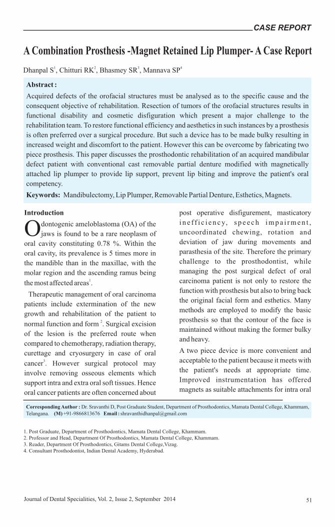
Abstract :
Acquired defects of the orofacial structures must be analysed as to the specific cause and the
consequent objective of rehabilitation. Resection of tumors of the orofacial structures results in
functional disability and cosmetic disfiguration which present a major challenge to the
rehabilitation team. To restore functional efficiency and aesthetics in such instances by a prosthesis
is often preferred over a surgical procedure. But such a device has to be made bulky resulting in
increased weight and discomfort to the patient. However this can be overcome by fabricating two
piece prosthesis. This paper discusses the prosthodontic rehabilitation of an acquired mandibular
defect patient with conventional cast removable partial denture modified with magnetically
attached lip plumper to provide lip support, prevent lip biting and improve the patient's oral
competency.
Keywords: Mandibulectomy, Lip Plumper, Removable Partial Denture, Esthetics, Magnets.
CASE REPORT
1. Post Graduate, Department of Prosthodontics, Mamata Dental College, Khammam.2. Professor and Head, Department Of Prosthodontics, Mamata Dental College, Khammam.3. Reader, Department Of Prosthodontics, Gitams Dental College,Vizag.4. Consultant Prosthodontist, Indian Dental Academy, Hyderabad.
A Combination Prosthesis -Magnet Retained Lip Plumper- A Case Report1 2 3 4Dhanpal S , Chitturi RK , Bhasmey SR , Mannava SP

prostheses. Magnetic technology is
constantly improving: currently available
magnets based on Nd-Fe-B are small (which
allows them to be incorporated into dentures)
and have attractive forces that enable them to
provide retention. Although magnets have
disadvantage of being corroded in long term
service, they are far superior to springs,
suction cups, clips, and studs in medical 4applications . This clinical report describes
the procedure associated with the fabrication
of a combination prosthesis which includes
removable partial denture with magnet
retained lip plumper.
Case Report
A 32 yr old male patient, name Balu Nayak
reported to the department of Prosthodontics,
Mamata Dental College with complaints of
missing teeth and disfigured face. Patient
gave a history of mandibular resection for
intra oral growth followed by bone graft at
NIMS Hospitals, Hyderabad. Extra oral
examination revealed a 22cm post surgical
scar, 6 cm below the right ear extending to
5cm beneath the left corner of his mouth. He
also presented with 1.5cm deep mentolabial
fold and puckering of chin (Fig. 1). On intra
oral examination, there was loss of residual
ridge and a saucer shaped defect extending
from the mesial aspect of the right mandibular
first molar to that of left mandibular first
molar (Fig. 2). Anteriorly the labial sulcus was
missing and mucosa of inner aspect of lower
lip and that covering the alveolar ridge was at
the same level. Labial frenum was missing,
and edentulous space appeared shrunken and
reduced. Support for the lower lip was lost and
was pulled inside.
Full complement of maxillary arch and molars
on the either side of the mandibular arch were
present. Class I molar relation was observed.
The panoramic radiograph revealed that the
base of the mandible was reconstructed with
bone graft (Fig. 3). On enquiry patient
revealed that a piece of bone was taken from
right leg.
The intermaxillary space in the region of the
surgical resection was excessive. To restore
such large space would require a heavier
prosthesis which might enhance resorption of
the grafted area. In addition to excessive
intermaxillary space, the skin over
mentolabial fold was pulled inside due to
cicatrisation and presented disfigured
appearance.
52Journal of Dental Specialities, Vol. 2, Issue 2, September 2014
CASE REPORTSravanthi
Fig. 1: Puckering of chin without prosthesis
Fig. 2: Loss of residual ridge and saucer shaped defect.
Fig. 3 : Panoramic radiograph revealing reconstructionwith bone graft

53Journal of Dental Specialities, Vol. 2, Issue 2, September 2014
Available treatment options to restore the
missing teeth were a fixed partial denture,
implant prosthesis and a removable partial
denture. Although the first two options could
result in an excellently stabilized prosthesis,
they would not have provided lip support and
restore normal facial profile. A removable
prosthesis with a modified acrylic flange
might provide the required support to the
lower lip. A diagnostic wax build up was tried
in increments in the intercanine region to
assess its aesthetic affect. Patient's approval
was sought. After the patient's acceptance was
obtained, a treatment plan was made to restore
edentulous area with six anterior teeth and left
first premolar. Because of uneven resorption
and healing of the residual ridge, a
compromise in arrangement of lower teeth
regarding the midline was made after
informing the patient.
To provide the lip support the labial flange is
needed to be modified without causing
unfavourable sequelae either to the soft tissues
or to the foundation. With this in mind, a single
unit bulky labial flange was avoided and a
sectional prosthesis jointed by magnets was
planned. Magnets (MAGFIT DX-800 Dental
Magne t i c A t t achmen t A ich i S t ee l
Corporation, Aichi- Ken, Japan) used in this
case had a Magnetic Field Leakage of 0.002T,
and attractive force of 7.7N which has superior
retentive force compared to other retentive
aids. A sectional prosthesis would also have an
advantage of easy retrieval whenever the
patient would not need lip supporting 5
component. Magnets were preferred over
other attachment devices owing to their easy
cleansability and simplicity, no difficulty
during placement by the patient because of its
automatic reseating nature, and long lasting 4
retention even with number of cycles.
Titanium alloy was chosen to provide metallic
frame work of removable partial denture.
Diagnostic impressions were made with
irreversible hydrocolloid (Zelgan 2002;
Dentsply-India, Gurgaon, India) and initial
survey was done. Mesial occlusal rests on 36
and 46 with embrasure clasps for 37, 38 and
47,48 were designed. Embrasure clasp
assemblies in this design would also provide
indirect retention. Because of restricted
lingual sulcus in anterior region, lingual bar
with extended minor connector over the ridge
area and into labial sulcus was considered
(Fig. 4).
This would provide enough surface for the
acrylic portion of the labial flange and fixing
of the magnet along with its yoke and outer lip
(MAGFIT DX-800 Dental Magnetic
Attachment Aichi Steel Corporation, Aichi-
Ken, Japan). Keeper of the magnet assembly
(MAGFIT DX-800 Dental Magnetic
Attachment Aichi Steel Corporation, Aichi-
Ken, Japan) would be in the plumper section
(Fig. 5).
CASE REPORTSravanthi
Fig. 4: Lingual bar with extended minor connector
Fig. 5 : Magnets and magnetic keepers in to the
denture base and template respectively

As per design occlusal rest seat preparation,
guiding planes and abutment tooth
modifications were included in mouth
preparation before a final impression was
made. A custom tray in acrylic resin was
constructed on the diagnostic cast. The
definitive cast was prepared from a dual
impression of the mandibular arch with
silicone impression material (AFFINIS
Precious, Regular; coltene whaledent). Metal
framework was fabricated and tried for
retention, stability and comfort. Acrylization
was done after necessary jaw relations and try-
in. On insertion it was seen that only function
could be restored but not the facial form
(Fig. 6).
Wax was added in increments on the labial
surface of the definitive prosthesis until
aesthetics was improved and accepted by the
patient. Added wax was indexed in the putty
which was further supported by dental plaster
(Fig. 7).
Wax was removed and cold cure clear acrylic
template was fabricated. Magnet keeper was
incorporated in the template and magnets in
the definitive prosthesis at equidistance from
midline. Factor II Inc. A-2186 silicone
elastomer (medical grade) was mixed in a 10:1
ratio by weight. Floccules, intrinsic strains,
sealant, thixotrophic agent were added in
proportions until desired color and
consistency were obtained. Care was taken to
minimize air entrapment during mixing and
the mix was loaded in the putty index and on to
the template and placed against the prosthesis
in situ on the master cast. Index was held until
complete set of silicon material (Fig. 8).
Modified partial denture (Fig. 9) was inserted
to check for occlusion in centric, phonetics,
and aesthetics (Fig. 10). The opinion on
patient's immediate relative was also
considered regarding aesthetics. Patient's
approval was obtained.
CASE REPORTSravanthi
54Journal of Dental Specialities, Vol. 2, Issue 2, September 2014
Fig. 6 : Frontal view after insertion ofremovable partial denture alone
Fig. 9: Removable partial denture and lip plumper Fig. 7: Wax added in incremental is indexed by Putty index
Fig. 8: Silicon material loaded within the putty indexand placed against removable partial denture
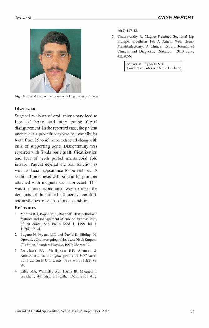
Discussion
Surgical excision of oral lesions may lead to
loss of bone and may cause facial
disfigurement. In the reported case, the patient
underwent a procedure where by mandibular
teeth from 35 to 45 were extracted along with
bulk of supporting bone. Discontinuity was
repaired with fibula bone graft. Cicatrization
and loss of teeth pulled mentolabial fold
inward. Patient desired the oral function as
well as facial appearance to be restored. A
sectional prosthesis with silicon lip plumper
attached with magnets was fabricated. This
was the most economical way to meet the
demands of functional efficiency, comfort,
and aesthetics for such a clinical condition.
References
1. Martins RH, Rapoport A, Rosa MP. Histopathologic
features and management of ameloblastoma: study
of 20 cases. Sao Paulo Med J. 1999 Jul 1;
117(4):171-4.
2. Eugene N. Myers, MD and David E. Eibling, M.
Operative Otolaryngology: Head and Neck Surgery. nd2 edition, Saunders Elsevier, 1997; Chapter 32.
3. Reichar t PA, Phi l ipsen HP, Sonner S .
Ameloblastoma: biological profile of 3677 cases.
Eur J Cancer B Oral Oncol. 1995 Mar; 31B(2):86-
99.
4. Riley MA, Walmsley AD, Harris IR. Magnets in
prosthetic dentistry. J Prosthet Dent. 2001 Aug;
86(2):137-42.
5. Chakravarthy R. Magnet Retained Sectional Lip
Plumper Prosthesis For A Patient With Hemi-
Mandibulectomy: A Clinical Report. Journal of
Clinical and Diagnostic Research 2010 June;
4:2582-6.
CASE REPORTSravanthi
55Journal of Dental Specialities, Vol. 2, Issue 2, September 2014
Fig. 10: Frontal view of the patient with lip plumper prosthesis
Source of Support: NILConflict of Interest: None Declared