Embed Size (px)
Citation preview

7,3�,4�-Trihydroxyisoflavone, a Metabolite of the SoyIsoflavone Daidzein, Suppresses Ultraviolet B-induced SkinCancer by Targeting Cot and MKK4*□S
Received for publication, May 24, 2010, and in revised form, February 28, 2011 Published, JBC Papers in Press, March 4, 2011, DOI 10.1074/jbc.M110.147348
Dong Eun Lee‡§¶1, Ki Won Lee§1, Sanguine Byun‡§¶, Sung Keun Jung‡§¶, Nury Song‡§¶, Sung Hwan Lim‡,Yong-Seok Heo�, Jong Eun Kim‡§¶, Nam Joo Kang**, Bo Yeon Kim‡‡, G. Tim Bowden§§, Ann M. Bode§,Hyong Joo Lee‡2, and Zigang Dong§3
From the ‡World Class University, Biomodulation Major, Department of Agricultural Biotechnology, §Food Science andBiotechnology Program, Seoul National University, Seoul 151-921, Republic of Korea, ¶The Hormel Institute, University ofMinnesota, Minnesota 55912, the �Department of Chemistry, Konkuk University, Seoul 143-701, Republic of Korea, the **School ofFood Science and Biotechnology, College of Agriculture and Life Sciences, Kyungpook National University, Daegu 702-701,Republic of Korea, the ‡‡Korea Research Institute of Bioscience and Biotechnology, Choongbuk 363-883, Republic of Korea, and the§§University of Arizona Cancer Center, Tucson, Arizona 85724
Nonmelanoma skin cancer is one of the most frequentlyoccurring cancers in the United States. Chronic exposure toUVB irradiation is a major cause of this cancer. Daidzein, alongwith genistein, is amajor isoflavone found in soybeans; however,little is known about the chemopreventive effects of daidzeinand itsmetabolites inUVB-induced skin cancer. Here, we foundthat 7,3�,4�-trihydroxyisoflavone (THIF), a major metabolite ofdaidzein, effectively inhibits UVB-induced cyclooxygenase 2(COX-2) expression through the inhibition of NF-�B transcrip-tion activity in mouse skin epidermal JB6 P� cells. In contrast,daidzein had no effect on COX-2 expression levels. Data fromWestern blot and kinase assays showed that 7,3�,4�-THIFinhibited Cot and MKK4 activity, thereby suppressing UVB-induced phosphorylation of mitogen-activated proteinkinases. Pull-down assays indicated that 7,3�,4�-THIF com-peted with ATP to inhibit Cot or MKK4 activity. Topicalapplication of 7,3�,4�-THIF clearly suppressed the incidenceand multiplicity of UVB-induced tumors in hairless mouseskin. Hairless mouse skin results also showed that 7,3�,4�-THIF inhibits Cot or MKK4 kinase activity directly, resultingin suppressed UVB-induced COX-2 expression. A dockingstudy revealed that 7,3�,4�-THIF, but not daidzein, easilydocked to the ATP binding site of Cot and MKK4, which islocated between the N- and C-lobes of the kinase domain.Collectively, these results provide insight into the biological
actions of 7,3�,4�-THIF, a potential skin cancer chemopre-ventive agent.
Nonmelanoma skin cancer is one of the most frequentlyoccurring cancers in the United States (1). UV irradiation fromsunlight is themajor etiologic factor in the development of non-melanoma skin cancers, including squamous cell carcinomasand basal cell carcinomas (2). Among the forms of solar irradi-ation, UVB (290–320 nm) exhibits highly mutagenic and car-cinogenic effects in animal experiments, as compared withUVA (320–400 nm) (3). Prolonged exposure to UVB irradia-tion causes the development of benign epidermal tumors, mostof which become skin carcinomas because UVB functions as acomplete carcinogen (4). Therefore, targeting UVB-inducedmolecular and signaling mechanisms might be effective ap-proaches for the chemoprevention of skin cancer.Cyclooxygenase 2 (COX-2)4 is an essential enzyme mediat-
ing the conversion of arachidonic acid to prostaglandin, theinducible isoform of cyclooxygenase (1). The inflammatoryprocess affects numerous human malignancies, including skincancer, by promoting epidermal hyperproliferation and hyper-plasia through the secretion of various inflammatory factors,such as prostaglandin E2 (2). In human skin, increased COX-2expression is observed in response to acuteUVB irradiation (5).Similarly, in the mouse model, COX-2 is overexpressed inhyperplastic skin, benign tumors, and malignant tumors fol-lowing chronic UVB irradiation (2). Indeed, celecoxib, a COX-2-selective inhibitor, in dietary or topical treatment in micefollowingUVB exposure, attenuated the number and volume oftumors (2). Therefore, regulating the expression of COX-2might be a potential protection strategy against skin cancer.Cot was initially identified in a human thyroid carcinoma cell
line and in moloney murine leukemia virus-induced rat T-celllymphomas (Tpl2) (6). Stimulation of Cot causes the activationof a p38 MAPK, JNKs, and transcription factors NF-�B and
* This study was supported, in whole or in part, by The Hormel Foundationand National Institutes of Health Grants CA077646, CA027502,CA111536, CA120388, R37CA081064, and ES016548. This work was alsosupported by World Class University Program Grant R31-2008-00-10056-0, World Class Institute Program Grant 2009-0093824, LeapResearch Program Grant 2010-0029233, and Basic Science ResearchProgram Grant 2009-0090797 through the National Research Founda-tion of Korea.
□S The on-line version of this article (available at http://www.jbc.org) containssupplemental Fig. 1.
1 Both authors contributed equally to this work.2 To whom correspondence may be addressed: Department of Agricultural
Biotechnology, Seoul National University, Seoul 151-742, Republic ofKorea. Tel.: 82-2-880-4860; Fax: 82-2-873-5095; E-mail: [email protected].
3 To whom correspondence may be addressed: The Hormel Institute, Univer-sity of Minnesota, 801 16th Ave., NE, Austin, MN 55912. Tel.: 507-437-9600;Fax: 507-437-9606; E-mail: [email protected].
4 The abbreviations used are: COX-2, cyclooxygenase 2; THIF, trihydroxyiso-flavone; MKK, mitogen-activated protein kinase kinase; MEM, minimumEagle’s medium; MOPS, 4-morpholinepropanesulfonic acid.
THE JOURNAL OF BIOLOGICAL CHEMISTRY VOL. 286, NO. 16, pp. 14246 –14256, April 22, 2011© 2011 by The American Society for Biochemistry and Molecular Biology, Inc. Printed in the U.S.A.
14246 JOURNAL OF BIOLOGICAL CHEMISTRY VOLUME 286 • NUMBER 16 • APRIL 22, 2011
by guest on March 22, 2019
http://ww
w.jbc.org/
Dow
nloaded from

nuclear factor of activated T cells (7). Numerous studies haveshown that Cot mediates cytokine production, such as TNF-�,and leads to several inflammatory diseases, such as rheumatoidarthritis. Recently, several studies have revealed that the activa-tion of Cot is observed in tumor cells, such as T cell neoplasiacell lines (7). Cot also positively regulates COX-2 expression byLPS in macrophages (8). However, the involvement of Cot inUVB-induced COX-2 expression in skin has not been reported.The mitogen-activated protein kinase kinase 4 (MKK4), a
dual-specificity kinase, plays a critical role in the SAPK signal-ing pathway. The SAPK pathways, including p38 MAPK andJNKs, is activated in response to environmental stressors andextracellular stimuli (9). MKK3 and MKK6 are specific up-stream kinases of p38, whereas MKK4 activates both p38 andJNKs (10–12). Phosphorylation of p38 and JNKs mediates theinflammatory response, thereby inducing the eukaryotic tran-scription factor NF-�B activation (13). In mouse skin, UV irra-diation activates p38 and JNKs, which contribute to the devel-opment of skin cancer (13). Therefore, regulating the upstreammolecules of p38 and JNKs is one possible strategy for the che-moprevention of skin carcinogenesis.Daidzein and genistein are naturally occurring isoflavones in
soy foods. In particular, genistein has received attention as apotential anticarcinogenic compound and has been intenselystudied, whereas daidzein and its metabolites have been lesswell studied. Here, we investigated the chemopreventive effectsof 7,3�,4�-trihydroxyisoflavone (THIF), a major metabolite ofdaidzein, against UVB-induced skin cancer. We report that7,3�,4�-THIF, but not daidzein, is anATP-competitive inhibitorof Cot and MKK4, and subsequently suppresses UVB-inducedCOX-2 expression in JB6 P�mouse epidermal cells. In amouseskin tumorigenesis model, 7,3�,4�-THIF strongly suppressedthe incidence, multiplicity, and volume of UVB-inducedmouseskin tumors. Consistent with the tumor data, 7,3�,4�-THIFclearly attenuated UVB-induced COX-2 expression in hairlessmouse skin. Furthermore, 7,3�,4�-THIF directly bound withCot or MKK4, resulting in the suppression of Cot or MKK4activity in hairless mouse skin.
MATERIALS AND METHODS
Chemicals—7,3�,4�-THIF was obtained from the IndofineChemical Co., Inc. (Hillsborough, NJ), and daidzein was pur-chased from Sigma. The antibodies against phosphorylatedERKs (Thr-202/Tyr-204), total ERKs, total p38, phosphorylatedNF-�B, phosphorylated c-Jun NH2-terminal kinases (JNKs;Thr-183/Tyr-185), and total JNKs were purchased from CellSignaling Technology (Danvers, MA). The antibody againstphosphorylated p38 (Tyr-180/Tyr-182) was from BD Biosci-ences, and Cot (Thr-209) was purchased from Invitrogen. TheCot1 and MKK4 kinase assay kits were obtained from Cell Sig-naling Technology. CNBr-Sepharose 4B, glutathione-Sephar-ose 4B, [�-32P]ATP, and the chemiluminescence detection kitwere purchased from Amersham Biosciences. The proteinassay kit was obtained from Bio-Rad. G418 and the luciferaseassay substrate were purchased from Promega (Madison, WI).Cell Culture—JB6 P� mouse epidermal (JB6 P�) cell lines
were cultured in monolayers at 37 °C in a 5% CO2 incubator inMEM containing 5% FBS, 2 mM L-glutamine, and 25 �g/ml
gentamicin. The cells were stably transfected with an NF-�Bluciferase reporter plasmid and maintained in MEM supple-mented with 5% FBS containing 200 �g/ml G418.UVB Irradiation—UVB irradiation was carried out in a UVB
chamberwith a transluminator emittingUVB light protons andfitted with a Kodacel K6808 filter (Eastman Kodak, Rochester,NY) that eliminates all wavelengths below 290 nm. This lamp isone of the most frequently used UVB sources for the study ofskin carcinogenesis. Irradiation energy was measured using aUVX radiometer (UVX-31) from UVP (Upland, CA).Luciferase Assay for COX-2 Promoter Activity or NF-�B
Transcription Activity—COX-2 (14) or NF-�B (15, 16) lucifer-ase reporter-transfected JB6 P� cells were constructed asdescribed earlier. The cells (8� 103), suspended in 100�l of 5%FBS/MEM, were added to each well of a 96-well plate and incu-bated at 37 °C in a humidified atmosphere of 5% CO2. Whencells reached 80 to 90% confluence, they were starved in 0.1%FBS/MEM for an additional 24 h. The cells were then treatedfor 1 h with 7,3�,4�-THIF or daidzein and then exposed to UVBfor 24 h. After treatment, cells were disrupted with 100 �l oflysis buffer (0.1 M potassium phosphate buffer (pH 7.8), 1%Triton X-100, 1 mM DTT, 2 mM EDTA), and the luciferaseactivity was measured using a luminometer (LuminoskanAscent, Thermo Electron).Western Blot Analysis—After the cells (1.5 � 106) were cul-
tured in a 10-cm dish for 48 h, they were starved in 0.1% FBS/MEM for an additional 24 h. They were then treated with7,3�,4�-THIF for 1 h before exposure to UVB (4 kJ/m2) andharvested 30min later. The harvested cells were disrupted, andthe supernatant fractions were boiled for 5 min. The proteinconcentration was determined using a dye-binding proteinassay kit according to the manufacturer’s protocol. Lysate pro-tein (30�g)was subjected to 10%SDS-PAGE and transferred toa polyvinylidene difluoride membrane. After blotting, themembrane was incubated with the specific primary antibody at4 °C overnight. Protein bands were visualized by a chemilumi-nescence detection kit after hybridization with a horseradishperoxidase-conjugated secondary antibody.In Vitro MKK4 and Cot1 Kinase Assays—The in vitro kinase
assays were performed in accordancewith the instructions pro-vided by Cell Signaling Technology. Briefly, for the MKK4assay, 40 ng/�l active MKK4 recombinant murine protein(55.8% purity and 964 units/mg activity) and 7,3�,4�-THIF (20,40, or 60 �M) were reacted at 30 °C for 10 min. For each reac-tion, 5 �l of 2 � kinase buffer (10 mM MOPS (pH 7.2), 5 mM
�-glycerol phosphate, 2 mM EGTA, 0.8 mM EDTA, 10 mM
MgCl2, 0.1 mMDTT), 5 �l of 250 �MATP, and 0.2 �g/�l of theinactive JNK2 were added. The mixtures were incubated at30 °C for 15min. A 5-�l aliquot was removed from the reactionmixture containing 10 �l of 2 mg/ml of ATF-2 substrate pep-tide, 5 �l of 2� kinase buffer, and 5 �l of 0.16 �Ci/�l [32P]ATPsolution, and incubated at 30 °C for 15 min. Then, 20-�l ali-quots were transferred onto p81 filter paper and washed threetimes with 1% phosphoric acid for 5 min per wash and oncewith acetone for 5 min. Radioactive incorporation was deter-minedusing a scintillation counter (LS6500; BeckmanCoulter).Each experiment was performed three times. For the Cot1assay, 150 ng/�l active Cot1 recombinant human protein (62%
7,3�,4�-Trihydroxyisoflavone Suppresses Skin Cancer
APRIL 22, 2011 • VOLUME 286 • NUMBER 16 JOURNAL OF BIOLOGICAL CHEMISTRY 14247
by guest on March 22, 2019
http://ww
w.jbc.org/
Dow
nloaded from
purity and 276 units/mg activity) and 7,3�,4�-THIF (20, 40, or 60�M) were reacted at 30 °C for 10 min. For each reaction, 5 �l of2� kinase buffer, 1 �g/ml of p65 as substrate, and 5 �l of 0.16�Ci/�l [32P]ATP solutionwere added.Themixturewas reactedat 30 °C for 15 min. Then, 20-�l aliquots were transferred ontop81 filter paper and washed three times with 1% phosphoricacid for 5min per wash and oncewith acetone for 2min. Radio-active incorporationwas determined using a scintillation coun-ter (LS6500, Beckman Coulter). Each experiment was per-formed separately three times.Ex Vivo MKK4 and Cot Immunoprecipitation and Kinase
Assays—JB6P� cells were cultured to 80% confluence and thenserum-starved in 0.1% FBS/MEM for 24 h at 37 °C. Cells wereeither treated with 7,3�,4�-THIF or left untreated for 1 h,exposed to 4 kJ/m2UVB, harvested after 30min, disruptedwithlysis buffer (20 mM Tris-HCl (pH 7.4), 1 mM EDTA, 150 mM
NaCl, 1 mM EGTA, 1% Triton X-100, 1 mM �-glycerophos-phate, 1 mg/ml leupeptin, 1 mM Na3VO4, 1 mM PMSF), andfinally centrifuged at 14,000 rpm for 10 min in a microcentri-fuge. The lysates, each containing 500 �g of protein, were usedfor immunoprecipitation with an antibody against MKK4 orCot and then incubated at 4 °C for 4 h. Protein A/G Plus-aga-rose beads were then added, and the mixture was continuouslyrotated overnight at 4 °C. The beads were then washed threetimes with kinase buffer (20mMMOPS (pH 7.2), 25mM �-glyc-erol phosphate, 5 mM EGTA, 1 mM sodium orthovanadate, 1mM DTT), and then a kinase reaction was performed in thesame manner as for in vitro kinase assays.In Vitro and exVivo Pull-downAssays—RecombinantMKK4
or Cot1 (2�g) or a JB6 P� cellular supernatant fraction (500�gprotein) was incubated with 7,3�,4�-THIF-Sepharose 4B (orSepharose 4B as a negative control) beads (100�l, 50% slurry) inreaction buffer (50mMTris-HCl (pH 7.5), 5mMEDTA, 150mM
NaCl, 1 mM DTT, 0.01% Nonidet P-40, 2 �g/ml BSA, 0.02 mM
PMSF, 1� protease inhibitor mixture). After incubation withgentle rocking overnight at 4 °C, the beads were washed fivetimes with buffer (50 mM Tris-HCl (pH 7.5), 5 mM EDTA, 150mM NaCl, 1 mM DTT, 0.01% Nonidet P-40, 0.02 mM PMSF),and the proteins bound to the beads were analyzed byimmunoblotting.Mouse Skin Tumorigenesis Analysis—SKH-1 hairless mice (5
weeks of age; mean body weight, 25 g) were purchased from theInstitute of Laboratory Animal Resources at Seoul NationalUniversity. Animals were acclimated for 1 week before thestudy and had free access to food and water. The animals werehoused in climate-controlled quarters (24 °C at 50% humidity)with a 12-h light/12-h dark cycle. Skin carcinogenesis wasinduced inmice receivingUVB irradiation over the course of 27weeks. TheUVB radiation source (Bio-Link cross-linker, VilberLourmat) emitted wavelengths with peak emission at 312 nm.SKH-1 mice were divided into four groups of 12 animals each.In the control group, the dorsal skin was topically treated with200 �l of acetone only. In the UVB group, the dorsal skin wastopically treated with 200 �l of acetone 1 h before UVB expo-sure. Themice in the third and fourth groups received a topicalapplication of 7,3�,4�-THIF (10 or 40 nmol, respectively) in 200�l of acetone 1 h beforeUVB irradiation. TheUVBdosewas0.18J/cm2 given three times/week for 27 weeks. The incidence of skin
tumors was recorded weekly, with a tumor defined as an out-growth of�1mm in diameter that persisted for 2 weeks ormore.Tumor incidence, multiplicity, and volume were recorded everyweek until the end of the experiment at the 27th week.In VivoWestern Blot Analysis—For in vivoWestern blotting,
mice received a topical application of 7,3�,4�-THIF (10 or 40nmol) in 200 �l acetone on their backs 1 h before UVB irradia-tion. To isolate protein frommouse skin, the dorsal skin of eachmouse was excised and placed on ice. Any fat was removed, andthe skin was placed in liquid nitrogen and immediately pulver-ized with a mortar and pestle. The pulverized skin was blendedon ice with a homogenizer (IKA T10 basic, IKA LaboratoryEquipment), and skin lysateswere centrifuged at 12,000 rpm for20 min. After the protein content was determined using theBio-Rad protein assay kit, 100 �g protein from mouse skinextract was subjected to 10% SDS-PAGE and transferred to aPVDF membrane (Amersham Biosciences). Membranes wereprocessed, and proteins were analyzed as described above forthe in vitroWestern blot assay.Short-term in Vivo Model—Imprinting control region mice
(5 weeks of age; mean body weight, 25 g) were purchased fromthe Institute of Laboratory Animal Resources at Seoul NationalUniversity. ICR mice were maintained under the same condi-tion as SKH-1 hairless mice described above. The dorsal areasof ICR mice were shaved before sample treatment. Each groupwas topically treated with 7,3�,4�-THIF (40 or 160 nmol) or daid-zein (160 nmol) in 200 �l of acetone 1 h before UVB (0.5 J/cm2)irradiation. The protein extract forWestern blot analysis was pre-pared as described above.In Vivo Kinase and Pull-down Assays—For the in vivo Cot or
MKK4 immunoprecipitation and kinase assay, mice weretreated with 7,3�,4�-THIF (10 or 40 nmol) in 200 �l of acetone,and dorsal skin was prepared as for in vivo Western blotting.Proteins were extracted as above and centrifuged at 12,000 rpmfor 20 min. 700 �g of protein from mouse skin extract wasmixed with protein-A/G beads (20 �l) for 1 h at 4 °C. The mix-ture was processed, and radioactive incorporation was deter-mined as for the ex vivo assay described above. Data are pre-sented as the mean of data points from five mice in each group.For the in vivo pull-down assay,mice received a topical appli-
cation of 200 �l of acetone alone or 7,3�,4�-THIF (10 or 40nmol, respectively) in 200 �l of acetone on their backs 1 h beforeUVB irradiation. Dorsal skin was prepared as described above forthe in vivo Western blotting, and proteins were extracted asdescribed above for the Cot or MKK4 immunoprecipitation andkinase assays. Then 500 �g of protein from mouse skin extractwere incubatedwith 7,3�,4�-THIF-Sepharose 4B (or Sepharose 4Balone as a control) beads (100 �l, 50% slurry) in reaction buffer asdescribed for the ex vivo pull-down assay. Beads were incubatedand washed, and proteins bound to the beads were analyzed byWestern blotting as described above.Molecular Modeling—The homology model structures of
Cot and MKK4 were generated by Geno3D using the coordi-nates of Mst1 and MKK7 (PDB codes 3COM and 2DYL),respectively. Insight II (Accelrys, Inc., San Diego, CA) was usedfor the docking study and structure analysis.Statistical Analysis—When necessary, data were expressed
as mean � S.D., and analysis of variance was used for multiple
7,3�,4�-Trihydroxyisoflavone Suppresses Skin Cancer
14248 JOURNAL OF BIOLOGICAL CHEMISTRY VOLUME 286 • NUMBER 16 • APRIL 22, 2011
by guest on March 22, 2019
http://ww
w.jbc.org/
Dow
nloaded from
statistical comparisons.Aprobability value ofp� 0.05was usedas the criterion for statistical significance.
RESULTS
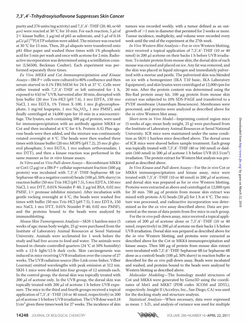
7,3�,4�-THIF Inhibits UVB-induced COX-2 Expression bySuppressing COX-2 Promoter Activity and NF-�B Transcrip-tional Activity in JB6 P�Cells,WhereasDaidzeinHasNoEffect—As aberrant COX-2 expression is intimately involved in multi-ple malignancies, including skin cancer (17), we first examined
the possible inhibitory effects of 7,3�,4�-THIF on UVB-inducedCOX-2 expression in JB6 P� cells. Treatment with 7,3�,4�-THIF dose-dependently inhibited UVB-induced COX-2expression in these cells (Fig. 1B, left panel), whereas daidzein,even up to 60�M, had no effect (Fig. 1B, right panel). Consistentwith the Western blotting results, 7,3�,4�-THIF strongly sup-pressedUVB-induced COX-2 promoter activity in JB6 P� cellsstably transfectedwith aCOX-2 luciferase reporter plasmid in adose-dependent manner (Fig. 1C, left panel), but daidzein had
FIGURE 1. Comparison of the inhibitory effects of 7,3�,4�-THIF and daidzein on UVB-induced COX-2 expression, COX-2 promoter activity, and NF-�B trans-activation in JB6 P�cells. A, the chemical structures of 7,3�,4�-THIF and daidzein. B, effect of 7,3�,4�-THIF or daidzein on UVB-induced COX-2 expression in JB6 P�cells.The cells were treated with 7,3�,4�-THIF or daidzein at the indicated concentrations for 1 h before exposure to UVB (4 kJ/m2) and harvested 4 h later. The levels of COX-2expression were then determined by Western blot analysis, as described under “Materials and Methods,” using specific antibodies against the corresponding COX-2and �-actin proteins. Data are representative of three independent experiments. The protein levels were quantified using an image analysis program to evaluate thedensity of each band on the immunoblot, and the fold value was calculated. C and D, effects of 7,3�,4�-THIF or daidzein on UVB-induced COX-2 (C) or NF-�B (D) activityin JB6 P� cells. In the luciferase assay, JB6 P� stably transfected with the COX-2 or NF-�B luciferase reporter plasmid were cultured as described under “Materials andMethods.” After reaching 80% confluence, the cells were cultured in 0.1% FBS/MEM and then treated with 7,3�,4�-THIF or daidzein at the indicated concentrations orleft untreated for 1 h before exposure to UVB (4 kJ/m2) and harvested 24 h later. COX-2 or NF-�B activity is expressed as the percent inhibition relative to cells treatedwith only UVB. Data are represented as the mean � S.D. of the luciferase activity calculated from three separate experiments. For C and D, the asterisk indicatessignificant differences between groups treated with UVB alone and the 7,3�,4�-THIF- or daidzein-treated groups. *, p � 0.05; **, p � 0.01; ***, p � 0.001).
7,3�,4�-Trihydroxyisoflavone Suppresses Skin Cancer
APRIL 22, 2011 • VOLUME 286 • NUMBER 16 JOURNAL OF BIOLOGICAL CHEMISTRY 14249
by guest on March 22, 2019
http://ww
w.jbc.org/
Dow
nloaded from

7,3�,4�-Trihydroxyisoflavone Suppresses Skin Cancer
14250 JOURNAL OF BIOLOGICAL CHEMISTRY VOLUME 286 • NUMBER 16 • APRIL 22, 2011
by guest on March 22, 2019
http://ww
w.jbc.org/
Dow
nloaded from
no effect (Fig. 1C, right panel). NF-�B is an important transcrip-tion factor regulating COX-2 expression in the skin (15). Thus,to investigate whether 7,3�,4�-THIF down-regulates UVB-in-duced COX-2 expression through the inhibition of the NF-�Btranscription factor, we measured NF-�B transcription activityby using cells stably transfected with the NF-�B luciferasereporter plasmid. 7,3�,4�-THIF significantly attenuated UVB-inducedNF-�B transcription activity (Fig. 1D, left panel). Com-pared with daidzein, treatment with 7,3�,4�-THIF at low con-centrations inhibited NF-�B transcription activity moreeffectively (Fig. 1D, right panel).7,3�,4�-THIF Suppresses UVB-induced Phosphorylation of
JNKs and p38 MAPK in JB6 P� Cells—JNKs and p38 are gen-erally referred to as stress-activated MAP kinases. We exam-ined whether 7,3�,4�-THIF blocks the activation of the JNKsand p38 pathways stimulated by UVB in JB6 P� cells. Westernblotting data showed that 7,3�,4�-THIF inhibited UVB-inducedphosphorylation of JNKs and p38 dose-dependently but had noeffect on the phosphorylation of ERKs (Fig. 2A, left panels).
Also, 7,3�,4�-THIF did not affect I�B kinase beta activity (datanot shown). Recently, several studies indicated that stimulationof Cot leads to the activation of JNKs, p38, and NF-�B (7).MKK3 and MKK6 activate p38, whereas MKK4 can activateboth JNKs and p38 (18). Thus, we further investigated theeffects of 7,3�,4�-THIF on the upstream regulatory proteins ofthe JNKs and p38 pathways. Our results revealed that 7,3�,4�-THIF inhibited both Cot and MKK4 activity in vitro (Fig. 2B,upper panels), but it did not affect the phosphorylation level ofMKK4 or Cot in JB6 P� cells (Fig. 2A, right panels). Consistentwith the results from the in vitro kinase assay, an ex vivo kinaseassay also revealed that 7,3�,4�-THIF inhibited UVB-inducedMKK4 and Cot activity in JB6 P� cells (Fig. 2C). In contrast,7,3�,4�-THIF had no effect on either the phosphorylation levelof MKK3/6 (Fig. 2A, right panels) or the in vitro kinase activityofMKK3 andMKK6 (Fig. 2B, lower panels).We next examinedwhether 7,3�,4�-THIF had an effect on the activity of variousMAP kinases. The in vitro kinase assay showed that 7,3�,4�-THIF had no effect on ERKs, p38�, or JNK1 kinase activity as
FIGURE 2. 7,3�,4�-THIF suppresses UVB-induced phosphorylation of JNKs and p38 through the direct inhibition of both Cot and MKK4. A, 7,3�,4�-THIFinhibited UVB-induced phosphorylation of JNKs and p38 in JB6 P� cells. Cells were treated with 7,3�,4�-THIF (0, 20, 40, or 60 �M) for 1 h before being exposedto UVB (4 kJ/m2) and harvested 30 min later. The cells were disrupted, and the levels of phosphorylated and total proteins were determined by Western blotanalysis, as described under “Materials and Methods,” using specific antibodies against the respective phosphorylated and total proteins. Data are represen-tative of three independent experiments that gave similar results. B, 7,3�,4�-THIF inhibited both Cot and MKK4 in vitro. In contrast, MKK3 and MKK6 activity wasnot affected by 7,3�,4�-THIF. The in vitro kinase assay was performed as described under “Materials and Methods,” and kinase activity is expressed as percentinhibition relative to the activity of the untreated kinase control. C, 7,3�,4�-THIF inhibited both Cot and MKK4 activity ex vivo. In the ex vivo Cot or MKK4 kinaseassay, cells were pretreated with 7,3�,4�-THIF at the indicated concentrations (0, 20, 40, or 60 �M) for 1 h and then exposed to UVB (4 kJ/m2) and harvested after30 min. Cells were used for immunoprecipitation, and the kinase assay was performed. Kinase activity is expressed as percent inhibition relative to cells treatedwith UVB only. The average 32P count was determined from three separate experiments, and the data are presented as mean � S.D. In the ex vivo kinase assays,the asterisks indicate a significant decrease in kinase activity between cells treated with 7,3�,4�-THIF and cells treated with UVB only. **, p � 0.01; ***, p � 0.001).D, 7,3�,4�-THIF did not affect ERKs or MSK1 activity in vitro. The in vitro kinase assay was performed as described under “Materials and Methods,” and kinaseactivity is expressed as percent inhibition relative to the activity of the untreated kinase control. For the in vitro kinase assays (B and D), the asterisk indicates asignificant decrease in kinase activity between the groups treated with active Cot (or MKK4) and 7,3�,4�,-THIF and the group treated with active Cot (or MKK4)alone. *, p � 0.05; **, p � 0.01; ***, p � 0.001.
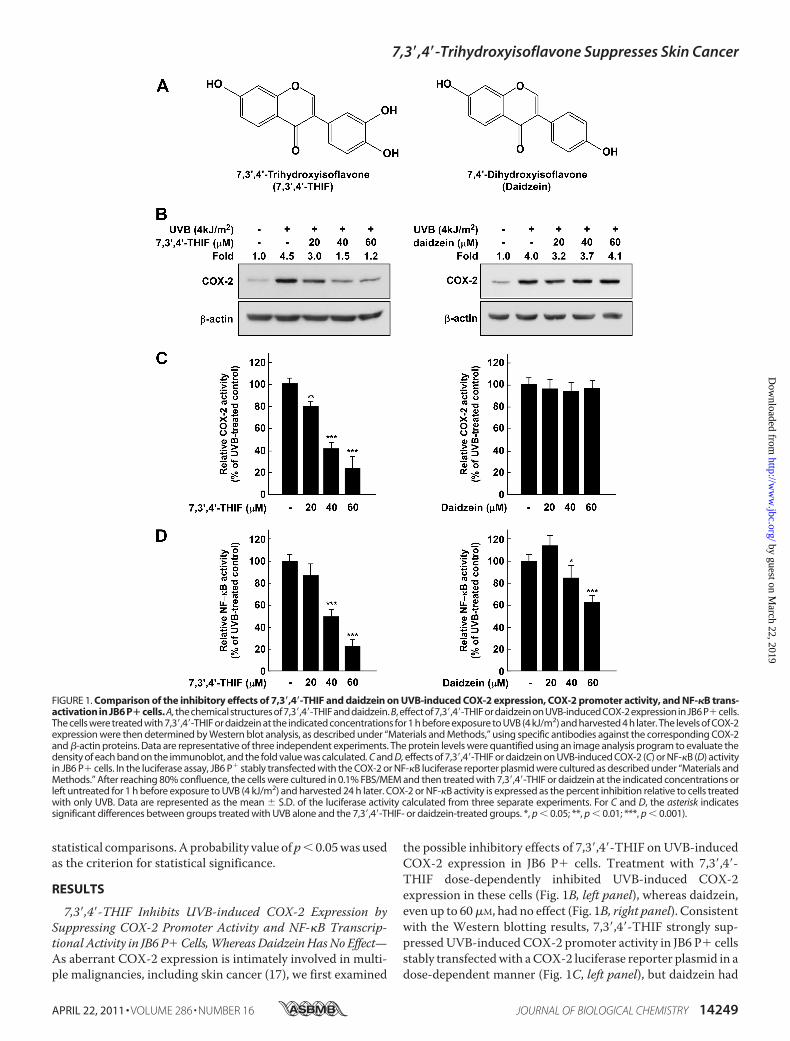
FIGURE 3. 7,3�,4�-THIF direct-binds with Cot and MKK4. A, 7,3�,4�-THIF specifically binds with Cot or MKK4 in vitro. The Cot (or MKK4) 7,3�,4�-THIF binding wasconfirmed by immunoblotting using an antibody against Cot (left panel) or MKK4 (right panel). 1st lane (input control), Cot, or MKK4 protein standard; 2nd lane(control), Sepharose 4B was used to pull down Cot or MKK4, as described under “Materials and Methods;” 3rd lane, 7,3�,4�-THIF-Sepharose 4B affinity beadswere used to pull down Cot or MKK4. B, 7,3�,4�-THIF directly binds with either Cot and MKK4 ex vivo. Binding was confirmed by immunoblotting using anantibody against Cot (left panel) or MKK4 (right panel). 1st lane (input control), whole cell lysates from JB6 P� cells; 2nd lane (control), a lysate of JB6 P� cellsprecipitated with Sepharose 4B beads; 3rd lane, whole cell lysates from JB6 P� cells precipitated by 7,3�,4�-THIF-Sepharose 4B affinity beads. C, 7,3�,4�-THIFcompetes with ATP to bind with Cot and MKK4. Active Cot or MKK4 (0.2 �g) was incubated with ATP at different concentrations (0, 10, or 100 �M) and 100 �lof 7,3�,4�-THIF-Sepharose 4B or 100 �l of Sepharose 4B (as a negative control) in reaction buffer to a final volume of 500 �l. The mixtures were incubated at 4 °Covernight with shaking. After washing, the pulled-down proteins were detected by Western blotting. 1st lane (input control), Cot or MKK4 protein standard; 2ndlane, (negative control), Cot or MKK4 bound with Sepharose 4B; 3rd lane (positive control), Cot (left panel) or MKK4 (right panel) binding with 7,3�,4�-THIF-Sepharose 4B. Each experiment was performed three times, and representative blots are shown.
7,3�,4�-Trihydroxyisoflavone Suppresses Skin Cancer
APRIL 22, 2011 • VOLUME 286 • NUMBER 16 JOURNAL OF BIOLOGICAL CHEMISTRY 14251
by guest on March 22, 2019
http://ww
w.jbc.org/
Dow
nloaded from
well as no effect on activity of MSK1, a downstream protein ofp38 and ERKs (Fig. 2D). These results indicate that the inhibi-tion of UVB-induced COX-2 expression by 7,3�,4�-THIF wasmainly caused by the suppression of both Cot and MKK4activity.7,3�,4�-THIF Directly Binds with Either Cot or MKK4 Com-
petitively Binds with ATP—To determine whether the inhibi-tion ofCot1 andMKK4 activities by 7,3�,4�-THIFwas caused bydirect interaction, we performed an in vitro pull-down assay.We detected Cot1 or MKK4 in 7,3�,4�-THIF-Sepharose 4Bbeads but not in Sepharose 4B beads alone (Fig. 3A). We alsoobserved ex vivo binding between 7,3�,4�-THIF and Cot orMKK4 in JB6 P� cell lysates (Fig. 3B). However, 7,3�4�-THIFdid not bind with MKK3 (data not shown). ATP treatmentblocked the binding ability of 7,3�,4�-THIF with Cot1 andMKK4 in a dose-dependent manner, suggesting that 7,3�,4�-THIF binds with Cot1 or MKK4 in competition with ATP (Fig.3C). These results indicate that the inhibition of Cot1 orMKK4activity by 7,3�,4�-THIF occurs through the direct binding of7,3�,4�-THIF with both proteins.7,3�,4�-THIF Represses UVB-induced Skin Tumorigenesis in
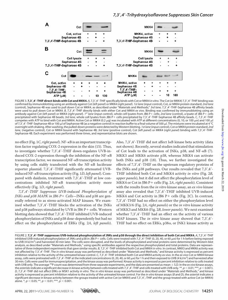
the SKH-1 Hairless Mouse Model—To evaluate the chemopre-ventive activity of 7,3�,4�-THIF in vivo, we studied the effects of7,3�,4�-THIF in the two-stage, UVB-induced skin carcinogene-
sis model. Data show that 7,3�,4�-THIF treatment suppressedUVB-induced skin cancer development in mice (Fig. 4A).Tumor incidence was also clearly attenuated by topical appli-cation of 7,3�,4�-THIF (10 or 40 nmol) prior to UVB irradiation(Fig. 4B). In the UVB-only treated mouse group, the onset oftumors was observed at the 16th week and reached 100% by the20th week. However, in the 10- or 40-nmol 7,3�,4�-THIF-treatedmouse groups, the onset of tumorswas delayed until the17th or 19th week and approached 100% only by the 24th week.Topical application of 7,3�,4�-THIF (10 or 40 nmol) prior toUVB irradiation also clearly suppressed tumor multiplicitythroughout the duration of the experiment (Fig. 4C). Further-more, the volume of tumors was markedly suppressed by7,3�,4�-THIF treatment (Fig. 4D). Collectively, these resultsreveal that 7,3�,4�-THIF may serve as an effective chemopre-ventive agent against UVB-mediated skin tumorigenesis.7,3�,4�-THIF Inhibits UVB-induced COX-2 Expression and
Cot and MKK4 Activity by Directly Binding with Cot or MKK4in SKH-1 Hairless Mouse Skin—To further confirm the inhibi-tory effect of 7,3�,4�-THIF on UVB-induced COX-2 expressionin an in vivomodel, we examined the level of COX-2 expressionin SKH-1 hairless mouse skin. Consistent with the results fromthe JB6 P� cells, the Western blotting data showed that thelevels of COX-2 expression in the 7,3�,4�-THIF-treated groups
FIGURE 4. 7,3�,4�-THIF inhibits UVB-induced skin carcinogenesis in the SKH-1 hairless mouse. Control mice (n � 12) received a topical treatment of 200 �lacetone (no UVB), and experimental mice (n � 12) were topically treated with 200 �l acetone before UVB (0.18 J/cm2) exposure (3 days/week for 27 weeks). Themice in the third and fourth groups received a topical application of 7,3�,4�-THIF (10 or 40 nmol, respectively, per mouse in 200 �l acetone) on the dorsal surface1 h before UVB (0.18 J/cm2) irradiation 3 days/week for 27 weeks. The incidence of skin tumors was recorded weekly, and tumors were defined as an outgrowthof �1 mm in diameter persisting for 2 weeks or longer. Tumor incidence and multiplicity were recorded every week until the end of the experiment at 27 weeks.A, appearance of skin tumors. B, 7,3�,4�-THIF retarded the incidence of skin tumors compared with the UVB only-treated group. C, 7,3�,4�-THIF strongly inhibitsUVB-induced tumor multiplicity in SKH-1 hairless mice. D, 7,3�,4�-THIF reduces UVB-induced tumor volume in SKH-1 hairless mice. At the end of the study, thedimensions of all tumors on each mouse were recorded. Tumor volumes were calculated using the hemiellipsoid model formula: tumor volume � 1/2(4�/3)(l/2)(w/2)h, wherein l is length, w is width, and h is height. The asterisks indicate a significant decrease in tumor incidence or volume between the grouptreated with UVB alone and the group treated with 7,3�,4�-THIF. **, p � 0.05; ***, p � 0.001.
7,3�,4�-Trihydroxyisoflavone Suppresses Skin Cancer
14252 JOURNAL OF BIOLOGICAL CHEMISTRY VOLUME 286 • NUMBER 16 • APRIL 22, 2011
by guest on March 22, 2019
http://ww
w.jbc.org/
Dow
nloaded from
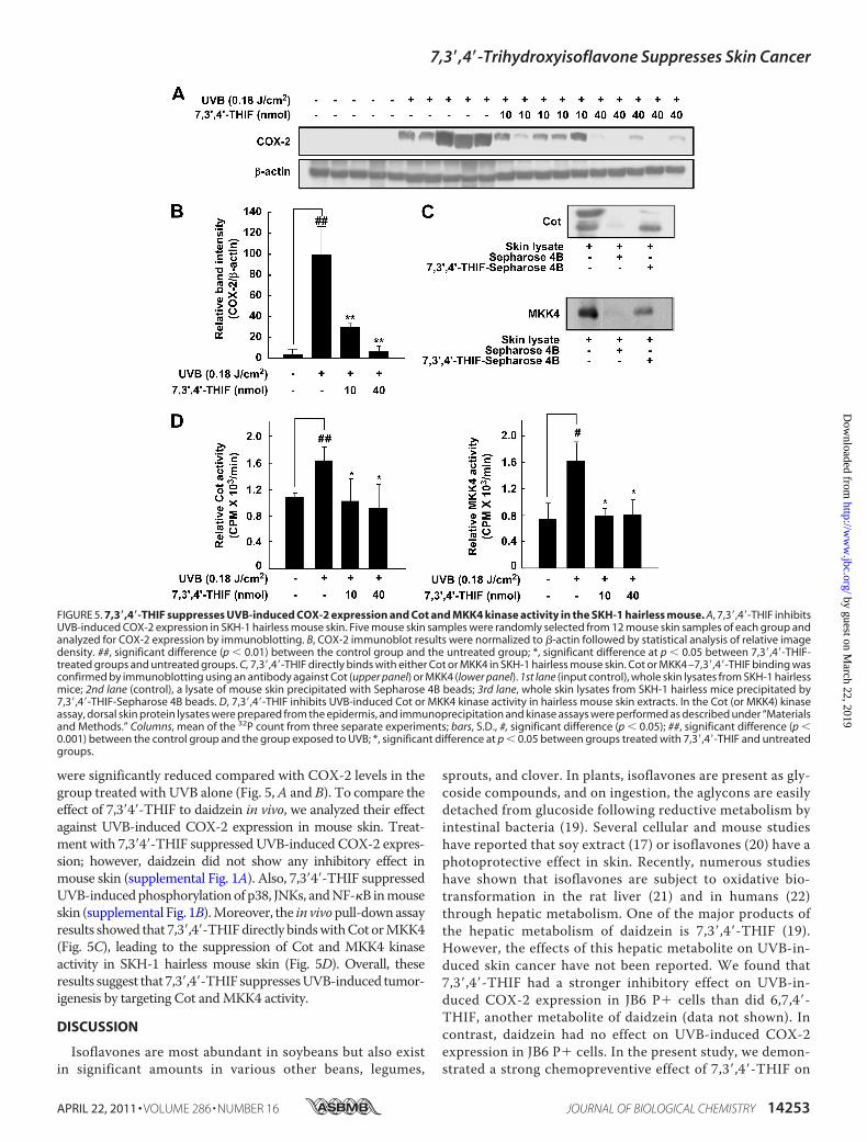
were significantly reduced compared with COX-2 levels in thegroup treated with UVB alone (Fig. 5,A and B). To compare theeffect of 7,3�4�-THIF to daidzein in vivo, we analyzed their effectagainst UVB-induced COX-2 expression in mouse skin. Treat-ment with 7,3�4�-THIF suppressed UVB-induced COX-2 expres-sion; however, daidzein did not show any inhibitory effect inmouse skin (supplemental Fig. 1A). Also, 7,3�4�-THIF suppressedUVB-inducedphosphorylationof p38, JNKs, andNF-�B inmouseskin (supplemental Fig. 1B).Moreover, the in vivopull-downassayresults showed that 7,3�,4�-THIFdirectly bindswithCot orMKK4(Fig. 5C), leading to the suppression of Cot and MKK4 kinaseactivity in SKH-1 hairless mouse skin (Fig. 5D). Overall, theseresults suggest that 7,3�,4�-THIF suppressesUVB-induced tumor-igenesis by targeting Cot andMKK4 activity.
DISCUSSION
Isoflavones are most abundant in soybeans but also existin significant amounts in various other beans, legumes,
sprouts, and clover. In plants, isoflavones are present as gly-coside compounds, and on ingestion, the aglycons are easilydetached from glucoside following reductive metabolism byintestinal bacteria (19). Several cellular and mouse studieshave reported that soy extract (17) or isoflavones (20) have aphotoprotective effect in skin. Recently, numerous studieshave shown that isoflavones are subject to oxidative bio-transformation in the rat liver (21) and in humans (22)through hepatic metabolism. One of the major products ofthe hepatic metabolism of daidzein is 7,3�,4�-THIF (19).However, the effects of this hepatic metabolite on UVB-in-duced skin cancer have not been reported. We found that7,3�,4�-THIF had a stronger inhibitory effect on UVB-in-duced COX-2 expression in JB6 P� cells than did 6,7,4�-THIF, another metabolite of daidzein (data not shown). Incontrast, daidzein had no effect on UVB-induced COX-2expression in JB6 P� cells. In the present study, we demon-strated a strong chemopreventive effect of 7,3�,4�-THIF on
FIGURE 5. 7,3�,4�-THIF suppresses UVB-induced COX-2 expression and Cot and MKK4 kinase activity in the SKH-1 hairless mouse. A, 7,3�,4�-THIF inhibitsUVB-induced COX-2 expression in SKH-1 hairless mouse skin. Five mouse skin samples were randomly selected from 12 mouse skin samples of each group andanalyzed for COX-2 expression by immunoblotting. B, COX-2 immunoblot results were normalized to �-actin followed by statistical analysis of relative imagedensity. ##, significant difference (p � 0.01) between the control group and the untreated group; *, significant difference at p � 0.05 between 7,3�,4�-THIF-treated groups and untreated groups. C, 7,3�,4�-THIF directly binds with either Cot or MKK4 in SKH-1 hairless mouse skin. Cot or MKK4 –7,3�,4�-THIF binding wasconfirmed by immunoblotting using an antibody against Cot (upper panel) or MKK4 (lower panel). 1st lane (input control), whole skin lysates from SKH-1 hairlessmice; 2nd lane (control), a lysate of mouse skin precipitated with Sepharose 4B beads; 3rd lane, whole skin lysates from SKH-1 hairless mice precipitated by7,3�,4�-THIF-Sepharose 4B beads. D, 7,3�,4�-THIF inhibits UVB-induced Cot or MKK4 kinase activity in hairless mouse skin extracts. In the Cot (or MKK4) kinaseassay, dorsal skin protein lysates were prepared from the epidermis, and immunoprecipitation and kinase assays were performed as described under “Materialsand Methods.” Columns, mean of the 32P count from three separate experiments; bars, S.D., #, significant difference (p � 0.05); ##, significant difference (p �0.001) between the control group and the group exposed to UVB; *, significant difference at p � 0.05 between groups treated with 7,3�,4�-THIF and untreatedgroups.
7,3�,4�-Trihydroxyisoflavone Suppresses Skin Cancer
APRIL 22, 2011 • VOLUME 286 • NUMBER 16 JOURNAL OF BIOLOGICAL CHEMISTRY 14253
by guest on March 22, 2019
http://ww
w.jbc.org/
Dow
nloaded from
UVB-induced skin cancer and suggested a molecular mech-anism and targets.The aberrant expression of COX-2 is a property of epithelial
cancers, including skin cancer, inmice and humans (23).More-over, COX-2-deficient mice have a reduced tumor incidenceand multiplicity response to 7,12-dimethylbenz(a)anthraceneand 12-O-tetradecanoylphorbol-13-acetate treatment (24, 25).
Therefore, the development of a natural inhibitor suppressingthe aberrant expression of COX-2 is a promising strategy in thechemoprevention of skin cancer. Our results showed that7,3�,4�-THIF exerts potent anti-tumor and anti-inflammatoryeffects. NF-�B is an important transcription factor in tumor-promoting processes such as inflammation and proliferation,and it is an important factor in the development of skin cancer.
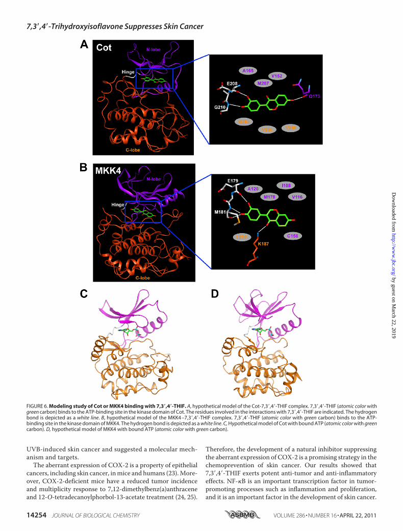
FIGURE 6. Modeling study of Cot or MKK4 binding with 7,3�,4�-THIF. A, hypothetical model of the Cot-7,3�,4�-THIF complex. 7,3�,4�-THIF (atomic color withgreen carbon) binds to the ATP-binding site in the kinase domain of Cot. The residues involved in the interactions with 7,3�,4�-THIF are indicated. The hydrogenbond is depicted as a white line. B, hypothetical model of the MKK4 –7,3�,4�-THIF complex. 7,3�,4�-THIF (atomic color with green carbon) binds to the ATP-binding site in the kinase domain of MKK4. The hydrogen bond is depicted as a white line. C, Hypothetical model of Cot with bound ATP (atomic color with greencarbon). D, hypothetical model of MKK4 with bound ATP (atomic color with green carbon).
7,3�,4�-Trihydroxyisoflavone Suppresses Skin Cancer
14254 JOURNAL OF BIOLOGICAL CHEMISTRY VOLUME 286 • NUMBER 16 • APRIL 22, 2011
by guest on March 22, 2019
http://ww
w.jbc.org/
Dow
nloaded from

Two estimatedNF-�Bbinding sites are present in the promoterregion of COX-2, and COX-2 expression is positively regulatedby NF-�B. Our results showed that 7,3�,4�-THIF inhibits UVB-induced COX-2 expression by attenuating COX-2 promoteractivity, which was attributed to the inhibition of NF-�B tran-scription activation.Research data indicate that MAP kinase phosphorylation
triggers signals from the cell surface, activating various tran-scription factors, thereby contributing to the regulation of tar-get gene expression. JNKs and p38 are generally recognized asstress-activatedMAP kinases. Several kinds ofMKKs can phos-phorylate JNKs, and p38-MKK3 and MKK6 activate p38,whereas MKK4 can activate both JNKs and p38 MAPK (18).Increased levels ofMKK4 are positively related to the increasedproliferation and invasion of several cancer cell lines (26). Ourresults showed that 7,3�,4�-THIF suppressed UVB-inducedphosphorylation of p38 and JNKs and was also effective atinhibiting MKK4 activity in vitro and ex vivo, resulting in theinhibition of COX-2 expression.The serine/threonine protein kinase Cot has been studied
mainly as a mediator of the inflammation or macrophage sig-naling pathways (27, 28). Recent studies showed that Cot isup-regulated in a variety of cancers and that Cot positively reg-ulates COX-2 expression and neoplastic cell transformationthrough transcriptional transactivation. Our previous studyshowed that Cot represents a novel serine/threonine kinasedirectly interacting with histone H3, resulting in the increasedcell transformation (29). Cot regulates NF-�B transcriptionalactivity by directly binding to p65 and triggering its phosphor-ylation (30). Our preliminary data showed that the Cot inhibi-tor down-regulated UVB-induced COX-2 expression by sup-pressing COX-2 promoter activity and NF-�B transcriptionactivity (data not shown). We then examined whether 7,3�,4�-THIF affected Cot activity, and the results showed that 7,3�,4�-THIF clearly suppressed Cot1 activity in vitro and ex vivo bydirectly binding with Cot1 competitively with ATP. Takentogether, these results indicate that the inhibition of COX-2expression by 7,3�,4�-THIF was attributable to the suppressionof Cot and MKK4 activity.The SKH-1 hairless mouse is an excellent model to study the
appearance of skin tumors induced by chronic UV radiation(31). The topical application of 7,3�,4�-THIF strongly sup-pressed the incidence, multiplicity, and volume of UVB-in-duced skin cancer development in the SKH-1 hairless mousemodel. Induction of COX-2 expression after skin exposure toUVB was substantial, and 7,3�,4�-THIF strongly inhibited theUVB-induced COX-2 expression in hairless mouse skin. Fur-thermore, 7,3�,4�-THIF directly bound with Cot or MKK4,resulting in suppression ofCot orMKK4activity in both JB6P�cells and hairless mouse skin. Collectively, these results suggestthat 7,3�,4�-THIF suppresses UVB-induced skin cancer bydirectly targeting Cot and MKK4.To investigate the molecular basis of Cot and MKK4 inhibi-
tion by 7,3�,4�-THIF, we carried out a docking study using thehomology model structures of the kinase domain of Cot,derived from the crystal structure of Mst1, which has 50%homology in amino acid sequence, and MKK4, derived fromthe crystal structure of MKK7 that has 66% homology. Homol-
ogymodelingwas used because neither the structure of Cot norMKK4 is available as yet. The kinase domains of Cot andMKK4consist of anN-lobe and aC-lobe. TheN-andC-lobes are linkedthrough a loop, which is called a “hinge region.” The backboneof this loop interacts with the adenine moiety of ATP throughhydrogen bonding. Considering the experimental result show-ing that 7,3�,4�-THIF is an ATP-competitive inhibitor of Cotand MKK4, we docked the compound to the ATP binding siteof each of the two kinases. 7,3�,4�-THIF easily docked to theCotor MKK4 ATP binding site, which is located between the N-and C-lobes of the kinase domain. In the model structure ofCot, complexed with 7,3�,4�-THIF, the hydroxyl group at the 3�and 4� positions of 7,3�,4�-THIF can form hydrogen bonds withthe backbone ofGlu-208 andGly-210 in the hinge region of Cot(Fig. 6A). The hydroxyl group at the 7 position could form ahydrogen bond with the backbone carbonyl group of Gln-173.In addition, the inhibitor would be sandwiched in the ATPbinding site by the side chains of the hydrophobic residues,including Ala-165, Met-207, and Val-152 from the N-lobe andMet-262, Val-260, and Val-269 from the C-lobe. In the modelstructure ofMKK4, complexedwith 7,3�,4�-THIF, the hydroxylgroups at the 3� and 4� positions on 7,3�,4�-THIF could formhydrogen bonds with the backbone of Glu-179 andMet-181 inthe hinge region of MKK4 (Fig. 6B). The carbonyl group at the4 position could make a hydrogen bond with the side chain ofLys-187. 7,3�,4�-THIF could form van derWaals interactions attheATP binding site with hydrophobic residues, includingAla-120, Met-178, Ile-108, Val-116, Cys-156, and Leu-236. Becauseof the lack of a hydroxyl group at the 3� position of daidzein, itsinteraction with the hinge regions of Cot and MKK4 would beweaker than that of 7,3�,4�-THIF, and thus daidzein could noteffectively inhibit either of these kinases. Further studies byx-ray crystallography to determine the inhibitor complex struc-tures could elucidate the exact bindingmode of 7,3�,4�-THIF toCot and MKK4.In summary, 7,3�,4�-THIF, but not daidzein, inhibits UVB-
induced COX-2 expression in JB6 P� cells. The inhibition ismediatedmainly by the blockage of the JNKs and p38 pathwaysactivation and subsequent suppression of NF-�B activity.7,3�,4�-THIF binds with Cot and MKK4 and strongly inhibitstheir kinase activity. The chemopreventive effect of 7,3�,4�-THIFwas confirmed in the hairlessmousemodel because the invivo data showed that 7,3�,4�-THIF strongly suppressed UVB-induced tumor incidence, multiplicity, and tumor volume.Consistent with the tumor data, 7,3�,4�-THIF clearly inhibitedUVB-induced COX-2 expression in hairless mouse skin, and7,3�,4�-THIF directly boundwithCot orMKK4, resulting in thesuppression of Cot or MKK4 activity in hairless mouse skin.Collectively, these results suggest that Cot and MKK4 are themain molecular targets of 7,3�,4�-THIF in the suppression ofUVB-mediated skin cancer. These results provide insight intothe biological actions of 7,3�,4�-THIF and the molecular basisfor the development of new chemoprotective agents.REFERENCES1. Athar, M., An, K. P., Morel, K. D., Kim, A. L., Aszterbaum,M., Longley, J.,
Epstein, E. H., Jr., and Bickers, D. R. (2001) Biochem. Biophys. Res. Com-mun. 280, 1042–1047
2. Rundhaug, J. E., and Fischer, S. M. (2008) Photochem. Photobiol. 84,
7,3�,4�-Trihydroxyisoflavone Suppresses Skin Cancer
APRIL 22, 2011 • VOLUME 286 • NUMBER 16 JOURNAL OF BIOLOGICAL CHEMISTRY 14255
by guest on March 22, 2019
http://ww
w.jbc.org/
Dow
nloaded from
322–3293. Katiyar, S. K., Korman, N. J., Mukhtar, H., and Agarwal, R. (1997) J. Natl.
Cancer Inst. 89, 556–5664. Buckman, S. Y., Gresham, A., Hale, P., Hruza, G., Anast, J., Masferrer, J.,
and Pentland, A. P. (1998) Carcinogenesis 19, 723–7295. Marwaha, V., Chen, Y. H., Helms, E., Arad, S., Inoue, H., Bord, E., Kishore,
R., Sarkissian, R. D., Gilchrest, B. A., and Goukassian, D. A. (2005) J. Biol.Chem. 280, 32379–32388
6. Yaomura, T., Tsuboi, N., Urahama, Y., Hobo, A., Sugimoto, K.,Miyoshi, J.,Matsuguchi, T., Reiji, K.,Matsuo, S., andYuzawa, Y. (2008)Nephrology13,397–404
7. Krcova, Z., Ehrmann, J., Krejci, V., Eliopoulos, A., and Kolar, Z. (2008)Biomed. Pap. Med. Fac. Univ. Palacky Olomouc. Czech Repub. 152, 21–25
8. Eliopoulos, A. G., Dumitru, C. D., Wang, C. C., Cho, J., and Tsichlis, P. N.(2002) EMBO J. 21, 4831–4840
9. Cuenda, A. (2000) Int. J. Biochem. Cell Biol. 32, 581–58710. Derijard, B., Raingeaud, J., Barrett, T.,Wu, I. H., Han, J., Ulevitch, R. J., and
Davis, R. J. (1995) Science 267, 682–68511. Han, J., Lee, J. D., Jiang, Y., Li, Z., Feng, L., and Ulevitch, R. J. (1996) J. Biol.
Chem. 271, 2886–289112. Brancho, D., Tanaka, N., Jaeschke, A., Ventura, J. J., Kelkar, N., Tanaka, Y.,
Kyuuma,M., Takeshita, T., Flavell, R. A., andDavis, R. J. (2003)Genes Dev.17, 1969–1978
13. Mantena, S. K., and Katiyar, S. K. (2006) Free Radic. Biol. Med. 40,1603–1614
14. Ouyang, W., Zhang, D., Ma, Q., Li, J., and Huang, C. (2007) Environ.Health Perspect. 115, 513–518
15. Jung, S. K., Lee, K. W., Byun, S., Kang, N. J., Lim, S. H., Heo, Y. S., Bode,A. M., Bowden, G. T., Lee, H. J., and Dong, Z. (2008) Cancer Res. 68,6021–6029
16. Kang,N. J., Lee, K.W., Lee, D. E., Rogozin, E. A., Bode, A.M., Lee, H. J., and
Dong, Z. (2008) J. Biol. Chem. 283, 20664–2067317. Chen, N., Scarpa, R., Zhang, L., Seiberg, M., and Lin, C. B. (2008) Pho-
tochem. Photobiol. 84, 1551–155918. Whitmarsh, A. J., and Davis, R. J. (2007) Oncogene 26, 3172–318419. Kulling, S. E., Lehmann, L., and Metzler, M. (2002) J. Chromatogr. B 777,
211–21820. Moore, J. O., Wang, Y., Stebbins, W. G., Gao, D., Zhou, X., Phelps, R.,
Lebwohl, M., and Wei, H. (2006) Carcinogenesis 27, 1627–163521. Kulling, S. E., Honig, D. M., Simat, T. J., and Metzler, M. (2000) J. Agric.
Food Chem. 48, 4963–497222. Kulling, S. E., Honig, D. M., and Metzler, M. (2001) J. Agric. Food Chem.
49, 3024–303323. Muller-Decker, K., and Furstenberger, G. (2007) Mol. Carcinog. 46,
705–71024. Muller-Decker, K., Neufang, G., Berger, I., Neumann, M., Marks, F., and
Furstenberger, G. (2002) Proc. Natl. Acad. Sci. U.S.A. 99, 12483–1248825. Tiano, H. F., Loftin, C. D., Akunda, J., Lee, C. A., Spalding, J., Sessoms, A.,
Dunson, D. B., Rogan, E. G.,Morham, S. G., Smart, R. C., and Langenbach,R. (2002) Cancer Res. 62, 3395–3401
26. Wang, X., Destrument, A., andTournier, C. (2007)Biochim. Biophys. Acta1773, 1349–1357
27. Babu, G., Waterfield, M., Chang, M., Wu, X., and Sun, S. C. (2006) J. Biol.Chem. 281, 14041–14047
28. Stafford, M. J., Morrice, N. A., Peggie, M. W., and Cohen, P. (2006) FEBSLett. 580, 4010–4014
29. Choi, H. S., Kang, B. S., Shim, J. H., Cho, Y. Y., Choi, B. Y., Bode, A.M., andDong, Z. (2008) FASEB J. 22, 113–126
30. Wittwer, T., and Schmitz, M. L. (2008) Biochem. Biophys. Res. Commun.371, 294–297
31. Lee, H. J., Kim, J. S., Song, M. S., Seo, H. S., Moon, C., Kim, J. C., Jo, S. K.,Jang, J. S., and Kim, S. H. (2009) Phytother. Res. 23, 399–403
7,3�,4�-Trihydroxyisoflavone Suppresses Skin Cancer
14256 JOURNAL OF BIOLOGICAL CHEMISTRY VOLUME 286 • NUMBER 16 • APRIL 22, 2011
by guest on March 22, 2019
http://ww
w.jbc.org/
Dow
nloaded from

Ann M. Bode, Hyong Joo Lee and Zigang DongLim, Yong-Seok Heo, Jong Eun Kim, Nam Joo Kang, Bo Yeon Kim, G. Tim Bowden, Dong Eun Lee, Ki Won Lee, Sanguine Byun, Sung Keun Jung, Nury Song, Sung Hwan
Suppresses Ultraviolet B-induced Skin Cancer by Targeting Cot and MKK4-Trihydroxyisoflavone, a Metabolite of the Soy Isoflavone Daidzein,′,4′7,3
doi: 10.1074/jbc.M110.147348 originally published online March 4, 20112011, 286:14246-14256.J. Biol. Chem.
10.1074/jbc.M110.147348Access the most updated version of this article at doi:
Alerts:
When a correction for this article is posted•
When this article is cited•
to choose from all of JBC's e-mail alertsClick here
Supplemental material:
http://www.jbc.org/content/suppl/2011/03/04/M110.147348.DC1
http://www.jbc.org/content/286/16/14246.full.html#ref-list-1
This article cites 31 references, 10 of which can be accessed free at
by guest on March 22, 2019
http://ww
w.jbc.org/
Dow
nloaded from


![150 ppp [7,3 Mo]](https://img.dokumen.tips/doc/110x75/586e0b751a28abd80f8c1d8a/150-ppp-73-mo.jpg)