Embed Size (px)
Citation preview
Reading Essentials Chapter 7 Cellular Structure and Function 69
Cop
yrig
ht ©
Gle
ncoe
/McG
raw
-Hill
, a d
ivis
ion
of T
he M
cGra
w-H
ill C
ompa
nies
, Inc
.
Cellular Structure and Function7
section ● Cell Discovery and Theory1
Before You ReadHave you ever looked at anything through a magnifying glass or a microscope? Describe on the lines below how the magnifying glass or microscope changed the object. In this section you will learn about some important discoveries made using microscopes.
Focus As you read, underline or highlight the main ideas in each paragraph.
1. Compare What is one thing that plants and animals have in common?
Read to LearnHistory of the Cell Theory
A cell is the basic structural and functional unit of all living things. The human body consists of trillions and trillions of cells. But cells are too small to see with the human eye. The invention of the microscope allowed scientists to discover that cells existed.
In 1665, an English scientist named Robert Hooke made a simple microscope. He used the microscope to look at a piece of cork, which is the dead cells of oak bark. Hooke saw small, box-shaped structures in the cork, which he called cellulae. Today, we call them cells.
In the late 1600s, Anton van Leeuwenhoek (LAY vun hook), a Dutch scientist, made another microscope. He examined pond water, milk, and other substances. He was surprised to fi nd living organisms in these substances.
What discoveries led to the cell theory?In 1838, German scientist Matthias Schleiden studied plants
under microscopes. He concluded that all plants are composed of cells. Another German scientist, Theodor Schwann, declared that animal tissues were made up of cells.
The microscope led to the discovery of cells.
What You’ll Learn■ the principles of the cell theory■ how compound light
microscopes differ from electron microscopes
■ how prokaryotic and eukaryotic cells differ
069-082 GHSB RESG-874600.indd 69069-082 GHSB RESG-874600.indd 69 3/17/06 1:12:26 PM3/17/06 1:12:26 PM

Cop
yrig
ht ©
Gle
ncoe
/McG
raw
-Hill
, a d
ivis
ion
of T
he M
cGra
w-H
ill C
ompa
nies
, Inc
.
What is the cell theory?Scientists continued to learn more about cells. Scientist
Rudolf Virchow proposed that cells divide to form new cells. He suggested that every cell came from a cell that already existed. The observations and ideas of the various scientists who studied cells are summarized as the cell theory. The cell theory is a fundamental idea of modern biology and includes the principles listed in the table below.
The Cell Theory
Principle Explanation
1. All living organisms are made up of one or more cells.
An organism can have one or many cells. Most plants and animals have many cells.
2. The cell is the basic unit of organization in living organisms.
Even in complex organisms such as humans, the cell is the basic unit of life.
3. All cells come from cells. Cells pass copies of their genetic material on to their daughter cells.
Cells contain hereditary information that passes from cell to cell during cell division.
Microscope TechnologyThe development of the microscope made the discovery of
cells possible. Improvements made to early microscopes have helped scientists learn much more about cells.
What is a compound light microscope?The modern compound light microscope uses a series of
glass lenses to magnify, or enlarge, an object. When visible light passes through each lens, it magnifi es the image of the previous lens. For example, two lenses that each magnify an image 10× result in a microscope that magnifi es the object 100×, as shown in the fi gure below.
Picture This2. Highlight the principle in
the cell theory that resulted from the discoveries of Matthias Schleiden and Theodor Schwann.
Picture This3. Calculate If each lens in
this example magnifi ed the image 20×, what is the total magnifi cation? (Show your work.)
70 Cellular Structure and Function
069-082 GHSB RESG-874600.indd 70069-082 GHSB RESG-874600.indd 70 3/17/06 1:12:29 PM3/17/06 1:12:29 PM

Cop
yrig
ht ©
Gle
ncoe
/McG
raw
-Hill
, a d
ivis
ion
of T
he M
cGra
w-H
ill C
ompa
nies
, Inc
.What is an electron microscope?
The best compound light microscopes only magnify an image about 1000×. Scientists needed more powerful microscopes to learn more about cells. The electron microscope was invented in the 1940s. It doesn’t use lenses. Instead, the transmission electron microscope (TEM) uses magnets to aim a beam of electrons at the image to be magnifi ed. Some TEMs can magnify an image 500,000×.
The scanning electron microscope (SEM) was a further improvement in technology. It produces a three-dimensional image of the cell. One problem with the TEM and SEM is that only nonliving cells can be examined. A more recent invention, the scanning tunneling electron microscope (STM), can magnify living cells.
Basic Cell TypesCells have different sizes, shapes, and functions, but all cells
have a plasma membrane. A plasma membrane is a boundary that helps control what enters and leaves the cell.
Some basic functions are common to most cells. For example, most cells have some form of genetic material that provides instructions for making substances that the cell needs. In addition, all cells break down molecules to generate energy for metabolism.
What are the two categories of cells?Scientists group cells into two broad categories based on
their internal structures. These categories are prokaryotic cells and eukaryotic cells.
Simple cells that have no specialized structures are known as prokaryotic (pro kar ee AW tik) cells. Cell functions in these simple cells occur in the plasma membrane. Most unicellular organisms, such as bacteria, are prokaryotic cells. Thus, they are called prokaryotes. Prokaryotic cells are believed to be similar to the fi rst cells on Earth.
Eukaryotic (yew kar ee AW tik) cells are the other category of cells. They are usually larger and more complex.Eukaryotic cells contain a nucleus and other structures called organelles. Organelles are specialized structures that carryout specifi c functions. The nucleus contains the genetic material for the cell. Organisms that are made up of eukaryotic cells are called eukaryotes. Eukaryotes can be unicellular or multicellular.
4. Draw Conclusions What is an advantage of viewing living cells?
5. Compare Which cells are more complex? (Circle your answer.)
a. prokaryotic cells b. eukaryotic cells
Reading Essentials 71 biologygmh.com
069-082 GHSB RESG-874600.indd 71069-082 GHSB RESG-874600.indd 71 3/17/06 1:12:31 PM3/17/06 1:12:31 PM

72 Chapter 7 Cellular Structure and Function Reading Essentials
Cop
yrig
ht ©
Gle
ncoe
/McG
raw
-Hill
, a d
ivis
ion
of T
he M
cGra
w-H
ill C
ompa
nies
, Inc
.
Read to Learn
7
section ● The Plasma Membrane2
Cellular Structure and Function
Before You ReadA window screen in your home allows air to pass through while keeping insects out. In this section, you will learn about a cell structure that has the same basic function. On the lines below, list some things you think would be allowed to pass into a cell and some things that would be kept out.
Function of the Plasma MembraneA cell’s survival depends on maintaining balance, called
homeostasis. The plasma membrane is the cell structure primarily responsible for homeostasis. It is the thin, fl exible boundary between the cell and its watery environment. Nutrients enter the cell and wastes leave the cell through the plasma membrane.
Selective permeability (pur mee uh BIH luh tee) of the plasma membrane allows some substances to pass through while keeping others out. The fi gure below shows selective permeability of the cell’s plasma membrane. The arrows show common substances that enter and leave the cell. The plasma membrane controls how, when, and how much of these substances enter and leave the cells.
Make Flash Cards Make a fl ash card for each question heading in this section. On the back of the fl ash card, write the answer to the question. Use the fl ash cards to review what you have learned.
Picture This 1. Highlight the items in
the fi gure that enter the cell through the plasma membrane. Circle the items that exit the cell.
A cell’s plasma membrane helps maintain homeostasis.
What You’ll Learn■ how the cell’s plasma membrane
functions■ the role of proteins,
carbohydrates, and cholesterol in the plasma membrane
069-082 GHSB RESG-874600.indd 72069-082 GHSB RESG-874600.indd 72 3/17/06 1:12:33 PM3/17/06 1:12:33 PM

Reading Essentials Chapter 7 Cellular Structure and Function 73
Cop
yrig
ht ©
Gle
ncoe
/McG
raw
-Hill
, a d
ivis
ion
of T
he M
cGra
w-H
ill C
ompa
nies
, Inc
.Structure of the Plasma Membrane
You have learned that lipids are large molecules madeup of glycerol and three fatty acids. A phospholipid(fahs foh LIH pid) is made up of glycerol, two fatty acids, and a phosphate group. The plasma membrane is made up of two layers of phospholipids arranged tail-to-tail in what is called a phospholipid bilayer. The phospholipid bilayer allows the plasma membrane to survive and function in its watery environment.
What is the structure of thephospholipid bilayer?
Each phospholipid has a polar head and two nonpolar tails. The phosphate group in the phospholipid makes it polar. The polar head is attracted to water because water is also polar. The nonpolar tails, made of the fatty acids, are repelled by water.
The phospholipid bilayer is arranged so that the polar heads can be closest to the water that is inside and outside the cell. Likewise, the nonpolar tails are farthest from the water because they are inside the phospholipid bilayer, as shown in the fi gure below. This bilayer structure is important for the formation and function of the plasma membrane.
2. Explain the purpose of the phospholipid bilayer.
Picture This3. Identify Circle one
phospholipid. Label itshead and tails.
How does the phospholipid bilayer function?The phospholipid bilayer forms a barrier that is polar on
the surface and nonpolar in the middle. Substances that can dissolve in water will not pass through the plasma membrane because they are stopped by the nonpolar middle. This allows the plasma membrane to separate the environment inside the cell from the environment outside the cell.
069-082 GHSB RESG-874600.indd 73069-082 GHSB RESG-874600.indd 73 3/17/06 1:12:37 PM3/17/06 1:12:37 PM
Cop
yrig
ht ©
Gle
ncoe
/McG
raw
-Hill
, a d
ivis
ion
of T
he M
cGra
w-H
ill C
ompa
nies
, Inc
.
What else is found in the plasma membrane?Cholesterol, proteins, and carbohydrates move among the
phospholipids in the plasma membrane. Proteins are found on both the inner surface and the outer surface of the plasma membrane. Proteins on the outer surface are called receptors because they send signals to the inside of the cell. Proteins on the inner surface anchor the plasma membrane to the cell’s internal support structure. These proteins give the cell its shape.
What are transport proteins?Proteins also create tunnels through the plasma membrane.
These proteins, known as transport proteins, move needed substances or waste materials through the plasma membrane. Transport proteins contribute to the selective permeability of the plasma membrane.
How does cholesterol help cells?Cholesterol molecules are nonpolar. They move among
the tails of the phospholipids. Cholesterol helps prevent the fatty-acid tails from sticking together, keeping the plasma membrane fl uid. Cholesterol also helps maintain homeostasis in a cell.
What substances help identifychemical signals?
Carbohydrates and proteins might stick out from the plasma membrane. They help the cell identify chemical signals from the environment. For example, carbohydrates in the plasma membrane might help disease-fi ghting cells identify and attack a potentially harmful cell.
What is the fl uid mosaic model?All the components of the plasma membrane are in
constant motion. Phospholipids can move sideways within the plasma membrane. Proteins, carbohydrates, and cholesterol molecules move among the phospholipids.
The phospholipid bilayer creates a sea in which all the other molecules fl oat. As the individual molecules move around, a pattern, or mosaic, is formed on the surface of the plasma membrane. This organization of the plasma membrane is called the fl uid mosaic model. It is fl uid because the molecules are moving and being rearranged. It is called a mosaic because scientists can observe clear patterns on the surface of the plasma membrane.
4. Defi ne the role of transport proteins.
5. Name three substances that move among the phospholipids of the plasma membrane.
biologygmh.com74 Cellular Structure and Function
069-082 GHSB RESG-874600.indd 74069-082 GHSB RESG-874600.indd 74 3/17/06 1:12:39 PM3/17/06 1:12:39 PM

Cellular Structure and Function
Reading Essentials Chapter 7 Cellular Structure and Function 75
Cop
yrig
ht ©
Gle
ncoe
/McG
raw
-Hill
, a d
ivis
ion
of T
he M
cGra
w-H
ill C
ompa
nies
, Inc
.
Read to LearnCytoplasm and Cytoskeleton
The environment inside the plasma membrane is a semifl uid material called cytoplasm. Scientists once thought the organelles of eukaryotic cells fl oated freely in the cell’s cytoplasm. As technology improved, scientists discovered more about cell structures. They discovered a structure within the cytoplasm called the cytoskeleton. The cytoskeleton is a network of long, thin protein fi bers that provide an anchor for organelles inside the cell. The cell’s shape and movement depend on the cytoskeleton.
Two types of protein fi bers make up the cytoskeleton.Microtubules are long, hollow protein cylinders that form a fi rm skeleton for the cell. They assist in moving substances within the cell. Microfi laments are thin protein threads that help give the cell shape and enable the entire cell or parts of the cell to move.
Cell StructuresAll chemical processes of a typical eukaryotic cell take place
in the organelles, which move around in the cell’s cytoplasm. Proteins are produced, food is transformed into energy, and wastes are processed in the organelles. Each organelle has a unique structure and function.
7
section ● Structures and Organelles3
Before You ReadFor cells to function correctly, each part must do its job. Members of families have jobs that help the whole family. On the lines below, list your family members and their jobs.
1. Name one cell function that takes place in organelles.
Identify the Parts Highlight each cell structure as you read about it. Underline the function of each part.
The eukaryotic cell contains organelles.
What You’ll Learndifferences in the structures of plant and animal cells
069-082 GHSB RESG-874600.indd 75069-082 GHSB RESG-874600.indd 75 3/17/06 1:12:42 PM3/17/06 1:12:42 PM

76 Chapter 7 Cellular Structure and Function Reading Essentials
Cop
yrig
ht ©
Gle
ncoe
/McG
raw
-Hill
, a d
ivis
ion
of T
he M
cGra
w-H
ill C
ompa
nies
, Inc
.
How are plant and animal cells different?The fi gure below shows a typical plant cell and a typical
animal cell. Note how many organelles are found in both types of cells. Also, note a few differences, such as the chloroplast that appears only in the plant cell. Observe that the vacuole in the plant cell is much larger than the vacuole in the animal cell.
Picture This 2. Highlight the names of
structures found in both plant cells and animal cells. Circle the names of structures that are found only in animal cells. Underline the names of structures that are found only in plant cells.
069-082 GHSB RESG-874600.indd 76069-082 GHSB RESG-874600.indd 76 3/17/06 1:12:44 PM3/17/06 1:12:44 PM

Reading Essentials Chapter 7 Cellular Structure and Function 77
Cop
yrig
ht ©
Gle
ncoe
/McG
raw
-Hill
, a d
ivis
ion
of T
he M
cGra
w-H
ill C
ompa
nies
, Inc
.What structure manages cell processes?
The nucleus is the cell’s managing structure. Most of the cell’s DNA is in the nucleus. DNA defi nes the cell and controls protein production. A nuclear envelope surrounds the nucleus. Substances pass through the nuclear envelope to move in and out of the nucleus.
Which organelle produces proteins?Ribosomes produce proteins and are made of two
components—RNA and protein. Ribosomes are produced in the nucleolus, a structure located inside the nucleus. Some ribosomes fl oat freely in the cytoplasm. They produce proteins that will be used by other cells. Other ribosomes attach to an organelle called the endoplasmic reticulum.
What attaches to rough endoplasmic reticulum?The endoplasmic reticulum, (en duh PLAZ mihk •
rih TIHK yuh lum) also called ER, is a membrane system of folded sacs and channels to which ribosomes are attached. There are two types of ER. The fi rst type is called rough endoplasmic reticulum. This is the area where ribosomes attach to the ER’s surface. The ribosomes appear to create bumps or rough places on the membrane. The second type, smooth endoplasmic reticulum, has no ribosomes attached. Smooth ER produces complex carbohydrates and lipids.
What is the purpose of the Golgi apparatus?Once proteins are created, they move to another organelle,
the Golgi (GAWL jee) apparatus. The Golgi apparatusmodifi es, sorts, and packs the proteins into sacs called vesicles. The vesicles fuse with the cell’s plasma membrane. There the vesicles release the proteins, which move through the plasma membrane to the environment outside the cell.
What is stored in vacuoles?Cells have vesicles called vacuoles that act as temporary
storage for materials in the cytoplasm. Vacuoles can store food and other material needed by a cell. They can also store wastes. Plant cells normally have one large vacuole. Animal cells might or might not have a few small vacuoles.
What are lysosomes?Lysosomes are vesicles that contain substances that digest
excess or worn-out organelles and food particles. Lysosomes also digest bacteria and viruses that enter the cell.
3. Explain What is the role of DNA?
069-082 GHSB RESG-874600.indd 77069-082 GHSB RESG-874600.indd 77 3/17/06 1:12:47 PM3/17/06 1:12:47 PM

78 Chapter 7 Cellular Structure and Function Reading Essentials
Cop
yrig
ht ©
Gle
ncoe
/McG
raw
-Hill
, a d
ivis
ion
of T
he M
cGra
w-H
ill C
ompa
nies
, Inc
.
What makes up a centriole?Centrioles are organelles made of microtubules that
function during cell division. They usually are found near the nucleus of the cell. Plant cells do not contain centrioles.
Which organelle produces energy?Cells need energy to survive. The organelles that convert
fuel particles such as sugars into usable energy are called mitochondria (mi tuh KAHN dree uh). A mitochondrion has an outer membrane and an inner membrane with many folds, as shown in the fi gure below. The membrane provides a large surface area for breaking the bonds of sugar molecules. Energy is produced when the bonds are broken.
How do plant and animal cells differ?In addition to mitochondria, plant cells contain chloroplasts.
Chloroplasts are organelles that capture light energy and convert it to chemical energy through a process called photosynthesis. Plants can use light energy from any light source—usually solar. Animal cells do not have chloroplasts and cannot use the Sun’s energy as fuel for cell processes.
Plants also have cell walls. The cell wall is a mesh of fi bers that surrounds the plasma membrane. It protects and supports the cell. Plant cell walls are made of a carbohydrate known as cellulose.
What are cilia and fl agella?Some animal cell surfaces have cilia or fl agella that extend
beyond the plasma membrane. Cilia are short projections that look like hairs. They move back and forth, similar to the motion of the oars of a rowboat. Flagella are longer projections that move in a whiplike motion. Both cilia and fl agella are composed of microtubules. They move cells through their watery environments. Cilia also move substances along the surface of the cell.
Picture This 4. Explain why the inner
membrane has many folds.
5. Recall What is another function of microtubules?
069-082 GHSB RESG-874600.indd 78069-082 GHSB RESG-874600.indd 78 3/17/06 1:12:49 PM3/17/06 1:12:49 PM

Chapter 7 Cellular Structure and Function 79
Cop
yrig
ht ©
Gle
ncoe
/McG
raw
-Hill
, a d
ivis
ion
of T
he M
cGra
w-H
ill C
ompa
nies
, Inc
.
biologygmh.com
Comparing CellsThe table below summarizes the structures of eukaryotic
plant and animal cells. The function of each structure is described. Note that prokaryotic cells lack most of the organelles found in eukaryotic cells.
Organelles at WorkThe structures in the cell work together to perform cell
functions. The synthesis of proteins is a major cell function, which begins in the nucleus. Protein synthesis continues with the ribosomes on the rough ER and the ribosomes that fl oat freely in the cytoplasm. Most proteins made on the rough ER are sent to the Golgi apparatus. There they are packaged in vesicles and sent to other organelles or out of the cell. Like each member of a soccer team, each cell structure has a specifi c task to do to make the cell function properly.
Cell Structure FunctionPresent in
Plant/Animal Cells
Cell wall protects and supports plant cells plant cells only
Centriole important in cell division animal cells only
Chloroplast site where photosynthesis occurs plant cells only
Cilia aids in moving the cell and moving substances along thesurface of the cell
some animal cells
Cytoskeleton a framework for the cell within the cytoplasm both
Endoplasmicreticulum (ER)
site of protein synthesis; where ribosomes attach both
Flagellum aids in moving and feeding the cell some animal cells
Golgi apparatus modifi es and packages proteins for distribution outside the cell both
Lysosome breaks down excess or worn-out substances in the cell animal cells only
Mitochondrion supplies energy to the rest of the cell both
Nucleus directs the production of proteins and cell division both
Plasma membrane controls the movement of substances in and out of the cell both
Ribosome produces proteins both
Vacuole stores materials temporarily plant cell—one large; animal cell—a few small
Picture This 6. Highlight the organelle
to which ribosomes attach.
7. Identify a major cell function.
069-082 GHSB RESG-874600.indd 79069-082 GHSB RESG-874600.indd 79 3/17/06 1:12:51 PM3/17/06 1:12:51 PM

Cellular Structure and Function
80 Chapter 7 Cellular Structure and Function Reading Essentials
Cop
yrig
ht ©
Gle
ncoe
/McG
raw
-Hill
, a d
ivis
ion
of T
he M
cGra
w-H
ill C
ompa
nies
, Inc
.
section ● Cellular Transport4
7
Before You ReadDescribe on the lines below how you would move a large box that weighs more than you do. Then read the section to learn how large particles move in and out of cells.
Read to LearnDiffusion
Substances dissolved in water move constantly and randomly. Imagine you place a drop of red ink on the left side and a drop of blue ink on the right side of a dish of water. The ink moves randomly through the water and turns the water purple as the colors mix. The ink has diffused in the water. Diffusion is the net movement of particles from an area where there are more particles of the substance to an area where there are fewer particles. Diffusion does not require additional energy because the particles are already in motion.
Concentration is the amount of a substance in an area.Diffusion continues until the concentrations are the same in all areas of the water. The dish of water has reached dynamic equilibrium, in which the particles continue to move randomly, but the overall concentration does not change.
What affects the rate of diffusion?Concentration, temperature, and pressure affect the
rate of diffusion. Diffusion occurs more quickly when the concentration, temperature, or pressure are high because the particles collide more often. The size and charge of a substance also affects the rate of diffusion.
Create a Quiz As you read this section, write quiz questions based on what you have learned. After you write the questions, answer them.
Cellular transport moves substances in and out of a cell.
What You’ll Learn■ the process of diffusion,
facilitated diffusion, and active transport
■ effect of hypotonic, hypertonic, or isotonic solutions on a cell
■ how large particles enter and exit a cell
Record Information Make a three-pocket Foldable from an 11 � 17 sheet of paper. As you read, record information about cellular transport on quarter sheets of notebook paper and store them in the appropriate pocket.
Diffusion PassiveTransport
ActiveTransport
069-082 GHSB RESG-874600.indd 80069-082 GHSB RESG-874600.indd 80 3/17/06 1:12:53 PM3/17/06 1:12:53 PM
Reading Essentials Chapter 7 Cellular Structure and Function 81
Cop
yrig
ht ©
Gle
ncoe
/McG
raw
-Hill
, a d
ivis
ion
of T
he M
cGra
w-H
ill C
ompa
nies
, Inc
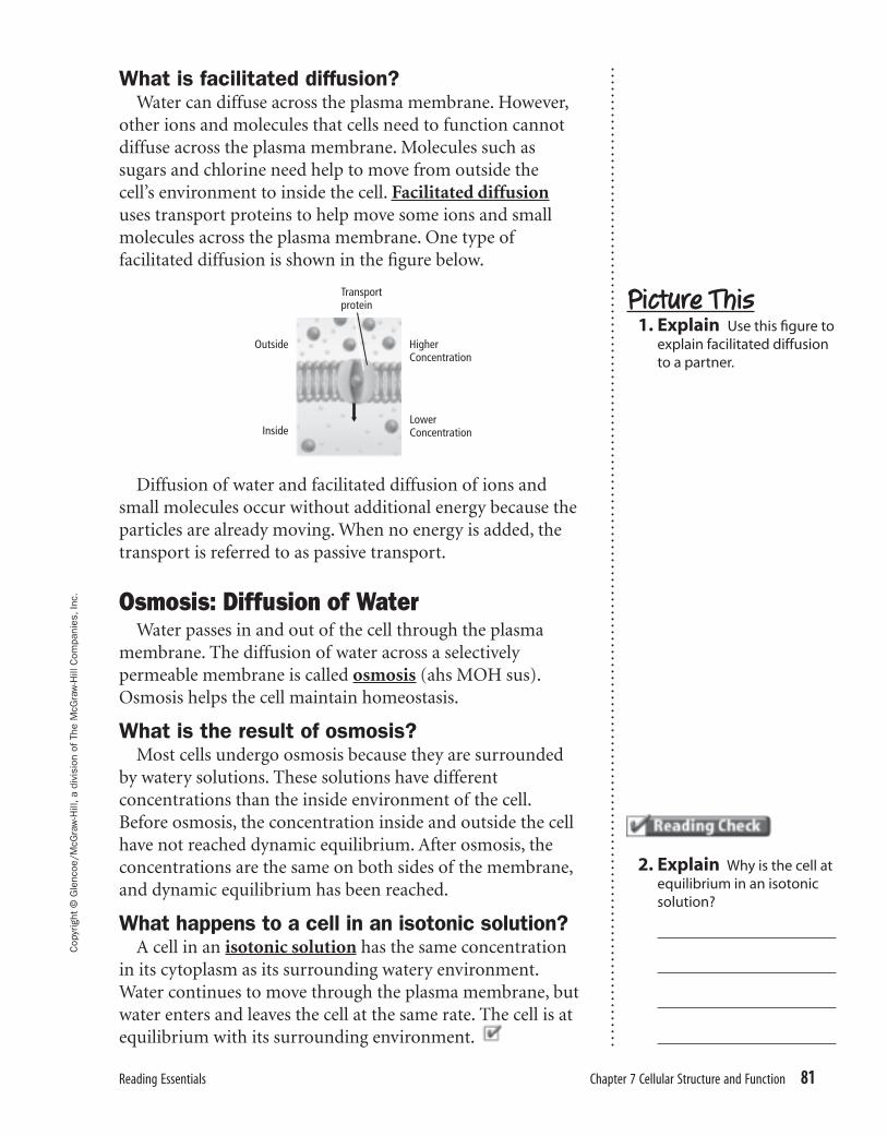
.What is facilitated diffusion?
Water can diffuse across the plasma membrane. However, other ions and molecules that cells need to function cannot diffuse across the plasma membrane. Molecules such as sugars and chlorine need help to move from outside the cell’s environment to inside the cell. Facilitated diffusion uses transport proteins to help move some ions and small molecules across the plasma membrane. One type of facilitated diffusion is shown in the fi gure below.
Diffusion of water and facilitated diffusion of ions and small molecules occur without additional energy because the particles are already moving. When no energy is added, the transport is referred to as passive transport.
Osmosis: Diffusion of WaterWater passes in and out of the cell through the plasma
membrane. The diffusion of water across a selectively permeable membrane is called osmosis (ahs MOH sus). Osmosis helps the cell maintain homeostasis.
What is the result of osmosis?Most cells undergo osmosis because they are surrounded
by watery solutions. These solutions have different concentrations than the inside environment of the cell. Before osmosis, the concentration inside and outside the cell have not reached dynamic equilibrium. After osmosis, the concentrations are the same on both sides of the membrane, and dynamic equilibrium has been reached.
What happens to a cell in an isotonic solution?A cell in an isotonic solution has the same concentration
in its cytoplasm as its surrounding watery environment. Water continues to move through the plasma membrane, but water enters and leaves the cell at the same rate. The cell is at equilibrium with its surrounding environment.
Picture This 1. Explain Use this fi gure to
explain facilitated diffusion to a partner.
2. Explain Why is the cell at equilibrium in an isotonic solution?
069-082 GHSB RESG-874600.indd 81069-082 GHSB RESG-874600.indd 81 3/17/06 1:12:56 PM3/17/06 1:12:56 PM
82 Chapter 7 Cellular Structure and Function
Cop
yrig
ht ©
Gle
ncoe
/McG
raw
-Hill
, a d
ivis
ion
of T
he M
cGra
w-H
ill C
ompa
nies
, Inc
.
biologygmh.com
3. Analyze Why does water move into a cell placed in a hypotonic solution?
Picture This 4. Label the cell structure
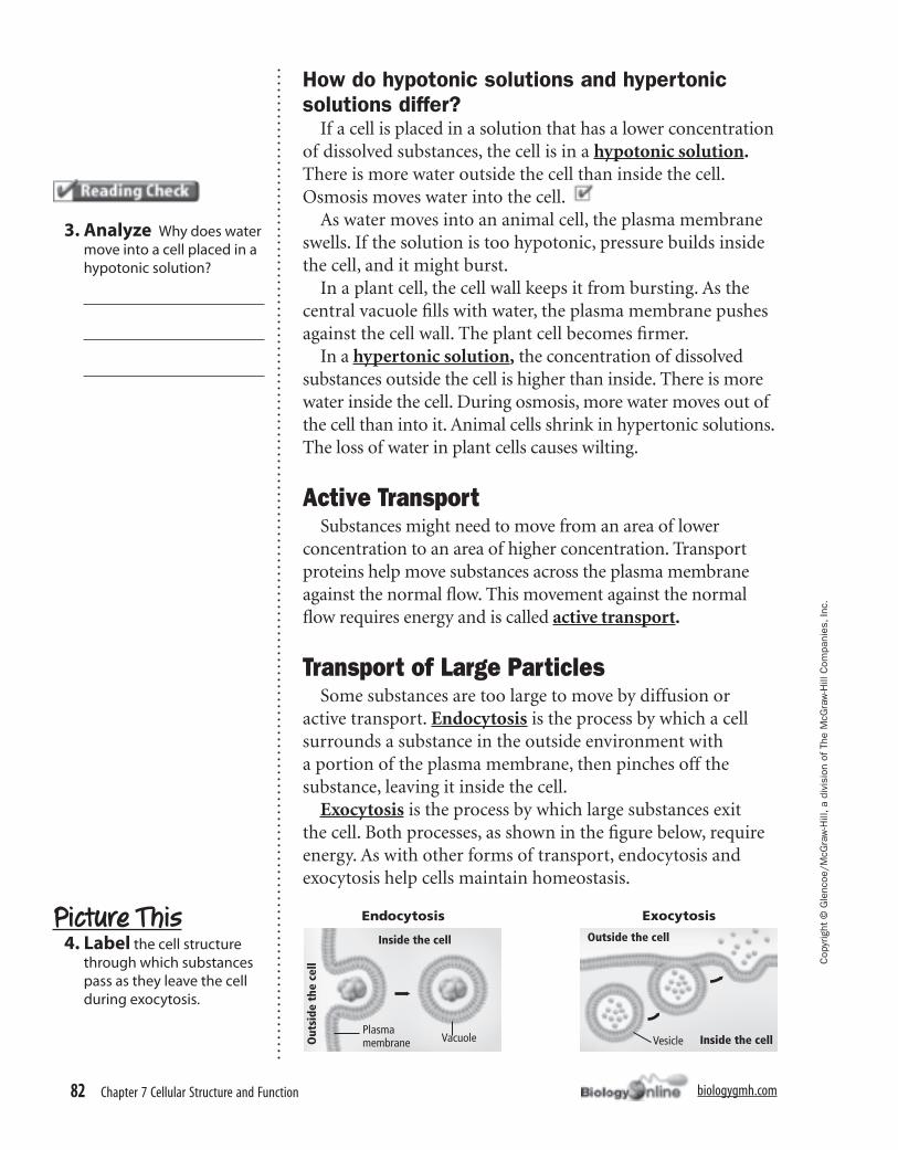
through which substances pass as they leave the cell during exocytosis.
How do hypotonic solutions and hypertonic solutions differ?
If a cell is placed in a solution that has a lower concentration of dissolved substances, the cell is in a hypotonic solution. There is more water outside the cell than inside the cell. Osmosis moves water into the cell.
As water moves into an animal cell, the plasma membrane swells. If the solution is too hypotonic, pressure builds inside the cell, and it might burst.
In a plant cell, the cell wall keeps it from bursting. As the central vacuole fi lls with water, the plasma membrane pushes against the cell wall. The plant cell becomes fi rmer.
In a hypertonic solution, the concentration of dissolved substances outside the cell is higher than inside. There is more water inside the cell. During osmosis, more water moves out of the cell than into it. Animal cells shrink in hypertonic solutions. The loss of water in plant cells causes wilting.
Active TransportSubstances might need to move from an area of lower
concentration to an area of higher concentration. Transport proteins help move substances across the plasma membrane against the normal fl ow. This movement against the normal fl ow requires energy and is called active transport.
Transport of Large ParticlesSome substances are too large to move by diffusion or
active transport. Endocytosis is the process by which a cell surrounds a substance in the outside environment with a portion of the plasma membrane, then pinches off the substance, leaving it inside the cell.
Exocytosis is the process by which large substances exit the cell. Both processes, as shown in the fi gure below, require energy. As with other forms of transport, endocytosis and exocytosis help cells maintain homeostasis.
069-082 GHSB RESG-874600.indd 82069-082 GHSB RESG-874600.indd 82 3/17/06 1:12:58 PM3/17/06 1:12:58 PM