Embed Size (px)
Citation preview
End Show
Slide 2 of 47
Copyright Pearson Prentice Hall
7-3 Cell Boundaries 7-3 Cell Boundaries

End Show
7-3 Cell Boundaries
Slide 3 of 47
Copyright Pearson Prentice Hall
7-3 Cell Boundaries
All cells are surrounded by a thin, flexible barrier known as the cell membrane.
Many cells also produce a strong supporting layer around the membrane known as a cell wall.

End Show
7-3 Cell Boundaries
Slide 4 of 47
Copyright Pearson Prentice Hall
Cell Membrane
What is the main function of the cell membrane?

End Show
7-3 Cell Boundaries
Slide 5 of 47
Copyright Pearson Prentice Hall
Cell Membrane
Cell Membrane
The cell membrane regulates what enters and leaves the cell and also provides protection and support.

End Show
7-3 Cell Boundaries
Slide 6 of 47
Copyright Pearson Prentice Hall
Cell Membrane
Cell Membrane
Outside of cell
Cell membrane
Inside of cell (cytoplasm)
Protein channel
Proteins
Lipid bilayer
Carbohydrate chains
End Show
7-3 Cell Boundaries
Slide 7 of 47
Copyright Pearson Prentice Hall
Cell Membrane
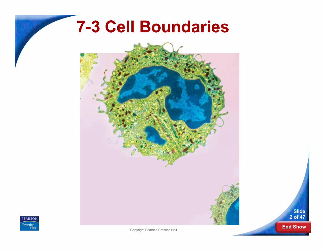
The composition of nearly all cell membranes is a double-layered sheet called a lipid bilayer.
Lipid bilayer

End Show
7-3 Cell Boundaries
Slide 8 of 47
Copyright Pearson Prentice Hall
Cell Membrane
The lipid bilayer gives cell membranes a flexible structure that forms a barrier between the cell and its surroundings.

End Show
7-3 Cell Boundaries
Slide 9 of 47
Copyright Pearson Prentice Hall
Cell Membrane
Most cell membranes contain protein molecules embedded in the lipid bilayer, some of which have carbohydrate molecules attached to them.
Protein channel
Proteins
Carbohydrate chains

End Show
7-3 Cell Boundaries
Slide 10 of 47
Copyright Pearson Prentice Hall
Cell Walls
What is the main function of the cell wall?

End Show
7-3 Cell Boundaries
Slide 11 of 47
Copyright Pearson Prentice Hall
Cell Walls
The main function of the cell wall is to provide support and protection for the cell.

End Show
7-3 Cell Boundaries
Slide 12 of 47
Copyright Pearson Prentice Hall
Cell Walls
Cell Wall
Cell walls are found in plants, algae, fungi, and many prokaryotes.
The cell wall lies outside the cell membrane.
Most cell walls are porous enough to allow water, oxygen, carbon dioxide, and certain other substances to pass through easily.

End Show
7-3 Cell Boundaries
Slide 13 of 47
Copyright Pearson Prentice Hall
Diffusion Through Cell Boundaries
Diffusion Through Cell Boundaries
Every living cell exists in a liquid environment.
The cell membrane regulates movement of dissolved molecules from the liquid on one side of the membrane to the liquid on the other side.

End Show
7-3 Cell Boundaries
Slide 14 of 47
Copyright Pearson Prentice Hall
Diffusion Through Cell Boundaries
Measuring Concentration
A solution is a mixture of two or more substances.
The substances dissolved in the solution are called solutes.
The concentration of a solution is the mass of solute in a given volume of solution, or mass/volume.

End Show
7-3 Cell Boundaries
Slide 15 of 47
Copyright Pearson Prentice Hall
Diffusion Through Cell Boundaries
What happens during diffusion?

End Show
7-3 Cell Boundaries
Slide 16 of 47
Copyright Pearson Prentice Hall
Diffusion Through Cell Boundaries
Diffusion
Particles in a solution tend to move from an area where they are more concentrated to an area where they are less concentrated.
This process is called diffusion.
When the concentration of the solute is the same throughout a system, the system has reached equilibrium.

End Show
7-3 Cell Boundaries
Slide 17 of 47
Copyright Pearson Prentice Hall
Diffusion Through Cell Boundaries

End Show
7-3 Cell Boundaries
Slide 18 of 47
Copyright Pearson Prentice Hall
Diffusion Through Cell Boundaries
There is a higher concentration of solute on one side of the membrane as compared to the other side of the membrane.

End Show
7-3 Cell Boundaries
Slide 19 of 47
Copyright Pearson Prentice Hall
Diffusion Through Cell Boundaries
Solute particles move from the side of the membrane with a higher concentration of solute to the side of the membrane with a lower concentration of solute. The solute particles will continue to diffuse across the membrane until equilibrium is reached.

End Show
7-3 Cell Boundaries
Slide 20 of 47
Copyright Pearson Prentice Hall
Diffusion Through Cell Boundaries
When equilibrium is reached, solute particles continue to diffuse across the membrane in both directions.

End Show
7-3 Cell Boundaries
Slide 21 of 47
Copyright Pearson Prentice Hall
Diffusion Through Cell Boundaries
Diffusion depends upon random particle movements. Therefore, substances diffuse across membranes without requiring the cell to use energy.

End Show
7-3 Cell Boundaries
Slide 22 of 47
Copyright Pearson Prentice Hall
Osmosis
What is osmosis?

End Show
7-3 Cell Boundaries
Slide 23 of 47
Copyright Pearson Prentice Hall
Osmosis
Osmosis
Osmosis is the diffusion of water through a selectively permeable membrane.

End Show
7-3 Cell Boundaries
Slide 24 of 47
Copyright Pearson Prentice Hall
Osmosis
How Osmosis Works
Movement of water
Dilute sugar solution (Water more concentrated)
Concentrated sugar solution (Water less concentrated)
Sugar molecules
Selectively permeable membrane

End Show
7-3 Cell Boundaries
Slide 25 of 47
Copyright Pearson Prentice Hall
Osmosis
Water tends to diffuse from a highly concentrated region to a less concentrated region.
If you compare two solutions, the more concentrated solution is hypertonic (“above strength”).
The more dilute solution is hypotonic (“below strength”).

End Show
7-3 Cell Boundaries
Slide 26 of 47
Copyright Pearson Prentice Hall
Osmosis
When concentrations of solutions are the same on both sides of a membrane, the solutions are isotonic (”same strength”).

End Show
7-3 Cell Boundaries
Slide 27 of 47
Copyright Pearson Prentice Hall
Osmosis
Osmotic Pressure
Osmosis exerts a pressure known as osmotic pressure on the hypertonic side of a selectively permeable membrane.

End Show
7-3 Cell Boundaries
Slide 28 of 47
Copyright Pearson Prentice Hall
Osmosis
Because the cell is filled with salts, sugars, proteins, and other molecules, it will almost always be hypertonic to fresh water.
If so, the osmotic pressure should produce a net movement of water into the cell. As a result, the volume of the cell will increase until the cell becomes swollen or bursts.

End Show
7-3 Cell Boundaries
Slide 29 of 47
Copyright Pearson Prentice Hall
Osmosis
Cells in large organisms are not in danger of bursting because they are bathed in fluids, such as blood, that are isotonic.
Other cells are surrounded by tough cell walls that prevent the cells from expanding even under tremendous osmotic pressure.

End Show
7-3 Cell Boundaries
Slide 30 of 47
Copyright Pearson Prentice Hall
Facilitated Diffusion
Facilitated Diffusion
Cell membranes have protein channels that act as carriers, making it easy for certain molecules to cross.

End Show
7-3 Cell Boundaries
Slide 31 of 47
Copyright Pearson Prentice Hall
Facilitated Diffusion
The movement of specific molecules across cell membranes through protein channels is known as facilitated diffusion.
Hundreds of different protein channels have been found that allow particular substances to cross different membranes.

End Show
7-3 Cell Boundaries
Slide 32 of 47
Copyright Pearson Prentice Hall
Facilitated Diffusion
Facilitated Diffusion
Protein channel
Glucose molecules

End Show
7-3 Cell Boundaries
Slide 33 of 47
Copyright Pearson Prentice Hall
Facilitated Diffusion
Although facilitated diffusion is fast and specific, it is still diffusion.
Therefore, facilitated diffusion will only occur if there is a higher concentration of the particular molecules on one side of a cell membrane as compared to the other side.

End Show
7-3 Cell Boundaries
Slide 34 of 47
Copyright Pearson Prentice Hall
Active Transport
Active Transport
Sometimes cells move materials in the opposite direction from which the materials would normally move—that is against a concentration difference. This process is known as active transport.
Active transport requires energy.

End Show
7-3 Cell Boundaries
Slide 35 of 47
Copyright Pearson Prentice Hall
Active Transport
Molecular Transport
In active transport, small molecules and ions are carried across membranes by proteins in the membrane.
Energy use in these systems enables cells to concentrate substances in a particular location, even when diffusion might move them in the opposite direction.

End Show
7-3 Cell Boundaries
Slide 36 of 47
Copyright Pearson Prentice Hall
Active Transport
Molecule to be carried
Active Transport

End Show
7-3 Cell Boundaries
Slide 37 of 47
Copyright Pearson Prentice Hall
Active Transport
Endocytosis and Exocytosis
Large molecules and even solid clumps of material may undergo active transport by means of the cell membrane.
Endocytosis is the process of taking material into the cell by means of infoldings, or pockets, of the cell membrane.
The pocket breaks loose from the outer portion of the cell membrane and forms a vacuole within the cytoplasm.

End Show
7-3 Cell Boundaries
Slide 38 of 47
Copyright Pearson Prentice Hall
Active Transport
Two examples of endocytosis are:
• phagocytosis
• pinocytosis
End Show
7-3 Cell Boundaries
Slide 39 of 47
Copyright Pearson Prentice Hall
Active Transport
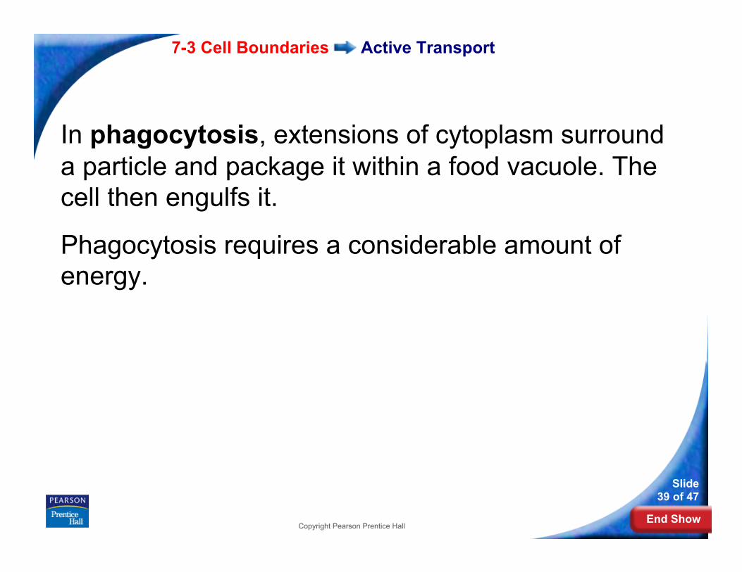
In phagocytosis, extensions of cytoplasm surround a particle and package it within a food vacuole. The cell then engulfs it.
Phagocytosis requires a considerable amount of energy.

End Show
7-3 Cell Boundaries
Slide 40 of 47
Copyright Pearson Prentice Hall
Active Transport
In pinocytosis, tiny pockets form along the cell membrane, fill with liquid, and pinch off to form vacuoles within the cell.

End Show
7-3 Cell Boundaries
Slide 41 of 47
Copyright Pearson Prentice Hall
Active Transport
Exocytosis
Many cells also release large amounts of material from the cell, in a process called exocytosis.
During exocytosis, the membrane of the vacuole surrounding the material fuses with the cell membrane, forcing the contents out of the cell.