Embed Size (px)
Citation preview

696 NATURE BIOTECHNOLOGY VOL 17 JULY 1999 http://biotech.nature.com
RESEARCH
Since plants express endogenous b-galactosidase activity, lacZ can-not be employed as a reporter gene1. Instead, the Escherichia coli b-glucuronidase gene (gusA, formerly uidA) has been developed as areporter gene for plants, and has been widely used for over adecade2. Both chromogenic and fluorogenic GUS substrates havebeen synthesized3, allowing rapid nonradioactive assays. The GUSenzyme is stable and active under a variety of conditions1, evenwhen fused to other sequences4.
The utility of GUS as a reporter, however, has been constrainedin three ways. First, many animal systems, and some plants andplant-associated bacteria express endogenous glucuronidase activi-ties2,3. Second, GUS activity is greatly reduced during tissue fixationby glutaraldehyde or formaldehyde, making it necessary to trade offretention of activity for preservation of tissue structure5. Third,both of these considerations drastically restrict the use of GUS as areporter gene in vertebrate systems6.
Enzymatic inactivation by aldehydes is largely due to the for-mation of Schiff bases with surface-accessible lysine residues7.While the removal of lysine residues by directed mutation mightrender an enzyme more resistant to fixatives, many surface lysinesare critical for function and cannot be readily changed. Thesequences of the E. coli 8, human9, mouse10, rat11, and dog12
homologs are known. Six of the 27 lysine residues in the E. coliprotein are conserved in the other species and thus are likelyessential. Moreover, to find what combination of the 27 lysineresidues could be changed in order to increase resistance to fixa-tives without abrogating enzyme activity would require con-structing and assaying a dauntingly large number of mutantenzymes. Therefore, in order to alter the surface chemistry ofGUS, either to avoid or to accommodate aldehyde modificationswithout loss of enzyme activity, we employed a random mutation-
al approach similar to those previously proven useful for alteringenzyme substrate specificity13 or thermostability14.
ResultsDirected evolution of glutaraldehyde-resistant variants. Randommutations were initially introduced into the gusA structural gene bymutagenic PCR15. Mutated PCR products were ligated into theexpression vector gusA-pBSD and transformed into E. coli. Whenthe library was induced on plates containing the chromogenic GUSsubstrate, 5-bromo-4-chloro-3-indolyl-b-D-glucuronide (X-gluc),approximately 80% of the colonies were visibly less green than con-trol colonies expressing only the chromosomal gene (seeExperimental protocol). b-Glucuronidase functions as a tetramer16,so it was likely that many of the mutations in the highly expressed,plasmid-borne library had a dominant negative effect on the func-tion of the chromosomal gene. This did not deter us from utilizingthis library for screening experiments, since successive rounds ofDNA shuffling should efficiently select against neutral or deleteri-ous mutations13.
Nine thousand replica-plated colonies, each expressing a ran-domly mutated gusA gene, were exposed to buffer containing 0.2%glutaraldehyde for 20 min. The colony remnants were then incubat-ed in buffer containing X-gluc and the histochemical indicator,nitroblue tetrazolium (NBT) (Fig. 1). The catalytic activity of thewild-type enzyme is greatly diminished under those conditions,indicating that the glutaraldehyde disrupts the cell membranes andcovalently modifies many intracellular proteins, including GUS (cf.Figs 2A and B). Of all the variants examined, only 10 colonies repro-ducibly exhibited greater catalytic activity than control coloniesexpressing wild-type gusA (Fig. 2B). The corresponding colonies onmaster plates were isolated, and their expression vectors purified
Directed evolution of the surfacechemistry of the reporter enzyme
b-glucuronidaseIchiro Matsumura1, John B. Wallingford2,3, Neeraj K. Surana1,4, Peter D. Vize2, and Andrew D. Ellington1*
1Institute of Cellular and Molecular Biology, ICMB A4800/MBB 3.424, 26th and Speedway, University of Texas, Austin, TX 78712. 2Center for DevelopmentalBiology, Department of Zoology, University of Texas, Austin, TX 78712. 3Present address: Department of Molecular and Cell Biology, University of California,
Berkeley, Berkeley, CA 94720. 4Present address: Division of Biology and Biomedical Sciences, Washington University, St. Louis, MO 63108. *Corresponding author(e-mail: [email protected]).
Receieved 29 December 1999; accepted 4 May 1999
The use of the Escherichia coli enzyme b-glucuronidase (GUS) as a reporter in gene expression stud-ies is limited due to loss of activity during tissue fixation by glutaraldehyde or formaldehyde. We havedirected the evolution of a GUS variant that is significantly more resistant to both glutaraldehyde andformaldehyde than the wild-type enzyme. A variant with eight amino acid changes was isolated afterthree rounds of mutation, DNA shuffling, and screening. Surprisingly, although glutaraldehyde is knownto modify and cross-link free amines, only one lysine residue was mutated. Instead, amino acid changesgenerally occurred near conserved lysines, implying that the surface chemistry of the enzyme wasselected to either accept or avoid glutaraldehyde modifications that would normally have inhibited func-tion. We have shown that the GUS variant can be used to trace cell lineages in Xenopus embryos understandard fixation conditions, allowing double staining when used in conjunction with other reporters.
Keywords: b-glucuronidase, reporter gene, in vitro evolution, directed evolution, DNA shuffling, Xenopus laevis
© 1999 Nature America Inc. • http://biotech.nature.com©
199
9 N
atu
re A
mer
ica
Inc.
• h
ttp
://b
iote
ch.n
atu
re.c
om

NATURE BIOTECHNOLOGY VOL 17 JULY 1999 http://biotech.nature.com 697
RESEARCH
and pooled. The variant gusA genes were amplified using the PCRand randomly recombined by DNA shuffling17. We then screened6,000 random recombinants in a second round for variants thatretained catalytic activity after a 20 min incubation in 1.0% glu-taraldehyde. Nine colonies contained variants that exhibited greaterresidual catalytic activity than the most resistant clone isolated inthe first round of screening (Fig. 2C). These variants were againpooled, amplified, and randomly recombined. Then, 6,000 recom-binants were screened in a third round for variants that retainedcatalytic activity after a 20 min incubation in 3.5% glutaraldehyde.Again, nine improved clones were isolated, one of which (GUSAR)reproducibly showed the greatest activity under the most stringentconditions (Fig. 2D).
In vitro characterization of GUSAR. To determine whether thecolony-lift assay significantly influenced the apparent fixative-resistant phenotype of GUSAR, activity assays were also carried outin cell extracts. The GUS-deficient strain, pREP4/GMS407, wastransformed with vectors that expressed either wild-type gusA, theGUSAR variant, or the lacZ a-fragment. Cell extracts from inducedcultures were treated with glutaraldehyde or formaldehyde for 20min at 23°C, and diluted 100-fold in buffer containing saturatingconcentrations of a GUS substrate, p-nitrophenyl b-D-glu-curonide (PNPG). The extracts containing wild type or GUSAR
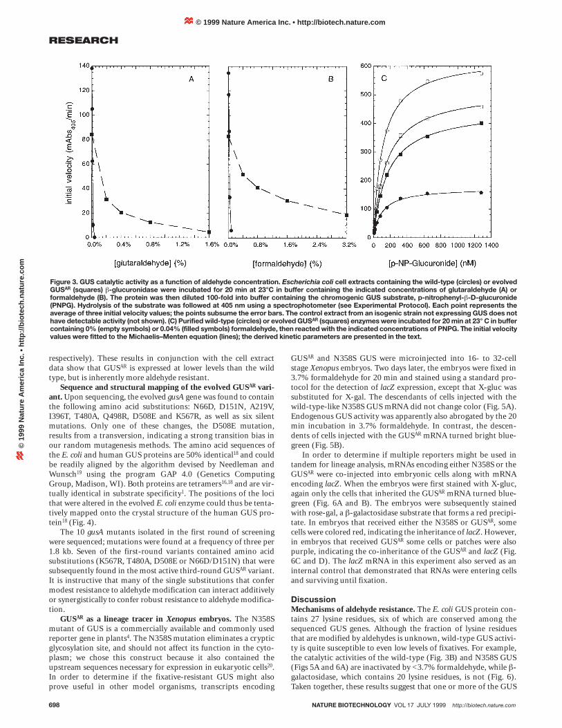
catalyzed the hydrolysis of PNPG; no hydrolysis was detected inthe negative control extracts, in which only the lacZ a-fragmentwas expressed (data not shown). Treatment of the extract contain-ing wild-type GUS with only 0.04% glutaraldehyde for 20 min at23°C reduced catalytic activity by 99.6 ± 0.24%. In sharp contrast,the GUSAR extract retained 78.1 ± 0.69% of its activity after treat-ment with a fivefold higher (0.2%) concentration of glutaralde-hyde (Fig. 3A).
Extracts were also separately treated with formaldehyde to assesswhether the fixative-resistant phenotype was specific to glutaralde-hyde. Again, the GUSAR variant exhibited much greater resistance tothe fixative than did the wild-type GUS. The wild-type extractretained only 4.6 ± 0.05% of its catalytic activity after treatmentwith 0.08% formaldehyde; the GUSAR extract retained 62.4 ± 0.40%activity after incubation with 0.4% formaldehyde (Fig. 3B). Todetermine how sequence and chemical modifications may haveinfluenced GUS activity, we conducted kinetic studies of the mutantenzyme (Fig. 3C). The wild-type and evolved gus genes were sub-cloned, expressed as fusion proteins with N-terminal hexahistidinetags, and purified by immobilized metal ion adsorption chromatog-raphy. Purified enzymes were assayed with varying concentrationsof PNPG (Fig. 3C). The kinetic parameters of the wild-type enzyme(KM for the complex with PNPG = 110 ± 2.9 mM; kcat = 920 ± 7.3 s-1)were very similar to those of the GUSAR variant (KM = 150 ± 3.9 mM;kcat = 750 ± 5.8 s-1). The kinetic parameters of wild-type and evolvedenzymes were also determined following reaction with a sublethalconcentration (0.04%) of formaldehyde for 20 min at 23°C.Formaldehyde had a larger effect on the turnover number (250 ± 17s-1 for the partially modified wild type, 650 ± 4.7 s-1 for the modifiedGUSAR variant), than on the Michaelis constants (99 ± 6.7 mM and170 ± 3.7 mM for the modified wild-type and GUSAR enzymes,
Figure 1. Screen for glutaraldehyde-resistant b-glucuronidase (GUS)function (sequence from top left). A library of randomly mutated b-glucuronidase genes (gusA) is subcloned into an inducibleexpression vector and transformed into Escherichia coli. Theresulting colonies are transferred to a nitrocellulose filter, which isoverlaid upon an agarose plate containing an inducer and incubatedfor 12–24 h at 37°C. The filter-bound colonies are incubated in buffercontaining glutaraldehyde, then transferred to buffer containing thehistochemical indicators of b-glucuronidase, X-gluc and NBT. Thebrief incubation in 4 M guanidine HCl (Gdn-HCl) arrests colordevelopment. Colonies that retain GUS activity are isolated from theoriginal plate and randomly recombined by DNA shuffling for the nextround of screening.
Figure 2. Detection of glutaraldehyde-resistant GUS activity.Escherichia coli cells transformed with vectors expressing the wild-type (A, B left), a pool of the ten glutaraldehyde-resistant variantsfrom round 1 (B right, C left) or the most resistant variants fromrounds 2 (C right, D left), or 3 (D right) were streaked ontononinducing plates. The colonies were propagated, induced, andtreated for 20 min with the indicated concentrations ofglutaraldehyde, then reacted with X-gluc and NBT, as described inthe legend to Figure 1.
A
B
C
D
© 1999 Nature America Inc. • http://biotech.nature.com©
199
9 N
atu
re A
mer
ica
Inc.
• h
ttp
://b
iote
ch.n
atu
re.c
om
698 NATURE BIOTECHNOLOGY VOL 17 JULY 1999 http://biotech.nature.com
RESEARCH
respectively). These results in conjunction with the cell extractdata show that GUSAR is expressed at lower levels than the wildtype, but is inherently more aldehyde resistant.
Sequence and structural mapping of the evolved GUSAR vari-ant. Upon sequencing, the evolved gusA gene was found to containthe following amino acid substitutions: N66D, D151N, A219V,I396T, T480A, Q498R, D508E and K567R, as well as six silentmutations. Only one of these changes, the D508E mutation,results from a transversion, indicating a strong transition bias inour random mutagenesis methods. The amino acid sequences ofthe E. coli and human GUS proteins are 50% identical18 and couldbe readily aligned by the algorithm devised by Needleman andWunsch19 using the program GAP 4.0 (Genetics ComputingGroup, Madison, WI). Both proteins are tetramers16,18 and are vir-tually identical in substrate specificity1. The positions of the locithat were altered in the evolved E. coli enzyme could thus be tenta-tively mapped onto the crystal structure of the human GUS pro-tein18 (Fig. 4).
The 10 gusA mutants isolated in the first round of screeningwere sequenced; mutations were found at a frequency of three per1.8 kb. Seven of the first-round variants contained amino acidsubstitutions (K567R, T480A, D508E or N66D/D151N) that weresubsequently found in the most active third-round GUSAR variant.It is instructive that many of the single substitutions that confermodest resistance to aldehyde modification can interact additivelyor synergistically to confer robust resistance to aldehyde modifica-tion.
GUSAR as a lineage tracer in Xenopus embryos. The N358Smutant of GUS is a commercially available and commonly usedreporter gene in plants4. The N358S mutation eliminates a crypticglycosylation site, and should not affect its function in the cyto-plasm; we chose this construct because it also contained theupstream sequences necessary for expression in eukaryotic cells20.In order to determine if the fixative-resistant GUS might alsoprove useful in other model organisms, transcripts encoding
GUSAR and N358S GUS were microinjected into 16- to 32-cellstage Xenopus embryos. Two days later, the embryos were fixed in3.7% formaldehyde for 20 min and stained using a standard pro-tocol for the detection of lacZ expression, except that X-gluc wassubstituted for X-gal. The descendants of cells injected with thewild-type-like N358S GUS mRNA did not change color (Fig. 5A).Endogenous GUS activity was apparently also abrogated by the 20min incubation in 3.7% formaldehyde. In contrast, the descen-dents of cells injected with the GUSAR mRNA turned bright blue-green (Fig. 5B).
In order to determine if multiple reporters might be used intandem for lineage analysis, mRNAs encoding either N358S or theGUSAR were co-injected into embryonic cells along with mRNAencoding lacZ. When the embryos were first stained with X-gluc,again only the cells that inherited the GUSAR mRNA turned blue-green (Fig. 6A and B). The embryos were subsequently stainedwith rose-gal, a b-galactosidase substrate that forms a red precipi-tate. In embryos that received either the N358S or GUSAR, somecells were colored red, indicating the inheritance of lacZ. However,in embryos that received GUSAR some cells or patches were alsopurple, indicating the co-inheritance of the GUSAR and lacZ (Fig.6C and D). The lacZ mRNA in this experiment also served as aninternal control that demonstrated that RNAs were entering cellsand surviving until fixation.
DiscussionMechanisms of aldehyde resistance. The E. coli GUS protein con-tains 27 lysine residues, six of which are conserved among thesequenced GUS genes. Although the fraction of lysine residuesthat are modified by aldehydes is unknown, wild-type GUS activi-ty is quite susceptible to even low levels of fixatives. For example,the catalytic activities of the wild-type (Fig. 3B) and N358S GUS(Figs 5A and 6A) are inactivated by <3.7% formaldehyde, while b-galactosidase, which contains 20 lysine residues, is not (Fig. 6).Taken together, these results suggest that one or more of the GUS
Figure 3. GUS catalytic activity as a function of aldehyde concentration. Escherichia coli cell extracts containing the wild-type (circles) or evolvedGUSAR (squares) b-glucuronidase were incubated for 20 min at 23°C in buffer containing the indicated concentrations of glutaraldehyde (A) orformaldehyde (B). The protein was then diluted 100-fold into buffer containing the chromogenic GUS substrate, p-nitrophenyl-b-D-glucuronide(PNPG). Hydrolysis of the substrate was followed at 405 nm using a spectrophotometer (see Experimental Protocol). Each point represents theaverage of three initial velocity values; the points subsume the error bars. The control extract from an isogenic strain not expressing GUS does nothave detectable activity (not shown). (C) Purified wild-type (circles) or evolved GUSAR (squares) enzymes were incubated for 20 min at 23° C in buffercontaining 0% (empty symbols) or 0.04% (filled symbols) formaldehyde, then reacted with the indicated concentrations of PNPG. The initial velocityvalues were fitted to the Michaelis–Menten equation (lines); the derived kinetic parameters are presented in the text.
© 1999 Nature America Inc. • http://biotech.nature.com©
199
9 N
atu
re A
mer
ica
Inc.
• h
ttp
://b
iote
ch.n
atu
re.c
om

NATURE BIOTECHNOLOGY VOL 17 JULY 1999 http://biotech.nature.com 699
RESEARCH
lysine residues is either itself critical for activity or presents a con-jugation site that leads to functional disruption. However, identi-fying which of the many lysine residues in GUS were responsiblefor inhibition by fixatives would have been a daunting task.Instead, we relied on a random mutagenesis to identify GUS variantswith catalytic activity resistant to aldehydes. Following three roundsof screening and amplification, we isolated an octuple-mutantGUSAR with catalytic activity resistant to roughly 80-fold higher lev-els of glutaraldehyde than the wild-type activity (Fig. 3A).
Surprisingly, only one of the amino acid substitutions, K567R,in the evolved GUSAR occurred at a lysine residue. Since AAA orAAG encodes lysine, the apparent transition bias in our random-mutagenesis method and the size of our initial library providedample opportunities for each lysine to conservatively mutate intoarginine (AGA or AGG). While it is possible that mutation of thissingle lysine was largely responsible for protection against aldehy-des, this explanation is unlikely. Three of the 10 clones isolated inthe first round of screening contain the K567R sequence substitu-tion, but none of these first-round isolates are as resistant as anyof the second-round isolates (Fig. 2C). The aldehyde resistance ofGUS progressively increased over three rounds of screening andselection, and the final product had accumulated seven additionalamino acid substitutions. The finding that amino acid substitu-tions that modulate protein function are dispersed in the primaryand tertiary structure of GUS is congruent with previous attemptsto evolutionarily engineer the physical and kinetic parameters ofenzymes. Experiments that directed an increase in the catalyticactivity of a p-nitrobenzyl esterase in organic enzymes yieldedmultiple sequence substitutions scattered throughout the tertiarystructure21. Site-directed mutation studies of T4 lysozyme haveshown that stabilizing amino acid changes, which occur in thecore of that enzyme, are additive in effect22.
Interestingly, the seven non-lysine amino acid substitutionsmapped onto the surface of the protein near lysine residues (Fig.4). Protein structure is more highly conserved than proteinsequence23, and since the primary sequences of the E. coli andhuman GUS enzymes are quite similar (48.5% identity8), it can beconservatively assumed that their tertiary structures also alignwell. Based on this assumption, we can advance hypothesesregarding the contributions of individual amino acid substitu-tions to aldehyde resistance. For example, Lys568 (E. coli number-ing) is conserved among the sequenced GUS genes, and is in theactive-site18. The Ca–Ca distance from Lys568 to the D508E sub-stitution is 3.97 Å (Fig. 4). Since the lysine side chain is 7 Å inlength, this adjacent sequence substitution might raise the pKa ofthe epsilon amino group of Lys568, thereby reducing its reactivitywith aldehydes. Similarly, the K567R substitutions already men-tioned is within 3.81 Å of the active-site lysine, and mutation toarginine may prevent modification that could sterically interferewith substrate binding or catalysis.
Similarly, b-glucuronidase is active only as a tetramer16,18, andlysines play a key role in its quaternary structure. To the extentthat modifications of interfacial lysines disrupt quaternary struc-ture and enzymatic function, adjacent amino acid substitutionscould render these lysines less reactive. In this regard, the loopcontaining D151N and three lysines is <5 Å away from the a-helixof the adjacent subunit containing T480A and three other lysines.These amino acid substitutions could also prevent structural andfunctional disruption by independently increasing the affinitybetween the subunits. For example, the A219V substitution alsomaps to the other interface, although it is not immediately adja-cent to any lysines.
Overall, it appears the surface chemistry of the enzyme hascoordinately evolved either to cause lysines to be less reactive or tofunctionally accommodate covalent modification of lysines. Ourresults suggest that there may be multiple possible routes by whichproteins could be adapted to function in a wide variety of fixativesor solvent systems. More importantly, they suggest a way of aug-menting protein chemistry by introducing amino acids with novelsurface conjugates.
Figure 4. Homology mapping of amino acid substitutions that conferaldehyde resistance. The crystal structure of a subunit of the Catrace of human GUS18 is shown. The amino acid sequences of the E.coli and human GUS proteins were aligned using the application GAP4.0 (Genetics Computer Group) and were found to be 48.5% identical.The positions of lysine residues are darkened, and the conservedlysines are labeled. The positions of amino acid substitutions in theevolved GUSAR E. coli protein are shown as balls.
Q498R
D508E
K567R
D151N
N66D
A219V
I396T
Figure 5. Expression of GUSAR in Xenopus embryos. Embryos at the16- to 32-cell stage were injected with 1 ng of mRNA encoding N358S(A) or GUSAR (B) and fixed two days later in 3.7% formaldehyde for 20min. GUS activity was detected using the chromogenic substrate X-gluc (light blue/green). The reddish-purple color of the cement glandof the embryo shown in (A) is from a natural pigment, and the bluecolor of the embryo shown in (B) is from GUSAR activity.
A B
© 1999 Nature America Inc. • http://biotech.nature.com©
199
9 N
atu
re A
mer
ica
Inc.
• h
ttp
://b
iote
ch.n
atu
re.c
om

GUSAR as a universal reporter gene. Since most naturallyoccurring b-glucuronidases are likely to be fixative-labile, the fix-ative-resistant GUSAR we have isolated should prove useful forexpanding the range and power of GUS staining techniques. Inaddition, it should be possible to develop methods for followingmultiple genes or cell lineages in parallel. Such methods generallyrely on protocols in which fixed tissues are reacted with antibodiesconjugated to dyes or reporter enzymes (for example, see ref. 24).
Reporter genes are very commonly used in Xenopus as cell lin-eage tracers, and have proved important for gene expression stud-ies in developing embryos25,26. Following mRNA microinjection,the fixative-resistant GUS could be specifically followed inXenopus relative to both background activity and the wild-typereporter. Moreover, a lineage trace in tandem with b-galactosidasedemonstrated the use of GUS in a multiple-reporter format. Theseexperiments pave the way for the practical development of two-enzyme reporter systems, and could potentially be combined witha b-lactamase reporter system developed by Raz et al.27 to createthree-enzyme reporter systems.
The results in Xenopus embryos are notable in that that no spe-cial precautions were taken to enhance gene expression or enzy-matic activity. In contrast to the transformation of reporter con-structs, microinjected reporter mRNAs do not replicate and theirdosage progressively decreases as messages are segregated or bro-ken down. Further, no attempt was made to increase the signalintensity of GUSAR by fusing it to a nuclear localization signal, aswas the case for the lacZ control. Nor were fluorescent or otherhighly sensitive commercially available GUS substrates3 utilized.In short, the evolved enzyme is itself robust enough so that newstaining techniques can easily be adapted from extant methods.
Experimental protocolMaterials. DNA-modifying enzymes, including restriction enzymes andVent polymerase, were purchased from New England Biolabs (Beverly,MA). Deoxyribonuclease I was from GIBCO-BRL (Gaithersburg, MD).
700 NATURE BIOTECHNOLOGY VOL 17 JULY 1999 http://biotech.nature.com
RESEARCH
Taq polymerase was expressed and purified asdescribed by Grimm and Arbuthnot28. DNAsequencing kits were from Perkin-Elmer/AppliedBiosystems (Foster City, CA). Cloning vectorpGEM-5 was from Promega (Madison, WI),pBluescript II SK(+) was from Stratagene (La Jolla,CA), and the regulatory vector pREP4 was fromQiagen (Chatsworth, CA). pGUS N358S was fromClontech (Palo Alto, CA), and pET28a(+) fromNovagen (Madison, WI). DNA purification columnswere purchased from Qiagen (Chatsworth, CA). X-gluc was from Gold Biotechnology (St. Louis, MO)and Butterfly nitrocellulose membranes fromSchleicher and Schuell (Keene, NH). The mMessagemMachine SP6 in vitro mRNA transcription kit wasfrom Ambion (Austin, TX). MicroSpin G-25Sephadex spin columns were from PharmaciaBiotech (Piscataway, NJ). Escherichia coli strainInvaF’ was from Invitrogen (Carlsbad, CA), W3110(ATCC No. 27325) from the ATCC (Rockville, MD),GMS407 from the E. coli Genetic Stock Center (NewHaven, CT), and BL21(DE3)pLysS from Novagen.Other chemicals, including glutaraldehyde, PNPGand NBT, were from Sigma Chemicals (St. Louis,MO).
Cloning of gusA. The E. coli gusA gene was ampli-fied from W3110 cells using Vent polymerase andthe primers 5¢-CCGGATCCTCTAGAGATGT-TACGTCCTGTAGAAACC-3¢ and 5¢-GCGAATTCTGCAGTCATTGTTTGCCTCCCTGCT-3¢ (XbaI and EcoRI sites underlined). The PCRproduct was blunt-end ligated into the EcoRV site ofpGEM-5 by standard methods29. Escherichia coliInvaF’ cells were transformed by the method
described by Inoue et al.30. The gusA gene was subcloned into pBluescriptII SK(+) using restriction endonucleases XbaI and EcoRI. The nucleotidesencoding the lacZ a-fragment that would normally have been locatedbetween the ribosome binding site and the gusA start codon were deleted byamplifying the remainder of the plasmid using primers 5¢-CCGGATCCTC-TAGAGATGTTACGTCCTGTAGAAACC-3¢ and 5¢-CGTCTAGAAGCT-GTTTCCTGTGTGAAATTG-3¢, digesting with XbaI, ligating, and trans-forming pREP4/InvaF’ (see below). The resultant construct placed the gusAgene under direct control of the lac promoter. The GUS expression vector wasnamed gusA-pBSD.
Library construction and screening. For the first round of screening, ran-dom mutations were introduced into the cDNA by mutagenic PCR15 usingprimers 5¢-CCCAGTCACGACGTTGTAAAA CGACG-3¢ and 5¢-ATGCTTC-CGGCTCGTATGTTGTGTGG-3¢, which anneal to the pBluescript II SK(+)vector outside of the boundaries of the gusA insert. The amplification reac-tion was carried out with 100 nM primers, 60 mM Tris-HCl pH 8.5, 15 mM(NH4)SO4, 3.2 mM MgCl2, 0.125 mM MnCl2, 0.2 mM dGTP, 0.2 mM dATP,0.4 mM dTTP, 0.4 mM dCTP, for 35 cycles of 94°C ́ 30 s, 72°C ´ 2 min. ThegusA-pBSD plasmid library was transformed into E. coli InvaF’ cells harbor-ing the lacI expression vector pREP4. The plasmid was unstable when propa-gated in E. coli InvaF’ without pREP4, probably because the lac repressor isnot present at high enough levels to limit expression of the gusA. For colony-lift assays, gusA-pBSD/pREP4/InvaF’ colonies were propagated on liquidLuria Broth supplemented with 25 mg/ml kanamycin and 100 mg/mL ampi-cillin (LB-kan/amp) + 0.4% glucose plates for 12 h at 37°C. The coloniesadsorbed to a nitrocellulose filter and were transferred colony side up to LB-kan/amp plates containing 0.5 mM isopropyl b-D-thiogalactopyranoside(IPTG), and induced at 37°C for 12–24 h. The nitrocellulose-bound colonieswere transferred to GUS buffer (50 mM sodium phosphate pH 7.0, 0.1%Triton X-100, 1 mM EDTA) containing 0.2% glutaraldehyde and incubatedfor 20 min at 23°C. The filters were then transferred to buffer containing 165mg/ml X-gluc and 330 mg/ml NBT and incubated for 10–30 min. The filterswere incubated briefly in 4 M guanidine hydrochloride to arrest color devel-opment. Those colonies on the master plate corresponding to the darkestcolony remnants on the filter were isolated and amplified.
For subsequent rounds of screening, the alleles were randomly recom-bined and mutated by DNA shuffling as described by Stemmer17. In short,the gusA variants were PCR amplified using the same primers as in the
Figure 6. Multiple marker staining of Xenopus embryos. Embryos were co-injected with 0.5ng of lacZ mRNA and 1 ng of either N358S GUS (A, C) or GUSAR (B, D), and subsequently fixedin 3.7% formaldehyde for 20 min. Following fixation, embryos were reacted with X-gluc (lightblue/green in all frames). As in Figure 5, embryos injected with mRNA encoding GUSAR (B)stained much more intensely than those injected with N358S GUS mRNA (A). All embryoswere subsequently rinsed free of X-gluc and stained with the chromogenic substrate for b-galactosidase, rose-gal (red in C and D).
A
B
C
D
© 1999 Nature America Inc. • http://biotech.nature.com©
199
9 N
atu
re A
mer
ica
Inc.
• h
ttp
://b
iote
ch.n
atu
re.c
om
NATURE BIOTECHNOLOGY VOL 17 JULY 1999 http://biotech.nature.com 701
RESEARCH
mutagenic PCR reactions already described, partially digested with DNaseI, and reassembled in a PCR reaction without primers. The products wereamplified in a PCR with primers, then subcloned back into gusA-pBSD forscreening. The second and third rounds were carried out in the same way,except that 1.0% and 3.5% glutaraldehyde was used to fix the colonies beforethe incubation in X-gluc and NBT. The most resistant round 3 clone was iso-lated and sequenced at the University of Texas, Institute of Cellular andMolecular Biology Core Facility using the Applied Biosystems protocol, via theprimers originally used for mutagenic PCR and two additional internalprimers: 5¢-CGCCGGGAATGGTGATTACC-3¢ and 5¢-CTGATGGTATCGGT-GTGAGCG-3¢.
In vitro characterization of enzyme activity. For the preparation of lysates,gusA-pBSD/pREP4/GMS407 cells were propagated at 37°C in LB-kan/amp.The gusA gene was induced by the addition of 0.5 mM IPTG to mid-log (OD600
= 0.3) cultures, and the induced cultures were grown overnight. Cells were cen-trifuged, resuspended in distilled water, centrifuged again, and resuspended inGUS buffer. Cells were lysed with the addition of 10 mM EDTA and 1 mg/mlchicken lysozyme. The insoluble fraction was centrifuged down, and the alde-hyde resistance of the GUS in the supernatant was determined as follows.Glutaraldehyde or formaldehyde was added to an aliquot of supernatant andthe mixture was incubated at 23°C for exactly 20 min. The mixture was thendiluted 1/100 into GUS buffer containing 0.5 mM PNPG. The hydrolysis of thesubstrate was followed for 1 min at 23°C at 405 nm in a Shimadzu UV-1601spectrophotometer. The absorption extinction coefficient of p-nitrophenolunder these conditions was 11.50 mM-1 cm-1. The initial rates of hydrolysiswere linear (data not shown).
To generate purified GUS enzymes, the wild-type and evolved gusA geneswere amplified by PCR with the primers: 5¢-GCTCTAGAGCATATGT-TACGTCCTGTAGAAACC-3¢ and 5¢-GCGAATTCTGCAGTCATTGTTTGC-CTCCCTGCT-3¢ and subcloned into the expression vector pET28a(+) usingthe restriction enzymes NdeI and EcoRI (sites underlined in primers). Theresultant genes were sequenced as described already to confirm that no addi-tional mutations had been introduced during amplification or cloning. Theexpression constructs were transformed into BL21(DE3)/pLysS. The trans-formed strains were propagated and induced, and the proteins purified bynickel chelate chromatography, as suggested by Novagen (Madison, WI). Theprotein preparations were judged to be >99% pure following SDS–PAGE andCoomassie Blue staining (data not shown). Purified protein concentrationswere determined via Bradford protein assays (Bio-Rad, Hercules, CA).
A 10 pmol quantity of purified GUS protein was preincubated for 20 min at23°C in 10 ml of GUS buffer (1 mM) containing 0% or 0.04% formaldehyde.Then, 5 ml of protein solution were added to 1 ml of buffer (5 nM) containingvarying concentrations of PNPG, and the initial velocity of each reaction wasdetermined as already described. The kinetic parameters of the wild-type andmutant enzymes were calculated by fitting the initial velocity values to theMichaelis–Menten equation using the application Kaleidagraph 3.0.5(Adelbeck Software, Reading, PA).
Expression of gusA in Xenopus embryos. The GUSAR gene was subclonedinto pGUS N358S; this placed the gene downstream of a Kozak sequence20, sothat its transcript could be recognized by eukaryotic translation systems.N358S GUS and GUSAR were subcloned into the Xenopus expression vectorp64TS. This plasmid provides in vitro-transcribed mRNAs with Xenopus glo-bin 5´- and 3´-untranslated regions and greatly increases the amount of proteintranslated from the mRNA31. Capped mRNA was produced by in vitro tran-scription32 of the clones already described using the Ambion mMessagemMaker SP6 protocol. In vitro transcriptions were also treated with DNase I,and the mRNA was purified using a Sephadex G-25 spin column to minimizenonspecific toxicity effects. Purified mRNAs were resuspended in sterile waterfor injections.
Female adult Xenopus were induced to ovulate with human chorionicgonadotropin, and eggs were fertilized in vitro. Embryos were dejellied in 3%cysteine solution and washed in 0.2´ MMR33. Embryos were then reared at13–18°C in 0.2´ MMR. Microinjections were performed as described34.Embryos were fixed in MEMFA (0.1 M MOPS, pH 7.4 / 2 mM EGTA/1 mMMgSO4 / 3.7% formaldehyde) for 20 min, and embryos were washed 5 ´ 5 minin 1´ PBS. GUS activity was detected using 1 mg/ml X-gluc in a solution of 1´PBS / 20 mM potassium ferricyanide / 20 mM potassium ferrocyanide / 2 mMMgCl2 / 0.02% NP-40 at 37°C for 2 h. b-Galactosidase was detected using thesame buffer but substituting rose-gal for X-gluc. Injection experiments repeat-ed on different days with different preparations of mRNA gave similar results(data not shown).
AcknowledgmentsWe thank the Office of Naval Research for funding. I.M. was supported by aNational Science Foundation/Alfred P. Sloan Postdoctoral Research Fellowship inMolecular Evolution (DBI-9750002). We thank Dr. Mary Berlyn of the E. coliGenetic Stock Center for sending us strain GMS407 and Ms. Sabine Bell for syn-thesizing oligonucleotides. Finally, we thank members of the Ellington group andDr. William Wu of Ambion for helpful discussion.
1. Jefferson, R.A., Burgess, S.M. & Hirsh, D. Beta-glucuronidase from Escherichia colias a gene-fusion marker. Proc. Natl. Acad. Sci. USA 83, 8447–8451 (1986).
2. Martin, T., Woehner, R-V., Hummel, S., Willmitzer, L. & Frommer, W.B. in GUSProtocols: using the GUS gene as a reporter of gene expression. (ed. Gallagher,S.R.). 23–43 Academic Press, New York; 1992).
3. Naleway, J.J. in GUS Protocols: using the GUS gene as a reporter of gene expres-sion. (ed. Gallagher, S.R.). 61–76 (Academic Press, New York; 1992).
4. Farrell, L.B. & Beachy, R.N. Manipulation of beta-glucuronidase for use as areporter in vacuolar targeting studies. Plant Mol. Biol. 15, 821–825 (1990).
5. Craig, S., in GUS Protocols: using the GUS gene as a reporter of gene expression(ed. Gallagher, S.R.) 115–124 (Academic Press, New York; 1992).
6. Jefferson, R.A. The GUS reporter gene system. Nature 342, 837–838 (1989).7. Habeeb, A.F.S.A. & Hiramoto, R. Reactions of proteins with glutaraldehyde. Arch.
Biochem. Biophys. 126:16–26 (1968).8. Schlaman, H.R., Risseeuw, E., Franke-van Dijk, M.E. & Hooykaas, PJ. Nucleotide
sequence corrections of the uidA open reading frame encoding beta-glu-curonidase. Gene 138, 259–260 (1994).
9. Oshima, A. et al. Cloning, sequencing, and expression of cDNA for human beta-glucuronidase. Proc. Natl. Acad. Sci. USA 84, 685–689 (1987).
10. D’Amore, M.A., Gallagher, P.M., Korfhagen, T.R. & Ganschow, R.E. The completesequence and organization of the murine beta-glucuronidase gene. Biochemistry27, 7131–7140 (1988).
11. Nishimura, Y. et al. Nucleotide sequence of rat preputial gland beta-glucuronidasecDNA and in vitro insertion of its encoded polypeptide into microsomal mem-branes. Proc. Natl. Acad. Sci. USA 83, 7292–7296 (1986).
12. Ray, J. et al. Cloning of canine beta-glucuronidase cDNA, mutation identification incanine MPS VII, and retroviral vector-mediated correction of MPS VII cells.Genomics 48, 248–253 (1988).
13. Zhang, J.H., Dawes, G. & Stemmer, W.P. Directed evolution of a fucosidase from agalactosidase by DNA shuffling and screening. Proc Natl Acad Sci USA 94,4504–4509 (1997).
14. Giver, L., Gershenson, A., Freskgard, P.O. & Arnold, F.H. Directed evolution of athermostable esterase. Proc. Natl. Acad. Sci. USA 95, 12809–12813 (1998).
15. Cadwell, R.C. & Joyce, G,F. Randomization of genes by PCR mutagenesis. PCRMethods Appl. 2, 28–33 (1992).
16. Blanco, C. & Nemoz, G. One step purification of Escherichia coli beta-glu-curonidase. Biochimie 69, 157–161 (1987).
17. Stemmer, W.P.C. DNA shuffling by random fragmentation and reassembly: in vitrorecombination for molecular evolution. Proc. Natl. Acad. Sci. USA 91:10747–10751(1994).
18. Jain, S., Drendel, W.B., Chen, Z.W., Mathews, F.S., Sly, W.S. & Grubb, J.H.Structure of human beta-glucuronidase reveals candidate lysosomal targeting andactive-site motifs. Nat. Struct. Biol. 3, 375–381 (1996).
19. Needleman, S.B. & Wunsch, C.D. A general method applicable to the search forsimilarities in the amino acid sequence of two proteins. J. Mol. Biol. 48, 443–453(1970).
20. Kozak, M. An analysis of 5¢-noncoding sequences from 699 vertebrate messengerRNAs. Nucleic Acids Res. 15, 8125–8148 (1987).
21. Moore, J.C. & Arnold, F.H. Directed evolution of a para-nitrobenzyl esterase foraqueous-organic solvents. Nat. Biotechnol. 14, 458–467 (1996).
22. Zhang, X.J., Baase, W.A., Shoichet, B.K., Wilson, K.P. & Matthews, B.W.Enhancement of protein stability by the combination of point mutations in T4lysozyme is additive. Protein Eng. 8, 1017–1022 (1995).
23. Holm, L. & Sander, C. Mapping the protein universe. Science 273, 595–603 (1996).24. van der Loos, C.M., Becker, A.E. & van den Oord, J.J. Practical suggestions for
successful immunoenzyme double-staining experiments. Histochem. J. 25, 1–13(1993).
25. Wallingford, J., Seufert, D., Virta, V. & Vize, P. p53 Activity is essential for normaldevelopment in Xenopus laevis. Curr. Biol. 7, 747–757 (1997).
26. Wallingford, J., Carroll, T. & Vize, P. Precocious expression of the Wilms’ tumorgene xWT1 inhibits embryonic kidney development in Xenopus laevis. Dev. Biol.202, 103–112 (1998).
27. Raz, E., Zlokarnik, G., Tsien, R.Y. & Driever, W. Beta-lactamase as a marker for geneexpression in live zebrafish embryos. Dev. Biol. 203, 290–294 (1998).
28. Grimm, E. & Arbuthnot, P. Rapid purification of recombinant Taq DNA polymeraseby freezing and high temperature thawing of bacterial expression cultures. NucleicAcids Res. 23, 4518–4519 (1995).
29. King, P.V. & Blakesley, R.W. Optimizing DNA ligations for transformations. FOCUS8, 30–32 (1986).
30. Inoue, H., Nojima, H. & Okayama, H. High efficiency transformation of Escherichiacoli with plasmids. Gene 96, 23–28 (1990).
31. Krieg, P.A. & Melton, D.A. Functional messenger RNAs are produced by SP6 in vitrotranscription of cloned cDNAs. Nucleic Acids Res. 12, 7057–7070 (1984).
32. Krieg, P. & Johnson, A. in A laboratory guide to RNA: isolation, analysis, and syn-thesis. (ed. Krieg, P.) 141–153 (Wiley-Liss, New York; 1996).
33. Peng, H.B. Solutions and protocols. Methods Cell Biology 36, 657–662 (1991).34. Vize, P., Melton, D., Hemmati–Brivanlou, A. & Harland, R. Asssays for gene function
in developing Xenopus embryos. 36, 367–387 (1991).
© 1999 Nature America Inc. • http://biotech.nature.com©
199
9 N
atu
re A
mer
ica
Inc.
• h
ttp
://b
iote
ch.n
atu
re.c
om


