Embed Size (px)
Citation preview

5/6/2013
1
The Ponseti Method:
Details, Tips and Tricks
Jose A. Morcuende, MD, PhD
The Ponseti Clubfoot Treatment Center
Vumedi Webinar May 6, 2013
Clubfoot = Static
Ippolito et al. 2009
DEVELOPMENTAL DEFORMITY
What is the Ponseti Method ?
Ponseti
Method
Manipulation
&
casting =
Tenotomy

5/6/2013
2
The Ponseti method is:
A specific method of manipulation
A specific method of casting
A specific method of preventing relapses
A specific method of treating relapses
Country Author Correction
USA Herzenberg et al, 2002 94 %
France Chotel et al, 2002 95 %
Turkey Goksan, 2003 95 %
USA Lehman et al, 2004 92 %
USA Colburn et al, 2004 95 %
USA Dobbs et al, 2004 100 %
Israel Segev et al, 2004 94 %
Malawi Tindal et al, 2005 98 %
Germany Radler et al, 2006 93 %
England Shack & Eastwood, 2006 98 %
India Gupta et al, 2007 100 %
Int. Symp. Iowa City, Oct 2012 (56,246) 97%
Simple Effective Fast Economical Long lasting
5/6/2013
3
• Precise position of the fingers
• Precise molding of the cast
• Clinical setting critical
Relaxed patient, no pain !!
Ponseti Cast Application
It can’t be do alone!
Parents
comforting and
distracting baby
Assistant
Operator at
end of table
5/6/2013
4
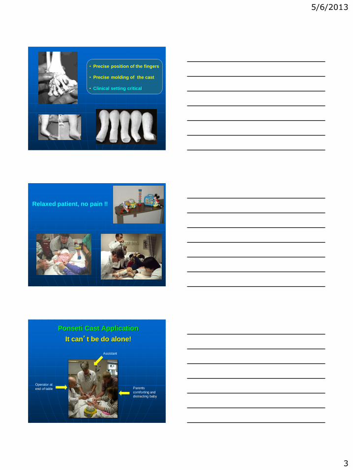
Manipulation technique
Localize lateral maleolus and talar head
Place thumb on the talar head
to block rotation
Elevate slightly the first metatarsal (no supinate the whole foot) to correct cavus
Cotton & Cast
Just one protective layer of cotton to allow good molding of the cast
5/6/2013
5
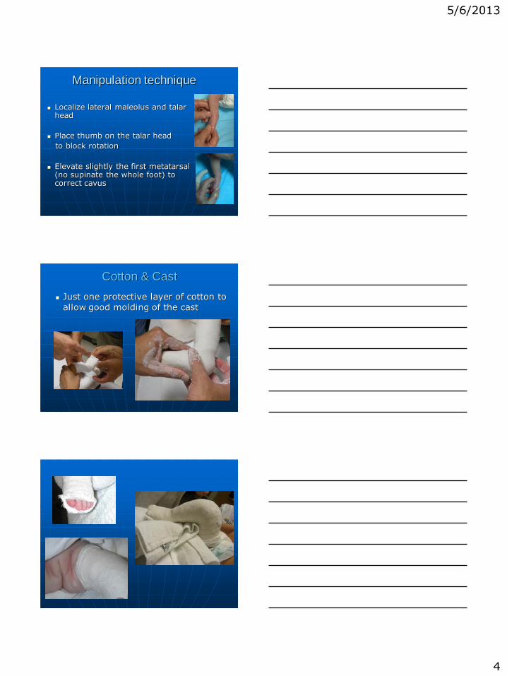
Rocker-bottom deformity
Overcorrection
5/6/2013
6
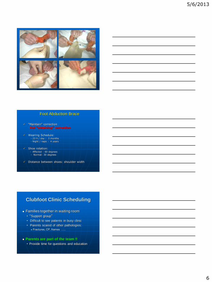
Foot Abduction Brace
“Maintain” correction
Not “obtaining” correction
Wearing Schedule: - 23 h / day : 3 months
- Night / naps : 4 years
Shoe rotation: - Affected : 60 degrees
- Normal: 30 degrees
Distance between shoes: shoulder width
Clubfoot Clinic Scheduling
Families together in waiting room
• “Support group”
• Difficult to see patients in busy clinic
• Parents scared of other pathologies:
Fractures, CP, frames …..
Parents are part of the team !!
• Provide time for questions and education

5/6/2013
7
THANK YOU !!
jose-morcuende @ uiowa.edu
http://www.ponseti.info

5/3/2013
1
Persistent and Recurrent
Clubfoot Deformity Following Treatment by
the Ponseti Method
W.B. Lehman, M.D.
Alice Chu, M.D.
Department of Pediatric Orthopaedic Surgery
New York Ponseti Clubfoot Center
Financial Disclosure
The authors have not received any financial support for
the preparation of this work.
Wallace B. Lehman
Alice Chu
Group 1 - Those maintained in an ankle-foot-orthosis (AFO) - 93% success rate
Group 2 - Those not maintained in an AFO (noncompliant) - Recurrence rate 50%
or greater
5/3/2013
2
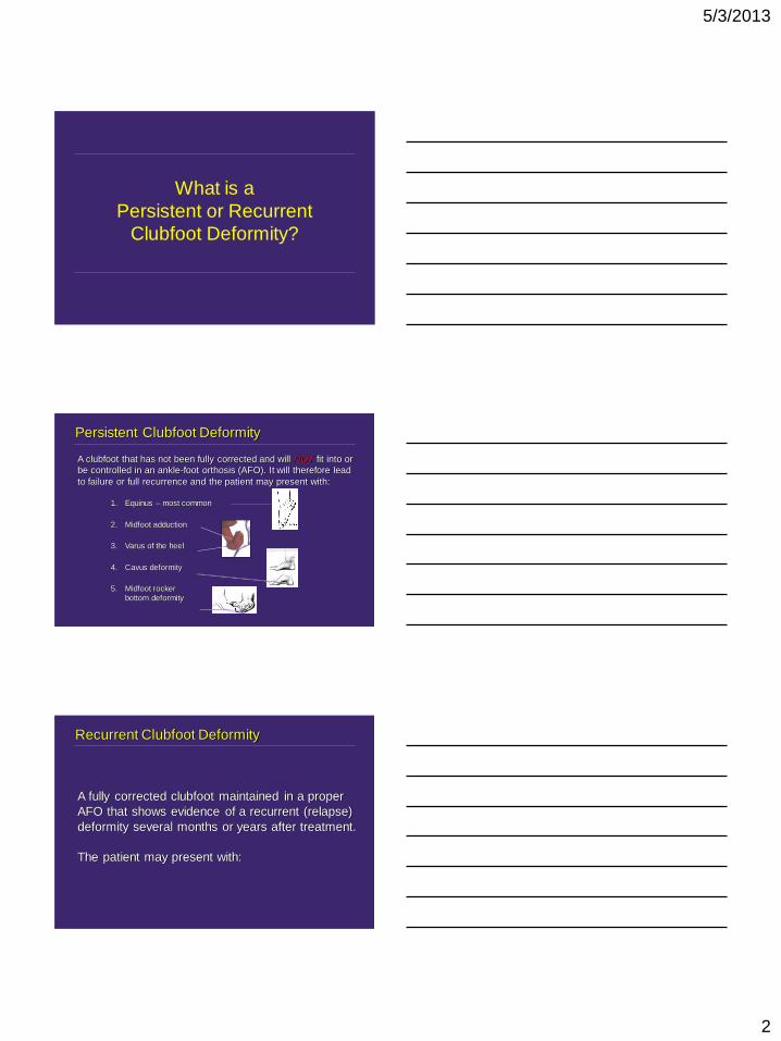
What is a
Persistent or Recurrent
Clubfoot Deformity?
Persistent Clubfoot Deformity
1. Equinus – most common
2. Midfoot adduction
3. Varus of the heel
4. Cavus deformity
5. Midfoot rocker
bottom deformity
A clubfoot that has not been fully corrected and will NOT fit into or
be controlled in an ankle-foot orthosis (AFO). It will therefore lead
to failure or full recurrence and the patient may present with:
Recurrent Clubfoot Deformity
A fully corrected clubfoot maintained in a proper
AFO that shows evidence of a recurrent (relapse)
deformity several months or years after treatment.
The patient may present with:

5/3/2013
3
1. Heel elevation – equinus
2. Rocker bottom deformity – midfoot dislocation (midfoot breach –
Noonan) through the talonavicular and calcaneocuboid joints
3. Cavus deformity
4. Varus of the calcaneus
5. Callus at lateral aspect of the foot
6. Inability to shoe correctly, i.e. due to recurrent deformity
7. Midfoot adduction – navicular not corrected over the talus
8. Forefoot adduction – adduction through Lisfranc’s joint
9. In-toeing gait – corrected clubfoot and flexible foot with overactive anterior
tibial tendon
10. Complete recurrence to original clubfoot deformity
Recurrent Clubfoot Deformity (cont.)
Recurrent Clubfoot Deformity (cont.)
1. Heel elevation – equinus
Recurrent Clubfoot Deformity (cont.)
2. Rocker bottom deformity – Chopart’s joint
5/3/2013
4
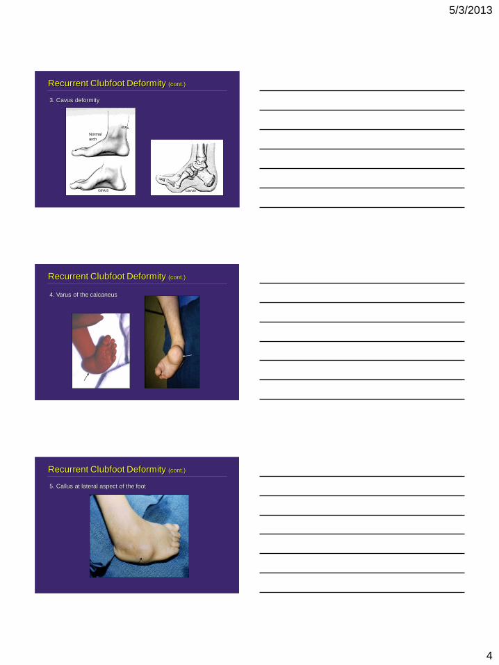
Recurrent Clubfoot Deformity (cont.)
3. Cavus deformity
cavus
Normal
arch
cavus
Recurrent Clubfoot Deformity (cont.)
4. Varus of the calcaneus
Recurrent Clubfoot Deformity (cont.)
5. Callus at lateral aspect of the foot
5/3/2013
5
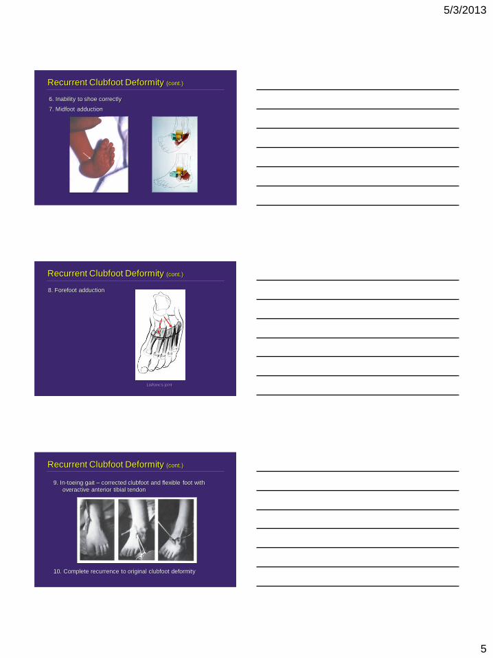
Recurrent Clubfoot Deformity (cont.)
6. Inability to shoe correctly
7. Midfoot adduction
8. Forefoot adduction
Recurrent Clubfoot Deformity (cont.)
Lisfranc’s joint
9. In-toeing gait – corrected clubfoot and flexible foot with
overactive anterior tibial tendon
10. Complete recurrence to original clubfoot deformity
Recurrent Clubfoot Deformity (cont.)

5/3/2013
6
Most Common Causes of Persistent or
Recurrent Clubfoot Deformities
1. Feet prone to persistent or recurrent deformities. Such as:
A. Clubfoot due to unrecognized syndrome -
B. Complex or atypical clubfoot
described by Morcuende and Ponseti
Short first toe
Bean-shaped foot Plantar crease
Ponseti, 1996
Oxford University Press
Ponseti technique will fail unless modified
i.e. arthrogryposis
C. Patient initially treated after seven months of age?
D. Unrecognized neurological clubfoot i.e. spastic or paralytic deformity
(Frick, S. Drop Toe Sign. CORR.2009)
E. Foot previously treated with “supposed” Ponseti technique that failed
(Helig: Current Management of Idiopathic Clubfoot Questionnaire.
JPO, 2003) (26.2% correction with the Ponseti technique)
Most Common Causes of Persistent or
Recurrent Clubfoot Deformities (cont.)
Most Common Causes of Persistent or
Recurrent Clubfoot Deformities (cont.)
2. Failure to maintain correction – usually a deformity that was
uncorrected and forced into an AFO
3. Noncompliance with regard to use of the DB bar (AFO?)
5/3/2013
7
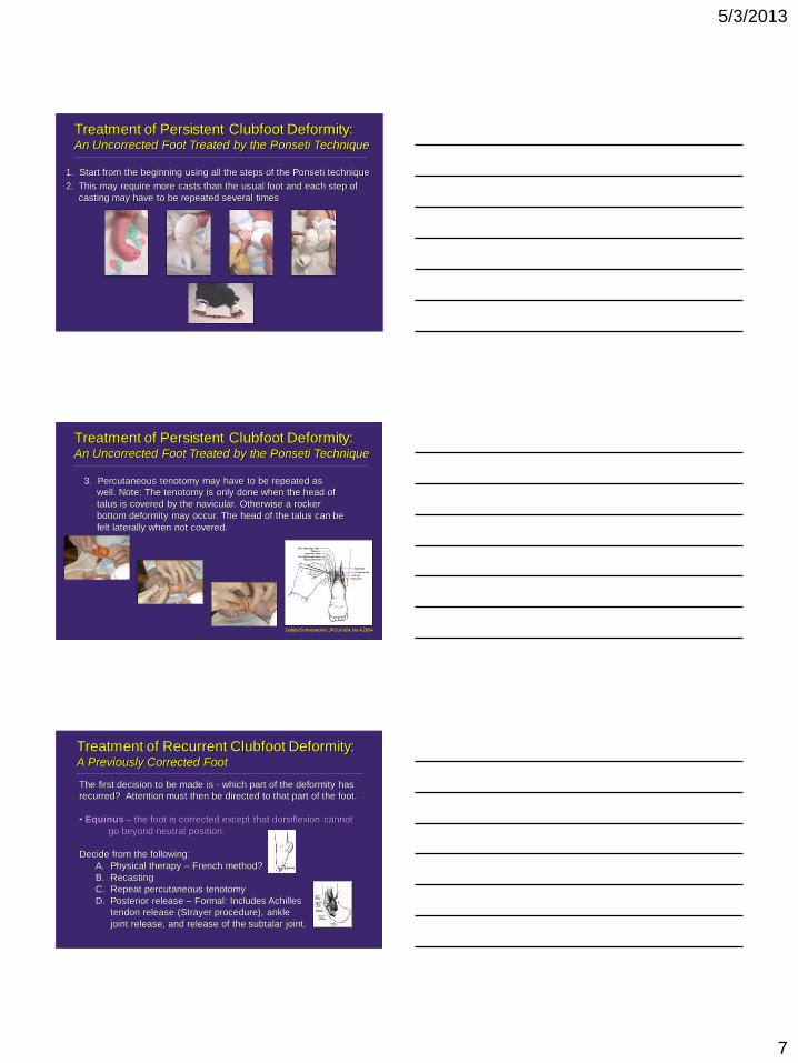
Treatment of Persistent Clubfoot Deformity: An Uncorrected Foot Treated by the Ponseti Technique
1. Start from the beginning using all the steps of the Ponseti technique
2. This may require more casts than the usual foot and each step of
casting may have to be repeated several times
Treatment of Persistent Clubfoot Deformity: An Uncorrected Foot Treated by the Ponseti Technique
3. Percutaneous tenotomy may have to be repeated as
well. Note: The tenotomy is only done when the head of
talus is covered by the navicular. Otherwise a rocker
bottom deformity may occur. The head of the talus can be
felt laterally when not covered.
Dobbs/Schoenecker; JPO,Vol24, No.4,2004
Treatment of Recurrent Clubfoot Deformity: A Previously Corrected Foot
The first decision to be made is - which part of the deformity has
recurred? Attention must then be directed to that part of the foot.
• Equinus – the foot is corrected except that dorsiflexion cannot
go beyond neutral position.
Decide from the following:
A. Physical therapy – French method?
B. Recasting
C. Repeat percutaneous tenotomy
D. Posterior release – Formal: Includes Achilles
tendon release (Strayer procedure), ankle
joint release, and release of the subtalar joint.

5/3/2013
8
Treatment of Recurrent Clubfoot Deformity (cont.)
A. Mild deformity may persist throughout life and may only require
intelligent neglect
B. If it occurs early after treatment, recast being sure to cover head of
talus, followed by maintenance with AFO fitted into a shoe for walking
Midfoot Adduction
Chopart’s joint
Treatment of Recurrent Clubfoot Deformity (cont.)
C. If persistent, may require release of adductor muscle, surgical release of
talonavicular joint, plus or minus a release of the first cuneiform-navicular joint
• Midfoot Adduction
D. In older children (5-10 years of age), it
may be necessary to perform a calcaneocuboid fusion or a first cuneiform
opening wedge osteotomy and a closing wedge cuboid osteotomy, or a complete
midtarsal osteotomy
E. If associated with heel varus, a closing
wedge osteotomy of the heel may be necessary in addition to the other
procedures – what we refer to as a triple osteotomy involving the cuneiform,
cuboid and calcaneus
Dwyer
McHale
1991
calcaneocuboid
fusion
Treatment of Recurrent Clubfoot Deformity (cont.)
F. Before maturity – soft tissue Lisfranc’s release (Heyman) Not effective
After maturity a transmetatarsal osteotomy or triple arthrodesis may
be necessary
• Metatarsus Adductus
Transmetatarsal osteotomy Triple arthrodesis
5/3/2013
9
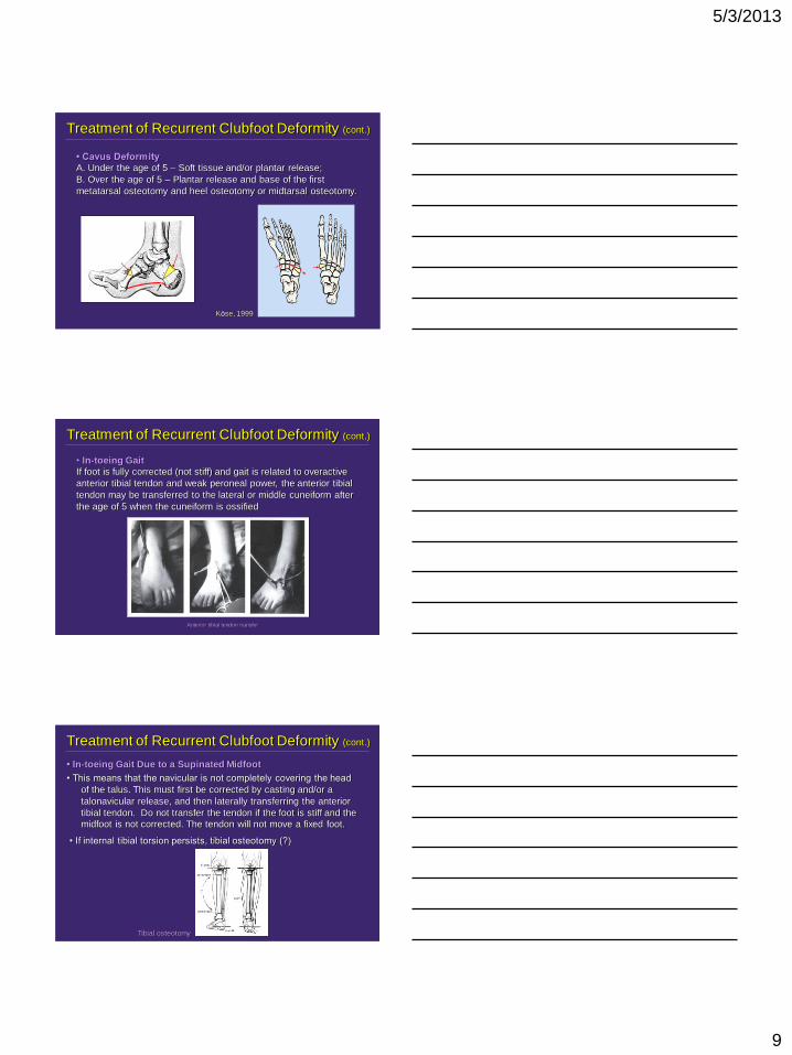
Treatment of Recurrent Clubfoot Deformity (cont.)
• Cavus Deformity
A. Under the age of 5 – Soft tissue and/or plantar release;
B. Over the age of 5 – Plantar release and base of the first
metatarsal osteotomy and heel osteotomy or midtarsal osteotomy.
Köse, 1999
Treatment of Recurrent Clubfoot Deformity (cont.)
• In-toeing Gait
If foot is fully corrected (not stiff) and gait is related to overactive
anterior tibial tendon and weak peroneal power, the anterior tibial
tendon may be transferred to the lateral or middle cuneiform after
the age of 5 when the cuneiform is ossified
Anterior tibial tendon transfer
Treatment of Recurrent Clubfoot Deformity (cont.)
• In-toeing Gait Due to a Supinated Midfoot
• This means that the navicular is not completely covering the head
of the talus. This must first be corrected by casting and/or a
talonavicular release, and then laterally transferring the anterior
tibial tendon. Do not transfer the tendon if the foot is stiff and the
midfoot is not corrected. The tendon will not move a fixed foot.
• If internal tibial torsion persists, tibial osteotomy (?)
Tibial osteotomy

5/3/2013
10
Treatment of Recurrent Clubfoot Deformity (cont.)
• Rocker Bottom Deformity
This is due to aggressive dorsiflexion of
the foot against a hindfoot contracture.
A. Stop treatment and allow the midfoot to correct itself or cast in
a vertical talus position
B. Release the hindfoot, Achilles tendon and posterior structures,
and cast in a congenital vertical talus position
C. A fixed rocker bottom deformity is difficult to treat and may
require aggressive management, similar to releasing a vertical
talus
Treatment of Recurrent Clubfoot Deformity (cont.)
• Major Recurrence
“A la carte” treatment as reported
by the late Dr. Henri Bensahel.
Each patient must be treated
individually, and in most cases
tendon releases, hindfoot releases,
and/or midfoot releases will do. It is
rarely necessary to perform a
complete subtalar release that was
popular in the past and very often
resulted in stiff, painful feet.
Treatment of Recurrent Clubfoot Deformity (cont.)
• Major Recurrence – gradual external fixator correction
5/3/2013
11
In summary:
Gentle manipulation of the clubfoot +/- percutaneous Achilles
tenotomy +/- anterior tibial tendon transfer (Ponseti technique)
will be effective if done properly in all clubfoot deformities at
least 80 - 90% of the time.
Persistence of clubfoot deformity is most likely due to
incomplete correction of the deformity.
Recurrent deformity has to be addressed depending upon
which part of the deformity recurs and when.
Bibliography for Persistent Clubfoot Deformity
Following Treatment by Ponseti Method.
1. Abdelgawad, A.A., Lehman, W.B., van Bosse, H.J.P., Scher, D.M. and Sala, D.A.: Treatment of idiopathic
clubfoot using the Ponseti method: minimum 2-year follow-up. J Pediatr Orthop B, 16:98-105, 2007.
2. Bensahel, H., Csukonyi, Z., Desgrippes,Y., and Chaumien, J.P: Surgery in residual clubfoot: One stage
medioposterior release “a la carte.” J Pediatr Orthop, 7:145-148, 1987.
3. Dobbs, M.B., Rudzki, J.R., Purcell, D.B., Walton, T., Porter, K.R. and Gurnett, C.A: Factors predictive of
outcome after use of the Ponseti method for the treatment of idiopathic clubfeet. J Bone Joint Surg (Am),
86A(1):22-27, 2004.
4. Koureas, G., Rampal, V., Mascard, E., Seringe, R. and Wicart, P: The incidence and treatment of rocker
bottom deformity as a complication of the conservative treatment of idiopathic congenital clubfoot. J Bone Joint
Surg (Br), 90B:57-60, 2008.
5. Lehman, W.: Revision Clubfoot Surgery. In Instructional Course Lecture, AAOS. J Bone Joint Surg (Am), Vol
84-A, No 2, p.303, Feb. 2002
6. Noonan, K.: Management of persistent clubfoot deformity following management via the Ponseti method.
AAOS Instructional Course Lecture Handout. Course title: Ponseti Clubfoot Method: Technical Skills Course
#7SK, San Diego, California, February 15, 2007.
7. Ponseti, I.V.: Congenital Clubfoot: Fundamentals of Treatment. Oxford University Press, London, 1996.
8. Ponseti, I.V, Morcuende, J.A, et al: Atypical clubfoot. In: Staheli, L, ed., Clubfoot: Ponseti Management (3rd
edition). Global-HELP Organization, 2009, http://global-help.org/publications/books.
Thank
You
Department of Pediatric Orthopaedic Surgery
New York Ponseti Clubfoot Center
5/3/2013
12
and
Goodbye
1
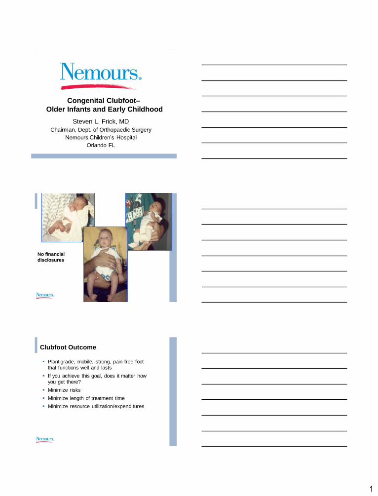
Congenital Clubfoot–
Older Infants and Early Childhood
Steven L. Frick, MD
Chairman, Dept. of Orthopaedic Surgery
Nemours Children’s Hospital
Orlando FL
No financial
disclosures
Clubfoot Outcome
Plantigrade, mobile, strong, pain-free foot that functions well and lasts
If you achieve this goal, does it matter how you get there?
Minimize risks
Minimize length of treatment time
Minimize resource utilization/expenditures
2
Does abnormal anatomy lead to
poor function and arthritis?
Data for hip and knee joints convincing
Surgical strategies – alter anatomy to normalize, improve function and delay arthritis
Is the foot different?
Data not as convincing
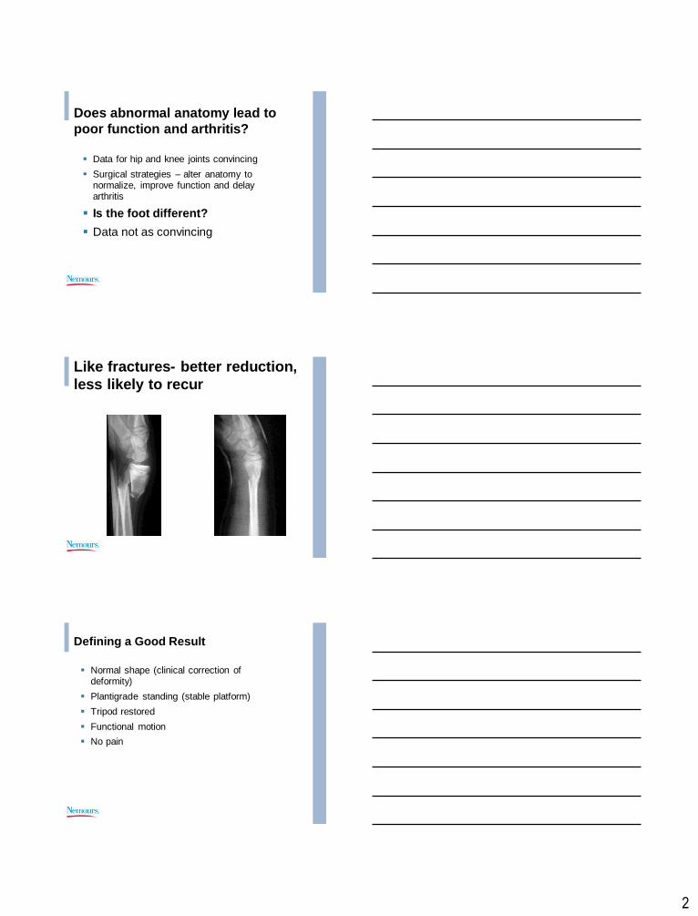
Like fractures- better reduction,
less likely to recur
Defining a Good Result
Normal shape (clinical correction of deformity)
Plantigrade standing (stable platform)
Tripod restored
Functional motion
No pain
3
5/1/2013
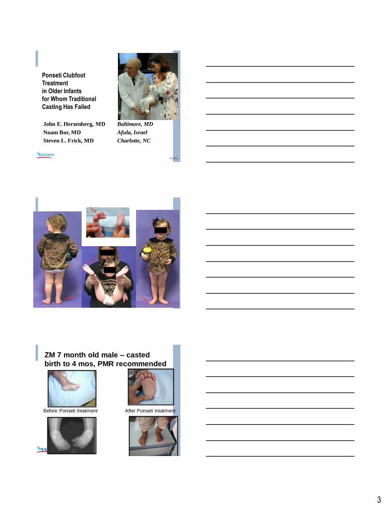
Ponseti Clubfoot
Treatment
in Older Infants
for Whom Traditional
Casting Has Failed
John E. Herzenberg, MD Baltimore, MD
Noam Bor, MD Afula, Israel
Steven L. Frick, MD Charlotte, NC
5/1/2013
5/1/2013
ZM 7 month old male – casted
birth to 4 mos, PMR recommended
Before Ponseti treatment After Ponseti treatment
4
Older infants and children
More challenging to cast
Hungry with a bottle
Experienced holder/cast roller
Often go every two weeks
If mild deformity and walking may use below knee, but usually above knee
Be patient
5/1/2013
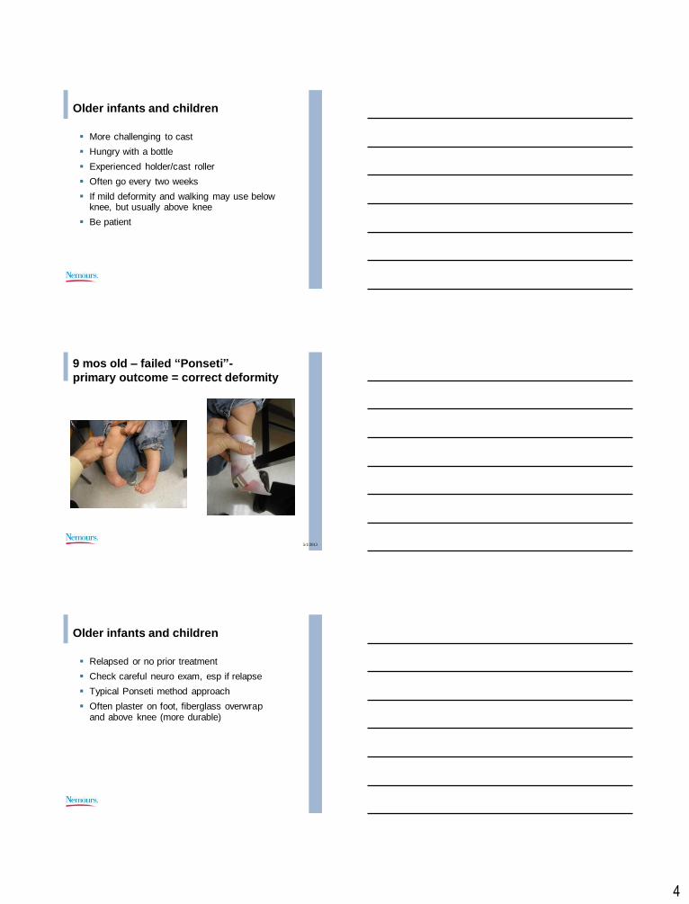
9 mos old – failed “Ponseti”-
primary outcome = correct deformity
Older infants and children
Relapsed or no prior treatment
Check careful neuro exam, esp if relapse
Typical Ponseti method approach
Often plaster on foot, fiberglass overwrap and above knee (more durable)

5
Older Clubfoot- sometimes SLC
5/1/2013
Too low
5/1/2013
After re-casting/tenotomies
6
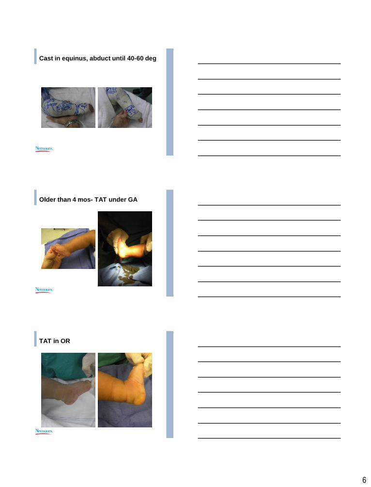
Cast in equinus, abduct until 40-60 deg
Older than 4 mos- TAT under GA
TAT in OR

7
At time of TAT in OR
Measure for braces
Casted with plaster and FG
3 weeks
If walking I just use FAO at night
Untreated 1 year old
multiple attempts to cast- noncompliant
Older patients
Ever effort to use Ponseti method
Be patient
Will take more casts
TAT or TAL in OR
Sometimes need a la carte releases
8
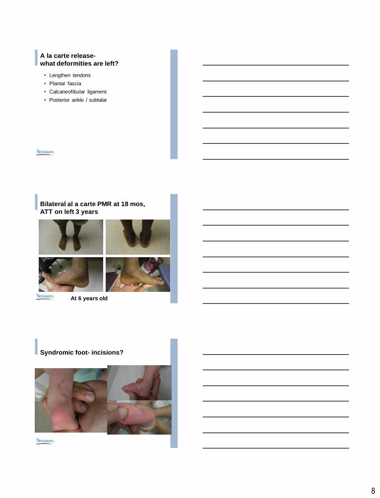
A la carte release-
what deformities are left?
Lengthen tendons
Plantar fascia
Calcaneofibular ligament
Posterior ankle / subtalar
Bilateral al a carte PMR at 18 mos,
ATT on left 3 years
At 6 years old
Syndromic foot- incisions?
9
Older patients- questions
What is the upper age limit for successful Ponseti method treatment?
Is TAT ok or do older kids need TAL?
How about repeat TAT vs TAL?
How many casts are ok? Is there a maximum?
Is a little rocker-bottom bad?
Is surgery that opens joints a sign of failure?

5/6/2013
1
Vumedi Clubfoot Webinar
Discussion Questions
Is the Ponseti Method the best we can do? The results of the Ponseti Method are obvious, nearly immediate, and are impressive to parents and practitioners. Some have suggested these criteria make the success of an intervention so self-evident that better evidence (EBM) is not needed. Our best long-term outcome study of Ponseti method patients involved only 45 patients, and 22% had a fair or poor outcome.
How much ankle dorsiflexion is enough? Most expert clubfoot surgeons using the Ponseti method perform Achilles tenotomy 90% of the time. The indications for tenotomy differ, with some using less than 10 degrees, while others use less than 15 or 20. How is this measured? And once the child is walking, how much ankle dorsiflexion is enough – 0 or plantigrade, 5, 10 or more degrees? We do not have validated information about this, and opinions seem to vary.

5/6/2013
2
While initial Achilles tenotomy in clubfoot seems to have few complications or functional implications, little is known about the indications for and long term results of repeat Achilles tenotomy or Achilles lengthenings after prior tenotomy. Is repeat TAT or TAL ok?
What is the best way to manage patients who do not respond as expected to Ponseti method manipulations and casting? What should be the approach for the rare patient who does not respond? Do we apply the principles of “al a carte” surgery and address the residual deformities specific to that foot? Most frequently in my experience this will mean hindfoot surgery to address persistent equinus and varus- what are the long term results for these patients?
How many patients with clubfoot who relapse have neurological abnormalities that contribute to propensity to relapse? Some authors recommend neurology referral, electrodiagnostic testing and/or neuro-imaging for relapsing clubfeet. What are the indications for these types of tests, and what information do the tests yield?
5/6/2013
3
What is the best approach for patients managed initially with the Ponseti method who relapse, are treated with repeat casting and then tibialis anterior transfer, but still have residual deformity leading to gait and functional abnormalities? What are the causes of persistent intoeing in clubfoot patients?