Embed Size (px)
Citation preview

4th European Symposium on Ultrasonic
Characterization of Bone
ESUCB 2011
Department of PhysicsUniversity of Jyvaskyla
Jyvaskyla, Finland20–21 June 2011

Scientific Committee
Local Organizer Petro Moilanen
Pascal Laugier (France)Frederic Padilla (France)Claus-C. Gluer (Germany)Reinhard Barkmann (Germany)Kay Raum (Germany)Michal Pakula (Poland)Emmanuel Bossy (France)
International Scientific Committee Keith A. Wear (USA)Jonathan J. Kaufmann (USA)Mami Matsukawa (Japan)Dean Ta (China)Jussi Timonen (Finland)Sulin Cheng (Finland)Jukka Jurvelin (Finland)Patrick Nicholson (UK)
Scientific Program Information
Oral Presentation
All the oral presentations will take place in the lecture hall FYS 1 in the Department of Physicsat the University of Jyvaskyla. The time reserved for each presentation is 15 minutes. The rec-ommended time for the actual presentation is 10 minutes, as approximately 5 minutes should bereserved for questions and discussion. Proper discussion and questions are essential for the students,who are expected to form a large part of the attendees.
30 minutes have been reserved for introductory and plenary presentations. 5 minutes of this shouldagain be used for questions and discussion.
The session chairs are responsible for maintaining the schedule and guiding the discussion. Theyshould also be present to organize the session 15 minutes before the scheduled time.
Poster Presentations
A space in the hallways directly outside the lecture hall will be reserved for the poster presentations.The length of the poster session will be 90 minutes and it will be during the last coffee break onMonday. Maximum size for the posters is one A0 sheet (portrait oriented).
At least one of the authors should be available to present the results of the poster during the postersession. Sufficient space will be reserved for all of the submissions, so posters need not be removedafter the session. The hallways reserved for poster sessions can be used by the conference attendeesat any time during the conference.
II

Social Program
Monday 20th - Barbeque party at the lakeside terrace of the Hotel Alba
After the scientific sessions on Monday we invite you to enjoy the company of your colleagues andthe taste of barbequed dishes. The barbeque party takes place at the terrace of the Hotel Alba,close to the conference venue. All participants are welcome.
Tuesday 21st - Gala dinner at the idyllic farmyard of Savutuvan apaja.
We hope that you will join us on a gala dinner on Tuesday after the scientific program. A bus willtransport all the enrolled to a historical country site about 12 km away from the city of Jyvaskyla.A finnish dinner will be served and you can enjoy the warm summer evening by the lake Paijanne.The lakeside scenery provides a beautiful setting in which you can follow the midnight sun, whichbarely sets at all at this time of the year. The bus leaves in front of the Hotel Alba at 7 p.m. andwe will return around 11 p.m.
Internet access
A free of charge WLAN access will be available during the whole conference. Detailed instructionsand personal password will be given to each conference participant.
III

Schedule
Time Sunday 19th Monday 20th Tuesday 21st8:30
9:00
9:30
10:00
10:30
11:00
11:30
12:00
12:30
13:00
13:30
14:00
14:30
15:00
15:30
16:00
16:30
17:00
19:00
20:00
16:00-20:00Registration
19:30-21:30 Get-together evening:
Barbeque at Hotel Alba
9:00 Opening and Welcome notePetro Moilanen
9:15 Invited lecture:Prof. Juliet Comspton
9:45 Session I:Bone Material and Structure
10:15 Coffee break
10:45 Session II-a:Bone Growth
11:00 Session II-b:Fractures and Healing
14:45 Poster session / Coffee break
16:15 Session IV:Ultrasonic Characterization of
Cancellous Bone
12:00 Lunch break:Restaurant Ylistö
13:30 Session III:Ultrasonic Propagation Models for
Cancellous Bone
8:30 Registration
19:00-23:00Gala dinner: Finnish evening
10:15 Coffee break
10:45 Session VI:Models for Ultrasonic
Characterization of Cortical Bone
9:00 Invited lecture:Prof. Claus-Christian Glüer
9:30 Session V:Clinical Quantitative Ultrasound
12:15 Lunch break:Restaurant Ylistö
17:00-17:45 Lab tour: Micro-CT laboratory
17:00-17:45 Lab tour: Micro-CT laboratory
16:00 Session VIII:New Technologies and Imaging
15:30 Coffee break
13:45 Session VII:Guided Waves and Ultrasonic
Characterization of Cortical Bone
V


Contents
Invited LectureMonday, June 20th 2011, 09:15, Chair: Sulin Cheng
09:15 Bone quality: a clinical perspective Juliet Compston . . . . . . . . . . . 2
Session I: Bone Material and StructureMonday, June 20th 2011, 09:45, Chair: Sulin Cheng
09:45 Most of variations of cortical bone elasticity at the mesoscale (millime-
ter scale) are determined by porosity variation in aged women. Mathilde
Granke, Quentin Grimal, Amena Saıed, Pierre Nauleau, Francoise Peyrin and Pascal
Laugier . . . . . . . . . . . . . . . . . . . . . . . . . . . . . . . . 4
10:00 Spatial distribution of tissue mineralization and anisotropic tissue elastic
constants in human femoral cortical bone Daniel Rohrbach, Sannachi Lakhs-
manan, Max Langer, Francoise Peyrin, Alf Gerisch, Quentin Grimal, Pascal Laugier
and Kay Raum . . . . . . . . . . . . . . . . . . . . . . . . . . . . . 6
Session II-a: Bone GrowthMonday, June 20th 2011, 10:45, Chair: Kay Raum
10:45 Elastic values of fibula children bone autotransplants Jean-Philippe Berteau,
Martine Pithioux, Helene Follet, Patrick Chabrand and Philippe Lasaygues . . . . 10
Session II-b: Fractures and HealingMonday, June 20th 2011, 11:00, Chair: Kay Raum
11:00 Noninvasive functional assessment of a rat femur model and phantoms us-
ing quantitative focused ultrasound (QUS): A pilot study. Daniel Rohrbach,
Bernhard Hesse, Bernd Preininger and Kay Raum . . . . . . . . . . . . . . 12
11:15 Using micro Brillouin scattering technique for the assessment of the elas-
tic properties of newly formed bone in the vicinity of an implant Vincent
Mathieu, Kenji Fukui, Mami Matsukawa, Masahiko Kawabe, Romain Vayron, Em-
manuel Soffer, Fani Anagnostou and Guillaume Haıat . . . . . . . . . . . . . 14
VII

11:30 The influence of callus mineralization degree on ultrasound axial trans-
mission measurements: an in vitro approach Christiano Bittencourt Machado,
Wagner Coelho de A. Pereira, Mathilde Granke, Maryline Talmant, Frederic Padilla
and Pascal Laugier . . . . . . . . . . . . . . . . . . . . . . . . . . . 15
11:45 Nonlinear ultrasound monitoring of four-point bending fatigue induced
micro-damage Sylvain Haupert, Sandra Guerard, Paul A. Johnson, David Mitton
and Pascal Laugier . . . . . . . . . . . . . . . . . . . . . . . . . . . 17
Session III: Propagation Models for Cancellous BonesMonday, June 20th 2011, 13:30, Chair: Michal Pakula
13:30 Investigation of fast and slow wave attenuation properties of cancellous
bone -application of Bayesian techinique to the experimentally observed
waveforms- Amber Nelson, Joseph J. Hoffman, Christian Anderson, Mark Holland,
Katsunori Mizuno, Yoshiki Nagatani, James G. Miller and Mami Matsukawa . . . 20
13:45 Role of absorption mechanisms for ultrasound attenuation in cancellous
bone: Macroscopic modeling and experiment Michal Pakula . . . . . . . 22
14:00 Two wave propagation in equine radius cancellous bone Keisuke Yamashita,
Katsunori Mizuno, Mami Matsukawa, Takahiko Otani, Hiroko Aida and Hirokazu
Tsubone . . . . . . . . . . . . . . . . . . . . . . . . . . . . . . . 23
14:15 In vitro ultrasonic characterization of human bones using focused trans-
ducers: a model-based approach Roberto Longo, Quentin Grimal, Josquin
Foiret, Pascal Laugier, Steve Vanlanduit and Patrick Guillaume . . . . . . . . . 24
14:30 Ultrasonic monitoring and parameters identification of simulated tissue
culture Guillermo Rus Carlborg, Nicolas Bochud and Juan Manuel Melchor . . . 25
Poster session / Coffee breakMonday, June 20th 2011, 14:45, Chair: Mami Matsukawa
1 Numerical simulation of cancellous bone remodeling using a finite-
difference time domain method Atsushi Hosokawa . . . . . . . . . . . . 28
2 Distribution of the longitudinal wave velocity in equine cortical bone –
effects of microstructure and HAp orientation Kazufumi Yamamoto, Daisuke
Suga, Tomohiro Nakatsuji, Mami Matsukawa, Takahiko Otani, Hirokazu Tsubone,
Hiroko Aida, Hironobu Hoshino and Yukihiro Matsuyama . . . . . . . . . . . 29
VIII
3 Ultrasound velocity estimation of cortical bones using axial transmission
technique with angle beams Rui Zheng, Lawrence H Le and Edmond Lou . . . 31
4 Relationships between broadband ultrasonic attenuation and microstruc-
ture of human trabecular bone Michal Pakula, Jalmari Pirhonen, Petro Moila-
nen, Pascal Laugier and Tuomas Turpeinen . . . . . . . . . . . . . . . . . 32
5 Numerical simulation reveals how the fast wave is generated in cancellous
bone Yoshiki Nagatani . . . . . . . . . . . . . . . . . . . . . . . . . 33
6 Separation technique for ultrasonic waveform propagated in cancellous
bone using wavelet transform analysis Sho Hasegawa, Yoshiki Nagatani, Kat-
sunori Mizuno and Mami Matsukawa . . . . . . . . . . . . . . . . . . . . 34
7 A crazy climber method for analyzing fundamental flexural guided wave
in ultrasonic axial transmission signals recorded by a low-frequency array
transducer Vantte Kilappa, Kailiang Xu, Petro Moilanen, Dean Ta and Jussi Timonen 35
8 Reproducibility of the new ultrasound technique for osteoporosis diag-
nostics at primary healthcare level Ossi Riekkinen, Aleksi Monkkonen, Jarkko
Kauppinen, Janne Karjalainen and Jukka Jurvelin . . . . . . . . . . . . . . 36
9 Bone phantoms for ultrasonic axial transmission studies Jalmari Pirhonen,
Petro Moilanen, Vantte Kilappa, Pasi Karppinen, Zuomin Zhao, Ari Liikkanen, Timo
Karppinen, Edward Hæggstrom, Risto Myllyla and Jussi Timonen . . . . . . . . 37
10 Volumetric quantification of repair cartilage and subchondral ossification
using ultrasound biomicroscopy Nils Mannicke, Martin Schone, Ronny Schulz,
Hannes Kuttner, Bastian Marquaß and Kay Raum . . . . . . . . . . . . . . 38
11 Micro Brillouin scattering study on the velocity anisotropy in a trabecula
Kenji Fukui and Mami Matsukawa . . . . . . . . . . . . . . . . . . . . . 39
12 A nonlinear stratified model to predict ultrasonic wave propagation in
trabecular bone Nicolas Bochud, Quentin Grimal, Sylvain Haupert, Pascal Laugier
and Guillermo Rus . . . . . . . . . . . . . . . . . . . . . . . . . . . 40
13 Age affects the bone healing kinetics in a rat osteotomy model Katrein
Sauer, Daniel Rohrbach, Patrick Strube, Bernd Preininger and Kay Raum . . . . 41
Session IV: Ultrasonic Characterization of Cancellous BoneMonday, June 20th 2011, 16:15, Chair: Jukka Jurvelin
16:15 A new perspective on ultrasound assessment of cancellous bone Christian
Langton . . . . . . . . . . . . . . . . . . . . . . . . . . . . . . . 44
IX

16:30 In situ ultrasound backscatter measurements of intact human proximal
femur Markus Malo, Ossi Riekkinen, Viktoria Prantner, Hanna Isaksson, Janne
Karjalainen, Juha Toyras and Jukka Jurvelin . . . . . . . . . . . . . . . . 45
16:45 Estimating mean trabecular bone spacing from ultrasonic backscattering
signals based on two fundamental frequency estimation methods Dean Ta,
Baiding Yang and Weiqi Wang . . . . . . . . . . . . . . . . . . . . . . 46
Invited lectureTuesday, June 21st 2011, 09:00, Chair: Jussi Timonen
09:00 Development strategies for clinical skeletal quantitative ultrasound ap-
proaches: challenges and opportunities Claus-Christian Glueer . . . . . . . 48
Session V: Clinical Quantitative UltrasoundTuesday, June 21st 2011, 09:30, Chair: Jussi Timonen
09:30 Multi-site bone ultrasound measurements in elderly women Janne Kar-
jalainen, Ossi Riekkinen, Juha Toyras, Mikko Hakulinen, Heikki Kroger, Toni Rikko-
nen, Kari Salovaara and Jukka Jurvelin . . . . . . . . . . . . . . . . . . . 50
09:45 Ability of low-frequency axial transmission ultrasound to discriminate ret-
rospective low-energy fractures Petro Moilanen, Mikko Maatta, Vantte Kilappa,
Leiting Xu, Timo Jamsa, Jussi Timonen and Sulin Cheng . . . . . . . . . . . 51
10:00 In vitro evidence that the circumferential wave guided by the femur cor-
tical shell can be used to predict femur strength Julien Grondin, Quentin
Grimal, Sandra Guerard, Reinhard Barkmann, Claus Gluer and Pascal Laugier . . 52
Session VI: Models for Ultrasonic Characterization of Cortical BoneTuesday, June 21st 2011, 10:45, Chair: Pascal Laugier
10:45 Excitability of axially transmitted guided waves in models of human long
bones Maryline Talmant, Petro Moilanen, Josquin Foiret, Jean-Gabriel Minonzio,
Pascal Laugier and Jussi Timonen . . . . . . . . . . . . . . . . . . . . . 56
11:00 Characterization of growing bone: curvature and anisotropy Cecile Baron . 57
11:15 Characterization of circumferential guided waves in the femur cortical
shell – Part II: Sensitivities of phase velocities to variations of porosity
and shell thickness Mathieu Chekroun, Quentin Grimal, Jean-Gabriel Minonzio
and Pascal Laugier . . . . . . . . . . . . . . . . . . . . . . . . . . . 58
X
11:30 Three-dimensional finite-difference time-domain simulations demonstrate
the sensivity of ultrasound measurements at the femoral neck to cortical
bone geometry Pierre Nauleau, Julien Grondin, Quentin Grimal, Klaus Engelke
and Pascal Laugier . . . . . . . . . . . . . . . . . . . . . . . . . . . 59
11:45 2D simulations of the impact of porosity and cortical thickness on the
wave propagation at the inferior femoral neck cortex Kerstin Rohde, Melanie
Daugschies, Quentin Grimal, Pascal Laugier, Claus-Christian Gluer and Reinhard
Barkmann . . . . . . . . . . . . . . . . . . . . . . . . . . . . . . 60
12:00 Advanced stress guided waves simulation to support the non-invasive as-
sessment of human long bones Giovanni Castellazzi, Alessandro Marzani, Luca
De Marchi, Nicolo Speciale, Annapaola Parrilli, Matilde Tschon and Gianluca Giavaresi 61
Session VII: Guided wavesTuesday, June 21st 2011, 13:45, Chair: Patrick Nicholson, Dean Ta
13:45 The impact of soft tissue on the propagation of ultrasonic guided waves
through a bone plate Lauren Stieglitz, Lawrence Le, Jeff Gu and Edmond Lou . 64
14:00 Axial transmission guided mode measurement with 1MHz clinical probe
on bone mimicking phantom Josquin Foiret, Jean Gabriel Minonzio, Maryline
Talmant and Pascal Laugier . . . . . . . . . . . . . . . . . . . . . . . 65
14:15 Signal processing for guided mode measurement in absorbing material
Jean-Gabriel Minonzio, Josquin Foiret, Maryline Talmant and Pascal Laugier . . . 66
14:30 Characterization of circumeferential guided waves in the femur cortical
shell – Part I : Measurements on a bone mimicking tube Pierre Nauleau,
Etienne Cochard, Jean-Gabriel Minonzio, Quentin Grimal, Claire Prada and Pascal
Laugier . . . . . . . . . . . . . . . . . . . . . . . . . . . . . . . . 67
14:45 Measurement of long cortical thickness using ultrasonic guided waves
based on two signal processing methods Xiaojun Song, Dean Ta and Weiqi
Wang . . . . . . . . . . . . . . . . . . . . . . . . . . . . . . . . 69
15:00 A wavelet-based processing method for simultaneously determining
broadband ultrasonic velocity and cortical bone thickness Philippe
LASAYGUES and Matthieu LOOSVELT . . . . . . . . . . . . . . . . . . 70
15:15 Multi-frequency approach of the first arriving signal (FAS) on long cortical
bones Thien-Ly PHAM, Maryline Talmant and Pascal Laugier . . . . . . . . . 71
XI

Session VIII: Novel Instrumentation and ImagingTuesday, June 21st 2011, 16:00, Chair: Reinhard Barkmann
16:00 Experimental assessment of the amount of bone surrounding an implant
with quantitative ultrasound Vincent Mathieu, Fani Anagnostou, Emmanuel
Soffer and Guillaume Haıat . . . . . . . . . . . . . . . . . . . . . . . . 74
16:15 Impact of the beam incidence angle on Quantitative Ultrasound (QUS)
measurements of the calcaneus Melanie Daugschies, Kerstin Rohde, Claus-
Christian Gluer and Reinhard Barkmann . . . . . . . . . . . . . . . . . . 75
16:30 Simultaneous quantitative ultrasound assessment of cartilage and sub-
chondral bone Jukka Liukkonen, Antti Aula, Juha Toyras and Jukka Jurvelin . . 76
16:45 Ultrasound imaging of long bones using Born scattering theory: in vitro
study Rui Zheng, Lawrence H Le, Mauricio D Sacchi and Edmond Lou . . . . . 77
Indexes
List of Authors . . . . . . . . . . . . . . . . . . . . . . . . . . . . . . . 79
XII

Invited LectureMonday, June 20th 2011, 09:15
Chair: Sulin Cheng
1

4th European Symposium on Ultrasonic Characterization of Bone19–21 June 2011, Jyvaskyla, Finland
Bone quality: a clinical perspective∗Juliet Compston
Medicine, University of Cambridge, Cambridge, United Kingdom∗[email protected]
Bone strength is determined by a number of inter-related variables which include bone mineraldensity (BMD), bone geometry and bone quality. Whilst BMD is a strong predictor of bone strengthand fracture risk in the untreated state, therapeutically induced changes in BMD explain only asmall proportion of the associated reduction in fracture, indicating that changes in bone qualitymay be more important in this context. The importance of bone quality is also evident fromdisease states in man, some of which are associated with increased fracture risk despite increasedbone density and even bone size.
Assessment of bone quality in vivo can be made using a number of approaches, including bio-chemical markers of bone turnover, bone histomorphometry and imaging techniques, for examplequantitative computed tomography (QCT) and magnetic resonance. In particular, the applicationof high-resolution QCT techniques to both the axial and appendicular skeleton enables more de-tailed characterisation of cortical and cancellous bone structure in vivo than has previously beenpossible and will provide new insights into the mechanisms underlying bone fragility in disease andthe effects of therapeutic interventions on bone structure and strength.
*Presenting author
2

Session I: Bone Material and StructureMonday, June 20th 2011, 09:45
Chair: Sulin Cheng
3

4th European Symposium on Ultrasonic Characterization of Bone19–21 June 2011, Jyvaskyla, Finland
Most of variations of cortical bone elasticity at themesoscale (millimeter scale) are determined by porosity
variation in aged women.∗Mathilde Granke1, Quentin Grimal1, Amena Saıed1, Pierre Nauleau1, Francoise Peyrin2, Pascal
Laugier1
1UPMC Univ Paris 06, CNRS, UMR 7623, Laboratoire d’Imagerie Parametrique, France2CREATIS INSERM U1044; CNRS 5220; INSA Lyon; Universite de Lyon; ESRF, France
At the mesoscale (i.e. a few millimeters), cortical bone can be described as two-phase material,which consists of pores and a relatively hard mineralized matrix. The cortical porosity is knownto influence the mesoscopic elasticity. Our objective was to determine whether the variations ofporosity are sufficient to predict the variations of bone mesoscopic anisotropic elasticity or if thebone matrix stiffness is an important factor to consider. Measurements were conducted on humanfemoral cortical bone (21 specimens taken from 10 women donors). A 50MHz scanning acousticmicroscope (SAM) was used to evaluate the bone matrix elasticity (reflected in impedance values)and porosity. Porosity evaluation with SAM was validated against Synchrotron Radiation microCTmeasurements. A standard contact ultrasonic method was applied to determine the mesoscopicanisotropic stiffness coefficients. The mesoscopic stiffness was found to be highly correlated (R2
= [0.72 – 0.84]) to the cortical porosity. Multivariate analysis including tissue elasticity did notprovide a better statistical model of mesoscopic stiffness variations. This work suggests that thecortical porosity accounts for most of the variations of mesoscopic elasticity, at least when theanalyzed porosity range is large (3-27 % in this work).
*Presenting author
4

4th European Symposium on Ultrasonic Characterization of Bone19–21 June 2011, Jyvaskyla, Finland
Figure 1: The comparison of our results with the predictions of a micromechanical modelindicates that cortical bone can be modeled with fixed matrix properties and a sample-dependent
porosity fraction.
5

4th European Symposium on Ultrasonic Characterization of Bone19–21 June 2011, Jyvaskyla, Finland
Spatial distribution of tissue mineralization andanisotropic tissue elastic constants in human femoral
cortical bone
Daniel Rohrbach1, Sannachi Lakhsmanan1, Max Langer2, Francoise Peyrin2, Alf Gerisch3,Quentin Grimal4, Pascal Laugier4, ∗Kay Raum1
1Julius-Wolff-Institut, Charite-Universitatsmedizin Berlin, Berlin, Germany2ESRF, Grenoble, France
3TU Darmstadt, Darmstadt, Germany4UPMC, Paris, France∗[email protected]
The spatial distribution of micro- and mesoscale anisotropic elastic properties and mineraliza-tion within a human femoral cortical bone shaft (female:72years) have been investigated. Cylindri-cally shaped punch biopsy samples (diameter: 4.4mm,N=56) were analyzed using SAM at 50MHz(Fig.1) and SR-µCT.
For all samples the average cortical porosity (Ct.Po), tissue elastic coefficients (cij) and theaverage tissue degree of mineralization (DMB) were derived from SAM and SR-µCT measurements.The volumetric bone mineral density (vBMD) and mesoscale stiffness coefficients (Cij) were derivedusing a rule of mixtures and an asymptotic homogenization approach, respectively. Variations ofthese properties were derived with respect to the anatomical location and relations between theseproperties were analyzed.
Variations of DMB (1.5%) had a minor effect on the variations of mesoscale elastic properties(11-21%). Multivariate regression revealed that the microscale coefficients cii and Ct.Po contributedapproximately equally to the variations of Cij . The correlations of vBMD with Cij were less than70 %. However, a combined in-vivo assessment of vBMD and Cii may lead to a precise and distinctprediction of variations of Ct.Po and tissue elasticity, e.g. in the course of pathologies or treatment.
*Presenting author
6

4th European Symposium on Ultrasonic Characterization of Bone19–21 June 2011, Jyvaskyla, Finland
Figure 1: Acoustic impedance image of a cross-section (a). The highlighted rectangle and lineindicate the region for the transverse sectional scan (b), and the cylinder measurement (c). Forbetter illustration the cylinder surface was unwrapped. The image segmentation and erosion is
shown in (d) for the small region highlighted in (c).
7


Session II-a: Bone GrowthMonday, June 20th 2011, 10:45
Chair: Kay Raum
9

4th European Symposium on Ultrasonic Characterization of Bone19–21 June 2011, Jyvaskyla, Finland
Elastic values of fibula children bone autotransplants∗Jean-Philippe Berteau1, Martine Pithioux2, Helene Follet3, Patrick Chabrand2, Philippe
Lasaygues1
1UPR 7051, LMA, Marseille, France2UMR 6233, CNRS & Universite de la Mediterranee, Marseille, France
3U831, INSERM, Lyon, France∗[email protected]
For cortical bone, few studies consider mechanical characteristic of growing process. In theliterature, the samples studied are close to cancellers cells (Baleani et al., 2008), or from cadaver(Currey, 1975) and the results are dispersive. We have focused our study to the most used bonefor cortical transplantation, the fibula bottom extremity. It is a two step method; first we haveevaluate the porosity and the osteonal orientation with histological image; second, we have testedcortical sample in ultrasonic bench with nominal frequency of 7 and 10 MHz. The first step shows alow porosity level and an osteonal orientation similar to an adult fibula, it is leading us to considerit as transversely isotropic and reliable to an ultrasonic transmission scanning. The second stepgives an average value of CL (longitudinal velocity) of 13 bone samples (extracted from childrenfrom 4 year old to 16) is 2818 m·s−1 (± 540) and of CT (transversal velocity) is 1804 m·s−1 (±213). These results are lower than adult, but not depending of children age. This is the first studywhich provides cortical bone ultrasonic velocity of children population.
*Presenting author
10
Session II-b: Fractures and HealingMonday, June 20th 2011, 11:00
Chair: Kay Raum
11
4th European Symposium on Ultrasonic Characterization of Bone19–21 June 2011, Jyvaskyla, Finland
Noninvasive functional assessment of a rat femur modeland phantoms using quantitative focused ultrasound
(QUS): A pilot study.∗Daniel Rohrbach1, Bernhard Hesse1, Bernd Preininger2, Kay Raum1
1JWI, AG Raum, Charite, Berlin, Germany2Charite, Berlin, Germany
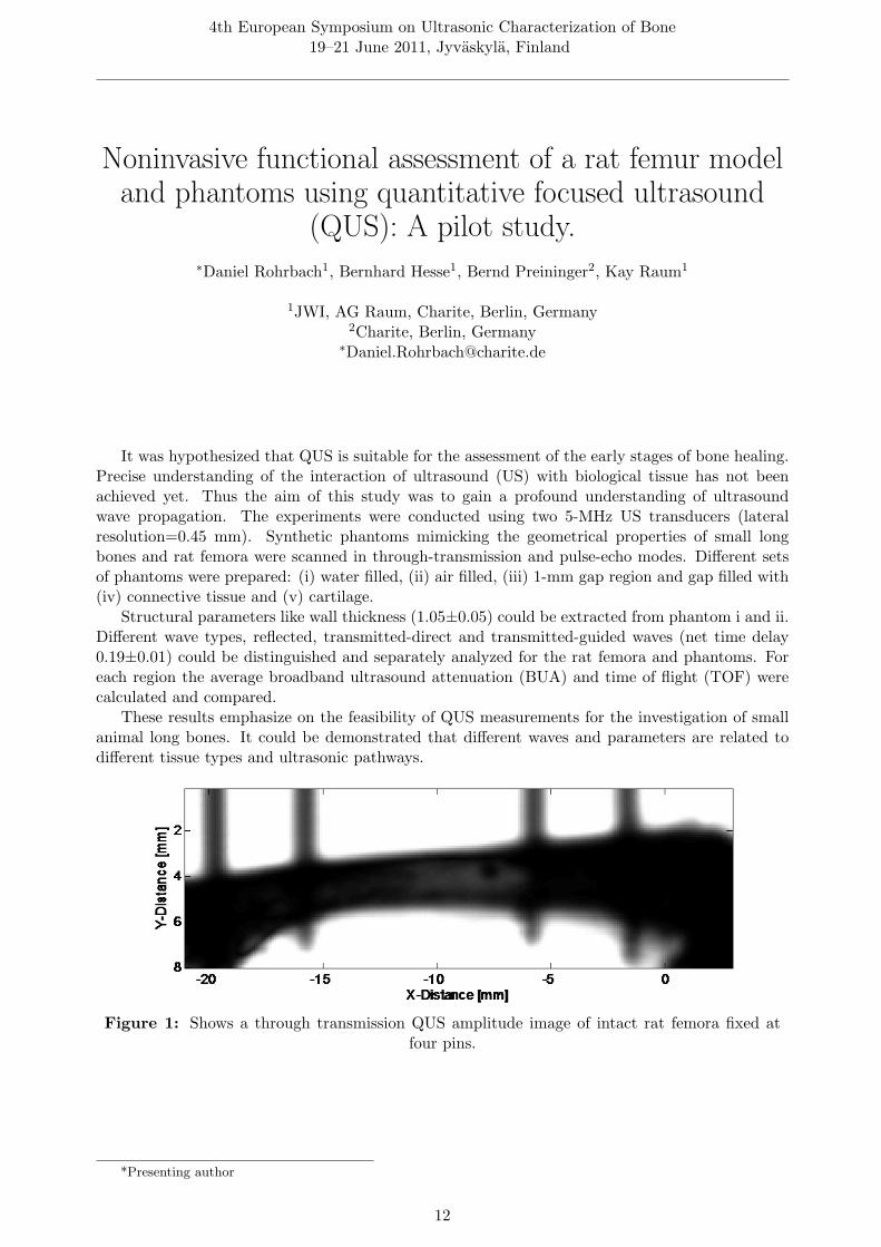
It was hypothesized that QUS is suitable for the assessment of the early stages of bone healing.Precise understanding of the interaction of ultrasound (US) with biological tissue has not beenachieved yet. Thus the aim of this study was to gain a profound understanding of ultrasoundwave propagation. The experiments were conducted using two 5-MHz US transducers (lateralresolution=0.45 mm). Synthetic phantoms mimicking the geometrical properties of small longbones and rat femora were scanned in through-transmission and pulse-echo modes. Different setsof phantoms were prepared: (i) water filled, (ii) air filled, (iii) 1-mm gap region and gap filled with(iv) connective tissue and (v) cartilage.
Structural parameters like wall thickness (1.05±0.05) could be extracted from phantom i and ii.Different wave types, reflected, transmitted-direct and transmitted-guided waves (net time delay0.19±0.01) could be distinguished and separately analyzed for the rat femora and phantoms. Foreach region the average broadband ultrasound attenuation (BUA) and time of flight (TOF) werecalculated and compared.
These results emphasize on the feasibility of QUS measurements for the investigation of smallanimal long bones. It could be demonstrated that different waves and parameters are related todifferent tissue types and ultrasonic pathways.
Figure 1: Shows a through transmission QUS amplitude image of intact rat femora fixed atfour pins.
*Presenting author
12

4th European Symposium on Ultrasonic Characterization of Bone19–21 June 2011, Jyvaskyla, Finland
Figure 2: Shows a transmitted time resolved signal (right) and a power spectrum (left) of aphantom. The guided and direct wave are separable. Reference signal is referred to be the powerspectrum measured in water and attenuation is the difference spectrum of the reference signal
and amplitude spectrum (power spectrum of signal).
13

4th European Symposium on Ultrasonic Characterization of Bone19–21 June 2011, Jyvaskyla, Finland
Using micro Brillouin scattering technique for theassessment of the elastic properties of newly formed
bone in the vicinity of an implant∗Vincent Mathieu1, Kenji Fukui2, Mami Matsukawa2, Masahiko Kawabe2, Romain Vayron3,
Emmanuel Soffer1, Fani Anagnostou1, Guillaume Haıat3
1Lab. Biomecanique et Bioingenierie Osteo Articulaire, CNRS, 75010 PARIS, France2Lab. Ultrasonic Electronics, Doshisha University, 610-0321 KYOTO, Japan
3Lab. Modelisation et Simulation Multi Echelle, CNRS, 94010 CRETEIL, France∗[email protected]
The use of titanium implants allowed considerable progress in dental and orthopaedic surgery.However, the reasons for implant failure remain difficult to understand. Remodeling phenomena atthe bone-implant interface play a predominant part in the osseointegration of the implant. In thisstudy, the use of micro-Brillouin scattering technique is proposed to assess the elastic properties ofnewly formed bone in the vicinity of the implant, at the micrometer scale. To do so, an originalanimal model was used, consisting in securing a coin-shaped titanium implant (see Fig. 1) incontact with a leveled surface of the tibia of a rabbit. This animal model allows the formation of acavity exclusively filled with newly formed bone after the implantation period. After the sacrifice,micro-Brillouin velocity measurements were performed in mature and newly formed bone regionsand an histological analysis was realized. The results show that significantly higher velocities areobtained in mature bone compared to newly formed bone, which is in agreement with the highermineral content observed in histological slices for mature bone. This multimodality study showsthe potentiality of the animal model and of micro-Brillouin scattering for the assessment of newlyformed bone elastic properties.
Figure 1: Image of a coin shaped titanium implant after 7 weeks of implantation on the tibiaof a New Zealand White rabbit.
*Presenting author
14

4th European Symposium on Ultrasonic Characterization of Bone19–21 June 2011, Jyvaskyla, Finland
The influence of callus mineralization degree onultrasound axial transmission measurements: an in vitro
approach
Christiano Bittencourt Machado1, Wagner Coelho de A. Pereira2, Mathilde Granke3, MarylineTalmant3, Frederic Padilla4, ∗Pascal Laugier3
1Biomechanics Laboratory – Campus Friburgo, Estacio de Sa University, Nova Friburgo – RJ,Brazil
2Biomedical Engineering Program, COPPE – Federal University of Rio de Janeiro, Rio de Janeiro– RJ, Brazil
3Laboratoire d’Imagerie Parametrique, Universite Pierre et Marie Curie, Paris, France4Department of Radiology, University of Michigan Medical Center, Ann Arbor, USA
This work aims at studying the effect of cortical bone mineralization on ultrasonic axial trans-mission measurements, using experiments and numerical simulations. A reverse fracture healingapproach is proposed, using a cortical bovine femur sample with a 3-mm fracture gap. A 3-mmthick cortical bone slice was extracted and submitted to a progressive chemical demineralizationduring 12 days. Each day, axial transmission measurements were achieved with the 3-mm thickcortical bone slice placed back inside the gap. Calcium concentrations in chemical solutions werealso quantified each day. Axial transmission measurements and FDTD simulations using a 1-MHzprobe were performed to estimate the time-of-flight of the first arriving signal (TOFFAS) (Figure1) in three configurations: (i) bone specimen intact; (ii) bone specimen with the fracture gap; (iii)bone specimen with the 3-mm thick cortical bone slice placed back at each stage of demineraliza-tion. A progressive increase in TOFFAS (p < 0.001) was observed until day 4 of demineralization(Figure 2), corresponding to the rapid initial loss of calcium. Experimental data were in goodagreement with numerical simulations and the results show the potential of axial transmission onthe consolidation follow-up, specifically on callus mineralization.
*Presenting author
15

4th European Symposium on Ultrasonic Characterization of Bone19–21 June 2011, Jyvaskyla, Finland
Figure 1: Experimental setup. The cortical slice is put inside the 3-mm fracture gap forultrasound measurements with a 1-MHz ultrasound probe. A fixing device is used to avoid
undesirable movements.
Figure 2: Experimental and simulated TOFFAS for intact, fractured and demineralized bone(from days 0 to 12). It can be also observed the calcium loss curve from the demineralization
process.
16

4th European Symposium on Ultrasonic Characterization of Bone19–21 June 2011, Jyvaskyla, Finland
Nonlinear ultrasound monitoring of four-point bendingfatigue induced micro-damage
∗Sylvain Haupert1, Sandra Guerard2, Paul A. Johnson3, David Mitton4, Pascal Laugier1
1UPMC – CNRS – LIP, Paris, France2Arts et Metiers ParisTech – LBM, Paris, France
3Geophysics Group, Los Alamos National Laboratory, Los Alamos, USA4Univ Lyon – Ifsttar – LBMC, Arts et Metiers ParisTech – LBM, Paris, France
Accumulation of bone micro-damage is suspected to lead to severe impairment of mechanicalproperties (toughness and stiffness) with an increase in skeletal fragility and fracture risk. Theobjective of the study was to evaluate the resonant ultrasound spectroscopy (NRUS) technique formeasuring micro-damage accumulation in cortical bone using mechanical fatigue. Sixteen humancortical bone specimens were machined as parallelepiped beams (50*4*2mm) to control damage lo-calization during four-point bending fatigue cycling and to unambiguously identify resonant modesfor NRUS measurement. During damage progression, load and displacement curves were recordedto extract secant (Esec), loading (Eload) and linear elastic beam theory (ELEBT) moduli. Thelatter has been shown previously to reflect micro-damage accumulation during four-point bend-ing mechanical fatigue. Before and between each damage step, nonlinear ultrasonic elastic (af)and dissipative (aQ) coefficients were monitored by NRUS. At the end of fatigue cycling, resultsshowed no significant variation of the linear elastic moduli (Esec and Eload). In contrast, meanvalues of both af and aQ increased significantly (50% and 20% respectively) while ELEBT meanvalue decreased significantly (43%). The results of this study suggest that non invasive monitoringfatigue micro-damage accumulation in cortical bone can be achieved using nonlinear ultrasoundtechniques.
*Presenting author
17


Session III: Propagation Models for Cancellous BonesMonday, June 20th 2011, 13:30
Chair: Michal Pakula
19

4th European Symposium on Ultrasonic Characterization of Bone19–21 June 2011, Jyvaskyla, Finland
Investigation of fast and slow wave attenuationproperties of cancellous bone -application of Bayesiantechinique to the experimentally observed waveforms-
Amber Nelson1, Joseph J. Hoffman1, Christian Anderson1, Mark Holland1, Katsunori Mizuno2,Yoshiki Nagatani3, James G. Miller1, ∗Mami Matsukawa2
1Department of Physics, Washington University in St. Louis, St. Louis, USA2Laboratory of Ultrasonic Electronics, Doshisha University, Kyotanabe, Japan
3Department of Electronics, Kobe City College of Technology, Kobe, Japan∗[email protected]
The goal of this study was to investigate the attenuation behavior of overlapping fast andslow waves in cancellous bone, using Bayesian parameters estimated from experimentally obtainedwaveforms. Simulated data with Bayesian parameters were generated using a propagation modelincluding both fast and slow waves. Two analysis methods for determining the attenuation asa function of propagation distance were applied to samples of two lengths. The first was a time-domain analysis using amplitudes of the first peak of mixed waveform. The second was a frequency-domain analysis using the separated fast and slow waves. In the time-domain analysis, the apparentattenuation of the fast wave is larger at the beginning and gradually decreases as the wave travelsfarther into the bone. The frequency-domain analysis gives almost constant attenuation coefficientsfor each wave mode without dependence on the propagation distance, but different values of samplesof different length. The simulated data tell us that the apparent dependence of the attenuation onpropagation distance can arise from analyzing the unseparated signal with time domain analysis.Phase cancellation at the face of a phase sensitive receiver can also yield attenuation that appearsto vary with propagation distance. Supported in part by NIH R01 AR057433
*Presenting author
20

4th European Symposium on Ultrasonic Characterization of Bone19–21 June 2011, Jyvaskyla, Finland
Figure 1: The attenuation behavior as a function of sample thickness results when applying,panel (a): the time-domain method to the unseparated fast wave, panel (b): the log spectralsubtraction technique to the entire sample wave, consisting of overlapping wave modes, panel(c): the time-domain method to the individual fast wave and slow wave, and panel (d): the log
spectral subtraction technique to the separated fast wave and slow wave.
21

4th European Symposium on Ultrasonic Characterization of Bone19–21 June 2011, Jyvaskyla, Finland
Role of absorption mechanisms for ultrasoundattenuation in cancellous bone: Macroscopic modeling
and experiment∗Michal Pakula
Institute of Mechanics and Applied Computer Science, Kazimierz Wileki University, 85-074Bydgoszcz, Poland
Evaluation of the relative contribution of physical mechanisms responsible for attenuation ofultrasonic wave in cancellous bone is one of the crucial issues from the point of view of modelingelastic wave propagation and related model-based identification of the structural and mechanicalproperties of bone material. Considering trabecular bone as a porous material filled with fluid, thewave attenuation may stem from: (i) intrinsic absorption in the fluid and solid phase, (ii) frictionat the fluid-solid interface as well as (iii) wave scattering by inhomogeneities (pores/trabeculaes).The commonly used for modeling ultrasound propagation in cancellous bone the macroscopic Biot’stheory will be discussed in context of its potential applicability for prediction of wave parameters:phase velocity and attenuation coefficient as functions of frequency. Since the model was introducedfor long wavelength range, the scattering effects are neglected, and the analysis will be focused onthe absorption mechanisms responsible for attenuation of ultrasonic waves in bone material. Thesuitability of the model will be verified by comparison of results of sensitivity analysis of the modelwith in vitro experimental ultrasonic data obtained for cancellous bones filled with different fluids.
*Presenting author
22
4th European Symposium on Ultrasonic Characterization of Bone19–21 June 2011, Jyvaskyla, Finland
Two wave propagation in equine radius cancellous bone∗Keisuke Yamashita1, Katsunori Mizuno1, Mami Matsukawa1, Takahiko Otani1, Hiroko Aida2,
Hirokazu Tsubone3
1Laboratory of Ultrasonic Electronics, Doshisha University, Japan2JRA, Equine Research Institute, Japan
3Tokyo University, Japan∗[email protected]
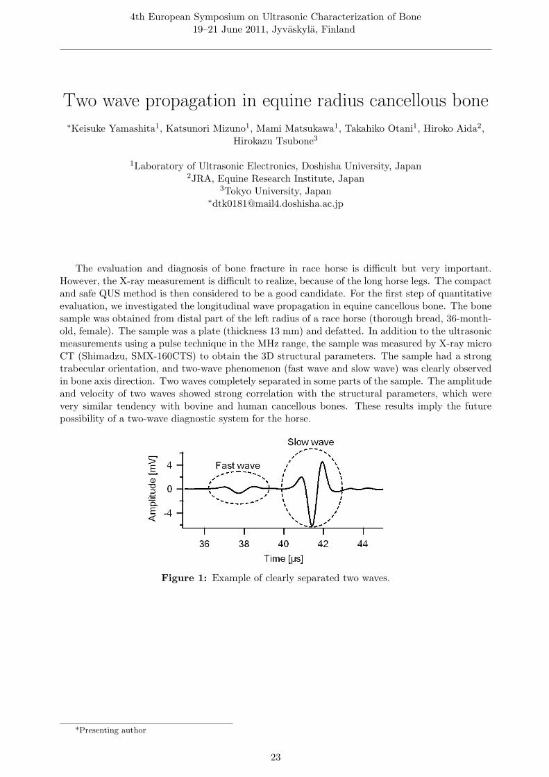
The evaluation and diagnosis of bone fracture in race horse is difficult but very important.However, the X-ray measurement is difficult to realize, because of the long horse legs. The compactand safe QUS method is then considered to be a good candidate. For the first step of quantitativeevaluation, we investigated the longitudinal wave propagation in equine cancellous bone. The bonesample was obtained from distal part of the left radius of a race horse (thorough bread, 36-month-old, female). The sample was a plate (thickness 13 mm) and defatted. In addition to the ultrasonicmeasurements using a pulse technique in the MHz range, the sample was measured by X-ray microCT (Shimadzu, SMX-160CTS) to obtain the 3D structural parameters. The sample had a strongtrabecular orientation, and two-wave phenomenon (fast wave and slow wave) was clearly observedin bone axis direction. Two waves completely separated in some parts of the sample. The amplitudeand velocity of two waves showed strong correlation with the structural parameters, which werevery similar tendency with bovine and human cancellous bones. These results imply the futurepossibility of a two-wave diagnostic system for the horse.
Figure 1: Example of clearly separated two waves.
*Presenting author
23

4th European Symposium on Ultrasonic Characterization of Bone19–21 June 2011, Jyvaskyla, Finland
In vitro ultrasonic characterization of human bonesusing focused transducers: a model-based approach
∗Roberto Longo1, Quentin Grimal2, Josquin Foiret2, Pascal Laugier2, Steve Vanlanduit1, PatrickGuillaume1
1Mechanical Engineering, Vrije Universiteit Brussel, Belgium2Laboratoire d’Imagerie Parametrique, Universite Pierre et Marie Curie-Paris6, Paris, France
Acoustic properties of bone are heterogeneous at the millimeter scale. This heterogeneity canbe proved in vitro by mapping the acoustic properties of bone slabs with ultrasound in the MHzrange. A problem when measuring human bone samples using ultrasound is to deal with thesmall surface they offer for ultrasonic investigation. Diffraction effects on the sample borders area limitation to measurement precision. The use of focused transducers is advantageous because itconcentrates the sound beam in a smaller region than plane transducers. A method was previouslydeveloped (R. Longo et al. IEEE UFFC, 2010) to measurement simultaneously speed of sound, massdensity, thickness and attenuation of bone slabs using plane transducers. The wave propagationmodel was presented in the frequency domain, for transmission measurements. This method wasnot applicable to human bone due to their small size. In this paper we show how the methodcan be adapted to measurements with focused transducers. The alignment of the emitter-receivertransducers (supported also by Laser Doppler Vibrometer wave fronts visualizations), the post-processing technique (based on the impulse response function) and the validation of the methodusing reference materials (Copper and Perspex) are also shown along this article.
*Presenting author
24
4th European Symposium on Ultrasonic Characterization of Bone19–21 June 2011, Jyvaskyla, Finland
Ultrasonic monitoring and parameters identification ofsimulated tissue culture
∗Guillermo Rus Carlborg1, Nicolas Bochud2, Juan Manuel Melchor3
1Dpt. Structural Mechanics, University of Granada, 18071, Spain2Dot. Structural Mechnaics, University of Granada, Granada, Spain3Dpt. Structural Mechanics, University of Granada, Granada, Spain
A monitoring Petri dish is tested for real-time measurement of mechanical properties of thinlayers of biomaterials, such as tissue culture or bone. To verify the sensitivity, a gelification processis monitored during approximately an hour, and validated numerically.
A layer of phantom gel is cultured on a Petri dish. The gel suffers consitency changes during aperiod in the order of magnitude of one hour. The device was excited by high-frequency ultrasonicburst waves at a central frequency of 20 MHz, and the signal was registered during a period of 5[µs] and a sampling rate of 400 [MHz]. The forward problem simulation of the experimental systemis proposed using a semi-analytical model of the ultrasonic wave interactions within the Petri dishand gel based on the transfer matrix formalism. An inverse problem is proposed for determiningthe sensitivity of the mechanical properties of the gel regarding the time evolution of the gelificationprocess.
This propagation model, combined with an inversion algorithm, allow to determine the timeevolution of the mechanical properties of the gel, such as the stiffness and the attenuation coefficent,and thus to interpret the gelification procedure.
*Presenting author
25

Poster session / Coffee breakMonday, June 20th 2011, 14:45
Chair: Mami Matsukawa
27

4th European Symposium on Ultrasonic Characterization of Bone19–21 June 2011, Jyvaskyla, Finland
Numerical simulation of cancellous bone remodelingusing a finite-difference time domain method
∗Atsushi Hosokawa
Department of Electrical and Computer Engineering, Akashi National College of Technology,674-8501 Akashi, Japan∗[email protected]
Cancellous bone remodeling, which is a couple of formation and resorption on the trabecular sur-face, was numerically simulated using a finite-difference time domain (FDTD) method with X-raymicrocomputed tomographic models. Assuming that the formation/resorption could be generatedon the trabecular surface where the local stress under the mechanical load was larger/smaller thanthe surrounding stress, the voxel elements in the cancellous bone models were added/removed. Anultrasound wave at 0.5 MHz was applied to bovine cancellous bone as the mechanical load, and thelocal stress was calculated using the FDTD method. The change in the trabecular structure dueto the remodeling was observed, as shown in Figs. 1 and 2. In Fig. 1, the bone volume fraction(BV/TV) decreases with the remodeling step, which means that the resorption rate was largerthan the formation rate. In Fig. 2, the degree of the trabecular orientation, that is the ratio of themean intercept length (MIL) of the trabeculae in the direction parallel to the ultrasound propaga-tion to MIL in the perpendicular direction, increases with the remodeling step. Consequently, thetrabecular orientation in the ultrasound direction became strong by the remodeling.
Figure 1: Variation in BV/TV with remod-eling step.
Figure 2: Variation in degree of trabecularorientation with remodeling step.
*Presenting author
28

4th European Symposium on Ultrasonic Characterization of Bone19–21 June 2011, Jyvaskyla, Finland
Distribution of the longitudinal wave velocity in equinecortical bone – effects of microstructure and HAp
orientation∗Kazufumi Yamamoto1, Daisuke Suga2, Tomohiro Nakatsuji2, Mami Matsukawa2, Takahiko
Otani2, Hirokazu Tsubone3, Hiroko Aida4, Hironobu Hoshino5, Yukihiro Matsuyama5
1Department of Orthopaedic Surgery, JA Shizuoka Kohseiren Enshu Hospital, 430-0929Hamamatsu city, Japan
2Doshisha University, Japan3The University of Tokyo, Japan
4Equine Research Institute Japan Racing Association, Japan5Hamamatsu University School of Medicine, Japan
Nowadays, diagnosis techniques to assess racehorse bones are urgently required to discoverbone disease (fatigue fracture etc.) early. We have then evaluated the ultrasonic wave properties inequine cortical bone for the future applicability of QUS to the assessment of racehorse bones. In thenanoscopic level, bone consists of hydroxyapatite (HAp) crystal. The preferred orientation of c-axisof HAp crystallites induces anisotropy of elastic properties in bone. The objectives of this study areto investigate the distribution of the longitudinal wave velocity in the axial direction, in relationto HAp crystallites orientation, microstructure, Bone Mineral Density (BMD). Three ring shapedcortical bone samples were made from equine third metacarpal bone. Longitudinal ultrasonic wavepropagation was investigated by using a conventional ultrasonic pulse system. HAp orientationwas obtained using X-ray diffraction. BMD was measured with DXA method. Microstructurewas classified in two types of Haversian structure. Fig. 1 shows velocity distribution. Regardlessof microstructure, significant correlation between velocity and HAp orientation was observed (R2
= 0.74-0.77; Fig. 2). There was no correlation between velocity and BMD. It was thought thatvelocity could reflect nanoscopic bone quality in the equine bone.
*Presenting author
29

4th European Symposium on Ultrasonic Characterization of Bone19–21 June 2011, Jyvaskyla, Finland
Figure 1: Distribution of longitudinal wave velocity in each three sample..
Figure 2: Relationship between wave velocity and HAp orientation..
30
4th European Symposium on Ultrasonic Characterization of Bone19–21 June 2011, Jyvaskyla, Finland
Ultrasound velocity estimation of cortical bones usingaxial transmission technique with angle beams
∗Rui Zheng1, Lawrence H Le2, Edmond Lou1
1University of Alberta, Canada2University of Alberta, T6G 2V2 / Edmonton, AB, Canada
Being able to obtain a reasonable background velocity profile is essential to image internalbone structures when Born Scattering theory is used. The cortical velocity can be estimated bymeasuring the direct wave (FAS) traveling from the ultrasound source to the receiving transducer.The radiation pattern of a compressional wave transducer has a main lobe with the maximumenergy strength perpendicular to the transducer surface. Therefore the ultrasound beam requirestilting to have more energy travelling toward the horizontal direction. Four angle wedges (30o, 45o,60o, and 70o) were used to acquire twelve data sets for three bone samples. The arrival times ofthe FAS were measured and linear regression was applied to find the best-fitted line. The velocitiesthus obtained were compared with the reflection-based measurements. The latter was obtainedby dividing the computed tomography determined thickness of the cortical layer by the one-waytraveling time between the cortical interfaces. Due to the irregularities of the bone surfaces, wefound the 60o wedge provided the least errors. For the three bone samples, the absolute errors are1.36%, 5.02% and 3.61% for velocity and 1.89%, 4.55% and 3.23% for thickness.
*Presenting author
31

4th European Symposium on Ultrasonic Characterization of Bone19–21 June 2011, Jyvaskyla, Finland
Relationships between broadband ultrasonic attenuationand microstructure of human trabecular bone
∗Michal Pakula1, Jalmari Pirhonen2, Petro Moilanen2, Pascal Laugier3, Tuomas Turpeinen2
1Institute of Mechanics and Applied Computer Science, Kazimierz Wielki University inBydgoszcz, 85-074 Bydgoszcz, Poland
2Department of Physics, University of Jyvaskyla, Jyvaskyla, Finland3Laboratoire d’Imagerie Parametrique, University Paris 6- CNRS UMR 7623, Paris, France
The study focused on elucidation of the influence of human trabecular bone microstructure onthe broadband ultrasonic attenuation (BUA). Ultrasonic assessments where performed on 30 humancancellous bone specimens filled with marrow, water and alcohol, successively by using three pairs ofcustom made transducers (OPTEL; D = 10 mm; fc = 0.5, 1 and 2 MHz). A 2D ultrasonic scanningmethod was applied and the BUA was recorded. In addition, bone volume fraction (BV/TV)was determined by x-ray microtomography (SkyScan 1072) at the same volumes as the ultrasonicassessments. BUA showed a strong correlation with BV/TV at 2 MHz for marrow (R2=0.90,p<1e-4) and water (R2=0.89, p<1e-4) saturated specimens. Significant correlations were also foundat 1 MHz for specimens with marrow (R2=0.71, p<1e-4), water (R2=0.87, p<1e-4) and alcohol(R2=0.87, p<1e-4). Interestingly, at 0.5 MHz the BUA well correlated with BV/TV for alcoholsaturated specimens (R2=0.81, p<1e-4), but not so well with marrow (R2=0.37, p<1e-4) andwater (R2=0.35, p<1e-4) saturated ones. Strong correlations between BV/TV and BUA for higherfrequencies were thus shown. Poor correlation at 0.5 MHz could be explained by another mechanismof attenuation appearing at lower frequencies or by challenges of signal analysis (Anderson et al.JASA, 2010).
*Presenting author
32

4th European Symposium on Ultrasonic Characterization of Bone19–21 June 2011, Jyvaskyla, Finland
Numerical simulation reveals how the fast wave isgenerated in cancellous bone
∗Yoshiki Nagatani
Department of Electronic Engineering, Kobe City College of Technology, 651-2194, Japan∗[email protected]
In cancellous bone, longitudinal waves often separate into fast and slow waves depending onthe alignment of bone trabeculae in the propagation path. We pointed that fast wave requiredcertain propagation distance for steady propagation [Nagatani et al., Ultrasonics (2008)]. However,the precise mechanism of fast wave propagation is not clarified yet. In this study, the detailedbehavior of fast wave generation was investigated using three-dimensional simulation technique.Using the virtual model whose central portion inside the specimen was assumed to be vacuum,relationship between the thickness of the margin and first positive peak amplitude of the fast wavewas investigated. As a result, fast wave was mainly originated from the soundwave that goesinto trabecular within 1 to 2 times of wavelength after entering the cancellous bone (Fig. 1). Inaddition, when the acoustic impedance of the liquid part was higher, the degradation of fast waveattenuation was higher caused by the leakage of soundwave from solid portion into liquid portion(Fig. 2). These interesting phenomena may show that the fast wave attenuation is caused by bothmulti-path effect, leakage, and absorption in media. These investigations help us understand theprecise behavior of soundwave inside cancellous bone.
Figure 1: Relationship between the thick-ness of the margin and first positive peak am-plitude of the fast waves. The amplitude offast wave gradually increases, then plateaus
at 1 to 2 times of wavelength.
Figure 2: Distribution of fast wave attenua-tion of specimen when the acoustic impedanceof liquid portion was changed. The degrada-tion rate of the attenuation depends on the
acoustic impedance of the liquid portion.
*Presenting author
33

4th European Symposium on Ultrasonic Characterization of Bone19–21 June 2011, Jyvaskyla, Finland
Separation technique for ultrasonic waveformpropagated in cancellous bone using wavelet transform
analysis
Sho Hasegawa1, ∗Yoshiki Nagatani1, Katsunori Mizuno2, Mami Matsukawa2
1Kobe City College of Technology, Japan2Doshisha University, Japan∗[email protected]
Ultrasonic waves targeted on cancellous bone separate into fast and slow waves according tothe alignment of bone trabeculae. The characteristics of the specimen, however, can sometimesresult in ambiguous wave separation. In this study, we proposed using wavelet transform as a newmethod of analyzing ultrasonic waveforms. The experimentally observed waveforms were used forthe analysis. Using continuous wavelet transform, two components were separated by fitting two-dimensional Gaussian function in scalogram (Fig. 1). Because the residual is distributed mainly inthe lower-frequency portion, the component may include a fast wave that passes through the shorterpath in the trabecula. The relationship between bone volume fraction and the peak amplitudeof the lower- and higher-frequency components showed a clear negative correlation. This resultderived from scalogram conflicts with the previous knowledge derived from time domain analysis.Therefore, it is natural to consider that the peak value of the lower-frequency component in thescalogram and the first positive peak of the fast wave in the time domain properly reflect physicalphenomena, respectively. We recognized lower-frequency component as the multiple reflections(forward scattering) in cancellous bone.
Figure 1: Example of scalogram. Figure (a) is a scalogram of the original waveform thatpropagated in cancellous bone; (b) is the fitted two-dimensional Gaussian function; (c) is theresidual of fitting. The residual may include the ”fast wave” that passes through the shorter
path in the trabecula.
*Presenting author
34

4th European Symposium on Ultrasonic Characterization of Bone19–21 June 2011, Jyvaskyla, Finland
A crazy climber method for analyzing fundamentalflexural guided wave in ultrasonic axial transmissionsignals recorded by a low-frequency array transducer
∗Vantte Kilappa1, Kailiang Xu2, Petro Moilanen1, Dean Ta2, Jussi Timonen1
1Department of Physics, University of Jyvaskyla, Jyvaskyla, Finland2Department of Electrical Engineering, Fudan University, Shanghai, China
In a recent study, we have introduced a 6-receiver low-frequency (100-400 kHz) array transducerfor ultrasonic axial transmission of the first arriving signal (FAS). Due to the low number ofreceivers, techniques based on two-dimensional fast Fourier transform (2D-FFT) cannot be usedto analyze modes like the fundamental flexural guided wave (Lamb A0) with this array. Thepurpose of the present work was thus to test the suitability of an alternative approach based on atime-frequency representation (TFR) of the signal. A method of Crazy Climbers (CC) was usedto enhance the separation of modal components in TFR. Time of flight of the highest amplitudecomponent was detected and separated from TFR, and these data were used to determine thegroup velocity by linear regression. The method was tested on acrylic plates and 41 human radiusspecimens with the soft tissue removed. Velocity measured in the phantoms was in accordancewith the group velocity of the Lamb A0 mode (Fig. 1). In the bone samples it was correlatedwith the cortical thickness (R=0.71, p<0.001) (Fig. 2) and bone mineral density (R ≈ 0.6). Inconclusion, CC-TFR analysis enabled assessment of the fundamental flexural guided mode with anarray transducer.
Figure 1: Group velocity of the extractedguided wave mode as a function of thickness,as determined by the CC method at 100 kHzfor acrylic-plates (markers). That of the A0
mode is shown by a dashed line.
Figure 2: Extracted guided wave velocityas a function of cortical thickness for humanradius specimens. Cortical thickness was as-
sessed by pQCT.
*Presenting author
35

4th European Symposium on Ultrasonic Characterization of Bone19–21 June 2011, Jyvaskyla, Finland
Reproducibility of the new ultrasound technique forosteoporosis diagnostics at primary healthcare level
∗Ossi Riekkinen1, Aleksi Monkkonen2, Jarkko Kauppinen2, Janne Karjalainen1, Jukka Jurvelin1
1Applied Physics, University of Eastern Finland, Kuopio, Finland2Savonia University of Applied Sciences, Finland
At present, there are no reliable methods to diagnose osteoporosis at the primary healthcarelevel. Therefore, partially, about 75% of osteoporotic patients are not diagnosed or treated. Foroptimal treatment decisions, novel diagnostic techniques for primary healthcare level should closelymatch with the axial DXA and the clinical risk factors (e.g. FRAX). The new in vivo pulse-echoultrasound (PE-US) technique for the measurement of cortical thickness could fulfil these aims.Changes in cortical thickness are known to indicate sensitively early bone pathology. In this study,we tested whether the PE-US technique is simple, fast and easy to use, as well as independent ofthe operator.
Fourteen inexperienced operators (healthcare students/professionals) were trained to use thePE-US technique (2,25 MHz) for in vivo measurement of the cortical bone thickness at the tibia ofone volunteer.
The reproducibility for the measurements of cortical bone thickness was good (CV 3.6%) be-tween the operators. The operators used 8 ± 2 min for training and 6 ± 3 min for the measurements.
Using the first PE-US prototype reproducibility of the technique was good. However, we be-lieve that the measurement time and usability of the technique will be improved with the secondprototype under development.
*Presenting author
36

4th European Symposium on Ultrasonic Characterization of Bone19–21 June 2011, Jyvaskyla, Finland
Bone phantoms for ultrasonic axial transmission studies
Jalmari Pirhonen1, ∗Petro Moilanen1, Vantte Kilappa1, Pasi Karppinen2, Zuomin Zhao3, AriLiikkanen2, Timo Karppinen2, Edward Hæggstrom2, Risto Myllyla3, Jussi Timonen1
1Department of Physics, University of Jyvaskyla, Jyvaskyla, Finland2Department of Physics, University of Helsinki, Helsinki, Finland
3Department of Electrical and Information Engineering, University of Oulu, Oulu, Finland∗[email protected]
Ultrasonic axial transmission enables multimodal assessment of human cortical bones such asthe radius and tibia. We consider bone, coated by soft tissue and filled with marrow, as an elasticwaveguide and model it by a fluid-coated and fluid-filled axisymmetric tube. The purpose of thepresent study was to develop bone phantoms, i.e. reference waveguides, consistent with this model.The phantoms were made from a homogeneous composite of aluminium oxide (Al2O3) and epoxyresin. Their manufacturing included mixing and de-airing of the components, molding and turningthe castings on a lathe, and drilling them into tubes. Phantoms were coated and/or filled bygelatin gel to mimic the soft tissue and bone marrow, respectively (Fig.1). Dependence of the firstarriving signal velocity (vFAS) on the amount of Al2O3, temperature and sample wall thickness(Fig.2) were evaluated by a 400kHz array ultrasonometer. Maximum of 2930m/s was achieved atroom temperature for 70% by mass of Al2O3, with a temperature dependence of -10 m s−1 ◦C−1.In addition, a 200kHz scanning ultrasonometer was used to record the velocity of a fundamentalflexural guided wave, showing reasonable agreement with theoretical models. A series of five bonephantoms with individual wall thickness (1-5mm) was developed.
Figure 1: A bone phantom with soft coating and softfilling
Figure 2: Thickness depen-dence of FAS velocity in coated
and filled phantoms
*Presenting author
37

4th European Symposium on Ultrasonic Characterization of Bone19–21 June 2011, Jyvaskyla, Finland
Volumetric quantification of repair cartilage andsubchondral ossification using ultrasound biomicroscopy
∗Nils Mannicke1, Martin Schone1, Ronny Schulz2, Hannes Kuttner2, Bastian Marquaß2, KayRaum1
1Julius Wolff Institute and Berlin-Brandenburg School for Regenerative Therapies, Charite –Universitatsmedizin Berlin, Berlin, Germany
2Translational Centre for Regenerative Medicine Leipzig, Leipzig, Germany∗[email protected]
A method is presented that enables quantification of repair tissue and subchondral ossificationvolumes in cartilage repair studies using ultrasound biomicroscopy. Involved is a semi-automaticdetection of surface and cartilage bone-boundaries that allow a reliable tracking of subchondralbone growth.
In Merino-sheep, the effect of different treatment approaches for chronic focal osteochondralcartilage 7-mm-diameter-lesions was studied. Treatment groups covered untreated control groups(N=16), collagen-type-I-scaffolds (N=8) and BmMSC-cell-seeded scaffolds (N=8). Convalescencewas 12 months. Immediately after explantation, time-resolved C-scans of defect area and surround-ing native tissue were recorded using a portable ultrasound biomicroscope equipped with a focussed40MHz transducer. 3D- surface and cartilage bone interface were approximated using a threshold-based analysis of the Apparent Integrated Backscatter (AIB) and manually refined by physicians.Positions of surface and bone interface outside the defect area served as input for a 2D polynomial.By comparison of the polynomial model and the actual position, subchondral ossification as wellas the occurence of bone cysts were quantified volumetrically.
Accurate determination of subchondral bone boundaries and its comparison with native areasallowed quantification of ossification and detection of bone cysts at a high spatial resolution. Thepresented method will help improving a non-destructive assessment of different treatment strategies.
Figure 1: Exemplary B-mode cross section. Displayed are defect borders as vertical lines,surface (light gray) and cartilage-bone interface (dark gray). Cartilage-bone boundary could notbe detected on the left side. A 2D polynomial was used for approximation of native boundaries
in the defect region, shown as dotted lines.
*Presenting author
38

4th European Symposium on Ultrasonic Characterization of Bone19–21 June 2011, Jyvaskyla, Finland
Micro Brillouin scattering study on the velocityanisotropy in a trabecula
∗Kenji Fukui, Mami Matsukawa
Laboratory of Ultrasonic Electronics, Doshisha University, Kyotanabe, Japan∗[email protected]
Ultrasonic wave properties in bone include various information from different scales, such asmicrostructure, soft tissues, composition of the bone matrix, etc. It is then very difficult to extractthe precise effect of each factor on the obtained properties. As for the cortical bone and trabecula,they are often treated as isotropic without the actual measurement of anisotropic properties. Wehave then investigated longitudinal wave velocities in a single trabecula using a micro-Brillouinscattering technique. This technique enables velocity measurements in a small area (diameter 8micron meter) without the effect microstructure in a sliced specimen. In addition, rotation of thespecimen can give us in plane wave velocities. Specimens used were obtained from the cancellousbone of a bovine femur (27-month-old). Rod type trabeculae were selected using X-ray CT image ofthe cancellous bone and carefully polished to thin specimens (thicknesses 50-70 micron meter). Inall specimens, measured velocities were in the range of 4.92-5.31e3 m/s, and showed weak uniaxialanisotropy. The maximum wave velocity direction was always similar to the direction of rod.The high velocities seem to come from high frequency range (around 13.7GHz) and few effect ofmicrostructure in a small area.
Figure 1: Velocity anisotropy in a trabecula. 0-180 direction indicates the direction of thetrabecular rod.
*Presenting author
39

4th European Symposium on Ultrasonic Characterization of Bone19–21 June 2011, Jyvaskyla, Finland
A nonlinear stratified model to predict ultrasonic wavepropagation in trabecular bone
∗Nicolas Bochud1, Quentin Grimal2, Sylvain Haupert2, Pascal Laugier2, Guillermo Rus1
1University of Granada, Granada, Spain2UPMC-CNRS, Paris, France
Ultrasound is regarded as a promising tool for bone quality assessment. In the past, many lineartechniques have been developed to image ultrasonic wave propagation velocity and attenuation intrabecular bone, among them methods based on stratified models or on Biot theory. However,these methods are almost insensitive to progressive induced damages. Recently, a relation betweendamage accumulation and trabecular bone density was obtained in vitro by nonlinear ultrasoundtechniques. Models of nonlinear wave propagation in bone are required to analyse the experimentalresults and to help quantify the in situ level of damage.
In this study, the complex architecture of trabecular bone is modeled as a stratified mediumof alternating solid (trabecular material) and liquid (marrow) layers. The volume fraction of solidphase represents bone volume fraction. The model accounts for the transmissions and reflectionsof the ultrasonic signal describing the nonlinear interactions of primary waves within bone. Theacoustic response is simulated with a method which extends the classical transfer matrix formalismto classical nonlinearity.
The effective nonlinearity of trabecular bone is evaluated regarding the local nonlinearity andthe volume fraction of trabecular material, and is characterized by the amplitude ratio between thesecond and first harmonic amplitude.
*Presenting author
40

4th European Symposium on Ultrasonic Characterization of Bone19–21 June 2011, Jyvaskyla, Finland
Age affects the bone healing kinetics in a rat osteotomymodel
Katrein Sauer1, Daniel Rohrbach1, Patrick Strube2, Bernd Preininger2, ∗Kay Raum1
1Julius-Wolff-Institut, Charite-Universitatsmedizin Berlin, Berlin, Germany2Center for Musculoskeletal Surgery, Charite-Universitatsmedizin Berlin, Berlin, Germany
The course of bone healing in animal models is conventionally monitored by µCT or histologyapproaches. Both techniques do not allow the determination of the material properties of theinvolved tissues. In this study, quantitative scanning acoustic microscopy was used at 50 MHzto investigate microstructural and elastic alterations of mineralized callus and cortical tissue aftertransverse femoral osteotomies in young (12 weeks, N = 5) and old (12 months, N= 13) rat.Analyses were performed after 6 weeks of consolidation with stabilization by either a rigid or asemi-rigid external fixator. The mean stiffness values in cortical and callus tissues were 33.05 ±7.56 GPa and 19.32 ± 5.93 GPa, respectively. The callus stiffness values in the young group weresignificantly (9.33 %) higher than those in the old group (F = 17.23). Porosity values in corticaland callus tissues were 2.8 ± 1.0 % and 53.9 ± 9.8 %, respectively. The callus porosity in theyoung group was significantly (13.3 %) lower than that in the old group (F = 11.27). These resultsindicate that callus healing is considerably delayed in old rat.
Figure 1: Exemplary grey-scale stiffness map obtained by SAM measurements. The overlaidcolors indicate segmented evaluation regions (red = excluded non-mineralized tissue and embed-
ding material; green = cortical tissue; blue = callus tissue).
*Presenting author
41


Session IV: Ultrasonic Characterization of CancellousBone
Monday, June 20th 2011, 16:15Chair: Jukka Jurvelin
43

4th European Symposium on Ultrasonic Characterization of Bone19–21 June 2011, Jyvaskyla, Finland
A new perspective on ultrasound assessment ofcancellous bone
∗Christian Langton
Physics, Queensland University of Technology, Brisbane 4001, Australia∗[email protected]
The measurement of broadband ultrasonic attenuation (BUA) in cancellous bone at the cal-caneus for assessment of osteoporosis was first described 25 years ago. The technique has beenextensively clinically validated and is utilized worldwide. However, there is still no fundamentalunderstanding of the dependence of BUA upon the material and structural properties of cancellousbone as well as the observed ’parabolic’ relationship between BUA and bone volume fraction.
It has recently been proposed that the primary BUA mechanism in cancellous bone is phasecancellation due to variations in transit time as detected over the phase-sensitive surface of thereceive ultrasound transducer [1]. A combined experimental and computer simulation study hassuccessfully demonstrated that lateral inhomogeneity of transit time has significant potential forphase cancellation to occur; relatively simplistic solid:liquid models exhibited lateral inhomogeneityranging from minimal, a single transit time, to maximal, a wedge being the ultimate case.
This has led to the development of Ultrasound Transit Time Spectral Analysis, with the po-tential to quantify both the bone volume fraction and ’structure’ of cancellous bone.
1. Langton C M; 2011; 25th Anniversary of BUA for the Assessment of Osteoporosis – Timefor a New Paradigm?; Engineering in Medicine; 225 (2),113-125
*Presenting author
44

4th European Symposium on Ultrasonic Characterization of Bone19–21 June 2011, Jyvaskyla, Finland
In situ ultrasound backscatter measurements of intacthuman proximal femur
∗Markus Malo, Ossi Riekkinen, Viktoria Prantner, Hanna Isaksson, Janne Karjalainen, JuhaToyras, Jukka Jurvelin
Department of Applied Physics, University of Eastern Finland, Kuopio, Finland∗[email protected]
Ultrasound reflection and backscatter parameters have been shown to relate to mechanicaland structural properties of trabecular bone. However, the potential of reflection and backscatterparameters in measurements of intact human proximal femur have not been studied. In this study,human femoral bones of 14 cadavers (3 females and 11 males, 49.4 ± 17.4 years, range: 17 – 78years) were measured at the neck and at the trochanter in the anterior posterior and lateral medialdirections. Ultrasound reflection coefficient (IRC) and apparent integrated backscatter (AIB) wereobtained and compared with structural microCT parameters for trabecular bone samples extractedfrom the same locations. Pearson correlation analysis was used to calculate linear correlationcoefficients. The IRC values showed no significant correlations with structural parameters, however,AIB correlated significantly with several structural parameters, e.g. the structure model index (r= 0.72, p < 0.01). AIB was also correlated significantly with age (r = 0.82, p < 0.01). The presentresults encourage further in vivo measurements. However, the overlying soft tissues that distortand attenuate the ultrasound pulse make in vivo pulse-echo measurements from the proximal femurmore challenging.
*Presenting author
45

4th European Symposium on Ultrasonic Characterization of Bone19–21 June 2011, Jyvaskyla, Finland
Estimating mean trabecular bone spacing fromultrasonic backscattering signals based on twofundamental frequency estimation methods
∗Dean Ta, Baiding Yang, Weiqi Wang
Department of Electronic Engineering, Fudan University, Shanghai, China∗[email protected]
The mean trabecular bone spacing(MTBS) estimated from ultrasonic backscattering signalscan indicate the change of cancellous bone microstructures. This paper first proposed a MTBSestimation method based on fundamental frequency estimation(SFE). The SFE first utilizes sim-plified inverse filter to remove the high frequency multiplicative interference of system response,then the combination of event-based instantaneous fundamental frequency estimation and improvedautocorrelation algorithm are applied to get the estimated fundamental frequency, from which theMTBS can be obtained. Then the paper proposed Hilbert transform- fundamental frequency esti-mation(HFE). Two methods were applied to backscattering signals from simulations and in vitrobovine trabeculae experiments. The results shows that the performances of two proposed methodsoutperform that of the Autoregressive(AR) and simplified inverse filter tracking(SIFT) algorithms.In the in vitro experiments, the results are in agreement with simulation ones, the most of esti-mates are more precise compared with reference MTBS values from micro-CT than AR and SIFTmethods. It can be concluded that SFE algorithm is more robust for MTBS estimation than ARcepstrum and SIFT algorithm with precise estimates and small estimated variance, and HFE provedto be a more robust algorithm than SFE method, especially for varying SNR and MTBS.
*Presenting author
46

Invited lectureTuesday, June 21st 2011, 09:00
Chair: Jussi Timonen
47

4th European Symposium on Ultrasonic Characterization of Bone19–21 June 2011, Jyvaskyla, Finland
Development strategies for clinical skeletal quantitativeultrasound approaches: challenges and opportunities
∗Claus-Christian Glueer
Medical Physics, Diagnostic Radiology, CAU Kiel, Kiel, Germany∗[email protected]
The history of skeletal applications of Quantitative Ultrasound (QUS) methods has been a rollercoaster ride. Starting with cortical elasticity, moving on with attenuation in trabecular bone, largestudies confirmed the predictive power of QUS approaches for fracture risk assessment. However,the lack of inclusion in drug studies, real and virtual quality issues hinders widespread acceptanceof QUS. The resurgence of technological innovations in skeletal QUS methods in the past few yearsis quite impressive. What are the topics for successful QUS approaches? In cortical bone wavepropagation can be simulated, measured and understood very accurately. More importantly, thefield of osteoporosis diagnostics is waiting for methods for in vivo assessment of material properties.Cortical porosity, mineralization, anisotropy are key determinants of bone strength independent ofbone density. Elasticity can be measured at the radius or the tibia but it is not clear yet whetherbone strength and toughness are accessible by ultrasound as well. Also the correlation with materialproperties of the main fracture sites needs to be established and thus QUS measurements at theproximal femur are relevant. However, most importantly: establishing the quality of QUS in theassessment of bone qualities is the challenge to face !
*Presenting author
48

Session V: Clinical Quantitative UltrasoundTuesday, June 21st 2011, 09:30
Chair: Jussi Timonen
49

4th European Symposium on Ultrasonic Characterization of Bone19–21 June 2011, Jyvaskyla, Finland
Multi-site bone ultrasound measurements in elderlywomen
∗Janne Karjalainen1, Ossi Riekkinen1, Juha Toyras1, Mikko Hakulinen2, Heikki Kroger3, ToniRikkonen4, Kari Salovaara4, Jukka Jurvelin1
1Department of Applied Physics, University of Eastern Finland, Kuopio, Finland2Department of Clinical Physiology and Nuclear Medicine, Kuopio University Hospital, Kuopio,
Finland3Department of Orthopaedics, Traumatology and Hand Surgery, Kuopio University Hospital,
Kuopio, Finland4Bone and Cartilage Research Unit, University of Eastern Finland, Kuopio, Finland
At present, no reliable diagnostic methods for osteoporosis are available at the primary healthcare level. In this study, multi-site pulse-echo (PE) ultrasound method is proposed.
Thirty elderly women were examined in vivo using PE ultrasound (at 2.25 and 5.0 MHz) mea-surements in proximal femur, distal radius, proximal and distal tibia. Further, areal bone mineraldensity (BMD) in the proximal femur was determined by axial DXA. First, we predicted the BMDat femoral neck (BMDneck) by ultrasound measurements in tibia combined with subject charac-teristics (density index, DI), Second, we tested the ability of ultrasound backscatter measurementsat proximal femur to discriminate between individuals with previously fractured hips from thosewithout fractures.
Cortical thickness at distal and proximal tibia combined with age and weight of the subject,provided a good estimate of BMDneck (r = 0.86, p < 0.001, n = 30). When inserted into FRAX(World Health Organization fracture risk assessment tool) the DI indicated the same treatmentproposal as the BMDneck with 86% sensitivity and 100% specificity. The ultrasound backscatterparameter combined with patient characteristics discriminated subjects with fracture from thecontrols.
The results indicate that ultrasound parameters, combined with patient characteristics, mayprovide means for osteoporosis diagnostics.
*Presenting author
50

4th European Symposium on Ultrasonic Characterization of Bone19–21 June 2011, Jyvaskyla, Finland
Ability of low-frequency axial transmission ultrasoundto discriminate retrospective low-energy fractures
Petro Moilanen1, ∗Mikko Maatta2, Vantte Kilappa1, Leiting Xu3, Timo Jamsa2, Jussi Timonen1,Sulin Cheng3
1Department of Physics, University of Jyvaskyla, Jyvaskyla, Finland2Department of Medical Technology, University of Oulu, Oulu, Finland
3Department of Health Sciences, University of Jyvaskyla, Jyvaskyla, Finland∗[email protected]
Low-frequency axial transmission ultrasound (LF-AT; fc=400 kHz) shows good sensitivity tocortical thickness and BMD deep in the endosteum of long cortical bones. Purpose of the presentstudy was to evaluate the ability of this method when assessing bone fragility retrospectively. Thefracture group (F) consisted of n=25 females (age 26-80 yrs) with a history of low-energy fracturesand the non-fracture group (NF) included n=100 females (age 27-86 yrs). Subjects with medicationor disease affecting bone metabolism were excluded. An LF-AT array probe was used to measurethe velocity of the first arriving signal (VFAS) in the midshaft radius and tibia. Site-matchedpQCT assessments were done by XCT2000 (Stratec Medizintechnik). In addition, areal BMD ofthe whole body, femoral neck and lumbar spine were assessed by DXA (Prodigy, GE). Odds ratios(OR), adjusted for the age and body mass index, were 1.80 (95% CI: 1.31-2.50) and 1.42 (1.10-1.83)and areas under the receiver operator characteristics curve (AUC) were 0.70 and 0.65 for the VFASradius and tibia, respectively. The pQCT and DXA measurements yielded similar or weaker results.These results thus show that LF-AT is able to discriminate low-energy fractures with similar orbetter accuracy than pQCT and DXA.
*Presenting author
51

4th European Symposium on Ultrasonic Characterization of Bone19–21 June 2011, Jyvaskyla, Finland
In vitro evidence that the circumferential wave guidedby the femur cortical shell can be used to predict femur
strength
Julien Grondin1, ∗Quentin Grimal1, Sandra Guerard2, Reinhard Barkmann3, Claus Gluer3,Pascal Laugier1
1Laboratoire d’imagerie parametrique, UPMC-CNRS, Paris, France2LBM, Arts et Metiers PARISTECH, Paris, France
3Medizinische Physik, Klinik fur Diagnostische Radiologie, UKSH, Kiel, Germany∗[email protected]
QUS at the proximal femur shows a good performance for hip fracture discrimination (Bark-mann et al, Osteoporosis Int. 2010). With the implemented signal processing, the techniqueessentially measures trabecular bone. However the first arriving signal was shown to be associatedwith the propagation of a circumferential wave guided in the cortical shell and to be independentfrom the trabecular compartment. Because cortical bone is a key factor to maintain femur neckmechanical integrity, we hypothesized that the measurement of the propagation time of the firstarriving signal (T) could be predictive of femur strength. For nine left femurs of women donors(mean age 83y.) we measured: (1) femur strength in one-legged stance configuration; (2) BMDfrom DXA; (3) T through the neck with two cylindrically focused transducers (0,5 MHz central fre-quency) which illumated a 5 mm-thick transverse neck cross-section. Significant relationships wereobserved between T and mechanical parameters: failure load: R2=0.79; elastic energy : R2=0.63;apparent stiffness R2=0.70. DXA was also well correlated to strength (R2=0,78 for failure load). Infuture work, we shall investigate whether a combination of independent ultrasound measurementsof the cortical and trabecular compartments at the upper femur enhance fracture risk prediction.
*Presenting author
52

4th European Symposium on Ultrasonic Characterization of Bone19–21 June 2011, Jyvaskyla, Finland
Figure 1: Relationships between measured first arriving signal time-of-flight and proximalfemurs mechanical characteristics.
53


Session VI: Models for Ultrasonic Characterization ofCortical Bone
Tuesday, June 21st 2011, 10:45Chair: Pascal Laugier
55

4th European Symposium on Ultrasonic Characterization of Bone19–21 June 2011, Jyvaskyla, Finland
Excitability of axially transmitted guided waves inmodels of human long bones
∗Maryline Talmant1, Petro Moilanen2, Josquin Foiret1, Jean-Gabriel Minonzio1, Pascal Laugier1,Jussi Timonen2
1Laboratoire d’Imagerie Parametrique, CNRS-UPMC, Paris, France2Department of Physics, University of Jyvaskyla, Jyvaskyla, Finland
Previously, the Jyvaskyla group has used an empty tube as the reference waveguide for inversionor identification of ultrasound measurements, but the role of the marrow has remained an openissue. The objective of this study was to test several reference waveguides, empty, fluid-filled andfluid-coated tubes, in the modelling of guided waves. Mode excitability in fluid-filled tubes wasevaluated using the normal mode expansion method (NME), with normal stress excited by thesource while receivers detected the normal displacement. Results are shown in Fig 1 as a gray-scaleimage in the frequency-wavenumber plane. For cross validation, a corresponding spectrum wasobtained by 2D-FFT of signals simulated by SimSonic3D. Related experiments were performedto evaluate the sensitivity properties of real transducers. Finally, the utility of group velocityfiltering on mode separation was analyzed. Introduction of absorption in the fluid and/or in thetube material is currently under way, and its relevance will be evaluated by experiments. Tuningof the excitation and detection of ultrasound signals will as well be analyzed so as to optimize themeasurement of A0/F11 properties.
Figure 1: Ultrasound spectrum as determined by the NME method (gray scale image) for afluid-filled tube. Corresponding dispersion curves are shown by solid lines.
*Presenting author
56
4th European Symposium on Ultrasonic Characterization of Bone19–21 June 2011, Jyvaskyla, Finland
Characterization of growing bone: curvature andanisotropy
∗Cecile Baron
ISM, CNRS/Aix-Marseille University, F-13288 Marseille, France∗[email protected]
In ultrasound characterization, long bones can be realistically modeled as tubes with orthorom-bic crystal structure. Nevertheless, numerous studies on ultrasound propagation in bones representlong bones as transversely isotropic (or even isotropic) waveguides (typically plates). This paperproposes a semi-analytical method to solve the wave equation in orthotropic waveguides. Based onan analytical solution, the matricant, explicitly expressed under the Peano series expansion form, itallows to consider a degree of anisotropy greater than the transverse isotropy with a curved geom-etry. In this work, we investigate how the curvature of long bone influences the wave propagationand how wave speeds are sensitive to components of the orthotropic stiffness matrix. The resultsconfirm that the plate model may be adequate to study guided waves propagation in tubes withouter radius to thickness ratio lower than 0.5 and beyond a minimal frequency value. Moreover thisstudy gains insight the role of the stiffness coefficients on the propagation of the first – extensionaland flexural – guided modes travelling in an orthotropic tube. This model may reveal particularlyappropriate to characterize growing bone for which the outer radius to thickness ratio is greaterthan 0.5 and the material symmetry is considered as orthotropic.
*Presenting author
57
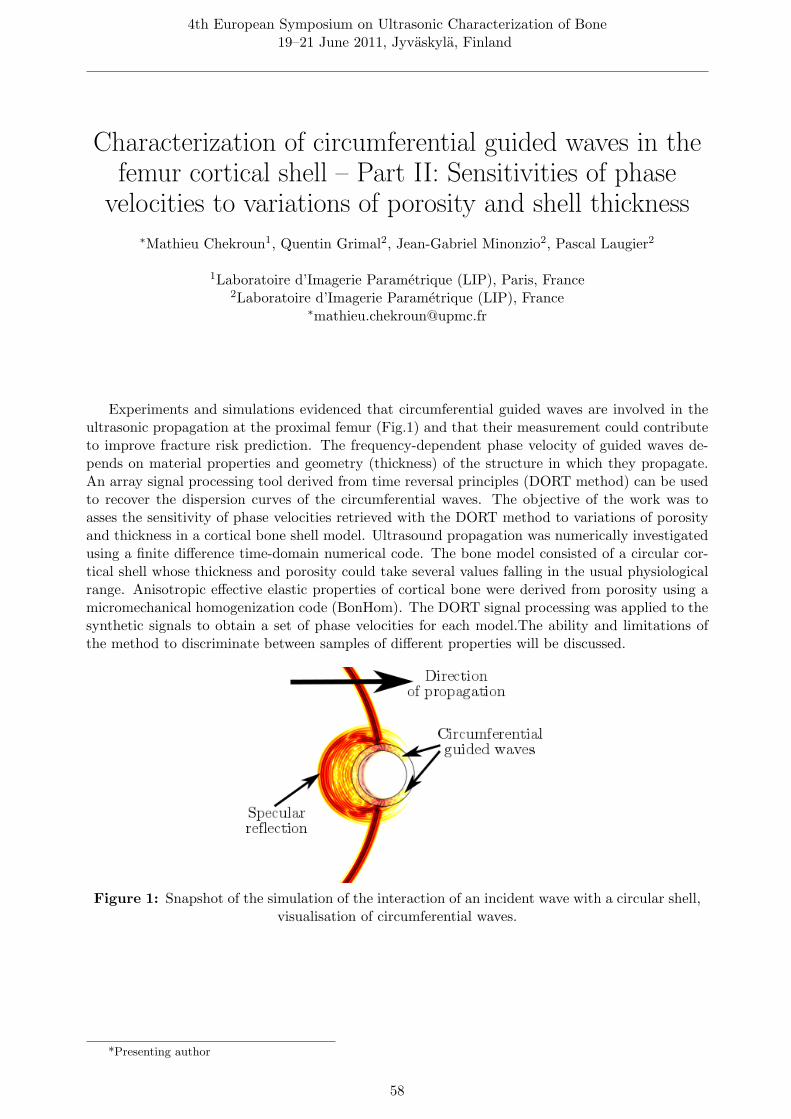
4th European Symposium on Ultrasonic Characterization of Bone19–21 June 2011, Jyvaskyla, Finland
Characterization of circumferential guided waves in thefemur cortical shell – Part II: Sensitivities of phase
velocities to variations of porosity and shell thickness∗Mathieu Chekroun1, Quentin Grimal2, Jean-Gabriel Minonzio2, Pascal Laugier2
1Laboratoire d’Imagerie Parametrique (LIP), Paris, France2Laboratoire d’Imagerie Parametrique (LIP), France
Experiments and simulations evidenced that circumferential guided waves are involved in theultrasonic propagation at the proximal femur (Fig.1) and that their measurement could contributeto improve fracture risk prediction. The frequency-dependent phase velocity of guided waves de-pends on material properties and geometry (thickness) of the structure in which they propagate.An array signal processing tool derived from time reversal principles (DORT method) can be usedto recover the dispersion curves of the circumferential waves. The objective of the work was toasses the sensitivity of phase velocities retrieved with the DORT method to variations of porosityand thickness in a cortical bone shell model. Ultrasound propagation was numerically investigatedusing a finite difference time-domain numerical code. The bone model consisted of a circular cor-tical shell whose thickness and porosity could take several values falling in the usual physiologicalrange. Anisotropic effective elastic properties of cortical bone were derived from porosity using amicromechanical homogenization code (BonHom). The DORT signal processing was applied to thesynthetic signals to obtain a set of phase velocities for each model.The ability and limitations ofthe method to discriminate between samples of different properties will be discussed.
Figure 1: Snapshot of the simulation of the interaction of an incident wave with a circular shell,visualisation of circumferential waves.
*Presenting author
58

4th European Symposium on Ultrasonic Characterization of Bone19–21 June 2011, Jyvaskyla, Finland
Three-dimensional finite-difference time-domainsimulations demonstrate the sensivity of ultrasoundmeasurements at the femoral neck to cortical bone
geometry∗Pierre Nauleau1, Julien Grondin1, Quentin Grimal1, Klaus Engelke2, Pascal Laugier1
1Laboratoire d’Imagerie Parametrique, UPMC Paris 6-CNRS UMR7623, Paris, France2Institute of Medical Physics, University of Erlangen-Nurnberg, Erlangen, Germany
The ability of QUS to predict fracture risk at the hip has been recently demonstrated. Ourgroup is investigating circumferential propagation of guided waves at the femoral neck as a mean toenhance fracture risk prediction. Two-dimensional numerical simulations have suggested a relation-ship between mechanical strength and propagation time of circumferential guided waves. This studyis an extension of our previous work to the three-dimensional case. The three-dimensional structureof eleven femoral necks was reconstructed from X-ray computed tomography. Three-dimensionalsimulations of a transverse transmission configuration were performed using a finite-difference time-domain code assuming constant material properties of bone. A transverse cross-section, in the mid-neck axis, was illuminated by the beam (5 mm-width) of a cylindrically focused transducer. Thetransmitted signal was processed to compute the time-of-flight (TOF) of the first arriving signal.Geometrical parameters, known as relevant indices of bone strength, (volume, V and moment ofinertia, MI) were calculated for the probed volume. Strong relationships between the geometry andTOF with R2-values of 0.78 (TOF vs. MI) and 0.61 (TOF vs. V) were consistent with our previoustwo-dimensional work. Three-dimensional numerical simulations are a powerful tool to investigateultrasounds interaction with the complex geometry of the proximal femur.
*Presenting author
59

4th European Symposium on Ultrasonic Characterization of Bone19–21 June 2011, Jyvaskyla, Finland
2D simulations of the impact of porosity and corticalthickness on the wave propagation at the inferior
femoral neck cortex∗Kerstin Rohde1, Melanie Daugschies1, Quentin Grimal2, Pascal Laugier2, Claus-Christian
Gluer1, Reinhard Barkmann1
1Medizinische Physik, UK S-H Kiel, Diagnostische Radiologie, Kiel, Germany2Laboratoire d’Imagerie Parametrique, UPMC, Paris, France
Cortical ultrasound propagation depends on cortical thickness and porosity. We performed 2D-simulations (SimSonic, LIP Paris) on an Xtreme-CT-Scan of a femoral neck cortex with artificiallymodified porosity, pore size (PS) and cortical cross-sectional area (CSA).
For the simulation sender and receiver (10 mm diameter, centre frequency 500 kHz) were posi-tioned at both sides of the inferior cortex. PS was varied in 4 steps (diameters 0.08-0.32 mm) ateach level of porosity and CSA, CSA in 3 steps (100%-74%) at each level of porosity and pore size,and the porosity in 4 steps (0%-15.8%) at each level of pore size and CSA. Using a multivariatemodel the impact of all variables and their interaction on the time-of-flight (TOF, at 10% of thesignal’s first maximum) was calculated.
All variables contributed significantly to the estimation of TOF (13.9micro-sec per % poros-ity; -2.03micro-sec per mm PS and -4.2micro-sec per % CSA). Only porosity and PS interactedsignificantly. The slope of the TOF-porosity regression line decreased with increasing PS (17.6 –11.3micro-sec/%). There was no significant impact of porosity and PS on the slope of the TOF-CSAregression line.
These results have to be checked in measurements on phantoms or specimens and in 3D.
*Presenting author
60

4th European Symposium on Ultrasonic Characterization of Bone19–21 June 2011, Jyvaskyla, Finland
Advanced stress guided waves simulation to support thenon-invasive assessment of human long bones
Giovanni Castellazzi1, ∗Alessandro Marzani1, Luca De Marchi2, Nicolo Speciale2, AnnapaolaParrilli3, Matilde Tschon3, Gianluca Giavaresi3
1DICAM, University of Bologna, Bologna, Italy2DEIS, University of Bologna, Bologna, Italy
3Laboratorio Studi Preclinici e Chirurgici, Istituto Ortopedico Rizzoli, Bologna, Italy∗[email protected]
Bone characterization via axial transmission can be fully exploited only once the complexitiesof guided wave propagation are unveiled. Generally, plate/cylindrical waveguide models are usedto identify the propagating modes.
Here, a numerical strategy for a more rigorous characterization of guided wave propagation inelongated bones is proposed.
First, from a computed tomography image a 3D finite element (FE) mesh of the problem isbuilt by converting voxels into elements. At this level, the soft tissues can be discharged and themechanical properties of bones obtained converting the Hounsfield units. If necessary, the meshcan be enhanced by smoothing the bone surface. Next, a representative 2D cross-section of thebone is used to set the guided wave equation by means of a Semi-Analytical Finite Element (SAFE)formulation. Via SAFE, the dispersive branches excited by forcing functions generally applied bymedical devices are finally obtained.
Such information is validated by passing the 3D mesh to a standard FE code, where wavespropagation is simulated, and then by processing the bone time-responses with a time-frequencywarped transform suitable for guided waves extraction.
These proposed strategy can support the research on non-invasive techniques based on stresswaves for the assessment of long bones.
*Presenting author
61

4th European Symposium on Ultrasonic Characterization of Bone19–21 June 2011, Jyvaskyla, Finland
Figure 1: (1) Voxel model of a human hand extracted from a CT image, (2) detail of the FEmesh of a bone segment with surface enhancement, (3) bone cross-section.
Figure 2: (4) SAFE mesh of constant strain triangles, (5) phase velocity dispersion curves, (6)wavestructure (red color) for the fundamental L1 mode at 100 kHz.
62

Session VII: Guided wavesTuesday, June 21st 2011, 13:45
Chair: Patrick Nicholson, Dean Ta
63

4th European Symposium on Ultrasonic Characterization of Bone19–21 June 2011, Jyvaskyla, Finland
The impact of soft tissue on the propagation ofultrasonic guided waves through a bone plate
∗Lauren Stieglitz1, Lawrence Le2, Jeff Gu3, Edmond Lou4
1Physics. Biomedical Engineering, University of Alberta, Edmonton, Canada2Radiology and Diagnostic Imaging, Physics, Biomedical Engineering, University of Alberta,
Edmonton, Canada3Physics, University of Alberta, Edmonton, Canada
4Biomedical Engineering, University of Alberta, Edmonton, Canada∗[email protected]
Recent studies have shown that guided waves in long bones and bone phantoms can successfullycharacterize bone properties. This work has great potential in the study of osteoporosis. For clinicalapplications of guided wave technology to be viable, we need to consider the layer of soft tissuecovering bone. Soft tissue is expected to change the dispersion characteristics of guided wave modes,and the impact of this layer has not yet been sufficiently examined. We analyzed the impact of asoft tissue layer on guided wave propagation through experimental work and theoretical modeling.Experimental studies were performed on a cortical bone plate with and without a soft tissue layerusing the axial transmission method. The effect of soft tissue was examined using dispersion curvesand time-frequency analysis. The experimental studies were validated using numerical modeling.Both the experimental and simulated results were shown to be consistent with the theoreticalpredictions. Guided wave modes were successfully identified and the modal changes due to thepresence of soft tissue were examined. The results showed that the wave propagation through thesoft tissue modulated the low frequency guided waves, and higher order guided wave modes wereexcited.
*Presenting author
64

4th European Symposium on Ultrasonic Characterization of Bone19–21 June 2011, Jyvaskyla, Finland
Axial transmission guided mode measurement with1MHz clinical probe on bone mimicking phantom
∗Josquin Foiret, Jean Gabriel Minonzio, Maryline Talmant, Pascal Laugier
LIP, UPMC Univ Paris 6, CNRS UMR 7623, Paris, France∗[email protected]
In axial transmission on long cortical bones, Lamb type guided waves are increasingly used withthe aim of bone characterization, i.e. determination of relevant properties such as thickness andporosity which are determinants of bone strength. The presence of interfering multimodal responsesin recorded signals requires sophisticated signal processing to disentangle individual modes and toevaluate their dispersive properties. Multi-emitters and multi-receivers signals were obtained usinga 1 MHz clinical probe with a large frequency bandwidth. Processing was made using an updatedversion of the singular value decomposition technique to overcome effect of absorption. Plateswith uniform thickness and made of transverse isotropic absorbing material were tested. Typicalcortical bone stiffness matrix and thicknesses (from 1 to 4 mm) were considered. On the 2.3mm bone mimicking plate (Fig. 1), five modes were identified after measurement along the fibersdirection: S0, A1, S1, S2, and S4. A method of determination of plate properties (elasticity andthickness) from experimental Lamb spectra was developed and extended to phantoms of tube andhuman radius geometry. In the example shown in Fig. 1, elastic coefficients after best fitting are(in GPa) c11 = 17.1, c13 = 6.8, c33 = 21, c55 = 4.2.
0 0.5 1 1.5 20
1
2
3
4
5
f (MHz)
k (r
ad.m
m−1 )
S0
A1
S2
S4
S1
Figure 1: experimental wavenumber k (circle) as a function of frequency f on a 2.3 mm thickbone mimicking plate measured along the fibers direction. Theoretical modes for best fittedmodel are shown with continuous and dashed lines (respectively symmetric and anti-symmetric
modes).
*Presenting author
65

4th European Symposium on Ultrasonic Characterization of Bone19–21 June 2011, Jyvaskyla, Finland
Signal processing for guided mode measurement inabsorbing material
∗Jean-Gabriel Minonzio, Josquin Foiret, Maryline Talmant, Pascal Laugier
Laboratoire Imagerie Parametrique, France∗[email protected]
Robust signal processing methods adapted to clinical measurements of guided modes are re-quired to assess bone properties such as cortical thickness and porosity. Recently, an approach basedon the singular value decomposition (SVD) of multidimensional signals recorded with an axial trans-mission array of emitters and receivers has been proposed for materials with negligible absorption.At each frequency f the most energetic singular vectors of the response matrix are interpreted as thesignal subspace basis. The so-called Norm-function corresponds to the projection into the signalsubspace of non attenuated plane waves (real wavenumber k). Guided modes are associated withNorm maxima in the (f-k) domain. In presence of absorption, the magnitude of the maxima ofthe Norm-function decreases, which degrades the ability to extract guided mode. The objective ofthe present study is to extend the method to the case of absorbing media. The Norm-function isextended considering attenuated plane waves (complex wavenumber). The maxima are enhancedand the order of magnitude of the attenuation of the guided mode are estimated. This methodis validated on a 2 mm thick PMMA plate (Fig. 1). Results (cL=2690m.s-1;cT=1380m.s-1;e=1.98mm) are consistent with the values obtained from transmission measurements (cL=2680m.s-1;cT=1320m.s-1;e=1.94mm).
0.5 1 1.5 20
1
2
3
4
5
f (MHz)
k (r
ad.m
m−1 )
S0
S1
S2
A3 A
5
S4
A0
A1
A2
Figure 1: real part of the guided mode wavenumbers for a 2-mm tick PMMA plate. Moreexperimental points are obtained with the extended Norm function (blue dots) than with theformer Norm function (red circles). A same threshold equal to 0.75 has been applied in the two
cases..
*Presenting author
66

4th European Symposium on Ultrasonic Characterization of Bone19–21 June 2011, Jyvaskyla, Finland
Characterization of circumeferential guided waves in thefemur cortical shell – Part I : Measurements on a bone
mimicking tube∗Pierre Nauleau1, Etienne Cochard2, Jean-Gabriel Minonzio1, Quentin Grimal1, Claire Prada2,
Pascal Laugier1
1Laboratoire d’Imagerie Parametrique, UPMC Paris 6-CNRS UMR7623, Paris, France2Laboratoires Ondes et Acoustique, ESPCI Universite Paris 7-CNRS UMR7587, Paris, France
There is evidence that circumferential guided waves are involved in the ultrasonic propagationat the proximal femur. The frequency-dependent guided waves phase velocities depend on materialproperties and geometry (thickness) of the structure in which they propagate. The measurementof guided waves at the proximal femur could contribute to improve fracture risk prediction. Amethod exists (DORT method, Prada and Fink, JASA 1998) to measure circumferential guidedwaves in hollow cylinder. It involves several signal processing steps which have to be modified inorder to measure bone. The objective of the work was to implement the method to measure atube-like bone phantom (cylindrical cross-section), with the typical material properties (elasticity,attenuation) and dimensions of upper diaphysis and neck. A focused array of 128 tranducers of1 MHz central frequency (for both emission and reception) and the phantom were immersed inwater. The DORT method parameters were optimized in order to retrieve as much information onthe phantom as possible. Three branches of guided waves modes were obtained and identified bycomparison with theoretical dispersion curves of a plate. This study shows that dispersion curvesof some circumferential waves in a bone phantom can be measured with a good precision.
*Presenting author
67

4th European Symposium on Ultrasonic Characterization of Bone19–21 June 2011, Jyvaskyla, Finland
Figure 1: Phase velocity dispersion curves : three branches of guided waves modes are obtained(blue circles). They can be identified using the theoretical curves for a plate (red points).
68

4th European Symposium on Ultrasonic Characterization of Bone19–21 June 2011, Jyvaskyla, Finland
Measurement of long cortical thickness using ultrasonicguided waves based on two signal processing methods
∗Xiaojun Song1, Dean Ta2, Weiqi Wang1
1Department of Electronic Engineering, Fudan University, Shanghai, China2Department of Electronic Engineering, Fudan University, Shanghai, 200433, China
The group velocities and other parameters of ultrasonic guided waves (GWs) are very sensitiveto the mechanical characteristic and they have the potential to assess the changes of cortical bone.But due to the mixed multiple modes, it’s a challenge to obtain the parameters for a single mode. Inthis report, two methods are applied to separate the superimposed GWs in the cortical bone. One isblind identification algorithm by joint approximate diagonalization of eigen-matrices (JADE) whichcan process the data fast, and the other is spatial time-frequency based blind source separation(TFBSS) method which can still work well in the poor SNR situation. In the simulation, a highlyagreement was found between the theoretical group velocities and estimated ones by both methods.And in the experiments of bovine bones, the cortical thickness of long bone was measured by thetwo methods. The results showed the estimated thickness had strong correlation with the real one.It suggests that both JADE algorithm and TFBSS method can be used to get single mode from thesuperimposed GW and have strong potential to assess long bones. The results also showed TFBSSmethod performed better than JADE algorithm when the noise was strong.
*Presenting author
69

4th European Symposium on Ultrasonic Characterization of Bone19–21 June 2011, Jyvaskyla, Finland
A wavelet-based processing method for simultaneouslydetermining broadband ultrasonic velocity and cortical
bone thickness∗Philippe LASAYGUES, Matthieu LOOSVELT
Wave & Imaging, Laboratory of Mechanics and Acoustics, UPR CNRS 7051, 13009 Marseille,France
The aim of the present study was to develop a method of simultaneously determining theultrasonic wave velocity in a cortical bone sample and the apparent thickness of the bone. Thewave propagation in these structures, which involves several processes, generates ultrasonic signalsforming packages with different time and frequency signatures. To correlate the complex physicalprocesses involved with the various packets forming the signals, it is advisable to separate the usefulinformation from the parasite information. After testing several possible methods with variablelevels of success, it was proposed to investigate a processing method, which can be optimizedby simultaneously analyzing the time and frequency parameters. For this purpose, an algorithmbased on the wavelet decomposition of the signals was used and suitable transmitted incidentwave correlated with the parameters of the experimental device was selected. The mathematicalproperties of the transmitted signal lend themselves well to this time and frequency based approach.This method, called the ”wavelet-based processing” method, can be used to determine the velocityof the ultrasonic wave and the wave path across the thickness of bone sample, independently (e.g.,without knowing each other), and also makes it possible to estimate the sub-wavelength size.
Figure 1: Position of the calipers used tomeasure the thicknesses of the bone sample
Figure 2: Thicknesses of the bone sampleand associate ultrasonic wave velocities
*Presenting author
70

4th European Symposium on Ultrasonic Characterization of Bone19–21 June 2011, Jyvaskyla, Finland
Multi-frequency approach of the first arriving signal(FAS) on long cortical bones
Thien-Ly PHAM, ∗Maryline Talmant, Pascal Laugier
Laboratoire d’Imagerie Parametrique, CNRS-UPMC, Paris, France∗[email protected]
In axial transmission studies on cortical bone, the velocity of the first arriving signal (VFAS)was shown to be able to discriminate osteoporotic patients from healthy subjects.
The study aims at elucidating the physical origin of the first arriving signal. Fifty humanradius specimens were measured with a bidirectional wideband axial transmission probe (frequencybandwidth 0.4-1.5 MHz). Individual frequency variations of VFAS were obtained by filtering theresponse signal using a bank of narrow pass-band filters at intervals of 100 kHz.
Two types of frequency dependence of VFAS were mainly observed. Increasing VFAS withfrequency was found consistent with Lamb type guided wave propagation in the samples. DecreasingVFAS with frequency was related to ray propagation of compressional bulk waves in the samples.The cortical thickness which defines the boundary between ray propagation and guided wavespropagation depends on the excitation frequency, the distance between emitter and receiver andthe elastic properties of the specific sample.
Such sorting of the propagation phenomenon related to FAS in cortical bone is crucial to designa procedure of bone properties determination, aiming at evaluating thickness and porosity fromfrequency dependent FAS velocity.
*Presenting author
71


Session VIII: Novel Instrumentation and ImagingTuesday, June 21st 2011, 16:00
Chair: Reinhard Barkmann
73

4th European Symposium on Ultrasonic Characterization of Bone19–21 June 2011, Jyvaskyla, Finland
Experimental assessment of the amount of bonesurrounding an implant with quantitative ultrasound
∗Vincent Mathieu1, Fani Anagnostou1, Emmanuel Soffer1, Guillaume Haıat2
1Lab. Biomecanique et Bioingenierie Osteo Articulaire, CNRS, 75010 PARIS, France2Lab. Modelisation et Simulation Multi Echelle, CNRS, 94010 CRETEIL, France
The prediction of the biomechanical stability of titanium implants is of primary importance inoral and maxillofacial surgery. Despite a routine clinical use, implant failures are still observedand may cause painful and expensive additional surgical interventions. The present in vitro studyproposes the use of Quantitative Ultrasound for the assessment of the amount and quality of bonesurrounding Ti-6Al-4V cylindrical implants. 4 groups of 10 rabbit femurs were considered, eachgroup corresponding to bone specimens prepared to obtain a controlled level of stability. The 10MHz ultrasonic response of each implant was recorded (see Fig. 1) and a quantitative indicator wascalculated, based on the decay of amplitude of the successive echoes. Mean values of the indicatorincrease from 11.6 to 18.6 (arbitrary units) when the anchorage depth of the implant decreases. Asignificant dependence of the indicator to the biomechanical stability of the implants was estab-lished. An empirical model enabled the understanding of propagation phenomena occurring in theimplant by considering mode conversion at the interfaces of the implant. The results demonstratethe potentiality of QUS techniques for the assessment of the biomechanical stability of an implant.
Figure 1: Contact probe positioning during the recording of the ultrasonic response of animplant.
*Presenting author
74

4th European Symposium on Ultrasonic Characterization of Bone19–21 June 2011, Jyvaskyla, Finland
Impact of the beam incidence angle on QuantitativeUltrasound (QUS) measurements of the calcaneus
∗Melanie Daugschies, Kerstin Rohde, Claus-Christian Gluer, Reinhard Barkmann
Medizinische Physik, Diagnostische Radiologie, UK-SH, Kiel, Germany∗[email protected]
Commercial devices of calcaneus QUS do not consider the irregular anatomical shape of thebone. Especially the direction of the ultrasound transmission is not well standardized. We built adevice in which the direction of propagation can be adjusted, and investigated in a small pilot-studythe impact of beam angle rotation on QUS variables. The scanner comprises two transducers of 10cm diameter, an receiver array with cells of 6mm * 6mm sizes and a single emitter, mounted on ac-arm opposite to each other and positioned on both sides of the calcaneus. Contact to the skinwas established using oil filled flexible membranes. SOS and signal amplitude were measured in twovolunteers with the c-arm mechanically rotated in two angles in a range of resp. ±15 degrees. SOSvaried 57 m/s resp. 39 m/s. We could locate a propagation direction where a minimum in SOS anda maximum in signal amplitude appear. This direction was roughly consistent in the subjects anddistinctly different from the direction usually used. Future tests will show if the reproducibility ofcalcaneus QUS measurements can be improved by our approach of tilting the angles of ultrasoundpenetration.
Figure 1: The C-arm can be rotated around a vertical and horizontal axis, see arrows.
*Presenting author
75

4th European Symposium on Ultrasonic Characterization of Bone19–21 June 2011, Jyvaskyla, Finland
Simultaneous quantitative ultrasound assessment ofcartilage and subchondral bone
∗Jukka Liukkonen, Antti Aula, Juha Toyras, Jukka Jurvelin
Department of Applied Physics, University of Eastern Finland, Kuopio, Finland∗[email protected]
In osteoarthritis, changes in cartilage and subchondral bone appear in parallel. Simultaneousanalysis of both tissues might yield diagnostically valuable information. Presently, we test quanti-tative ultrasound (QUS) to evaluate the cartilage and subchondral bone in situ in a bovine kneeas well as during in vivo arthroscopy of human knee joint. In situ the pulse-echo measurements(5MHz) were conducted through the articular cartilage, apparent integrated backscatter (AIB) wascalculated for the subchondral bone and compared with mineral density, recorded with a pQCTscanner. During clinical arthroscopy potential of QUS to detect changes in subchondral bone re-lated to osteochondritis dissecans (OCD) was evaluated by using a ultrasound system equippedwith an ultrasound catheter (dia=3mm, frequency=9MHz). Ultrasound enabled quantitative liveimaging of articular cartilage and subchondral bone. Statistically significant correlation was foundbetween the AIB and mineral density of the subchondral plate. During arthroscopy, cartilage-boneinterface and subchondral bone were visualized and OCD changes were diagnosed. In conclusion,QUS has potential to become a quantitative in vivo method for simultaneous assessment of ar-ticular cartilage and subchondral bone during arthroscopy. For further improvement, effect ofultrasound attenuation in the overlying cartilage should be determined and eliminated from thebone measurement.
Figure 1: Ultrasound enables quantitative live imaging of articular cartilage and subchondralbone during clinical arthroscopy..
*Presenting author
76

4th European Symposium on Ultrasonic Characterization of Bone19–21 June 2011, Jyvaskyla, Finland
Ultrasound imaging of long bones using Born scatteringtheory: in vitro study
∗Rui Zheng1, Lawrence H Le2, Mauricio D Sacchi1, Edmond Lou1
1University of Alberta, Canada2University of Alberta, T6G 2V2 / Edmonton, AB, Canada
Osteoporosis will lead to cortical thinning and micro-structural deterioration in bones. Ultra-sound imaging has the potential to measure the elasticity and estimate fracture risks. An ultrasoundimaging method based on Born scattering theory was implemented to reconstruct the internal struc-tures of long bones. With the source and receiver pairs at the same location, reflection data setswere collected along the axial direction of four long bone samples. The signal processing steps wereapplied to enhance signal-to-noise ratio and extract major reflections for inversion. The CT imageprovides the thickness measurements and a reference for comparison. The reconstructed resultsdemonstrate that the major tissue interfaces can be recovered and mostly comparable to the refer-ence. Especially the top cortical/marrow interface is faithfully reconstructed among all interfacesand can be used to estimate the thickness of the cortex. The sectional mean thickness is a reliableindicator for thickness measurement. The absolute errors between the reconstructed thickness andCT-thickness are as low as 1.89%, 4.55% and 3.23% for the three samples with approximate corticalthickness of 5mm, 4mm and 3mm respectively.
*Presenting author
77


List of Authors
AAida Hiroko, 23, 29Anagnostou Fani, 14, 74Anderson Christian, 20Aula Antti, 76
BBarkmann Reinhard, 52, 60, 75Baron Cecile, 57Berteau Jean-Philippe, 10Bittencourt Machado Christiano, 15Bochud Nicolas, 25, 40
CCastellazzi Giovanni, 61Chabrand Patrick, 10Chekroun Mathieu, 58Cheng Sulin, 51Cochard Etienne, 67Coelho de A. Pereira Wagner, 15Compston Juliet, 2
DDaugschies Melanie, 60, 75De Marchi Luca, 61
EEngelke Klaus, 59
FFoiret Josquin, 24, 56, 65, 66Follet Helene, 10Fukui Kenji, 14, 39
GGerisch Alf, 6Giavaresi Gianluca, 61Gluer Claus-Christian, 48, 52, 60, 75Granke Mathilde, 4, 15Grimal Quentin, 4, 6, 24, 40, 52, 58–60, 67Grondin Julien, 52, 59Gu Jeff, 64Guerard Sandra, 17, 52Guillaume Patrick, 24
HHæggstrom Edward, 37Haıat Guillaume, 14, 74Hakulinen Mikko, 50Hasegawa Sho, 34
Haupert Sylvain, 17, 40Hesse Bernhard, 12Hoffman Joseph J., 20Holland Mark, 20Hoshino Hironobu, 29Hosokawa Atsushi, 28
IIsaksson Hanna, 45
JJohnson Paul A., 17Jurvelin Jukka, 36, 45, 50, 76Jamsa Timo, 51
KKarjalainen Janne, 36, 45, 50Karppinen Pasi, 37Karppinen Timo, 37Kauppinen Jarkko, 36Kawabe Masahiko, 14Kilappa Vantte, 35, 37, 51Kroger Heikki, 50Kuttner Hannes, 38
LLakhsmanan Sannachi, 6Langer Max, 6Langton Christian, 44Lasaygues Philippe, 10, 70Laugier Pascal, 4, 6, 15, 17, 24, 32, 40, 52, 56,
58–60, 65–67, 71Le Lawrence, 64Le Lawrence H, 31, 77Liikkanen Ari, 37Liukkonen Jukka, 76Longo Roberto, 24LOOSVELT Matthieu, 70Lou Edmond, 31, 64, 77
MMalo Markus, 45Marquaß Bastian, 38Marzani Alessandro, 61Mathieu Vincent, 14, 74Matsukawa Mami, 14, 20, 23, 29, 34, 39Matsuyama Yukihiro, 29Melchor Juan Manuel, 25Miller James G., 20Minonzio Jean-Gabriel, 56, 58, 65, 66, 67
79
Mitton David, 17Mizuno Katsunori, 20, 23, 34Moilanen Petro, 32, 35, 37, 51, 56Myllyla Risto, 37Mannicke Nils, 38Maatta Mikko, 51Monkkonen Aleksi, 36
NNagatani Yoshiki, 20, 33, 34Nakatsuji Tomohiro, 29Nauleau Pierre, 4, 59, 67Nelson Amber, 20
OOtani Takahiko, 23, 29
PPadilla Frederic, 15Pakula Michal, 22, 32Parrilli Annapaola, 61Peyrin Francoise, 4, 6PHAM Thien-Ly, 71Pirhonen Jalmari, 32, 37Pithioux Martine, 10Prada Claire, 67Prantner Viktoria, 45Preininger Bernd, 12, 41
RRaum Kay, 6, 12, 38, 41Riekkinen Ossi, 36, 45, 50Rikkonen Toni, 50Rohde Kerstin, 60, 75Rohrbach Daniel, 6, 12, 41Rus Carlborg Guillermo, 25Rus Guillermo, 40
SSacchi Mauricio D, 77Saıed Amena, 4Salovaara Kari, 50Sauer Katrein, 41Schulz Ronny, 38Schone Martin, 38Soffer Emmanuel, 14, 74Song Xiaojun, 69Speciale Nicolo, 61Stieglitz Lauren, 64Strube Patrick, 41Suga Daisuke, 29
TTa Dean, 35, 46, 69Talmant Maryline, 15, 56, 65, 66, 71Timonen Jussi, 35, 37, 51, 56Tschon Matilde, 61Tsubone Hirokazu, 23, 29Turpeinen Tuomas, 32Toyras Juha, 45, 50, 76
VVanlanduit Steve, 24Vayron Romain, 14
WWang Weiqi, 46, 69
XXu Kailiang, 35Xu Leiting, 51
YYamamoto Kazufumi, 29Yamashita Keisuke, 23Yang Baiding, 46
ZZhao Zuomin, 37Zheng Rui, 31, 77
80