Embed Size (px)
Citation preview

THE JOURNAL OF BIOLOGICAL CHEMISTRY 0 1991 by The American Society for Biochemistry and Molecular Biology, Inc.
Vol. 266, No. 31, Issue of November 5 , pp. 21232-21236,1991 Printed in U. S. A.
39-kDa Protein Modulates Binding of Ligands to Low Density Lipoprotein Receptor-related Protein/a2-Macroglobulin Receptor*
(Received for publication, July 3, 1991)
Joachim Herz$#, Joseph L. Goldstein$, Dudley K. Stricklandn)), Y. K. HoS, and Michael S. Brown$ From the $Department of Molecular Genetics, University of Teras, Southwestern Medical Center, Dallas, Texas 75235 and the llBiochemistry Laboratory, American Red Cross, Rockville, Maryland 20855
A 39-kDa protein of unknown function has previ- ously been reported to copurify with the low density lipoprotein receptor-related protein (LRP)/aZ-macro- globulin receptor. In this study we demonstrate that a recombinant 39-kDa fusion protein can reversibly bind to the 616-kDa subunit of the LRP/a2-macroglob- ulin receptor. This interaction inhibits the binding and uptake of the receptor’s two known ligands: 1) @-mi- grating very low density lipoproteins activated by en- richment with apoprotein E and 2) a2-macroglobulin activated by incubation with plasma proteases or meth- ylamine. A potential in vivo role of the 39-kDa protein is to modulate the uptake of apoE-enriched lipoproteins and activated az-macroglobulin in hepatic and extra- hepatic tissues.
The low density lipoprotein receptor-related protein/az- macroglobulin receptor (hereafter designated LRP),‘ a large glycoprotein with a molecular mass of - 600 kDa (l), occupies the surface of hepatocytes and many other cells (reviewed in Ref. 2). In isolated cells this protein is responsible for the binding and endocytosis of two activated macromolecules: 1) @-migrating very low density lipoproteins (B-VLDL) activated by enrichment with apolipoprotein E (hereafter referred to as E/@-VLDL) (3) and 2) a2-macroglobulin activated by incu- bation with plasma proteases or methylamine (hereafter re- ferred to as a2-macroglobulin*) (4, 5). In the body LRP is proposed to mediate the hepatic clearance of cholesteryl ester- rich remnant lipoproteins and plasma proteases complexed with a2-macroglobulin (1-5).
After the cDNA for LRP was isolated by homology cloning (I), the protein was purified from rat liver by immunoaffinity chromatography (3). LRP is synthesized as a single chain of approximately 600 kDa that is cleaved into two subunits of
* This work was supported in part by research Grants HL20948, HL30200, and GM42581 from the National Institutes of Health, by the Lucille P. Markey Charitable Trust, and by the Perot Family Foundation. The costs of publication of this article were defrayed in part by the payment of page charges. This article must therefore be hereby marked “advertisement” in accordance with 18 U.S.C. Section 1734 solely to indicate this fact.
5 Recipient of a Lucille P. Markey Scholar Award and supported by the Syntex Scholar Program.
I( Recipient of Research Career Development Award HL02113 from the National Institutes of Health.
The abbreviations used are: LRP, low density lipoprotein recep- tor-related protein; LDL, low density lipoprotein; VLDL, very low density lipoprotein; apo, apolipoprotein; Hepes, 4-(2-hydroxyethyl)- 1-piperazineethanesulfonic acid; DTT, dithiothreitol; FH, familial hypercholesterolemia homozygote; GST, glutathione S-transferase; a2-macroglobulin*, a*-macroglobulin activated with methylamine; SDS-PAGE, sodium dodecyl sulfate polyacrylamide gel electropho- resis.
approximately 515 and 85 kDa which remain associated with each other as they travel to the cell surface (6). The 515-kDa NH2-terminal subunit contains the binding sites for apoE and for a2-macroglobulin*. It is composed of multiple cysteine- rich repeats of the complement type that are known to bind lipoproteins and multiple cysteine rich repeats of the growth factor type separated by a cysteine-poor region that contains the tetrapeptide sequence YWTD (1). The 515-kDa subunit remains attached to the membrane through interaction with the 85-kDa subunit which contains the membrane spanning region and cytoplasmic tail. The latter contains two copies of the sequence NPXY, which are believed to direct the receptor to coated pits on the cell surface (1, 7).
LRP was also purified from human placenta (8) and rat liver (9) by virtue of its ability to adhere to an affinity column containing a2-macroglobulin*. In addition to the 515- and 85- kDa subunits, these preparations contained a polypeptide described as 39-kDa (8,lO) or 40-kDa (9, ll), which could be removed from the complex by treatment with heparin. After removal, this peptide rebinds with high affinity ( K d = 18 nM) to the 515-kDa subunit (4, 10, 11). The 39-kDa protein was subsequently recognized to be present in immunoaffinity- purified rat hepatic LRP as well.’
cDNA cloning and peptide sequencing experiments revealed that the mature human 39-kDa protein comprises 323 residues with a calculated mass of 37,714 daltons (10). It is preceded by a 34-residue hydrophobic sequence that appears to be a somewhat atypical cleaved signal peptide. The human 39-kDa protein is the equivalent of a rat protein that was previously identified in kidney as a component of the Heymann nephritis antigen gp330 (12) and is also the equivalent of a mouse protein termed heparin-binding protein 44 (13). The high molecular weight component of the Heymann nephritis anti- gen complex is related in amino acid sequence to LRP and the LDL receptor (14).
Pulse labeling and surface labeling studies indicate that the 39-kDa protein associates with the 515-kDa subunit of LRP immediately after its synthesis and that the two travel to- gether to the cell surface where the 39-kDa protein remains attached by virtue of its association with LRP (10,ll). There was no evidence that the 39-kDa protein is secreted from the cell. The function of the 39-kDa protein remains unknown.
In the current paper we report that the 39-kDa protein, produced as a recombinant fusion protein with bacterial glu- tathione S-transferase, is a potent inhibitor of the binding of both EIP-VLDL and a2-macroglobulin* to LRP. In intact cells this protein blocks the enhancement of cholesteryl es- terification that is normally produced by E/P-VLDL, and it blocks the uptake and degradation of a2-macroglobulin*.
J. Herz, R. C. Kowal, M. S. Brown, and J. L. Goldstein, unpub- lished observations.
21232
39-kDa Subunit of LRP/a2-Macroglobulin Receptor 21233
While this work was in progress, Moestrup and Gliemann (11) observed that the 39-kDa protein blocks the binding of a2-macroglobulin* to LRP immobilized on nitrocellulose fil- ters, and Strickland et al. (10) showed that this protein exists on the cell surface of human fibroblasts. Together, these data raise the possibility that the 39-kDa protein is a physiologic regulator of the cellular uptake of cholesterol-enriched rem- nant lipoproteins and cr2-macroglobulin*.
EXPERIMENTAL PROCEDURES
Materials-@-VLDL were prepared from the plasma of rabbits fed for 4 days with a 2% (w/w) cholesterol, 10% (v/w) coconut oil diet as described (15). The ratio of total cholesterol to protein in the @- VLDL ranged from 8.7 to 12 in different preparations. Recombinant human apoE (E3 isoform; r-apoE) obtained from Escherichia coli (Lot No. 16-074-R07) (16) was kindly provided by Tikva Vogel (Biotech- nology General, Rehovot, Israel). az-Macroglobulin activated with methylamine (a2-macroglobulin*) was prepared as described previ- ously (8) and radiolabeled by the Iodogen method (17). Partially purified LDL receptor from bovine adrenal cortex (18) and LRP from rat and rabbit liver (3,6) were prepared as described in the indicated references. We obtained isopropylthio-@-D-galactoside from Bethesda Research Laboratories, bovine serum thrombin from Boehringer Mannheim, suramin from Mobay Chemical Corp., glutathione-aga- rose from Sigma, and protein A-sepharose from Pharmacia LKB Biotechnology Inc.
Buffers-Buffer A contains 20 mM Hepes at pH 7.6, 100 mM KCl, 0.2 mM EDTA, 20% (v/v) glycerol, 1 mM DTT, and 0.5 mM phenyl- methylsulfonyl fluoride. Buffer B contains 50 mM Tris-chloride at pH 8, 80 mM NaCI, 2 mM CaC12, 0.1% (v/v) Triton X-100, and 15% (v/v) newborn bovine serum. Buffer C has the same composition as Buffer B except that the newborn bovine serum was replaced with 5% bovine serum albumin. Buffer D contains 50 mM glycine-acetate at pH 4.5, 80 mM NaCl, 2 mM CaC12, and 0.1% Triton X-100.
Preparation of pH 10.5 Rat Kidney Membrane Extract Containing 39-kDa Protein-All steps were carried out at 4 "C. Rat kidney membranes were prepared by a modification of the method of Hub- bard et al. (19). Whole kidneys (100 g) in 300 ml of buffer containing 50 mM Tris-C1 at pH 7.4, 2 mM MgClp, 0.25 M sucrose, and 0.5 mM phenylmethylsulfonyl fluoride were homogenized in a Dounce ho- mogenizer and centrifuged successively at 500 X g for 10 min, 3000 X g for 10 min, and 200,000 X g for 45 min. The membranes were then resuspended in 1 M Na2C03 at pH 10.5, passed through multiple needles of decreasing diameter, and centrifuged at 200,000 X g for 45 min. The supernatant containing the released membrane-associated proteins was collected, dialyzed against 5 mM Hepes, 50 mM NaCl at pH 7.4, and stored at -20 "C.
NH2-terminal Sequence Analysis of Rat Liver 39-kDa Protein- Purified rat liver LRP fraction (3, 6) containing the 39-kDa protein was subjected to nonreducing SDS-PAGE and transblotted onto polyvinylidene difluoride paper (20). The 39-kDa protein band was identified by Coomassie staining, and NHz-terminal sequence analy- sis was performed on an Applied Biosystems 470A Protein Sequencer. The obtained amino acid sequence was: NH2-YSREKNEPEMAA-
cDNA Cloning of 39-kDa Protein-Approximately lo5 plaques from a rat brain cDNA library (Stratagene) constructed in the lambda-Zap vector were screened with a 32P-end-labeled degenerate oligonucleo- tide derived from the mature NHZ-terminal amino acid sequence of rat liver 39-kDa protein (see above) using standard methods (21). The primary colonies were verified with a 32P-end-labeled oligonucle- otide, 5'-GGAGGAGTTCCGCATGGAGAAGCTG-3', derived from published sequences (10, 12, 13). One positive plaque was identified, and the plasmid containing the cDNA insert was excised using helper phage according to the manufacturer's instructions. The plasmid contained a single 3.3-kilobase pair insert corresponding to the long mRNA species described previously for the 39-kDa protein (10).
Production of 39-kDa Fusion Protein-The Salmonella japonicum glutathione S-transferase (GST)-39-kDa expression plasmid (22) was constructed using the modified vector PGEX-KG (kindly provided by Doug Andres and Jack Dixon, Department of Biochemistry, Uni- versity of Michigan School of Medicine, Ann Arbor, MI). This vector is derived from PGEX-ST (Pharmacia) and contains an extended multiple cloning site separated from the thrombin cleavage site by a polyglycine stretch. The mature 39-kDa cDNA sequence was engi- neered into this vector using a polymerase chain reaction cloning
KR-COOH.
strategy. Amplification was performed from the cloned cDNA using the following oligonucleotides: 5"AGCGGTGGAATTCTAATCG- AAGGTCGTTACTCGCGAGAGAAGAATGAGCCCGAGATG-3'and 5'-TCAGAGCTCATTGTGCCGAGCCCTTG-3'. Polymerase chain reaction amplification (23) was carried out in a Perkin-Elmer Cetus Thermal Cycler in buffer recommended by the manufacturer using the following parameters: 10 cycles of denaturation at 95 "C for 1 min, annealing at 68 "C for 1 min, extension at 72 "C for 2 min. The location of the EcoRI site was chosen to insert the cDNA into the correct translational reading frame. The 3' end of the 39-kDa cDNA fragment was ligated to a filled-in Sal1 site of the PGEX-KG poly- linker. The mature NH2 terminus (YSREKN) is preceded by cleavage sites for both thrombin (encoded by vector) and factor Xa (encoded by polymerase chain reaction oligonucleotide). Cleavage with factor Xa allows the generation of the 39-kDa protein without additional NHp-terminal amino acids, whereas cleavage with thrombin generates an NH2-terminal polyglycine extension.
DH5a bacteria harboring the pGEX-39-kDa expression construct were grown at 37 "C to an optical density of 0.4-0.5 at 600 nm. Expression was induced by the addition of isopropylthio-@-D-galac- toside to a final concentration of 0.01% (w/v), and the cultures were grown for another 4-6 h at 30 "C. Bacteria were harvested by low speed centrifugation at 4 "C and resuspended in 1% of the original volume in 15% (w/v) sucrose, 50 mM Tris chloride, and 50 mM EDTA at pH 8. Lysozyme (1 mg/ml) was added and the bacterial suspension placed on ice for 30 min after which 2% of the original volume of water containing 0.2% (v/v) Triton X-100 and 0.5 mM phenylmeth- ylsulfonyl fluoride were forcibly injected into the suspension. The tubes were vigorously shaken several times, and the lysate was passed sequentially through 18-, 22-, and 25-gauge needles. DTT (final concentration 1 mM) and glutathione-agarose (5-ml bed volume, equilibrated in Buffer A) were added, and the mixture was incubated at 4 "C overnight on a rotating wheel. The agarose beads were spun down at low speed and washed three times in Buffer A. The beads were then resuspended in Buffer A and packed into a column of 1- cm diameter. After further washing with a total volume of 50 ml of Buffer A, bound GST or GST-39-kDa fusion protein were eluted with Buffer A containing 25 mM glutathione (re-equilibrated to pH 7.5 with NaOH). Fractions of 1 ml were collected, and the protein- containing fractions were pooled and dialyzed against either 20 mM Hepes, 50 mM NaCl at pH 7.4 (for thrombin cleavage) or 150 mM NaCl, 0.2 mM EDTA at pH 8 (for ligand blotting and tissue culture experiments). Recombinant GST-39-kDa fusion protein was radio- labeled with '"I using the Iodogen method (17).
The 39-kDa protein was cleaved from the GST-39-kDa fusion protein by incubation of approximately 5 mg of the fusion protein for 15 h at 37 "C with 3 units of thrombin in a final volume of 2 ml containing 20 mM Hepes, 150 mM NaCI, and 2.5 mM CaCI2 at pH 7.4.
Antibodies-Monoclonal antibodies IgG-C7 (directed against the bovine LDL receptor) (17) and IgG-11H4 (directed against the COOH-terminal epitope of LRP) (3) were prepared as described in the indicated references. Monoclonal antibody IgG-2E1 (IgG subclass 1) was prepared as described (24) except that the LRP antigen was purified from rat instead of rabbit.
Polyclonal rabbit anti-LRP antibody has been described previously (3). Polyclonal rabbit anti-39-kDa antibody was prepared by coupling a peptide consisting of the NHz-terminal 10 amino acid residues of the rat 39-kDa protein to maleimide-activated keyhole limpet hemo- cyanin (Pierce Chemical Co.) according to the manufacturer's instruc- tions. The sequence of the peptide was NH2-YSREKNEPEMC- COOH (see above). The cysteine residue at the COOH-terminal end was added to mediate the coupling reaction. An initial injection of peptide complex in complete Freund's adjuvant (100 pg/rabbit) into multiple subcutaneous and intradermal sites of two NZW rabbits was followed by boosts of 75 pg of peptide complex at 4-week intervals. Blood was collected on day 7 after each boost.
Affinity-purified polyclonal rabbit anti-mouse and goat anti-rabbit IgG (Organon Teknika Corp., Durham, NC) were radiolabeled with lz5I by the Iodogen method (17).
Blot Analysis-Ligand blotting with apoE-enriched p-VLDL was carried out with biotinylated @-VLDL and '251-labeled streptavidin as describedpreviously (15) except that no @-mercaptoethanol was added to the blotting buffer.
Immunoblot analysis was performed with nitrocellulose filters that were blocked for 1 h in Buffer B, followed by incubation with primary antibody in the same buffer for 1 h at room temperature. After four washes (5 min each) in the same buffer without bovine serum, the filters were incubated with '251-labeled secondary antibody as de-

21234 39-kDa Subunit of LRP/an-Macroglobulin Receptor
scribed in the legends. After four washes, the filters were mounted on cardboard and exposed to x-ray film.
Cells-Diploid human skin fibroblasts were grown in monolayer a t 37 "C in a 5% CO, atmosphere and set up for experiments as described (3). The FH 984 and FH 549 fibroblasts were from French-Canadian subjects who are both homozygous for a >lO-kilobase deletion that removes the promoter and first exon of the LDL receptor gene (25). NRK cells (clone SA6 obtained from J. E. DeLarco, National Cancer Institute, National Institutes of Health, Bethesda, MD) were grown in monolayer at 37°C in a 10% COZ atmosphere and set up for experiments as described (6).
Cholesterol Esterification Assay-On day 0, 4-5 X lo' cells were seeded into 60-mm petri dishes and grown in 10% (v/v) fetal calf serum as described (3, 15). On day 7, each monolayer received a final volume of 2 ml of medium A (Dulbecco's modified Eagle's medium (without glutamine) supplemented with 1 mM P-mercaptoethanol and 5% fetal calf serum containing a mixture of P-VLDL and r-apoE). The P-VLDLlapoE mixture was preincubated for 1 h at 37 "C in 0.4 ml medium A before addition to the culture medium (3, 15). After the indicated time, cells were pulse-labeled with 0.2 mM ["Cloleate bound to albumin at a specific activity of 8822 to 9140 dpm/nmol and harvested for measurement of cholesteryl ["Cloleate and in some experiments ["Cltriglycerides (3, 15).
RESULTS
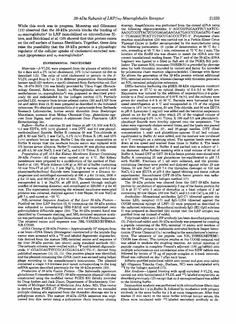
In preliminary experiments we found that rat kidney mem- branes are a rich source of 39-kDa protein, presumably be- cause of its association with the Heymann nephritis antigen gp330 (12). We also found that the 39-kDa protein could be extracted from kidney membranes by washing with a pH 10.5 buffer. Fig. 1 shows an experiment in which we tested the ability of the 39-kDa protein in the pH 10.5 eluate to bind to rat LRP. Three different potential targets were subjected to SDS gel electrophoresis: partially purified LDL receptor (lanes 1, 4, and 7), partially purified LRP (lanes 2, 5, and 8), and the pH 10.5 supernatant from kidney membranes (lanes 3, 6, and 9). The proteins were transferred to nitrocellulose and incubated with various ligands: a panel of monoclonal antibodies against the LDL receptor and the 515- and 85-kDa subunits of LRP (lanes 1 3 ) , the pH 10.5 eluate followed by a rabbit anti-39-kDa antiserum (lanes 4-6), and the anti-39- kDa antiserum alone (lanes 7-9). Bound antibodies were
" X b n a l kDa Anli-39 kDa Anttodies pH 10.5 Fraction
1 I 2 1 3 141 51 6 171 8 I 9
LRP-515 + 0
LDLR+ LRP-85+ - 39kDa+ .
FIG. 1. Binding of rat kidney 39-kDa protein to LRP im- mobilized on nitrocellulose. Approximately 200 pg of partially purified bovine adrenal LDL receptor (lanes I , 4, and 7), rat liver LRP (lanes 2, 5, and 8), and rat kidney pH 10.5 membrane extract (lanes 3, 6, and 9 ) were subjected to electrophoresis on nonreducing 3-8% SDS-PAGE and blotted to nitrocellulose as described under "Experimental Procedures." Lanes 1-3 were incubated with a mixture of three monoclonal antibodies (5 pg/ml each) directed against the bovine LDL receptor (IgG-C7), rat LRP-515 (IgG-ZEl), and LRP-85 (IgG-11H4). Bound IgG was detected with lzsI-labeled rabbit anti- mouse IgG (lo6 cpm/ml). Lanes 4-6 were first incubated for 30 min in buffer B containing 400 pg/ml of rat kidney, pH 10.5 membrane extract, washed in buffer B, and then incubated for 30 min with a 1:250 dilution of rabbit anti-39-kDa antiserum. The filter was sub- sequently washed, and bound IgG was detected with 12sI-labeled goat anti-rabbit IgG (lo6 cpm/ml). Lanes 7-9 were treated in the same way as lanes 4-6 except that no membrane extract was added. All three blots were exposed to Kodak XAR film for 15 h a t -70 "C. The gel was calibrated with myosin (200 kDa), phosphorylase B (97 kDa), and bovine serum albumin (68 kDa).
visualized with lZ5I-labeled second antibodies followed by au- toradiography.
As shown in Fig. 1, the panel of monoclonal antibodies stained the LDL receptor (lane I ) and the two subunits of LRP (lane 2 ) but not the pH 10.5 eluate (lane 3). The 39- kDa protein in the pH 10.5 eluate bound to the 515-kDa subunit of LRP (lane 5 ) . It did not bind to the 85-kDa subunit of LRP (lane 5 ) or the LDL receptor (lane 4 ) . The anti-39- kDa antiserum bound to a 39-kDa band that was present in the LRP preparation (lane 8 ) and in the pH 10.5 eluate (lane 9) but not in the LDL receptor preparation (lane 7).
To perform further studies of the 39-kDa protein, we used recombinant DNA techniques to prepare a fusion protein containing bacterial glutathione S-transferase at the NHz terminus and the 39-kDa protein at the COOH terminus. The two proteins are separated by a polyglycine stretch and a thrombin cleavage site. Fig. 2 shows a Coomassie-stained gel of the 70-kDa bacterial fusion protein after purification on a glutathione affinity column (lane 1 ). Thrombin cleavage gen- erated the 39-kDa protein plus the glutathione S-transferase (lane 2).
To screen for agents that would release the 39-kDa protein from LRP, we prepared '2sI-labeled GST-39-kDa protein and incubated it with LRP that had been subjected to SDS gel electrophoresis and transferred to nitrocellulose (Fig. 3). Binding was stable at pH 4.5 as well as pH 8 (lanes A and C). At either pH most of the bound GST-39-kDa protein was released with 20 mM EDTA (lanes B and D). It was also largely released by treatment with DTT (lane E ) and with polyanionic suramin (lane F) . Binding was totally abolished if the LRP was preincubated with DTT prior to and main- tained in DTT during the binding step (lane G ) . Thus, the
18.
68 - - <GST-39&
1 2
45 - - < 3 9 m
31 - " <GST
FIG. 2. SDS-PAGE of recombinant 39-kDa protein. Bacte- rially expressed recombinant GST-39-kDa fusion protein (3 pg, lane I ) and recombinant GST-39-kDa fusion protein after thrombin cleav- age (3 pg, lane 2 ) were separated on SDS-PAGE (Bio-Rad Minipro- tean System) under nonreducing conditions. After fixation, proteins were stained with Coomassie Blue R250. The positions of migration of molecular weight markers are indicated.
FIG. 3. Release of bound 12"I-labeled GST-39-kDa fusion protein from LRP-515. '*'I-Labeled GST-39-kDa protein (see Fig. 2, lane I ; lo6 cpm/ml; 815 cpm/ng) in buffer C was incubated for 30 min with replicate nitrocellulose strips containing partially purified LRP and LDL receptor (see Fig. 4). The strips were then washed three times with Buffer C at pH 8 or Buffer D at pH 4.5 without albumin and containing the indicated addition (20 mM EDTA, 10 mM dithiothreitol, or 10 mM suramin). Lane G was preincubated for 30 min with 10 mM DTT in Buffer C without serum albumin and then incubated with "'I-GST-39-kDa fusion protein in the presence of DTT and washed with DTT-containing buffer. The filters were then exposed together to Kodak XAR film for 1 h at -70 "C.

39-kDa Subunit of LRP/a2-Macroglobulin Receptor 21235
binding seems to require the intact disulfides in LRP and to be sensitive to polyanions. The failure of release at pH 4.5 may reflect a true property of LRP, or it may reflect a property conferred by the ligand blot assay, which is performed after electrophoresis under conditions of partial denaturation.
To determine whether the 39-kDa protein could bind to the 600-kDa precursor form of LRP, we performed a pulse-chase experiment (Fig. 4). NRK cells were pulse-labeled with a mixture of [35S]cysteine and ["S]methionine (Translabel) for 1 h, after which they were washed and allowed to incubate without labeled amino acids (chase period). At various times extracts were prepared and divided into aliquots. One aliquot was incubated with an antibody to LRP, and the complex was isolated on protein A-Sepharose. The other aliquot was in- cubated with the GST-39-kDa fusion protein, and complexes were isolated on glutathione-agarose beads. At zero time of chase, the anti-LRP antibody formed a complex with the 600- kDa precursor form of LRP and the biosynthesized 39-kDa protein ( l a n e 1). The GST-39-kDa protein also formed a complex with the 600-kDa precursor that could be isolated on glutathione-agarose ( l a n e 2). After longer times of chase, the 600-kDa precursor was progressively converted to the 515- and 85-kDa subunits, which were isolated by treatment with the antibody. These subunits were also isolated by treatment with the GST-39-kDa fusion protein bound to glutathione- agarose beads, albeit less efficiently. These data indicate that the GST-39-kDa fusion protein can bind to the 600-kDa precursor form of LRP as well as to the 515-kDa subunit. Endogenous biosynthetically labeled GST also bound to the glutathione-agarose beads and was co-precipitated (lanes 2,4, 6, 8, and 10).
The GST-39-kDa fusion protein blocked the binding of E/ P-VLDL to the 515-kDa subunit of LRP as visualized on ligand blots (Fig. 5). For this experiment, a partially purified preparation of rat liver LRP, which also contains the LDL receptor, was subjected to SDS gel electrophoresis and trans- ferred to nitrocellulose, which was then incubated with bio- tinylated 8-VLDL. The bound lipoprotein was visualized with '2sI-labeled streptavidin. Addition of apoE to the P-VLDL allowed it to bind to LRP and also increased the binding to the LDL receptor ( l a n e B ) . When the strips were preincubated with varying concentrations of GST-39-kDa fusion protein,
LRP-600, LRP-515'
LRP-85 - 39 kDa -
GST - CHASE 1 0 Wrnn 1 hr I 2hr I 4hr I
FIG. 4. Association of 39-kDa protein with biosynthetically labeled LRP-600 and LRP-515. NRK cells were pulse-labeled for 1 h at 37 "C with 50 pCi/ml of Translabel (ICN) as described (6). After the indicated chase period, the cells were lysed, and the deter- gent extracts were incubated for 1 h a t 4 "C with 100 pg of polyclonal anti-LRP IgG or 100 pg of GST-39-kDa fusion protein as indicated. After incubation with protein A-Sepharose (1) or glutathione-agarose (22), the beads were washed, treated with neuraminidase (6) to facilitate the separation of LRP-600 and LRP-515, and boiled in SDS sample buffer, after which the eluted proteins were separated on 3- 8% SDS-PAGE under nonreducing conditions. The gel was exposed t o Kodak XAR film for 15 h a t -70 "C. A longer exposure for 60 h showed clearly the presence of the 85-kDa subunit in lanes 4, 6, 8, and IO. The gel was calibrated with the molecular weight markers described in the legend to Fig. 1 plus ovalbumin (45 kDa), carbonic anhydrase (31 kDa), and soybean trypsin inhibitor (21 kDa).
FIG. 5. Inhibition of E/B-VLDL binding to LRP by GST-39- kDa fusion protein as detected by ligand blot analysis. Partially purified rat liver LRP was subjected to nonreducing SDS-PAGE and transferred to nitrocellulose as described in Ref. 15 and under "Ex- perimental Procedures." Replicate strips (each containing - 35 pg of protein) were incubated with the indicated amount of GST or GST- 39-kDa fusion protein for 30 min at room temperature, after which the strips were washed and incubated with 10 pg/ml of biotinylated 8-VLDL and the indicated amount of r-apoE-3. Bound 8-VLDL was detected with lZsI-labeled streptavidin (lo6 cpm/ml), after which the strips were exposed to Kodak XAR film for 4 h at -70 "C with an intensifying screen. The gel was calibrated with molecular weight markers as described in the legend to Fig. 1.
(u
0)
- - 0 " L I
'L - GST or GST-39 kDa Fusion Protein (pg/ml)
FIG. 6. Inhibition by 39-kDa protein of E/@-VLDL-stimu- lated cholesteryl ['4C]oleate formatkn in FH fibroblasts lack- ing LDL receptors. On day 7, each monolayer received 1.5 ml of medium A containing the indicated concentration of GST (0) or GST-39-kDa fusion protein (0). After incubation for 1 h at 37 "C, each dish received 0.5 ml of medium A containing j3-VLDL at a final concentration of 40 pg protein/ml and r-apoE at 50 pg/ml. After 4 h a t 37 "C, the cells were pulse-labeled for 2 h a t 37 "C with 0.2 mM ["Cloleate, and their content of cholesteryl ["Cloleate was measured. Each value is the average of duplicate or triplicate incubations. A blank value of 0.13 nmol h" mg protein" corresponding to the amount of cholesteryl [14C]oleate formed in parallel dishes receiving 8-VLDL without apoE was subtracted from each value.
the binding of 0-VLDL to LRP was abolished, with a 50% reduction at a concentration of about 0.3 pg/ml (lanes C-H) . The GST-39-kDa fusion protein produced only a slight de- crease in binding of 8-VLDL to the LDL receptor even at concentrations as high as 30 pg/ml ( l a n e H). Glutathione S- transferase itself had no effect (lanes I and J).
To determine whether the inhibition of /3-VLDL binding to LRP has functional consequences, we employed the choles- terol esterification assay, which relies on the observation that human fibroblasts do not incorporate [14C]oleate into choles- teryl esters unless they have an exogenous source of choles- terol (26). Lipoproteins such as LDL or P-VLDL can stimulate cholesteryl esterification in intact cells only if they bind to receptors and are taken up by endocytosis, delivering choles- terol to the cell. As shown in Fig. 6, E/B-VLDL stimulated cholesteryl ester synthesis in fibroblasts that are homozygous for a null allele at the LDL receptor locus. Previous studies have shown that this stimulation depends in large part on the binding of the E/@-VLDL to LRP (3,15). When the GST-39- kDa fusion protein was present, the stimulation of cholesteryl ester synthesis was greatly, but not completely, reduced. In a total of four experiments, we found that the maximal inhibi-

21236 39-kDa Subunit of LRP/a2-Macroglobulin Receptor
tion by the GST-39-kDa protein varied between 70 and 88% (average 80%). GST itself had no effect (Fig. 6). To confirm the specificity of the inhibition, we measured the incorpora- tion of [14C]oleate into triglycerides, a cellular reaction that is not influenced by 8-VLDL (Fig. 7). The GST-39-kDa protein had no effect on this reaction even though it inhibited ["C]oleate incorporation into cholesteryl esters by 85% in the same experiment.
The GST-39-kDa fusion protein did not inhibit cholesterol esterification that was stimulated by 25-hydroxycholesterol (Table I), ruling out an effect of this protein on the cholesteryl esterification reaction itself. Moreover, the GST-39-kDa pro- tein did not inhibit the LDL-mediated stimulation of choles- terol esterification in normal fibroblasts (Table 11), an event that is mediated by the LDL receptor (26). In data to be reported elsewhere, we found that the GST-39-kDa protein did not reduce the amount of LRP on the cell surface as judged by the cell's undiminished ability to bind a monoclonal antibody directed against the 515-kDa subunit. These exper- iments were performed with rabbit fibroblasts to which the
$ .- 125-1
I -
GST-39 kDa Fusion Protein (pg/rnl)
FIG. 7. Lack of inhibition by 39-kDa protein of incorpora- tion of [14C]oleate into ["Cltriglycerides in FH fibroblasts lacking LDL receptors. On day 7, each monolayer received 1.5 ml of medium A containing the indicated concentration of GST-39-kDa fusion protein. After incubation for 1 h at 37 "C, each dish received 0.5 ml of medium A containing P-VLDL at a final concentration of 30 pg protein/ml and r-apoE at 40 pg/ml. After 4 h at 37 "C, the cells were pulse-labeled for 2 h at 37 "C with 0.2 mM [14C]oleate, and their content of cholesteryl [14C]oleate (0) and [14C]triglycerides (0) were measured. Each value is the average of duplicate or triplicate incu- bations. The "100% of control values" for cholesteryl ["CC]oleate and ["C]triglycerides were 4.3 and 25.6 nmol h" mg protein", respec- tively. A blank value of 0.36 nmol h" mg protein" corresponding to the amount of cholesteryl [14C]oleate formed in parallel dishes receiv- ing P-VLDL without apoE was subtracted from each of the cholesteryl ["Cloleate values. The value for ['4C]triglycerides formed by cells receiving 0-VLDL without apoE was 22.0 nmol h-' mg protein".
TABLE I Sterol-mediated stimulation of cholestetyl [14C]oleate formation in
FH fibroblasts: lack of inhibition by GST-39-kDa fusionprotein On day 7, each monolayer received 1.5 ml of medium A containing
10 pg/ml of GST-39-kDa fusion protein as indicated. After incubation for 1 h at 37 "C, each dish received 0.5 ml of medium A containing the indicated addition: P-VLDL at a final concentration of 30 pg protein/ml with or without 40 pg/ml r-apo E or 20 pglml25-hydrox- ycholesterol plus 10 pg/ml cholesterol. After 4 h at 37 "C, the cells were pulse-labeled for 2 h at 37 "C with 0.2 mM [14C]oleate and their content of cholesteryl [14C]oleate measured. Each value is the average of dudicate or tridicate incubations. ND. not determined.
["CIOleate incorporated into cholesteryl ["C]oleate
No GST-39-kDa +GST-39-kDa protein protein
nmol h" mg protein"
Addition to Medium
0-VLDL 0.36 ND P-VLDL + ApoE 4.3 0.83 Sterols 6.4 6.6
TABLE I1 LDL receptor-dependent stimulation of cholesteryl [14C]oleate
formation in normal fibroblasts: lock of inhibition by GST-39-kDa fusion protein
On day 0, 2.5 X lo4 cells from a stock culture of normal human fibroblasts were seeded into 60-mm petri dishes and grown in 10% (v/v) fetal calf serum as described (29). On day 5, each monolayer was switched to 2 ml of medium containing 10% (v/v) human lipo- protein-deficient serum (29). On day 7, each monolayer received 2 ml of medium A containing the indicated concentration of GST-39-kDa fusion protein. After incubation for 1 h at 37 "C, P-VLDL at a final concentration of 30 pg/ml was added to the indicated dish. After 4 h at 37 "C, the cells were pulse-labeled for 2 h at 37 "C with 0.2 mM ['4C]oIeate, and their content of cholesteryl ["C]oleate was measured. Each value represents a single incubation.
Addition to medium G s T - ~ ~ - ~ D ~ ["CIOleate - cholesteryl ["C]oleate
8-VLDL protein
gg protein/ml pg/ml nmol h" mg protein" 0 0 0.24 0 30 0.31
30 0 16.7 30 1 18.2 30 3 15.5 30 10 16.6 30 30 16.6
16 (mg I ml) ?
FIG. 8. Inhibition by anti-LRP polyclonal antibody of the uptake and degradation of '261-a2-macroglobulin* in FH fibro- blasts lacking LDL receptors. On day 7, each monolayer received 2 ml of Dulbecco's modified Eagle's medium (without glutamine) containing 2 mg/ml bovine serum albumin and the indicated concen- tration of either nonimmune IgG (0) or polyclonal anti-LRP IgG (0) as indicated. After 2 h at 37 "C, each dish received 25 pg/ml of lZ5I- a*-macroglobulin* (310 cpm/ng protein). After incubation for 5 h at 37 "C, cells were chilled to 4 "C, and the total cellular content of lZ5I- a2-macroglobulin* (A) and the total amount of 1251-a2-macroglobulin* degradation products excreted into the medium ( B ) were determined. Each value is the average of duplicate incubations.
monoclonal antibody is directed (24). Moreover, pulse-chase experiments in NRK cells excluded the possibility that exog- enously added GST-39-kDa fusion protein leads to acceler- ated degradation of LRP (data not shown).
Before studying the effect of the GST-39-kDa protein on the cellular uptake and degradation of '251-~2-macroglobu1in* (methylamine-activated) in human fibroblasts, we performed an experiment to confirm that this process was being mediated by LRP. For this purpose we preincubated the FH fibroblasts with varying concentrations of a polyclonal anti-LRP anti- body that causes the degradation of LRP in intact cells (3,4). As shown in Fig. 8, the antibody caused a parallel decline in the uptake and rate of degradation of '251-a2-macroglobulin*. Fig. 9 shows that the GST-39-kDa fusion protein inhibited the uptake (Fig. 9A) and degradation (Fig. 9B) of '251-a2- macroglobulin*, whereas GST itself had no effect.
DISCUSSION
The current data demonstrate that a fusion protein con- taining the LRP 39-kDa subunit binds to the 515-kDa subunit

39-kDa Subunit of LRP/a2-Macroglobulin Receptor 21237
6. Degradation
0 5 10 3 0 0 5 10 30 GST or GST-39 kDa Fusion Protein (w / ml)
FIG. 9. Inhibition by 39-kDa protein of the uptake and deg- radation of '251-cr~-macroglobulin+ in FH fibroblasts lacking LDL receptors. On day 7, each monolayer received 2 ml of Dulbec- co's modified Eagle's medium (without glutamine) containing 2 mg/ ml bovine serum albumin and the indicated concentration of either GST (0) or GST-39-kDa fusion protein (0) as indicated. After 15 min at 37 "C, each dish received 15 pg/ml '251-cy2-macroglobulin* (461 cpm/ng). After incubation for 5 h at 37 "C, cells were chilled to 4 "C, and the total cellular content of 12SI-cy2-ma~roglob~1in* (A) and the total amount of '251-a2-macroglobulin* degradation products excreted into the medium ( B ) were determined. Each value represents a single incubation except for the control values (no added 39-kDa protein), which are the mean of triplicate incubations.
and blocks the binding of both E/P-VLDL and a,-macroglob- ulin* to LRP. In human fibroblasts the 39-kDa fusion protein reduces the ability of E/@-VLDL to stimulate cholesterol esterification, presumably as a result of impaired binding and uptake by LRP. In addition, the fusion protein markedly inhibits the high affinity uptake and degradation of az-mac- roglobulin*.
The observation that the 39-kDa protein blocks binding of E/@-VLDL as well as a,-macroglobulin* raises the possibility that all three ligands bind to the same site on the 515-kDa subunit of LRP. Other data contradict this simple hypothesis. The 515-kDa subunit contains three types of sequences, all of which are shared with the LDL receptor: ligand-binding cys- teine-rich repeats, growth factor-type cysteine-rich repeats, and cysteine-poor YWTD spacers (2). It is likely that E/P- VLDL binds to the ligand-binding repeats, which are known to be the binding sites for apoE in the LDL receptor (27).
Hussain et al. (28) have demonstrated partial cross-com- petition between c~~-macroglobulin* and E/@-VLDL for bind- ing to LRP, but only at high concentrations, suggesting that the competition results from steric hindrance at neighboring but not identical binding sites. It is possible that the 39-kDa protein binds to both classes of binding sites. Alternatively, the 39-kDa protein may bind to only one site, causing a conformational change that obliterates both sites. It is of interest that neither the 39-kDa protein nor az-macroglobu- lin* binds to the LDL receptor even though the LDL receptor contains all of the types of sequences that are present in LRP- 515.
If the 39-kDa protein blocks binding of az-macroglobulin* to LRP, how is it possible that the 39-kDa protein was isolated on an az-macroglobulin* affinity column? And if the 39-kDa protein is associated with LRP on the cell surface, how can the surface receptors bind az-macroglobulin* and E/@-VLDL? These paradoxes would be explained if LRP contains multiple binding sites for the 39-kDa protein and if occupancy of a high proportion of these sites is necessary to prevent the binding of the other ligands. If LRP in its native form were undersaturated with the 39-kDa protein, it could still bind the two ligands, and the complex could still be isolated on an c~2-macroglobulin* affinity column. Purified LRP was esti- mated to contain a 1:l molar ratio of 39-kDa protein to 515-
kDa subunit as revealed by inspection of electrophoretograms (11). It will be important in the future to perform careful in vitro binding studies to determine whether a ratio higher than 1:l is necessary to inhibit ligand binding.
The concentration of GST-39-kDa fusion protein that gave 50% maximal inhibition of E/@-VLDL-stimulated cholesterol esterification and of a,-macroglobulin* uptake and degrada- tion was about 1 pg/ml (Figs. 6, 7, and 9), which is equivalent to a 39-kDa protein concentration of 15 nM. Although this value varied severalfold with different preparations of fusion protein, each individual preparation of 39-kDa fusion protein inhibited both processes at similar concentrations. Strickland et al. (4) reported previously that biologically purified 39-kDa protein bound to LRP with a half-maximal concentration of 18 nM, which is in the same range as the concentrations of recombinant fusion protein that inhibited binding and uptake of the two ligands.
The 39-kDa protein inhibited the E/@-VLDL-mediated cholesteryl ester synthesis by a maximum of about 80%, whereas it inhibited the degradation of '251-a2-ma~r~glob~1in* by more than 95%. These data raise the possibility that FH homozygote fibroblasts lacking LDL receptors have a binding site in addition to LRP that leads to cholesterol delivery to cells. If such a site exists, it is only about one-fifth as active as LRP under the conditions of these assays, and its specificity is unknown.
We do not yet know whether the 39-kDa protein associates with LRP inside the cell and whether it assists in the assem- bly, processing, or transport of LRP. Detailed immunochem- ical studies will be required to answer these questions.
Acknowledgments-Wen-Ling Niu, Judy Webber, and Debra Noble provided excellent technical assistance. We thank Carolyn Moomaw in Clive Slaughter's laboratory for NHz-terminal sequence analysis, Richard Gibson for invaluable assistance with animals, Izabela Lez- nicki in Thomas Sudhof's laboratory for help with cDNA library screening, and Robert Kowal for helpful comments.
REFERENCES
1. Herz, J., Hamann, U., Rogne, S., Myklebost, O., Gausepohl, H., and Stanley, K. K. (1988) EMBO J. 7,4119-4127
2. Brown, M. S., Herz, J., Kowal, R. C., and Goldstein, J. L. (1991) Curr. Opin. Lipidol. 2, 65-72
3. Kowal, R. C., Herz, J., Goldstein, J. L., Esser, V., and Brown, M. S. (1989) Proc. Natl. Acad. Sci. U. S. A. 86,5810-5814
4. Strickland, D. K., Ashcom, J. D., Williams, S., Burgess, W. H., Midiorini. M.. and Arnaves. W. S. 11990) J. Biol. Chem. 265. 17401-17404 '
5. Kristensen, T., Moestrup, S. K., Gliemann, J., Bendtsen, L., Sand, O., and Sottrup-Jensen, L. (1990) FEBS Lett. 276,151- 155
- . .
6. Herzi J., Kowal, R. C., Goldstein, J. L., and Brown, M. S. (1990) EMBO J. 9,1769-1776
7. Chen, W.-J., Goldstein, J. L., and Brown, M. S. (1990) J. Biol. Chem. 265,3116-3123
8. Ashcom, J. D., Tiller, S. E., Dickerson, K., Cravens, J. L., Ar- graves, W. S., and Strickland, D. K. (1990) J. Cell Biol. 110,
9. Moestrup, S. K., and Gliemann, J. (1989) J. Biol. Chem. 264,
10. Strickland, D. K., Ashcom, J. D., Williams, S., Battey, F., Behre, E., McTigue, K., Battey, J. F., and Argraves, W. S. (1991) J. Biol. Chem. 266,13364-13369
1041-1048
15574-15577
11. Moestrup, S. K., and Gliemann, J. (1991) J. Biol. Ckm. 266, 14011-14017
12. Pietromonaco, S., Kerjaschki, D., Binder, S., Ullrich, R., and Farquhar, M. G. (1990) Proc. Natl. Acad. Sci. U. S. A. 87, 1811-1815
13. Furukawa, T., Ozawa, M., Huang, R-P., and Muramatsu, T. (1990) J. Bwchem. (Tokyo) 108,297-302
14. Raychowdhury, R., Niles, J. L., McCluskey, R. T., and Smith, J. A. (1989) Science 244, 1163-1166

21238 39-kDa Subunit of LRPlcYz-Macroglobulin Receptor
15.
16.
17.
18.
19.
20.
Kowal, R. C., Herz, J., Weisgraber, K. H., Mahley, R. W., Brown, M. S., and Goldstein, J. L. (1990) J. Biol. Chem. 2 6 5 , 10771- 10779
Vogel, T., Weisgraber, K. H., Zeevi, M. I., Ben-Artzi, H., Levanon, A. Z., Rall, S. C., Jr., Innerarity, T. L., Hui, D. Y., Taylor, J. M., Kanner, D., Yavin, Z., Amit, B., Aviv, H., Gorecki, M., and Mahley, R. W. (1985) Proc. Natl. Acad. Sci. U. S. A. 82,8696- 8700
Beisiegel, U., Schneider, W. J., Goldstein, J. L., Anderson, R. G. W., and Brown, M. S. (1981) J. Biol. Chem. 2 5 6 , 11923-11931
Schneider, W. J., Goldstein, J. L., and Brown, M. S. (1985) Methods Enzymol. 109,405-417
Hubbard, A. L., Wall, D. A., and Ma, A. (1983) J. Cell Biol. 9 6 ,
Aebersold, R. H., Leavitt, J., Saavedra, R. A., Hood, L. E., and Kent, S. B. H. (1987) Proc. Natl. Acad. Sci. U. S. A. 84,6970-
217-229
22. 23.
24.
25.
26.
27.
28.
Cloning: A Laboratory Manual, Cold Spring Harbor Laboratory Press, Cold Spring Harbor, NY
Smith, D. B., and Johnson, K. S. (1988) Gene (Amst.)67,31-40 Saiki, R. K., Gelfand, D. H., Stoffel, S., Scharf, S. J., Higuchi,
R., Horn, G. T., Mullis, K. B., and Erlich, H. A. (1988) Science
Herz, J., Kowal, R. C., Ho, Y. K., Brown, M. S., and Goldstein,
Hobbs, H. H., Brown, M. S., Russell, D. W., Davignon, J., and
Goldstein, J. L., Dana, S. E., and Brown, M. S. (1974) Proc. Natl.
Russell. D. W.. Brown. M. S.. and Goldstein. J. L. (1989) J. Biol.
239,487-491
J. L. (1990) J. Biol. Chem. 2 6 6 , 21355-21362
Goldstein, J. L. (1987) N. Engl. J. Med. 3 1 7 , 734-737
Acad. Sci. U. S. A. 71, 4288-4292
Chem. 264,'21682-21688 '
. I
Hussain. M. M.. Maxfield. F. R.. Mas-Oliva. J.. Tabas. I.. Ji. Z- S., Innerarity,' T. L., and Mahley, R. W. (1991) J. Biol. 'Chkm. 266.13936-13940
6974 29. Goldstein, J. L., Basu, S. K., and Brown, M. S. (1983) Methods 21. Sambrook, J., Fritsch, E. F., and Maniatis, T. (1989) Molecular Enzymol. 98,241-260