Embed Size (px)
Citation preview
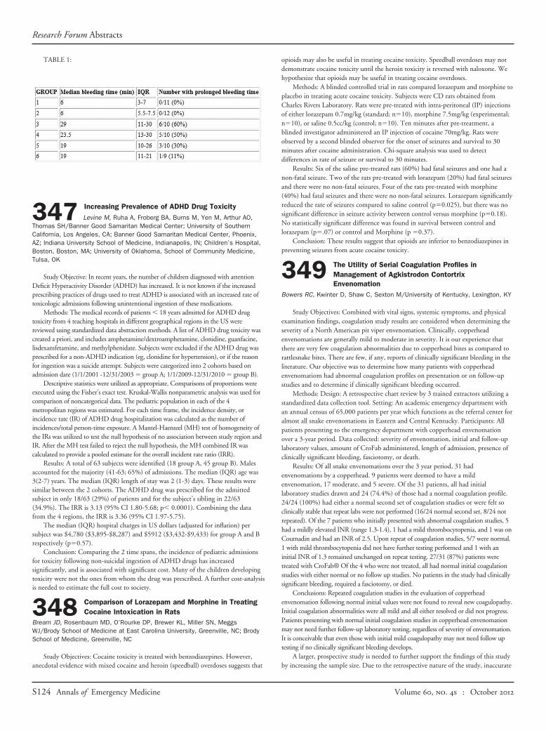
TABLE 1:
347 Increasing Prevalence of ADHD Drug ToxicityLevine M, Ruha A, Froberg BA, Burns M, Yen M, Arthur AO,
Thomas SH/Banner Good Samaritan Medical Center; University of SouthernCalifornia, Los Angeles, CA; Banner Good Samaritan Medical Center, Phoenix,AZ; Indiana University School of Medicine, Indianapolis, IN; Children’s Hospital,Boston, Boston, MA; University of Oklahoma, School of Community Medicine,Tulsa, OK
Study Objective: In recent years, the number of children diagnosed with attentionDeficit Hyperactivity Disorder (ADHD) has increased. It is not known if the increasedprescribing practices of drugs used to treat ADHD is associated with an increased rate oftoxicologic admissions following unintentional ingestion of these medications.
Methods: The medical records of patients � 18 years admitted for ADHD drugtoxicity from 4 teaching hospitals in different geographical regions in the US werereviewed using standardized data abstraction methods. A list of ADHD drug toxicity wascreated a priori, and includes amphetamine/dextroamphetamine, clonidine, guanfacine,lisdexamfetamine, and methylphenidate. Subjects were excluded if the ADHD drug wasprescribed for a non-ADHD indication (eg, clonidine for hypertension), or if the reasonfor ingestion was a suicide attempt. Subjects were categorized into 2 cohorts based onadmission date (1/1/2001 -12/31/2003 � group A; 1/1/2009-12/31/2010 � group B).
Descriptive statistics were utilized as appropriate. Comparisons of proportions wereexecuted using the Fisher’s exact test. Kruskal-Wallis nonparametric analysis was used forcomparison of noncategorical data. The pediatric population in each of the 4metropolitan regions was estimated. For each time frame, the incidence density, orincidence rate (IR) of ADHD drug hospitalization was calculated as the number ofincidences/total person-time exposure. A Mantel-Haenszel (MH) test of homogeneity ofthe IRs was utilized to test the null hypothesis of no association between study region andIR. After the MH test failed to reject the null hypothesis, the MH combined IR wascalculated to provide a pooled estimate for the overall incident rate ratio (IRR).
Results: A total of 63 subjects were identified (18 group A, 45 group B). Malesaccounted for the majority (41-63; 65%) of admissions. The median (IQR) age was3(2-7) years. The median (IQR) length of stay was 2 (1-3) days. These results weresimilar between the 2 cohorts. The ADHD drug was prescribed for the admittedsubject in only 18/63 (29%) of patients and for the subject’s sibling in 22/63(34.9%). The IRR is 3.13 (95% CI 1.80-5.68; p� 0.0001). Combining the datafrom the 4 regions, the IRR is 3.36 (95% CI 1.97-5.75).
The median (IQR) hospital charges in US dollars (adjusted for inflation) persubject was $4,780 ($3,895-$8,287) and $5912 ($3,432-$9,433) for group A and Brespectively (p�0.57).
Conclusion: Comparing the 2 time spans, the incidence of pediatric admissionsfor toxicity following non-suicidal ingestion of ADHD drugs has increasedsignificantly, and is associated with significant cost. Many of the children developingtoxicity were not the ones from whom the drug was prescribed. A further cost-analysisis needed to estimate the full cost to society.
348 Comparison of Lorazepam and Morphine in TreatingCocaine Intoxication in Rats
Bream JD, Rosenbaum MD, O’Rourke DP, Brewer KL, Miller SN, MeggsWJ/Brody School of Medicine at East Carolina University, Greenville, NC; BrodySchool of Medicine, Greenville, NC
Study Objectives: Cocaine toxicity is treated with benzodiazepines. However,anecdotal evidence with mixed cocaine and heroin (speedball) overdoses suggests that
opioids may also be useful in treating cocaine toxicity. Speedball overdoses may notdemonstrate cocaine toxicity until the heroin toxicity is reversed with naloxone. Wehypothesize that opioids may be useful in treating cocaine overdoses.
Methods: A blinded controlled trial in rats compared lorazepam and morphine toplacebo in treating acute cocaine toxicity. Subjects were CD rats obtained fromCharles Rivers Laboratory. Rats were pre-treated with intra-peritoneal (IP) injectionsof either lorazepam 0.7mg/kg (standard; n�10), morphine 7.5mg/kg (experimental;n�10), or saline 0.5cc/kg (control; n�10). Ten minutes after pre-treatment, ablinded investigator administered an IP injection of cocaine 70mg/kg. Rats wereobserved by a second blinded observer for the onset of seizures and survival to 30minutes after cocaine administration. Chi-square analysis was used to detectdifferences in rate of seizure or survival to 30 minutes.
Results: Six of the saline pre-treated rats (60%) had fatal seizures and one had anon-fatal seizure. Two of the rats pre-treated with lorazepam (20%) had fatal seizuresand there were no non-fatal seizures. Four of the rats pre-treated with morphine(40%) had fatal seizures and there were no non-fatal seizures. Lorazepam significantlyreduced the rate of seizures compared to saline control (p�0.025), but there was nosignificant difference in seizure activity between control versus morphine (p�0.18).No statistically significant difference was found in survival between control andlorazepam (p�.07) or control and Morphine (p �0.37).
Conclusion: These results suggest that opioids are inferior to benzodiazepines inpreventing seizures from acute cocaine toxicity.
349 The Utility of Serial Coagulation Profiles inManagement of Agkistrodon ContortrixEnvenomation
Bowers RC, Kwinter D, Shaw C, Sexton M/University of Kentucky, Lexington, KY
Study Objectives: Combined with vital signs, systemic symptoms, and physicalexamination findings, coagulation study results are considered when determining theseverity of a North American pit viper envenomation. Clinically, copperheadenvenomations are generally mild to moderate in severity. It is our experience thatthere are very few coagulation abnormalities due to copperhead bites as compared torattlesnake bites. There are few, if any, reports of clinically significant bleeding in theliterature. Our objective was to determine how many patients with copperheadenvenomatiom had abnormal coagulation profiles on presentation or on follow-upstudies and to determine if clinically significant bleeding occurred.
Methods: Design: A retrospective chart review by 3 trained extractors utilizing astandardized data collection tool. Setting: An academic emergency department withan annual census of 65,000 patients per year which functions as the referral center foralmost all snake envenomations in Eastern and Central Kentucky. Participants: Allpatients presenting to the emergency department with copperhead envenomationover a 3-year period. Data collected: severity of envenomation, initial and follow-uplaboratory values, amount of CroFab administered, length of admission, presence ofclinically significant bleeding, fasciotomy, or death.
Results: Of all snake envenomations over the 3 year period, 31 hadenvenomations by a copperhead. 9 patients were deemed to have a mildenvenomation, 17 moderate, and 5 severe. Of the 31 patients, all had initiallaboratory studies drawn and 24 (74.4%) of those had a normal coagulation profile.24/24 (100%) had either a normal second set of coagulation studies or were felt soclinically stable that repeat labs were not performed (16/24 normal second set, 8/24 notrepeated). Of the 7 patients who initially presented with abnormal coagulation studies, 5had a mildly elevated INR (range 1.3-1.4), 1 had a mild thrombocytopenia, and 1 was onCoumadin and had an INR of 2.5. Upon repeat of coagulation studies, 5/7 were normal.1 with mild thrombocytopenia did not have further testing performed and 1 with aninitial INR of 1.3 remained unchanged on repeat testing. 27/31 (87%) patients weretreated with CroFab® Of the 4 who were not treated, all had normal initial coagulationstudies with either normal or no follow up studies. No patients in the study had clinicallysignificant bleeding, required a fasciotomy, or died.
Conclusions: Repeated coagulation studies in the evaluation of copperheadenvenomation following normal initial values were not found to reveal new coagulopathy.Initial coagulation abnormalities were all mild and all either resolved or did not progress.Patients presenting with normal initial coagulation studies in copperhead envenomationmay not need further follow-up laboratory testing, regardless of severity of envenomation.It is conceivable that even those with initial mild coagulopathy may not need follow uptesting if no clinically significant bleeding develops.
A larger, prospective study is needed to further support the findings of this studyby increasing the sample size. Due to the retrospective nature of the study, inaccurate
Research Forum Abstracts
S124 Annals of Emergency Medicine Volume , . : October

classification of envenomation severity is a possibility. Some patients did not havefollow up studies performed so their outcome was based on clinical findings alone.
350 Comparison Between Children and Adult SnakebiteVictims: Analysis of 64,210 Victims
Morris AC, Morgan DL, Blair HW/Scott and White Memorial Hospital, Temple,TX; Texas A&M Health Science Center, Temple, TX; Central Texas PoisonCenter, Temple, TX
Study Objective: Thousands of patients with snakebites present to emergencydepartments each year in the USA. About one-third of these snakebite victims are lessthan 18 years of age. Previous studies have demonstrated some differences betweenpediatric and adult snakebite victims. Our goal was to evaluate the demographics,associated circumstances, and medical sequelae of pediatric patients of snakebitescompared to adult patients using a very large number of victims.
Methods: This study was an observational, case-control study of telephone callsto all US poison centers (National Poison Data System) for all snakebites of humansfrom 2000 to 2009. Clinical outcome was classified as no effects, minor, moderate,major, or death.
Results: There were 19,790 (30.8%) victims under 18 years and 44,420 (69.2%)victims 18 years and older. The average ages of the pediatric and adult victims were9.6 years and 39.6 years, respectively. Of the pediatric victims, 22.3% were under theage of 6 years, and 2.8% were under the age of 2 years. The percentages of pediatricand adult males were similar: 70.2% and 70.4%. Unknown snake types were morecommon in the pediatric group (40.2%) than in the adult group (33.2%) (OddsRatio� 1.35 [95% CI: 1.31-1.40]). Children were much less likely to be bitten by avenomous snake (28.7%) than adults (51.3%) (OR�0.38 [0.37-0.40]). The pediatricvictims were about half as likely as adult victims to be bitten by copperheads (OR�0.54 [0.51-0.57]), rattlesnakes (OR�0.40 [0.38-0.42]), or coral snakes (OR�0.60[0.51-0.70]). All 50 states reported pediatric snakebites during the studied timeperiod. Florida and Texas had the most bites for both children and adults, 21.7% and25.8% respectively. Alaska (4 pediatric and 5 adult bites) and Hawaii (1 pediatric and3 adult bites) reported the fewest bites in both groups. The 6-month period fromApril to September accounted for the most snake bites in both age groups, 83.4% and82.6% respectively. The least common months for pediatric bites were December andJanuary (total 2.4%), and for adults bites were January and February (total 2.4%).Children were more likely to have a “dry bite” or no clinical effects (4.0%) thanadults (2.6%) (OR� 1.56 [1.42-1.71]). The most common clinical effects for boththe pediatric victims and the adult victims were graded as minor (38.9% and 33.6%)(OR�1.26 [1.22-1.30]). Children had fewer moderate effects than adults (21.2%and 28.7%) (OR�0.67[0.64-0.70]). Children were less likely than adults to havemajor effects (1.9% and 3.2%) (OR�0.59 [0.52-0.66]) or death (OR�0.29[0.09-0.97]). There were 3 pediatric deaths (1 indirectly reported) and 23 adult deathsduring the study period.
Conclusion: This is the largest comparison of pediatric victims of snakebites toadult victims in the US. Children were more likely than adults to be bitten by anunknown snake type or non-venomous snake. They were also more likely than adultsto have “dry bites” or only minor clinical effects. There were no significant differencesin sex, month, or geographical location. This information may be useful for planningthe prevention and treatment of snakebites of pediatric victims.
351 The Effect of an Alcohol Withdrawal TreatmentProtocol on Alcohol Withdrawal Syndrome PatientOutcomes
De La Calzada-Jeanlouie M, Gatt J, Gurr D, Pizzuti J, Loftus A, Castaneda J, Ji E,Rosenberg D, Su MK, Lee DC/North Shore University Hospital, Manhasset, NY
Background: On October 1, 2010, the Department of Internal Medicine at asuburban, academic hospital implemented its first defined clinical alcoholwithdrawal syndrome (AWS) treatment protocol. After recognizing the lackof standardization and inexperience of health care providers in aggressivelytreating AWS, a protocol was developed to assist with the treatment of AWS. Theprotocol takes into consideration variables associated with AWS such as whetherthe AWS patient was high or low risk and the dosage of benzodiazepinesadministered, and it incorporates a standardized CIWA-Ar protocol.
Study Objecties: This study aimed to investigate the impact of a new AWStreatment protocol on patient outcomes in terms of hospital length of stay (LOS)and the frequency of revisits to the hospital for AWS within 30 days. Wehypothesize that patient outcomes will have improved through decreased LOS
and a decreased frequency of hospital revisits for AWS after the implementationof the protocol.
Methods: A retrospective case-control study of AWS patients who wereadmitted to the department of internal medicine from the emergency departmentwas conducted. A convenience sample of patients admitted during a 3-monthperiod before implementation of the protocol (06/01/2010 - 08/31/2010) wascompared to a convenience sample of those during a 3-month period afterimplementation (12/01/2010 - 02/28/2011). We reviewed admitted patients�18 years who had an AWS-related primary diagnosis. Patients admitted inSeptember, October and November of 2010, during which the protocol wasbeing introduced, were excluded. Primary outcome measures included LOS andre-visits to the hospital for AWS within 30 days of discharge. Descriptivestatistics and Welch’s t-test were used to analyze LOS among AWS patientsbefore (control group) and after (case group) the implementation of the protocol.Chi-square tests were used to compare the frequency of re-visits to the hospitalamong the case and control groups.
Results: Thirty-six subjects were reviewed in the control group (66% male; 20-76years old; mean age � 45.3); 28 subjects were reviewed in the case group (64% male;22-80 years old; mean age � 48.4). There was no significant difference in LOSbetween the case and control groups (p � 0.93). Mean LOS among control patientswas 6 days (95% CI: 3.84 - 8.17); mean LOS among case patients was 6.12 days(95% CI: 4.64 - 11.12). There was no correlation between case/control group and thefrequency of revisits to the hospital for the treatment of AWS within 30 days(p � 0.79).
Conclusion: Implementing an AWS protocol did not show a statisticallysignificant difference in AWS patient outcomes as measured by LOS and re-visits tothe hospital for AWS within 30 days of discharge. This brings attention to the needfor aggressive management of acute alcohol withdrawal. Our results suggest that adefined protocol may not be superior to symptom-triggered therapy. Limitationsinclude small sample size; further research should examine hospitals with a highervolume of AWS patients over a longer period of time. Additional strategies need to beevaluated to optimize the treatment of acute AWS.
352 Effect of High-Carbohydrate Meals on the Rate ofEthanol Metabolism
Jones JS, Judge B, Boss J, Kurtz W, Hagert R, Postema J, Del Camp A, BrownA/Grand Rapids MEP/ Michigan State University, Grand Rapids, MI; MSUCollege of Human Medicine, Grand Rapids, MI; University of Michigan MedicalSchool, Ann Arbor, MI
Study Objectives: Drinking alcoholic beverages together with or after a meal iswidely known to diminish the intensity and duration of ethanol intoxicationcompared with drinking the same dose on an empty stomach. The most commonexplanation for this observation is a delayed absorption of ethanol from the gut,resulting in lower and later occurring peak blood-ethanol concentration. However,recent research suggests a specific food-induced effect on the activity of alcoholmetabolizing enzymes in intoxicated patients. The objective of this current study wasto determine the effect of eating a meal on the rate of ethanol elimination (clearance)from the blood over a 4-hour time period.
Methods: This was a prospective, clinical study using a convenience sample ofpatients admitted to the emergency department (ED) with acute alcohol intoxication.The study took place at a single urban U.S. academic medical center over a 12-monthstudy period. Exclusion criteria included patients presenting with a social or medicalproblem other than alcohol-related such as other substance abuse or an acute medicalcondition such as trauma which requires emergent intervention. Demographic datawas collected to determine patient characteristics, diagnostic testing, interventions,and time spent in the ED. To estimate the blood alcohol we used an approvedautomated handheld breath alcohol instrument that was calibrated daily. Whenpatients were able to eat, they were given a standard high-calorie meal. We obtainedhourly alcohol readings before and after eating for at least 4 hours. Ethanolelimination from the blood over time was determined from these measurements.Concentration-time profiles of ethanol were plotted for the data collected from EDsubject sessions.
Results: Twenty-six subjects completed the study; 25 (96%) were male. Themean age was 48 �/� 9.3 years. The mean length of stay in the ED was 9.0hours. Breath alcohol concentration on arrival was 326 mg/dL. Subjects weregiven a meal as soon as they were able to feed themselves. Between 50%-100% ofthe meal was consumed for an average of 443 calories (range 294-600).Comparison of mean alcohol elimination rates before eating (21 mg/dL/hr) and 2
Research Forum Abstracts
Volume , . : October Annals of Emergency Medicine S125