Embed Size (px)
Citation preview

THE BRAIN OF THE ZEUQLODONTIDR. 61 5
33. The Brain of the Zeuglodonlida (Cetacea). By RAY- MOND A. DART, MSc., M.B., CILM., Professor of Anatomy, University of the Witwatersrand, Johnnnes- b u r g ; with a Note on the Skulls from which the Endocranial Casts were tak9n. By C. W. ANDRRWS, D.Sc., F.R.S., Y.Z.S., British Museum of Natural History.
[ h e i r e d May 18,1993 : h a d June la, 1023.3
(Text-figures 1-24.)
T A B L ~ 09 C o w ~ m ~ s . Piige
1. Introduction ......................................................... 2. Material ............ 3. The two Endocraninl Ca nied by S
Remains ...........................................................
.....................
a. Z ~ U . & J ~ O ~ rensitivus, sp. nov. (M. 12133.) b. Zeuglodon rlliotsmithii, MY. nov.
a. Prozmiglodon atror Andiews. 6. Zewgcglodnn intermedius, sp. nov. e. ZetegZodon ssirir Dames. (M. 10228.)
6. Table of Measurements of the Endocranial Casts 6. The Zoological Position of the Zeuglodontidm ............... 7. Comparison of Zcugldont and Prosqualodont Biaias 8. Tlie Trigemiuus aiid the Law of Infiltration in Cerebellar
Evolution ......................................................... 8. Note on the Skulls from which the Endocranial Casts
were t u h , by C. W. ANDILEW~, 1).Sc, F.R.S. .........
(M. 12088.) 4. The three Kndocranial Carts fonniug a Phyletic Series ...
(M. 9266.) (M. 10173.)
.........
LO. Bibliography .........................................................
816 818
818
847
834 636 888
843
8B 864,
1. INTUODUCTION. The Cetacea nre connected with the marine Carnivores
through the genus Zmgloclon, ns Huxley has shown, and the point3 of resemblance are so marked that the affinity cannot be doubted” (0. C. Marsh, 1877).
It has been suggested, in the past, that these water-living forms, which preserve in their skeletal parts characteristics linking them with the ancestors of modern Carnivom, were the actual ancestors of the whales and the dolphins, i. e., the Cetacea. Tlie Zeaglodonts were therefore grouped together as the Archsocati. This suggeadion of n Carnivore (or Creodont) ancestry seems to have been universolly admitted tinti1 Matthew and Gregory (1 910) pointed out various characteristics which

616 PROF. R. A. DART ON TIIE
seem to link the Zeaglodontidac more closcly with Insectivore antecedents. In the nietintime, the intimacy of the relationsliip between Zeuglodontidle and Cetacea has been reaffirmed by numerous observelx, including amongst many others Abel (1905- 1913) and Winge (1919, 1921).
Previous investigation of the group has naturally centred around the osteological remainct, and has shown (Andrews, 1907) that they probably arose “ o n the northern shores of the Ethiopian land in the early part uf the Eocene period,” and th:it “ b y the end of the Middle Eocene the true Zeuglodonts had come into existence, and had sprwld rapidly over the earth, their remains being found in the upper part of the Eocene of Americii, England, and New Zealand.”
Since the appearance of Professor G. Elliot Smith’s important contribution to the study of the form of the entlocranial cast (1903) no new feature of the cerebral anatomy of thebe fornis has been recognised, so far as I have discovered, although Stroiner (1908) hns figured what is apparently a uniqne iiiiturd endo- cranial cast in a form which he styles ZeugEodon osiris.
Shortly after the publication of Professor Elliot Smith’s article, Mr. H. J. L. Bendnell gave him an almost perfect natural endo- cranial cast of a Zeuglodont collected in the Egyptian Fayuin a t the locality known as the Gar-el-Gehannem. Professor Elliot Smith kindly placed it at my disposal some time ago. I was nssistetl in the “ developnient ” of this natural cast by Professor D. M. S. Watson, who c.wefiilly removed most of the aclherent bone and matrix, giving the result which has been accurately reproduced by Miss Davison in text-figs. 1, 2, and 3 . This specimen lins since been given to the British Museum by Professor G. Elliot Smith and is distinguished by the collection number M. 12123. The majority of the remaining figures have been drawn hy Mr. Poulton, artist to the Anatomical Institute of University College, London.
Owing to the courtesy of the British Museum officials, of Dr. A. Smith Woodward, and especially of Dr. C. W. Andrews, who has assisted me at every turn in this work, I have been able to draw upon the paleontological resources of that institntion. The Zeuglodont material there has been examined and extremely satisfactory casts made of the brain-cases, through the skilful work of Mr. L. E. Parsons.
The conclusion of this research would have been impossible apart from the courtesies that have been extended to me not only by the above-mentioned persons, but also by Sir Arthur Keith and Mr. Burne of the Roytil College of Surgeons. To one and all I tender my grateful thanks.
2. MATERIAL.
One of the most important results t h a t has emerged from the research is the definiteness with wliicli we can now detemiine
I3RAIB OF TIIE ZEUULODOXTIDE. 617
certain different Zeuglodont species. The species Zeugbrlon osiris Dames, 1894, was originally kuown from its lower jaw and certain other skeletal remains. Stromer (1903) correctly identified the skull of this creature, having in his possession an almost complete head. I n a later paper, however, Stromer (1 908) has caused con- fusion by referring an entirely different Zeuglodont to the same species. It became necessary, therefore, to discover which form wtually was Zewglodon osiris and, working with this information, to arrange our series.
I n this matter two lines of evidence have substantiated our findings. Elliot Smith (1903) described an imperfect endo- cranial cast (M. 8150) which was known to have come froin a skull (M. 8150) determined as ZeugZodon osiris Dames by Dr. Andrews in Egypt at that time. Fortunately, casts both of the cranial cavity and of the skull itself vvere amongst the British Museum material. I n addition there were two skulls in the Museum, both of which were termed Zeuglodon osiris. A more intimate survey of these two slrulls (M. 10228 and M. 10173) 1111s rendered necessary their separation into two different species. Further, by means of the matrix in which they are embedded, Dr. Andrews is able to recognise them as probably coming from two entirely different beds of the Middle Eocene epoch. The first (M. 10228) possesses the same characters as M. 8150, and is actually Zeuglodon osiris Dames (see text-fig. 14) from the Qiisr- el-Sagha Series. These two skulls (M. 8150 and M. 10228) con- form entirely to the description ,of that type given by Stromer (1903).
This primary orientation having been achieved, it became apparent that the second skull (M. 10173), hitherto classed indiscriminately as ZeuglorZo~z osiris, was not Zeu$odon o s i ~ i s but another form altogether. The matrix in which it is embedded shows that i t probably comes from the Birket-el-Qurun Series (Operculina-Nunimulite beds). Its cliarncters are intermediate between those of Zeuglodon osiris of the Carolia beds and Prozatc- glodon atrox Andrews ( M . 9266) from the Ravine beds. The endocranial casta confirm these facts ; indeed, it was the serial conformity of the casts which led us to a re-examination of the osteological features.
Therefore, three of the endocranial casts here described come from slrulls whose osteological features are known. Further, US a group, these three skiills and their casts may be conveniently regarded as a phyletic series. I n view of this fact, Dr. C. W. Andrews has kindly written a n account of the osteologicnl features of the new form (M. 10173), which we will a l l provi- sionally Zeuglodon internzedius, sp. riov., and has stated its geo- logical horizon to have probably been the Birket-el-Qurun series (vide p. 3.5, ‘The Topography and Geologyof the Fayum Province of Egypt,’ H. J. L. Beadnell, Survey Department, Cairo, Egypt, 1905).

618 PROF. R. A. DART OX TlIE
3. TEE Two ENDOCRANIAL CASTS UNACCOMPANIED BY OSSEOUS REMAINS.
Unfortunately there are no bony remains associated with the na turd cast (M. 12123) spoken of at the outset. This cast, although belonging to some Zeuglodont, is so entirely different from any one of the phyletic series that it certainly comes from a species of Zeuglodont not known in the British Museum colIection. Lack of information prevents our associating it with any other Zeoglodont hitherto described. This cast presents characters which indicate its affinity with the natural cast (M. 12066) described by Elliot Smith (1903)-which also was unaccompnniecl by any osseoiis remains-rather than with any member of this so-ctdlecl phyletic series. This natural cast does not come froin another member of the same species as that described by Elliot Smith, nor is it an endocraninl cast of Zeuglodoit osiris. Com- piirison with a duplicate (M. 12066) of the natural cast described by Elliot Smitli and with Zeuglodon osiris (M. 10228) denion- stmtes a wider degree of separation from both of these than could be accounted for on IL specific differentiation alone; we may be dealing here with different genera.
I n the case of t h a e casts (M. 13123 and M. 18066) the absence of knowledge concerning the skeleton and the exact horizon from which they have come makes it an invidious matter to establish new species. At the same time it is necessary, in order to avoid confusion in description, to associate some name with each of these casts since they represent at least different species, if not genera. Since we owe to Professor Elliot Smith our first detailed account of the brain in the Archsoceti, and especially the recog- nition that “ the differences ” ( i . e., between the endocranial cast of Zeugglodon osiris and the natural cast (M. 12066) in his hands) ‘‘ are sufficiently pronounced to indicate a generic distinction between the two specimens”; I propose to term the form from which this type of cast is derived Zeuglodmt elliotsmithii, sp. nov. The second natural cnst (M. 12123)-the one which Professor Elliot Smith has given to me for dwcription-I will call Zeuglodon seneitiuus, sp. iiov., since it was in this cast that a marked hypertrophy of the trigeminal apparatus was first recognised.
3. a. ZEUQLODON BENSTTIVUS, sp. nov. (M. 12123.) The most complete endocranial cast of II Zeuglodont yet
discovered is that represented by Stromer (1908) in situ in the sknll of what he has there called Zeuglodon osiris. It is not the Zeuglodon osiris of his earlier (1903) paper, but more nearly resembles the Prozeuyldm atrox Andrews or is intermediate between Prozeuglodos atrox and the Zeuglodon interrnedizrs of this paper.
The peculiar resemblance of the general configuration of the fore-brain aud olfactory peduncles in the Zeuglodont Lrain to

BRAIN OF THE ZEUl3LODONTIDB. 619
the corresponding regions of the reptilian brain was emphasized by Elliot Stnith (1903), and is shown to distinct advantage in the lateral aspect of the natural endocranial cast (text-fig. 1) here under deucription.
This appearance is accentuated by the apparently reptilian “ lobus olfactorius ” formed by the anterior part of the cerebrum in Stromer’s specimen. This particular resemblance is probably superficial. Insterrrl of passing with n grrttlual expansion into the front end of the hemisphere as the stalk of a pear is attached to the smaller end of the frui t , the olfactory peduncle in the Zeuglodont is attached to the hemisphere on its ventral silrface in the characteristically mrtmma1in.n fashion, in front of the area of which the still prominent tubercula olfactoria form a part (see text-fig. 3). Behind the position of the attachment of the olfa.ctory peduncle there is a n obvious depremion in the lower and nnterior part of the la.tei-a1 aspect of the cerebral hemispheres, which 1 consider to be a definite Sylvian depression (in the sense
Text-figure 1. ry&dS” ce”cor,al
Laternl view of iratutnl endocranial east of Zeuglodon senritiuacs, sp. nov. M. 19123. About & nat. size.
that that term is used in the lower Mammalia). This view is corroborated by the fact that the groove is occupied by the large middle cerebral vessels. Stromer’s spcimon shows not merely :L large portion of the endocranial cast, but also a mould of the whole interior of the brain-case, including the olfactory peduncle from its origin in the olfactory bulb i n its insertion into the brain. The length of the olfactory peduncle there is more than double the antero-posterior length of the cerebral hemispheres themselves, while the relative size and strape of the b u l k terminal dilatation is clearly rlistinguishable.
I Rm able to recognise in this cast (M. 12123) that the optie nerves formed the bwal angles of Elliot Smith’s “ prismatic olfactory peduncle” (wide “anterior view” inset to text-fig. 2). Although this involves a considerable reduction of the actual dimensions of the olfactory peduncles in cross-section, as stated by Elliot Smith, i t is still obvious that the sense of smell was of significant impoi tanae to the Archaeoceti.

6 a0 PROF. R. A. DART ON TRE
Zeuglodon ist mindestens zu den hemianosmatischen Sdage- thieren zu rechneu im Gegenslrtze zu den atuosmetischen Zahn- u11d Bartenwalen und ist auch durch normal verlaufenden Nasen- rachengiinge von letzteren unterschieden,” says Stromer, I n his insistence on the retention of a sense of smell by these creatures, he is justified even though his term ‘* hernianosmatic” seems philologically meaningless.
Along the dorsal border of the lateral aspect of the cast the olfactory peduncle appears t o merge into the sagittal sinus, which is continued over the cerebrum towards the anterior border of the cerebellum, where it dips into the ‘L median tentorial depres- sion ” and receives veins from the cerebellar burface.
The cerebrum on either side is seen to be boiinded by the sagittal sinus above and by the cerebellum posteriorly (which excludes all view of the mid-brain region from the dorsal or lateral aspect). Ventrally there is to be seen anterior to the region of the cerebellum designated ‘( paraflocculus,’’ a rounded roll-like structure continuous anteriorly with the ophthalmic and maxillary divisions of the trigeminal nerve. This bulging mnss is a huge Gasserian ganglion which rivals the cerebral hemisphere in size, helps to mould the lateral contour of the brain-case, and gives rise to its three propartionately large trunks. Coursing over the lateral aspect of the Gasserian ganglion (well illustrated in text-fig. 1) are the ,middle cerebral vessels. Between these confines (sagittal sinus, above, cerebellum behind, and Gasserinn ganglion below) in Zeuglodon senaitivus the cerebrum bulges out, displaying a perfectly smooth hemispherical sufface with the exception of the previously noted SylviRn depression.
Behind the cerebrum lies the apparently irregular mass of the cerebellum elevated to a height of approximately 15 mm. above the cerebrum. From this it is separated by n tentorial sulcus, which is of especial depth in the mid-line, forming IL “ median tentorial depression.” The most obtrusive feature of the cere- bellum from the lateral aspect is unquestionably the lobus floccu- lark (consisting of the flocculus and the paraflocculus).
Elliot Smith haa called this region the ‘‘ paraflocculus ” in his description of the Zeuglodont brain, and this name is retained in these figures. It seems that in most aquatic mammals the para- flocculus is the portion of the lobus floccularis which undergoes greatest expansion. The term paraflocculus may therefore be regarded, for the purposes of this paper, as synonymous wit11 the lobus floccularis.
Despite Bolk‘s association of the paraflocculus wi th tail-move- ments, bears which have no tail (as Elliot Smith has pointed out to me) have, nevertheless, a well-marked paraflocculus. But, whereas the paraflocculus is present in most land-mammals, it becomes especially hypertrophied in all marine Mammalia irre- spective of the stock from which they have sprung. Thus creatures so divergent ss Otaria and Momchus (Carnivore), Trichechus n m ~ t t c s (of ‘CJngnlate origin), and Phomwa (probably

BRAIJ OF THE XEUQLODONTIDR. 621
of Insectivore origin) all .possess hypertrophied paraflocculi. This convergent hypertrophy of the same cerebeilar region must arise from some muse common to all. Ferrier was one of the first to emphnsise the functions of the cerebellum ns piimnrily concerned with equilibmtion. Ingvar (1918) has uupported this conception, and he finds a certain pattern of equilibmtory locrtlisatisn in the organ. This pattern is that of a “compass,” a lesion in any given part of the cerebellum entailing B defect in co-ordinating muscle-movement in such a way as to resist frilling in that direction ; i. e., the aninisl falls in the direction indicated by the sit,e of the lesion. This theory seem$ ndequste to account for the pnratlocciilar exparision exhibited by marine MammaIia, for
Text-figme 2.
Dorsnl and autcrior v i e w of natural endoclrnial cast of Zcvglodan smsitiuus, M, 12123. About Q nat. sire.
SP. nov.
the parafloccnlus is situated entirely laterally in the cerebellum, and these creatures are under the necessity of resisting con- t.inuously the tendency to ‘‘ rolling,” which the fluid medium posttultttes. I n brief, it is a mechanism evolved to preserve un ew % keel.
The “ pnraflocculus ” is bounded above by the region which in the subsequent acconnt is referred to a s the lobus medius. Anteriorly, the paraflocculus abuts on the cerebrum and the Gasserian ganglion j inferiorly, upon the eighth nerve and the petrous temporal and the casts of the foramen lacerum medium and the foramen lacerum posterius (“jugular leash” of the figures). Posteriorly, it is in contact with the exoccipital bone.

G ZIL PROF. It. A. DART ON THE
Medially from this region can be seen an elevation of the lobns msdius of the cerebellum, which is the most posterior structure from the lateral aspect. It probably corresponds to the region which gives rise to the cerebellar tonsil of human anatomy.
Text-fig. 2 presents all those features emphasized by Elliot Smith (1903) in his specimen (Zeuglodon elliotsniithii of this paper). This natural cast reproduces very faitbfully the convo- lutional pattern of the cerebellum and the position of the vnrious meningeal vessels, particularly the sagittal sinus. This latter
Text-figure 3.
,O/factory peduncle
t/ar
of
'Medu/la ob/onyata.
Veirtral view of natural eudocranial cast of Zeuglodon seiruitiaics, sp. nov. M. 18123. About t nat. size.
structure was described by Elliot Smith (1903) as the " dorsal rostrurn." The knob-like elevations along its coiirse are nppa- rently veins elitering it from the diploe of the skull. They bear mi extraordinary resemblance to human Pacchionian bodies. Posterior to the prominence of the sagittal sinus and separating it from the cerebellum is again seen the deep '* tentorial medinn depression " already referred to, from which the tentorial sulcns runs Intorally on ench side separating cerebrum nnd cerebellniii. From this aspect, too, the cerebellum i s sezn in complete detail-

BRAIN OF THE ZEUQLODONTIDB. 628
the enlai-ged lohus flocculeris on either side and in the mid-region the so-called lobiis medius. The lobus medius of the cerebellum i u shown to be markedly asymmetrical. A bilaterally symmetrical projection into the posterior part of the tentorial median depres- sion possibly represents the lobulus simplex of Bolk. I f so, it is very emall, eLnd the Jobus anticus is completely hidden from sight underneath it.
The hemming in laterally and anteriorly of the hemispheres ab their ventral margins by the Gasserian ganglie is obvious from this aspect-forming a unique arrsngoment for a mammd. I n no otlier mammal have these ganglia been described as visible from the dorsal aspect of the brain. The ganglia therefore are peculiar RS constituting a factor in the modelling of the roof of the cranial cavity.
The ventral surface of the natural cast (text-fig. 3) gives im- portant confirmation of the inferences drawn already. Through this cast we are able to infer the extent and size of the huge Gasserian ganglia. With these must be related a correspondingly extensive area of grey matter in the medulla oblongata and cord (substantia gelatinosa Rolandi). The resemblance of this aspect of the casts to the ventral surface of the brain of Or~ti thorhpchz~s (which is the only mammal that provides a suitable comparison, by virtue of its similar functional specialisation) is undoubtedly the clue in this arrangement. Anteriorly, the ophthalniic trunk of tho trigeminus overlaps the optic nerves and olfactory peduncles. The course of all three structures is p a l l e l for some distance until the region of the tuberculum olfactorium is reached. The maxillary division of the trigeminus is there attached to the ganglion. Between the ganglion and the tuberculurn the optic nerve (not visible 8s a distinct elevation on the surface of the cast) must have skirted the laterd aspect of the tuberculum till it reached its posterior margin, where it bent medially to meet its fellow of the opposite side in the optic chiasma, the position of which a n he recognised at the posterior end of an “inter-tuber cular sulcu~l” lying in the mid-line between the two tuhercula. Dii-ectly posterior to this point there is a single median elevation present in the three casts. It is obviously the site of the strongly-marked tuber cinereurn and hypophysis. Behind this region the Unsserian ganglia diverge, giving place Grst of all to @
slight central depression, then to a broad flat surface. This wide divergence and a bulging of the cast on either side of the mid-line may be due in large measure to the forward jutting of a tuberculum quinti upon either side correlated in si2e with the ellormaus Gmserian ganglia. In the cast we have no indication that an elevation due to a pons Varolii, as such, wm present, end probably it possessed no larger pons than the Prototherian. The ‘‘ bulging af the cast ” postero-medial to the Gwseiian ganglion may there- fore be interpreted as the upper portion of the tuberoulum quinti. Behind it lies a transversely running depremion in the region where the pons would normally lie. It is possible that this

624 PROF. R. A. DART ON THE
depression is due to the constricting influence of a sinall pons, such as was probably present. I n other words, the substantia gelatinosa expanded anterior and posterior to this site to form the promi- nences evident in the cast. More posteriorly the base of the bruin passes graduzlly into the medulla oblongata. I n many mammals (e. g., Ungulata) the transverse width of the medulla oblongata is very appreciable, but in these creatures the trans- verse width is never so great (relatively) as in animals (e. g., O m i - thorhynchits) which rely to a greater extent upon the ‘‘ fifth nerve sense.” The great increase in transverse width foilnd in the Zeuglodontida is to be regarded as a corroboration of the obser- vation already made concerning the size of the tuberculnm quinti. In the cast there is no trace of the origin of either the tlhird, fourth, or sixth cranial nerves. The origin of the seventh and eighth is also obscure, though the latter is exposed for a portion of its course through the petrous portion of the temporal bone. The cast ends abruptly before the emergence of the l X , X, and XI complex of cranial nerves. The study of the skeletal parts
Text-figure 4.
Posterior view of natural eiidocraiiinl Cast of Zoicglodon sensitivus, sp. 1 3 0 ~ .
31.12123. About + nrrt size.
confirms what has been stated concerning the splaying apart of the Gasserian ganglia (and incidentalIy of the posterior parts of the cerebral hemispheres and the lobi flocculares of the cerebellum) by the expansion of the tubercula quinti.
No new anatomical feature from this aspect (text-&. 4) is revealed. The natural cast has been severed transversely at the hinder end of the cerebellum. The important fact to be remem- bered is that the lobus anticus aiid lobus posticiis are probably totally hidden from external view ; and the central mass of the cerebellum, as exhibited in dorsal or posterior view, is composed entirely of the lobus medius (of Elliot Smith), while laterally on either side lies the lobus floccularis. Posteriorly to the acoustic nerve and ventrally from the lobus floccularis (‘i paraflocculus” of the text-figures) is to be seen an ill-defined mass, which represents the cast of the jugular vein and the associated nervsstructures, which finally emerge at the foramen lacerum posterius. I have called this region the ‘i jugular leash.”

BRAIN OF THE ZEUQLODONTIDd3. 625
3. b. ZEUQLODON ELLIOTSYITHII, sp. nov. (M. 12066.) Turning now to the lateral view of Zeuglodon elliotsmithii,
M. 12086 (text-fig. 5), and comparing it, with ZezylaEon semi- tiuwr (text-fig. 1) we recognise in both the general Zeuglodont characters, which may b summarised as including a hyper- trophiod cerebellum tending to grow forwards over the cere- abrum, hypertrophied trigeminltl appnrntus, and R. diminutive reptilian-like cerebrum bounded by the two former behind rrud below, and an unusually well-marked sagittal sinus above and in the mid-line.
It will be noted that in Zeuglodon elliotmithii the fore brain is very flnttened and slopes upwards and bwkwqrds evenly towards the cerebellum, that the cerebellum is only slightly elevated (1-10 mm.) above the flattened cerebrum, and thnt nppearg almost vertical from the lateral aspect. I n all these features this brain contrasts strongly with that of Zeuglodon Remi- tivus. Further, the trigeminal apparatus, although hypertrophic,
Text-figure 5.
Lateral view of endocranial cast of Zezlglodon eZZiotstnithi, sp. nov. M. 12066. About Q. nat. uize.
does not seem quite so pronounced as in Zeuglodon sensitivus, although the fragmented nature of the cast, in this region, renders a f ind statement upon this particular point impossihle.
The features to which reference has been made are to be recog- nised from the dorsal aspect 81~0. Note fnrthw from this aspect that the lobn8 medius cerebelli of Zeuglodora elliotsmithii is sym- metrical, whereas the other is markedly asymmetrical (cf. text- fig. 2), a poiat which is also clearly evidenced in the posterior views (text-figM. 4 and 7). As these figures have been drawn to emle it is clear that many
features, such as the general flattening of Zewglodon elliotsmithii, shown by the increased bi-parafloccular width, its general triangular outline as seen from the dorsum, and its slender transversely- elongated cerebellum mark the brain as characteristic and distinct fiom that of Zeuglodoii sensitivw. These facts nre brought out by a comparison of the table of brain-measurements (vide infm). Thus both the greater width and length of the cerebral hemi- spheres emphasize the flatness already referred to, while both the

626 PROF. R. A. DART ON TEE
total length and height of the brain-mass were consid’ernbly smaller in this species.
The bulk of the endocranial cast (average of five seprirnte measurements by water displacement) of Zeug2odo.n elliotsnzithii
Text-3gure 6.
Dorsal view of endmnial cast of Zeuglodon elliotsmitltii. M. 1206f3. Abont $ not. size.
WRS 300c.c. Elliot Smith gives (1903) 410c.c. aa &be bulk of tliis “ natural cast (including that of a consid~mble quantity of matrix attaohed to the base of the bmin and mme smdl frag- ments of bone),” but states that the mtual weight of the brain
Text-5p1-e 7. Lobus medh
Posterior view of endoctnnial cast of Zeu,9lodon elliotsnnithii. XU. lX188. Aboat .t mat. siae.
wns probably 4‘nearer 300 grammes.” In this latter estimnte I aiu iuclined to agree. But whereas the bulk of Zeuglodon elliot- smithii could not have been very much greater than 300 c.c., t,hat of the naturd cast of atbglodon sen8itivlte (in which the medulla

BRAIN O F THE ZEUGLODOXTIDE. 627
oblongata is absent) was 490 c.c., or nearly 200 C.C. larger. This is of some interest not only in demonstrating the specific (if not generic) difference between these two Zeuglodonts but also in showing that. the brain-capacity of certain Zeuglodonts was con- siderably greater than that of some existing Cetacea-because the brain-weight of Xogia (Elaswell) is only 455 grammes.
All these distinctions between these two forms are certainly not to be accounted for by a difference in the sex or age of the individuals. They should therefore be separated as distinct species, as has been done here, until fresh data shall be forth- coming when it may be possible to associate one or the other of these forms, Zeuglodon sensitivua or Zeuglodon e&otemithii, with the species now recognised from skeletal parts alone, such as Zeuglodon Zittdi.
4. THE THREE ENDOCRANIAL CASTS FORMING A PHYLETIC SERUW.
In the ‘ Descriptive Catalogue of the Tertiary Vertebrata of the Fayum of Egypt,’ Dr. Andrews described briefly the out- standing features of this cast, pointing out its correspondence with Elliot Smith’s general account of the Archoeoceti (1903). It is unfortunate that in this important cast from the Iowest bed
Text-figure 8.
4. a. PROZEUULODON ATROX Andrews. (M. 9265.)
Midd/e ceebral vessels:
Gassedan gangiim:
Lateral view of endocranial cast of Prozeuglodon atrox Andrews. M. 8286. About + nat. aize.
of the Middle Eocene there is no reproduction of the anterior portion of the cranial cavity, because this region would supply important data concerning the degree of trigeminal specialisation in this early representative of the group-data which now must be inferred from the evidence afforded by other regions.
It is astonishing to find in this earliest-known Zeuglodont an extravagantly expanded cerebellum. It is the dominant portion of the brain from lateral, dorsal, or pooberior W@S. It rise8 a t
PBOO. ZOOL. S0~.-1923, NO. XLI. 41

628 PROF. R. A. DART ON THE
certain points taken along the tentorial sulcus to a height of 35mm. above the cerebral surface and nowhere is it less than
Text-figure 9.
Meddla &/ongata.
Dorsal view of endocranial cast of 4. a. Prozeuglodon atrox Andrews. Y. 9266. About & nat. size.
20:mm. higher than the cerebral surface. This one charackeristic immediately brands this brain (however Zeuglodont in type) as something entirely distinct from what has hitherto been described
Text-figure 10. Lobus medws cerebelh
Posterioriview of endocranial cast of Prozeuglodon a t r m Andrews. M. 9286. About nat oize.
in the literature. The size of the cerebellum is immense and the size is not confined to one part of the organ only ; the parafloc-

BRAIN OF TEE ZEUGLODONTIDE. 629
culus is hypertrophied, but so, too, is the lobus medius and, in addition, the latter is markedly asymmetrioal.
The lateral aspect is instructive as revealing the relative size of the cerebrum and the cerebellum. Coursing over the latter from the top to lateral angle is a ridge, probably indicating the site of a venous channel, which converges upon the jugular leash posterior t o the acoustic nerve.
From the posterior aspect we are impressed with the vertical 46 lie 1, of the paraflocculi and their size relative to the lobub: medius. The massive “jugular leash ” undoubtedly accommo- dates the venous channels and the posterior cerebral nerves. The dimensions of the medulla oblongata should be noted for comparison with the later forms to be described.
These facts are rendily appreciated from the figures.
Text-figure 11. In$ertubercu/ar su/rus.
k b r r cinebeam
Ventral view of endocrauial cnst of Prorsuglodon atroa: Andrews. M. 9866. About + nat. &e.
The ventral view of Prozeuglodon atrox should be compared with text-fig. 3. It will then be recognised that the structures on the basal surface of Prozeuglodon atrm are entirely comparable with those found on the basal surface of Zeugkdon semit ims. It is typically Zeuglodont in character-a fact which we will find to be of great significance in comparing these endocranial casts with that of Prosqualodon. No features call for special attention at this stage of the discussion other than those noted in the figures themselves.
4. b. ZEUGLODON INTERMEDIUS, sp. nov. (M. 10173.) Zeuylodon internzedius from the middle beds of the Middle
Eocene shows from the lateral aspect features comparable in 41”

630 PROF. R. A. DART OR TBE
most points with those of Proreziglodon citro.~, save for the more marked sagittal sinus, larger Gasserian gmglia, and a greater backward thrust of the paraflocculus.
From the dorsal nspect it is evident that there is a greater-
Text-figure 12.
Lateral view of endocranial cast of Zeuglodo~a intemnedizts, sp. nov. M. 10173. About + nnt. size.
lateral thrust of the paraflocculi also, II fact still more emphasized in the posterior view. The prominence of the sagittal sinus is
Test-figure 13.
L)Ord view of endoclanid cast of Zeugludon intermoddus, sp. nov. 1.10173. About $ nat. sire.
well shown from the dorsal aspect also, but the median tentorial' depression is not quite so pronounced.
An arresting feature of this cast is the transversely running furrow subdividing the central mass of the cerebellum and

BRAIN OF THE ZEUOLODONTIDAE. 631
disappearing laterally below the paraflocculi. Here it separates the paraflocculus above from the structures of the jugular leash below. It is due to a kind of secondary tentorial bony projection on the interior aspect of the occipital bone. The presence of this bony s t ru t is perhaps t o be associated with the increasing width of the occipital region. This furrow is wider and more dorso- ventrnlly situated in the cast of Prozeugloclon atrox ; whereas in Zeuglodon osiris (vide infra) it is still transverse but very wide nnd scalloped out, showing t11a.t with the extreme width of the skull of Zeuglodon osiris the bone has become greatly reinforced in thickness internally. It is well to note Ruch cases as theso
Test-figure 14. Lobus medtus cerr6elh
Posterior vier of endocranial cast of Zeuglodon intertnedius, sp. IIOV. M. 10173. About + nat. size.
where the modelling of the skull undoubtedly influences the shape of the endocranial contents, because it shows that the inter- play of factors (i. e., brain upon skull and skull upon brain) in cavity-modelling is ceaseless and reciprocal.
Coincident with the lateral expansion of the brain-substance there has been no widening of the medulla oblongata or the aperture of the foramen magnum (vide Tables), but the thickness of the medulla oblongata has decreased somewhat and so it presents a somewhat more ellipsoidal transverse section.
The cubical capacities of these two casts was 790 C.C. for Prozezcglodon atrox and 785 C.C. for Zeuylodon intermedius. As the cast of Prozeuylodon atrox wtts very incomplete anteriorly it is evident that its cubic bulk must have been considerably over
The degree of likeness between these two forms both cranially and endocranially is so great that they may be regarded as very closely related phyletically. Because of this the decreased volume of the brain of Zeuglodon intermedius, in accordance with the 4‘ law of increasing brain weight ” expressed by Marsh, indicates
aoo C.C.
632 PROF. R. A. DART ON TlfE
a degenerating condition in this later Zeuglodont type. When we realise that pari pclssu with this decrease in volume there is an actual increase in the lateral tlirusting of the brain-mass and with it of the Gasserian ganglia, and a relative flattening of the mass of medulla oblongata, i t seems clear that this degeneration is primarily trigeminal in nature.
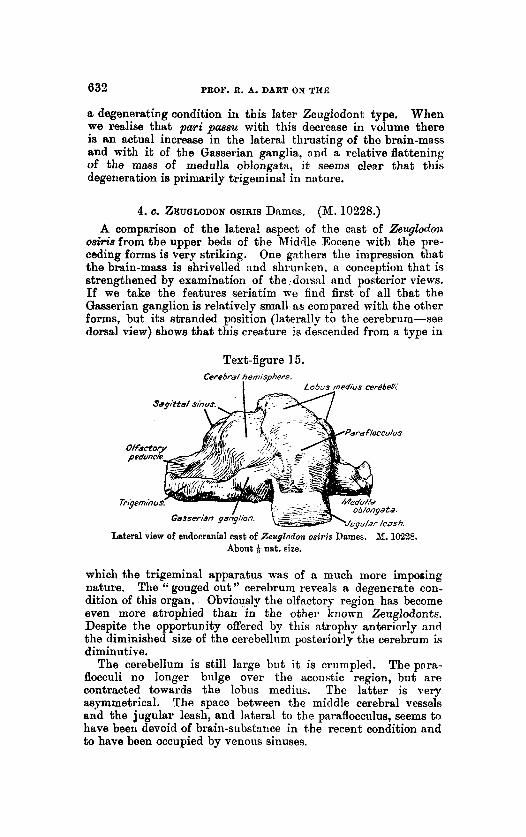
4. c. ZEUGLODON OSIRIS Dames. (M. 10228.) A comparison of the latera! aspect of the cast of ZeuglotJoia
osi& from the upper beds of the Middle Eocene with the pre- ceding forms is very striking. One gathers the impression that the brain-mass is shrivelled and shrunken. a conception that is strengthened by examination of the, doi-snl and posterior views. If we take the features seriatim we find first of all that the Gasserian ganglion is relatively small as compared with the other forms, but its stranded position (laterally to the cerebrum-see dorsal view) shows that this creature is descended from a type in
Text-figure 15. Cerebra1 hemisphere.
I Lobus rncdius rerebefi;
Lateral view of endocranial cast of Zeuglodon o s i ~ i s Dames. M. 10228. Abouc + nat. size.
which the trigeminal apparatus was of a much more imposing nature. The “gouged out ” cerebrum reveals a degenerate con- dition of this organ. Obviously the olfactory region has become even more atrophied than in the other known Zeuglodonts. Despite the opportunity offered by this ntrophy anteriorly and the diminished size of the cerebellum posteriorly the cerebrum is diminutive.
The pnra- flocculi no longer biilge over the acoustic region, but are contracted towards the lobus medius. The latter is very asymmetrical. The space between the middle cerebral vessels and the jugular leash, and lateral to the paraflocculus, seems to have been devoid of brain-substance in the recent condition and to have been occupied by venom sinuses.
The cerebellum is still large but it is crumpled.

BRAIN OF THE ZEUOLODONTIDAC. 633
The lobus medius, or area cremens, is not only very aaym- metrical but is poorly developed,and the paraflocculi form by far the greatest part of the cerebellar mass. The posterior view affords perhaps the completest picture of this atrophic brain, for here we recognise the general flattening out or ‘‘ cake-like ” appearance of
Text-figure 16.
Dorsal view of endocranial cast of Zeuglodon osiris Dames. M. 10228. Ahout + nat. size.
this brain (which came from a perfect skull) and also the extremely long but narrow cross-section of the medulla oblongata. This view of the medulla oblongata coupled with the lateral view
Text-figure 17.
Posterior view of endocranial cast of Zeuglodolz osiris Dames. N. 10228. About nat. size.
of this region corroborate the inference we have already drawn that in Zeuglodo0l.t oairis we have the degewerate ofspring of a specialised race.
The degree of this degeneration is perhaps most graphically shown if we compare the cubic capacity of this brain (48Oc.c.)

5. TAB- OF MEASUREMENTS OF THE ENDOCRANIAL CASTS.
I Greatcat width of cerebrum ........................ I I Greatest int,er-panifloceular width ............... I Length of cerebral liemispliere (measured to 1 tlie tentorial sulcus) .............................. ~ Height of ccrebellor projection above the cere-
brum along teutorial sulcus ..................... a. in tho region of the
Width of middle lobc Area B ............ b. in the regiou of the 1 Area C ............
Width of medulla oblongata a t fommeu magnum .............................................
! Height of medulla oblongata a t foramen 1 magnum ............................................. Greatest height of braiu-mass from base to
lobus medius cerebelli ........................... Greatest antero-posterior diameter of cerebd-
lnm measured midway along tentorialsulcus .
1
of thecerebellum ...
i
M. iaias. 2. sensitimts.
87 mm.
136 mm.
66 mm.
10-20 mm.
91 mm.
65 mm.
-
.-
73 mm.
30 mm.
j Greatest length of brain-mass ..................... 100 ~11111. (appro..).
Cubic capacity ........................ 1 490c.c. i -
M. 12066. 2. elliotsmithi.
92 nun.
126 mm.
___-
68 mm.
1-10 mm.
100 mm.
63 mm.
66 mm.
28 mm.
97 mm. (appros.).
310 C.C.
M. 102%. 2. osiria.
94mm. (approx.).
192 mm.
62 mm.
15-20 mm.
Wmm. (approx.).
67 mm.
4Q mm.
20 mm.
66 mm.
41 mm.
100 mm. (appros.)
480 C.C.
M. 10173. 2. internrediris.
90 m u ~ . (api~rox.).
172 rum.
----
47 mm.
20-26 nim.
96 mm.
64 mm.
67 mm.
34 mm.
96 mm.
43 mm.
108 min.
786 C.C.
---
M. M66. I’roz. atrox.
95 mni. (approx.).
153 mm.
42 mm. (npprox.).
2036 ~ I U .
114 mm.
47 mm.
68 mm.
37 mm.
96 mm.
60 mm.
110 mm. (approx.)
800+ C.C.

BRAIN O F THE ZEUGLODONTIDE. 638
with those of the foregoing which are almost twice the size. An examination of the tables and the diagrams of the skulls will show that this discrepancy in brain-capacity is not due to any diminished bodily size in Zeuylodoia osiris, for although the skull may be somewhat shorter it is absolutely wider than those of the other two forms.
One can only conclude that we have here an obvious dwindling in brain-substance which has affected not only the trigeminal and olfactory regions but with them the cerebrum, cerebellum, and the medulla oblongata. The whole evidence goes to show that the devolutional potentialities exhibited in the specidisations (chiefly trigeminai) of Prozeu!~lodoiz and the group it typifies have found in Zeugloclon osiiis (M, 10228) their logical finale.
6. THE ZOOLOQICAL POSITION OF THE ZEUULODONTIDIE. Certain facts arising from our study of these endocranial casts
claim our immediate attention. The first of these is the tri- geminal specialisation. I n all of the Zeuglodonts (although coming from different horizons) the essential features noted in Zeuglodon semitivus apply. I n all, the Gasserian ganglion assists in modelling the roof of the cranial cavity. The inference is justified that the group, as a whole, rested from the outset upon this specialisation for its subsequent achievements.
It seems to be a law of general evolutionary application that specialisation of one “sense” ontails as its corollary the atrophy of one or more of the other avenues of sense-perception. Thus. birds become specialised as to sight but lose their appreciation of am11 ; the same iEc true, though in different ways, for Teleostean fishes and for Primates. I n Omithmhynchus, which specialises in its ‘‘ fifth nerve sense,” we find a relative atrophy of both smell and sight. I n Zeuglodontids the sense of smell has certainly been largely lost as a result of their adoption of a water habitat, and it seems likely that sight also was of diminished value.
To the loss of smell and the abortion of the b a d (olfactory) parts of the cerebrum with their effect in limiting the longitudinal exten- sion of the hemispheres and consequently of the cranial cavity, reference was made by Elliot Smith (1902 and 1903). A relative loss of sight, with a resulting diminution of the mid-brain and thalamic regions, may assist in accounting for the smallness of the cerebrum in Prozeuglodoia and albo for its lack of growth in the two later forms from the upper horizons. It assists in the nnder- standing of the propinquity of the cerebellum to-or, rather, its overgrowth over-the cerebrum, and affords further reason for the diminished longitudinal extension of the cerebral axis (because of a wasting mesencephalon and optic thalamus and the con- sequences entailed thereby) throughout the series.
B u t it seems to me that the lateral expansion not merely of the brain-stem itself but also of the cerebellum has been provoked mainly by the trigeminus, and that R specialisation of this nerve hag been the most significant factor in determining the queer

636 PROF. R. A. BART ON THE
contour of the Zeuglodont brain. Of course, the enlarged cere- bellum has played a role in the lateral expansion of the cranial cavity, but tho extraordinary dimensions, even for an Eocene animal, of this cerebellum can only be explained by the tribute coming from trigeminal nerves whose Gasserian ganglia rival the cerebral cortex in size. Hence the influence of the cerebellum is fundamentally trigeminal in origin.
It is true then that, although the tr ipminus has played the chief part in this expansion, the internal factors include also an olfactory and a visual factor and may well have been assisted in Zeuglodonts, .as Andrews (1908) suggested, by the ‘‘ pressure on the anterior end, more or less in the direction of the long axis,” during motion through the water, “and during very rapid movement this pressure must be considerable.”
Now a reduction in value of the sense of smell and possibly of sight and a compensatory dependence upon the sense of touch in the muzzle-i. e . , a certain degree of trigeminal specialisxtion- are equally to be anticipated in the ancestors of modern Cetacea. Might it not be that the Zeuglodontids, in this respect as in their osteologicd conformity, are to be regarded as ancestral to Cetacea? This might be admitted if it were not for the demonstration of the already-marked specialisation of Prozeuglodoii atrox and the gradual deterioration by specialisation within the phyletic series as already discussed. I n this light tho passage of Cetacean ancestors through a “ trigeminal” stage in evolution can only be cited as convergence, or may be explained by the hypothesis that the Zeuglodontids and the true ancestors of Cetacea may have had a common ancestry in the earliest Eocene.
Despite the positive views concerning the ancestral relation- ship of Zeuglodontids to Cetacea stated a t the outset, it must not be assumed that palsontologists are agreed concerning this interpretation. As early as 1877 Marsh mid : ‘‘ That the connection (between
Zeuglodon and Cetacea) was a direct one, however, is hardly probable, since the diminutive brain, large number of simple teeth, and reduced limbs in the whales all indicate them to be an old type which doubtless branched off from the more primitive stock leading to the Carnivores.”
Weber (1886, p. 243) also referred to Zez~glodo?~ as “einen verun- gliickten Versuch Cetnceen herauszubilden,” and although there have been wavering opinions by many since, which even Weber himself has shared, many, such as Miiller, Fraas, and Stromer, have enunciated the same view.
Fraas, in 1905, summed up the evidence to that date available and showed that the Zeuglodontide, arising from some possible Creodont stock, have passed through some stnge corresponding to his Eocetus (Mesocetiis) and, by differentiation, have produced various forms, including the gigantic ones which generally have marked the acme of evolutionary progress in many groups of animals, after which the race has disappeared. He has felt it

BRAIN OF THE ZEUQLODONTIDR. 637
impossible to regard such later gigantic forms as ancestral to the later Squalodon series of animals, which appear to be more definitely related to the progenitors of the modern Cetacea. According to Winge (1919, 1921) Fraas ‘I considered both Proto- C6tUS and with it other Zeuglodonts as a side branch from the Carnivores which did not lead in the direction of true whales.”
Stromer (1908, p. 174) also stdies: “ lch halte also einstweilen fur geboten die Zeuglodontide fur eine iihnliche Parallelreihe der Denticeten anzusehen, wie sie neuerdings innerhalb vieler engemn Saugethier-abtheilungen nochgewiesen wurden. Sie hatten schon im Obereozan ihre Bliitezeit unter Entwickelung von Riesen- formen wahrend die anderen fast gleichartig aber vie1 weiter sich differenzierend langsamer sich entwickelten und wieder in mehrere Zweige auseinandergingen, die auch in vieler Beziehung einander parallel fortliefen. I n diesem Sinne also nehme ich wie Weber (1886, S. 243) Zeuglodon als 6 einen verungliickten Versuch CetRceen hemuszubilden ’ halte aber fiir noch nicht beweisen d a s alle Archeweti so auf zufassen waren.”
Abel, in his later works (1913). does not appear to have any doubt that the Zeuglodontida lie off the direct line, with the possible exception of the so-called Microzeuglodontida. Because of their great specialisation, the degree of which would entirely unfit them for such an evolution, I am entirely in agreement with the opinion that no Zeuglodont here examined can be regarded as ancestral to Cetacea.
It is a curious fact that these highly specinlised animals have so wide R geological distribution, and one in favour of their marine life. This wide distribution may have occurred before the trigeminal specialisation, so characteristic of all these Fay um forms, was attained. This makes the examination of the endo- cranial casts in Zeuglodontidae found elsewhere in Europe, in America, and in Australia of the greatest importance because the facts put forward demonstrate our lack of knowledge concerning the evolutionary liistory of the whales and dso because “ the distribution of the Zeuglodontidoe and other shallow-water fauna” has been used (Stromer, 1906, and Andrews, 1907) to indicate the location of the shore-lines of previous land-connections between the Old nnd New Worlds. I consider that in the demonstration of this tactile specialisation in Zeuglodonts their restriction to a shore-line distribution is shown to be highly probable.
The parallel origin of the Zeuglodontidae and the Sirenia- probably in the Lower Eocene-is very striking. The grade of cerebral organisation in both is approximately similar, but the cerebellum of Zeuglodontide seems to indicate that its life was somewhat less retired than that of Sirenia. Andrews (1907) has shown that ‘‘ freedom from competition and, to some extent, from powerful enemies, would offer exceedingly favourable conditions for the rapid spread and multiplication ” of these groups in the Eocene seas. Thus both groups became widely disseminated. but

638 PROF. R. A. DART ON THE
whereas the Sirenia hare persisted as “ living fossils,” despite their humble grade of intelligence, the Zeuglodontids have long since disa.ppeared.
This disappearance is to be correlated with the fact that, whereas the Sirenia chose an eminently retired and sluggish mode of existence, the Zeuglodontidie were more active. They came into more direct conflict with other marine forms and, specialised as they were, did so at a disadvantage and were over- whelmed in the struggle for existence.
The present study therefore indicates that the scepticism of Marsh and Fraas was entirely justified and that Winge (1921) is incorrect in regarding the Zeuglodontidae as ancestral to Cetacea. The origin, dispersal (into so many strikingly different forms and all over the globe), and disappearance of this group geologically is a demonstration of the conception put forward by Marsh (1877) that “ I n every primitive type which was destined t o survive many geological changes there seems to have been a tendency to throw off lateral branches which become highly specialised, and soon die out, because they are unable to adapt themselves to new conditions.”
7. COMPARISON O F ZEUGLODONT AND PROSQUALODONT BRAISS. The above paper was practically ready for the press when
Prof. Elliot Smith received from Tasmania, through the great courtesy of Prof. Flynn of the Zoological Department in the University at Hobart, a splendid cast of the cranial cavity of Prosqualodon davidi which he has recently described. Plaster replicasof the skull from which this cast was made were pre- sented by him to the British Museum of Natural History and to the Zoological Department of University College, London. I have had the privilege of studying these casts and the endocranial cast.
The lateral view of this cast (text-fig. 18) reveals an astound- ing likeness to the Zeuglodont endocranial cast. We meet with the same cerebellar enlargement, expanding forwards over the cerebrum, and quite an enlarged trigeminus.
It is obvious that such striking similarities would not exist i t i the absence of some close relationship between these forms, but the data will reveal that this relationship is not a filial one as i t has frequently been conceived.
It is to be noted that the parnfloeculus is recognizable laterally, but is submerged by a widely and generally expanded cerebellum. The cerebrum is emancipating itself from the cerebellar growth as its great height posteriorly shows. Anteriorly the rapidly dwindling olfactory apparatus is attached in a ‘‘ nipple-like ” fashion to the cerebrum and considerably ventral to it is n well-marlred optic chiasma. The middle cerebral vessels revekl a course comparable with that seen in the Zeuglodonts ancl posteriorly there is a well-marlred jugular leash.

BRAIN O F THE ZEUGLODONTIDB. 639
The dorsal view corroborates what we have already detected from the lateral aspect. The wide expanse of the “area crescens ” cerebelli is the most obtrusive feature and after that the expanding cerebrum. The double olfactory peduncle is clearly seen but no Gasserian ganglion is visible (text-fig. 19).
The posterior view is instructive in revealing an enlarged oval transverse section of the medulla oblongata and still further illustrates the nature of t,he cerebellar expansion (text-fig. 20).
Perhaps the most informative of all is the ventral view, because we are able to recognise the true size of the trigeminus (which is quite large), the relatively enormous width of the base of the brain, the marked development of the optic chiaama, and the relative atrophy of the olfactory apparatus. There is an entire ttbsence in this brain of anything corresponding to the tuber- culum olfactorium or “ intertubercular sulcus ” of the Zeuglo- donts, but we find a ridge on either side of the middle line medial
Text-figure 18.
Lateral view of endocranial cast of Prospualodon daoidi Rgnn. About + not. size.
to the trigeminal region which may be due to the ca.rotid artery and accompanying venous sinuses. The tuber cinereum is not apparent in the cast, but must lie between these ridges (text-
Prosqua2odon chvidi Flynn comes from the Miocene deposits of Tasmania, and the characters which it presents linking it to the Zeuglodonts might well be interpreted PE illustrating its origin from the Zeuglodont family if our information were not so complete as it is now.
The actual bulk of this endocranial cast is approximately 750 c.c., i. e., not quite so great as that of Zescglodon intermedim. This fact itself is sufficient to show that the Eocene form which gave rise to ~ o s q w ~ o d o n must have possessed a brain-capcity very much less t h m bhat of Prozeuglodon atrox or of Zeuglodm iq i temdiw. This follows from the well-known “lawof increasing brain weight” put forward by Marsh and eupported by all
fig. 21).

680 PROF. R. A. DART ON THE
paleontological endocranial investigation up to the present time.
Quite apart from this there is ample evidence against the view that a Zeuglodont, wen one so primitive as Prozezqlodon, could have given rise to Proaqualodon. This is very evident if we call to mind those features pointed out in detail for Zeuglodon sen& tivwr (c'ids text-fig. 3 ) and repeated by all the Zeuglodonts. It was shown in that case that the site of the insertion of the olfactory peduncle in the Zenglodonta hnd been drawn, as it were, during atrophy under the fore brain on to the basal aspect. This contraction of the area between the olfactory peduncles and the tuber cinereum demonstrated for us the relative atrophy of the
Text-figure 19.
Dorsal view of endocinnid cast of Prosqualodon davidi Flynn. About + nat. sire.
optic chiasmatic region in Zeuglodonts, and, fortunately, tlie cast of P r o z e q l o d m ahox in this crucial region is sufficiently perfect to illustrate the fact that these changes were already well marked i n the Zeuglodonts of the earliest Middle Eocene deposits of the Fayum (vide text-fig. 11).
I n the w e of Prosqualodon davirli, on the other hand, we find .that the course of its evolution has been entirely different, There is evidence here of an initiai expanding of this basal area between the insertion of the olfactory peduncle (vide text-fig. 21) m d the tuber cinereurn (which lies somewhere between the 6 6 carotid ridges l'). Crossing this wide interspace in A.oaqwlodoo?t, &vidi we find in the well-defined optic chiasma the evidence of

BRAL?. OF THE ZEUGLODONTIDE. 641
the retention of elaborate visual capacities in the Miocene-an utter impossibility in the offspring of the Zeuglodonts here described at any epoch.
It is of some value to appreciate how great a degree of trigeminal specialisation is compatible with future evolution. For it is evident that Prosqualodon davidi has an enlarged trigeminus, even though we do not find such gross enlargement of the Gasserian ganglia a~ in the Zeuglodonts. It has already been indicated that “ a certain degree of trigeminal specialisation” is to be expected in the ancestors of Cetacea. It seems unques- tionable that the initial widening of the Prosqualodont, as well as that of the Zeuglodont, brain and medulla oblongata is due not merely to the passive recession of the sense of smell but rather to the active hypertrophy of the trigeminal apparatus, which in a n aquatic mammal provides so much more information
Text-figure 20. Lo6us med& rere6e/h:
@ym!y expanded)
Posterior view of endocranial cast of Prospalodon davidi Flynn. About + nat. size.
concerning food, friends, and foes :than do the senses of smell or sight.
Prosqualodon davidi teaches us therefore that the evolution of the Cetacean stock, while it depended to some degree upon an initial trigeminal specialisation, was not effeoted by any sudden reliance upon this sense to the neglect of other important senses, but depended upon an orderly and ‘‘ balanced” correlation of this hypertrophy with a concurrent aggrandisement of the visunl and auditory senses. I n this connection it is significant that the most .expanded portions of the fore brain in Prosqualodon davidi appear t o be the “ occipital ” and “ temporal ’? regions ; i. e., posteriorly and laterally, where one may reasonably conclude that these senses were finding cerebral representation. Whether Fraas is corned in believing that even the more primitive Protocetw

642 PROF. R. A. DART ON THE
is also off the main line of Cetacean evolution would probably be indicated by an examination of the endocranial casts with regard to these details.
The conformation of the district between the olfactory peduucles and the chiasma-in fact, the whole “ mamma-like ” appearance of the antero-ventral portion of the fore brain in Prosqualodon davidi, contrasting as it does with the same region in the Zeuglodonts-is consonant with the homologous region in true Cetacert. A comparison of this region with the ventral aspect of the brain in the fetal Monodon or adult Kogia (vide figures in Elliot Smith’s account, 1903) demonstrates the truth of the conception that the ancestors of true Cetacea went through
Text-figure ZJ.
Ventral view of endocranial cast of Prosqualodon davidi Flynn. About 4 nat. size.
a stage of ‘‘ expansion of the basal regions of the brain” (witness the “desert region” of Brow in Cetacea) not displayed by Zeuglodonts but evident enough in Prosqualodon.
What this expanded “desert region ” or tuberculum olfactorium in whales exactly signifies, we are not as yet able to definitely state. Since this area is the site of the palaeostriatal cortex overlying the palaeostriatum (Dart, 1920) it follows that the persistence of a lnrge “desert region” is dependent on factors other then olfactory, i. e., factors which have determined the size of the paleostriatum itself. The consideration of what these factors actually mny be lies outside the mope of this article, but the essential point affecting our argument here is that these

BRAIN OF THE ZEUQLODONTIDIE. 043
fwtors, whatever their nature, played a n enhanced rdle in the ancestors of Uetacea, whereas they were recessive and negligible in the Zeuglodontidse.
The evidence afforded by this cast therefore supports the conclusions which we drew in the earlier part of this paper concerning the deflection of the Zeuglodont line from the course of true Cetncenn erolrition, but supports the conception that Sqzcalodon has at least close alfinities with the true Cetacean stock.
8. THE TRIOEMINUS AND THE LAW OF INFILTRATION IN CEREBELLAR EVOLUTION.
. I n conclusion, it is necessary to call to mind certain general facts about the cerebellum which appear to throw light upon the strange and generalised hypertrophy of that oigan in this group.
We know that animals with very sensitive whiskers and bristles (such as certain Rodentia) depend largely for their power of equilibration upon stimuli which arise in nerve-endings situated in relation with the proximal end of these whisker-like structures and affected by their faintest movement. Such stimuli are communicated to the cerebellum by way of the trigeminus. I n Omithorhynohb.rcs, instead of bristles, there is a remarkahle development of special receptive end organs in the delicate snout served by the trigeminus. The bristles round the mouth-parts of the Birenia may be active in a similar way in Rddition to their function as tactile end organs.
The more recent researches of Ingvar (1918) have placed the morphological survey of the cerebellum by Elliot Smith npon an even firmer basis: for he has extended his research into the Reptilian and Avian series and has shown that here, too, the same sulci and three lobes are distinguishable. Such a division into three lobes does not nppear to obtain in Fishes or Amphibia. It seems therefore that the advent of the middle lobe synchroiiises with the origin of n neopallium in the cerebral cortex. The researches of Haller (1900), of Unger (1906), and of Crosby (1917) have shown that the first clearly defined neopnllium primordium makes its appenraiice in the Reptilia, rind in these creatures tho tripartite structure of the cerebellum is also clearly to be recognised for the first time.
Recognising the relationship which the development of the neopallium has to the expansion of the cerebellum in Mammalia it is, at first, most disconcerting to find in a group of Eocene mammals with such ill-developed neopallium a cerebellum of extravagant proportions which h ~ s not merely obliterated all traces of the mid-brain from the surface but threatens to cover the cerebrum also by its forward expansion.
The cerebellum is usually relatively large in Eocene mammals and yet such a bizarre arrangement as is present in Zeuglodonts can have only one explanation. W e may eliminate the neopnlliiim
PROC. ZOOL. S0~.-1923, NO. XLTI. 42

644 PROF. R. A. DART ON THE
aa an explanation because of its slight development and the doubtful presence of a pons Varolii, and it is equally unlikely tha t the spino-cerebellar contribution could have been SO great as to account for bhe whole of the cerebellar expansion. But we do know that 80 diminutive and primitive a mammal as Omaithorhpchus, with a well-developed trigeminal apparatus can and does possess not only a n unexpectedly large neopallium, but also a relatively large convoluted cerebellum. The absolute and the relative size of the Gasserian ganglia in Zetqloclon sensitiutts, and indeed in all Zeuglodontidse here represented, are far more striking than they are in Ornithow5ynxhus. There can be no doubt t h a t the afferent impulses reaching the cerebellum from the anterior end of the body in Zeuglotlonticin: were of particular value in supplying these animals with information concerning disturbances of equilibration. The distribution of the trigeminal tract within the cerebellum is not fully known, but it is believed by many investigators that in Mammalia there is a tract for conveying trigeminal impulses to the cerebellum. The huge cerebellum in the Zeuglodontidze may well be due to the fact that, by a rapid and hypertrophic development, tho trigeminus provided it with very precise information concerning its position in space and hence afforded to this primitive creature R, ready solution of the problem of equilibration in a fluid medium.
I have said that the distribution of a direct trigeminal tract within the cerebellum is not fully known ; it would be nearer the truth to state that it is frequently affirmed but is sometimes denied. Since such denictl exists it is valuable to put forward other supporting evidence. It seems to me that the denial of the existence of a direct trigeminal connection with the cere- bellum can only come from an imperfect appreciation of the developmental history of the cerebellnm and n consequent failure to recognise its extent in the brain-axis-or else from a tendency, very manifest during t h e last two decades, to regard the cere- bellum as nn overgrown part of the vestibular apparatus.
As regards the development of the cerebellum it must be remembered that tho cerebellar ridge first appears in early embryos much further forward in the hind brain (metencephalon) than would be anticipated on the “ vestibular ” hypothesis. Even in human developmental history, the nerve wliicli is more obviously associated with the cerebellum is the trigeminal nnd not the vestibular. Ingvar (1918) finds that ‘‘ Die Basis (cere- belli) ist frontalwarts gerichtet. Von dem ventralen Rande dieser Basis laufen die kriiftig entwickelten N. N. Trigemini atis ” ; or (on p. 343) “ Ventrrtlwiirts grenzen die Cerebcllar- wiilste an die Insertionstelle des Trigeminns.”
It is not absolutely certain what actual neuroinere of the hind brain gives rise to the cerebellum, nor is that question pertinent to the present discassion. The embryological facts significant here are: (1) that the cerebellum arises in the most anterior portion of the hind-brain roof, and (2) that the territoiy of t h

RRAIN OF TRE ZEUGLODONTIDR. 645
neural tube in this vicinity is associated in the vertebrate embryo with the trigeminus (cf. Wilson and Hill, 1902 ; Streeter, 1908), while the acoustico-facinlis mas4 lies entirely posterior to the trigeminus and is related to a different territory which lies medial to it. Hence, any encroachment of vestibular fibres from its territory of the neural tube into the territory which is related to the trigeminus is a secondary phenomenon whether con- sidered phylogenetically or ontogenetically, even though such fibres be direct fibres. I n other words, the appearance of vesti- bular fibres in the cerebellar region is an intrusion just as certainly rts is the appearance of the mesencephalic fibres of the trigeminal in the miti-brrtin, or of thalamic fibres in the fore brain. ' The simplest cerebella reproduce more or less faithfully this
condition characteristic of the embryos of all Vertebrates. They are mere ridges roofing the most anterior portion of the fourth ventricle and are consequently a link between trigeminal terri- tories and contain decimations of the trochlear and trigeminal nerves (Herrick, 1914; Lamell, 1920; Palmgren, 1921).
I n the simplest cerebella iiatrzlsions of alien fibres are already found, even as we find alien fibres in the simplest-known fore brain and tectuin opticum. These intrusions occur in the cere- bellum in such a way that just as we find the olfactory apparatus is relegated to t,he periphery of the fore brain and the optic fibres to the periphery of the mid-brain, SO we find t h a t the trigeminal territory forms the true fringe of the cerebellum. Consequently, Beccari, Ingvar, and others (Kappers, 1921) have found a direct distribution of the trigeminus to the cerebellum (e.s/., in Reptilirt).
Topographically, the contribution of the vestibular nerve to the cerebellum is slwa.ys surrounded by the trigeniinal territory and the fibres proceeding to the cerebellum therefrom. Ingvar (1918) has shown (in Mnmmalia) n distribution of the vestibular com- ponent mainly in the flocculus, lingula, and nodulue, and thus pwipherally to the still more recent intrusions of spino-cerebellar contributions. On the other hand, spino- and olivo-cerebellar fibres have not been demonstrated in such peripherally lying parts such as the lobus floccularis, and it is to be noted (vide Kappers, 1921) that this region is not dependent upon a cerebro- pontine contribution ; whereas the clinical researches of Winkler have corroborated the conception that the latest portions of the cerebellum in the phylogenetic sense are more centi-ally situated.
This '' laminar " arrangement of the cerebellar constitution, a s it might be termed, although not so cleady defined as the some- what similar arrangement of successive fibrsintrusions into the fore brain, is nevertheless present. It is to be expected also from the fact that those tracts, which later in phylogeny become incorporated with any region, attain this incorporation by infil- tration and a spreading apart of the pre-existing mechrtnism.
42'

646 PROF. R. A . DART ON THE
The arrangement may be expressed by stating that the vestibular, olivary, spinal, and cerebro-pontine contributions successively aome to reach the aerebellum by penetrating a territory whicli was originally, and so ancestrally, trigeminal.
As has been suggested already, this phenomenon of “pene- tration ” may be illustrated equally well by the iiz$ltvatiofae of the mid-brain, thalamus, or fore brain, or by the injiltvation of the hippocampel commissure by the oorpus callosurii in the fore brain of Mammnlia as shown by Elliot Smith. I n biief, if we accept the doctrine of the segmental arrangement of the neural tube elements, i t is to be anticipated that the principle finds illus- tration in the development of the majority of the inter-segmeritR1 and supra-segmental apparatuses.
The language of neiirolopy is devoid of any term which describes conveniently this uniformity of behaviour iii tlie laying- clown of subsequent formations in the neural tube. It should prove of service therefore to descriptive neurology to recognise in this uniformity tho consistent working of a generti 1 principle which underlies the whole :irchitecture of the brain aiitl wliicli we may term for convenience the Law of Iv$Ztrafioi~.
Ry an inverse reasoning, if this law is correctly conceived, n peripherul or fringing nrrmgement of the trigeminal territory itself, and of fibres known t o proceed to the rerebelliim from it, corroborates the identi5cation of the trigeininal npparatus with the cerebellum and the function of equilibrium, niore primitively even than the vestibular apparatus.
These matters have been neglected by those who assert that the auricle of the cerebellum, 01- lobus floccularis (Knppers, 1921), is the oldest part of the cerebellum. It is not the auricle, but the anterior medullary velum whiah would better deserve this appella- tion. The “ veutibiilar ” hypothe& has been attiactive because it has seemed to demonstrate the fact that the cerebellum was 6 ‘ equilibratory” from the beginning. But this hypotliesis ten& to ignore the equilibratory potential of all tactile sense, it fails to nccount for the existence of a cerebellum (Cpclostonies) wliicli has no auricle, and does not nccount in any w:iy for tho striking relationship which the trigeminal nerve always hiis to the cere- bellum in all Vertebrata.
The fifth nerve displays a relationsliip to that segment of the neural tube, in which the cerebellum becomes tlevelopecl, wliicli is paralleled only by the relationship of the clfnctory nerve to t h t segment of the tube in which the prosenceplialon, and of the optic nerve to that segment m wliich the niebenceplialoii is cleve- loped. The oltlost part of the organ is the trigeininal territory itself just as the olfactory sensoriuni was tlie oldest represent:rti\ e of a fore brnin and the visual nensorium of a mid-brain.
I have discovered that Spitzka (1886) put forward t~ somewliat similar view concerning the cevebellnm n e d y forty yeais ngo .- a * I aiu i n c h e d to consider i t as a homologue oP the gelatinous nucleus of the fifth pair, and as in a primitive relation uitli that

BRAIN OF TBE LEUOLODON’l’IDiE. 647
nerve ; that subsequently t h e auditory nerve entered into con- nection with it, and that, increasing in dimension with its in- creasing iieural connections, it attained the high development found 111 the liumau cerebellum through tile spiiial and cerebral tritcts tliat i~ r0 cletuched into its meclullnry substance.” Tliere- fore the present stateiiient of this relationship, although inore detailed, cannot be regarded as a novelty.
If we consider the nature of the influences at work in the production of tile early chortlate brain, i t is evident that the more anterior segments ware conceriied in the production of the olfactory ant1 visual specialisations. These specialisations were bought at the price of successive anterior segments of the primitive vertebrate sltin and neural tube. When these anterior segments lost by their specialisation tlie capacity for the appre- ciation of presszvre, the succeeding segment wliicli retained this power grew forward invading tlie territory of the more specialised segnients lying anterior to it. Such an invasion meant an in- crease or hypertrophy of this (trigeminal) segment and :t special- isation within i t of the tuotile function. How exuberant has been its respoiise to this tlemand is demonstrated by the fact tliat- amongst Illammalia, for instarice-it subserves tlie tactile sense for tlie entire region anterior to the second cervical nerve, wi th the exception of the vestigial somatic elements of the vagus (Arnolcl’s nerve).
The information conveyed by the trigeminal system proved extremely valuable, because it told the aninial so much about what moved in space around it uncl also about its own position in spuce. Hence the central processes from the trigeminal gaiiglion assumed a sudden reflex ascendelicy over an increasing iiuriiber of the succeeding segmental nerves through the radix tlescendens and the substilntia gelatinosa. Bolandi in most Vertebratii. Even in Man the descendiiig root of the trigeminal represents one of the most impressive features of the medulla oblongata. Always a striking feature in Maniiiialia, this reflex dominaiice is shown to its best advantage (in living forms) in Omithorhy~~chzu ; in this creature tlie trigeminal seeins to exert t~ reflex control over practically the whole of the spinal cord.
Thus there has been a downward encroachment of the trige- ininus upon lower segments of the metlulln ob1ongat:i in a fashion comparable with the upward encroachment of the vestibular iierve into the trigeniinal territory or of the mesencephalic root of the trigeminal upoii optic territory. It is a further example of the Law of I~jiltration. Seemingly this encroachment afforded the bridge whereby the spiiio-cerebellar systems were conducted to the originally t r i g e m i d territory of the cerebellum. For the spinal fibres naturally lay peripheid to tlie infiltrating descending root of the trigeminus, and as the spinal fibres coursed towards the cerebellum tiley canie to embrace the trigeminus trunk before piercing its territory to enter the cerebellum. The diflerentiation into anterior and posterior spiuo-carebellair tracts einerged from

648 DR. C. W. ANDBEWS ON THE
a relatioilship primarily not to the cerebellum as such, but to the trigeminus.
So, in addition to the fringing above mentioned, a graphic picture of the central role which the trigeininal has played in the origin of the cerebellum is afforded by the relation of t h e anterior and posterior spino-cerebellar tracts t o the trigeminal nerve (in most Vertebrata) and by the central position of the trigeminus in the pons (in higher Mammdia).
Considering the nature of the impulses it conveyed. it is not surprising that the trigeminal nerve, which has had such diverse evolutionary possibilities in all vertebrate groups, should have been very intimately concerned in the emergence and evolution of the cerebellum in Vertebratrt. Nor is it surprising that we should find illustrated in the Zenglodontidre a group of prirnitive mammals taking to a water life at a stage so plastic that the trigeminal apparatus should becouie sufficiently specialised and hypertrophic to determine, through its moulding of the cere- bellum and the spinal cord, the whole course of evolution (or devolution) of its central nervous system.
9. NOTE ON THE SKULLS FROM WHICH THE ENDOCRANIAL CASTS By C. W. ANDREWS, DESCRIBED BY DR. DART WERE TAKEN.
D.Sc.: F.R.S. (British Museum of Katural History.) (Published by permission of the Trustees.)
Unfortunately, from the nature of the case, the skulls belonging to the two natural casts above described are unknown, having been destroyed by weathering, but those from which the remaining casts were made are preserved and are here discussed. Four such skulls are known. Of these, two [M. 8150 (a cast) and M. 102281 certainly belong to Zeuglodon osiris Dames, from the Qasr-el- Sagha beds (Upper Mokattani=Bartonian), north of Lake Birket- el-Qurun in the Fayum. One (M. 9266) is the skull of Prozeugbdon atrox Andrews, from the lower part of the Birket- el-Qurun beds in a valley twelve kilometres W.S.W. of the Gar-el-Gehannem, lying to the west of the lake. Thih last speci- men may be regarded as the paratype of the species and is described and figured in the British Museum Catalogue of the Fossil Vertebrata of the Fayum (1906, p. 243). The fourth skull (M. 10173), forming the middle term of the series, seems from the iiltture of the matrix to have been obtained from the Birket- el-Qurun beds a.t the western end of the lake from a n horizon intermediate between those from mhich the other species were found. This, however, in the absence of definite informatioii from the collector iR not certain. This drull appears to belong to a hitherto-undescribed species which has been called Zeecglodon intermedim by Dr. Dart, the characters of which are given below.
Numerous other Zeuglodont remains have been collected from various horizons in Egypt, and there is some difference of opinioii

as to their determination and relationship to one another. For the purpcses of the present paper these may be disregarded and the series represented by Prozeuglodon atrox, Zeuglodon inter- medius, and Zeuglodon osilis alone considered. The characters of the skulls of these species completely support Dr. Dart’s conclu- sion, founded on the endouranid easts, that these three species represent terms in a phyletic series.
The skull of Prozeuglodon has been described in detail in the catalogue (Zoc. cit.), so that here it will only be necessary to refer to a few characters easily comparable with those of the other fornis. The occipital surface (text-fig. 22) above the foramen magnum is wider than high (if the height* be taken as 100, the widtli is 110) and it is only gently concave from side to side. The foramen magnum and the occipital condyles are both large. The convexity of the portion of the cranial wall formed by the parietal
Text-figure 22.
Occipital surface of skull of Prozeuglodota atrox Aiidrews. M. 9266. About + lint. size.
and squarnosul is greater than in the later forms, while the sagittal crest is much less developed. The posterior border of the narial opening is over the space between the first and second premolars and the snout is less prolonged anteriorly than in the hter forms. Some of the above peculiarities may be dependent to some extent on the circumstance that the skull described is not quite adult.
The type-skull M. 10173 of Zeuglodon. intermedim (text-fig. 23 j is not complete, the zygomatic arch and the terminal portion of the snout being missing; the teeth also are mucth broken. I n it the width of the occipital surface (text-fig. 23) is much less in proportion to its height# (height 100, width 66), and it is much more concave from side to side than in Proauglodon.
* In each case the height is measured from the upper border of tho fornman magiium to the point of uniou of the sagittal and lambdoidti1 crests.

660
But the conoavity is regular and the middle portion of the lamb- doidal cre& is not suddenly reflected backwtircls as in Zeuglodm osiris. The occipital condyles and foramen magnum are relatively smaller than i n Prozewglodon, the occipital crest more developed, and tho convexity of the Irttternl cranial surface less marked. The snout was more slender aiid more prolonged in front of the narial opening thnn in Prozeuylodon.
This skull, both in the form of its occipital surface nnd in other points, closely resembles that described by Stromer 8s ~ e z b f / l o t h l , oeiris (in his paper of 1908) and distinguished by him as Mn.9. It is very different from that described by the same writer RB
Zeuglodon o&& in 1903 and called in his later paper Mn.1. This latter seems to be the true Zeuglodow osiris, its lower jaw
Text-figure 23.
m. c. w. ANDREWS 0s THB
Occipital surface of skull of Zeuglodon interntedius, sp. n. M. 10173. About f Mt. size.
agreeing very closely with that of the type-specimen of the species as described and figured by Dames (1894). Mn.9 is either the present species (Zeugbodon internzetlius) or a form between it and Prozeuglodon.
The skulls of Zeuglodon osiris from which endocranial casts have been made are two in number. One (M.8150) is very incomplete and reveals only the dorsal contour of the brain. The endocranial cast of this skull was figured and described by Prof. Elliot Smith (1903). The other skull (M. 10228) is a nearly perfect specimen, and in order that the endocranial cast miglit be made it was skilfiilly cut into sections and the matrix removed by Mr. L. E. Parsons (text-fig. 24).
The occipital region of this skull agrees exactly with Stronier'ij

BKULLB OF EEUGLODONTIDE. 661
specimen Mn.1. It is remarkable for its extreme narrown888 compai*ed with its height, the lateral portions of the lambdoid~rl crestA being sharply deflected backwards so that the deeply con- cave occipital surface has the appearance of being pinched in a t the sides. This charracter is also well shown in the cast of the second skull (M. 8150). If the height be taken as 100 the width is ahout 44. The occipital condylee and foramen magnum are
Text-figure 24.
Lambdm7 crest
Occipital surface of skull of Zeuglodon osiris Dames. 1.10828. About 4 nat. size.
still smaller than in Zeuglodon ilztevmedizis and the convex sagittal crest is much higher. The side walls of the skull show little traces of convexity. The snout seems to have been rela- tively narrower and perhaps the nostril opened rather further back than in Zeuglodom inte?.4nRdizla.
The cliangea occurring in the stages described are :-
(1) Increasing narrowness and concavity of the occipital surfam above the foramen magnum, due mainly to the folding back of the high lambdoidal crests ;
(2) increased height of the sagittal crest ; (3) the reduction in the size in the condyles and foramen
magnum ; (4) decreasing convexity of the lateral cranial walls accom-
panying the depression of the upper portion of the brain ;
(5 ) the increase in length and slenderness of the snout and the probable recemion, to some extent, of the narial opening.
Changes (1) and ( a ) result in the increase of the surface for the attachment of the jaw-muscles, and are no doubt correlated with the increasing size and strength of the mandible.
I n Proaeuylodon the two pobterior upper premolars have a laige

662 PROF. R. A. DART ON TEE
poshro-internal buttress supported by II large distinct root. In the two species of zszcglodon this buttress is reduced in size and probably had ?o distinct mot, though from the state of preservation of the speeimen this cannot be definitely attled.
Some dimensions in millimetres of the skull in the three forins are given below. approximate only :-
These in some instances must be regarded
Length of parietal portion of ..................... 157 app. sagittal crest 126 i I
4 . iiitermeditur (M. 10173.) --
560
4.53
180
108
112
283
62
-------- Length from condyles to tip of
snout ................................. Length. from lambda to nasal
opening .............................. Height of occipital surface from
foramen magnum to lambda ... Least width of occi ital surface
between lambdoiJ cmta ... ...,
Prossuglodon. (M. 9266.)
673
803 *PP.
120
1%
2. osiris. (M. 10228.)
643 app.
467
166
69
92
293
46
170
10. BIBLIOQWHY. EL, 0.--“ Die Ursache der Asymmetrie des Zahnwalachiidels.” Sitzunghericht.
der Kais. Ahd. der Wiss. in Wien. 1902. - “ E h Stammtype der Delphiniden aus dem Mioeiin der Halbinsel Tamau.” Jahrbuch der k.k. Qeol. Reichanstalt, Bd. iv. Hft. 2. 1905.
“ Cetaceen Studien 1. Mittheilung, Jan. 109 ; Cetaceen Studien 11. Mit- theilung, Mar. 1909 ; Cetaoeen Studien 111. Bfittheilnng, Jau. 1912.” Bus der Sitsungaberichten der &is. Akad. der Wissensch. in Wieu. - “ Die Vorfahren der firtenwale: Denksch. der MatLNaturwiss.. K h s . der Kak Akad. der Wise.. in Wien, Bd. xc. 1913.
AXDEBWE, C. W.-“Extinct Vertebrates from Egypt.” Part I. Geol. Mag. 1901,
-- “Extinct Vertebrates from Egypt.” Part 11. Geol. Mag. 1901, p. 436. I‘ Extinct Vertebrates from Egypt.’’ Part 111. Geol. Mag. 1902, p. 291. - “ Further Noten on the Mammals of the Eocene of Egypt.” Geol. Mag.
1904, pp. 109,157.211. - “A Descriptive Catalogue of the Tertiary Vertebrata of the Faydm of Egypt.” Brit. Mus. of Nat. Hist. London, 1906. -- “The recentlydimvered Tertiary Vertabrata of Egypt.” The 6mith- nonian Report for 1906, pp. 296-307. - - “Zsug2odrm wanklyni.” Quart. Jooru. Qeol. SOC. vol. Ixiii. pp. 124-127. 1907.
p. aoo.

BRAIN OF THE ZEUOLODONTIDI. 653
ANDBEWS, C. W.-“ M d e l of Skull end Mandible of Promuglodon atroa.” Gml.
- “ A Description of New Species of Zeuglodoiit nnd of Leathery Tiirtle Proc. Zml. SOC. London,
ANTHONY, R.-“ Lc Hegoplodon de la Hope.” Bull. du Mua. d‘hist. naturelle,
BEADIELL, H. J. L.-“The Faydm Depression.” Geol. Mng. 1901, p. 640. BLACK, DAVIDBOII.-“ Endocirnial Casts of the Giraffidm.” Journ. of Comp. Neur.
vol. xxv. (1916) p. 341. -- *‘Notes on Endocranial Casts of Okapi, etc.” Annbmical Hecord,
vol. is. BOLK, L.-‘ Das Cerebellum der Saugethiere.’ Jena, 1906. I h D L E Y , 0. C.-“ The Mammalian Cerebellum.” Jouiu. of Annt. & P h p
vol. xxrviii. 19% p. 448, & vol. xxxiv. l(106, p. 99. BUBME~STEB, H.-“ Bemerknngen iiber Zeuglodon cetoides (Owen’s), Basilosauros
(Harlan’s), Hydrarcho8 (Koch’a).” Juniheft der allgemein. Lit. Zeitsch. Hallc, 1847.
CABIJS, C. G., and others.-‘Uutersuchungcn iiber das (unter den Namen Hvdrarchos von Dr. A. C. Koch) grosse fossile Bkelett.’ Dresden anh Leipic, 1847. -- ‘ I Das Kopfskelett des Zeuglodor hydrarclros.” Nova Ada Acad Cm. bop.-Carol. Nat. Cur. vol. xxii. 1848.
CLABK, W. I%.-‘‘ Cerebellum of PetronryaonJEzr~atatilir.“ Journ. of Anat. & Phys.
COPE, E. i).-“ Brain of Procamelw occidmtalis.“ Amer. Philos. Society, 1879. DAMEY, W.--PaLeontol. Abhandl. N. F. Bd. i. 1895, p. 189. EDINQEB, 0.-’ Nervose Zentml-organe.’ Leipaig, 1896. E~CEBICHT, D. FB.-ZOOI. Anat. Phys. Untersuchungen iiber die nordischen Wal-
FBAAS, E.-“ Neue Zeuglodunten nus dem uuterem Mitteleociin vom Mokattam bei
- Jahreshefte Verein. vaterland. Naturkuiide Wiirttemberg. Stuttgart,
Mag., 1908, pl. ix. p. 209.
from the Eocene of Southern Nigeria.” 1919, p. 309.
1909, p. w.
v01. XI. 1906-1906, p. 318.
thiere. Leipaig, 1849.
Cairo.” Palaout. Abhandl., 11. s., vol. vi.
1906, y. 347. GBEQOBY, W. K.-“The Orders of hfammds.”
vol. xxvii. 1910. Bull. Amer. MUS. Nat. Hist.
Hsaurcx, C. .I UDSON.-“ Cerebellum of Urodele Amphibia.” Jonm. of Comp.
INQVAB, SVEN.-“ Zur Phylo- uud Ontogenese des Kleinhirns, etc.” Folia Neuro-
KAPPEBS, C. U. ABIENY.-‘ Die vergleichende Anatomie des Nervensy8tems, etc.’
“Cerebral Localisation and the Sigiifficnnce of Sulci.” Report of Anatomy and Embryology Section : Internat. Congress of Mediciiie. London, 1913.
KUKENTHAL, W.-“ Vergleich auat.und entwick.Untersucli au Walthiere.” Jenaische Denkschriften, vol. iii. Jena, 1893.
LABWLL, OLAB.--“ The Cerebellum of Amblystmna.” Journ. of Comp. Nenr. 19m.
M A ~ Y H , 0. C.-“ Introduction and Succession of Vertebrate Life in Americu.” Airier. ASS. for Adv. of Science. Nashville, Tenn., 1877.
MOODIE, Ron L.-“A Fish Brain from the Cod Measures.” OSBOBN, H. F.-“ Correlation between Tertiary Mammalian Horizons of Europe nud
America.” Annals N.Y. Acad. Sci. vol. xiii. No. 1. July 1900. PALMEB, R. W.-‘‘ Brain and Brain-case of Anoylothadunt.” Proc. % ~ l . SOC. of
London, pt. iv. p. 878 et ueq. 1918. PALMOBEN, A.-“Studies on the Mid-braiu niid Cerebellum.” Acta Zoologica,
Haft 1-2, Arg. 2. 19ai.
Neur. 1914.
biologica, Bd. xi. 1918.
m i .
Journ. Neur. 1916.

666
PUTXRR, 0.-“ hie Augen der Waseersangethieir?.” Zool. Jalrrbuoh, Abtlwil. fur
Scnu~rs, H. vox W.-“Skull of Kogiu breuiceps Blaiuv.” l3ull. Amer. Mus. Sat .
SHELLEXEAR, J. L.-“ Blood-supply of the Dentate Nucleus of the Cerebellum.”
SIIIXAZONO, 6.-“ Das Kleinhirn der Vogel.” Aich. fur mikr. Anat. 19. ELLIOT SMITH, G.-“The Brain in the Edentata.” Trans. Linn. SOC. hi idon,
vol. vii. pt. vii. pp. 380 at seg. 1899. ‘Catalogue of the Roy. Coll. of Surgeons,’ 2nd Edit., vol. xi. Pliyaio-
logical Series, 1902. -- ‘‘ The Primary Subdivisions of tho Mainxfialian Cerebellum.” Journ.
Anat. & Phys. vol. xxxvi., 1 1 . s., vol. xvi. p. 381. “ The Brain of tho Archseoceti.” Pi.00. Roy. Boc. vol. Ixti. 1803, p. 822. -- “ The Morphology of the Hnman Cerebellum.” Review of Neurology
3- “Further Observations on the Natu id Mode of Subdiviiion of the
SPITZKA, E. C.-“ Architecture of the Brnin.” Jouvn. of Nervous and Mental
STEBBTEE, G. L.-“ The Nuclei,of Origin of the Cranial Nerves in the 10-mni.
Y T E O ~ ~ E B , I.-Beitriige xur Palseont. Oesterreich. Ung. M. xv. 1903, p. 66. Heitritge xur Palsont. Oesterreich. Ung. Bd. xxi. 1908, p. 106.
-- “ Ueber die Bedeutung der f‘ossileu Wirbelthiere Afrikas fur die Tliier- geographic." Verhaudl. d. Dsutsch. ZOO~. Qesellsch. p. 204, 1006 (Narburg meeting).
STMINGTON, J.-“Relations of Inner Surface of Craninm to the Surface of the Brain.” Ediu. Medical Journal.
TRUE, I!’. W.-“The Fossil Cetacean, Dormdm 8wrattu Gibbes.” Bull. Mns. &nip. Zool. Harvard College, vol. lii. 1808, p. 63.
WBSEB, MAX.-‘ Die Siiugethiere,’ 19% pp. 680-684. W I N C ~ E , H.-“ Udsigt over Hvalerues indbyrdes GlaegtskaL,” Vidensk. Medd. fra
h S k . natiirh. b’orau., Bd. lax. 1919. (Tvaiia. hy G. M. Miller, Jr., Smithsonian Miacellnueous Collectious, vol. lxxii. No. 8. Pobli- cation 2060. 1921.)
THE BRAIN OF THlC ZBUOLOUDNTIDIE.
Auat., etc. Bd. xxxrii. p. 99 e t seg. Jeiiii, 1902.
Hist. vol. vii. 1917, p. 361.
‘ The Lancet,’ i. p. 1046. 1922.
1902.
and Paychihtry, 1903.
Mammalian Cerebellum.’’ Anat. Anseig. Bd. xxiii. (1903), y. 368.
Diseases, 1888.
Human Embryo. Auat. Rec. vol. ii. No. 3, p. 111. June 1908.