Embed Size (px)
Citation preview

3. CRETACEOUS CALCISPHAERULIDS FROM DSDP LEG 41, EASTERN NORTH ATLANTIC
Uwe Pflaumann, Geologisch-Palaontologisches Institut der Universitàt Kiel, Olshausenstr. 40/60, 23 Kiel,Federal Republic of Germany
andValery A. Krasheninnikov, Geological Institute of the USSR Academy of Sciences, Moscow, USSR
ABSTRACT
Nine new species, one new forma, two other forms in opennomenclature, and two previously known species of Pithonella,attributed to the incertae sedis family Calcisphaerulidae aredescribed from Lower and Upper Cretaceous sediments from theNorth Atlantic Ocean off northwest Africa collected during Leg 41at Sites 369 and 370 (Figure 1) Features of isolated specimens suchas shape, wall structure, number of layers forming the wall, crystalshape, and arrangement, as well as presence or absence of pores areused to establish new species and groups.
The stratigraphically and regionally restricted occurrences andranges of the species here described have to be confirmed byadditional investigations on further material from other regions tostate the stratigraphic and paleoecologic value of these micro-fossils.
INTRODUCTION
Bolli (1974) described new species of the incertaesedis family Calcisphaerulidae from Jurassic andCretaceous sediments of the Eastern Indian Ocean. Leg41 sediments also yielded calcisphaerulids which couldbe isolated. Because of their stratigraphic value thecalcisphaerulids from Leg 41 have been treated by thesame methods as described by Bolli (1974).
It became clear during shipboard investigations thatthere are various species in the Cretaceous materialrecovered. Calcisphaerulids can be distinguished bylight microscope using their shape and surface textureas criteria. They are sometimes fairly common in the>63 µm fraction.
The shore-based investigations had been done byscanning microscope (Stereoscan Mark 2, Cambridge,Inc.) at the Kiel University.
The main goal of this paper is to show the strati-graphic range and geographic distribution of calci-sphaerulids and to encourage additional investigationsof these fossils both in land and oceanic sediments.
OCCURRENCESCalcisphaerulids were found in sediments from Hole
369A (26°35.55'N, 14°59.56'W), in Cores 36 to 38,dated by foraminifera as Campanian to Maestrichtian;at Site 370 (32°5O.25'N, 10°46.56'W), in Cores 20 to 24,dated as Albian to Cenomanian; 28, CC (late Aptian toearly Albian), in Core 32 (Barremian), in Cores 35 to 39(Hauterivian), and in Cores 40 through 48 (lateValanginian to early Hauterivian).
The sediments from Site 370 which containcalcisphaerulids are dark greenish nanno-bearingclaystone and shale, and greenish silty clay, claystone,and marlstone. The calcisphaerulids from Site 369
30° 25° 20° 15° 10°
Figure 1. Location of Leg 41 sites.
range from bluish-white to greenish-gray nanno-bearing marl and nanno marlstone.
817
U. PFLAUMANN, V. A. KRASHENINNIKOV
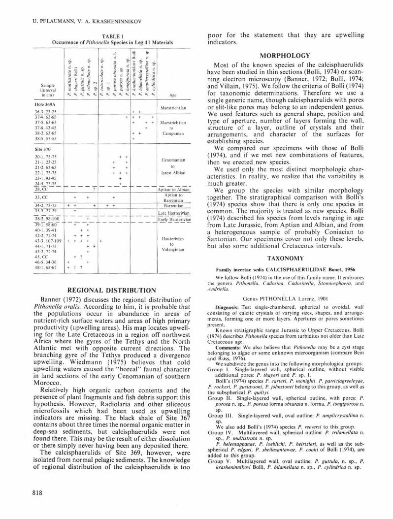
TABLE 1Occurrence of Pithonella Species in Leg 41 Materials
Sample(Interval
in cm)
Hole 369A
36-3, 23-25374,63-6537-5,63-6537-6,434538-2,63-6538-5,53-55
Site 370
20-1,73-7521-1,23-2521-2,63-6522-1,73-7523-1,93-9524-5, 73-7528, CC
33, CC
34-2, 73-7535-5,27-29
38-2,98-10039-1,58-6040-1,394142-2, 72-7443-3, 107-10944-1,71-7345-2, 72-7445, CC46-5,34-3648-1,65-67
mul
tistr
ata
n.
sp.
thay
eri
Bol
ligu
ttul
a n
. sp
.tr
ilam
ella
ta n
. sp
.s
p.2
α, a, o, a, a.
1
+ +
+ + +
+
+
+ +
+ +
+ + +
+ + + +
+ +
+
+ ?
+ +
+ ? ?
tuhe
rcul
ata
n.
sp.
sp. 1
poro
sa o
btur
ata
n. f.
poro
sa n
. sp.
long
ipor
osa
n. s
p.
a, a, a, a, a.
+
+ +
+ +
+ +
+ + +
+
+
+
+ +
+
kras
heni
nnik
ovi
Bolli
bila
mel
lata
n.
sp.
ampl
icry
stal
lina
n. s
p.cy
lindr
ica
n. s
p.
a, a, a, ft,
+ ++ + ++ + +
++ ++
Age
Maestrichtian
Maestrichtianto
Campanian
Cenomanianto
latest Albian
Aptian to AlbianAptian toBarremianBarrem ian
Late HauterivianEarly Hauterivian
Hauterivianto
Valanginian
REGIONAL DISTRIBUTIONBanner (1972) discusses the regional distribution of
Pithonella ovalis. According to him, it is probable thatthe populations occur in abundance in areas ofnutrient-rich surface waters and areas of high primaryproductivity (upwelling areas). His map locates upwell-ing for the Late Cretaceous in a region off northwestAfrica where the gyres of the Tethys and the NorthAtlantic met with opposite current directions. Thebranching gyre of the Tethys produced a divergenceupwelling. Wiedmann (1975) believes that coldupwelling waters caused the "boreal" faunal characterin land sections of the early Cenomanian of southernMorocco.
Relatively high organic carbon contents and thepresence of plant fragments and fish debris support thishypothesis. However, Radiolaria and other siliceousmicrofossils which had been used as upwellingindicators are missing. The black shale of Site 367contains about three times the normal organic matter indeep-sea sediments, but calcisphaerulids were notfound there. This may be the result of either dissolutionor there simply never having been any deposited there.
The calcisphaerulids of Site 369, however, wereisolated from normal pelagic sediments. The knowledgeof regional distribution of the calcisphaerulids is too
poor for the statement that they are upwellingindicators.
MORPHOLOGY
Most of the known species of the calcisphaerulidshave been studied in thin sections (Bolli, 1974) or scan-ning electron microscopy (Banner, 1972; Bolli, 1974;and Villain, 1975). We follow the criteria of Bolli (1974)for taxonomic determinations. Therefore we use asingle generic name, though calcisphaerulids with poresor slit-like pores may belong to an independent genus.We used features such as general shape, position andtype of aperture, number of layers forming the wall,structure of a layer, outline of crystals and theirarrangements, and character of the surfaces forestablishing species.
We compared our specimens with those of Bolli(1974), and if we met new combinations of features,then we erected new species.
We used only the most distinct morphologic char-acteristics. In reality, we realize that the variability ismuch greater.
We group the species with similar morphologytogether. The stratigraphical comparison with Bolli's(1974) species show that there is only one species incommon. The majority is treated as new species. Bolli(1974) described his species from levels ranging in agefrom Late Jurassic, from Aptian and Albian, and froma heterogeneous sample of probably Coniacian toSantonian. Our specimens cover not only these levels,but also some additional Cretaceous intervals.
TAXONOMY
Family incertae sedis CALCISPHAERULIDAE Bonet, 1956
We follow Bolli (1974) in the use of this family name. It embracesthe genera Pithonella, Cado.<sina, Cadosinella, Stomiosphaera, andAndriella.
Genus PITHONELLA Lorenz, 1901
Diagnosis: Test single-chambered, spherical to ovoidal, wallconsisting of calcite crystals of varying sizes, shapes, and arrange-ments, forming one or more layers. Apertures or pores sometimespresent.
Known stratigraphic range: Jurassic to Upper Cretaceous. Bolli(1974) describes Pithonella species from turbidites not older than LateCretaceous age.
Comments: We also believe that Pithonella may be a cyst stagebelonging to algae or some unknown microorganism (compare Beinand Russ, 1976).
We subdivide the genus into the following morphological groups:Group I. Single-layered wall, spherical outline, without visible
additional pores: P. thayeri and P. sp. I.Bolli's (1974) species P. carteri, P. monighti, P. patriciagreeleyae,
P. rockeri, P. gustavsoni, P. johnstonei belong to this group, as well asthe subspherical P. quiltyi.Group II. Single-layered wall, spherical outline, with pores: P.
porosa n. sp., P. porosa forma obturata n. forma, P. longiporosa n.sp.
Group III. Single-layered wall, oval outline: P. amplicrystallina n.sp.We also add Bolli's (1974) species P. veeversi to this group.
Group IV. Multilayered wall, spherical outline: P. trilamellata n.sp., P. multistrata n. sp.P. helentappanae, P. loeblichi, P. heirtzleri, as well as the sub-
spherical P. edgari, P. sheilasantawae, P. cooki of Bolli (1974), areadded to this group.Group V. Multilayered wall, oval outline: P. guttula, n. sp., P.
krasheninnikovi Bolli, P. bilamellata n. sp., P. cylindrica n. sp.
818

CRETACEOUS CALCISPHAERULIDS
TABLE 2Ranges of SEM Investigated Pithonella Species in Jurassic and Cretaceous Sediments
(according to Banner, 1972, Bolli, 1974, and this paper)
eou
uCD03
CJ
Q .Q .
3
?tac
eou
òCD
assi
cJu
r
CD
Q .
Z>
Maestrichtian
Campanian
Santonain
Coniacian
Turonian
Cenomanian
Albian
Aptian
Barremian
Hauterivian
Valanginian
Berriasian
Tithonian
Kimmeridgian
Oxfordian
ri B
olli
hti
Bo
lli
V B
olli
q
mcn
ig
cart
el
I °c -2j "5 "55: c PO <
Pith Pith
II
sp-
stra
ta n
.m
ulti
-2OS
0
11•1
la n
. sp.
gu
ttu
-2δ0
1•1
sp.
ella
ta n
.tr
ilarr
-2
c•0
Pit
h
• i
11••
sp.
2
-2
0
Pit
h
mm
1••
sp.
zula
ta n
.tu
be
r
CO
13)c•o
Pit
h
I
sp.
1
JO
"cTi
c•n
Pit
h
1
c
CO
a o
btu
rai
po
ros
co
αic•ci
Pit
h
m
111
Bo
lli
I
he
len
CO
~δr•r i
Pit
h
|
enza
e B
en
on
ar
1)r-CΛ
Pit
h
|
chi
Bo
lli
, ^
"rhr•
o
Pit
h
oCD
'5Oy>
gus
ta\
CO
Q)
c->
Pit
h
ri B
olli
CD
sJO
Qle•0
Pit
h
soni
Bo
ll
s-2
0
Pit
h
Bo
lli
1CO
1
she
ila
δO
Pit
hB
olli
edg
ar
CO
Cl)Co
Pit
h
i B
olli
qui
/ty
S3
Cllc•
Pit
h
-si
Bo
lli
CO
Cllc•
Pit
hB
olli
.ε
c
.CO
q i
rn
•c
leri
Bo
llih
eir
tz
CO
Cllc•α
Pit
h
l l l l l l l • • ••
ae B
olli
iag
reel
eyp
a tr
ie
"δr•Cl
Pit
h
Bo
llisp
. A
JO
Qlc•Cl
Pit
h
3 n.
sp
.
|
CO
"cTi
0
Pit
h
sp.
ooro
sa n
.
1-2
Pit
t
ann)
(Ka
ufm
ova
lis
-2δ
o
Pit
h
|
ton
ei
Bo
John
s
-2
cü
Pith
mI
•
Bol
lico
oki
-2CD
co
•1•
Bo
llisp
. B
-2
£
Pith
•I
•
Bo
llisp
. C
-2CD
o
Pith
•I
•
Bo
llisp
. D
-2CD
cü
Pith
1I
// B
olli
or ^
cP
kras
h
JO
0)c•o
Pith
1
1
I
a n.
sp.
crys
tal/i
r
|
c-
Pith
1
n
irica
n. s
cyfin
t
-2δ
o
Pit
h
1
ds
ella
ta n
.b
ilam
-2"05co
•g
11
The criteria used to establish these groups include test shape,presence or absence of main or slit-like apertures, and quantity ofdifferent layers.
Pithonella thayeri Bolli(Plate 1, Figures 1-5)
Pithonella thayeri Bolli, 1974, p. 853, pi. 1, fig. 9-12, pi. 8, fig. 9-12,pi. 9, fig. 1-12, pi. 21, fig. 3.Description of species: Test globular to subspherical, composed of
a single-layered wall of about 6 µm in thickness which is formed bycrystals 5 to 12 µm in diameter. The outer surface is very roughalthough single crystals seen on the outer surface are mostly rounded.Apertures were not seen with one exception of a questionablespecimen (Plate 1, Figure 3), whose outer surface is not as coarse as istypical. Its aperture measures 25 µm in diameter and is surrounded byradially oriented plate-like crystals 3 × 2 × 2 µm in size. However,the crystal size and shape away from the aperture takes on more theoutline of a normal P. thayeri. We have not observed an inner surfacebecause all broken specimens were filled with sediment.
Sometimes the interspaces between coarse crystals are not filledwith finer ones so that the surface pseudopores remain open.Diagenesis may be a major factor controlling the outline of the outertest surface so it is possible that our Pithonella thayeri is only avariant of another species.
There are affinities to Pithonella trilamellata n. sp., but the multi-layered character is missing. Intergrades are assigned to Pithonellatuberculata n. sp., which is recorded in only one sample and it has adouble-layered texture. The outer surface is similar to the double-layered P. loeblichi. (Dimensions: diameter 45-80 µm.)
Occurrence: Section 370-48-1 to Sample 370-33, CC.Age: Valanginian to Barremian.Bolli (1974) recorded this species from Upper Oxfordian to
Tithonian sediments off NW-Australia.
Pithonella sp. 1(Plate 1, Figures 6a, b)
Description of species: Test globular, the outer surface formed byobliquely arranged, nearly smooth large crystals of different sizes.
819

U. PFLAUMANN, V. A. KRASHENINNIKOV
This arrangement suggests there may have been a diageneticinfluence. There seems to be remnants of very small crystals betweenthe large ones like that of the inner layer of Pithonella guttula n. sp.
Only one sample (370-34-2) yielded specimens of this type, so it isquestionable whether or not this is a separate species.
Dimensions: Diameter around 50 µm.Occurrence: Section 370-34-2, rare.Age: Barremian.
Pithonella porosa n. sp.(Plate 2, Figures 3, 5; Plate 3, Figures 1, 2)
Derivatio nominis: porosa (Latin): with pores.Holotype: Plate 2, Figures 4a, b.Paratypes: Plate 2, Figures 5a, b, c; 6a, b, c; 7.Type locality: DSDP Leg 41, Sample 370-24-5, 73-75 cm.Type level: Latest Albian to Cenomanian.Diagnosis: Test globular, consisting of a single calcite layer,
penetrated by irregularly distributed rounded to elongate pores.Description: Test globular, consisting of one layer of about 6 µm
in thickness. The crystal growth, as seen from the outside, variesgradually from the interior (where the diameters visible are around0.2 µm) to the outer surface (where the diameters measure between 2and 5 µm). The crystals at the outer surface have a fairly irregular,closely packed arrangement. Pores of about 4 µm diameter areirregularly distributed but with nearly equal distances between them(12 to 15 µm). The shapes of the pores vary between circular andelongate, the longest slits observed measure 8 µm in length. The poresare sometimes surrounded by rosette-like crystals, which may be ofdiagenetic origin.
A main aperture of about 20 µm diameter is present, in rare cases,showing no special patterns of crystal structure or arrangement in itssurroundings.
Specimens from the deeper parts of Site 370 have pores that areclosed and not visible at the outer surface. In these specimens, theouter surface consists of calcite crystals with diameters between 1 and7 µm without a visible special pattern. We describe these forms asPithonella porosa forma obturata, new form.
Dimensions of holotype: Diameter 74 µm.Dimensions of paratypes: Diameter 65-75 µm.Occurrence: DSDP Leg 41 Site 370, Sections 24-5 to 20-1.Age: Latest Albian {Rotalipora apenninica Zone) to Ceno-
manian/Turonian.Pithonella porosa n. sp. differs from Pithonella longiporosa by
having an outer surface that is more coarsely crystalline and roundedpores being more common than in Pithonella longiporosa. Inaddition, the slit-like pores are less strongly marked.
Pithonella porosa n. sp. forma obturata n. forma(Plate 3, Figures 1-3)
Derivatio nominis: obturata (Latin): obturated.Type specimen: Plate 3, Figures la, b.Type locality: DSDP Leg 41, Site 370, Sample 21-2 (63-65 cm).Type level: Latest Albian to Cenomanian.Diagnosis: A new form of Pithonella porosa n. sp., with closed
pores.Description: Test globular, distinct aperture not recorded, wall
consisting of one layer, formed by crystals which are randomlydistributed in a radial array. As observed from the outer surface, theirsizes vary between 1 and 7 µm in diameter, without visible specialpatterns.
Occasionally specimens with a more or less circular "opening"were found. The diameter of the openings range up to 40% of thediameter of the test. These openings are considered artifacts becausethe borders have irregular outlines and no special wall structuresaround the opening were detected. The inner surface of the test isformed like that of Pithonella porosa n. sp.: small crystals about 0.5µm diameter form a relatively rough but even inner surface. Pores of5 µm diameter are visible but closed by rough crystals whichsometimes grow into the interior with crystals elevating the normalinner surface. The pores are numerous and the distances betweenthem are about 4 to 5 µm.
There are some specimens in Sample 370-21-2, 63-65 cm, whichshow pore-like openings of about 3 µm diameter on the outer surface.These specimens show an intergradation to Pithonella porosa n. sp.,the differences being only additional, possibly diagenetic crystalgrowth.
Dimensions: Diameter 65-80 µm.Occurrence: Sections 370-34-2 to 370-21-1.Age: Barremian to Cenomanian/Turonian.
Pithonella longiporosa n. sp.(Plate 2, Figures I, 2; Plate 3, Figure 3)
Derivatio nominis: longiporosa (Latin): with elongated pores.Holotype: Plate 2, Figures la, b, c.Paratypes: Plate 2, Figures 2a-c; Figures 3a, b.Type locality: DSDP Leg 41, Sample 370-21-1, 23-25 cm.Type level: Latest Albian to Cenomanian.Diagnosis: Test spherical, single-layered, penetrated by rounded
and slit-like pores.Description: Test spherical, consisting of one layer of about 5 µm
thickness. Crystals visible on the outer surface somewhat irregularbetween 1 and 5 µm in diameter. The crystals are oriented randomlylooking perpendicular to the outer surface or sometimes oblique likea pile. Their arrangement is closely packed. There are two types ofpores visible on the outer surface: rounded ones with diameters up to7 µm, and elongated, slit-like pores, up to 20 µm in length and 2 to 3µm in width. The elongated pores are arranged nearly at right anglesto one another, like meridians and parallels although direct polescould not be detected. The distances between the rounded and theslit-like pores are nearly equal. In some cases, it looks like ameridional arrangement of the rounded pores in the same manner asshown by the slit-like pores. A prominent pore or aperture was notencountered.
Dimension of holotype: Diameter 78 µm.Dimensions of paratypes: Diameter 65-75 µm.Occurrence: Sections 370-22-1 to 370-20-1.Age: Uppermost Albian to Cenomanian/Turonian.Remarks: Pithonella longiporosa differs from Pithonella porosa in
its strongly elongated, slit-like pores.
Pithonella amplicrystallina n. sp.(Plate 3, Figures 4-7)
Derivatio nominis: amplus (Latin): big, wide; crystallinus (Latin):crystalline.
Holotype: Plate 3, Figures 5a-c.Paratypes: Plate 3, Figures 4a-c, 6, 7a-d.Type locality: DSDP Leg 41, Sample 369A-37-6, 43-45 cm.Type level: Maestrichtian.Diagnosis: Test ovoidal, single-layered, composed of elongated
calcite crystals with the long axis nearly parallel to the test surface.Description: Test ovoidal, composed of a single layer of elongated
calcite crystals, which are relatively loosely arranged with the longaxis parallel or slightly oblique to the test surface. The thickness ofthe smallest diameters of the crystals is about 1 µm, the length about 4µm, the thickness of the wall is about 3 µm. An aperture is present inall investigated specimens. It is located symmetrically at one end ofthe test parallel to the longest axis. The diameter of the aperturevaries between 15 and 20 µm. There are no significant differences inthe wall structure around the aperture relative to other parts of thetest.
Dimensions of holotype: Long axis 90 µm, diameter 68 µm.Dimensions of paratypes: Long axis about 90 to 96 µm, diameter
68 to 72 µm.Occurrence: 369A-37-5.Age: Maestrichtian.This species differs from Pithonella krasheninnikovi Bolli mainly
by a larger size of the crystals forming the surface and the mono-lamellar wall. The inner layer of rough crystals apparent in Pithonellakrasheninnikovi could not be detected. Pithonella veeversi is alsoelongated and single-layered, but its outer surface is built up ofsubtriangular crystals and its aperture is larger.
Pithonella tuberculata n. sp.(Plate 1, Figures 7, 8)
Derivatio nominis: tuberculata (Latin): with tubercules.Holotype: Plate 1, Figures 7a-c.Paratype: Plate 4, Figure 8.Type locality: DSDP Leg 41, Sample 370-43-3, 107-109 cm.Type level: Late Valanginian to early Hauterivian.Diagnosis: Test double-layered, subspherical, with single pores
and big tubercules on the outer surface.
820

CRETACEOUS CALCISPHAERULIDS
Description: The subspherical tests show a very rough surfaceunder the light microscope. Magnifications by SEM reveals a surfaceformed by small crystals of about 0.5 to 1 µm in diameter. Therugosity is a result of big tubercules, composed of larger crystalsarranged in piles. Single pores of 3 to 6 µm in diameter are visible atthe outer surface, but they have no special position. The wall iscomposed of at least two layers; the outer one is 1 µm in thickness andis formed by oblique radially grown large crystals. The inner layer isbuilt up of very small, randomly orientated, elongate crystals withdiameters around 0.1 to 0.2 µm. The thickness of the inner layer isabout 2 µm.
Dimensions of holotype: Diameter around 50 µm.Occurrence: Sample 370-43-3, 107-109 cm, rare.Age: Late Valanginian-early Hauterivian.Remarks: This species is similar to Pithonella thayeri, but differs
from that by the tubercules, the somewhat smaller crystals on thesurface, and in having a multilayered wall. It differs from Pithonellaguttula n. sp. by having tubercules and in its globular outline.
Pithonella trilamellata n. sp.(Plate 6, Figures 3-5, 7; Plate 7, Figures 1, 2)
Derivatio nominis: trilamellata (Latin): three-layered.Holotype: Plate 6, Figures 3a-c.Paratypes: Plate 6, Figures 4, 5, 7.Type locality: DSDP Leg 41, Sample 370-33, CC.Type level: Barremian to Aptian.Diagnosis: Test globular, consisting of three main calcite layers,
the outermost consisting of plate-like crystals, the intermediate one iscomposed of randomly oriented stick-like crystals, and the innermostlayer is built up of randomly oriented, stick-like, fine crystals.
Description: Test globular to slightly ovoidal, consisting of threemain layers. The outermost layer is 3 to 4 µm in thickness and isformed by large crystals of up to 10 µm in diameter at the outersurface. The surface of the crystals is plate-like so that the outersurface of the test looks like a pavement. The thickness of theintermediate layer reaches up to 4 µm. There is a transition to anothertype of crystals at the base of the outer layer which have diameters of1 µm and which are oriented oblique to the radius. They are arrangedcrosswise at different angles with empty interspaces between them.The base of the medium layer is formed by a lining, probably oforganic matter (Plate 7, Figure 2). The surface of the inner layer issometimes difficult to detect. The outer surface of the inner layer isseen as a polished cutting of randomly oriented and relatively looselypacked, very fine crystals where corrosion has removed this coating.The diameter of these crystals is 0.2 to 0.5 µm. The thickness of theinner layer is about 5 µm.
Single, large, well-rounded apertures are present, but not visible inall specimens.
Dimensions of holotype: Diameter 80 µm.Dimensions of paratypes: Diameter 50 to 80 µm.Occurrence: DSDP Leg 41, Samples 370-46-2, 34-36 cm, to 370-
33, CC.Age: Valanginian to Barremian, and possibly ? Aptian-Albian.Remarks: Pithonella trilamellata n. sp. strongly resembles the
three-layered Pithonella helentappanae Bolli from the Albian offnorthwest Australia. Differences are given in the thickness of theinnermost layer, which in Pithonella trilamellata n. sp. is nearly thesame as the outer layer, whereas in Pithonella helentappanae the innerlayer is very thin. The characteristic outermost test surface ofPithonella trilamellata n. sp. cannot be seen in the figures given byBolli (1974) for Pithonella helentappanae.
Our species is quite similar to Pithonella multistrata n. sp., but inthe latter the outermost layer is missing and the boundary betweenthe intermediate and inner layer is more pronounced by differentcrystal sizes in P. trilamellata. In some cases, only the innermost layeris represented. These specimens have a very glassy surface under thelight microscope. They resemble to some degree Pithonellapatriciagreeleyae Bolli, but in the cross-section figured by Bolli (1974)the latter species has thicker and more compacted crystals.
Pithonella multistrata n. sp.(Plate 7, Figures 3-6)
Derivatio nominis: multistrata (Latin): composed of many layers.Holotype: Plate 7, Figures 3a-f.Paratypes: Plate 7, Figures 4a, b, 5a, b, 6a, b.Type locality: DSDP Leg 41, Sample 370-43-3, 107-109 cm.
Type level: Valanginian to Hauterivian.Diagnosis: Test globular, wall consisting of several different
calcite layers, separated by organic linings. Outer layer coarselycrystalline, crystals oriented oblique radially, inner layer finelycrystalline, crystals randomly grown.
Description: Test globular, consisting of several layers. The outersurface is formed by crystals which are arranged like a pavement withsome interspaces. The crystal size, as seen from the outer surface, isaround 2 µm, but smaller crystals of around 1 µm also exist as domore rarely, larger ones with diameters up to 6 µm. The latter aredistributed in irregular patterns across the outer surface. The surfacehas a mat appearance under the light microscope. The outer layer isabout 7 µm in thickness. The crystals are loosely arranged andoriented crosswise oblique to the radius of the test. The angle betweenthe crystals is about 60 . Sometimes a very thin coating which caneasily be removed at the outer surface is observed. It is considered tobe the residual of an outermost layer, similar to that of Pithonellatrilamellata n. sp.
The outer layer can be divided using crystal diameters into anouter part with diameters of around 1 µm, and an inner part of 1 µmin thickness where the crystals have diameters below 0.2 µm. Theinner surface of the outer layer is smooth. The boundary betweenthese two parts is not sharp, but rather the different crystals areintermingled. A residual of a thin lining of possibly organic matterwas found between the outer layer and the inner layer. The outersurface of the inner layer has the same outline as the inner surface ofthe outer layer.
The inner layer is about 7 to 8 µm in thickness and composed ofirregularly arranged crystals of 0.2 µm in diameter and up to 1 µm inlength. The inner surface of the inner layer is only partly visible andhas a structure similar to the cross-section. The inner layer to theinterior is covered by a flexible lining which shows no structures athigh magnifications under the SEM. It is believed to be organicmatter. An aperture of about 20 µm diameter has been observed insome specimens.
Dimensions: Diameter 40 to 70 µm.Occurrence: Sections 370-43-3, 370-34-2, (?) 370-48-1, rare.Age: Valanginian to Barremian.Taxonomy: The outer surface and the cross-section of the outer
layer are very similar to those of Pithonella johnstonei Bolli (1974, pi.6, fig. 5, 6, 8) from the Senonian, but the crossing angle of crystals inPithonella multistrata is around 60°, not 90° as in Pithonellajohnstonei. Additionally, Pithonella johnstonei is described as a single-layered form without an inner lining. Pithonella quiltyi from theAlbian is also closely related, but an inner lining was not described byBolli (1974).
Pithonella guttula n. sp.(Plate 8, Figures 1-4)
Derivatio nominis: guttula (Latin): small drop.Holotype: Plate 8, Figures la-e.Paratypes: Plate 8, Figures 2-4.Type locality: DSDP Leg 41, Sample 370-40-1, 39-41 cm.Type level: Valanginian to Hauterivian.Diagnosis: Test ovoidal, wall consisting of several layers; the outer
one formed by big plate-like crystals and the intermediate layer iscomposed of finer crystals, oblique radially oriented with a sharpboundary with the innermost layer. The inner layer is composed ofrandomly oriented, fine, needle-like crystals.
Description: Test generally subspherical to drop-like. Somespecimens have been distinguished with a knob-like protrusion at onepole, but without a visible aperture. The surface structure in thatregion is not different from that of other parts of the test. The wallconsists of several layers. The outer surface is formed by irregularlydistributed crystals of various sizes between 4 and 16 µm in diameteron the surface. As seen in cross-section, the outer layer is about 7 µmin thickness with large crystals oriented oblique to the radius. Belowthese big plate-like crystals, there are smaller ones of more stick-likeoutline also oriented oblique to the radius and arranged in pilestructures. The boundary between the large crystals and the smallerones is transitional, whereas the boundary with the inner layer issharply marked by an even layer of a thickness too small to measure.Because there are no structures visible at high SEM magnifications,this layer is believed to be organic matter. Below this lining there isthe inner layer formed by loosely arranged, needle- to plate-likecrystals with diameters of about 0.2 µm and lengths up to 3 µm. Theirorientation is random (Plate 8, Figures lb, c). The thickness of the
821

U. PFLAUMANN, V. A. KRASHENINNIKOV
inner layer is more than 5 µm, but we have not yet recorded a goodsection through the inner layer.
The big crystals at the outer surface are isolated and the spacesbetween them are open or filled by smaller crystals, sometimes ofmore stick-like shape. Although the outer surface is composed ofquite large components, its general surface is smooth, like a looselyarranged pavement.
Dimension of holotype: Length 88 µm, diameter 72 µm.Dimension of paratypes: Length 85 to 97 µm, diameter 67 to 82
µm.Occurrence: Sample 370-45, CC to Section 370-40-1.Age: Valanginian to Hauterivian.Remarks: Pithonella guttula n. sp. differs from Pithonella
trilametlata n. sp. in its ovoidal (not spherical) outline and in its finercrystalline innermost layer. Pithonella multistrata n. sp. lacks thecoarse crystalline outer layer and is globose, not drop-Hke.
Pithonella krasheninnikovi Bolli(Plate 4, Figures 1-6)
02Pithonella krasheninnikovi Bolli, 1974, p. 856, pi. 7, fig. 1-5,pi. 18, fig. 10-12, pi. 19, fig. 1-12, pi. 20, fig. 1-4, pi. 24, fig. 1,2.Description: Test ovoidal, consisting of two layers. The outer layer
(2 to 5 µm in thickness) is formed by irregularly arranged smallcrystals with the longest axis parallel or slightly oblique to the outersurface. The smallest diameters are around 0.8 to 1.2 µm. Externally,the crystals are clearly separated from each other with straight edges.Their sizes range 0.6 to 1 × 1.6 × 3 to 4 µm. Some larger crystals of1.6 to 2.6 µm in diameter may occur in the outer layer. A somewhatporous surface is given by the loosely arranged small crystals.
The inner layer is about 6 µm in thickness and consists of heavycrystals. The aperture is positioned a bit asymmetrically at one end,its size varies between 20 and 35 µm in diameter. Occasionally, it issurrounded by thick crystals of the inner layer. In tliose cases, theouter layer may have been partly removed. We encounteredspecimens in some samples whose outer layer was partly or totallyremoved.
Pithonella krasheninnikovi has been described as distinctlyelongate with a width/length ratio of approximately 1:2. Variabilityof our specimens is between 1:2 and 1:1.2.
Dimensions: Length up to 110 µm, thickness: around 60 µm.Occurrence: Sections 369A-38-5 to 369A-37-4.Age: Campanian to early Maestrichtian.Remarks: Pithonella krasheninnikovi differs from Pithonella
bilamellata n. sp. in the more elongated shape of the test and in themore tangentially oriented crystals of the outer layer. Pithonellamcknighti has a similar surface, but coarser crystals, and is single-layered. Pithonella robinsoni is double-layered and ovoidal likePithonella krasheninnikovi, but the outer layer is composed of twodifferent crystal types. Pithonella edgari is double-layered as well, butthe inner layer is composed of crystals with shape and arrangementsimilar to the outer layer, and not of heavy crystals like those ofPithonella krasheninnikovi.
Pithonella krasheninnikovi was initially described by Bolli (1974)from the Upper Cretaceous, probably Coniacian to Santonian, fromDSDP Leg 27 Site 260 in the Eastern Indian Ocean off northwestAustralia. Its range is now extended to include early Maestrichtian.
Pithonella cylindrica n. sp.(Plate 5, Figures 1-4)
Derivatio nominis: cylindrus (Latin): cylinder.Holotype: Plate 5, Figures 4a-c.Paratypes: Plate 5, Figures 1-3.Type locality: DSDP Leg 41, Sample 369A-37-4, 63-65 cm.Type level: Campanian to Maestrichtian.Diagnosis: Test elongated ovoidal with nearly cylindrical flanges,
composed of two layers of crystals; the outer formed by obliquetangentially arranged small crystals, and the inner layer composed ofdensely packed large crystals.
Description: Test of elongated shape, the flanges being nearlycylindrical and the poles subspherical. The wall is built up of twolayers. The outer layer consists of small crystals, the longest axis ofwhich are oriented oblique tangentially to the surface. The smallerdiameters of the plate-like crystals are between 0.6 and 1.6 µm. Thelongest diameters are about 2 µm. The thickness of the outer layer isabout 2 µm.
The inner layer is composed of one layer of large, densely packedeuhedral crystals, radially oriented. The inner layer is about 3 to 4 µmin thickness. An aperture is asymmetrically positioned at one pole.
Dimensions of holotype: Length 100 µm, diameter 62 µm.Dimensions of paratypes: Length 96 to 108 µm, diameter 60 to 64
µm.Occurrence: DSDP Leg 41, Samples 369A-37-4, 63-65 cm, 369A-
37-5, 63-65 cm.Age: Campanian to Maestrichtian.Remarks: Pithonella cylindrica n. sp. is distinguished from
Pithonella krasheninnikovi by its elongated shape with nearlycylindrical flanges, whereas Pithonella krasheninnikovi has a morespindle-like outline.
Pithonella bilamellata n. sp.(Plate 5, Figures 5, 6; Plate 6, Figures 1, 2)
Derivatio nominis: bi (Latin): double, lamellata (Latin): layered.Holotype: Plate 5, Figures 6a-d.Paratypes: Plate 5, Figure 5; Plate 6, Figures 1, 2.Type locality: DSDP Leg 41, Sample 369A-36-3, 23-25 cm.Type level: Early Maestrichtian.Diagnosis: Test ovoidal, consisting of two layers, the outer formed
by oblique radially oriented, small crystals, the inner formed bydensely packed, large crystals, radially oriented.
Description: Test ovoidal, consisting of two layers of calcitecrystals. The outer layer is composed of short, somewhat irregularlyradially arranged crystals which tend to have an oblique position.They are clearly separated from each other, sometimes with inter-spaces, producing a pseudoporous surface. The short diameters of thecrystals are around 0.8 µm, and their longest axis is about 2 µm,which is also the thickness of the outer layer.
The thickness of the inner layer is up to 8 µm. The inner layer isformed by heavy, densely packed, radially arranged, crystals. Theboundary between the two layers is sharp. Sometimes the outer layeris partly removed.
A wide opening with irregular borders at one end may be anaperture.
Dimensions of holotype: Length 104 µm, diameter 68 µm.Dimensions of paratypes: Length up to 110 µm, diameter 60 to 64
µm.Occurrence: DSDP Leg 41, Hole 369A, Sections 38-2 to 36-3.Age: Upper Cretaceous, Campanian to Maestrichtian.Lithology of the samples: Nanno-bearing marl and nanno-
marlstone.Remarks: Pithonella bilamellata n. sp. differs from P. krashenin-
nikovi in having a more shortened oval shape, and in more radiallyarranged crystals of the outer layer. P. robinsoni has an outer layerconsisting of two different crystal types, which we have notencountered in the new species. The inner layer in P. edgari is not ascoarsely crystalline as in P. bilamellata n. sp.
Pithonella sp. 2(Plate 8, Figure 5)
Description: Test globular to slightly ovoidal. The outer surfaceshows a pavement-like structure. Crystals of about 4 to 10 µm indiameter are intermixed with some smaller ones to form anirregularly polygonal pattern. The texture of the surface is rathereven, although the edges of the crystals have holes producing asomewhat porous outline in the SEM views. Distinct apertures werenot found. The species is given in open nomenclature, because we donot know the precise structure of the wall. It may be a variant ofPithonella trilamellata.
Dimensions: Diameter 65 to 95 µm.Occurrence: DSDP Leg 41, Samples 370-44-1, 71-73 cm and 370-
34-2, 73-75 cm, rare.Age: Valanginian to Barremian.Remarks: The range falls in the range of Pithonella trilamellata.
We found some other specimens with very smooth surfaces (comparePlate 8, Figure 6). Identification is difficult because these forms maybe only parts of specimens with the outer layers removed.
ACKNOWLEDGMENTS
We wish to acknowledge the Deep Sea Drilling Project forproviding the opportunity to participate on Leg 41 of GlomarChallenger and the Geological Institute of the Academy of
822

CRETACEOUS CALCISPHAERULIDS
Sciences of the USSR and the Geological-PaleontologicalInstitute of the Kiel University for leave.
Technical help included that from Mrs. C. Schulz, Mr. W.Reimann, and Mr. W. Rosier. The English text was revised byJ. Gardner.
This contribution was supported by Deutsche Forschungs-gemeinschaft (German research society).
REFERENCES
Banner, F.T., 1972. Pithonella ovalis from the early Ceno-manian of England: Micropaleontology, v. 18, p. 278.
Bein, A. and Reiss, Z., 1976. Cretaceous Pithonella fromIsrael: Micropaleontology, v. 22, p. 83.
Bolli, H.M., 1974. Jurassic and Cretaceous Calcisphaerulidaefrom DSDP Leg 27, Eastern Indian Ocean. In Veevers,
J.J., Heirtzler, J.R., et al., Initial Reports of the Deep SeaDrilling Project, Volume 27: Washington (U.S. Govern-ment Printing Office), p. 843.
Villain, J.-M., 1975. "Calcisphaerulidae" (incertae sedis) duCretacé superieur du Limbourg (Pays-Bas), et d'autreregions: Palaeontogr., A, v. 149, p. 193.
Wiedmann, J., 1975. Faunenprovinzen der westmediterranenKreide: Abstracts of papers, Palaont. Ges. 45. Jahresver-samml. Hannover.
EXPLANATIONS TO PLATES
Figures with the same number but different lettersbelong to the same specimen. Following the magnifica-tion is the gun potential and the number of the scan-ning electron microscope photograph.
823

U. PFLAUMANN, V. A. KRASHENINNIKOV
PLATE 1
Figure 1 Pithonella thayeri Bolli.Sample 41-370-35-5, 27-29 cm.la. Lateral view, showing the irregular distribution of subrounded
large and small crystals. ×500, 20 kv, No. 53003.lb. Detail of the surface from the central part of Figure la; note the
pseudopores. X2500, 20 kv, No. 53004.
Figure 2 Pithonella thayeri Bolli.Sample 41-370-24-5, 73-75 cm.2a. Lateral view, showing pustule-like engrown large crystals between
smaller ones with plate-like surfaces. ×500, 20 kv, No. 53308.2b. Detail of the surface. The surface is smooth between the large
crystals and may be an artificial coating of gelatine during mount-ing procedure. ×2500, 20 kv, No.52309.
Figure 3 Pithonella thayeri ?Bolli.Sample 41-370-35-5, 27-29 cm.3a. Apertural view of a specimen, which may be P. thayeri. The
aperture is surrounded by small crystals. ×500, 20 kv, No. 52990.3b. Detail of the lateral outer surface, showing irregularly shaped pore-
like openings between crystals of different sizes. X2500, 20 kv, No.52991.
Figure 4 Pithonella thayeri Bolli.Sample 41-370-35-5, 27-29 cm.4a. Lateral view to a specimen with partly removed wall. ×500, 20 kv,
No. 52998.4b. Detail of the broken wall, large crystals are partly grown on very
small ones. The ultrastructure of the surface of the filling is anegative of the inner surface of the wall, but could not be resolvedat higher SEM magnifications. X2500, 20 kv, No. 53000.
Figure 5 Pithonella thayeri Bolli.Sample 41-370-34-2, 73-75 cm.5a. Lateral view of a specimen with very large plate-like crystals
irregularly distributed. ×500, 20 kv, No. 53656.5b. Detail of the surface with signs of corrosion. Note the small holes
on the big crystals. ×2500, 10 kv, No. 53655.
Figure 6 Pithonella sp. 1.Sample 41-370-34-2, 73-75 cm.6a. Specimen with oblique and irregularly imbricated large crystals at
the surface. ×500, 10 kv, No. 53671.6b. Detail of the outer surface, with twin crystallization. ×2500, 10 kv,
No. 53670.
Figure 7 Pithonella tuberculata n. sp. (holotype).Sample 41-370-43-3, 107-109 cm.7a. Lateral view. The holes are enlarged due to corrosion. ×500, 10 kv,
No. 53746.7b. Detail of the outer surface. The wall is multilayered. To the lower
right of the hole, the outer portion of the intermediate crystallinelayer is removed, showing a fine crystalline surface of the innerlayer. X2500, 10 kv, No. 53747.
7c. Detail of some pustules formed by an agglomeration of largecrystals, smoothly textured similar to the outer surface of the areabetween the pustules. X25OO, 10 kv, No. 53748.
Figure 8 Pithonella tuberculata n. sp.Sample 41-370-43-3, 107-109 cm.Lateral view to a paratype. X500, 10 kv, No. 53749.
824

CRETACEOUS CALCISPHAERULIDS
PLATE 1
825

U. PFLAUMANN, V. A. KRASHENINNIKOV
PLATE 2
Figure 1 Pithonella longiporosa n. sp. (holotype).Sample 41-370-21-1, 23-25 cm.la, lc. View from different sides of the same test, angle between the
views 77°. The slit-like pores and the connecting lines between therounded ones cross each other at nearly right angles. ×500, 10 kv,(a) No. 53538, (c) No. 53549.
lb. Detail of the outer surface. The crystals show very irregular edgesand holes which may be due to corrosion of the outermost layer.The crystal size is larger around the pores and the orientation is atnearly right angles to the pore margin (lower left). X25OO, 10 kv,No. 53539.
Figure 2 Pithonella longiporosa n. sp.Sample 41-370-21-1, 23-25 cm.2a. Artificially broken specimen. ×500, 10 kv, No. 53530.2b. View to the broken wall, showing a monolamellar layer of
irregularly oblique-oriented crystals. X2500, 10 kv, No. 53533.2c. View of the inner surface of the wall showing the smooth fine
crystals. The arrangement of crystals around the pores is nottypical. X25OO, 10 kv, No. 53531.
Figure 3 Pithonella longiporosa n. sp.Sample 41-370-21-1, 23-25 cm.3a. Lateral view of a specimen looking at a break, not aperture. ×500,
10 kv, No. 53540.3b. Cross-break of the wall, showing the single layer of more or less
radially oriented crystals. X25OO, 10 kv, No. 53541.
Figure 4 Pithonella porosa n. sp. (holotype).Sample 41-370-24-5, 73-75 cm.4a. Peripheral view showing a large opening and narrow slit-like
rounded pores. ×500, 20 kv, No. 53012.4b. Transverse section of the wall showing a single layer of oriented
crystals. ×2500, 20 kv, No. 53013.
Figure 5 Pithonella porosa n. sp.Sample 41-370-23-1, 93-95 cm.5a. Artificially broken specimen showing the inner surface and the wall
structure. A marcasite crystal is attached at the upper left. ×500, 10kv, No. 53715.
5b. Cross-section of the monolamellar wall. The more or less radiallyoriented crystals have increasingly larger diameters toward theouter surface. Compare Figure 5c. X25OO, 10 kv, No. 53716.
5c. Detail of the inner surface. The diameters of the crystals in thatview are nearly equal, the pores are partly closed by large engrowncrystals. X25OO, 10 kv, No. 53717.
Figure 6 Pithonella porosa n. sp.Sample 41-370-22-1, 73-75 cm.6a. Lateral view. ×500, 10 kv, No. 53731.6b. Detail of the outer surface around an elongated pore. X2500, 10 kv,
No. 53732.
Figure 7 Pithonella porosa n. sp.Sample 41-370-20-1, 73-75 cm.Lateral view to a specimen with a circular main aperture. ×500, 10 kv,No. 52401.
826

CRETACEOUS CALCISPHAERULIDS
PLATE 2
827

U. PFLAUMANN, V. A. KRASHENINNIKOV
PLATE 3
Figure 1 Pithonella porosa n. sp. forma obturata n. forma.Sample 41-370-21-2, 63-65 cm.la. View of a specimen with very small pores.
The distances between the pores are nearlythe same as those in Figure lb. ×500, 10 kv.No. 53518.
lb. Detail of the outer surface showing theirregular arranged large and small crystals,without any special arrangement in thevicinity of the pores. X25OO, 10 kv, No.53517.
Figure 2 Pithonella porosa n. sp. forma obturata n. forma.Sample 41-370-22-1, 73-75 cm.2a. Lateral view without visible pores. ×500, 10
kv, No. 52725.2b. Detail of the outer surface with cobblestone
crystals of different sizes. X2500, 10 kv, No.52726.
Figure 3 Pithonella porosa n. sp. forma obturata n. forma.Sample 41-370-22-1, 73-75 cm.Detail of a broken specimen, showing the mono-lamellar wall structure and the fine crystallineinner surface of the wall. ×25OO, 10 kv,No. 53736.
Figure 4 Pithonella amplicrystallina n. sp.Sample 41-369A-37-6, 43-45 cm.4a. Lateral view. ×500, 20 kv, No. 53257.4b. Detail of the apertural region. X25OO, 20 kv,
No. 53258.4c. Distal view. ×500, 20 kv, No. 53257.
Figure 5 Pithonella amplicrystallina n. sp. (holotype).Sample 41-369A-37-5, 43-45 cm.5a. Lateral view. ×500, 20 kv, No. 53249.5b. Oblique view to the apertural area, with a
monolamellar wall. X25OO, 20 kv, No.53250.
5c. Detail of the lateral outer surface. X25OO, 20kv, No. 53248.
Figure 6 Pithonella amplicrystallina n. sp.Sample 41-369A-37-6, 43-45 cm.Detail of the lateral outer surface showing thenearly equal size of the crystals as occurs in Figure5c. X2500, 20 kv, No. 53239.
Figure 7 Pithonella amplicrystallina n. sp.Sample 4I-369A-37-5, 63-65 cm.7a. Lateral view. ×500, 20 kv, No. 53195.7b. Detail of the lateral outer surface. X25OO, 20
kv, No. 53194.7c. Detail of the monolamellar wall at the distal
end. X2500, 20 kv, No. 53196.7d. Distal view. The aperture is enlarged by frag-
mentation. X500, 20 kv, No. 53208.
828

CRETACEOUS CALCISPHAERULIDS
PLATE 3
829

U. PFLAUMANN, V. A. KRASHENINNIKOV
PLATE4
Figure 1 Pithonella krasheninnikovi Bolli.Sample 41-369A-37-5, 63-65 cm.la. Lateral view. ×500, 20 kv, No. 53201.lb. Distal view. ×500, 20 kv, No. 53213.lc. Detail of the lateral outer surface. X2500, 20
kv, No. 53202.
Figure 2 Pithonella krasheninnikovi Bolli.Sample 41-369A-37-4, 63-65 cm.Distal view of a specimen with outer layerremoved. ×500, 20 kv, No. 53193.
Figure 3 Pithonella krasheninnikovi Bolli.Sample 41-369A-38-2, 63-65 cm.3a. Lateral view. ×500, 10 kv, No. 53297.3b. Detail of the distal end showing the thin
outer layer of fine crystals covering the innerlayer of coarse crystals. The edges of theaperture are irregularly bordered indicatingpart of the wall has been removed. ×500, 10kv, No. 53304.
3c. Distal view showing an aperture or a brokensection of the test at the distal end. ×500, 10kv, No. 53303.
3d. Detail of the lateral outer wall, looselyarranged fine crystals partly surroundingirregular polygons. X25OO, 10 kv, No. 53296.
Figure 4 Pithonella krasheninnikovi Bolli.Sample 41-369A-38-5, 53-55 cm.4a. Lateral view. The outer layer is mostly
removed. ×500, 10 kv, No. 53510.4b. Detail of the lateral surface. The heavy
crystals of the inner layer are covered by thefine crystalline outer layer. ×2500, 10 kv,No. 53511.
4c. Distal view. The aperture is filled withsediment. ×500, 10 kv, No. 53512.
Figure 5 Pithonella krasheninnikovi Bolli.Sample 41-369A-38-5, 53-55 cm.5a. Lateral view. The outer layer is totally
removed, so that only the inner layer ofcoarse crystals is visible. At the distal ends,the size of the srystals as seen from thesurface is reduced in comparison to thelateral parts. ×500, 10 kv, No. 53509.
5b. Detail of the lateral wall showing the largecrystals of the inner layer which are some-what corroded. ×2500, 10 kv, No. 53508.
Figure 6 Pithonella krasheninnikovi Bolli.Sample 41-369A-37-4, 63-65 cm.6a. Lateral view of a broken specimen. At the
left side, the outer layer is partly removed.X500, 20 kv, No. 53154.
6b. Deta i l of the l a t e ra l sur face . Thearrangement of the crystals is relativelydense, but similar to that in Figure 3d.Polygonal pseudopores remain open. X2500,20 kv, No. 53155.
6c. Oblique view of the inner surface of the innerlayer composed of densely packed euhedralcrystals. X2500, 20 kv, No. 53156.
830

CRETACEOUS CALCISPHAERULIDS
PLATE 4
831

U. PFLAUMANN, V. A. KRASHENINNIKOV
PLATE 5
Figure 1 Pithonella cylindrica n. sp.Sample 41-369A-37-4, 63-65 cm.la. Lateral view. ×500, 20 kv, No. 53169.lb. Detail of the outer surface. The outer layer is
partly removed, giving a view of the coarsecrystals of the inner layer. ×2500, 20 kv, No.53168.
lc. Detail of the lateral surface with looselyarranged crystals of the outer layer. X2500,20 kv, No. 53167.
Figure 2 Pithonella cylindrica n..sp.Sample 41-369A-37-5, 63-65 cm.2a. Lateral view. X500, 20 kv, No. 53207.2b. Detail of the lateral surface. The size of the
crystals of the outer layer is larger than thosein Figure lc. X2500, 20 kv, No. 53206.
Figure 3 Pithonella cylindrica n. sp.Sample 41-369A-37-4, 63-65 cm.3a. Lateral view. ×500, 10 kv, No. 54250.3b. Detail of the apertural region showing a
section through the coarse crystalline innerlayer. X2500, 10 kv, No. 54252.
3c. Detail of the lateral outer surface. The outerlayer is partly removed. X2500, 10 kv,No. 54251.
Figure 4 Pithonella cylindrica n. sp. (holotype).Sample 41-369A-37-4, 63-65 cm.4a. Lateral view. X500, 20 kv, No. 53152.4b. Detail of the artificially enlarged aperture
showing the bilamellar wall, a fine crystallineouter layer, and a coarse crystalline innerlayer. X2500, 20 kv, No. 53188.
Figure 5 Pithonella bilamellata n. sp.Sample 41-369A-36-3, 23-25 cm.Detail of the apertural region showing thebilamellate wall. X2500, 10 kv, No. 54052.
Figure 6 Pithonella bilamellata n. sp. (holotype).Sample 41-369A-36-3, 23-25 cm.6a. Oblique lateral view. ×500, 10 kv,
No. 54080.6b. Detail of the bilamellate wall and the inner
surface of the test. X2500, 10 kv, No. 54078.6c. Detail of the lateral outer surface. X2500, 10
kv, No. 54081.6d. Detail of the inner surface. X2500, 10 kv,
No. 54079.
832

CRETACEOUS CALCISPHAERULIDS
PLATE 5
833

U. PFLAUMANN, V. A. KRASHENINNIKOV
PLATE 6
Figure 1 Pithonella bilamellata n. sp.Sample 41-369A-36-3, 23-25 cm. Same specimen as Plate 5, Figure 5.la. Distal view of the aperture. ×500, 10 kv, No. 54050.lb. Detail of the lateral part of the outer layer. X25OO, 10 kv,
No. 54051.
Figure 2 Pithonella bilamellata n. sp.Sample 41-369A-36-3, 23-25 cm.2a. Lateral view. ×500, 10 kv, No. 54061.2b. Detail of the lateral part of the outer surface. X25OO, 10 kv,
No. 54060.
Figure 3 Pithonella trilamellata n. sp. (holotype).Sample 41-370-33, CC.3a. View of the holotype. Note the circular form of the break which
may follow the natural pattern. The outer wall lamella is partlyremoved. ×500, 10 kv, No. 53319.
3b. Detail of the multilayered wall. The inner, finely-crystalline layer iscovered on both sides by a thin organic(?) lining, which did notshow any details at magnifications up to 20,000×. The interior (inthe shadow) is filled with sediment and large euhedral crystals.X25OO, 10 kv, No. 53318.
3c. Detail of the outer surface of the outer wall. Large crystals leavingopen interspaces between them form the pseudopores. X25OO, 10kv, No. 53317.
Figure 4 Pithonella trilamellata n. sp.Sample 41-370-38-2, 98-100 cm.4a. View of a specimen with the outer layer almost totally removed.
X500, 10 kv, No. 52424.4b. Detail of the outer surface of the inner layer. The crystal growth is
terminated by the lining which has been removed. X25OO, 10 kv,No. 52425.
Figure 5 Pithonella trilamellata n. sp.Sample 41-370-44-1, 71-73 cm.5a. Partly corroded specimen with a small aperture. Note the large
crystals on the outer layer with smooth plate-like surfaces moredensely arranged in comparison to Figure 3a. ×500, 10 kv, No.53614.
5b. Detail of the broken external lamella showing the gradationaltransition between the oblique radially oriented small crystals andthe heavy crystals of the outer part. Also note the spherical limita-tion by organic(?) lamella between external and internal layers.X25OO, 10 kv, No. 53615.
Figure 6 Pithonella sp. aff. trilamellata n. sp.Sample 41-370-48-1, 65-67 cm.6a. Lateral view. ×500, 10 kv, No. 53757.6b. Detail of the surface, which is probably an inner layer. X25OO, 10
kv, No. 53756.
Figure 7 Pithonella trilamellata n. sp.Sample 41-370-33, CC.7a. View of a specimen whose outer layer is totally removed. The
surface of the inner layer has a smooth texture but is corroded.X500, 10 kv, No. 53315.
7b. Detail of the outer surface of the inner layer showing denselyarranged, radially oriented small crystals. ×2500, 10 kv, No. 53316.
834

CRETACEOUS CALCISPHAERULIDS
PLATE 6
835
U. PFLAUMANN, V. A. KRASHENINNIKOV
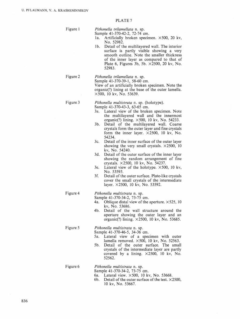
PLATE 7
Figure 1 Pithonella trilamellata n. sp.Sample 41-370-42-2, 72-74 cm.la. Artificially broken specimen. ×500, 20 kv,
No. 52982.lb. Detail of the multilayered wall. The interior
surface is partly visible showing a verysmooth outline. Note the smaller thicknessof the inner layer as compared to that ofPlate 6, Figures 3b, 5b. X2500, 20 kv, No.52983.
Figure 2 Pithonella trilamellata n. sp.Sample 41-370-39-1, 58-60 cm.View of an artificially broken specimen. Note theorganic(?) lining at the base of the outer lamella.X500, 10 kv, No. 53639.
Figure 3 Pithonella multistrata n. sp. (holotype).Sample 41-370-43-3, 63-65 cm.3a. Lateral view of the broken specimen. Note
the multilayered wall and the innermostorganic(?) lining. ×500, 10 kv, No. 54233.
3b. Detail of the multilayered wall. Coarsecrystals form the outer layer and fine crystalsform the inner layer. X2500, 10 kv, No.54234.
3c. Detail of the inner surface of the outer layershowing the very small crystals. X2500, 10kv, No. 54240.
3d. Detail of the outer surface of the inner layershowing the random arrangement of finecrystals. X2500, 10 kv, No. 54237.
3e. Lateral view of the holotype. X500, 10 kv,No. 53593.
3f. Detail of the outer surface. Plate-like crystalscover the small crystals of the intermediatelayer. X2500, 10 kv, No. 53592.
Figure 4 Pithonella multistrata n. sp.Sample 41-370-34-2, 73-75 cm.4a. Oblique distal view of the aperture. X525, 10
kv, No. 53686.4b. Detail of the wall structure around the
aperture showing the outer layer and anorganic(?) lining. X2500, 10 kv, No. 53685.
Figure 5 Pithonella multistrata n. sp.Sample 41-370-46-5, 34-36 cm.5a. Lateral view of a specimen with outer
lamella removed. X500, 10 kv, No. 52563.5b. Detail of the outer surface. The small
crystals of the intermediate layer are partlycovered by a lining. X2500, 10 kv, No.52562.
Figure 6 Pithonella multistrata n. sp.Sample 41-370-34-2, 73-75 cm.6a. Lateral view. X500, 10 kv, No. 53668.6b. Detail of the outer surface of the test. X2500,
10 kv, No. 53667.
836

CRETACEOUS CALCISPHAERULIDS
PLATE 7
837

U. PFLAUMANN, V. A. KRASHENINNIKOV
PLATE 8
Figure 1 Pithonella guttula n. sp. (holotype).Sample 41-370-40-1, 39-41 cm.la. Lateral view of a specimen with outer layer
partly removed. ×500, 10 kv, No. 53337.1 b. Detail of the different layers of the wall. The
outer layer has coarse crystals and the innerlayer has fine, needle-like crystals, separatedby a smooth, organic(?) lining. X25OO, 10 kv,No. 53339.
lc. Detail of a cross-section through the wall,showing the short needle-like crystals of theouter layer covered by and intergrading intolarge crystals of the outermost layer butbelow the organic(?) lining of the longneedles of the inner layer. X25OO, 10 kv, No.55645.
Id. Another part of the fracture and surface ofthe inner layer below the lining. X25OO, 10kv, No. 55646.
le. Detail of the outer surface of the test. Notethe partially not filled interspaces betweenthe large crystals. X2500, 10 kv, No. 53338.
Figure 2 Pithonella guttula n. sp.Sample 41-370-42-2, 72-74 cm.2a. Lateral view of a specimen with partly
removed outer layers. ×500, 10 kv, No.52962.
2b. View of a section of the outer lamella. Notethe oblique orientation of the large crystals.X25OO, 10 kv, No. 52964.
2c. View of the multilayered wall. Lower leftshows crystals of the outer layer; lower rightshows the organic(?) lining. In the middle ofthe picture, fine needle-like crystals of theinner layer are seen and upper right showsparts of an innermost lining or filling.X25OO, 10 kv, No. 52963.
Figure 3 Pithonella guttula n. sp.Sample 41-370-40-1, 39-41 cm.3a. Lateral view. ×500, 10 kv, No. 53348.3b. Detail of the outer surface of the test. The
boundaries between the plate-like crystalsurfaces are irregular but smooth. X2500, 10kv, No. 53349.
Figure 4 Pithonella guttula n. sp.Sample 41-370-44-1, 71-73 cm.4a. Lateral view. ×500, 10 kv, No. 53608.4b. Detail of the outer surface of the test. × 2500,
10 kv, No. 53607.
Figure 5 Pithonella sp. 2.Sample 41-370-34-2, 73-75 cm.5a. Lateral view. ×500, 10 kv, No. 53664.5b. Detail of the outer surface of the test. × 2500,
10 kv, No. 53663.
Figure 6 Pithonella sp. indet.Sample 41-370-28, CC.6a. Lateral view of a specimen whose outer layer
is probably missing. ×500, 10 kv, No. 53690.6b. Detail of the surface of the specimen. X2500,
10 kv, No. 53691.
838

CRETACEOUS CALCISPHAERULIDS
PLATE 8
839