Embed Size (px)
Citation preview
Ministry of health of Ukraine
Ukrainian Medical Stomatological Academy
It is ratified
on meeting of chair
surgical stomatology
and maxillofacial surgery
with plastic and reconstructive
surgery of the head and neck
«28» August 2019
Protocol № 1 28.08.2019
The head of chair________ Avetikov D.S.
METHODICAL RECOMMENDATIONS
FOR INDIVIDUAL WORK OF STUDENTS
DURING PREPARATION TO PRACTICAL (SEMINAR) LESSON
Educational discipline Surgical stomatology
Module № 6
Theme of the lesson №2
Explorer anaesthetizing for maxilla and mandible and
supporting soft tissues. Applique and infiltration
anaesthetizing of tissues of maxillo-facial area.
Pneumocardial reanimation. Physiotherapy of
omplications, related to anaesthetizing.
Course V
Faculty Stomatological
Poltava – 2019
1. ACTUALITY OF THEME.
Necessity of knowledge of methods of local anesthesia due to the fact that the majority of
operations in outpatient clinics is carried out on modern methods of local anesthesia. The future
doctor should thoroughly use these methods to prevent possible complications local and General
nature.
2. CONCRETE AIMS:
2.1. Analyze the possibility of selecting a particular method of local anesthesia.
2.2. To explain the mechanism of pain and pain relief.
2.3. Propose a definition of the concept of «local anesthesia».
2.4. Classify the types of local anesthesia.
2.5. Interpret methods of local anesthesia in surgical stomatology clinic.
2.6. To draw the scheme of the mechanism of local anesthetics.
2.7. To analyze the effect of local anesthetics different chemical groups.
2.8. Plan cardiopulmonary resuscitation.
2.9. Classify complications arising in the course of local anesthesia.
2.10. Offer physiotherapy methods used for treatment of the patients with complications of local
anesthesia.
3. BASIC KNOWLEDGE, ABILITIES, SKILLS, WHICH ARE NECESSARY FOR STUDY
THEMES (intradisciplinary integration)
4. TASKS FOR INDEPENDENT WORK DURING THE PREPARATION TO THE
LESSON.
4.1. A list of main terms, parameters, characteristics, which should learn the student in
preparation for the lesson:
Terms Definition
Anesthesia complete loss of sensation body or parts thereof in connection with the
loss of conductivity sensitive nerve impulses in nerves at different levels.
Full anesthesia unit of all kinds of sensitivity.
Partial anesthesia of the block of a certain kind of sensitivity.
Analgesia absence of the sensation of pain.
General anesthesia General anesthesia in which the patients complete loss of the whole
sensitivity with different levels of human consciousness.
Local anesthesia type of anesthesia in which there is a loss of sensitivity to any particular
part of the body as a result of blocking the transmission of nerve
impulses.
Anesthetics medicinal substances possessing ability to induce anesthesia.
Local anaesthetics medicinal substances blocking the perception of pain in a limited area.
Infiltrative type of
anaesthesia
local anesthesia, blocking local anesthetic transmission of nerve
impulses at the level of pain receptors and nerve branches.
Conduction
anesthesia
local anesthesia, blocking the transmission of nerve impulses at the level
of the nerve or nerve plexus.
The names of the previous disciplines The received skills
1. Ethics and deontology. Install psychological contact with the patient.
2. Normal anatomy. Apply knowledge of innervation of the maxillofacial
region at carrying out of local anesthesia.
3. Pharmacology Describe the pharmacokinetics and pharmacodinamic,
indications and contraindications for use of local
anesthetics, features modern drugs for local anesthesia.
4. Pathological physiology. Describe the mechanisms of pain and to interrupt the
pain impulse.
5. Propedeutics of internal diseases. Write scheme cardiopulmonary resuscitation.
Premedication drug preparation with action, influence on the psycho-emotional sphere
of a patient to save adaptive mechanisms and the prevention of
complications of a General nature.
Potencial local
anesthesia
local anesthesia in the face then the patient.
Stem anesthesia local anesthesia, which blocks the main trunk
4.2.Тheoretical questions to the lessons:
1. Give the definition of local anesthesia. Name the classification of local anesthesia in dentistry.
2. What are the indications, contra-indications to local anesthesia.
3. Name the funds for the local anesthesia, their properties.
4. What is the mechanism of local anesthetics?
5. Name the vasoconstrictor substance, their properties, doses, indications for use.
6. What substances are used for potentiated of local anesthesia in dentistry? Name the indications
for their use.
7. What local anaesthetics belong to the group of esters?
8. What local anaesthetics belong to the group of amides?
9. What local and General осложнеия arise during and after local anesthesia?
10. Name the features of topographic anatomy structure pterygiod-palatal fossa, maxillary nerve
and its branches, especially their innervation in the maxillofacial region.
4.3. Practical works (task) which are executed on lesson:
1. Reception and curation of patients in the surgical Department of dental polyclinic and
maxillofacial hospital under the guidance of a teacher.
2. Collect the medical history, a clinical examination, make a preliminary diagnosis of the patient.
3. Choose instrumental and madical equippingor instruments for local anesthesia.
4. Choose local anesthesia for surgery on maxilla or lower jaw.
5. Run on the testimony of local anesthesia for the upper or lower jaw, the patient is under the
supervision of a teacher.
6. Make medical documentation surgeon dentist (patient records) under the guidance of a teacher.
7. Choose a local anesthetic and a local anesthetic to perform sphenopalatine anesthesia and
surgery on the upper jaw, the patient is indicated.
8. To make pterygopalatine anesthesia, the patient is under the supervision of a teacher.
9. To render first medical aid to the patient on the testimony of local and systemic complications of
local anesthesia under the guidance of a teacher.
10. Causes, pathogenesis, clinics and first-aid of syncope (faint).
11. Causes of shock.
12. Types of anaphylactic shock. Pathogenesis, clinic, diagnostics, emergency therapy in case of
anaphylactic shock Prevention of anaphylactic shock.
13. Intoxication anaesthetics local action: causes, prevention, treatment.
14. Intoxication adrenaline: causes, prevention, treatment.
15. Causes, clinical features, diagnosis, emergency measures in collapse.
16. Damage to the blood vessels. Postinjection, hematoma. Treatment. Prevention.
17. Damage to the sensory nerves - branches of the trigeminal nerve. Treatment. Prevention.
18. Damage to the motor nerves. Treatment. Prevention.
19. Postinjection, pain and swelling. Treatment. Prevention.
20. Postinjection, contracture of the lower jaw. Treatment. Prevention.
21. Tissue necrosis, which an(a)esthetize. Treatment. Prevention.
22. Breakdown of the injection needle. Tactics of actions of the doctor. Prevention.
23. Ischemic tissues after local anesthesia. Possible consequences.
24. Postinjection, abscesses and phlegmons. Prevention activities.
25. Investigation of the introduction of aggressive liquids instead of anesthetic solutions.
Therapeutic management. Prevention activities.
5. TABLE OF CONTENTS OF THEME:
Local anesthesia is any technique to induce the absence of sensation in part of the body,
generally for the aim of inducing local analgesia, that is, local insensitivity to pain, although other
local senses may be affected as well. It allows patients to undergo surgical and dental procedures
with reduced pain and distress. In many situations, such as cesarean section, it is safer and therefore
superior to general anesthesia. It is also used for relief of non-surgical pain and to enable diagnosis
of the cause of some chronic pain conditions. Anesthetists sometimes combine both general and
local anesthesia techniques.
The following terms are often used interchangeably:
Local anesthesia, in a strict sense, is anesthesia of a small part of the body
such as a tooth or an area of skin.
Regional anesthesia is aimed at anesthetizing a larger part of the body such as
a leg or arm.
Conduction anesthesia is a comprehensive term, which encompasses a great
variety of local and regional anesthetic techniques.
Medical
A local anesthetic is a drug that causes reversible local anesthesia and a loss of nociception.
When it is used on specific nerve pathways (nerve block), effects such as analgesia (loss of pain
sensation) and paralysis (loss of muscle power) can be achieved. Clinical local anesthetics belong
to one of two classes: aminoamide and aminoester local anesthetics. Synthetic local anesthetics are
structurally related to cocaine. They differ from cocaine mainly in that they have no abuse potential
and do not act on the sympathoadrenergic system, i.e. they do not produce hypertension or local
vasoconstriction, with the exception of Ropivacaine and Mepivacaine that do produce weak
vasoconstriction.
Regional nerve blockade, or more commonly nerve block, is the interruption of signals
traveling along a nerve, usually for the purpose of pain relief. Local anesthetic nerve block (often
referred to as simply "nerve block") is a short-term block, usually lasting hours or days, involving
the injection of an anesthetic and/or corticosteroid onto or near a nerve. A neurolytic
block (deliberate temporary damage to nerve fibers) produces a block that may persist for weeks,
months or indefinitely.Neurectomy (cutting through or removal of a nerve or a section of a nerve)
usually produces a permanent block.
Local anesthetics vary in their pharmacological properties and they are used in various
techniques of local anesthesia such as:
Topical anesthesia (surface)
Infiltration
Plexus block
Epidural (extradural) block
Spinal anesthesia (subarachnoid block).
Non-medical local anesthetic techniques
Local pain management that uses other techniques than analgesic medication include:
Transcutaneous electrical nerve stimulation, which has been found to be ineffective for
lower back pain, however, it might help with diabetic neuropathy.
Pulsed radiofrequency, neuromodulation, direct introduction of medication and nerve
ablation may be used to target either the tissue structures and organ/systems responsible for
persistent nociception or the nociceptors from the structures implicated as the source of
chronic pain.
Techniques
Local anesthetics can block almost every nerve between the peripheral nerve endings and
the central nervous system. The most peripheral technique is topical anesthesia to the skin or other
body surface. Small and large peripheral nerves can be anesthetized individually (peripheral nerve
block) or in anatomic nerve bundles (plexus anesthesia). Spinal anesthesia and epidural anesthesia
merges into the central nervous system.
Injection of local anesthetics is often painful. A number of methods can be used to decrease
this pain including buffering of the solution with bicarb and warming.
Clinical techniques include:
Surface anesthesia - application of local anesthetic spray, solution or cream to the skin or a
mucous membrane. The effect is short lasting and is limited to the area of contact.
Infiltration anesthesia - injection of local anesthetic into the tissue to be anesthetized.
Surface and infiltration anesthesia are collectively topical anesthesia.
Field block - subcutaneous injection of a local anesthetic in an area bordering on the field to
be anesthetized.
Peripheral nerve block - injection of local anesthetic in the vicinity of a peripheral nerve to
anesthetize that nerve's area of innervation.
Plexus anesthesia - injection of local anesthetic in the vicinity of a nerve plexus, often inside
a tissue compartment that limits the diffusion of the drug away from the intended site of
action. The anesthetic effect extends to the innervation areas of several or all nerves
stemming from the plexus.
Epidural anesthesia - a local anesthetic is injected into the epidural space where it acts
primarily on the spinal nerve roots. Depending on the site of injection and the volume
injected, the anesthetized area varies from limited areas of the abdomen or chest to large
regions of the body.
Spinal anesthesia - a local anesthetic is injected into the cerebrospinal fluid, usually at the
lumbar spine (in the lower back), where it acts on spinal nerve roots and part of the spinal
cord. The resulting anesthesia usually extends from the legs to the abdomen or chest.
Intravenous regional anesthesia (Bier's block) - blood circulation of a limb is interrupted
using a tourniquet (a device similar to a blood pressure cuff), then a large volume of local
anesthetic is injected into a peripheral vein. The drug fills the limb's venous system and
diffuses into tissues where peripheral nerves and nerve endings are anesthetized. The
anesthetic effect is limited to the area that is excluded from blood circulation and resolves
quickly once circulation is restored.
Local anesthesia of body cavities (e.g. intrapleural anesthesia, intraarticular anesthesia)
Transincision (or Transwound) catheter anesthesia, wherein a multilumen catheter is
inserted through an insicion or wound and aligned across it on the inside as the incision or
wound is closed, providing continuous administration of local anesthetic along the incision
or wound.
Dental anesthesia (or dental anaesthesia) is a field of anesthesia that includes not only
local anesthetics but sedation and general anesthesia.
Local anesthetic agents in dentistry
The most commonly used local anesthetic is lidocaine (also called xylocaine or lignocaine),
a modern replacement for novocaine and procaine. Its half-life in the body is about 1.5–2 hours.
Other local anesthetic agents in current use include articaine (also called septocaine or ubistesin),
marcaine (a long-acting anesthetic), and mepivacaine. A combination of these may be used
depending on the situation. Also, most agents come in two forms: with and without epinephrine or
other vasoconstrictor that allow the agent to last longer and also controls bleeding in the tissue
during procedures. Usually the case is classified using the ASA Physical Status Classification
System before any anesthesia is given.
Types of local anesthesia in dentistry
Nerve block — a common form of local dental anesthesia; blocks the reception of pain in
one region of the mouth at a time.
Infiltration given inferior to the root the tooth involved in the dental work; used usually for
minor procedures such as restorations.
Palatal block given into the hard palate using pressure anethesia; useful in anesthetizing the
palate side of the maxillary teeth.
Intraosseous an injection of local anesthetic given directly into the osseous (bone) structure
of the tooth for more involved dental procedures such as surgery or endodontic therapy
(root canals).

Intrapulpal an injection of local anesthetic given directly into the pulp of the tooth to
completely desensitize the tooth.
Pressure anesthesia — pressure with a cotton swab in the area to distract the nerve sensation
of pain when the needle enters certain areas such as palatal tissue.
Electrical nerve blocks— a technology that involves using electrical current to block the
reception or generation of pain signals; the pain control can be transient.
Acupuncture or accupressure An alternative to chemical or electrical blocks, but is rarely
used.
Inferior alveolar nerve anesthesia (or anaesthesia), also known as the inferior alveolar
nerve block or IANB, is a technique for dental anesthesia, used to cause numbness to the areas of
the face innervated by the inferior alveolar nerve; namely, the lower lip and the teeth and gingiva of
the mandible. This procedure attempts to anaesthetise the inferior alveolar nerve prior to it entering
the mandibular foramen.
The inferior alveolar nerve is a branch of the mandibular nerve, which is itself the third
branch or division of the trigeminal nerve, the fifth (V) cranial nerve.
Symptoms of anesthesia
Administration of anesthesia near the mandibular foramen causes blockage of the inferior
alveolar nerve and the nearby lingual nerve by diffusion (includes supplying the tongue). This
causes patients to lose sensation in:
their mandibular teeth on one side (via inferior alveolar nerve block)
their lower lip and chin on one side (via mental nerve block)
and parts of their tongue and lingual gingival tissue on one side except on the cheek side of
the mandibular molars (via lingual nerve block); a buccal block will anesthetize this later
tissue area.
Another symptom is harmless numbness and tingling of the body of the tongue and floor of
the mouth, which indicates that the lingual nerve, a branch of the mandibular nerve, is anesthetized.
Another symptom that can occur is ―lingual shock‖ as the needle passes by the lingual nerve during
administration. The patient may make an involuntary movement, varying from a slight opening of
the eyes to jumping in the chair. This symptom is only momentary, and anesthesia will quickly
occur.
Complications
The most common adverse affect of this injection is accidental self-inflicted trauma after
the procedure, either by biting the lip or tongue or by thermal burn caused by inadvertent
drinking of fluid that is too hot. This classically occurs in children or those with learning
disability.
A blood vessel may be punctured accidentally and a hematoma or "blood blister" may occur
that will heal over time.
If needle is positioned too posteriorly, anasthetic may be put into parotid gland, that may
cause transient facial paralysis of the facial nerve or cranial Nerve VI. Symptoms of this
temporary loss of the use of the muscles of facial expression include the inability to close
the eyelid and the drooping of the labial commissure on the aected side for a few hours.
Also if the needle is placed too medially the medial pterygoid muscle can be injected,
resulting in trismus.
The sphenomandibular ligament may act as a barrier to the agent if the injection is given too
shallow and the lingual nerve is only anethetized.
This injection can rarely cause needle tract infections of the pterygomandibular space. This
is because the mouth contains many types of bacteria which are normally harmless by virtue
of the physical barrier that the mucosa presents. However, if they are inoculated into the
tissues during an injection, they can become pathogenic (disease causing).
Mental nerve is a general somatic afferent (sensory) nerve which provides sensation to the
anterior aspects of the chin and lower lip as well as the buccal gingivae of the mandibular anterior
teeth and the premolars. It is a branch of the posterior trunk of the inferior alveolar nerve, which is
itself a branch of the mandibular division of the trigeminal nerve (CN V).
The nerve emerges at the mental foramen in the mandibula, and divides beneath
the Depressor anguli oris muscle into three branches:
one descends to the skin of the chin.
two ascend to the skin and mucous membrane of the lower lip.
These branches communicate freely with the facial nerve.
The lingual nerve is a branch of the mandibular division of the trigeminal nerve (CN V3),
which supplies sensory innervation to the tongue. It also carries fibers from the facial nerve, which
return taste information from the anterior two thirds of the tongue.
The anterior superior alveolar branch (anterior superior dental branch), of
considerable size, is given off from themaxillary nerve just before its exit from the infraorbital
foramen; it descends in a canal in the anterior wall of the maxillary sinus, and divides into branches
which supply the incisor and canine teeth.
It communicates with the middle superior alveolar branch, and gives off a nasal branch,
which passes through a minute canal in the lateral wall of the inferior meatus, and supplies
the mucous membrane of the anterior part of the inferior meatus and the floor of the nasal cavity,
communicating with the nasal branches from the sphenopalatine ganglion.
Dental considerations for this nerve are important. The anterior superior alveolar usually
innervates all anterior teeth, premolars, and the Mesiobuccal root of the first maxillary molar,
especially if there is an anatomical absence of a middle superior alveolar nerve. It is the innervation
of the one root of the first maxillary molar that makes any procedure done around it important,
especially when trying to anesthetize the location.
The posterior superior alveolar branches (posterior superior dental branches) arise
from the trunk of the maxillary nerve just before it enters the infraorbital groove; they are generally
two in number, but sometimes arise by a single trunk.
They descend on the tuberosity of the maxilla and give off several twigs to the gums and
neighboring parts of the mucous membrane of the cheek.
They then enter the alveolar canals on the infratemporal surface of the maxilla, and, passing
from behind forward in the substance of the bone, communicate with the middle superior alveolar
nerve, and give off branches to the lining membrane of the maxillary sinus and gingival and dental
branches to each molar tooth from a superior dental plexus; these branches enter the apical
foramina at the roots of the teeth.
The posterior superior alveolar nerve innervates the second and third maxillary molars, and
two of the three roots of the maxillary first molar (all but the mesiobuccal root). When giving a
Posterior Superior Alveolar nerve block, it will anesthetize the mesialbuccal root of the maxillary
first molar approximately 72% of the time.
The middle superior alveolar nerve is a nerve that drops from the infraorbital portion of
the maxillary nerve to supply thesinus mucosa, the roots of the maxillary premolars, and
the mesiobuccal root of the first maxillary molar. It is not always present; and in the majority of
cases it is non existent with the posterior superior alveolar nerve innervating the premolars and
molars alone.
MOST COMMON LOCAL ANESTHETIC PROCEDURE
The Inferior alveolar nerve anaesthesia or block or IANB (sometimes referred to wrongly as
the mandibular block) probably is anesthetized more often than any other nerve in the body. An
injection blocks sensation in the inferior alveolar nerve, which runs from the angle of the mandible
down the medial aspect of the mandible, innervating the mandibular teeth, lower lip, chin, and parts
of the tongue, which is effective for dental work in the mandibular arch. To anesthetize this nerve,
the licensed dental professional inserts the needle somewhat posterior to the most distal mandibular
molar on one side of the mouth. The lingual nerve is also anesthetized through diffusion of the
agent to produce a numb tongue as well as anesthetizing the floor of the mouth tissue, including
that around the tongue side or lingual of the teeth.
Several nondental nerves are usually anesthetized during an inferior alveolar block. The
mental nerve, which supplies cutaneous innervation to the anterior lip and chin, is a distal branch of
the inferior alveolar nerve. When the inferior alveolar nerve is blocked, the mental nerve is blocked
also, resulting in a numb lip and chin. Nerves lying near the point where the inferior alveolar nerve

enters the mandible often are also anesthetized during inferior alveolar anesthesia, such as affecting
hearing (auriculotemporal nerve).
The facial nerve lies some distance from the inferior alveolar nerve within the
submandibular salivary gland, but in rare cases anesthetic can be injected far enough posteriorly to
anesthetize that nerve. The result is a transient facial paralysis, with the injected side of the face
having temporary loss of the use of the muscles of facial expression that include the inability to
close the eyelid and the drooping of the labial commissure on the affected side for a few hours,
which disappears when the anesthesia wears off.
In contrast, the superior alveolar nerves are not usually anesthetized directly because they
are difficult to approach with a needle. For this reason, the maxillary arch is usually anesthetized
locally for dental work by inserting the needle beneath the oral mucosa surrounding the teeth so as
to anesthetize the smaller branches.
Allergic reactions to anaesthesia
The incidence of life-threatening hypersensitivity reactions occurring during surgery and
anaesthesia is around one in 10,000 procedures. Serious allergic reactions to anesthetic medications
are rare and a usually attributable to factors other than the anesthetic. Neuromuscular blocking
agents, natural rubber latex, and antibiotics are the most common causes of serious allergic
reactions during surgery.[1]
The mortality rate from these reactions is about 3–6%.
Successful immediate treatment requires prompt recognition by the attending anaesthetist,
or in the US, the attending anesthesiologist or nurse anesthetist . Adrenaline (epinephrine) remains
the mainstay of treatment, with corticosteroids and antihistamines providing limited benefit in the
acute situation.
Subsequent investigation aims to determine the responsible agent to allow its future
avoidance. Skin testing is often useful to identify potentially cross-reactive compounds and
appropriate therapeutic alternatives. This is done weeks after the initial reaction to allow the
immune system to reset itself. However, skin testing can be misleading in giving false positive and
false negative results.
Lidocaine (INN) /ˈlaɪdɵkeɪn/, xylocaine, or lignocaine is a common local anesthetic and
antiarrhythmic drug. Lidocaine is used topically to relieve itching, burning and pain from skin
inflammations, injected as a dental anesthetic or as a local anesthetic for minor surgery.
Indications
The efficacy profile of lidocaine as a local anesthetic is characterized by a rapid onset of
action and intermediate duration of efficacy. Therefore, lidocaine is suitable for infiltration, block
and surface anesthesia. Longer-acting substances such as bupivacaine are sometimes given
preference for subdural and epidural anesthesias; lidocaine, on the other hand, has the advantage of
a rapid onset of action. Epinephrine (aka adrenaline) vasoconstricts arteries reducing bleeding and
also delays the resorption of lidocaine, almost doubling the duration of anaesthesia. For surface
anesthesia several formulations are available that can be used e.g. for endoscopies, before
intubations etc. Buffering the pH of lidocaine makes local freezing less painful.
Topical lidocaine has been shown in some patients to relieve the pain of postherpetic
neuralgia (a complication of shingles), though there is not enough study evidence to recommend it
as a first-line treatment. It also has uses as a temporary fix for tinnitus. Although not completely
curing the disorder, it has been shown to reduce the effects by around two thirds.
Lidocaine is also the most important class 1B antiarrhythmic drug: it is used intravenously
for the treatment of ventricular arrhythmias (for acute myocardial infarction, digoxin poisoning,
cardioversion or cardiac catheterization).
However, amiodarone has been replacing lidocaine as the first-line pharmacologic
management of ventricular tachycardia.
A routine prophylactic administration is no longer recommended for acute cardiac
infarction; the overall benefit of this measure is not convincing.
Lidocaine has also been efficient in refractory cases of status epilepticus.
Inhaled lidocaine can be used as an antitussive (cough suppressor) acting peripherally below
the larynx.
Lidocaine has also proved effective in treating jellyfish stings, both numbing the affected
area and preventing further nematocyst discharge.
Contraindications
Contraindications for the use of lidocaine include:
Heart block, second or third degree (without pacemaker)
Severe sinoatrial block (without pacemaker)
Serious adverse drug reaction to lidocaine or amide local anaesthetics
Concurrent treatment with quinidine, flecainide, disopyramide, procainamide (Class I
antiarrhythmic agents)
Prior use of Amiodarone hydrochloride
Hypotension not due to Arrhythmia
Bradycardia
Accelerated idioventricular rhythm
Pacemaker
Porphyria, especially Acute Intermittent Porphyria (AIP); lidocaine has been classified as
porphyrogenic although clinical evidence suggests that it is not.
]
Adverse drug reactions (ADRs) are rare when lidocaine is used as a local anesthetic and is
administered correctly. Most ADRs associated with lidocaine for anesthesia relate to administration
technique (resulting in systemic exposure) or pharmacological effects of anesthesia, and allergic
reactions only rarely occur.
Systemic exposure to excessive quantities of lidocaine mainly result in central nervous
system (CNS) and cardiovascular effects – CNS effects usually occur at lower blood plasma
concentrations and additional cardiovascular effects present at higher concentrations, though
cardiovascular collapse may also occur with low concentrations. CNS effects may include CNS
excitation (nervousness, tingling around the mouth (also known as circumoral paraesthesia),
tinnitus, tremor, dizziness, blurred vision, seizures) followed by depression, and with increasingly
heavier exposure: drowsiness, loss of consciousness, respiratory depression and apnoea.
Cardiovascular effects include hypotension, bradycardia, arrhythmias, and/or cardiac arrest – some
of which may be due to hypoxemia secondary to respiratory depression.
ADRs associated with the use of intravenous lidocaine are similar to toxic effects from
systemic exposure above. These are dose-related and more frequent at high infusion rates
(≥3 mg/minute). Common ADRs include: headache, dizziness, drowsiness, confusion, visual
disturbances, tinnitus, tremor, and/or paraesthesia. Infrequent ADRs associated with the use of
lidocaine include: hypotension, bradycardia, arrhythmias, cardiac arrest, muscle twitching, seizures,
coma, and/or respiratory depression.
Overdosage with lidocaine can be a result of excessive administration via topical or
parenteral routes, accidental oral ingestion of topical preparations by children, accidental
intravenous (rather than subcutaneous, intrathecal or paracervical) injection or prolonged use of
subcutaneous infiltration anesthesia during cosmetic surgical procedures. These occurrences have
often led to severe toxicity or death in both children and adults. Lidocaine and its two major
metabolites may be quantified in blood, plasma or serum to confirm the diagnosis in potential
poisoning victims or to assist in the forensic investigation in a case of fatal overdosage. It is
important in the interpretation of analytical results to recognize that lidocaine is often routinely
administered intravenously as an antiarrhthymic agent in critical cardiac care situations. Treatment
with intravenous lipid emulsions (used for parental feeding) to reverse the effects of local
anaesthetic toxicity is becoming more commonplace.
Insensitivity to lidocaine
Relative insensitivity to lidocaine is genetic. In hypokalemic sensory overstimulation,
relative insensitivity to lidocaine has been described in people who also have attention deficit
hyperactivity disorder. In dental anesthesia, a relative insensitivity to lidocaine can occur for
anatomical reasons due to unexpected positions of nerves. Some people with Ehlers-Danlos
syndrome are insensitive to lidocaine.
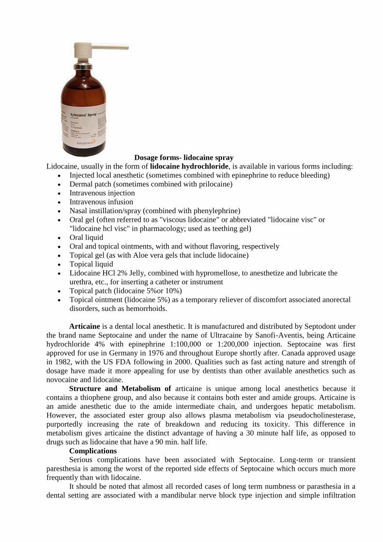
Dosage forms- lidocaine spray
Lidocaine, usually in the form of lidocaine hydrochloride, is available in various forms including:
Injected local anesthetic (sometimes combined with epinephrine to reduce bleeding)
Dermal patch (sometimes combined with prilocaine)
Intravenous injection
Intravenous infusion
Nasal instillation/spray (combined with phenylephrine)
Oral gel (often referred to as "viscous lidocaine" or abbreviated "lidocaine visc" or
"lidocaine hcl visc" in pharmacology; used as teething gel)
Oral liquid
Oral and topical ointments, with and without flavoring, respectively
Topical gel (as with Aloe vera gels that include lidocaine)
Topical liquid
Lidocaine HCl 2% Jelly, combined with hypromellose, to anesthetize and lubricate the
urethra, etc., for inserting a catheter or instrument
Topical patch (lidocaine 5%or 10%)
Topical ointment (lidocaine 5%) as a temporary reliever of discomfort associated anorectal
disorders, such as hemorrhoids.
Articaine is a dental local anesthetic. It is manufactured and distributed by Septodont under
the brand name Septocaine and under the name of Ultracaine by Sanofi-Aventis, being Articaine
hydrochloride 4% with epinephrine 1:100,000 or 1:200,000 injection. Septocaine was first
approved for use in Germany in 1976 and throughout Europe shortly after. Canada approved usage
in 1982, with the US FDA following in 2000. Qualities such as fast acting nature and strength of
dosage have made it more appealing for use by dentists than other available anesthetics such as
novocaine and lidocaine.
Structure and Metabolism of articaine is unique among local anesthetics because it
contains a thiophene group, and also because it contains both ester and amide groups. Articaine is
an amide anesthetic due to the amide intermediate chain, and undergoes hepatic metabolism.
However, the associated ester group also allows plasma metabolism via pseudocholinesterase,
purportedly increasing the rate of breakdown and reducing its toxicity. This difference in
metabolism gives articaine the distinct advantage of having a 30 minute half life, as opposed to
drugs such as lidocaine that have a 90 min. half life.
Complications Serious complications have been associated with Septocaine. Long-term or transient
paresthesia is among the worst of the reported side effects of Septocaine which occurs much more
frequently than with lidocaine.
It should be noted that almost all recorded cases of long term numbness or parasthesia in a
dental setting are associated with a mandibular nerve block type injection and simple infiltration
injections are generally thought to be immune from such complications. For this reason many
dentists have abandoned using articaine for mandibular nerve blocks.
Clinical use Articaine is often used by dentists for patients in whom lidocaine is not very effective. In
people with hypokalemic sensory overstimulation lidocaine is not very effective and articaine
works well.
Local anesthetic toxicity
While generally safe, local anesthetic agents can be toxic if used in excessive doses or
administered improperly. Even when administered properly, patients may still experience
unintended reactions to local anesthetics.
Excessive doses may be unintentionally administered in several ways.
1. Repetitive (small) doses of local anesthetic to achieve an adequate level of
anesthesia may lead to eventual administration of toxic doses.
2. Injection of anesthesia in a confined space may result in excessive fluid
pressure that may damage nerves.
3. Doses intended for epidural or intra-support-tissue administration may be
accidentally delivered as intravascular injection, resulting in accelerated systematic
absorption.
The toxic effects of local anesthetics can be classified by localized and systemic effects.
A cause of local toxicity is allergic reaction to para-aminobenzoic acid (PABA). These
reactions range from urticaria to anaphylaxis.
PABA is a metabolic product of the degradation of Ester class of local anesthetics, such as
procaine (Novocaine), benzocaine, and, to a lesser degree, amide class anesthetics such as
lidocaine, and prilocaine. It is also a metabolic by-product of pramod methylparaben, a preservative
in multi-dose vials of lidocaine. When allergic response to injected anesthetics does occur, it is
most likely due to the ester class local anesthetics. The amide class of local anesthetics is far less
likely to produce allergic reaction.
Use of topical anesthetics for relief of eye pain can result in severe corneal damage. See
abuse of anesthetics for ocular pain relief page.
Systemic toxicity of local anesthetics can be described by the direct effects on the immune
system, blood (hematologic), central nervous system, and cardiovascular system.
Immune system
As noted previously, allergic reaction to metabolic break-down of anesthetic agents and
preservatives (PABA) can cause anaphylaxis.
Hematologic
Methemoglobinemia is a process where iron in hemoglobin is altered, reducing its oxygen-
carrying capability, which produces cyanosis and symptoms of hypoxia. Benzocaine, lidocaine, and
prilocaine all produce this effect, especially benzocaine.
Central Nervous System
Systemic toxic reactions to locally administered anesthetics are progressive as the level of
the anesthetic agent in the blood rises. Initial symptoms suggest some form of central nervous
system excitation such as a ringing in the ears (tinnitus), a metallic taste in the mouth, or tingling or
numbness of the mouth. Advanced symptoms include motor twitching in the periphery followed by
grand mal seizures, coma, and eventually respiratory arrest.
Cardiovascular effects are primarily those of direct myocardial depression and
bradycardia, which may lead to cardiovascular collapse.
At extremely high levels, cardiac
arrhythmia or hypotension and cardiovascular collapse occur.
6. MATERIALS FOR SELF-CONTROL:
A. Questions for self-control:
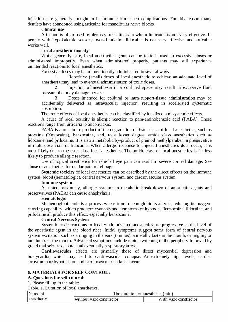
1. Please fill up in the table:
Table. 1. Duration of local anesthetics.
Name of
anesthetic
The duration of anesthesia (min)
without vazokonstrictor With vazokonstrictor
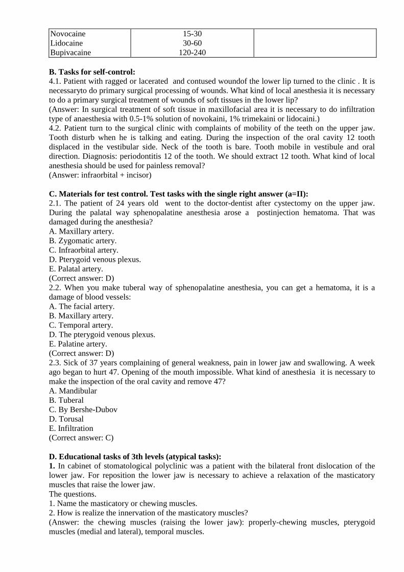
Novocaine
Lidocaine
Bupivacaine
15-30
30-60
120-240
B. Tasks for self-control:
4.1. Patient with ragged or lacerated and contused woundof the lower lip turned to the clinic . It is
necessaryto do primary surgical processing of wounds. What kind of local anesthesia it is necessary
to do a primary surgical treatment of wounds of soft tissues in the lower lip?
(Answer: In surgical treatment of soft tissue in maxillofacial area it is necessary to do infiltration
type of anaesthesia with 0.5-1% solution of novokaini, 1% trimekaini or lidocaini.)
4.2. Patient turn to the surgical clinic with complaints of mobility of the teeth on the upper jaw.
Tooth disturb when he is talking and eating. During the inspection of the oral cavity 12 tooth
displaced in the vestibular side. Neck of the tooth is bare. Tooth mobile in vestibule and oral
direction. Diagnosis: periodontitis 12 of the tooth. We should extract 12 tooth. What kind of local
anesthesia should be used for painless removal?
(Answer: infraorbital + incisor)
C. Materials for test control. Test tasks with the single right answer (a=II): 2.1. The patient of 24 years old went to the doctor-dentist after cystectomy on the upper jaw.
During the palatal way sphenopalatine anesthesia arose a postinjection hematoma. That was
damaged during the anesthesia?
A. Maxillary artery.
B. Zygomatic artery.
C. Infraorbital artery.
D. Pterygoid venous plexus.
E. Palatal artery.
(Correct answer: D)
2.2. When you make tuberal way of sphenopalatine anesthesia, you can get a hematoma, it is a
damage of blood vessels:
A. The facial artery.
B. Maxillary artery.
C. Temporal artery.
D. The pterygoid venous plexus.
E. Palatine artery.
(Correct answer: D)
2.3. Sick of 37 years complaining of general weakness, pain in lower jaw and swallowing. A week
ago began to hurt 47. Opening of the mouth impossible. What kind of anesthesia it is necessary to
make the inspection of the oral cavity and remove 47?
A. Мandibular
B. Тuberal
C. By Bershe-Dubov
D. Torusal
E. Infiltration
(Correct answer: C)
D. Educational tasks of 3th levels (atypical tasks): 1. In cabinet of stomatological polyclinic was a patient with the bilateral front dislocation of the
lower jaw. For reposition the lower jaw is necessary to achieve a relaxation of the masticatory
muscles that raise the lower jaw.
The questions.
1. Name the masticatory or chewing muscles.
2. How is realize the innervation of the masticatory muscles?
(Answer: the chewing muscles (raising the lower jaw): properly-chewing muscles, pterygoid
muscles (medial and lateral), temporal muscles.
The chewing muscles that raise the lower jaw and provide its mobility in the horizontal plane, they
are innervated by the nerves( of the same name). They are part of a small portion of the third
branch of the trigeminal nerve with a predominantly impellent innervation.)
2. The patient with an open fracture of the lower jaw in the right corner of between 8 and 7 teeth
with displacement of bone fragments. It is necessary a reposition and a fixation of fragments of the
lower jaw. Osteosynthesis of the lower jaw. The operation will be conducted under local
anesthesia.
Question.
1. Using a painless anesthesia perform the operation?
2. What concentrations solutions novocaine or lidocaine You apply?
(Answer: For a smooth execution of the operation, it is advisable to block III branch of the
trigeminal nerve in the oval holes or run anesthesia by Bershe-Dubov. In the time of surgery spend
infiltration anaesthesia soft tissues.
For conductive anesthesia using 2% solution of novokaini or lidocaine. For branches anaesthesia
soft tissues using 0.5-1% solution of these anesthetics.
3. In the dental clinic patient with the aim of mouth cavity sanation. During the inspection of the
oral cavity revealed: 36 tooth completely destroyed. Roots below the level of the gums, signs of
acute inflammation no. Diagnosed: chronic periodontitis 36 tooth. It is necessary to removal of
roots of the tooth 36.
Question:
1. How is innervation of 36 tooth?
2. How is the innervation of the alveolar mucosa shoot in the field of tooth 36, which is removed?
(Answer: 36 tooth innervations with lower inferior alveolar nerve.
The innervation of the alveolar mucosa shoot in the field of tooth 36: with vestibular side - cheek
nerve, with the lingual side - lingual nerve.)