Embed Size (px)
Citation preview
MODULE Haemostasis
Hematology and BloodBank Technique
222 HEMATOLOGY AND BLOOD BANK TECHNIQUE
Notes
24
HAEMOSTASIS
24.1 INTRODUCTION
Haemostasis means “arrest of bleeding”. During haemostasis several mechanismsinteract to slow blood flow, block the vessel wall defect with a platelet plug(primary haemostasis), convert fibrinogen to a jelly like fibrin clot (coagulationof blood) and later re-establish the flow of blood through a mechanism of slowclot lysis (fibrinolysis).
OBJECTIVES
After reading this lesson, you will be able to:
describe the 4 phases of Haemostasis
describe the coagulation of blood
explain Von Willebrand’s Disease
explain the causes of thrombocytopenia
discuss bleeding Time test
interpret PT/APTT/TT
explain INR
describe D – dimmer
24.2 HAEMOSTASIS
Haemostasis means “arrest of bleeding”. During haemostasis several mechanismsinteract to slow blood flow, block the vessel wall defect with a platelet plug(primary haemostasis), convert fibrinogen to a jelly like fibrin clot (coagulationof blood) and later re-establish the flow of blood through a mechanism of slowclot lysis (fibrinolysis). These complex physiological processes may be dividedinto phases:
223
Haemostasis MODULEHematology and Blood
Bank Technique
HEMATOLOGY AND BLOOD BANK TECHNIQUE
Notes
A. Vascular Phase.
B. Platelet Phase.
C. Coagulation Phase and
D. Fibrinolytic Phase.
During haemostasis all the phases interact. It is convenient to consider themunder these headings when investigating a patient with haemostatic problems.
A. The Vascular phase
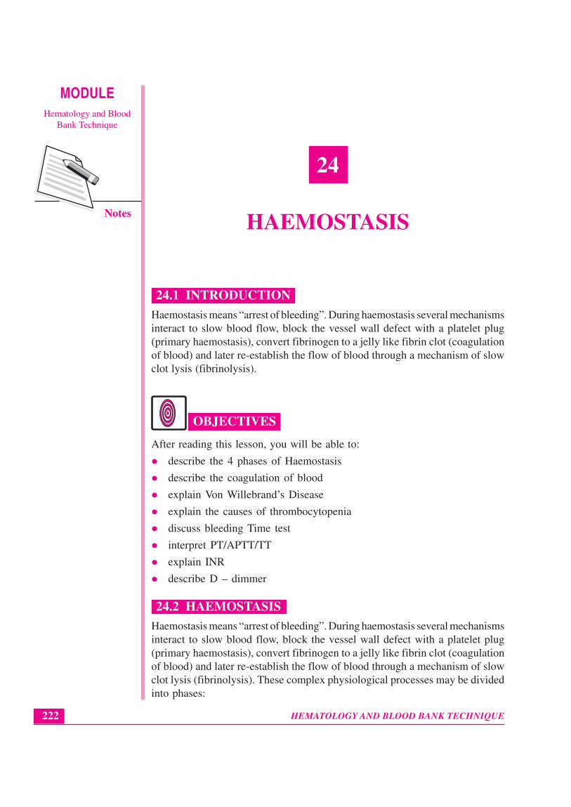
The blood vessel plays a major role in maintaining the blood in a liquid state.The entire surface of the vessel wall is lined by a single layer of endothelial cells(EC) which rest on a basement membrane. The blood inside the blood vesselis not in touch with the sub-endothelial tissues. The EC secrete a major proteincalled the von Willebrand factor (VWF) into the plasma and sub-endothelium.External to the basement membrane lies the sub-endothelium rich in collagen,elastin, fibronectin, tissue factor (TF), VWF etc. External to the sub-endotheliumlies the smooth muscle layer of variable thickness depending on the type ofvessel. External to the smooth muscle layer is the adventitial layer. Figure 24.1.
Fig. 24.1: The Blood Vessel Structure
Table I summarizes the role of the Vascular Phase.
Table I. Role of Vascular Phase in Haemostasis.
Blood vessel Property Function
Endothelial cell Non reactive surface Smooth blood flowPlatelets are repelled
VWF 1. Binds to platelet GPIb-IX and GPIIb-IIIa and mediates platelet adhesion tovessel wall.
2. Carries FVIII and prevents its earlydegradation.
Antiplatelet activity Negative charge on EC repels platelets,prostacyclin production and localdegradation of ADP
MODULE Haemostasis
Hematology and BloodBank Technique
224 HEMATOLOGY AND BLOOD BANK TECHNIQUE
Notes
Anticoagulant 1. Heparan sulphate on EC +property antithrombin III inactivate serine
proteases ie, thrombin, FXIa, FIXa etc.
2. Thrombin + Thrombo-modulin on ECactivate Protein C and S whichinactivates FVa and FVIIIa
Fibrinolytic activity EC releases plasminogen activator
Sub endothelial Collagen, elastin, Activate plateletstissues VWF etc
Tissue factor Complexes with FVII to form tissuethromboplastin for extrinsic pathway
Smooth muscle Vasoconstriction Regulates blood flowand dilatation
Table I:- EC – endothelial cell, VWF – von Willebrand factor, GP – glycoprotein
B. The Platelet Phase
Platelets are formed from megakaryocytes in bone marrow. The normal plateletcount is 150- 450 x109/l. The life span of the platelet is 8 - 9 days. On a stainedblood smear platelets are anucleate, round or discoid bodies, 1 – 2 mm in sizewith fine purple pink granules. The platelet has a phospholipid cell membraneinto which are inserted glycoproteins (GP) which act as major cell receptors andantigens of the platelets. The role of platelets in haemostasis may be summarizedas follows:
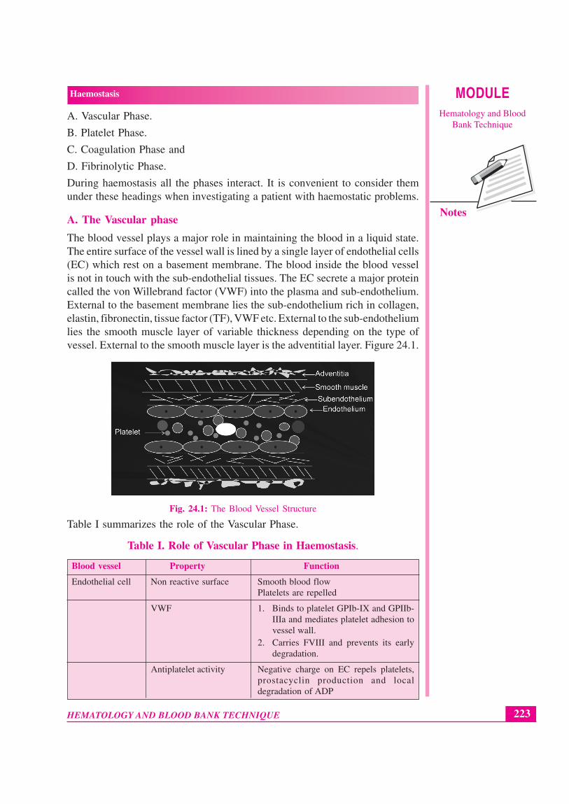
1. Adhesion. When EC is damaged platelets adhere to sub-endothelial tissues.Normal platelet number, presence of the receptor GPIb-IX complex on theplatelet membrane (receptor for VWF), VWF in plasma and normalstructure of collagen are necessary for adhesion. Figure 24.2
Fig. 24.2: Platelet Adhesion
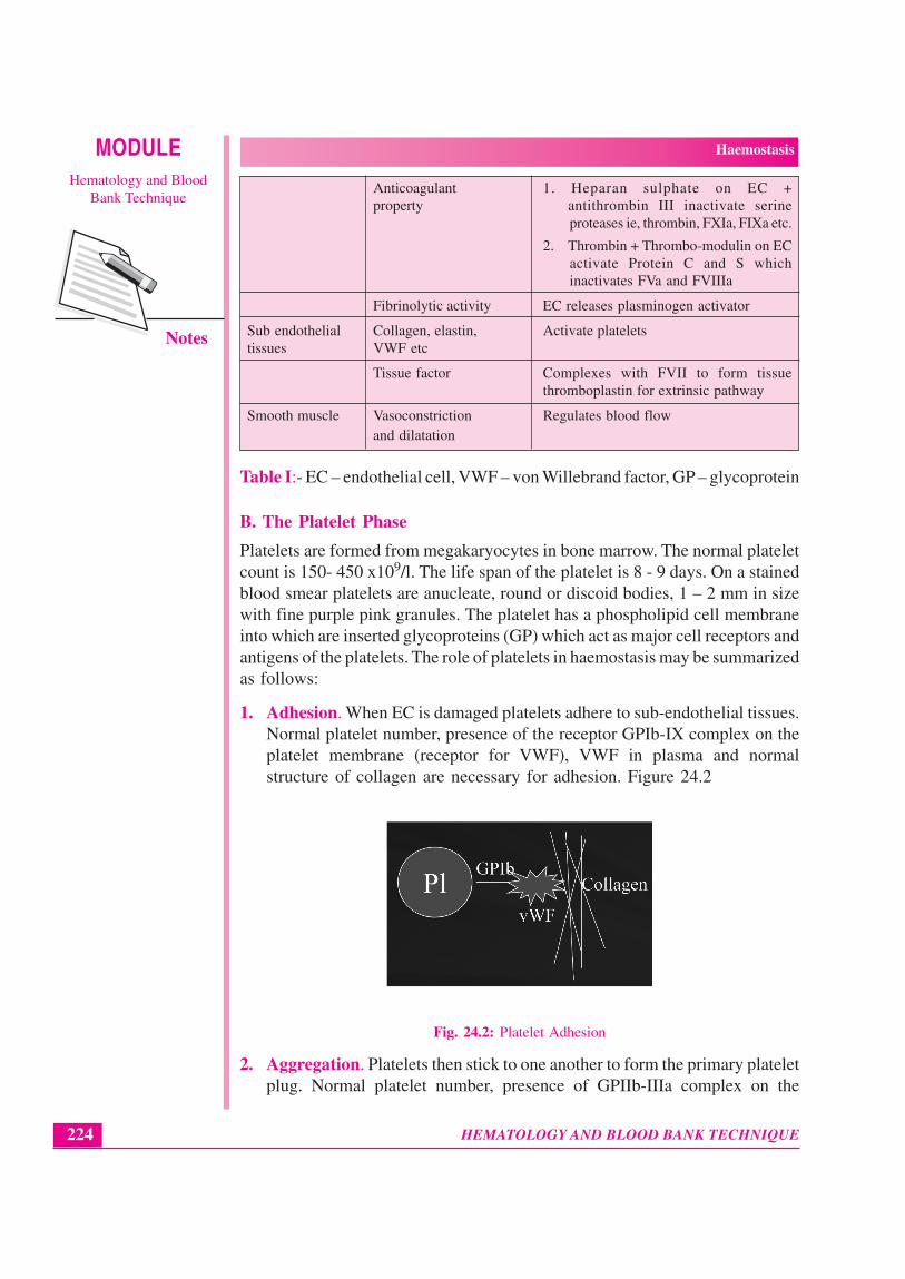
2. Aggregation. Platelets then stick to one another to form the primary plateletplug. Normal platelet number, presence of GPIIb-IIIa complex on the
225
Haemostasis MODULEHematology and Blood
Bank Technique
HEMATOLOGY AND BLOOD BANK TECHNIQUE
Notes
platelet surface (receptor for fibrinogen), plasma fibrinogen and calciumions are necessary for platelet aggregation. Figure 24.3.
Fig. 24.3: Platelet Aggregation
3. Platelet Release reaction. As platelets aggregate they release their a andd granule contents which further sustain the aggregation response.
4. The phospholipid surface for the formation of coagulation complexesduring blood coagulation is provided by the plasma membrane of theplatelets.
5. Clot retraction. The GPIIb-IIIa complex on platelets anchor fibrin strandsand pull them together to ensure a strong fibrin clot.
6. Wound healing. Platelet derived growth factor (PDGF) released from agranules promotes fibroblast proliferation and healing.
The platelet plug that is formed provides primary haemostasis. This must bereinforced with fibrin deposition to sustain haemostasis.
Table II lists some of the abnormal platelet - vessel wall interactions whichresult in bleeding.
Table II Abnormalties in Primary Haemostasis
Defect Condition
Abnormal collagen (adhesion defect) Ehlers Danlos syndrome, Marfanssyndrome, Senile purpura
Absence of GPIb-IX (adhesion defect) Bernard Soulier syndrome
Absence of VWF ( adhesion and Von Willebrand’s diseasecoagulation defect)
Absence of GPIIb-IIIa (aggregation Glanzmann,s thrombasthenia, drugdefect) induced.
Absence of FI (aggregation + Afibrinogenemiacoagulation defect)
MODULE Haemostasis
Hematology and BloodBank Technique
226 HEMATOLOGY AND BLOOD BANK TECHNIQUE
Notes
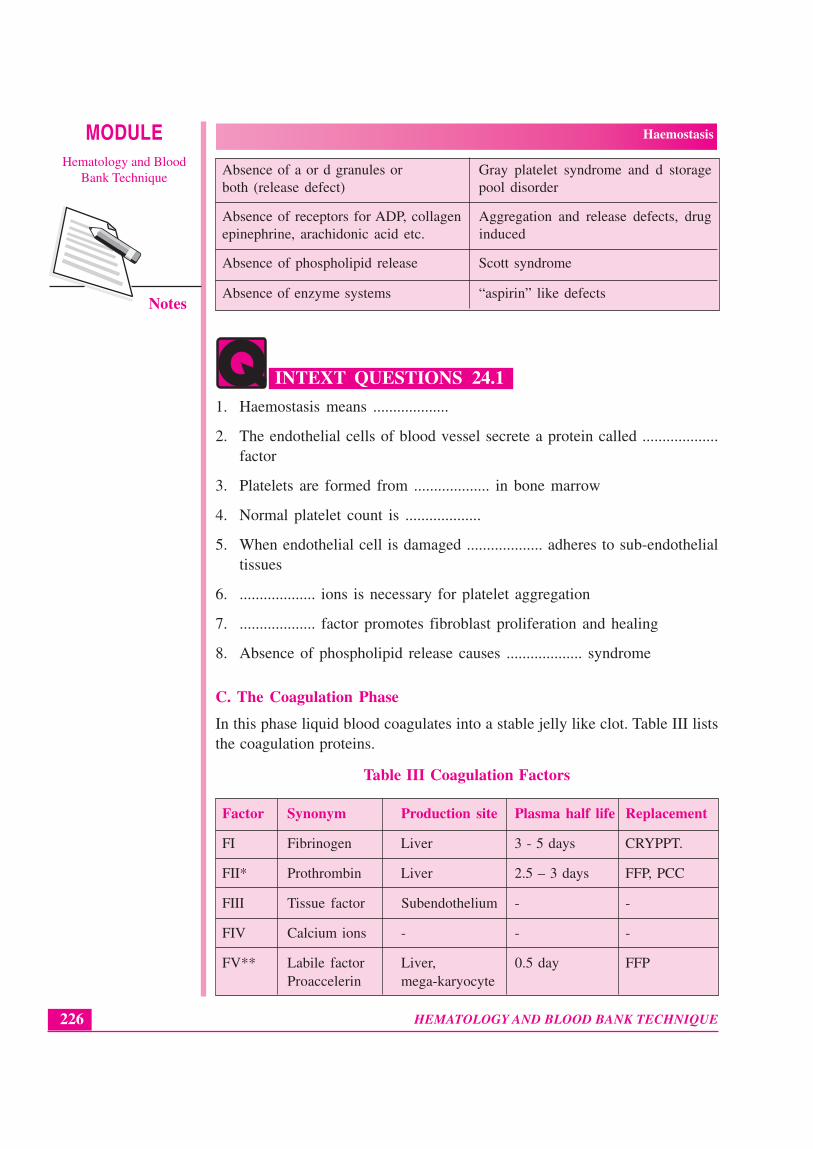
Absence of a or d granules or Gray platelet syndrome and d storageboth (release defect) pool disorder
Absence of receptors for ADP, collagen Aggregation and release defects, drugepinephrine, arachidonic acid etc. induced
Absence of phospholipid release Scott syndrome
Absence of enzyme systems “aspirin” like defects
INTEXT QUESTIONS 24.1
1. Haemostasis means ...................
2. The endothelial cells of blood vessel secrete a protein called ...................factor
3. Platelets are formed from ................... in bone marrow
4. Normal platelet count is ...................
5. When endothelial cell is damaged ................... adheres to sub-endothelialtissues
6. ................... ions is necessary for platelet aggregation
7. ................... factor promotes fibroblast proliferation and healing
8. Absence of phospholipid release causes ................... syndrome
C. The Coagulation Phase
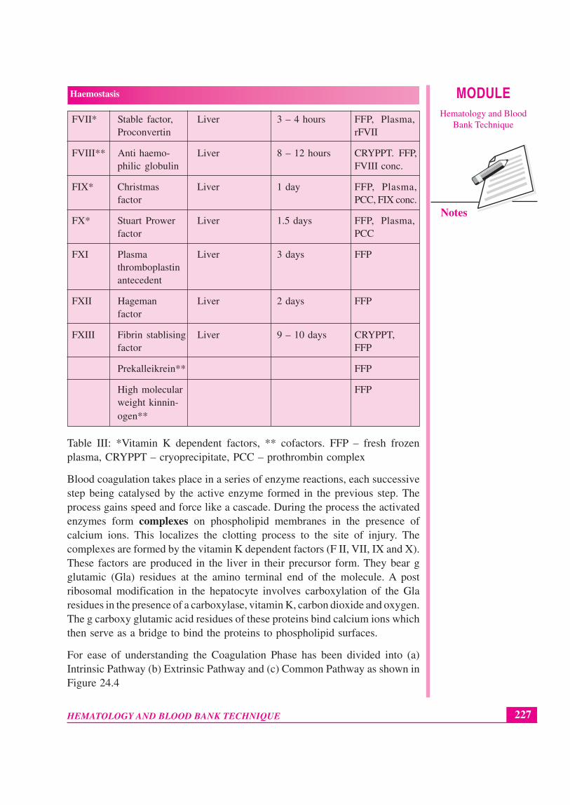
In this phase liquid blood coagulates into a stable jelly like clot. Table III liststhe coagulation proteins.
Table III Coagulation Factors
Factor Synonym Production site Plasma half life Replacement
FI Fibrinogen Liver 3 - 5 days CRYPPT.
FII* Prothrombin Liver 2.5 – 3 days FFP, PCC
FIII Tissue factor Subendothelium - -
FIV Calcium ions - - -
FV** Labile factor Liver, 0.5 day FFPProaccelerin mega-karyocyte
227
Haemostasis MODULEHematology and Blood
Bank Technique
HEMATOLOGY AND BLOOD BANK TECHNIQUE
Notes
FVII* Stable factor, Liver 3 – 4 hours FFP, Plasma,Proconvertin rFVII
FVIII** Anti haemo- Liver 8 – 12 hours CRYPPT. FFP,philic globulin FVIII conc.
FIX* Christmas Liver 1 day FFP, Plasma,factor PCC, FIX conc.
FX* Stuart Prower Liver 1.5 days FFP, Plasma,factor PCC
FXI Plasma Liver 3 days FFPthromboplastinantecedent
FXII Hageman Liver 2 days FFPfactor
FXIII Fibrin stablising Liver 9 – 10 days CRYPPT,factor FFP
Prekalleikrein** FFP
High molecular FFPweight kinnin-ogen**
Table III: *Vitamin K dependent factors, ** cofactors. FFP – fresh frozenplasma, CRYPPT – cryoprecipitate, PCC – prothrombin complex
Blood coagulation takes place in a series of enzyme reactions, each successivestep being catalysed by the active enzyme formed in the previous step. Theprocess gains speed and force like a cascade. During the process the activatedenzymes form complexes on phospholipid membranes in the presence ofcalcium ions. This localizes the clotting process to the site of injury. Thecomplexes are formed by the vitamin K dependent factors (F II, VII, IX and X).These factors are produced in the liver in their precursor form. They bear gglutamic (Gla) residues at the amino terminal end of the molecule. A postribosomal modification in the hepatocyte involves carboxylation of the Glaresidues in the presence of a carboxylase, vitamin K, carbon dioxide and oxygen.The g carboxy glutamic acid residues of these proteins bind calcium ions whichthen serve as a bridge to bind the proteins to phospholipid surfaces.
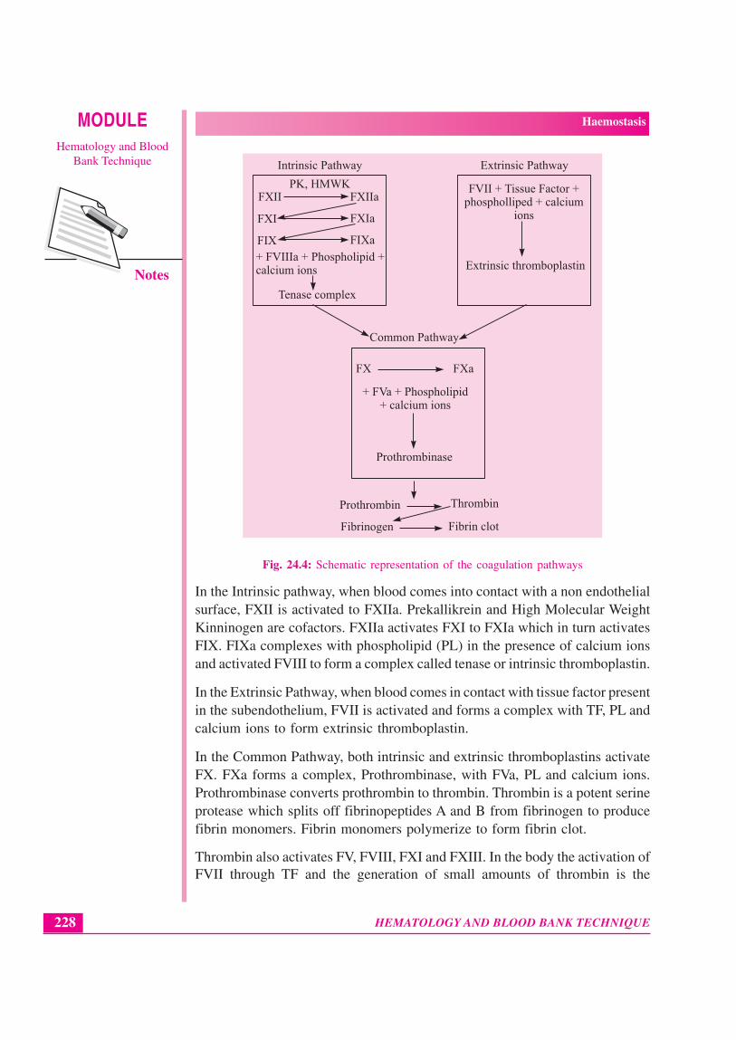
For ease of understanding the Coagulation Phase has been divided into (a)Intrinsic Pathway (b) Extrinsic Pathway and (c) Common Pathway as shown inFigure 24.4
MODULE Haemostasis
Hematology and BloodBank Technique
228 HEMATOLOGY AND BLOOD BANK TECHNIQUE
Notes
Intrinsic Pathway Extrinsic Pathway
PK, HMWKFXII FXIIa
FXI
FIX
FXIa
FIXa
+ FVIIIa + Phospholipid +calcium ions
Tenase complex
FVII + Tissue Factor +phospholliped + calcium
ions
Extrinsic thromboplastin
Common Pathway
FX FXa
+ FVa + Phospholipid+ calcium ions
Prothrombinase
Prothrombin
Fibrinogen
Thrombin
Fibrin clot
Fig. 24.4: Schematic representation of the coagulation pathways
In the Intrinsic pathway, when blood comes into contact with a non endothelialsurface, FXII is activated to FXIIa. Prekallikrein and High Molecular WeightKinninogen are cofactors. FXIIa activates FXI to FXIa which in turn activatesFIX. FIXa complexes with phospholipid (PL) in the presence of calcium ionsand activated FVIII to form a complex called tenase or intrinsic thromboplastin.
In the Extrinsic Pathway, when blood comes in contact with tissue factor presentin the subendothelium, FVII is activated and forms a complex with TF, PL andcalcium ions to form extrinsic thromboplastin.
In the Common Pathway, both intrinsic and extrinsic thromboplastins activateFX. FXa forms a complex, Prothrombinase, with FVa, PL and calcium ions.Prothrombinase converts prothrombin to thrombin. Thrombin is a potent serineprotease which splits off fibrinopeptides A and B from fibrinogen to producefibrin monomers. Fibrin monomers polymerize to form fibrin clot.
Thrombin also activates FV, FVIII, FXI and FXIII. In the body the activation ofFVII through TF and the generation of small amounts of thrombin is the
229
Haemostasis MODULEHematology and Blood
Bank Technique
HEMATOLOGY AND BLOOD BANK TECHNIQUE
Notes
dominant pathway. Once thrombin is generated the process is sustained throughthe intrinsic pathway.
FXIIIa acts on the fibrin clot in the presence of calcium ions to stabilize it.
The extrinsic and common pathways are tested “in vitro” by the Prothrombintime (PT) and the intrinsic and common pathways are tested by the activatedpartial thromboplastin time (APTT). The final conversion of FI to fibrin is testedby the Thrombin Time (TT). Since FXIIIa acts only after fibrin formation, itsactivity is not evaluated by PT or APTT.
Disorders of the coagulation pathway due to a single factor deficiency areusually inherited disorders. Haemophilia A or FVIII deficiency is the mostcommon example. Multifactor deficiencies are usually acquired as seen insevere liver disease, disseminated intravascular coagulation or massive transfusionof bank blood
D. The Fibrinolytic Phase.
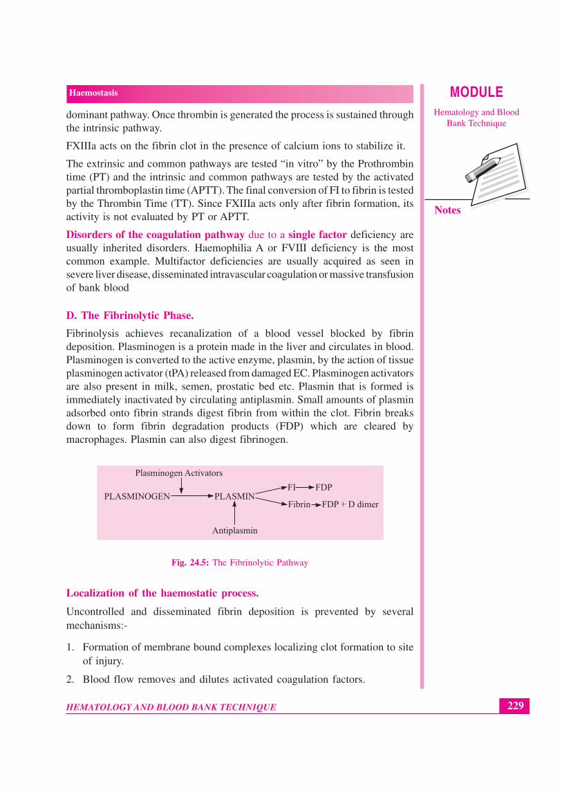
Fibrinolysis achieves recanalization of a blood vessel blocked by fibrindeposition. Plasminogen is a protein made in the liver and circulates in blood.Plasminogen is converted to the active enzyme, plasmin, by the action of tissueplasminogen activator (tPA) released from damaged EC. Plasminogen activatorsare also present in milk, semen, prostatic bed etc. Plasmin that is formed isimmediately inactivated by circulating antiplasmin. Small amounts of plasminadsorbed onto fibrin strands digest fibrin from within the clot. Fibrin breaksdown to form fibrin degradation products (FDP) which are cleared bymacrophages. Plasmin can also digest fibrinogen.
PLASMIN
Plasminogen Activators
PLASMINOGENFI FDP
Fibrin
Antiplasmin
FDP + D dimer
Fig. 24.5: The Fibrinolytic Pathway
Localization of the haemostatic process.
Uncontrolled and disseminated fibrin deposition is prevented by severalmechanisms:-
1. Formation of membrane bound complexes localizing clot formation to siteof injury.
2. Blood flow removes and dilutes activated coagulation factors.

MODULE Haemostasis
Hematology and BloodBank Technique
230 HEMATOLOGY AND BLOOD BANK TECHNIQUE
Notes
3. Presence in plasma of normal inhibitor proteins which inactivate theactivated coagulation factors. These are
(a) Antithrombin III formed by the liver and complexed to heparansulfate and inactivates the serine proteases – Thrombin, FXIa, FXa,FIXa, FVIIa.
(b) Proteins C and S which are vitamin K dependent factors whichinactivate FVa and FVIIIa.
(c) Antiplasmin inactivates plasmin.
(d) Tissue factor pathway inhibitor inhibits tissue factor.
Abnormalties of Haemostasis may result in Bleeding (Congenital or Acquired)and Thrombosis (Arterial or Venous)
INTEXT QUESTIONS 24.2
1. The extrinsic and common pathways are tested by ................ test
2. The intrinsic and common pathways are tested by ................ test
3. Final conversion of F1 to fibrin is tested by ................ test
4. Abnormalities of Haemostasis may result in ................ and ................
24.3 BLEEDING DISORDERS
All patients who present with bleeding need to be investigated. It is also goodpractice to screen patients for a bleeding tendency prior to subjecting apatient to any form of surgery. Preoperative evaluation of haemostasis occursat two levels – an adequate history and screening tests.
History. Specific questions should be directed to
(a) Is there a history of a bleeding tendency? If so, what was the age of onsetof the problem? Onset in childhood is associated with congenital causes.Recent onset suggests an acquired disorder eg. drug induced or liver disease.
(b) What is the type of bleeding? Bleeding into the skin and from mucosalsurfaces – bruising, epistaxis, gum bleeding, menorrhagia suggest a vascularor platelet related problem. Bleeding into subcutaneous tissues, haematomaformation and haemarthrosis suggest a coagulation factor deficiency.
(c) Delayed wound healing, bleeding from umbilical stump, intracranialbleeds and frequent abortions are associated with FI and FXIII defects.

231
Haemostasis MODULEHematology and Blood
Bank Technique
HEMATOLOGY AND BLOOD BANK TECHNIQUE
Notes
(d) Assess severity of bleeding tendency from frequency of episodes, days ofhospitalization, transfusion requirements.
(e) Detailed history of drug intake especially of antiplatelet agents should betaken.
(f) Family history. In general haemophilia A and B are sex linked recessivedisorders, von Willebrand’s disease is autosomal dominant and all othercoagulation factor deficiencies and platelet disorders are autosomal recessivein nature.
24.4 SCREENING TESTS FOR HAEMOSTASIS
These tests detect most abnormalities in haemostasis. If found to be abnormalthe patient is referred for definitive tests which may be available only inspecialized laboratories.
24.4.1 Blood Samples to be taken for screening tests are:
(a) 1 – 2 ml blood in EDTA anticoagulant for complete blood count. Plateletcount done from a finger prick is not reliable.
(b) 4.5 ml venous blood mixed with 0.5ml of 3.2% sodium citrate for plasmaclotting tests.
Blood must be taken from a clean venepuncture. If sampling is to be done froma central line, it must be flushed with normal saline, the first 4 – 5 ml of blooddiscarded and then the sample for tests must be drawn. The sample must beprocessed within 1 – 2 hours.
24.4.2 Tests to be done are:-
24.4.2.1 Platelet count. All platelet counts must be verified by a blood smear.
24.4.2.2 Bleeding Time. (BT) This measures the platelet vessel wall interaction.Two methods are available – the Template method (normal 2-9minutes) andthe Ivy method ( normal 2 – 6minutes). The BT is very operator dependent.It is accepted now that the BT is not indicated as a routine screening testand that a history taken well is just as reliable. The BT is a poor predictorof abnormal surgical bleeding and need not be done prior to surgery.Indications for doing BT are assessment of platelet function 4 –5 days afterstopping asprin, assessing platelet function when patient has mildthrombocytopenia ( platelet count > 50x109/l), diagnosis of VWD andplatelet dysfunction and to evaluate response to FFP, cryoprecipitate ordesmopressin in VWD prior to surgery.
24.4.2.3 Prothrombin time (PT) is the time taken for citrated plasma to cloton the addition of tissue thromboplastin and calcium chloride. It measures
MODULE Haemostasis
Hematology and BloodBank Technique
232 HEMATOLOGY AND BLOOD BANK TECHNIQUE
Notes
the extrinsic and common pathways and will detect abnormalities of FI, II,V, VII and X. A control plasma is always run with the test. PT is consideredprolonged if the test and control values show a greater than 3 seconddifference. The normal PT by the manual technique is 12 - 14 seconds. Amild elevation of the PT is an early indication of deranged hepatocelluardysfunction. For patients on oral anticoagulants the PT is reported as theINR or International Normalised Ratio to avoid problems that arise fromthe use of thromboplastins of varying sensitivities. Commercialthromboplastins are assigned an ISI (International Sensitivity Index) valueafter comparisons with a WHO standard. The ISI is used to calculate theINR. The INR is calculated as:
INR = (Patient’s PT ÷ Mean normal PT)ISI The normal INR is 0.9 – 1.2.
24.4.2.4 Activated partial thromboplastin time (APTT) has replaced the oldand insensitive Clotting Time (CT) test. The APTT measures all thecoagulation factors except FVII and FXIII. The normal manual APTT is 30– 35". A greater than 5 second difference from the control is significant.A persistently short APTT (<25") indicates a hypercoagulable state. TheAPTT is used to monitor heparin therapy.
24.4.2.5 Thrombin time (TT) measures clottable fibrinogen. The normal TTis 12 –14" and a greater than 2 seconds difference from control is significant.TT is increased in deficiency of FI, dysfibrinogenemia, in the presence ofheparin and with elevated levels of FDP.
24.4.2.6 Correction studies. When the PT/APTT/or TT are prolonged acorrection study is performed by mixing equal parts of the test plasma andcontrol plasma and repeating the test with the mixture. If the prolonged timeis corrected, the study indicates a factor deficiency. Lack of correctionindicates the presence of an inhibitor in the patient’s plasma. Commoninhibitors are heparin, lupus anticoagulant or an antibody to one of thecoagulation factors.
24.4.2.7 Tests of fibrinolysis. For screening purposes it is sufficient to detectincreased fibrinolysis in a patient using latex agglutination kits or ELISAtechniques which demonstrate the presence of fibrin degradation products(FDP) and D-dimers.
24.4.2.8 Screening test for FXIII activity. Since FXIII acts after clotformation, the plasma clotting tests do not measure FXIII activity. Ascreening test called clot solubility in 5M urea or 1% acetic acid isperformed to screen for FXIII activity.
233
Haemostasis MODULEHematology and Blood
Bank Technique
HEMATOLOGY AND BLOOD BANK TECHNIQUE
Notes
INTEXT QUESTIONS 20.3
1. INR stands for ..................
2. Normal INR is ..................
3. Normal prothrombin tine is ..................
4. Activated partial thromboplastin time measures are coagulation factorsexcept .................. & ..................
5. Normal Activated partial thromboplastin time is ..................
6. Normal thrombin time is ..................
24.5 COMMON BLEEDING PROBLEMS
24.5.1 Vascular Disorders
Many disorders of abnormal blood vessel structure are present as inherited oracquired conditions. Many of these disorders are present as syndromes andrecognized because of typical signs. They present with easy bruising andpurpura.
Von Willebrand’s Disease
This is an inherited disorder of vascular dysfunction. The VWF is normallyproduced by the endothelial cells and secreted into plasma. The VWF has twofunctions (1) to mediate platelet adhesion to subendothelial collagen and (2) tocarry FVIII and prevent it from being destroyed. VWD is an autosomal dominantdisorder and occurs in both males and females.
Laboratory Diagnosis
1. Haemoglobin, PCV, RBC count are normal unless there is blood loss.
2. Platelet count is normal, morphology is normal.
3. Bleeding time is increased
4. Prothrombin time is normal, Thrombin time is normal
5. APTT is increased
6. FVIII activity is decreased
7. FXIII is normal
Treatment is infusion of FFP or cryoprecipitate both of which contain VWF.
MODULE Haemostasis
Hematology and BloodBank Technique
234 HEMATOLOGY AND BLOOD BANK TECHNIQUE
Notes
24.5.2 Platelet Disorders
Thrombocytopenia is the most commonly encountered haemostatic problem.The availability of platelet concentrates for replacement therapy has madesurgery possible for such patients.
Causes of Thrombocytopenia
A. Failure of Platelet Production in bone marrow.
Aplastic anaemia, Leukemia, Radiation, chemotherapy, Alcholism, Megaloblasticanaemia, marrow infiltration by tumour, lymphoma.
B. Increased destruction of platelets
(a) Immune causes – ITP (immune thrombocytopenic purpura), SLE,drug induced, post transfusion, malaria, viral infections, neonatal.
(b) DIC
(c) Hemolytic uremic syndrome, TTP
(d) Sepsis
C. Abnormal distribution of platelets – splenomegaly
D. Dilutional – massive transfusion
Laboratory diagnosis
1. Complete blood counts shows thrombocytopenia
2. Platelet morphology may be abnormal depending on the cause for lowplatelets
3. Bleeding Time variably prolonged
4. Plasma clotting tests are normal unless there is factor consumption
5. Bone marrow examination to determine cause of thrombocytopenia
Disorders of Platelet Function are given in Table II
Coagulation Factor Deficiency: The deficiency of a single factor is usually dueto an inherited deficiency. The commonest factor deficiency is Haemophilia Awhich is FVIII deficiency. Haemophilia B or deficiency of FIX is less common.Both haemophilias present with haematoma formation and haemarthrosis andare inherited as sex linked recessive disorders. Clinically severe form of thedisorder has less than 1% factor activity and is associated with spontaneousbleeding by one year of age. Moderate deficiency has 2 – 5% factor activity andis associated with bleeding following trauma and haemarthrosis. Mild form ofthe disease has 5 – 15% factor activity and is asymptomatic unless the patientis subjected to trauma or surgery. The APTT will detect these patients whereasthe CT will definitely miss them.
235
Haemostasis MODULEHematology and Blood
Bank Technique
HEMATOLOGY AND BLOOD BANK TECHNIQUE
Notes
Laboratory Diagnosis
1. Haemoglobin, PCV, RBC normal or variably deranged.
2. MCV, MCH,MCHC,RDW normal
3. WBC count normal
4. Platelet count normal
5. Bleeding Time normal
6. PT normal
7. APTT prolonged
8. TT normal
9. FVIII/FIX assay.
Rare disorders of other coagulation factors are autosomal recessive and mayinvolve any of the coagulation factors.
Acquired Disorders
(a) Liver disease – Most of the coagulation factors are made in the liver andhence chronic liver disease is associated with multifactor deficiencycharacterized by prolonged PT, APTT, low fI and increased D-dimers.
(b) Vitamin K deficiency.
Vitamin is a fat soluble vitamin which is needed for the normal formationof the Vitamin K dependent factor – FII,FVII, FIX and FX. Vitamin Kneeds bile for absorption. Deficiency is seen in
(a) Newborns: This is called haemorrhagic disease of the new born. Itoccurs because the liver of the newborn is immature and mother’smilk lacks vitamin K. PT and APTT are markedly prolonged and TTis normal
(b) Obstructive jaundice
(c) Liver disease.
(d) Oral anticoagulants used to prevent thrombosis are vitamin antagonists
(c) Thrombotic Disorders
Thrombosis in blood vessels may be arterial (eg heart attack, stroke) orvenous (eg Deep vein thrombosis). The conditions that are associated withthrombosis are usually acquired. They may rarely be inherited due to thedecreased levels of naturally occurring inhibitors in blood. Thrombosisoccurs as a result of abnormalities in blood vessels like atherosclerosis,stasis or pooling of blood, inflammation of blood vessels and abnormalitiesin blood flow due to viscosity, increased levels of fibrinogen, FVIII etc.
MODULE Haemostasis
Hematology and BloodBank Technique
236 HEMATOLOGY AND BLOOD BANK TECHNIQUE
Notes
WHAT HAVE YOU LEARNT
Haemostasis means arrest of bleeding
Phases of Haemostasis are vascular phase, platelet phase, coagulation phaseand fibrinolytic phase
Blood vessel plays a major role in maintaining the blood in the liquid state
The entire surface of the vessel is lined by Endothelial cells (EC)
Endothelial cells secrete a major protein called Von Willebrand factor intoplasma and sub-endothelium
Platelets are formed from megakaryocytes in bone marrow and normalcount is 150 – 450 X 10 9/L
When endothelial cells is damaged platelets adhere to sub endothelial cells
Platelet derived growth factor released from á granules promotes fibroblastproliferation and healing
Blood coagulation takes place in a series of enzyme reactions eachsuccessive step being catalysed by active enzyme formed in previous step
Coagulation phase has been divided into Intrinsic pathway, Extrinsicpathway and common pathway
Extrinsic and common pathway is tested by Prothrombin Time (PT)
Intrinsic and common pathway is tested by Activated Thromboplastin Time(APTT)
Final conversion of FI to fibrin is tested by Thrombin time (TT)
Hemophilia A or FVIII deficiency is a disorder of coagulation pathway dueto single factor
Abnormalities of haemostasis may result in Bleeding & Thrombosis
Bleeding time measures platelet vessel wall interaction and is a poorpredictor of abnormal surgical bleeding
Prothrombin time (PT) is the time taken for citrated plasma to clot on theaddition of tissue thromboplastin and calcium citrate
Normal PT is 12-14 seconds and for patients on oral anticoagulants, PT isreported as INR or International Normalised Ratio and normal INR is 0.9– 1.2
Activated partial thromboplastin time (APTT) measures all coagulationfactors except FVII and FXIII. Normal APTT is 30-35 seconds
Thrombin time (TT) measures clottable fibrinogen and normal TT is 12 –14 seconds
237
Haemostasis MODULEHematology and Blood
Bank Technique
HEMATOLOGY AND BLOOD BANK TECHNIQUE
Notes
TERMINAL QUESTIONS
1. Draw and describe the coagulation pathway.
2. Short notes on:
(a) Platelet function
(b) Haemophilia
(c) Prothrombin time
ANSWERS TO INTEXT QUESTIONS
24.1
1. Arrest of bleeding
2. Von Willebrand
3. Megakaryocytes
4. 150-450x109/l
5. Platelets
6. Calcium
7. Platelet derived growth
8. Scott
24.2
1. Prothrombin time
2. Activated thrombopastin time
3. Thrombin time
4. Bleeding and thrombosis
24.3
1. International Normalised Ratio
2. 0.9 – 1.2
3. 12-14 seconds
4. FVII & FXIII
5. 30 – 35 seconds
6. 12 – 14 seconds