Embed Size (px)
Citation preview
2 Drug Disposition and Response
Robert B. Raffa
Objectives
� Review basic principles of pharmacokinetics related to the absorption, distribution, metab-
olism, and elimination of drugs and nutrients.
� Discuss factors that can affect these processes.
� Review basic principles of pharmacodynamics and the quantification of drug and nutrient
action.
� Highlight potential pharmacokinetic and pharmacodynamic sites of drug–nutrient
interactions.
Key Words: Bioavailability; elimination; pharmacokinetics; pharmacodynamics
1. INTRODUCTION
This chapter presents an overview of drug disposition (pharmacokinetics) anddrug action (pharmacodynamics) as a basis for understanding drug–nutrient inter-actions. Pharmacokinetics is the term used to describe the disposition of a drugthroughout the body – that is, the drug’s absorption, distribution, metabolism, andexcretion (ADME). Pharmacodynamics is the term used to describe a drug’s effectand how that effect is produced (its mechanism of action). A drug–nutrient inter-action is medically significant if either the patient’s response to the drug or thepatient’s nutritional status is affected adversely. Therefore, this chapter highlightsprocesses that can contribute to either outcome.
2. PHARMACOKINETICS
A substance can produce an effect only if it can reach its physiological target(s) insufficient concentration. Hence, the extent and rate of disposition of a drug or anutrient is important for understanding or predicting the magnitude or the durationof their effect or possible interaction. Several factors affect the absorption anddistribution of drugs and nutrients.
From: Handbook of Drug-Nutrient InteractionsEdited by: J.I. Boullata, V.T. Armenti, DOI 10.1007/978-1-60327-362-6_2
� Humana Press, a part of Springer ScienceþBusiness Media, LLC 2009
27

2.1. Absorption
The route by which a substance is introduced into the body affects its pharma-cokinetics (1,2,3).
2.1.1. SYSTEMIC ROUTES
Systemic routes of administration are those that deliver the substance with theintent of producing a systemic (on the whole system) effect, rather than a local effect(for example, on the skin). A subdivision of the systemic route of administration isparenteral, which refers to systemic routes other than alimentary routes (e.g., oral,sublingual, buccal, or rectal). Systemic routes of administration provide anopportunity for drug–nutrient interaction at several levels, including the rate atwhich drug substance or nutrient is available for absorption (e.g., dissolution rate,degree of ionization, adsorption); the extent of plasma protein binding; and the rateor the route of metabolism.
Oral (PO) administration is generally the simplest, most convenient, safest(because of slower onset of drug effect and ability to reverse a mistake), and oftenmost economical route of administration. Most drugs are well absorbed from thegastrointestinal (GI) tract. The rate and extent of absorption is a function of thephysiochemical properties of the substance (e.g., water or lipid solubility), itsformulation (e.g., tablet, capsule, liquid, slow-release reservoir, matrix), excipients,physiological environment (e.g., high acidity of the stomach), andmetabolism in thegut wall. Alteration of any of these features, for example, as a result of change indiet, lifestyle, age, or health status, can affect absorption. Nutrients and foodstuffscan affect the absorption of a drug by binding to it or by altering the physiologicenvironment (e.g., pH of the stomach). The simple act of food ingestion, or even itsanticipation, can release digestive enzymes that inactivate certain drugs(e.g., penicillins).
The intravenous (IV) route of administration delivers the drug directly into thebloodstream. The drug is then delivered to the heart and from there to the generalcirculation. The IV route bypasses problems of absorption from theGI tract, allowsfor rapid adjustment of dose to effect, can be used even if the patient is unconscious,and avoids the ‘‘first-pass effect’’ (see below). The related intraarterial route ofadministration, although used much less commonly than IV administration, isuseful when infusion of a high concentration of drug into a specific target is desired.Examples include chemotherapeutic agents for the treatment of certain cancers andvasodilators for the treatment of Raynaud’s syndrome.
Subcutaneous (SC) administration involves drug delivery into the tissue beneaththe skin and its subsequent entry into the blood perfusing the tissue. Absorptionfollowing SC administration is generally rapid, depending on blood perfusion of aparticular site, and the rate of absorption can be accelerated (e.g., by heating orvasodilators) or slowed (e.g., by cooling, vasoconstrictors, or slow-releaseformulations).
Intramuscular (IM) administration is generally rapid because of the high vascu-larity of muscles. It also provides space for drug depots, such as sustained-releaseformulations, provided a patient has sufficient skeletal muscle.
28 Part I / Approaching Drug–Nutrient Interactions
Inhalation provides one of the most rapid routes of drug administration due tothe large surface area and high vascularity of the lungs, provided adequate doses ofthe active drug reach the distal airway.
Other systemic routes include intraperitoneal, which is particularly useful for theadministration of drugs to small animals because it provides a rapid, convenient,and reproducible technique, and transdermal, because of its convenience and use forextended drug delivery.
2.1.2. TOPICAL ROUTES
Topical routes of administration are generally used for the purpose of local drugaction and are generally not sites of drug–nutrient interactions (a possible exceptionis the reduction of ultraviolet light exposure by sunscreen lotions and the resultingdecreased activation of vitamin D). However, if the skin is damaged (such as byabrasions or burns) or if transmucosal passage is significant, the drug does notremain localized to the site of application and administration is akin to systemicadministration with the attendant opportunity for a drug–nutrient interaction.
2.1.3. OTHER ROUTES
Direct application of drugs for localized effects to the eye (ophthalmic adminis-tration), ear (otic administration), nerves (intraneural administration), spinal cord(e.g., epidural or intrathecal administration), or brain (e.g., intracerebroventricularadministration) does not often lead to significant interactions, but any substancethat alters the drug’s access to specialized compartments particularly via commontransporters (e.g., through the blood–brain barrier) can alter the magnitude or theduration of the drug effect.
2.1.4. FACTORS THAT AFFECT ABSORPTION
The rate and extent of absorption of a drug or a nutrient is influenced by thecharacteristics of the drug or the nutrient and by the characteristics of the patient atthe time of administration (4). For example, the rate of dissolution depends on howthe product is formulated and also on the person’s state of health and other factors,such as diet.
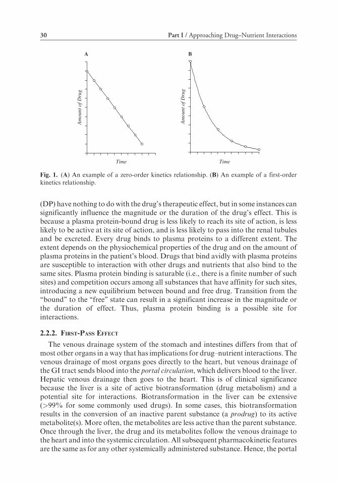
The absorption (and elimination) of substances generally follows eitherzero-order kinetics – i.e., a constant amount is absorbed (or eliminated) per unittime (Fig. 1a) – or first-order kinetics – i.e., a constant fraction is absorbed (oreliminated) per unit time (Fig. 1b). Most currently used drugs follow first-orderkinetics.
2.2. Distribution
Once a drug or a nutrient enters the bloodstream, it might bind to a plasmaprotein (e.g., albumin). In addition, the drug or the nutrient usually must pass somebiological barrier in order to reach its site of action.
2.2.1. PLASMA PROTEIN BINDING
Depending on their physicochemical characteristics, drug molecules (D) canform weak, reversible bonds with plasma proteins (P) according to the generalequilibrium interaction represented as D + P, DP (5). Drug–protein complexes
Chapter 2 / Drug Disposition and Response 29
(DP) have nothing to dowith the drug’s therapeutic effect, but in some instances cansignificantly influence the magnitude or the duration of the drug’s effect. This isbecause a plasma protein-bound drug is less likely to reach its site of action, is lesslikely to be active at its site of action, and is less likely to pass into the renal tubulesand be excreted. Every drug binds to plasma proteins to a different extent. Theextent depends on the physiochemical properties of the drug and on the amount ofplasma proteins in the patient’s blood. Drugs that bind avidly with plasma proteinsare susceptible to interaction with other drugs and nutrients that also bind to thesame sites. Plasma protein binding is saturable (i.e., there is a finite number of suchsites) and competition occurs among all substances that have affinity for such sites,introducing a new equilibrium between bound and free drug. Transition from the‘‘bound’’ to the ‘‘free’’ state can result in a significant increase in the magnitude orthe duration of effect. Thus, plasma protein binding is a possible site forinteractions.
2.2.2. FIRST-PASS EFFECT
The venous drainage system of the stomach and intestines differs from that ofmost other organs in a way that has implications for drug–nutrient interactions. Thevenous drainage of most organs goes directly to the heart, but venous drainage ofthe GI tract sends blood into the portal circulation, which delivers blood to the liver.Hepatic venous drainage then goes to the heart. This is of clinical significancebecause the liver is a site of active biotransformation (drug metabolism) and apotential site for interactions. Biotransformation in the liver can be extensive(>99% for some commonly used drugs). In some cases, this biotransformationresults in the conversion of an inactive parent substance (a prodrug) to its activemetabolite(s). More often, the metabolites are less active than the parent substance.Once through the liver, the drug and its metabolites follow the venous drainage tothe heart and into the systemic circulation. All subsequent pharmacokinetic featuresare the same as for any other systemically administered substance. Hence, the portal
A B
Time
Am
ount
of D
rug
Am
ount
of D
rug
Time
Fig. 1. (A) An example of a zero-order kinetics relationship. (B) An example of a first-orderkinetics relationship.
30 Part I / Approaching Drug–Nutrient Interactions
circulation introduces a special influence on distribution during a substance’s‘‘first-pass’’ into the circulation (6). Oral administration of drugs results in thelargest first-pass effect. Drugs that are administered IV are not subjected to a first-pass effect.
The extent of first-pass metabolism is an important consideration in drug design,formulation, and dosage regimen. For drugs that undergo high first-pass metab-olism, small changes in the rate or the extent of biotransformation (as may occurwith interactions) can result in large changes in systemic blood levels. Changes inbiotransformation can result from changes in GI and liver function or on hepaticdrug-metabolizing enzymes brought about by other drugs, nutrients, or foodcomponents.
2.2.3. BLOOD–BRAIN BARRIER
Many drugs have only limited ability to enter the brain because of their physico-chemical properties. The morphologic basis for the blood–brain barrier includestight junctions between the epithelial cells lining the brain capillaries and transportmechanisms that pump substances out of the brain. In general, the blood–brainbarrier restricts the passage of substances that are either too hydrophilic (watersoluble) or too lipophilic (fat soluble). Nutritionally required substances can beactively transported across the blood–brain barrier (7).
The permeability of the blood–brain barrier depends on such factors as age,disease, and other influences, including nutritional state. Plasma protein binding isalso a factor, since drug molecules highly bound to plasma proteins are less able totraverse the blood–brain barrier. Hence, drug interaction at the level of plasmaprotein binding can affect blood–brain barrier passage.
2.2.4. BIOLOGICAL MEMBRANES
Biological membranes are bilayer, phospholipid matrices containing cholesterol,proteins, and other constituents. Drugs can be transported around or through thesemembranes, depending on the properties of the drug and the composition of theparticular membrane (see Chapter 3). Some mechanisms of drug transport are asfollows (8):
Passive diffusion. If a drug is sufficiently lipid soluble, it can diffuse down itsconcentration gradient (energy is not required, hence the diffusion is ‘‘passive’’). Forweak acids (HA , H+ + A�) and weak bases (BH+ , H+ + B), it is thenonionized form (HA and B respectively) that is more lipid soluble. Simple diffusionoccurs according to Fick’s law:
dQ
dt¼ �DA
dC
dx;
where the flux of drug across a membrane is determined by the diffusion constant(D), the surface area (A), and the drug concentration (C). This type of diffusionfavors molecules in the uncharged form and is a function of the pH of the environ-ment at themembrane and the pKa of the drug according to relationships termed theHenderson–Hasselbach equations:
Chapter 2 / Drug Disposition and Response 31
pKa ¼ pHþ logHA
A�
� �
for weak acids and
pKa ¼ pHþ logBHþ
B
� �
for weak bases. As described by these equations, absorption of weak acids(e.g., aspirin) is favored over weak bases in the low pH of the stomach. However,the total amount of absorption is usually greater in the intestines due to the muchhigher surface area. Conversely, the absorption of weak bases is favored in the smallintestine (higher pH). Renal excretion follows the same pattern. Weak acids areusually excreted in alkaline urine; weak bases are excreted faster in acidic urine.
Filtration. Some vascular bed capillaries have pores or channels that allow thepassage of low-molecular-weight substances, whether they are polar or nonpolar.Such capillaries serve as molecular sieves (filters) that exclude molecules larger thana certain size.
Carrier-mediated (facilitated) diffusion. Transport of some substances acrossmembranes, although by diffusion down a concentration gradient, is facilitatedby membrane-associated molecules (carriers). This type of diffusion is generallyselective for molecules having specific structures or another property. If the con-centration of drug or nutrient exceeds the number of carriers, the process becomessaturated and any further increase in drug or nutrient concentration will notincrease the rate of their passage across the membrane.
Active transport. Some molecules are transported across biological membranesagainst their concentration gradient. Transport in this direction – ‘‘up’’ a concen-tration gradient – is not favored thermodynamically and, hence, does not occurspontaneously. It requires input of energy, which is commonly supplied by coupledbiochemical reactions that, for example, convert ATP to cAMP (catalyzed by Na+/K+-ATPase). Active transport is similar to carrier-mediated (facilitated) diffusionin that transport is mediated by a membrane-associated macromolecule (pump); itis saturable; and it is usually selective for certain drugs or nutrients (based on size,shape, or other characteristic). It differs in its requirement for energy and the abilityto pump against a concentration gradient.
Endocytosis. Some drugs and nutrients can be transported across biologicalmembranes by becoming entrapped (in ‘‘pits’’) and internalized (in ‘‘vesicles’’) withvarying degrees of selectivity. For example, sucrose and insulin can be internalizedin this manner.
2.2.5. BIOAVAILABILITY
Due to the multiple barriers to absorption, the amount of a drug that enters thesystemic circulation is less than the amount administered (with the exception of IVadministration). The proportion (expressed either as fraction or percent) of anadministered drug dose that reaches the systemic circulation is referred to as thedrug’s bioavailability. Factors that affect a drug’s bioavailability include the
32 Part I / Approaching Drug–Nutrient Interactions

first-pass effect, the solubility and stability, and the formulation of the drug product(including the quality control of its manufacture). In addition, a person’s dietarypatterns, nutritional status, and state of health can affect a drug’s bioavailability.
2.2.6. FACTORS THAT AFFECT DISTRIBUTION
Multiple factors affect the distribution of substances in the body. Some arerelated to the substance itself, such as its physical characteristics (e.g., size, solu-bility) and its chemical characteristics (e.g., ability to form bonds with plasmaproteins or other biochemical substances). Other factors are related to the state ofthe physiological system, such as concentration of plasma proteins, lipid content ofbarrier or target tissues, cardiac output, capillary permeability in target or othertissues, and many others. Many of these factors are a function of age, disease, orother influences.
2.3. Metabolism
Drugs and nutrients are often biotransformed (metabolized) to other substances(metabolites) by a variety of biochemical reactions in a variety of locations through-out the body (9). Almost all tissues can metabolize drugs, but the liver, GI tract,and lungs are the major sites of drugmetabolism of most drugs in humans. The liverplays a predominant role in drug metabolism for two reasons: first, because of itsstrategic location relative to the portal circulation and second, because it containshigh levels of enzymes capable of metabolizing foreign substances (see Chapter 4).In general, but not always, metabolites are less active andmore water soluble (whichfavors excretion in the urine) than the parent substance. In some instances, activemetabolites are formed from inactive parent drugs, in which case the parent istermed a prodrug. The most common chemical reactions that metabolize drugsand nutrients can be conveniently categorized into two broad types: reactions thatalter the basic chemical structure of the parent molecule – Phase 1 reactions– andreactions that result in the attachment of some endogenous substance to the parentmolecule – Phase 2 or conjugation reactions.
2.3.1. PHASE 1 REACTIONS
Phase 1 reactions often occur in the cytosol, mitochondria, and microsomes(a subcellular component containing membrane-associated enzymes on the smoothendoplasmic reticulum) of cells of the liver and other organs.
2.3.1.1. Oxidation. Oxidation (e.g., the addition of oxygen or the removal ofhydrogen from the parent molecule) is a common Phase 1-type reaction. Micro-somal oxidation is a common mechanism of metabolism of many drugs andnutrients because these substances typically have chemical structures that makethem susceptible to oxidation reactions. There is an extensive system (family) ofenzymes that are capable of catalyzing oxidation reactions. Primary components ofthis system are cytochrome P-450 reductase and the many isozymes of cytochromeP-450 (CYP). Examples of microsomal oxidation reactions are C-oxidation orC-hydroxylation of aliphatic or aromatic groups;N- orO-dealkylation;N-oxidationorN-hydroxylation; sulfoxide formation; deamination; and desulfuration. Examples
Chapter 2 / Drug Disposition and Response 33

of nonmicrosomal enzymes having important roles in the metabolism of endogenousand exogenous substances include alcohol- and aldehyde dehydrogenase; xanthineoxidase; tyrosine hydroxylase; and monoamine oxidase.
The family of CYP enzymes is particularly important in studying metabolismbecause of themany drugs and nutrients that aremetabolized by these enzymes and,in addition, the potential for drug–nutrient interactions (10). For example, it isestimated that over 90% of presently used drugs are metabolized by one or more ofthe CYP enzymes. Of the most commonly used drugs, about 50% are metabolizedby the CYP3A subfamily; about 25% by the CYP2D6 isozyme; about 15% by theCYP2C9 isozyme; and about 5%by the CYP1A2 isozyme. Because the enzymes aresaturable, and can be induced or inhibited, there is significant potential forinteractions.
2.3.1.2. Reduction. Reduction reactions (e.g., the addition of hydrogen or theremoval of oxygen from the parent molecule) occur both in microsomal and non-microsomal fractions of hepatic and other cells. Examples of such reactions includenitro-, azo-, aldehyde-, ketone-, and quinone reduction.
2.3.1.3. Hydrolysis. Hydrolysis-type reactions can occur in multiple locationsthroughout the body, including the plasma. Examples of some nonmicrosomalhydrolases include esterases, peptidases, and amidases.
2.3.2. PHASE 2 REACTIONS
The coupling (conjugation) of an endogenous substance to a drug or a nutrientmolecule typically alters its three-dimensional shape sufficiently to result in adecrease in biological activity. Conjugation also typically results in an increase inwater solubility of the substance, which decreases the amount that is reabsorbedthrough renal tubules and thereby enhances the fraction that is excreted in the urine.Conjugation with glucuronic acid (glucuronidation) is the most common conjuga-tion reaction in humans. Other Phase 2 reactions include glycine-, glutamate-, orglutathione-conjugation;N-acetylation (acetyl coenzymeA as acetyl donor);O-, S-,or N-methylation (S-adenosylmethionine as methyl donor); and sulfate or sulfani-late formation (30-phosphoadenosine 50-phosphosulfate as the sulfate donor).
2.3.3. SEQUENCE OF METABOLISM
It is common for a drug to be metabolized through several biotransformationreactions, resulting in the production and the elimination of several or manymetabolites, each having its own pharmacokinetic and pharmacodynamic charac-teristics. It is also common for a substance to undergo a Phase 2-type reactionfollowing a Phase 1-type reaction, but this sequence is not a requirement. It ispossible for a Phase 2 reaction to precede a Phase 1 reaction.
2.3.4. INDUCTION OR INHIBITION
Many of the enzymes involved in the biotransformation of drugs and nutrientscan be induced (increased in number and activity) or inhibited (reduced in numberand activity) by a variety of chemical substances, including themselves and otherdrugs or nutrients (11). Induction results in an enhanced metabolism of molecules
34 Part I / Approaching Drug–Nutrient Interactions

that are biotransformed by the same pathway and results in a decrease in the level ofparent molecule and increase in the level of metabolites. The biological effect will bedecreased if the parent is more active than its metabolites and increased if the parentis a prodrug. The opposite occurs with enzyme inhibition.
2.3.5. FACTORS THAT AFFECT METABOLISM
Multiple factors can affect metabolism (12), including genetics (polymor-phisms); the chemical properties of the drug or the nutrient (which determinestheir susceptibility to the various types of metabolic reactions); the route of admin-istration (which affects, for example, the extent of the first-pass effect); dose (whichcan exceed the capacity of substrates for conjugation reactions); diet (which can alsoaffect the capacity of substrates for conjugation reactions); age and disease (whichcan affect hepatic function); and others.
2.4. Elimination
The biological effects of exogenous substances are terminated by the combinedprocesses of redistribution,metabolism, and excretion – i.e., elimination (13). Severalfactors affect the rate and extent of elimination, and accumulation occurs if the rate ofabsorption and distribution of a drug or a nutrient exceeds the rate of elimination.
2.4.1. ROUTES OF ELIMINATION
In humans, the kidney is the major route for elimination ofmany drugs, partly dueto the fact that the kidneys receive about 20–25%of the cardiac output. Other sites ofelimination include the lungs, the feces, and (usually to a lesser, but no less important,extent) sweat, saliva, blood loss, gastric fluid, breast milk, semen, and others.
Size exclusion prevents plasma proteins – and drug molecules that are bound tothem – from passing through the glomerulus of a healthy kidney. The fate of asubstance that passes into the nephron depends on the substance’s physicochemicalproperties. Lipophilic substances (such as the nonionized form of weak acids orbases) are more likely to be reabsorbed through the wall of the nephron and backinto the circulation. Hydrophilic substances (such as the ionized form of weak acidsor bases) are more likely to be excreted in the urine. The pH dependence ofionization is exploited clinically by adjusting the urine pH. Some substances areactively transported across the wall of the nephron either into or out of the lumen ofthe nephron. Such transport processes are generally saturable and, thus, arepossible sites of drug–nutrient interactions.
2.4.2. RATE OF ELIMINATION
The elimination of most current drugs follows first-order kinetics (i.e., ‘‘expo-nential decay’’) in which the drug concentration at any time t (Ct) is related to theoriginal drug concentration (Co) by the equation Ct= Co e�kt. In first-orderelimination, equal fractions of drug are eliminated in equal times and Co is reducedby 50% in one half-life (t1/2). Other drugs are eliminated by zero-order (linear)kinetics. In zero-order elimination, equal amounts of drug are eliminated in equaltimes. In both cases, elimination is a function both of the substance and of thecondition of the patient.
Chapter 2 / Drug Disposition and Response 35

2.4.3. CLEARANCE
The rate of elimination (mass/time) of a substance is equal to its concentration(mass/volume) times the ‘‘clearance’’ (volume/time). Clearance is the volume of acompartment (e.g., blood) per unit of time that is ‘‘cleared’’ of the substance due toelimination (e.g., metabolism or excretion). The equation that relates renal plasmaclearance (Cl), rate of excretion (Re), drug concentration in plasma (Cp), and drugconcentration in urine (Cu) is:
CICp ¼ CuRe:
2.4.4. EFFECT OF MULTIPLE DOSING
When a drug is administered according to a fixed-interval schedule, the rate ofaccumulation is predictable from the dose and half-life. For example, following therepeated IV dosing of a drug having first-order elimination kinetics, the mean drugconcentration (Cav) can be estimated from the dose (D) and the fraction of drugremaining (F) by the equation
Cav ¼ �D = lnF:
The upper (Cmax) and lower (Cmin) bounds can be estimated by
D=ð1� F Þ andFD=ð1� F Þ;
respectively (Fig. 2). The actual clinical results depend on the patient’s individualcharacteristics.
2.4.5. FACTORS THAT AFFECT ELIMINATION
In addition to the factors already cited, elimination can be accelerated by enzymeinduction, increases in urine flow, or by change in urine pH and can be slowed byrenal impairment, change in pH, or other patient-specific factors.
2.5. Pharmacogenetics
Pharmacogenetics (pharmacogenomics) is the study of how a person’s geneticmakeup (genotype) influences the way they respond to a drug (their phenotype inthis regard) and the role genetic differences play in interindividual variability of
Con
cent
rati
on
Time
Cmax
Cav
Cmin
Fig. 2. An example of multiple dosing of a drug having first-order elimination kinetics. Cmax,Cav, and Cmin are described in the text.
36 Part I / Approaching Drug–Nutrient Interactions

response to drugs. Many genes that encode drug-metabolizing enzymes,transporters, and receptors are now known to be genetically polymorphic – definedas the ability of a gene to assume multiple forms, where the least common alleleoccurs in >1% of the population. The variation can be in the gene promoter, thecoding region (exons), the noncoding region (introns), or an untranslated genesequence. A polymorphism in any region can lead to faulty protein structure orexpression and there are numerous clinical examples of polymorphic enzymesaltering a drug’s disposition or effect. Single nucleotide polymorphisms (SNPs)are defined as mutations that involve a single DNA base substitution. SNPs arethe most common variants in the human genome.
Knowledge of a person’s phenotype can facilitate better choice of therapeuticapproach and the design of more optimal drug regimens, particularly in patientswho may not be achieving the expected effect of a drug.
3. PHARMACODYNAMICS
A substance produces a biological effect by modification or interaction withongoing physiological processes. In some cases the target is foreign (e.g., bacteriaor viruses) or aberrant (cancer cells). Inmost other cases, the target is part of normalphysiology (e.g., enzymes or receptors). Drug actions are quantified and evaluatedusing dose–response curves.
3.1. Mechanisms of Action
In the broadest sense, drug effects can be categorized into fourmajormechanisms(14). They can kill invading organisms (e.g., antibiotics or antivirals), they can killaberrant cells (e.g., many cancer chemotherapies), they can neutralize acids(antacids), and they can modify physiological processes.
3.1.1. ANTIBIOTICS/ANTIVIRALS
Antibiotics and antivirals target biochemical processes of invading organisms.For example, penicillins, cephalosporins, carbapenems, and monobactams, whichhave chemical structures that contain a b-lactam ring, disrupt cell walls or inhibittheir synthesis. Sulfonamides and trimethoprim act on enzymatic pathways,resulting in the inhibition of folic acid synthesis. Aminoglycosides, tetracyclines,chloramphenicol, and erythromycin interfere with mechanisms involved in thesynthesis of bacterial proteins. Quinolones inhibit bacterial DNA gyrase. Mostantivirals work by inhibiting viral replication. In all cases, the clinical utility issignificantly increased when the drug exhibits selectivity for biochemical processesessential to the invading organism, but not essential to humans.
3.1.2. CANCER CHEMOTHERAPY
Much current cancer chemotherapy (antineoplastic agents) involves the use ofsubstances that are cytotoxic. In general, current antineoplastic drugs can bedivided into four major classes: alkylating agents, antimetabolites, alkaloids, andantibiotics. Alkylating agents bind covalently to DNA, thereby impeding replica-tion and transcription, leading to cell death. Antimetabolite drugs compete withcritical precursors of RNAandDNA synthesis, thereby inhibiting cell proliferation.
Chapter 2 / Drug Disposition and Response 37

Alkaloids inhibit microtubular formation and topoisomerase function, therebyblocking cell division and DNA replication. Certain antibiotics inhibit RNA andDNA synthesis. Many patients receive combinations of these drugs.
3.1.3. ANTACIDS
Excess gastric acidity is reduced by treatment with antacids, which are weak basesthat convert gastric (hydrochloric) acid to water and a salt. Most antacids in currentuse contain aluminum hydroxide, magnesium hydroxide, sodium bicarbonate, or acalcium salt.
3.1.4. MODULATION
The chemical nature of cellular function and communication within and betweencells allows for modulation by endogenous chemical substances (drugs andnutrients). The targets of such modulation include enzymes, DNA, and a varietyof other molecules involved in the synthesis, storage, metabolism, or elimination ofendogenous substances.
3.2. Receptors
Many drugs interact with macromolecular components of cells that then initiate achainof events that lead to thedrug’s effect. In the commonlyusedanalogy, the receptoris likea light switch.Abetteranalogy is thata receptor is likeadimmerswitch, since thereis generally some basal level of activity. A receptor also serves to limit the access to theswitch to only a select number of specific molecules (by ‘‘lock-and-key’’ fit).
3.2.1. OCCUPATION THEORY
Receptors are activated when specific molecules (drugs) form weak intermo-lecular bonds with them – the magnitude of such a drug’s effect is related to thenumber (or the fraction of the total) of receptors that are ‘‘occupied’’ (15). Theformation of drug–receptor complexes is usually reversible such that the reactionbetween drug molecule (D) and receptor molecule (R) is an equilibrium reactionthat can be described and characterized – as any other chemical equilibriumreaction – according to the equation D + R , DR. The ‘‘driving force’’ for thereaction to proceed in the direction of drug–receptor complex depends on theGibb’s free energy difference (DG) according to DG ¼ �RT lnKeq, where R is aconstant, T is the temperature (Kelvin), and Keq is the equilibrium constant (16).
3.2.2. AGONISTS AND ANTAGONISTS
The vast majority of chemical substances do not fit a binding site on any receptor.Chemicals that do bind to receptors are said to do so with a certain affinity, themagnitude of which is given by the reciprocal of the equilibrium constant andtermed the ‘‘dissociation constant’’ (often designated as KD). Only a subset ofsubstances that bind to receptors are capable of eliciting an effect through thereceptor, i.e., have intrinsic activity. Substances that have affinity and intrinsicactivity are termed agonists and substances that have affinity, but not intrinsicactivity, are termed antagonists. Antagonists competitively or noncompetitivelyinhibit the access of agonists to their receptors. Since receptors mediate the effects
38 Part I / Approaching Drug–Nutrient Interactions
of endogenous agonists such as neurotransmitters, hormones, and peptides, antag-onist drugs – although lacking intrinsic activity in vitro – can produce biologicaleffects in vivo by attenuating the signal of the endogenous agonist.
3.2.3. SIGNAL FIDELITY
One of the major functions of receptors is to provide the necessary fidelity foraccurate and reliable communication between neurons or other cells. The ‘‘lock-and-key’’ requirement restricts access only to molecules of specific three-dimensionalshape. The fit is sufficiently flexible, however, that certain molecules (such as drugs)having three-dimensional shapes similar to the endogenous ligand can bind to theirreceptors (with greater or lesser affinity and intrinsic activity).
3.2.4. ‘‘UP-’’ AND ‘‘DOWNREGULATION’’
The number of receptors expressed at any given time is the difference between thenumber synthesized and the number destroyed or internalized and, thus, is afunction of the age, health, and other characteristics of the individual. Repeatedexposure to an agonist or an antagonist can alter the number of expressed receptors.The change in receptor number is often interpreted as the body’s attempt tocounteract excess action of an agonist or an antagonist and an effort to reestablishhomeostasis. More permanent change in receptor number can result from drugeffects at the level of the gene.
3.3. Signal Transduction
Signal transduction refers to the postreceptor sequence of events that lead toan agonist’s effect. Transduction mechanisms can be divided broadly into twotypes: ionotropic, in which activation of the receptor leads directly to influx ofions, and metabotropic, in which activation of the receptor actuates a series ofbiochemical second messengers that mediate the response (17).
3.3.1. LIGAND-GATED ION CHANNELS
Located on the membranes of excitable cells, ligand-gated ion channel receptors(LGICRs) are comprised of segments of transmembrane proteins that form pores ofspecific size and shape that allow the passage of certain ions. The magnitude or therate of flow of ions through the membrane is regulated by the binding of ligand tothe receptor. LGICRs usually display selectivity for ions (e.g., Na+, K+, Ca2+, orCl�) and can be composed of subunits that can be expressed or coupled in differentways in different cells, thus mediating a variety of effects. Examples of LGICRs arethe nicotinic cholinergic, GABAA, glutamate, and glycine receptors.
3.3.2. GPCRS
The G-protein-coupled receptors (GPCRs) typically include seven transmem-brane regions, an N-terminal extracellular region, and a C-terminal intracellularregion (18). A group of guanosine triphosphate (GTP) protein subtypes arecoupled to the receptor. Ligand activation of a GPCR induces GDP–GTPexchange and modulation of associated second messengers such as adenylatecyclase, phosphoinositide pathways, and ion channels. Multiple G-proteinsubtypes allow for selective responses (19).
Chapter 2 / Drug Disposition and Response 39
3.3.3. TYROSINE KINASE RECEPTORS
Tyrosine kinase receptors span the cell membrane and their self-containedcatalytic domain functions as an enzyme. Examples include receptors for certaingrowth factors and insulin.
3.3.4. NUCLEAR RECEPTORS
A large group of intracellular receptors, referred to as the nuclear receptors, areligand-dependent transcription factors that regulate gene expression. Along withcoreceptors and cofactors, the activation or the inhibition of these receptorsinfluences the synthesis and regulation of proteins (e.g., enzymes and receptors)and other cellular components.
3.4. Dose–Response Curves
The relationship between the dose of a drug and its corresponding response is auseful measure of effect from both a mechanistic and a practical standpoint. Forexample, given a reaction scheme of the form D + R , DR, it follows that theshape of the dose–response curve should be hyperbolic, something that is observedfor many drugs (19). In addition, certain features of a dose–response curve canyield clinically valuable information, such as a measure of relative potency orefficacy (20).
Several ways of displaying a dose–response curve are described in the followingsections. The type of display can affect certain mathematical (statistical) analyses ofthe data (for details, see ref. (21)).
3.4.1. QUANTAL
A ‘‘quantal’’ dose–response curve is one in which the dependent variable, usuallyplotted on the ordinate (y-axis), is measured as an all-or-none outcome (e.g., thenumber of patients with systolic blood pressure greater than 140 mm Hg).
3.4.2. GRADED
A ‘‘graded’’ dose–response curve is one in which the dependent variable ismeasured using a continuous scale (e.g., systolic blood pressure in mm Hg). Aswith a quantal dose–response curve, the set of points on rectangular coordinatesderived from plotting the measured effect against the administered dose typicallyforms a pattern that approximates a rectangular hyperbola (Fig. 3A).
3.4.3. LOG
For practical, and now partly unnecessary but historical, reasons, dose–responsecurves are commonly constructed by plotting the response against the logarithm(base 10) of the dose. The shape of such curves becomes sigmoidal or ‘‘S shaped’’ (Fig.3B). This has become so customary that such a plot is often called a dose–responsecurve, although log(dose)–response curve is more precise.
3.4.4. POTENCY AND EFFICACY
The dose of the drug estimated to produce 50% effect is termed the ED50 (orequivalent) for a quantal dose–response curve and the D50 (or equivalent) fora graded dose–response curve. Potency is a comparative term that refers to the
40 Part I / Approaching Drug–Nutrient Interactions

amount of substance that is required to produce a specified level of effect (Fig. 4A).Efficacy is a term that refers to a substance’s ability to achieve a certain degree ofresponse under specified conditions (Fig. 4B). Potency and efficacy are independentcharacteristics.
3.4.5. ANTAGONISM
Antagonists, though lacking intrinsic activity, can produce effects when given toa patient because they attenuate the action of an endogenous agonist involved in apathway that is tonically active. For example, antagonists of the muscariniccholinergic receptor attenuate the parasympathetic influence on heart rate, with
A B
potency efficacy
Dose
Res
pons
e
Dose
Res
pons
e
Fig. 4. (A) Potency is indicated by the location of a dose–response curve along the x-axis. (B)Efficacy is indicated by the maximal-attainable level of effect under specified conditions.
A B
Dose
Res
pons
e
Dose
Res
pons
e
Fig. 3. (A) A dose–response curve on rectangular coordinates. (B) Quantal or graded dose–response data plotted against log10(dose).
Chapter 2 / Drug Disposition and Response 41

consequent increase in heart rate due to the less-opposed influence of the sympa-thetic subdivision. Such ‘‘effects’’ of an antagonist can also be characterized by adose–response curve.
4. CONCLUSION
Drugs are substances taken to defend against invading organisms or to correctaberrant physiological processes, while nutrients are substances taken for main-tenance of normal physiological processes. Both types of substance are desirable.Both types of substances also have chemical compositions that, by nature or bydesign, interact with common sites within the body. Therefore, drug–nutrientinteractions can occur. The interactions can be deleterious to the intended actionof the drug or to the nutritional status of the patient. Either outcome is undesirable.The principles of drug disposition and response outlined in this chapter provide thebasis for understanding, or predicting, such interactions. They further provide afoundation for the more detailed treatments of these interactions presentedthroughout the rest of this book.
Take Home Points
� In the broadest sense, drugs and nutrients share the feature of being chemicalsubstances that – within a proscribed concentration range – produce a beneficialphysiological effect.
� Drugs and nutrients share several common sites of transport within the body(absorption, distribution, metabolism, and elimination), each of which representsa potential site of drug–nutrient interaction.
� Drugs and nutrients produce their effects through similar pharmacodynamicmechanisms (e.g., enzymes and receptors), which can be sites of drug–nutrientinteraction.
� The clinical significance of a pharmacokinetic or a pharmacodynamic drug–nutrientinteraction can be highly dependent on the individual patient – i.e., a function ofpatient’s general health, nutritional status, age, etc.
� The basic principles of pharmacokinetics and pharmacodynamics provide a basisfor understanding the occurrence and treatment of drug–nutrient interactions.
REFERENCES
1. Raffa RB, Rawls SM, Portyansky-Beyzarov E. Netter’s Illustrated Pharmacology. Philadelphia,PA: Elsevier, 2005.
2. Rang HP, Dale MM, Ritter JM, et al. Pharmacology. New York: Churchill Livingstone,1995:74–79.
3. Jacob LS. NMS Pharmacology. 4th ed. Philadelphia, PA: Williams & Wilkins, 1996:3–4.
4. Xie H-G, Kim RB, Wood AJJ, et al. Molecular basis of ethnic differences in drug disposition andresponse. Annu Rev Pharmacol Toxicol 2001;41:815–850.
5. Pratt WB. The entry, distribution, and elimination of drugs. In: Pratt WB, Taylor P, eds. Principlesof Drug Action: the Basis of Pharmacology. 3rd ed. New York: Churchill Livingstone,
1990:231–236.
42 Part I / Approaching Drug–Nutrient Interactions
6. Holford NHG, Benet LZ. Pharmacokinetics & pharmacodynamics: dose selection & the timecourse of drug action. In: Katzung BG, ed. Basic & Clinical Pharmacology. 7th ed. Stamford, CT:Appleton & Lange, 1998:34–49.
7. de Boer AG, van der Sandt ICJ, Gaillard PJ. The role of drug transporters at the blood–brainbarrier. Annu Rev Pharmacol Toxicol 2003;43:629–656.
8. Levine RR. Pharmacology: Drug Actions and Reactions. 5th ed. New York: Parthenon,1996:51–74.
9. Benet LZ, Kroetz DL, Sheiner LB. Pharmacokinetics: the dynamics of drug absorption, distri-bution, and elimination. In: Hardman JG, Limbird LE, eds-in-chief. Goodman & Gilman’s thePharmacological Basis of Therapeutics. 9th ed. New York: McGraw-Hill, 1996:11–16.
10. Lin JH, LuAYH. Inhibition and induction of cytochrome P450 and the clinical implications. ClinPharmacokinet 1998;35:361–390.
11. Park BK, Kitteringham NR, Pirmohamed M, et al. Relevance of induction of human
drug-metabolizing enzymes: pharmacological and toxicological implications. Br J Clin Pharmacol1996;41:477–491.
12. Lin JH, Lu AYH. Interindividual variability in inhibition and induction of cytochrome P450
enzymes. Annu Rev Pharmacol Toxicol 2001;41:535–567.13. Shargel L, Yu ABC. Applied Biopharmaceutics and Pharmacokinetics. 3rd ed. Stamford, CT:
Appleton & Lange, 1993:265–292.14. Raffa RB. Mechanisms of drug action. In: Raffa RB, ed. Quick Look Pharmacology. Madison,
CT: Fence Creek, 1999:14–15.15. Boeynaems JM, Dumont JE. Outlines of Receptor Theory. Amsterdam: Elsevier/North-Holland,
1980:1–226.
16. Raffa RB. Drug-Receptor Thermodynamics: Introduction and Applications. Chichester, UK;John Wiley & Sons, 2001:1–781.
17. Roerig SC. Drug receptors and signaling. In: Raffa RB, ed. Quick Look Pharmacology.Madison,
CT: Fence Creek, 1999:16–17.18. Strader CD, Fong TM, Tota MR, et al. Structure and function of G protein-coupled receptors.
Annu Rev Biochem 1994;63:101–132.
19. Gudermann T, Kalkbrenner F, Schultz G. Diversity and selectivity of receptor-G proteininteraction. Annu Rev Pharmacol Toxicol 1996;36:429–459.
20. Tallarida RJ, Jacob LS. The dose–response relation in pharmacology. New York:Springer-Verlag, 1979:1–207.
21. Tallarida RJ. Drug synergism and dose-effect data analysis. Boca Raton, FL: Chapman & Hall/CRC, 2000:1–247.
Chapter 2 / Drug Disposition and Response 43

http://www.springer.com/978-1-60327-363-3