Embed Size (px)
Citation preview

The expression and prognostic value of the peripheral cannabinoid receptor
in hematological malignancies
Nazik Durdu-Rayman
UITNODIGING
Voor het bijwonen van de openbare verdediging van het
proefschrift:
The expression and prognostic value of the peripheral
cannabinoid receptor in hematological malignancies
De openbare verdediging zal plaatsvinden op
23 september 2011 om 9:30 uurErasmus Universiteit,
Senaatszaal A locatie Woudestein.
Burgemeester Oudlaan 503062 PA Rotterdam
U bent van harte uitgenodigd voor de receptie na a oop van de
promotie
Paranimfen:Emine Kiliç
Behiye Salkml-Özcan
Nazik Durdu-RaymanBinnengracht 47 3162 WD RhoonTel: 0641518756
The expression and prognostic value of the peripheral cannabinoid receptor in hem
atological malignancies N
azik Durdu-R
ayman

The expression and prognostic value of the
peripheral cannabinoid receptor
in hematological malignancies
Nazik Durdu-Rayman

Publication of this thesis was fi nancially supported by:
Layout: Legatron Electronic Publishing, RotterdamPrinted by: Ipskamp Drukkers B.V.
ISBN/EAN: 978-94-6191-006-6
© N. Durdu-Rayman, 2011All rights reserved. No part of the material protected by this copyright notice may be reproduced or utilized in any form or by any electronic, mechanical, or other means, now known or hereafter invented, including photocopying and recording, or in any information storage and retrieval system without prior written permission of the author.
The work in this thesis was performed at the Departments of Hematology and Pathology Erasmus MC Rotterdam.This work was partly funded by the Dutch Cancer Society (Koningin Wilhelmina Fonds) and the Revolvung Fund (MRace) of the Erasmus University Rotterdam.

The expression and prognostic value of the peripheral cannabinoid receptor
in hematological malignancies
De expressie en prognostische waarde van de perifere cannabinoid receptor bij hematologische maligniteiten
Proefschrift
ter verkrijging van de graad van doctor aan de
Erasmus Universiteit Rotterdam
op gezag van de
rector magnifi cus
Prof.dr. H.G. Schmidt
en volgens besluit van het College voor Promoties.
De openbare verdediging zal plaatsvinden op
23 september 2011 om 9:30 uur
Nazik Durdu-Rayman
geboren te Sarıkaya Turkije

PROMOTIECOMMISSIE
Promotoren Prof.dr. P. Sonneveld Prof.dr. H.R. Delwel
Overige leden Prof.dr. H. Hooijkaas Prof.dr. A. Hagenbeek Dr. D. de Jong
Copromotor Dr. K.H. Lam

Babam içinVoor mijn vader


TABLE OF CONTENTS
Chapter 1 Introduction 9
Chapter 2 The peripheral cannabinoid receptor Cb2, frequently 25expressed on AML blasts, either induces a neutrophilic differentiation block or confers abnormal migration properties in a ligand-dependent manner
Chapter 3 Distinct expression profi les of the peripheral cannabinoid 51receptor in lymphoid tissues depending on receptor activation status
Chapter 4 The expression of the peripheral cannabinoid receptor on cells 71of the immune system and Non-Hodgkin’s lymphomas
Chapter 5 Prognostic relevance of immunohistochemical sub 91classifi cation of Diffuse Large B-cell Lymphoma in two prospective phase III clinical trials
Chapter 6 The expression of the peripheral cannabinoid receptor CB2 113has no effect on clinical outcome in Diffuse Large B-cell Lymphomas
Chapter 7 Discussion 135
Summary 145
Samenvatting 149
List of abbreviations 153
Curriculum Vitae 155
Dankwoord 157
Color fi gures 162


Chapter 1
Introduction

10 | Chapter 1
GENERAL ASPECTS OF NON-HODGKIN’S LYMPHOMA
Non-Hodgkin’s lymphoma’s (NHLs) are a heterogeneous group of hematological malignancies with a large variation in clinical presentation, morphological appearance and prognosis. The NHLs make up the largest group (40-50%) of all hematological malignancies. In 2007, 17.700 people in the Netherlands had a non-Hodgkin’s lymphoma. Of these cases, 1.2/1000 was male and 1.2/1000 was female. The number of newly diagnosed NHL was 2800. In the same year, 1061 succumbed to the disease (585 male, 476 female)1. NHLs almost always arise from cells of the immune system resulting, in either B-cell or T-cell lymphomas. Most (approximately 85%) NHLs arise from their normal B-cell counterparts whereas a minority (approximately 15%) is derived from T-cells. Of the NHLs, approximately 65% arise in lymph nodes (nodal type), whereas the remaining 35% can arise in any organ (extra-nodal type). The most recent WHO classifi cation contains about 50 different (clinico-pathological) entities. Each entity is considered to have a normal physiological counterpart refl ecting the various differentiation stages in the lymphoid organs or bone marrow1. Diffuse large B-cell lymphoma (DLBCL) as defi ned by the World Health Organization (WHO) 2008 classifi cation is the most frequent lymphoma subtype comprising approximately 40% of the aggressive NHL. More than 50% of the patients are at least 60 years of age. The addition of the anti-CD20 antibody rituximab to cyclophosphamide, doxorubicin, vincristine and prednisone (CHOP) or other CHOP-like regimens (R-CHOP) has improved the clinical outcome signifi cantly, the 3 year overall survival (OS) being approximately 30 – 40% without rituximab, and 60 – 80% with rituximab. Nevertheless, the survival of both elderly and young patients with NHL still remains unsatisfactory, making improvement of the current therapeutic strategies necessary2,3,4,5,6,7,8,9.
In DLBCL, the international prognostic index (IPI) is one of the most useful tools to distinguish patients with a good from those with a poor prognosis10,11. However, it is a clinical classifi cation system insuffi cient in itself to fully encompass the widely variable biological aspects of this malignancy. Therefore, biological classifi cation systems have been developed in order to improve outcome prediction. The most recently described for this purpose employs DNA micro-array techniques. Using gene expression profi ling (GEP), two subtypes of DLBCL have been identifi ed: a germinal center B-cell (GCB)

Introduction | 11
subtype with a relatively favorable clinical outcome and a ABC (activated B-cell) subtype with a relatively unfavorable clinical outcome12,13,14. GEP is still not a readily accessible technique in daily clinical practice. Therefore, histological subclassifi cation systems using immunohistochemistry were subsequently designed as a substitute. Hans et al published the proof of principle demonstrating the possibilities by using only 3 antibodies (CD10, BCL6 and MUM1) in an algorithm which could identify patients with a favorable GCB or a non-favorable ABC phenotype. The outcome correlated highly with the GEP results on the very same tissue samples15. However, these fi ndings could not always be confi rmed by other investigators, indicating the need for either (additional) novel prognostic markers and/or improvement of the technique16,17.
GENERAL ASPECTS OF ACUTE MYELOID LEUKEMIA
In Western Europe, the mean incidence of acute myeloid leukemia (AML) is approximately 2-3/100000. In the Netherlands, approximately 450 new cases of AML are being diagnosed each year. AML occurs in all age groups, but the incidence rises with higher age. It is the most common type of acute leukemia in adults. Two-thirds of the people diagnosed with AML cases are 60 years or older. The 5-year overall survival of patients younger than 60 years old is approximately 35 – 40%, but this drops dramatically to only 10% when patients are older than 60 years. Thus, age is an important parameter predicting the clinical outcome of AML. In addition, intrinsic cell biological factors such as cytogenetic and molecular determinants are also important in predicting a favorable or non-favorable outcome of AMLs10. The disease is characterized by a large variation in clinical, morphologic, cytogenetic, immunophenotypic and molecular features. Analogous to NHL, maturation and differentiation arrest at various stages of myelopoiesis is seen in this disease with accumulation of immature cell types in the bone marrow. Up till now, it is the predominant immature myeloid cell which determines the AML subtype, but this is expected to change in the near future, as the molecular pathogenesis is being unraveled10. To classify myeloid leukemias, various methods to determine the characteristics of the malignant immature myeloid cell are being used. These include morphology, cytochemistry, immunophenotyping, cytogenetic and molecular genetic studies.

12 | Chapter 1
The type and percentage of blasts which still forms the basis for diagnosis and classifi cation of myeloid neoplasms are determined with morphological analysis, including the study of peripheral blood, bone marrow aspirates and/or trephine biopsies. Cytochemistry and other special stains are used to further determine the lineage of the blasts. This characterization process is refi ned using multi parameter fl owcytometry or immunohistochemistry, resulting in a more specifi c classifi cation. Immunophenotypic characteristics of AML sub types are commonly quite heterogeneous, probably due to genetic diversity. Expression of markers such as CD7, CD9, CD11b, CD14, CD56 and CD34 have been reported to be associated with adverse prognosis, although controversial results have been reported regarding the clinical relevance of these antigens10. Additional genetic studies, such as gene rearrangement analysis by RT-PCR or FISH and mutation detection by gene sequencing or allelic PCR, may be performed in order to further characterize (defi ne) AML subtypes. This is illustrated by the fact that mutation analysis has revealed novel important diagnostic and prognostic subtypes. Examples of mutations in AML which have a diagnostic and prognostic impact are RAS, JAK2, PTPN11, NMP1, CEBPA, RUNX1, KIT and FLT3. Nevertheless, as in NHL, ongoing research is still necessary to defi ne novel genes or their products to enable more patient-tailored treatment modalities2,3.
THE DISCOVERY OF THE CANNABINOID RECEPTOR 2 (CB2)
GENE AS A POTENTIAL ONCOGENE
The heterogenic prognoses of both NHL and AML are based on host-related factors and specifi c genetic or biologic alterations. Many approaches have been tried to unravel the genetic basis of the cell biological processes which determine the clinical outcome. Based on the results of these attempts, several clinical or biological classifi cation systems have been designed to improve the prediction of outcome in these malignancies10. Retroviral insertional mutagenesis (RIM) is one of the more effective approaches to identify biologic/genetic factors i.e. transforming genes in leukemia and lymphoma. By means of RIM, several proto-oncogenes involved in leukemia and lymphoma have been identifi ed18,19,20. Mice injected with a retrovirus develop leukemias and lymphomas within a few months. The ability of these retroviruses to cause malignancy is based on the fact that they integrate into the host genome and interfere with normal transcription and or translation of their target genes. This process in turn may lead to gene over-expression or,

Introduction | 13
in case of tumor suppressor genes, gene inactivation, both having a transforming effect which in a number of cases ultimately lead to cancer. The identifi cation of the target proto-oncogenes and tumor suppressor genes is done by identifying the same genomic locus in independent tumors, i.e. the common virus integration sites (cVIS). Using this technique, well-known proto-oncogenes and tumor suppressor genes such as EVI1 and P53 respectively, have been found21,22,23. Using RIM, the group of Delwel has identifi ed a cVIS, EVI11 (ecotropic virus integration 11) in mice. Further research has demonstrated that viral insertions also occurred in the gene encoding the peripheral cannabinoid receptor Cb2, resulting in aberrant mRNA expression, which in turn leads to over-production of the protein24.
CANNABINOID RECEPTORS AND THEIR LIGANDS
The cannabinoid receptors are members of the family of seven trans membrane G-protein-coupled receptors (GPCR) (Figure 1). Two cannabinoid receptors have been identifi ed using radiolabelled ligand binding assays, RT-PCR and immunohistochemistry. Experiments with mainly rodents have uncovered that the central cannabinoid receptor Cb1, is mainly located in the central and peripheral nervous system25,26,27. More specifi cally, Cb1 was identifi ed by radio-labeling assays with the synthetic cannabinoid compound CP55,940. This compound showed specifi c binding sites in rat brain tissue28. Further research has lead to the identifi cation and cloning of the receptor responsible for this binding29. Since this receptor was only present in brain tissues, it was named the central cannabinoid receptor (Cb1). Subsequently, another cannabinoid receptor was also identifi ed and cloned in the human promyelocytic cell-line HL6030. Further research indicated the peripheral cannabinoid receptor Cb2 to be mainly found on cells of the immune system. Thus, it was called the peripheral cannabinoid receptor (Cb2). The human gene encoding CB1 is located on chromosome 6q14-15 and the gene encoding CB2 is located on chromosome 1p36. The main known/identifi ed endogenous ligands of these receptors, the so called endocannabinoids are anandamide, which is found in brain 31 and 2-arachidonoylglycerol (2-AG), which is found in canine gut and brain32,33. Also natural ligands such as delta9-THC extracted from the plant Cannabis sativa and synthetic ligands such as CP55,940 have been identifi ed34. These ligands have different binding affi nities for the receptors and this may result in different downstream effects. The cannabinoid ligands can function

14 | Chapter 1
as an agonist, an antagonist or an inverse agonist of the Cb1/Cb2 receptors and may exert several functions with neuromodulatory, cardiovascular and reproductive effects35,36,37. Modulation of immune and infl ammatory response and last but not least inhibition of cancer cell growth have also been reported38,39,40,41,42,43,44,45,46,47,48,49,50.
Figure 1 | Schematic overview of the peripheral cannabinoid receptor: a seven trans-membrane receptor with an intracellular C-terminal part (COO-) and an extracellular N-terminal part (NH3+). The antibodies used were directed against these C-and N-terminal parts of this receptor.
EXPRESSION AND FUNCTIONS OF THE PERIPHERAL
CANNABINOID RECEPTOR IN IMMUNE CELLS
Cb2 mRNA has been detected in the spleen, thymus, tonsils and bone marrow51,52,53. Moreover, mRNA expression analysis in human hematopoietic cells showed that B-cells express CB2 abundantly, followed in decreasing order by NK-cell, monocytes, neutrophils and T-cells54. Protein expression in human immune cells has for the fi rst time been described by Carayon et al., using a polyclonal antibody (Ab) raised against the C-terminus of the human CB2 receptor. CB2 protein was mainly found in the mantle zones (MZs) of secondary follicles of tonsils, areas where mainly naïve B-cells reside. Also, modulation of this receptor during B-cell differentiation has been demonstrated55. Up till now, this is

Introduction | 15
the only study in which the human CB2 receptor expression in developing B-cells in the immune system was described. As already stated above, the peripheral cannabinoid receptor belongs to the family of seven transmembrane G-protein-coupled receptors (GPCRs). GPCRs are crucial to many cellular functions, such as proliferation, maturation, survival, apoptosis and migration56,57,58. The mechanisms through which cannabinoids mediate immunosuppression is still under investigation and can generally be categorized into four pathways: apoptosis, inhibition of proliferation, suppression of cytokine and chemokine production and induction of T regulatory cells (T regs)59. In general, cannabinoids exert their effects through inhibition of adenylate cyclase activity. This blocks forskolin-stimulated cAMP activation, leading to decreased activity of protein kinase A and subsequently to lesser binding of transcription factors to CRE (cyclic-AMP responsive element) resulting in dysfunction in IL-2 production. This may result in immune modulatory effects60. Cannabinoid ligands such as THC, trigger apoptosis of cultured immune cells (macrophages and lymphocytes) through the regulation of Bcl-2 and caspase activity61. Also, THC interferes with MAP kinase signaling pathway, which plays an important role in apoptosis of Jurkat leukemia T-cell line. Specifi cally, THC inhibited the MAPK/MEK/ERK signaling pathway, which resulted in translocation of Bad into mitochondria, and eventually apoptosis. This apoptosis could be signifi cantly decreased by CB2 antagonist SR14452862. Some cannabinoid ligands may interfere with the immune response resulting in impairment of macrophage functions, alteration of CD4/CD8 T-cell ratio and immune globulin productions, down regulation of NK-cell activity or perturbation of macrophage/T-cell cooperation63,64,65,66. Various effects of marijuana components on the proliferation of spleen, lymph node and thymus immune cells in vitro and in vivo have been described. For example, these components suppress the induction and cytolytic function of murine immune cells. Inhibiting capabilities of THC on macrophage co-stimulatory activity have also been described67,68,69. Immunomodulation by cannabinoids is absent in mice defi cient for the cannabinoid receptor Cb270 and formation of B-and T-cell subsets in mice require the presence of the Cb2 receptor71. An immunomodulatory function of this receptor in human immune cells has been described earlier 51. It has been described that cannabinoids enhance human B-cell growth at nanomolar concentrations72. Also, the endogenous cannabinoid ligand 2-AG can act as a chemo attractant in vivo for dendritic cells73. Also, moderate proliferation of virgin and germinal center B-cells of the tonsil occurred upon stimulation with the synthetic cannabinoid agonist CP55,940. This proliferation only occurred when the cells were co-

16 | Chapter 1
stimulated with CD40 monoclonal antibody51. Exposure to CD40 activating agents has already been described as crucial for the functions of other GPCRs such as CXCR4. Only pretreatment of germinal center B-cells (GC) with CD40 resulted in migration of these cells upon stimulation with the CXCR ligand stromal-derived factor74. In mouse, Cb2 mRNA expression in splenic B-cells was augmented following immune cell activation with CD4075, indicating a crucial cross-talk of the CB2 and CD40 receptors. One recent study reports that Cb2 mediates immunoglobulin class switching from IgM to IgE in cultures of murine-purifi ed B lymphocytes76.
EXPRESSION AND FUNCTION OF THE PERIPHERAL
CANNABINOID RECEPTOR IN MYELOID CELLS
To investigate the potential functions of the Cb2 receptor, migration assays were carried out in Cb2-overexpressing cells (32D/GCF-R/Cb2-EGFP) using the potent endocannabinoid ligand 2-arachidonoyl glycerol (2-AG) as a Cb2 agonist. Furthermore, these cells are also a powerful in vitro model for studying granulocytic differentiation. Two distinct biological effects of the Cb2 receptor have been observed in hematopoietic cells expressing the peripheral cannabinoid receptor. The ligand used determined the effect observed: stimulation with endocannabinoid 2-AG results in migration and exposure to a synthetic ligand CP55,940 induces a block of neutrophilic differentiation77,78. Thus, binding of distinct ligands to Cb2 receptors results in activation of distinct effector functions. This ligand dependent downstream effect may be explained by the presence of distinct complexes in which Cb2 is present39. Low CB2 mRNA expression has been demonstrated on normal human neutrophils50. Also, low protein expression using fl owcytometry was detected on human neutrophils79 and CB2 mRNA was detected on the human promyelocytic cell line HL-6080. Several distinct effects were observed using different agonists of the Cb2 receptor, such as signifi cant induction of migration upon stimulation with the endocannabinoid 2-AG71. It has also been demonstrated that 2-AG induces a rapid transient increase in intracellular free calcium (2+) concentrations in HL-60 cells81. Also, in murine leukemia cell lines which (over)express the Cb2 receptor, the major effect is stimulation of migration67. Moreover, the Cb2 receptor induces a reversible block in neutrophilic differentiation in this murine cell line upon stimulation with a synthetic cannabinoid ligand68. In a human eosinophilic

Introduction | 17
leukemia cell line, the endocannabinoid 2-AG induces migration82. Furthermore, the same ligand enhances adhesion of HL-60 cells differentiated into macrophage like cells83.
CB2 EXPRESSION IN HUMAN MALIGNANT LYMPHOMA AND
ACUTE MYELOID LEUKEMIA
In human hematopoietic tissues, the decreasing rank order of CB2 mRNA expression is: B-cells, natural killer cells, monocytes, neutrophils, CD8+ T-cells, and CD4+ T-cells50. CB2 protein expression was detected in B-cell areas, i.e. the marginal zone and mantle zone of secondary follicles of tonsils51. To our knowledge no detailed studies have been carried out which describe the protein expression of the CB2 receptor in NHLs. Using RIM, Cb2 was identifi ed as the target gene in the cVIS EVI11 in Cas-Br-M MuLV-induced myeloid leukemias, indicating that Cb2 may act as a proto-oncogene in leukemogenesis. Aberrations in (the expression of) the gene that encodes the Cb2 receptor leading to aberrant expression of this receptor may be a critical event in the transformation of normal myeloid cells into leukemic cells21,25. The group of Delwel has previously shown that Cb2 is highly expressed in myeloid cell lines containing a retroviral insertion in the Cb2 gene67. Overexpression of the Cb2 receptor in myeloid precursor cells is related with impairment of neutrophilic development, one of the major characteristic of myeloid leukemia68. So far, no further detailed studies have been carried out which describe the expression of CB2 in normal human myeloid precursor and AML cells.
AIM OF THE THESIS
The aim of this thesis is to investigate the expression of the Cb2 receptor and its potential as a novel diagnostic and/or prognostic marker in hematological malignancies. More specifi cally, we have investigated the expression of the CB2 receptor in normal and malignant hematopoietic and immune tissues with a focus on DLBCL. In order to address the question whether CB2 can serve as a prognostic marker, we studied CB2 expression using histological material of well-defi ned patient groups drawn from randomized phase III clinical trials and correlated the results with the clinical outcome of these patients. We have also studied expression of Cb2 (CB2) in vitro under

18 | Chapter 1
various physiological conditions and investigated the effects of CB2-receptor ligands, using cell culture systems of malignant lymphoma and myeloid cell lines. In chapter 2 we studied the CB2 expression in normal bone marrow and primary AMLs. Subsequently we also determined the function of the receptor in differentiation or stimulation of migration using a Cb2 transfected 32-D cell line with different Cb2 specifi c agonists. In chapter 3, we have investigated which cells in primary and secondary follicles in normal human lymphoid tissue expressed CB2 and whether CB2 receptors in the distinct cell populations were active or inactive. Immunohistochemical studies were done using two antibodies: an N-terminal specifi c anti-CB2 antibody and a C-terminal specifi c anti-CB2 antibody which only detected phosphorylated inactive receptors. The function of CB2 expression has been investigated in migration assays using an endocannabinoid 2-AG in a B-lymphoma cell line. In chapter 4, we subsequently have studied the CB2 protein expression in various B- and T-cell NHL subtypes and compared the results with the CB2 protein expression in their normal physiological counterparts. In chapter 5, we have attempted to clarify the controversies about the prognostic value of various immunohistochemical classifi cation systems in order to test this model for its usefulness as a prognostic marker in DLBCL. To this end, we have investigated a known immunohistochemical model using CD10, BCL-6 and MUM1 in a specifi c algorithm to sub-classify DLBCL in two randomized phase III clinical trials. In chapter 6 we have investigated the prognostic value of the CB2 expression in DLBCL using a single center study population. In chapter 7 (general discussion), we evaluated the prognostic value of the CB2 expression as determined by immunohistochemistry We hypothesize that although the protein expression itself does not have prognostic value in DLBCL, targeting the receptor with antagonists or agonists may be of interest in the treatment of NHL as well as in AML and therefore constitute a subject for further research.

Introduction | 19
REFERENCES
1. Gommer AM (RIVM), Poos MJJC (RIVM). Cijfers non-Hodgkin lymfomen (prevalentie, incidentie en sterfte) uit de VTV 2010 In: Volksgezondheid Toekomst Verkenning, Nationaal Kompas Volks-gezondheid. Bilthoven.
2. Bob Löwenberg, Gert Ossenkoppele, Theo de Witte en Marc Boogaerts. Handboek Hematologie. De Tijdstroom 2008.
3. Swerdlow SH, Campo E, Harris NL, et al. WHO Classifi cation of Tumours of Haematopoietic and Lymphoid Tissues. 4th ed. Lyon: 2008.
4. Coiffi er B, Lepage E, Briere J, Herbrecht R, Tilly H, Bouabdallah R, et al. CHOP chemotherapy plus rituximab compared with CHOP alone in elderly patients with diffuse large-B-cell lymphoma. N Engl J Med 2002; 346:235-242.
5. Feugier P, Van Hoof A, Sebban C, et al. et al. Long-Term Results of the R-CHOP Study in the Treatment of Elderly Patients With Diffuse Large B-Cell Lymphoma: A Study by the Groupe d’Etude des Lymphomes de l’Adulte. J Clin Oncol 2005;23:4117-4126.
6. Habermann TM, Weller EA, Morrison VA, et al. Rituximab-CHOP versus CHOP Alone or With Maintenance Rituximab in Older Patients With Diffuse Large B-Cell Lymphoma. J Clin Oncol 2006;24:3121-3127.
7. Pfreundschuh M, Trumper L, Osterborg A, et al. CHOP-like chemotherapy plus rituximab versus CHOP-like chemotherapy alone in young patients with good-prognosis diffuse large-B-cell lymphoma: a randomised controlled trial by the MabThera International Trial (MInT) Group. Lancet Oncol 2006; 7:379-391.
8. A predictive model for aggressive non-Hodgkin’s lymphoma. The International Non-Hodgkin’s Lymphoma Prognostic Factors Project. N Engl J Med 1993.
9. Pfreundschuh M, Schubert J, Ziepert M, Schmits R, Mohren M, Lengfelder E, et al. Six versus eight cycles of bi-weekly CHOP-14 with or without rituximab in elderly patients with aggressive CD20+ B-cell lymphomas: a randomised controlled trial (RICOVER-60). Lancet Oncol 2008; 9:105-16.
10. Jaffe ES, Harris NL, Stein H, Vardiman JW. World Health Organization Classifi cation of Tumours: Pathology and Genetics of Tumours of Haematopoietic and Lymphoid Tissues.IARC Press, Lyon, 2001.
11. Jaffe ES. The 2008 WHO classifi cation of lymphomas: implications for clinical practice and translational research.Hematology Am Soc Hematol Educ Program. 2009:523-31. Review.
12. Alizadeh AA, Eisen MB, Davis RE, Ma C, Lossos IS, Rosenwald A, Boldrick JC, Sabet H, Tran T, Yu X, Powell JI, Yang L, Marti GE, Moore T, Hudson J Jr, Lu L, Lewis DB, Tibshirani R, Sherlock G, Chan WC, Greiner TC, Weisenburger DD, Armitage JO, Warnke R, Levy R, Wilson W, Grever MR, Byrd JC, Botstein D, Brown PO, Staudt LM. Distinct types of diffuse large B-cell lymphoma identifi ed by gene expression profi ling. Nature. 2000 Feb 3;403(6769):503-11.
13. Rosenwald A, Wright G, Chan WC, Connors JM, Campo E, Fisher RI, Gascoyne RD, Muller-Hermelink HK, Smeland EB, Giltnane JM, Hurt EM, Zhao H, Averett L, Yang L, Wilson WH, Jaffe ES, Simon R, Klausner RD, Powell J, Duffey PL, Longo DL, Greiner TC, Weisenburger DD, Sanger WG, Dave BJ, Lynch JC, Vose J, Armitage JO, Montserrat E, López-Guillermo A, Grogan TM, Miller TP, LeBlanc M, Ott G, Kvaloy S, Delabie J, Holte H, Krajci P, Stokke T, Staudt LM; Lymphoma/Leukemia Molecular Profi ling Project. The use of molecular profi ling to predict survival after chemotherapy for diffuse large-B-cell lymphoma. N Engl J Med. 2002 Jun 20;346(25):1937-47.

20 | Chapter 1
14. Wright G, Tan B, Rosenwald A, Hurt EH, Wiestner A, Staudt LM. A gene expression-based method to diagnose clinically distinct subgroups of diffuse large B cell lymphoma. Proc Natl Acad Sci U S A. 2003 Aug 19;100(17):9991-6. Epub 2003 Aug 4.
15. Hans CP, Weisenburger DD, Greiner TC, Gascoyne RD, Delabie J, Ott G, Müller-Hermelink HK, Campo E, Braziel RM, Jaffe ES, Pan Z, Farinha P, Smith LM, Falini B, Banham AH, Rosenwald A, Staudt LM, Connors JM, Armitage JO, Chan WC. Confi rmation of the molecular classifi cation of diffuse large B-cell lymphoma by immunohistochemistry using a tissue microarray. Blood. 2004 Jan 1;103(1):275-82. Epub 2003 Sep 22.
16. De Paepe P, Achten R, Verhoef G, Wlodarska I, Stul M, Vanhentenrijk V, Praet M, De Wolf-Peeters C. Large cleaved and immunoblastic lymphoma may represent two distinct clinicopathologic entities within the group of diffuse large B-cell lymphomas. J Clin Oncol. 2005 Oct 1;23(28):7060-8. Epub 2005 Aug 29.
17. Veelken H, Vik Dannheim S, Schulte Moenting J, Martens UM, Finke J, Schmitt-Graeff A. Ann Oncol. Immunophenotype as prognostic factor for diffuse large B-cell lymphoma in patients undergoing clinical risk-adapted therapy.2007 May;18(5):931-9. Epub 2007 Mar 29.
18. Jonkers J, Berns A. Retroviral insertional mutagenesis as a strategy to identify cancer genes. Biochim Biophys Acta. 1996 May 16;1287(1):29-57.
19. Joosten M, Vankan-Berkhoudt Y, Tas M, Lunghi M, Jenniskens Y, Parganas E, Valk PJ, Löwenberg B, van den Akker E, Delwel R. Large-scale identifi cation of novel potential disease loci in mouse leukemia applying an improved strategy for cloning common virus integration sites.Oncogene. 2002 Oct 17;21(47):7247-55.
20. Valk PJ, Hol S, Vankan Y, Ihle JN, Askew D, Jenkins NA, Gilbert DJ, Copeland NG, de Both NJ, Löwenberg B, Delwel R. The genes encoding the peripheral cannabinoid receptor and alpha-L-fucosidase are located near a newly identifi ed common virus integration site, Evi11.J Virol. 1997 Sep;71(9):6796-804.
21. Mucenski ML, Taylor BA, Ihle JN, Hartley JW, Morse HC 3rd, Jenkins NA, Copeland NG. Identifi cation of a common ecotropic viral integration site, Evi-1, in the DNA of AKXD murine myeloid tumors.Mol Cell Biol. 1988 Jan;8(1):301-8.
22. Morishita K, Parker DS, Mucenski ML, Jenkins NA, Copeland NG, Ihle JN. Retroviral activation of a novel gene encoding a zinc fi nger protein in IL-3-dependent myeloid leukemia cell lines.Cell. 1988 Sep 9;54(6):831-40.
23. Dreyfus F, Sola B, Fichelson S, Varlet P, Charon M, Tambourin P, Wendling F, Gisselbrecht S. Rearrangements of the Pim-1, c-myc, and p53 genes in Friend helper virus-induced mouse erythroleukemias.Leukemia. 1990 Aug;4(8):590-4.
24. Valk PJ, Delwel R. The peripheral cannabinoid receptor, Cb2, in retrovirally-induced leukemic transformation and normal hematopoiesis.Leuk Lymphoma. 1998 Dec;32(1-2):29-43. Review.
25. Herkenham M, Lynn AB, Little MD, Johnson MR, Melvin LS, de Costa BR, Rice KC.Cannabinoid receptor localization in brain.Proc Natl Acad Sci U S A. 1990 Mar;87(5):1932-6.
26. Herkenham M, Lynn AB, Johnson MR, Melvin LS, de Costa BR, Rice KC. Characterization and localization of cannabinoid receptors in rat brain: a quantitative in vitro autoradiographic study. J Neurosci. 1991 Feb;11(2):563-83.
27. Tsou K, Brown S, Sañudo-Peña MC, Mackie K, Walker JM. Immunohistochemical distribution of cannabinoid CB1 receptors in the rat central nervous system. Neuroscience. 1998 Mar;83(2):393-411.
28. Devane WA, Dysarz FA 3rd, Johnson MR, Melvin LS, Howlett AC. Determination and characterization of a cannabinoid receptor in rat brain.Mol Pharmacol. 1988 Nov;34(5):605-13.

Introduction | 21
29. Matsuda LA, Lolait SJ, Brownstein MJ, Young AC, Bonner TI. Structure of a cannabinoid receptor and functional expression of the cloned cDNA.Nature. 1990 Aug 9;346(6284):561-4.
30. Munro S, Thomas KL, Abu-Shaar M. Molecular characterization of a peripheral receptor for cannabinoids.Nature. 1993 Sep 2;365(6441):61-5.
31. Devane WA, Hanus L, Breuer A, Pertwee RG, Stevenson LA, Griffi n G, Gibson D, Mandelbaum A, Etinger A, Mechoulam R. Isolation and structure of a brain constituent that binds to the cannabinoid receptor.Science. 1992 Dec 18;258(5090):1946-9.
32. Mechoulam R, Ben-Shabat S, Hanus L, Ligumsky M, Kaminski NE, Schatz AR, Gopher A, Almog S, Martin BR, Compton DR, et al. Identifi cation of an endogenous 2-monoglyceride, present in canine gut, that binds to cannabinoid receptors.Biochem Pharmacol. 1995 Jun 29;50(1):83-90.
33. Sugiura T, Kondo S, Sukagawa A, Nakane S, Shinoda A, Itoh K, Yamashita A, Waku K. 2-Arachidonoylglycerol: a possible endogenous cannabinoid receptor ligand in brain. Biochem Biophys Res Commun. 1995 Oct 4;215(1):89-97.
34. Devane WA, Dysarz FA 3rd, Johnson MR, Melvin LS, Howlett AC. Determination and characterization of a cannabinoid receptor in rat brain.Mol Pharmacol. 1988 Nov;34(5):605-13.
35. Di Marzo V, Melck D, Bisogno T, De Petrocellis L. Endocannabinoids: endogenous cannabinoid receptor ligands with neuromodulatory action.Trends Neurosci. 1998 Dec;21(12):521-8.
36. Kunos G, Járai Z, Varga K, Liu J, Wang L, Wagner JA. Cardiovascular effects of endocannabinoids � the plot thickens.Prostaglandins Other Lipid Mediat. 2000 Apr;61(1-2):71-84.
37. Paria BC, Ma W, Andrenyak DM, Schmid PC, Schmid HH, Moody DE, Deng H, Makriyannis A, Dey SK. Effects of cannabinoids on preimplantation mouse embryo development and implantation are mediated by brain-type cannabinoid receptors.Biol Reprod. 1998 Jun;58(6):1490-5.
38. De Petrocellis L, Melck D, Palmisano A, Bisogno T, Laezza C, Bifulco M, Di Marzo V. The endogenous cannabinoid anandamide inhibits human breast cancer cell proliferation.Proc Natl Acad Sci U S A. 1998 Jul 7;95(14):8375-80.
39. Bisogno T, Maurelli S, Melck D, De Petrocellis L, Di Marzo V.Biosynthesis, uptake, and degradation of anandamide and palmitoylethanolamide in leukocytes.J Biol Chem. 1997 Feb 7;272(6):3315-23.
40. Di Marzo V, Bisogno T, De Petrocellis L, Melck D, Orlando P, Wagner JA, Kunos G. Biosynthesis and inactivation of the endocannabinoid 2-arachidonoylglycerol in circulating and tumoral macrophages.Eur J Biochem. 1999 Aug;264(1):258-67.
41. Klein TW, Newton C, Friedman H. Cannabinoid receptors and immunity.Immunol Today. 1998 Aug;19(8):373-81. Review.
42. Lau RJ, Tubergen DG, Barr M Jr, Domino EF, Benowitz N, Jones RT. Phytohemagglutinin-induced lymphocyte transformation in humans receiving delta9-tetrahydrocannabinol. Science. 1976 May 21;192(4241):805-7.
43. Lee M, Yang KH, Kaminski NE.Effects of putative cannabinoid receptor ligands, anandamide and 2-arachidonyl-glycerol, on immune function in B6C3F1 mouse splenocytes.J Pharmacol Exp Ther. 1995 Nov;275(2):529-36.
44. Facci L, Dal Toso R, Romanello S, Buriani A, Skaper SD, Leon A.Mast cells express a peripheral cannabinoid receptor with differential sensitivity to anandamide and palmitoylethanolamide. Proc Natl Acad Sci U S A. 1995 Apr 11;92(8):3376-80.
45. Zheng ZM, Specter S, Friedman H. Inhibition by delta-9-tetrahydrocannabinol of tumor necrosis factor alpha production by mouse and human macrophages. Int J Immunopharmacol. 1992 Nov;14(8):1445-52.

22 | Chapter 1
46. Flygare J, Gustafsson K, Kimby E, Christensson B, Sander B. Cannabinoid receptor ligands mediate growth inhibition and cell death in mantle cell lymphoma. FEBS lett. 2005 Dec 19; 579(30):6885-9.
47. Rieder SA, Chauhan A, Singh U, Nagarkatti M, Nagarkatti P. Cannabinoid-induced apoptosis in immune cells as a pathway to immunosuppression. Immunobiology. 2010 Aug;215(8):598-605.
48. Gustafsson K, Wang X, Severa D, Eriksson M, Kimby E, Merup M. Expression of cannabinoid receptors type 1 and type 2 in non-Hodgkin lymphoma: Growth inhibition by receptor activation. Int J Cancer. 2008 Sep 1;123(5):1025-33.
49. Do Y, McKallip RJ, Nagarkatti M, Nagarkatti PS. Activation through Cannabinoid Receptors 1 and 2 on Dendritic Cells Triggers NF-kB-Dependent Apoptosis: Novel Role for Endogenous and Exogenous Cannabinoids in Immunoregulation. J Immunol. 2004. Aug 15;173(4):2373-82.
50. McKallip RJ, Lombard C, Fisher M, Billy R. Martin BR, Ryu S, Grant S, Prakash S. Nagarkatti PS, Nagarkatti M. Targeting CB2 cannabinoid receptors as a novel therapy to treat malignant lymphoblastic disease. Blood 2002 Jul 15;100(2):627-34.
51. Galiègue S, Mary S, Marchand J, Dussossoy D, Carrière D, Carayon P, Bouaboula M, Shire D, Le Fur G, Casellas P. Expression of central and peripheral cannabinoid receptors in human immune tissues and leukocyte subpopulations.Eur J Biochem. 1995 Aug 15;232(1):54-61.
52. Pettit DA, Anders DL, Harrison MP, Cabral GA. Cannabinoid receptor expression in immune cells. Adv Exp Med Biol. 1996;402:119-29.
53. Schatz AR, Lee M, Condie RB, Pulaski JT, Kaminski NE. Cannabinoid receptors CB1 and CB2: a characterization of expression and adenylate cyclase modulation within the immune system. Toxicol Appl Pharmacol. 1997 Feb;142(2):278-87.
54. Bouaboula M, Rinaldi M, Carayon P, Carillon C, Delpech B, Shire D, Le Fur G, Casellas P. Cannabinoid-receptor expression in human leukocytes. Eur J Biochem. 1993 May 15;214(1):173-80.
55. Carayon P, Marchand J, Dussossoy D, Derocq JM, Jbilo O, Bord A, Bouaboula M, Galiègue S, Mondière P, Pénarier G, Fur GL, Defrance T, Casellas P. Modulation and functional involvement of CB2 peripheral cannabinoid receptors during B-cell differentiation. Blood. 1998 Nov 15;92(10):3605-15.
56. Dhanasekaran N, Heasley LE, Johnson GL. G protein-coupled receptor systems involved in cell growth and oncogenesis. Endocr Rev. 1995 Jun;16(3):259-70.
57. Neptune ER, Bourne HR. Receptors induce chemotaxis by releasing the betagamma subunit of Gi, not by activating Gq or Gs. Proc Natl Acad Sci U S A. 1997 Dec 23;94(26):14489-94.
58. Wettschureck N, van der Stelt M, Tsubokawa H, Krestel H, Moers A, Petrosino S, Schütz G, Di Marzo V, Offermanns S. Forebrain-specifi c inactivation of Gq/G11 family G proteins results in age-dependent epilepsy and impaired endocannabinoid formation. Mol Cell Biol. 2006 Aug;26(15):5888-94.
59. Amcaoglu-Rieder S, Chauhan A, Singh U, Nagarkatti I, Nagarkatti P. Cannabinoid- induced apoptosis inimmune cells as a pathway to immunosuppression. Immunobiology 215 (2010) 598– 605. Review.
60. Condie R, Herring A, Koh WS, Lee M, Kaminski NE. Cannabinoid inhibition of adenylate cyclase-mediated signal transduction and interleukin 2 (IL-2) expression in the murine T-cell line, EL4.IL-2. J Biol Chem. 1996 May 31;271(22):13175-83.
61. Zhu W, Friedman H, Klein TW. Delta9-tetrahydrocannabinol induces apoptosis in macrophages and lymphocytes: involvement of Bcl-2 and caspase-1. J. Pharmacol Exp Ther. 1998 Aug;286(2):1103-9.

Introduction | 23
62. Jia W, Hegde VL, Singh NP, Sisco D, Grant S, Nagarkatti M, Nagarkatti PS.Delta9- tetrahydrocannabinol-induced apoptosis in Jurkat leukemia T cells is regulated by translocation of Bad to mitochondria. Mol Cancer Res. 2006 Aug;4(8):549-62.
63. Berdyshev EV. Cannabinoid receptors and the regulation of immune response. Chem Phys Lipids. 2000 Nov;108(1-2):169-90. Review.
64. Lopez-Cepero M, Friedman M, Klein T, Friedman H. Tetrahydrocannabinol-induced suppression of macrophage spreading and phagocytic activity in vitro.J Leukoc Biol. 1986 Jun;39(6):679-86.
65. Matias I, Pochard P, Orlando P, Salzet M, Pestel J, Di Marzo V. Presence and regulation of the endocannabinoid system in human dendritic cells. Eur J Biochem. 2002 Aug;269(15):3771-8.
66. Miller AM and Stella N. CB2 receptor-mediated migration of immune cells: it can go either way. Br J Pharmacol. 2008 Jan;153(2):299-308.
67. Pross S, Klein T, Newton C, Friedman H. Differential effects of marijuana components on proliferation of spleen, lymph node and thymus cells in vitro. Int J Immunopharmacol. 1987;9(3):363-70.
68. Klein TW, Kawakami Y, Newton C, Friedman H. Marijuana components suppress induction and cytolytic function of murine cytotoxic T cells in vitro and in vivo. J Toxicol Environ Health. 1991 Apr;32(4):465-77.
69. Chuchawankul S, Shima M, Buckley NE, Hartmann CB, McCoy KL. Role of cannabinoid receptors in inhibiting macrophage costimulatory activity. Int Immunopharmacol. 2004 Feb;4(2):265-78.
70. Buckley NE, McCoy KL, Mezey E, Bonner T, Zimmer A, Felder CC, Glass M, Zimmer A. Immunomodulation by cannabinoids is absent in mice defi cient for the cannabinoid CB(2) receptor. Eur J Pharmacol. 2000 May 19;396(2-3):141-9.
71. Ziring D, Wei B, Velazquez P, Schrage M, Buckley NE, Braun J. Formation of B and T cell subsets require the cannabinoid receptor CB2. Immunogenetics. 2006 Sep;58(9):714-25. Epub 2006 Aug 19.
72. Derocq JM, Ségui M, Marchand J, Le Fur G, Casellas P. Cannabinoids enhance human B-cell growth at low nanomolar concentrations. FEBS Lett. 1995 Aug 7;369(2-3):177-82.
73. Maestroni GJ. The endogenous cannabinoid 2-arachidonoyl glycerol as in vivo chemoattractant for dendritic cells and adjuvant for Th1 response to a soluble protein. FASEB J. 2004 Dec;18(15):1914- 6.
74. Corcione A, Ottonello L, Tortolina G, Facchetti P, Airoldi I, Guglielmino R, Dadati P, Triunes M, Sozzani S, Dallegri F, Pistoia V. Stromal cell-derived factor-1 as a chemoattractant for follicular center lymphoma B cells. J Natl Cancer Inst. 2000 Apr 19;92(8):628-35.
75. Lee SF, Newton C, Widen R, Friedman H, Klein TW. Downregulation of cannabinoid receptor 2 (CB2) messenger RNA expression during in vitro stimulation of murine splenocytes with lipopolysaccharide. Adv Exp Med Biol. 2001;493:223-8.
76. Agudelo M, Newton C, Widen R, Sherwood T, Nong L, Friedman H, Klein TW. Cannabinoid receptor 2 (CB2) mediates immunoglobulin class switching from IgM to IgE in cultures of murine-purifi ed B lymphocytes. J Neuroimmune Pharmacol. 2008 Mar;3(1):35-42. Epub 2007 Sep 27.
77. Jordà MA, Verbakel SE, Valk PJ, Vankan-Berkhoudt YV, Maccarrone M, Finazzi-Agrò A, Löwenberg B, Delwel R. Hematopoietic cells expressing the peripheral cannabinoid receptor migrate in response to the endocannabinoid 2-arachidonoylglycerol. Blood. 2002 Apr 15;99(8):2786-93.
78. Jordà MA, Lowenberg B, Delwel R. The peripheral cannabinoid receptor Cb2, a novel oncoprotein, induces a reversible block in neutrophilic differentiation. Blood. 2003 Feb 15;101(4):1336-43.
79. Graham ES, Angel CE, Schwarcz LE, Dunbar PR, Glass M. Detailed characterisation of CB2 receptor protein expression in peripheral blood immune cells from healthy human volunteers using fl ow cytometry. Int J Immunopathol Pharmacol. 2010 Jan-Mar;23(1):25-34.

24 | Chapter 1
80. Kishimoto S, Gokoh M, Oka S, Muramatsu M, Kajiwara T, Waku K, Sugiura T. 2-arachidonoylglycerol induces the migration of HL-60 cells differentiated into macrophage-like cells and human peripheral blood monocytes through the cannabinoid CB2 receptor-dependent mechanism. J Biol Chem. 2003 Jul 4;278(27):24469-75.
81. Sugiura T, Kondo S, Kishimoto S, Miyashita T, Nakane S, Kodaka T, Suhara Y, Takayama H, Waku K. Evidence that 2-arachidonoylglycerol but not N-palmitoylethanolamine or anandamide is the physiological ligand for the cannabinoid CB2 receptor. Comparison of the agonistic activities of various cannabinoid receptor ligands in HL-60 cells. J Biol Chem. 2000 Jan 7;275(1):605-12.
82. Oka S, Ikeda S, Kishimoto S, Gokoh M, Yanagimoto S, Waku K, Sugiura T.2- Arachidonoylglycerol, an endogenous cannabinoid receptor ligand, induces the migration of EoL-1human eosinophilic leukemia cells and human peripheral blood eosinophils J Leukoc Biol. 2004 Nov;76(5):1002-9.
83. Gokoh M, Kishimoto S, Oka S, Metani Y, Sugiura T. 2-Arachidonoylglycerol, an Endogenous cannabinoid receptor ligand, enhances the adhesion of HL-60 cells differentiated into macrophage-like cells and human peripheral blood monocytes.FEBS Lett. 2005 Nov 21;579(28):6473-8.

Chapter 2
The peripheral cannabinoid receptor Cb2, frequently expressed on AML blasts, either induces a neutrophilic differentiation block or confers abnormal migration properties in a ligand-dependent manner
Meritxell Alberich Jordà, Nazik Rayman, Marjolein Tas, Sandra E. Verbakel, Natalia Battista1,
Kirsten van Lom, Bob Löwenberg, Mauro Maccarrone2 and Ruud Delwel
The Department of Hematology, ErasmusMC, Rotterdam, The Netherlands; 1Department
of Experimental Medicine and Biochemical Sciences, University of Rome Tor Vergata, Italy; 2Department of Biomedical Sciences, University of Teramo, Italy, and IRCCS C. Mondino,
Mondino-Tor Vergata- Santa Lucia Center for Experimental Neurobiology, Italy.
This work was supported by the Dutch Cancer Foundation Koningin Wilhelmina Fonds.
Blood 2004; 104:526-534
The Department of Hematology, ErasmusMC, Rotterdam, The Netherlands; 1Department
of Experimental Medicine and Biochemical Sciences, University of Rome Tor Vergata, Italy;2Department of Biomedical Sciences, University of Teramo, Italy, and IRCCS C. Mondino,
Mondino-Tor Vergata- Santa Lucia Center for Experimental Neurobiology, Italy.
This work was supported by the Dutch Cancer Foundation Koningin Wilhelmina Fonds.
Blood 2004; 104:526-534

26 | Chapter 3
ABSTRACT
Cb2, the gene encoding the peripheral cannabinoid receptor, is located in a common virus integration site and is overexpressed in retrovirally-induced murine myeloid leukemias. Here we show that this G protein-coupled receptor (GPCR) is also aberrantly expressed in a high percentage of human acute myeloid leukemias. We investigated the mechanism of transformation by Cb2 and demonstrate that aberrant expression of this receptor on hematopoietic precursor cells results in distinct effects depending on the ligand used. Cb2-expressing myeloid precursors migrate upon stimulation by the endocannabinoid 2-arachidonoylglycerol and are blocked in neutrophilic differentiation upon exposure to another ligand, CP55,940. Both effects depend on the activation of Gαi proteins and require the MEK/ERK pathway. Downregulation of cAMP levels upon Gαi activation is important for migration induction, but is irrelevant for the maturation arrest. Moreover, the highly conserved G protein interacting DRY-motif, present in the second intracellular loop of GPCRs, is critical for migration, but unimportant for the differentiation block. This suggests that the Cb2-mediated differentiation block requires interaction of Gαi proteins with other currently unknown motifs. This indicates a unique mechanism by which a transforming GPCR, in a ligand-dependent manner, causes two distinct oncogenic effects, i.e. altered migration and block of neutrophilic development.

The effect depends on the ligand| 27
INTRODUCTION
Using retroviral insertional mutagenesis we recently demonstrated that Cb2, the gene encoding the peripheral cannabinoid receptor, is located in a common virus integration site (Evi11) in Cas-Br-M MuLV-induced myeloid leukemias, suggesting that Cb2 is a proto-oncogene involved in transformation1,2. Cb2 encodes a seven transmembrane (7TM) protein, belonging to the family of Gαi protein-coupled receptors (GαiPCRs)3. This receptor is normally expressed in areas enriched for B lymphocytes, i.e. marginal zone of the spleen, in the cortex of lymph nodes, in the nodular corona of Peyer’s patches and in the mantle zones of secondary follicles in tonsils3-6. Cb2 receptor is involved in B cell differentiation and migration of splenic B lymphocytes, suggesting a role for this receptor in the immune response5,7. The natural activator of Cb2 has been demonstrated to be 2-arachydonoylglycerol (2-AG)7-11, although a number of alternative Cb2 ligands have been reported (for review see Howlett et al12). Acute myeloid leukemia (AML) is characterized by an accumulation of immature non-functional cells in the bone marrow and blood13. Myeloid leukemia is considered to be a multigenetic disease involving cooperation between several disease genes14-16. The genetic abnormalities in AML may result in aberrant expression of proto-oncogenes or inactivation of tumor suppressor genes, and consequently leukemia cells escape from regulatory signals, resulting in altered proliferation, aberrant survival and a maturation arrest. Our previous observation that Cb2 is overexpressed in myeloid cell lines containing a retroviral insertion nearby Cb2 suggest that it maybe involved in leukemic transformation in certain mouse leukemias. In the present study we demonstrate that CB2 receptor is aberrantly expressed in several human myeloid cell lines and primary AML samples, whereas normal bone marrow precursor cells do not express this GPCR. We generated a Cb2-EGFP fusion construct17, which was introduced into murine normal bone marrow cells and into 32D/G-CSF-R cells. 32D/G-CSF-R cells proliferate in vitro in the presence of IL-3 and are capable to terminally differentiate towards mature neutrophils upon G-CSF stimulation. Furthermore, this cell line is a useful in vitro model to study molecular mechanisms involved in granulocytic differentiation18-20 and to perform functional analysis of transforming genes causing a block of neutrophilic differentiation17,21. The Cb2-EGFP fusion protein appears fully functional, since the Cb2-expressing marrow cells and 32D/G-CSF-R/Cb2 cells migrate in response to the endocannabinoid 2-AG. In the present study we assessed whether 2-AG was capable of inducing a neutrophilic differentiation block of 32D/G-CSF-R/Cb2 cells. We demonstrate

28 | Chapter 3
that the endocannabinoid 2-AG, although being a potent stimulator of migration of Cb2-expressing cells, could not block G-CSF-induced neutrophilic differentiation. Next, we tested whether an other potent cannabinoid ligand CP55,94022,23, could affect neutrophilic differentiation of Cb2-expressing cells. Interestingly, CP55,940 failed to induce migration but evoked a complete arrest of neutrophilic differentiation. Classical signaling by GPCRs is based on transduction of extracellular signals to downstream effectors via intracellular, heterotrimeric G protein complexes, which comprise α, β and γ subunits24,25. The recruitment of G proteins to GPCRs may require several motifs present in 7TM receptors. A well-characterized domain involving G protein recruitment and activation is the so-called DRY motif26-29. The DRY (asp-arg-tyr) box is a highly conserved region in 7TM receptors, located N-terminally in the second intracellular loop of most GPCRs. To analyse whether the DRY motif present in Cb2 receptor is crucial for migration and/or block of differentiation, we generated two different DRY mutants, i.e. DRA-Cb2 and DAY-Cb2. Finally we demonstrated that MEK/ERK signaling is involved in both Cb2 functions whereas downregulation of the intracellular cAMP levels is only required for migration.
METHODS
Cannabinoid ligands, cytokines and inhibitors of intracellular signaling
The Cb2 ligands 2-arachidonoylglycerol (2-AG), anandamide (AEA), WIN 55,212-2, cannabinol, cannabidiol, D8-tetrahydrocannabinol (D8-THC) and D9-tetrahydro-cannabinol (D9-THC) were obtained from Sigma (Zwijndrecht, The Netherlands). N-palmitoylethanolamine (PEA) and N-acylethanolamine (POEA) were from ICN Biomedicals (Zoetermeer, The Netherlands) and CP55,940 from Pfi zer (Groton, CT). Cb1 inverse agonist SR141716 and Cb2 inverse agonist SR144528 were kindly donated by Dr. Casellas (Sanofi Recherche, Montpellier, France). Recombinant human stromal cell-derived factor (SDF-1α) was obtained from R&D systems (Uithoorn, The Netherdands). Murine IL-3 was obtained from an IL-3 producing CHO cell line and G-CSF was from Amgen (Thousand Oaks, CA). Dibutyryl cyclic AMP (dbcAMP) and U0126 (MEK inhibitor) were from Kordia Life Science (Leiden, The Netherlands), whereas PD98059 (MEK inhibitor) was obtained from Omnilabo International (Breda, The Netherlands). The inhibitors were dissolved in DMSO and added to the cultures at the indicated concentrations, and were refreshed daily.

The effect depends on the ligand| 29
Cb2-EGFP expression construct, site directed mutagenesis and infection of
32D/G-CSF-R cells
A Cb2-EGFP fusion construct was generated and cloned into pLNCX (Clontech, Palo Alto, CA) as described previously17. A QuikChangeTM Site-Directed Mutagenesis Kit was used to mutate the DRY motif present in Cb2-EGFP receptor as indicated by the supplier (Stratagene Europe, Amsterdam, The Netherlands). The primers 5’-GCTGTTGACCGCGCCCTATGTCTGTG-3’ and 5’-CACAGACATAGGGCGC-GGTCAACAGC-3’ were used to mutate the wt DRY motif into a DRA motif, and the primers 5’-CCGCTGTTGACGCCTACCTATGTCTG-3’ and 5’-CAGACATAGGTAG-GCGTCAACAGCGG-3’ to mutate the wt DRY motif into a DAY motif (Figure 5). The Cb2-EGFP and the DRY mutated constructs were verifi ed by nucleotide sequence. These expression constructs were transfected into Phoenix cells type E (gift from G. Nolan, Stanford, CA) and the viral supernatants were used for infection of 32D/G-CSF receptor (32D/G-CSF-R) cells. Single clones were obtained using limiting dilution in 96 well microtiter trays (Becton Dickinson, Mountain View, CA) and infected clones were selected on 0.8 mg/ml G418 (Gibco, Breda, The Netherlands). Cb2-EGFP fusion protein and DRY-mutants expression was analyzed by Leica DMRXA microscopy (Leica Microsystems, Rijswijk, The Netherlands) and fl ow cytometric analysis of EGFP fl uorescence.
Flow cytometric analysis
32D/G-CSF-R cells transduced with Cb2-EGFP17, as well as with Cb2-EGFP DRA mutant, Cb2-EGFP DAY mutant and EGFP control were analyzed by fl ow cytometric analysis by means of EGFP fl uorescence (FACScan fl ow cytometer, Becton Dickinson, Mountain View, CA) as described previously17. The myeloid cell lines HL60 (ATCC CCL 240), MV 4-11 (ATCC CRL 9591), U937 (ATCC CRL 1593), KG1 (ATCC CCL 246), KG1a (ATCC CCL 246.1), K562 (ATCC CCL 243) and NB-430 and ME-131, as well as primary AML samples and CD34+ cells were used for immunofl uorescence analyses. Bone marrow AML samples at diagnosis and healthy volunteers were obtained after informed consent. Blast from AML patients and healthy bone marrow specimens were isolated from the samples by Ficoll-Hypaque (Nygaard, Oslo, Norway) centrifugation32. The cells were then cryopreserved as described33. Normal umbilical cord blood CD34 cells were purifi ed using magnetic cell sorting system (MACS cell isolation Kits, Miltenyi Biotec GmbH, Bergisch Gladbach, Germany). In brief, cells were thawed, washed twice in RPMI1640 medium (Life Technologies, Breda,

30 | Chapter 3
The Netherlands) and cultured in this medium suplemented with penicillin (100 U/ml), streptomycin (100 ng/ml) and 10% fetal calf serum (Life Technologies, The Netherlands) for one hour at 37ºC and 10%CO2. After washing cells were incubated on ice with the polyclonal N-terminal anti-CB2 antibody (1:50) (Affi nity Bioreagents Inc, CO, USA) for 1 hour, followed by 30 minutes incubation with the FITC-conjugated secondary rabbit antibody (1:200) GAR-FITC/IgG (Nordic Immunological Labs, Tilburg, The Netherlands). In case of dual staining, cells were incubated next for 30 minutes with phycoerythrin (PE)-conjugated primary or secondary control antibodies (IgG/GAR-FITC and IgG1-PE). CD34 PE and CD14 PE were obtained from Becton Dickinson (NJ, USA), CD33 PE and IgG1 PE were from Beckman Coulter (CA, USA), and CD66 PE was obtained from CLB Laboratories (Amsterdam, The Netherlands). Cells were washed twice with Phosphate Buffer Saline (PBS), resuspended in 500 ml PBS containing 0.5% BSA (Bovine serum albumine) and analyzed using a FACScan fl ow cytometer (Becton Dickinson Mountain View, CA, USA).
Ligand binding analysis
[3H]2-AG was synthesized from 1,3-dibenzyloxy-2-propanol and [3H]arachidonic acid (200 Ci/mmol, ARC Inc., St. Louis, MO), as reported34, and [3H]CP55,940 (5-(1,1’-dimethyheptyl)-2-[1R,5R-hydroxy-2R-(3-hydroxypropyl) cyclohexyl]-phenol; 126 Ci/mmol) was from NEN DuPont de Nemours (Köln, Germany). Membrane fractions were prepared from the different clones (100x106/test) as reported35, and were used in rapid fi ltration assays with the synthetic cannabinoid [3H]CP55,940. Apparent dissociation constant (Kd) and maximum binding (Bmax) values of [3H]CP55,940 were calculated from saturation curves through nonlinear regression analysis with the Prism 3 program (GraphPAD Sofware for Science, San Diego, CA). Binding of [3H]2-AG was evaluated with the same fi ltration assays used for [3H]CP55,940, and apparent Kd and Bmax values were calculated through nonlinear regression analysis of saturation curves35. In all experiments, unspecifi c binding was determined in the presence of 10 μM nonlabeled agonist. Data reported are the mean (± S.D.) of at least three independent determinations, each in duplicate. Statistical analysis was performed by the nonparametric Mann-Whitney test the InStat 3 program (GraphPAD Software for Science).

The effect depends on the ligand| 31
In vitro proliferation and neutrophilic differentiation of 32D/G-CSF-R cells
The 32D/G-CSF receptor (32D/G-CSF-R) cell line19 was cultured in RPMI1640 medium (Life Technologies, Breda, The Netherlands) supplemented with penicillin (100 IU/ml), streptomycin (100ng/ml), 10% Fetal Calf Serum (FCS) and human G-CSF (100 ng/ml) for nine days. Cell counting was performed using a CASY1/TTC cell counter (Schärfe System, Germany) and the cell density was readjusted to 2 x 10⁵ cells/ml daily. Morphological analysis was done by microscopy on May-Grünwald-Giemsa stained cytospins (Shandon Holland, Amsterdam, The Netherlands).
Migration assay
Migration assays were performed using 5 mm pore size and 6.5 mm diameter transwells (Corning Costar, Amsterdam, The Netherlands) as previously described7. In brief, cells were washed twice with Hank’s Balanced Salt Solution (HBSS) medium, resuspended in 100 ml of migration medium (Iscove's Modifi ed Dulbecco's Medium (IMDM) +0.5% BSA) and placed in the upper chamber of the transwells. In the lower chamber 600 ml of migration medium with or without ligand were placed. After 4 hours of incubation at 37ºC and 5% CO2 the upper chamber was removed and the numbers of migrated cells were determined using a CASY1/TTC cell counter (Schärfe System, Germany). Cb1- and Cb2-specifi c antagonists (100 nM) were added to the upper chamber when tested. PD98059, U0126 and dbcAMP were added to the cells, incubated 30 minutes at 37ºC, and then transferred to the upper well.
Cb2-EGFP retroviral vectors, virus production and infection of mice bone
marrow progenitor cells
Cb2-EGFP was obtained by Eco47III/NotI digestion from pEGFP-N1 vector and subcloned as a blunt fragment into HpaI site of pBabe retroviral vector. Correct insertion of Cb2-EGFP was verifi ed by nucleotide sequencing. The expression constructs were transfected into Phoenix cells type E (gift from G. Nolan, Stanford, CA) and the virus-containing supernatants were used for infection of bone marrow progenitor cells as described previously21. Transduction effi ciency was determined by FACS analysis of EGFP fl uorescence. To study migration, cells were cultured in Cell Gro medium supplemented as before plus 2.5 μg/ml puromycin (Sigma, Zwijndrecht, The Netherlands) for 4 – 5 days and then used in a migration assay. Bone marrow suspension cultures were performed in RPMI1640 medium supplemented with 10% FCS, 2.5 μg/ml puromycin (Sigma, Zwijndrecht, The Netherlands) and human G-CSF (100 ng/ml). Cultures were carried

32 | Chapter 3
out in the presence or absence of CP55,940, the Cb1 inverse agonist, the Cb2 inverse agonist or combinations of these agents. Cell countings were performed every 3 – 4 days and cytospins were prepared for morphological analysis.
RESULTS
CB2 is frequently expressed on human acute myeloid leukemia cells but absent
on normal myeloid precursors
Cb2 is frequently targeted in retrovirally-induced leukemia, resulting in overexpression of this receptor1,7. To investigate whether CB2 may be involved in human malignancies as well, we studied expression of this receptor on malignant and normal myeloid precursor cells using specifi c antibodies and fl ow cytometric analysis. High receptor levels were observed in HL60, NB4 (Figure 1A), U937 and MV 4-11 (data not shown). The cell lines KG1, KG1a, K562 and ME-1 did not show any CB2 protein expression (data not shown). High CB2 expression was observed on AML blasts in 14/30 patient samples. Two typical examples are demonstrated in Figure 1B. Flow cytometric analysis of CD34 purifi ed fractions from normal bone marrow revealed no expression of CB2 on these cells (Figure 1B). Moreover, double labeling of normal marrow cells using CB2 specifi c antibodies in combination with CD34, CD33, CD66 or CD14, revealed no detectable CB2 levels on myeloid cells at any differentiation stage (Figure 1C). These data suggest that CB2 expression on myeloid leukemia cells in humans, as well as in mice, is an abnormal feature.
CP55,940 mediates a decrease of neutrophilic differentiation and 2-AG-
induces migration of Cb2-expressing bone marrow precursors
To study the mechanism of transformation by this GPCR we introduced the Cb2 gene fused in frame to EGFP as previously described 7 into percoll separated normal murine bone marrow cells. Cb2-expressing bone marrow cells migrated signifi cantly in response to the endocannabinoid 2-AG (Figure 2). These 2-AG migrated CB2-EGFP expressing bone marrow cells when placed in an in vitro colony assay were capable of generating high numbers of G-CSF, GM-CSF and IL3 stimulated colonies (data not shown). 2-AG-induced migration could be fully abolished by addition of Cb2 inverse agonists, whereas Cb1 inverse agonist did not affect migration. EGFP control infected bone marrow cells weakly migrated upon 2-AG stimulation. The low numbers of 2-AG migrated EGFP-

The effect depends on the ligand| 33
Figure 1 | CB2 expression in human acute myeloid leukemia cells and normal myeloid precursors. (A) Flow cytometric analysis of a representative CB2 positive and a CB2 negative myeloid cell line. Staining was performed using a CB2 N-terminal antibody followed by FITC-conjugated secondary rabbit antibody. (B) CB2 cell surface expression analysis on primary AML patient samples and normal CD34+ bone marrow cells using the CB2 N-terminal antibody. (C) Immunophenotyping of normal total bone marrow using fl ow cytometric analysis.

34 | Chapter 3
Figure 2 | 2-AG-induced migration of Cb2- or EGFP-transduced murine bone marrow cells. Cb2- or EGFP-transduced bone marrow precursors were exposed to medium with or without 300 nM 2-AG. Cells were placed in the upper well in the presence or absence of 100 nM of either Cb2 (C2) or Cb1 (C1) inverse agonist. Data represent the mean values of three independent experiments. Error bars indicate SD.
expressing control cells were not capable of forming any colonies in vitro. Although we did not investigate which cell types were 2-AG responsive, our previous data7 would suggest that these cells may be B-lymphocytes. In vitro culture using IL-3 or G-CSF revealed no effect of 2-AG on proliferation or differentiation of Cb2-expressing marrow cells (data not shown). We next studied whether another well known Cb2 agonist, i.e. CP55,940, had an effect on marrow precursors expressing Cb2. A decrease in neutrophilic differentiation, although statistically not signifi cant (ANOVA test, p=0.11), of Cb2-expressing cells was observed when cultured in suspension with G-CSF plus CP55,940 as compared to cultures with G-CSF only (Table 1). Moreover, addition of Cb2 inverse agonist recovered the appearance of mature neutrophils (Table 1), whereas the Cb1 inverse agonist had no effects (data not shown). EGFP-transduced bone marrow cells do not show any response to CP55,940 or inverse agonists in an in vitro differentiation assay (Table 1).
The endocannabinoid 2-AG stimulates migration and CP55,940 induces a full
block of neutrophilic differentiation of Cb2-expressing 32D/G-CSF-R cells
To further analyze the effects of Cb2 and the distinct ligands in detail Cb2-EGFP or EGFP constructs were introduced into 32D/G-CSF-R cells. 8 Cb2-expressing clones and 8 EGFP control clones were fi rst cultured in the presence of G-CSF and different concentrations of 2-AG (100 nM-1 μM). 2-AG did not affect neutrophilic differentiation at any of the concentrations tested (Figure 3A and B). On the other hand, 2-AG showed to be an effi cient stimulator of migration of the 32D/G-CSF-R/Cb2 cells as determined

The effect depends on the ligand| 35
in a transwell assay (Figure 3A). This effect was receptor specifi c, since 2-AG-induced migration was fully counteracted by the Cb2 inverse agonist SR144528 but not by the Cb1 inverse agonist SR141716 (Figure 3A). In contrast, the other Cb2 ligand CP55,940 fully blocked G-CSF-induced neutrophilic differentiation of Cb2-expressing 32D/G-CSF-R clones (Figures 3A and B). CP55,940 did not affect maturation of EGFP control 32D/G-CSF-R cells (Figure 3B). Addition of Cb2 inverse agonist to the G-CSF/CP55,940 containing cultures completely restored neutrophilic differentiation of Cb2-expressing 32D/G-CSF-R cells, whereas the Cb1 inverse agonist had no effect (Figure 3A). Titration experiments revealed that pM concentrations of CP55,940 were suffi cient to signifi cantly stimulate a differentiation block (Figure 3C). Using 100 nM of CP55,940 and different concentrations of Cb2 inverse agonist we observed that neutrophilic differentiation of Cb2-expressing cells could be recovered in a dose dependent manner (Figure 3D). In contrast to 2-AG, CP55,940 could not induce migration of Cb2-expressing cells (Figure 3A).
Table 1 | Morphologic analysis of Cb2- and EGFP-transduced murine bone marrow cells cultured for 6 days.
CP55,940-mediated block of differentiation and 2-AG-induced migration are
pertussis toxin (PTX) sensitive
Cb2 receptor belongs to the GαiPCR sub-family. To study whether Cb2 requires Gαi proteins to stimulate migration or block neutrophilic differentiation we used PTX, a molecule that prevents heteromere formation between the G protein and the receptor. We observed

36 | Chapter 3
Figure 3 | Effects of distinct cannabinoids on the G-CSF-induced neutrophilic differentiation and migration of 32D/G-CSF-R cells. (A) Morphologic analysis of May-Grünwald-Giemsa stained cytospins of one representative Cb2-expressing clone cultured in G-CSF and 100 nM of 2-AG or CP55,940 (CP), in the presence or absence of 1μM Cb2 (C2) or Cb1 (C1) inverse agonist. In vitro migration of 32D/G-CSF-R/Cb2 cells upon 2-AG or CP stimulation. 100nM C2 or C1 were added to the upper well when tested. (B) Two representative Cb2- and two representative EGFP-expressing clones were cultured in G-CSF and 100 nM 2-AG or CP. (C) Differential counts of a representative CP titration experiment in the presence of G-CSF (day 8 of culture). White bars represent blast cells, black bars intermediate forms and gray bars terminally differentiated neutrophils. (D) Differential counts of a 32D/G-CSF-R/Cb2 clone cultured with G-CSF, 100 nM of CP and different concentrations of C2.

The effect depends on the ligand| 37
full differentiation of Cb2-expressing 32D/G-CSF-R cells when PTX (100 ng/ml) was added to the G-CSF/CP55,940 cultures (Figure 4A and B). Addition of PTX to the EGFP control clones had no effect on the neutrophilic differentiation of these cells (Figure 4A and B). Moreover, 2-AG-induced migration of 32D/G-CSF-R/Cb2 cells was completely abolished by 300ng/ml PTX (Figure 4C).
Mutation of the DRY-motif in Cb2 causes a reduced migration response to
2-AG but does not affect CP55,940-mediated block of differentiation
To assess whether the Cb2 DRY motif is important to recruit and activate G proteins in the CP55,940-mediated block of differentiation and/or the 2-AG-induced migration, 32D/G-CSF-R cells were infected with retrovirus carrying different Cb2-EGFP DRY-mutants. Distinct constructs, i.e. Cb2-DRY (w.t), Cb2-DRA mutant and Cb2-DAY mutant were generated (Figure 5A), introduced into 32D/G-CSF-R cells and studied in transwell and differentiation assays. Following G418 selection six 32D/G-CSF-R clones for each construct were obtained. Expression of the distinct Cb2-EGFP variants introduced into 32D/G-CSF-R cells was analyzed by means of fl uorescence microscopy and fl ow cytometric analysis. A representative clone for each transfected construct is shown in Figure 5B. Equal levels of fl uorescense were detected in the three Cb2 clone types and receptor membrane distribution was similar in all cell types (Figure 5B). Binding of 2-AG and CP55,940 to the different clones was assessed by ligand binding assays. In Figure 5A it is indicated that receptor levels (Bmax), as well as affi nities (Kd) for 2-AG and CP55,940 on 32D/G-CSF-R cells were comparable between clones transduced with the distinct constructs. Cb2 mutants cultured in the presence of G-CSF plus CP55,940 showed a block in neutrophilic differentiation (Figure 5C and D), comparable to the differentiation block observed with the non-mutated Cb2 wt transduced cells. This block of differentiation was reversible by the Cb2 inverse agonist, but not by addition of Cb1 inverse agonist (Figure 5D). In contrast, the 2-AG-induced migration of 32D/G-CSF-R/Cb2-mutants was signifi cantly reduced in comparison to the non-mutated Cb2 control clones (Figure 5E). The reduced levels of 2-AG-induced migration could still be abolished by addition of Cb2, but not Cb1, inverse agonist (Figure 5E).

38 | Chapter 3
Figure 4 | Effect of pertussis toxin (PTX) on the CP55,940-evoked block of differentiation and the 2-AG-induced migration of 32D/G-CSF-R cells. (A) Two representative Cb2- and EGFP-expressing 32D/G-CSF-R clones were cultured for 8 days in the presence of G-CSF plus CP55,9940 (CP) with or without PTX (100 ng/mL). (B) Differential counts of a representative Cb2- and EGFP-expressing 32D/G-CSF-R clone at day 7 of culture in the presence of G-CSF with or without CP and PTX. White bars represent blast cells, black bars intermediate forms and gray bars terminally differentiated neutrophils. (C) Effect of 300 ng/mL PTX on 2-AG-induced migration of 32D/G-CSF-R/Cb2 cells. PTX was added to the cells and pre-incubated for 1 hour at 37°C before placing the cells in the upper chamber of a transwell assay. Values indicate the average of three representative clones. Error bars indicate SD.

The effect depends on the ligand| 39
Figure 5 | Mutation of the DRY motif in Cb2 and analysis of 32D/G-CSF-R/Cb2-mutant clones. (A) Location of the DRY motif in Cb2. Introduced mutations are indicated in bold. Right box shows the results of ligand binding assays. The dissociation constant (Kd) for CP55,940 is expressed in pM and for 2-AG in nM. Maximum binding (Bmax) is expressed as fmol/mg protein. Data were pooled from independent experiments performed on two clones of the same cell type. (B) Flow cytometric analysis of representative 32D/G-CSF-R clones expressing the distinct constructs. Upper right inserts show cell fl uorescence distribution in the infected cells by microscopy. Original magnifi cation of inserts x 63. (C) 4 representative 32D/G-CSF-R clones expressing Cb2 mutants cultured in G-CSF with or without CP, Cb2 (C2) and Cb1 (C1) inverse agonist (100 nM). Counts were carried out on day 8 of culture. White bars represent blast cells, black intermediate forms and gray terminally differentiated neutrophils. (D) In vitro migration of cells containing a DRY, DRA or DAY motif. Cells were exposed to medium with 300 nM 2-AG or control medium. 100 nM C1 or C2 were added to the upper chamber. The percentage of migration is the average of three clones.

40 | Chapter 3
dbcAMP interferes with migration but not with the neutrophilic differentiation
block of Cb2-expressing 32D/G-CSF-R cells
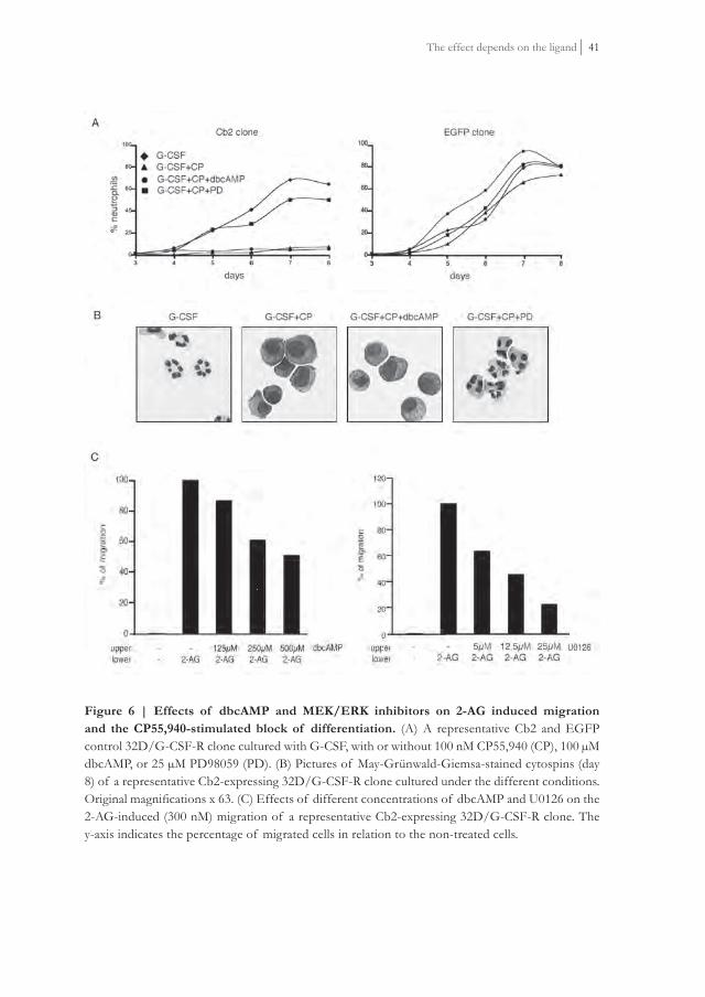
Since activation of GαiPCRs inhibits adenyl cyclase activity, we investigated whether downregulation of the intracellular cAMP levels was necessary to drive the distinct Cb2 effects. Addition of dbcAMP, a cAMP analog, to the G-CSF plus CP55,940 containing cultures did not recover neutrophilic differentiation of 32D/G-CSF-R/Cb2 cells (Figure 6A and B). dbcAMP did not alter neutrophilic maturation of EGFP control clones (Figure 6A and B). Increasing concentrations of dbcAMP partially blocked 2-AG-induced migration of Cb2-expressing 32D/G-CSF-R cells (Figure 6C). Thus, downregulation of intracellular cAMP levels seems to be partially responsible for Cb2-mediated migration but appears unimportant for the block of neutrophilic differentiation following Cb2 receptor stimulation.
Interference of CP55,940-mediated block of differentiation as well as 2-AG-
induced migration by MEK/ERK pathway inhibitors
We next studied whether signaling via MEK/ERK pathway is critical for the distinct Cb2-mediated effects. MEK inhibitors, PD98059 (Figure 6A and B) or U0126 (data not shown), fully recovered neutrophilic differentiation of 32D/G-CSF-R/Cb2 cells cultured with G-CSF plus CP55,940. MEK inhibitors did not alter differentiation of EGFP control clones (Figure 6A and B). Addition of U01 26 to transwell assays revealed a dose dependent inhibition of 2-AG-induced migration of Cb2-expressing 32D/G-CSF-R cells (Figure 6C). The same results were observed when the cells were exposed to PD98059 in a transwell assay (data not shown). This effect appeared highly specifi c since stimulation of migration of 32D/G-CSF-R cells by SDF1, the ligand for CXCR4, could not be inhibited by U0126 (data not shown). These data indicate that MEK/ERK signaling is critical in 2-AG-induced migration as well as for CP55,940-induced block of neutrophilic differentiation.
The effect depends on the ligand| 41
Figure 6 | Effects of dbcAMP and MEK/ERK inhibitors on 2-AG induced migration and the CP55,940-stimulated block of differentiation. (A) A representative Cb2 and EGFP control 32D/G-CSF-R clone cultured with G-CSF, with or without 100 nM CP55,940 (CP), 100 μM dbcAMP, or 25 μM PD98059 (PD). (B) Pictures of May-Grünwald-Giemsa-stained cytospins (day 8) of a representative Cb2-expressing 32D/G-CSF-R clone cultured under the different conditions. Original magnifi cations x 63. (C) Effects of different concentrations of dbcAMP and U0126 on the 2-AG-induced (300 nM) migration of a representative Cb2-expressing 32D/G-CSF-R clone. The y-axis indicates the percentage of migrated cells in relation to the non-treated cells.

42 | Chapter 3
DISCUSSION
The peripheral cannabinoid receptor gene, Cb2, encodes a seven transmembrane (7TM) G protein-coupled receptor (GPCR)3. Using retroviral insertional mutagenesis we identifi ed Cb2 as the target gene in the Evi11 locus, indicating that Cb2 may be a proto-oncogene involved in leukemogenesis1,2. We previously observed that Cb2 is highly expressed in myeloid cell lines containing a retroviral insertion in Cb21,7, and here we showed that CB2 is overexpressed in several human myeloid leukemia cell lines. Interestingly, here we report that CB2 is frequently overexpressed in AML blast, whereas normal bone marrow fractions are CB2 negative. It is unclear why CB2 is so highly expressed in particular AML samples and cell lines. We did not fi nd any correlation between the different morphologic AML subtypes and CB2 expression levels. Likewise, no correlation was found between the origin of the distinct cell lines and Cb2 protein levels. Real-time PCR studies revealed high levels of CB2 mRNA in the CB2 positive cell lines and not in the negative lines, suggesting altered transcription (unpublished observation). The cause of these differences in transcription remains to be elucidated. When overexpressed in the myeloid precursors Cb2 induces a block in neutrophilic development17 and stimulates migration of Cb2-expressing cells in vitro7. The endocannabinoid 2-AG is the most potent agonist capable of inducing migration of cannabinoid receptor expressing cells7,9-11. Here we report that, 2-AG has no effect on the G-CSF-induced differentiation of 32D/G-CSF-R/Cb2 cells in vitro, whereas another well described ligand CP55,94022,23, stimulates a neutrophilic differentiation block. The fact that the Cb2 inverse agonist SR144528 could fully counteract the two functions demonstrates receptor specifi city. To our knowledge this is the fi rst example of a GPCR that, when overexpressed in myeloid precursors cells, causes two different effects depending on the ligand. Normal murine bone marrow precursors aberrantly expressing Cb2 show a moderate but steady impairment of maturation when cultured with G-CSF plus CP55,940. Leukemia is a multigenic disease, meaning that a combination of genetic defects is required to obtain a full leukemia15,16. For instance, we previously demonstrated that aberrant Cb2 expression frequently coincides with aberrant expression of Evi136,37, another transforming gene shown to be involved in impairment of neutrophilic development38-40. The observation that in 32D/G-CSF-R cells Cb2 overexpression causes a complete block of neutrophilic differentiation most likely refl ects cooperation between Cb2 and other genetic defects present in this myeloid precursor cell line. Interestingly, Evi1 retroviral insertions as well as overexpression have been reported for 32D cells41. It would be of
The effect depends on the ligand| 43
interest to study whether overexpression of Cb2 in combination with Evi1 in normal marrow precursors would lead to a more severe block of neutrophilic differentiation. The experiments presented in this manuscript demonstrate that Cb2 receptor may evoke two distinct biological effects depending on the ligand used for stimulation, i.e. migration upon 2-AG presentation and block in neutrophilic differentiation following exposure to CP55,940. How the interaction of distinct Cb2 ligands to Cb2 receptor may result in activation of different processes in the same cell is an interesting question to be addressed. Several examples exist of one receptor interacting with distinct ligands. The cytokines IL-3, IL-5 as well as GM-CSF interact with different receptor complexes which all share a common beta-receptor chain. The specifi city of these distinct ligands for the different complexes is determined by the ligand specifi c receptor alpha chains42,43. Similarly, IL-2, IL4, IL-7, IL-9 and IL-15 each interact with unique receptor complexes which all share a common gamma chain44,45. The distinct effects that we observed upon 2-AG or CP55,940 stimulation may be explained by stimulation of two distinct receptor complexes which both contain the Cb2 receptor. For a long time it was generally believed that GPCRs function as monomers. However, GPCRs may be involved in high molecular weight complexes formed by homo- as well as hetero-dimers. Interestingly dimer formation may affect receptor-ligand binding, signaling and function46-48. Another intriguing observation is that agonist presentation can promote or decrease receptor dimerization in several GPCRs49-51. In addition, receptor heterodimerization between GPCRs may result in generation of novel ligand binding sites 52, or novel ligand binding properties53. GPCRs may even interact with non-G protein-coupled receptors, such as tyrosine kinase receptors54. In this respect it is interesting to note that a functional interaction between CB1 receptor and fi broblast growth factor receptor as well as with the insulin-like growth factor 1-receptor has been demonstrated55,56. Our observation that binding of distinct ligands to Cb2 receptors evoked different effects, may be explained by the Cb2 receptor being present in distinct complexes depending on the ligand, or acting as a monomer with one agonist and as a di- or multimere with the other. Cb2 belongs to the family of GPCRs3,57,58, suggesting that signaling upon receptor stimulation may require G proteins. The inhibition of migration by PTX administration, as well as the ineffi cient chemotaxis of cells expressing DRY-Cb2 mutants, indicate that Cb2 receptor requires G protein activity to induce migration upon 2-AG stimulation. In additon, most chemokine receptors belong to the subgroup of GαiPCRs59-61, suggesting that adenyl cyclase inactivation and cAMP downmodulation via Gαi proteins may be critical for induction of migration. The decrease of 2-AG-induced migration of 32D/G-CSF-R/

44 | Chapter 3
Cb2 cells following dbcAMP administration demonstrates that this pathway is critical for Cb2-mediated cell motility. On the other hand, our data show that downregulation of the intracellular cAMP levels is not the only pathway involved in migration of Cb2-expressing cells. Transwell studies using MEK inhibitors demonstrate that this route plays a critical role in 2-AG-stimulated chemotaxis as well. Previous studies demonstrated that stimulation of cannabinoid receptors Cb1 and Cb2 causes phosphorylation of ERK and consequently activation of this pathway62-64. Although critical for Cb2-induced chemotaxis, MEK/ERK signaling is not a prerequisite for GPCR-induced migration in general. For instance, SDF-1-induced migration of 32D/G-CSF-R/Cb2 cells, that endogenously express CXCR4 receptor, is insensitive to the addition of MEK inhibitors (data not shown). We clearly demonstrate that the 2-AG-induced migration involves MEK/ERK pathway, but the mechanism of activation remains unclear. Multiple examples show that MEK/ERK signaling route may be activated through the βγ complex65, although G protein-independent manners of ERK activation have been proposed as well for GPCRs65,66. Summarizing, Cb2-mediated migration depends on at least two distinct signaling pathways which both appear to be indispensable (Figure 7).
Figure 7 | Schematic representation of Cb2 signaling. Signal transduction pathways linked to Cb2 leading to the 2-AG-induced migration and the CP55,940-evoked block of neutrophilic differentiation.
As with migration induction, our data suggest that multiple pathways are involved in the Cb2-induced block of differentiation (Figure 7). MEK/ERK signaling has been shown to be critical, since MEK inhibitors fully recovered G-CSF-induced differentiation in the presence of CP55,940. Previously, we demonstrated as well involvement of PI3-K

The effect depends on the ligand| 45
in the Cb2-mediated block of differentiation17. On the other hand, intracellular cAMP downregulation is unimportant for this effect. Moreover, the DRY motif, which is critical for the 2-AG-induced migration was completely unnecessary for the induction of a maturation arrest. These experiments would suggest that G protein signaling is dispensable for the Cb2-induced differentiation block. However, we demonstrated that the CP55,940-evoked block in differentiation of 32D/G-CSF-R/Cb2 cells could be fully reversed by the addition of PTX. Therefore, these data suggest that activated Cb2 receptors may induce the proper signals via G proteins, but since we observed that the DRY motif is unnecessary for the CP55,940-induced block of differentiation, we suggest involvement of another currently unknown G protein interaction domain in Cb2. Multiple GPCRs have previously been reported to have transforming abilities, e.g. the α1B-adenergic67, thrombin68, and serotonin 1 C receptors69 and the receptor encoded by the Mas oncogene70,71. In contrast to Cb2, which interacts with Gαi subunits, a large number of these previously identifi ed transforming GPCRs can associate with Gαs subunits72. An interesting question to be addressed is whether transformation of myeloid precursor cells by Cb2 is a feature unique for this particular GPCR or whether the peripheral cannabinoid receptor is a paradigm for a novel class of transforming GPCRs. The fact that we have shown that stimulation of migration as well as the interference with differentiation by Cb2 are PTX sensitive, meaning involvement of Gαi, may indicate that other Gαi interacting GPCRs with transforming abilities may exist. Interestingly, using retroviral insertional mutagenesis we and others recently identifi ed among a large panel of novel leukemia disease genes, four GPCR encoding genes73,74, unpublished observation. Three of those genes encode GPCR that may interact with Gαi subunits, i.e. endothelial differentiation gene 3-R (Edg3-R), chemokine-R7 (CCR7) and vomeronasal1-R (V1-R). Introduction of these transforming receptors or other potentially interesting GPCRs into 32D/G-CSF-R cells will be a valid approach to answer the question whether Cb2 is a paradigm for a novel class of transforming GPCRs.
ACKNOWLEDGMENTS
We thank Prof. Dr. I. P. Touw (ErasmusMC, Rotterdam) for donation of the 32D/G-CSF-R cell line. We thank Dr. P. Casellas (Sanofi Recherche, Montpellier, France) for donation of the Cb1- and Cb2- inverse agonist. We thank I. van Ostaijen, E. de Wee and C. M. Dicke for technical assistance, and J. Prasher for critical reading of the manuscript. We thank K. van Rooyen for preparation of the fi gures.

46 | Chapter 3
REFERENCES
1. Valk PJ, Hol S, Vankan Y, et al. The genes encoding the peripheral cannabinoid receptor and alpha-L-fucosidase are located near a newly identifi ed common virus integration site, Evi11. J Virol. 1997;71:6796-6804.
2. Valk PJ, Delwel R. The peripheral cannabinoid receptor, Cb2, in retrovirally-induced leukemic transformation and normal hematopoiesis. Leuk Lymphoma. 1998;32:29-43.
3. Munro S, Thomas KL, Abu-Shaar M. Molecular characterization of a peripheral receptor for cannabinoids. Nature. 1993;365:61-65.
4. Lynn AB, Herkenham M. Localization of cannabinoid receptors and nonsaturable high-density cannabinoid binding sites in peripheral tissues of the rat: implications for receptor-mediated immune modulation by cannabinoids. J Pharmacol Exp Ther. 1994;268:1612-1623.
5. Carayon P, Marchand J, Dussossoy D, et al. Modulation and functional involvement of CB2 peripheral cannabinoid receptors during B-cell differentiation. Blood. 1998;92:3605-3615.
6. Galiegue S, Mary S, Marchand J, et al. Expression of central and peripheral cannabinoid receptors in human immune tissues and leukocyte subpopulations. Eur J Biochem. 1995;232:54-61.
7. Alberich Jorda M, Verbakel SE, Valk PJ, et al. Hematopoietic cells expressing the peripheral cannabinoid receptor migrate in response to the endocannabinoid 2-arachidonoylglycerol. Blood. 2002;99:2786-2793.
8. Sugiura T, Kodaka T, Nakane S, et al. Evidence that the cannabinoid CB1 receptor is a 2-arachidonoylglycerol receptor. Structure-activity relationship of 2-arachidonoylglycerol, ether-linked analogues, and related compounds. J Biol Chem. 1999;274:2794-2801.
9. Sugiura T, Kondo S, Kishimoto S, et al. Evidence that 2-arachidonoylglycerol but not N-palmitoylethanolamine or anandamide is the physiological ligand for the cannabinoid CB2 receptor. Comparison of the agonistic activities of various cannabinoid receptor ligands in HL-60 cells. J Biol Chem. 2000;275:605-612.
10. Gonsiorek W, Lunn C, Fan X, Narula S, Lundell D, Hipkin RW. Endocannabinoid 2-arachidonyl glycerol is a full agonist through human type 2 cannabinoid receptor: antagonism by anandamide. Mol Pharmacol. 2000;57:1045-1050.
11. Sugiura T, Waku K. 2-Arachidonoylglycerol and the cannabinoid receptors. Chem Phys Lipids. 2000;108:89-106.
12. Howlett AC, Barth F, Bonner TI, et al. International Union of Pharmacology. XXVII. Classifi cation of cannabinoid receptors. Pharmacol Rev. 2002;54:161-202.
13. Lowenberg B, Downing JR, Burnett A. Acute myeloid leukemia. N Engl J Med. 1999;341:1051-1062.
14. Vogelstein B, Kinzler KW. The multistep nature of cancer. Trends Genet. 1993;9:138-141.
15. Hunter T. Cooperation between oncogenes. Cell. 1991;64:249-270.
16. Adams JM, Cory S. Oncogene co-operation in leukaemogenesis. Cancer Surv. 1992;15:119-141.
17. Alberich Jorda M, Lowenberg B, Delwel R. The peripheral cannabinoid receptor Cb2, a novel oncoprotein, induces a reversible block in neutrophilic differentiation. Blood. 2003;101:1336-1343.
18. de Koning JP, Soede-Bobok AA, Schelen AM, et al. Proliferation signaling and activation of Shc, p21Ras, and Myc via tyrosine 764 of human granulocyte colony-stimulating factor receptor. Blood. 1998;91:1924-1933.

The effect depends on the ligand| 47
19. Dong F, van Buitenen C, Pouwels K, Hoefsloot LH, Lowenberg B, Touw IP. Distinct cytoplasmic regions of the human granulocyte colony-stimulating factor receptor involved in induction of proliferation and maturation. Mol Cell Biol. 1993;13:7774-7781.
20. Ward AC, Smith L, de Koning JP, van Aesch Y, Touw IP. Multiple signals mediate proliferation, differentiation, and survival from the granulocyte colony-stimulating factor receptor in myeloid 32D cells. J Biol Chem. 1999;274:14956-14962.
21. Erkeland SJ, Valkhof M, Heijmans-Antonissen C, et al. The gene encoding the transcriptional regulator Yin Yang 1 (YY1) is a myeloid transforming gene interfering with neutrophilic differentiation. Blood. 2003;101:1111-1117.
22. Little PJ, Compton DR, Johnson MR, Melvin LS, Martin BR. Pharmacology and stereoselectivity of structurally novel cannabinoids in mice. J Pharmacol Exp Ther. 1988;247:1046-1051.
23. Kuster JE, Stevenson JI, Ward SJ, D’Ambra TE, Haycock DA. Aminoalkylindole binding in rat cerebellum: selective displacement by natural and synthetic cannabinoids. J Pharmacol Exp Ther. 1993;264:1352-1363.
24. Hamm HE. The many faces of G protein signaling. J Biol Chem. 1998;273:669-672.
25. Lambright DG, Noel JP, Hamm HE, Sigler PB. Structural determinants for activation of the alpha-subunit of a heterotrimeric G protein. Nature. 1994;369:621-628.
26. Alewijnse AE, Timmerman H, Jacobs EH, et al. The effect of mutations in the DRY motif on the constitutive activity and structural instability of the histamine H(2) receptor. Mol Pharmacol. 2000;57:890-898.
27. Moore SA, Patel AS, Huang N, et al. Effects of mutations in the highly conserved DRY motif on binding affi nity, expression, and G-protein recruitment of the human angiotensin II type-2 receptor. Brain Res Mol Brain Res. 2002;109:161-167.
28. Rasmussen SG, Jensen AD, Liapakis G, Ghanouni P, Javitch JA, Gether U. Mutation of a highly conserved aspartic acid in the beta2 adrenergic receptor: constitutive activation, structural instability, and conformational rearrangement of transmembrane segment 6. Mol Pharmacol. 1999;56:175-184.
29. Shibata T, Suzuki C, Ohnishi J, Murakami K, Miyazaki H. Identifi cation of regions in the human angiotensin II receptor type 1 responsible for Gi and Gq coupling by mutagenesis study. Biochem Biophys Res Commun. 1996;218:383-389.
30. Lanotte M, Martin-Thouvenin V, Najman S, Balerini P, Valensi F, Berger R. NB4, a maturation inducible cell line with t(15;17) marker isolated from a human acute promyelocytic leukemia (M3). Blood. 1991;77:1080-1086.
31. Yanagisawa K, Horiuchi T, Fujita S. Establishment and characterization of a new human leukemia cell line derived from M4E0. Blood. 1991;78:451-457.
32. Boyum A. Isolation of leucocytes from human blood. Further observations. Methylcellulose, dextran, and fi coll as erythrocyteaggregating agents. Scand J Clin Lab Invest Suppl. 1968;97:31-50.
33. Delwel R, Touw I, Bot F, Lowenberg B. Fucose binding lectin for characterizing acute myeloid leukemia progenitor cells. Blood. 1986;68:41-45.
34. Margonelli A, Angelici, G., Attina, M., Cartoni, A., Finnazzi-Agro, A., and Maccarrone, M. New effective chemical radiosynthesis of (2)-[5,6,8,9,11,12,14,15-3H(N)] arachidonoyl-glycerol (2-AG), a putative endogenous cannabinoid ligand. J Labelled Cpd Radiopharm. 1999;42:787-789.
35. Maccarrone M, Lorenzon T, Bari M, Melino G, Finazzi-Agro A. Anandamide induces apoptosis in human cells via vanilloid receptors. Evidence for a protective role of cannabinoid receptors. J Biol Chem. 2000;275:31938-31945.

48 | Chapter 3
36. Joosten M, Valk PJ, Vankan Y, de Both N, Lowenberg B, Delwel R. Phenotyping of Evi1, Evi11/Cb2, and Evi12 transformed leukemias isolated from a novel panel of cas-Br-M murine leukemia virus-infected mice. Virology. 2000;268:308-318.
37. Joosten M, Valk PJ, Jorda MA, et al. Leukemic predisposition of pSca-1/Cb2 transgenic mice. Exp Hematol. 2002;30:142-149.
38. Hirai H. The transcription factor Evi-1. Int J Biochem Cell Biol. 1999;31:1367-1371.
39. Nucifora G. The EVI1 gene in myeloid leukemia. Leukemia. 1997;11:2022-2031.
40. Morishita K, Parganas E, Matsugi T, Ihle JN. Expression of the Evi-1 zinc fi nger gene in 32Dc13 myeloid cells blocks granulocytic differentiation in response to granulocyte colony-stimulating factor. Mol Cell Biol. 1992;12:183-189.
41. Khanna-Gupta A, Lopingco MC, Savinelli T, Zibello T, Berliner N, Perkins AS. Retroviral insertional activation of the EVI1 oncogene does not prevent G-CSF-induced maturation of the murine pluripotent myeloid cell line 32Dcl3. Oncogene. 1996;12:563-569.
42. Scott CL, Begley CG. The beta common chain (beta c) of the granulocyte macrophage-colony stimulating factor, interleukin-3 and interleukin-5 receptors. Int J Biochem Cell Biol. 1999;31:1011-1015.
43. Bagley CJ, Woodcock JM, Stomski FC, Lopez AF. The structural and functional basis of cytokine receptor activation: lessons from the common beta subunit of the granulocyte-macrophage colony-stimulating factor, interleukin-3 (IL-3), and IL-5 receptors. Blood. 1997;89:1471-1482.
44. Malek TR, Porter BO, He YW. Multiple gamma c-dependent cytokines regulate T-cell development. Immunol Today. 1999;20:71-76.
45. Leonard WJ, Noguchi M, Russell SM, McBride OW. The molecular basis of X-linked severe combined immunodefi ciency: the role of the interleukin-2 receptor gamma chain as a common gamma chain, gamma c. Immunol Rev. 1994;138:61-86.
46. Rios CD, Jordan BA, Gomes I, Devi LA. G-protein-coupled receptor dimerization: modulation of receptor function. Pharmacol Ther. 2001;92:71-87.
47. Gomes I, Jordan BA, Gupta A, Rios C, Trapaidze N, Devi LA. G protein coupled receptor dimerization: implications in modulating receptor function. J Mol Med. 2001;79:226-242.
48. Angers S, Salahpour A, Bouvier M. Dimerization: an emerging concept for G protein-coupled receptor ontogeny and function. Annu Rev Pharmacol Toxicol. 2002;42:409-435.
49. Rocheville M, Lange DC, Kumar U, Sasi R, Patel RC, Patel YC. Subtypes of the somatostatin receptor assemble as functional homo- and heterodimers. J Biol Chem. 2000;275:7862-7869.
50. AbdAlla S, Zaki E, Lother H, Quitterer U. Involvement of the amino terminus of the B(2) receptor in agonist-induced receptor dimerization. J Biol Chem. 1999;274:26079-26084.
51. Cvejic S, Devi LA. Dimerization of the delta opioid receptor: implication for a role in receptor internalization. J Biol Chem. 1997;272:26959-26964.
52. Maggio R, Vogel Z, Wess J. Coexpression studies with mutant muscarinic/adrenergic receptors provide evidence for intermolecular “cross-talk”; between G-protein-linked receptors. Proc Natl Acad Sci U S A. 1993;90:3103-3107.
53. Jordan BA, Devi LA. G-protein-coupled receptor heterodimerization modulates receptor function. Nature. 1999;399:697-700.

The effect depends on the ligand| 49
54. Maudsley S, Pierce KL, Zamah AM, et al. The beta(2)-adrenergic receptor mediates extracellular signal-regulated kinase activation via assembly of a multi-receptor complex with the epidermal growth factor receptor. J Biol Chem. 2000;275:9572-9580.
55. Bouaboula M, Perrachon S, Milligan L, et al. A selective inverse agonist for central cannabinoid receptor inhibits mitogen-activated protein kinase activation stimulated by insulin or insulin-like growth factor 1. Evidence for a new model of receptor/ligand interactions. J Biol Chem. 1997;272:22330-22339.
56. Williams EJ, Walsh FS, Doherty P. The FGF receptor uses the endocannabinoid signaling system to couple to an axonal growth response. J Cell Biol. 2003;160:481-486.
57. Howlett AC. Cannabinoid inhibition of adenylate cyclase. Biochemistry of the response in neuroblastoma cell membranes. Mol Pharmacol. 1985;27:429-436.
58. Felder CC, Joyce KE, Briley EM, et al. Comparison of the pharmacology and signal transduction of the human cannabinoid CB1 and CB2 receptors. Mol Pharmacol. 1995;48:443-450.
59. Frade JM, Mellado M, del Real G, Gutierrez-Ramos JC, Lind P, Martinez AC. Characterization of the CCR2 chemokine receptor: functional CCR2 receptor expression in B cells. J Immunol. 1997;159:5576-5584.
60. Smit MJ, Verdijk P, Van Der Raaij-Helmer EM, et al. CXCR3-mediated chemotaxis of human T cells is regulated by a Gi- and phospholipase C-dependent pathway and not via activation of MEK/p44/p42 MAPK nor Akt/PI-3-kinase. Blood. 2003;15:15.
61. Nagasawa T, Kikutani H, Kishimoto T. Molecular cloning and structure of a pre-B-cell growth-stimulating factor. Proc Natl Acad Sci U S A. 1994;91:2305-2309.
62. Kobayashi Y, Arai S, Waku K, Sugiura T. Activation by 2-arachidonoylglycerol, an endogenous cannabinoid receptor ligand, of p42/44 mitogen-activated protein kinase in HL-60 cells. J Biochem (Tokyo). 2001;129:665-669.
63. Galve-Roperh I, Rueda D, Gomez del Pulgar T, Velasco G, Guzman M. Mechanism of extracellular signal-regulated kinase activation by the CB(1) cannabinoid receptor. Mol Pharmacol. 2002;62:1385-1392.
64. Derkinderen P, Valjent E, Toutant M, et al. Regulation of extracellular signal-regulated kinase by cannabinoids in hippocampus. J Neurosci. 2003;23:2371-2382.
65. Marinissen MJ, Gutkind JS. G-protein-coupled receptors and signaling networks: emerging paradigms. Trends Pharmacol Sci. 2001;22:368-376.
66. Brzostowski JA, Kimmel AR. Signaling at zero G: G-protein-independent functions for 7-TM receptors. Trends Biochem Sci. 2001;26:291-297.
67. Allen LF, Lefkowitz RJ, Caron MG, Cotecchia S. G-protein-coupled receptor genes as protooncogenes: constitutively activating mutation of the alpha 1B-adrenergic receptor enhances mitogenesis and tumorigenicity. Proc Natl Acad Sci U S A. 1991;88:11354-11358.
68. Whitehead I, Kirk H, Kay R. Expression cloning of oncogenes by retroviral transfer of cDNA libraries. Mol Cell Biol. 1995;15:704-710.
69. Julius D, Livelli TJ, Jessell TM, Axel R. Ectopic expression of the serotonin 1c receptor and the triggering of malignant transformation. Science. 1989;244:1057-1062.
70. Janssen JW, Steenvoorden AC, Schmidtberger M, Bartram CR. Activation of the mas oncogene during transfection of monoblastic cell line DNA. Leukemia. 1988;2:318-320.
71. Young D, Waitches G, Birchmeier C, Fasano O, Wigler M. Isolation and characterization of a new cellular oncogene encoding a protein with multiple potential transmembrane domains. Cell. 1986;45:711-719.

50 | Chapter 3
72. Seifert R, Wenzel-Seifert K. Constitutive activity of G-protein-coupled receptors: cause of disease and common property of wild-type receptors. Naunyn Schmiedebergs Arch Pharmacol. 2002;366:381-416.
73. Suzuki T, Shen H, Akagi K, et al. New genes involved in cancer identifi ed by retroviral tagging. Nat Genet. 2002;32:166-174.
74. Joosten M, Vankan-Berkhoudt Y, Tas M, et al. Large-scale identifi cation of novel potential disease loci in mouse leukemia applying an improved strategy for cloning common virus integration sites. Oncogene. 2002;21:7247-7255.

Chapter 3
Distinct expression profi les of the peripheral cannabinoid receptor in lymphoid tissu es depending on receptor activation status
Nazik Rayman1, King H. Lam2, Jon D. Laman3, Peter J. Simons4, Bob Löwenberg1,
Pieter Sonneveld1 and Ruud Delwel1
1Department of Haematology, 2Department of Pathology, Josephine Nefkens Institute
and 3Department of Immunology, Erasmus Medical Centre, Rotterdam, The Netherlands, 4BioCeros BV, Amsterdam, The Netherlands
The Journal of Immunology 2004, 172:2111-2117

52 | Chapter 3
ABSTRACT
Using two distinct anti-CB2 receptor antibodies, we investigated the expression patterns of the peripheral cannabinoid receptor CB2 in human secondary lymphoid organs. Immunohistochemical analysis using an N-terminal specifi c anti-CB2 antibody revealed high protein expression in the germinal centres (GCs) of secondary follicles. A C-terminal specifi c anti-CB2 antibody, which only recognises a non-phosphorylated inactive receptor, showed positivity in the mantle zones (MZs) and marginal zones (MGZs) of the secondary follicles where resting cells reside, and in the primary follicles. In contrast, no positivity was observed in GCs using the C-terminal antibody, suggesting that active CB2 receptors are mainly present on cells in the GCs. Dual immunohistochemical analysis revealed that B-lymphocytes express the CB2 protein abundantly. In contrast to B-cells in the MZ or MGZ, CB2 expressing cells in the GCs co-express the co-stimulatory membrane protein CD40 and the proliferation marker Ki-67. Using the human Raji B-cell line as a model, we demonstrate in a transwell assay that moderate migration occurs upon stimulation of the CB2 receptor with the endocannabinoid 2-arachidonoylglycerol, which is enhanced by CD40 co-stimulation. Our fi ndings, that GC-related cells express active CB2 and that CB2-dependent migration requires CD40 co-stimulation, suggests that CB2 is involved in B-cell activation.

Peripheral cannabinoid receptors on B-lymphocytes | 53
INTRODUCTION
Two cannabinoid receptors, which are expressed in distinct organ systems, have previously been identifi ed. The central cannabinoid receptor CB11 is primarily expressed in brain, whereas the peripheral cannabinoid receptor CB22 is mainly present in immune tissues. CB2 mRNA has been detected in human haematopoietic cells, with B-cells expressing the highest transcript levels3,4. In fact, using a polyclonal antibody (PoAb) raised against the C-terminus of the human CB2 receptor, Carayon et al demonstrated expression of the CB2 protein in the mantle zones (MZs) of secondary follicles of tonsils5. These investigators reported that there was no CB2 staining on B-lymphocytes in the germinal centres of the secondary follicles. Importantly, the antibody used recognised CB2 receptors in a non-phosphorylated inactive state5,6. The observation that no expression was evident on B-cells in the germinal centres (GCs), could mean that the cells in the GCs did not express CB2 receptors at all or that CB2 receptors were present on these cells, but in an active form undetectable with this antibody. Insight into the distribution of active versus inactive CB2 receptors on cells of the immune system may provide understanding of the functional aspects of this receptor in its physiological environment. Using another anti-CB2 antibody raised against the N-terminus of the CB2 receptor in combination with the previously reported C-terminal specifi c anti-CB2 antibody, we therefore have investigated the distribution of CB2 receptors within normal lymphoid organs. We have demonstrated different staining patterns depending on the CB2 antibody used, implying distinct distribution of active versus non-active receptors. The inactive receptors were found in the MZs of secondary follicles, whereas active peripheral cannabinoid receptors were present in the GCs. Next, dual immunohistochemistry was applied to investigate the immunophenotype of the CB2-expressing cells. The data show that whether present in an active or inactive form CB2 was mainly expressed on B-lymphocytes. Moreover, in the GCs of secondary follicles the active form of CB2 was observed in cells expressing both the co stimulatory molecule CD40 and the proliferation marker Ki-67, suggesting an immunomodulatory function of the CB2 receptor in the GCs. We therefore assessed the functional relation between CB2 and CD40. We have demonstrated that the major function of the CB2 receptor on splenic B-cells is stimulation of migration, upon exposure to the endocannabinoid 2-arachidonoylglycerol (2-AG)7. Using the Raji B-lymphoma cell line as a model, we investigated the involvement of CD40 on CB2 mediated effects on B-cells. We have found that migration is a major function of the CB2 receptor upon stimulation with 2-AG, which is signifi cantly augmented following CD40 stimulation, suggesting

54 | Chapter 3
cross talk between the two receptors. Our investigations suggest a critical role for CB2 in migration of B-cells and the GC-response.
MATERIALS AND METHODS
Antibodies, ligands and cell lines
The polyclonal human anti-C-terminal-CB2 receptor antibody and the synthetic peptide derived from the predicted amino acid-sequence of the carboxyterminus of the receptor (Y-P-D-S-R-D-L-D-L-S-D-C) were kindly provided by P. Case llas (Sanofi -Synthelabo Recherche, Montpellier, France). The anti-CB2 receptor PoAb raised against the fi rst 33 amino acid residues of the N-terminus of the receptor was purchased from Affi nity Bioreagents Inc (ABR, CO, USA). The non-stimulatory/non inhibitory CD40 control antibody (nCD40), the anti-CD40 stimulatory antibody (sCD40/clone 7) and the anti-CD40 inhibitory antibody (iCD40/5D12) were kindly donated by Dr. L. Boon (PanGenetics BV, Amsterdam, The Netherlands). Other antibodies used for immunohistochemical analysis are listed in Table 1. The Raji cell line, donated by I.Touw (Department of Haematology Erasmus MC, Rotterdam, The Netherlands) was cultured in RPMI 1640 medium (Life Technologies, Breda, The Netherlands) and supplements: penicillin (100 U/ml), streptomycin (100 ng/ml) and 10% fetal calf serum (Life Technologies). The Chinese Hamster Ovary (CHO) cell line and CHO cell line transfected with CB2 (CHO-CB2), kindly donated by P.Casellas, were cultured in MEM-alpha Medium (Life Technologies) and supplements. The CB2 cannabinoid ligand 2-arachidonoylglycerol (2-AG) was obtained from Sigma (Zwijndrecht, The Netherlands). The CB1-specifi c inverse agonist SR141716 and CB2- specifi c inverse agonist SR 144528 were kindly donated by P. Casellas.
Immunohistochemistry
Single staining
Formalin-fi xed, paraffi n embedded normal lymph node, spleen and thymus were retrieved from the fi les of the department of Pathology at the Erasmus MC. Five μm thick sections were deparaffi nized, rehydrated and endogenous peroxidase was blocked with 3% H2O2 in methanol for 20 minutes at room temperature (RT). Heat induced antigen retrieval (required for staining with the C-terminal anti-CB2 antibody) was achieved by boiling the sections for 20 minutes at 100°C in citrate buffer (10 mM, pH 6.0) in a Micromed T/T Mega microwave oven (Salm and Kipp, Breukelen, The Netherlands). Sections were
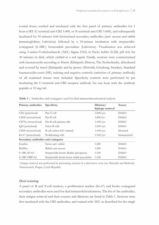
Peripheral cannabinoid receptors on B-lymphocytes | 55
cooled down, washed and incubated with the fi rst panel of primary antibodies for 1 hour at RT (C-terminal anti-CB2 1:400, or N-terminal anti-CB2 1:600), and subsequently incubated for 10 minutes with biotinylated secondary antibodies (anti- mouse and rabbit immunoglobins; Labvision) followed by a 10-minute incubation with streptavidin-conjugated (S-ABC) horseradish peroxidase (Labvision). Visualisation was achieved using 3-amino-9-ethylcarbazole (AEC; Sigma USA) in NaAc buffer (0.2M, pH 4.6) for 30 minutes in dark, which yielded in a red signal. Finally, sections were counterstained with haematoxylin according to Harris (Klinipath, Duiven, The Netherlands), dehydrated and covered by imsol (Klinipath) and by pertex (Histolab, Göteborg, Sweden). Standard haematoxylin-eosin (HE) staining and negative controls (omission of primary antibody) of all examined tissues were included. Specifi city controls were performed by pre incubating the C-terminal anti-CB2 receptor antibody for one hour with the synthetic peptide at 10 mg/ml.
Table 1 | Antibodies and conjugates used for dual immunohistochemical analysis.
Primary antibodies Specifi city Dilution/Epitope retrieval1
Source
CD3 (polyclonal)CD20 (monoclonal)CD79a (monoclonal)IgD (polyclonal)CD40 (monoclonal)Ki-67 (monoclonal)
Pan-T cellPan B cellPan B cell; plasma cellsNaive B cellsB-cell subset (GC-related)Proliferating cells
1:600/yes1:400/no1:100/yes1:200/yes1:100/yes1:100/yes
DAKODAKODAKODAKODonatedImmunotech2
Secondary antibodies and conjugates
SwarbioRaMbioS-ABCAP-kitS-ABC HRP-kit
Swine-anti -rabbitRabbit anti-mouseStreptavidin-biotin alkaline phospataseStreptavidin biotin horse radish peroxidase
1:2001:2001:1001:100
DAKODAKODAKODAKO
1Antigen retrieval was performed by pretreating sections in a microwave oven (see Materials and Methods). 2Immunotech, Prague, Czech Republic.
Dual staining
A panel of B and T-cell markers, a proliferation marker (Ki-67) and biotin conjugated secondary antibodies were used for dual immunohistochemistry. The list of the antibodies, their antigen retrieval and their sources and dilutions are listed in Table 1. Sections were fi rst incubated with the CB2 antibodies, and stained with AEC as described for the single

56 | Chapter 3
staining procedure. Sections were individually incubated with the primary antibodies listed in Table 1 for 30 minutes, washed and incubated with the biotin conjugated secondary antibodies for 30 minutes at RT. An S-ABC conjugated alkaline phosphatase kit (DAKO, Glostrup, Denmark) was used to enhance the signal. Visualisation of the signal was achieved using a solution containing Fast Blue BB salt (4-benzoylamino-2, 5-diethoxylbenzene diazoniumchloride; Sigma), naphtol-AS-MX phosphate (Sigma) and levamisole hydroxychloride (Acros Organics, Geel, Belgium) in Tris/HCl buffer (0.2M, pH 8.0). Sections were incubated for 30 minutes in dark with this solution, which fi nally resulted in blue staining of cells. In case of co-expression/ co-staining, the mixture of red and blue signals resulted in a dark purple staining of cells. Cells were counter-stained with haematoxylin according to Harris (Klinipath) and covered by pertex.
Flowcytometric analysis
Flow cytometric analysis was carried out using a FACScan fl ow cytometer (Becton Dickinson Mountain View, CA). In brief, cells were washed with Hanks balanced salt solution (HBSS) (HANKS, Invitrogen Corporation, Paisley, U.K.) and PBS, incubated on ice with the polyclonal N-terminal anti-CB2 antibody (1:50) or the nCD40 monoclonal antibody (1:100) for an hour, washed twice with PBS and incubated with the FITC-conjugated secondary rabbit (1:200 GAR-FITC/IgG; Nordic Immunological Labs, Tilburg, The Netherlands) or mouse antibody (1:200 GAM-FITC; ITK Diagnostics, Uithoorn, The Netherlands) for 30 minutes. Finally, cells were washed twice with PBS and resuspended in 500 ml PBS and analysed.
Immunofl uorescence microscopy and cytospins
After fl owcytometric analysis cytospins of cells stained with the N-terminal CB2 were made and analysed for membrane expression. To investigate cytoplasmatic CB2 expression, cells were fi xed in 4% paraformaldehyde at 4ºC for 20 minutes, washed twice in PBS and incubated for 1 hour at RT with the N-terminal CB2 antibody in a permeabilising PBG-T solution (0.1M phosphate buffer; 0.5%BSA; 0.2% gelatine; 0.05% Triton X100). Then cells were washed twice in PBG-T and incubated with the FITC-conjugated secondary rabbit antibody (1:200) for 1 hour at RT. Cells were washed again and embedded in vectashield. (twice in PBG-T and twice in milliQ, embedded in vectashield) (Brunschwig Chemie, Amsterdam, The Netherlands) and visualised with an immunofl uorescence microscope (Leica Mikroskopie & Systeme GmbH, Solms, Germany).

Peripheral cannabinoid receptors on B-lymphocytes | 57
Migration assay
Migration assays were performed using 6.5mm-diameter transwells with a 5 mm pore size (Corning Costar, Amsterdam, The Netherlands). In brief, cells were washed twice with HBSS medium, resuspended in 100 ml of migration medium (Iscoves modifi ed Dulbecco medium (IMDM) plus 0.5% BSA) and placed in the upper chamber of a transwell with or without CB1- or CB2-specifi c inverse-agonists (100nM). In the lower chamber, 600 ml migration medium with or without ligand (2-AG 300nM) was placed. After 4 hours of incubation at 37ºC and 5% CO2, the upper chamber was removed and the numbers of migrated cells were determined using a CASY1/TTC cell counter (Schärfe System, Reutlingen, Germany).
RESULTS
Flowcytometric analysis of CB2 transduced CHO cells
Two polyclonal antibodies directed to the peripheral cannabinoid receptor (CB2) were used in this study to investigate expression of this receptor in human immune tissues. Specifi city of the antibodies was confi rmed using CHO cells transfected with human CB25. CB2 membrane staining was demonstrated with the antibody directed to the N-terminal extra cellular domain using non-permeabilised CHO-CB2 cells (Figure 1A). Membrane bound and cytoplasmic CB2 expression was demonstrated in permeabilised CB2 expressing CHO cells (Figure 1B). An antiserum recognising the intracellular C-terminal part of CB2 was also highly positive in permeabilised CHO-CB2 cells (Figure 1C). As expected, no positivity was observed when the latter antibody was used on non-permeabilised CHO-CB2 cells (data not shown). Remarkably, a lower signal was evident when the N-terminal specifi c antibody (Figure 1A and B) was used as compared to the analysis with the C-terminal specifi c anti-CB2 (Figure 1C), which may suggest that the latter antibody binds CB2 with a higher affi nity or that the differences in staining by the CB2 antibodies may be the result of differences in epitope accessibility. No positivity at all was observed using the CB2 antibodies in non-transduced CHO-cells (Figure 1D and E). Importantly, the C-terminal anti-CB2 antibody only recognises the receptor in an inactivated non-phosphorylated state5,6. The N-terminal CB2 antibody on the other hand, recognises the receptor in both an active and inactive state.

58 | Chapter 3
Figure 1 | CB2 protein expression in CHO and CHO-CB2 cells. CB2 membrane expression (A) and intra cellular CB2 protein expression (B) was shown in CHO-CB2-cells transfected with human CB2 (CHO-CB2) using the N-terminal anti-CB2 antibody. Intra cellular CB2 protein in CHO-CB2 cells using the C-terminal anti-CB2 antibody (C) was also investigated. CB2 protein expression was analysed in non-CB2-transduced CHO cells using the N-terminal (D) and C-terminal (E) anti-CB2 antibodies respectively. Intra cellular CB2 was analysed in permeabilised cells (see Materials and Methods). The fi lled areas show CB2 protein expression, the grey lines represent background staining (only secondary Ab).
CB2 receptor expression in primary and secondary follicles in spleen and
lymph node
Primary follicles
Immunohistochemical staining of spleen sections using a C-terminal (inactive CB2 receptor) or N-terminal anti-CB2 receptor antibody (non-discriminating) showed CB2 expression in the follicular B-cell areas. In primary follicles, the cells were highly positive using the C-terminal specifi c CB2 antibody (Figure 2A and B). Weaker expression was observed using the N-terminal CB2 antibody (Figure 2C and D). These data suggest that mainly inactive CB2 receptors are present on cells in the primary follicles. Specifi city was evident, since binding of the C-terminal antibody was completely blocked when the antibody was pre-incubated with its specifi c peptide (Figure 2E). No staining was observed when only the secondary antibody was used (Figure 2F). Exactly the same results were obtained when normal lymph nodes were studied (data not shown).

Peripheral cannabinoid receptors on B-lymphocytes | 59
Figure 2 | CB2 expression in primary follicles of the human spleen. CB2 expression is demonstrated using the C-terminal (A,*5; B *20) and the N-terminal (C, *5; D, *20) anti-CB2 antibodies respectively. Specifi city of the C-terminal anti-CB2 antibody was confi rmed by pre-incubation with a specifi c C-terminal peptide (E; 5*). Staining with only the secondary antibody (F; 5*) served as a negative control. P=primary follicle; red staining=C-or N-terminal CB2 antibody staining; blue staining=counterstaining with haematoxylin. See page 162 for color fi gure.
Secondary follicles
CB2 expression patterns in the secondary follicles of lymph nodes were also determined using the same antibodies. The C-terminal CB2 specifi c antibody mainly recognised cells present in the MZ and the MGZ of the secondary follicles (Figure 3A-C). The N-terminal specifi c CB2 receptor antibody only weakly recognised cells in these zones (Figure 3E). On the other hand, using this antibody, strong staining was observed in the GCs of the secondary follicles (Figure 3D-F), whereas only weak staining was found in those regions

60 | Chapter 3
using the C-terminal antibody (Figure 3A-C). Strong binding of the CB2 N-terminal specifi c antibody suggests high receptor numbers present on the cells in the GCs. These results also suggest that cells in the GC mainly express active CB2, whereas inactive CB2 is present on cells in the MZ and MGZ of the secondary follicles. Remarkably, most prominent staining with the two antibodies occurred in the cytoplasm of the cells (Figure 3C and F).
Figure 3 | Single immunohistochemical analysis in primary and secondary follicles of the human lymph node. C-terminal anti-CB2 antibody staining pattern is depicted in fi gures (A; 1.25*), (B; 10*) and (C; 20*). N-terminal anti-CB2 antibody staining pattern is shown in fi gures (D; 1.25*), (E; 10*) and ((F;20*, insert *40). P= primary follicle; s= secondary follicle; MZ=mantle zone; MGZ=marginal zone; GC=germinal centre. Black rectangles represent magnifi cations of

Peripheral cannabinoid receptors on B-lymphocytes | 61
areas of interest. Red staining=C-or N-terminal CB2 staining; blue staining=counterstaining with haematoxylin. See page 163 for color fi gure.
Differential CB2 receptor expression on B-cell subsets
Inactive CB2 receptors on resting B-cells
Dual-immunohistochemistry using B-cell specifi c antibodies (Table 1) has been carried out in combination with the anti-CB2 receptor antibodies. First, we studied the cells carrying inactive CB2 receptors. Almost all cells in the primary follicles and in the MZ and MGZ of secondary follicles, co-expressed (dark purple) CD79a (pan B-cell marker; blue) and the inactive CB2 (red) on their surfaces (Figure 4A). The same results were obtained with another pan B-cell marker, CD20 (data not shown). Similarly, signifi cant expression of inactive CB2 (red membrane/cytoplasmic staining) was evident in IgD expressing resting follicular B-cells (dark purple membrane staining) in primary follicles and MZ/MGZ of secondary follicles (Figure 4B). No staining was observed when the primary antibodies were omitted in the immunohistochemical analysis (data not shown).
CB2 receptors on activated B-cells in the germinal centres (GC)
Next, we investigated the immunophenotype of the CB2 receptor expressing cells in the GC of the secondary follicles in lymph nodes. CB2 receptor expressing cells in the GC (as demonstrated with low staining with C terminal CB2 antibody and strong staining with N-terminal antibody) showed strong positivity with the pan B-cell marker CD79a (Figure 4C). These cells also expressed CD20 (data not shown), but were mostly IgD negative (Figure 4D). As expected the CD79a positive cells in the GC showed no co-staining with the C-terminal CB2 antibody (Figure 4E). Altogether, these data suggest the abundant expression of active CB2 receptors on B-cells in the GCs. To investigate whether the cells in the GC, expressing an active CB2 receptor, indeed represent activated B-lymphocytes engaged in the GC response, we analysed co-expression of CD40 or Ki-67 with CB2. The cells in the GC showing intense staining with anti-N-terminal CB2 were highly CD40 and Ki67 positive as well (Figure 5A and B). No co-staining was observed using anti-Ki-67 and the C-terminal specifi c CB2 antibody (Figure 5 C).
CB2 receptor specifi c migration is enhanced by CD40 specifi c stimulation
Co-expression of CD40 and active CB2 receptors on B-cells in the GC, prompted us to investigate a functional interaction between those two molecules. CB2 has been described as a G-protein-coupled-receptor (GPCR) involved in migration following exposure to

62 | Chapter 3
the endocannabinoid, 2-arachidonoylglycerol (2-AG)7. We studied CB2/CD40 interaction using the Raji B-cell line as a model. First CD40 membrane expression on Raji was demonstrated by fl owcytometry (Figure 6A). Using the N-terminal specifi c CB2 antibody, protein expression was observed in the cytoplasm of these cells (Figure 6C), as was observed by the immunohistochemical analysis (Figure 3). CB2 membrane expression was undetectable (Figure 6B).
Figure 4 |CB2 coexpression analysis in normal human lymph nodes. Co expression was analysed in primary and secondary follicles using the C-terminal anti-CB2 antibody with the pan B-cell marker CD79a (A; 10*) or with IgD (B; 20*). Using the N-terminal anti-CB2 antibody, co expression of CD79a expressing cells (C; 10* and 40*), or IgD (D; 10*) expressing cells was also analysed. Detailed co expression analysis of CD79a positive GC cells using the C-terminal anti-CB2 antibody is shown in fi gure (E; 10*). Open and fi lled arrows indicate coexpression; red
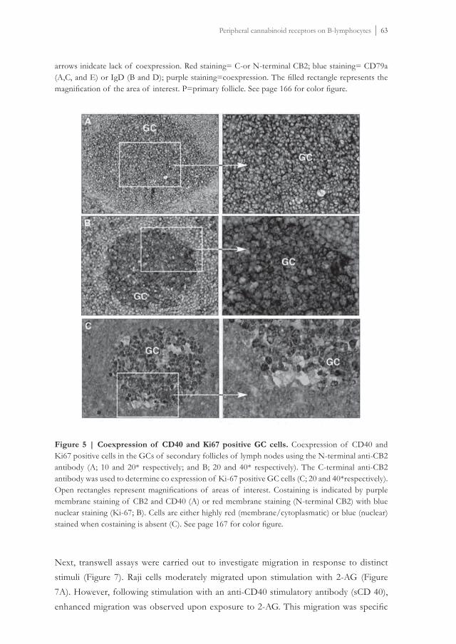
Peripheral cannabinoid receptors on B-lymphocytes | 63
arrows inidcate lack of coexpression. Red staining= C-or N-terminal CB2; blue staining= CD79a (A,C, and E) or IgD (B and D); purple staining=coexpression. The fi lled rectangle represents the magnifi cation of the area of interest. P=primary follicle. See page 166 for color fi gure.
Figure 5 | Coexpression of CD40 and Ki67 positive GC cells. Coexpression of CD40 and Ki67 positive cells in the GCs of secondary follicles of lymph nodes using the N-terminal anti-CB2 antibody (A; 10 and 20* respectively; and B; 20 and 40* respectively). The C-terminal anti-CB2 antibody was used to determine co expression of Ki-67 positive GC cells (C; 20 and 40*respectively). Open rectangles represent magnifi cations of areas of interest. Costaining is indicated by purple membrane staining of CB2 and CD40 (A) or red membrane staining (N-terminal CB2) with blue nuclear staining (Ki-67; B). Cells are either highly red (membrane/cytoplasmatic) or blue (nuclear) stained when costaining is absent (C). See page 167 for color fi gure.
Next, transwell assays were carried out to investigate migration in response to distinct stimuli (Figure 7). Raji cells moderately migrated upon stimulation with 2-AG (Figure 7A). However, following stimulation with an anti-CD40 stimulatory antibody (sCD 40), enhanced migration was observed upon exposure to 2-AG. This migration was specifi c

64 | Chapter 3
since the CB2 receptor inverse agonist (SR144528) completely abolished this effect. The
Figure 6 | CD40 and CB2 expression in Raji cell line. Flowcytometry of Raji cells determining CD40 expression (A). CB2 membrane expression on non-permeabilised cells (B) and intra cellular CB2 protein in permeabilised cells (C) was analysed using the N-terminal specifi c anti-CB2 receptor antibody. The fi lled areas represent CD40 (A) or CB2 (B) positive cells; the grey lines (A and B) represent background staining (only secondary antibody). The green dots represent CB2 protein in the cytoplasm; blue counter stain with diamino-2-phenylindole refl ects the nuclei of Raji cells. See page 162 for color fi gure.
CB1 receptor specifi c antagonist (SR 141716) did not infl uence the sCD40-enhanced migration (Figure 7A). An inhibitory CD40 specifi c antibody (iCD40), when added in 10-fold excess, completely abolished the sCD40 (stimulatory antibody) enhanced 2-AG induced migration of Raji-cells (Figure 7B). Non-stimulatory CD40 antibodies (nCD40) serving as a control did not enhance 2-AG induced migration (Figure 7B). CB2 membrane expression did not detectably increase following exposure to the CD40 stimulatory antibody, as determined by fl owcytometry (data not shown).

Peripheral cannabinoid receptors on B-lymphocytes | 65
Figure 7 | Migration of Raji cells in response to distinct stimuli. Cells were stimulated for 24 hours in the presence or absence of sCD40 monoclonal antibody (MoAb) (K7; 100nM), sCD40 MoAb (100nM) plus iCD40 (5D12; 1000 nM) or nCD40 (100nM). Migration was investigated in the presence or absence of the CB2 ligand 2-AG (300nM). The upper wells contained either non-stimulated or stimulated cells in the presence or absence of CB1 (SR141716; 100nM) or CB2 specifi c inverse agonists (SR 144528; 100nM). Y-axis shows percentage of migration from an input of 1 х 105 cells. A, represents the mean migration with the Ses of the means of three individual experiments.

66 | Chapter 3
DISCUSSION
In this study we investigated the distribution CB2 receptors as well as their activation status in human immune tissues using two distinct anti-CB2 antibodies. Specifi city of a C-terminal and a N-terminal specifi c CB2 antibody was demonstrated in CHO cells transfected with human CB2. We demonstrated that CB2 protein was present on the membrane as well as in the cytoplasm of these cells (Figures 1A-E). Applying these antibodies in single immunohistochemical staining, we observed distinct expression patterns in B-cell areas in human lymphoid organs. In conjunction with previous studies5 we found CB2 receptors in the MZs using the anti-C-terminal CB2 antibody. We also observed CB2 positivity in primary follicles and in the MGZs of secondary follicles, using this antiserum. As the C-terminal specifi c antibody only recognizes non-phosphorylated CB2, apparently inactive CB2 receptors are present on B-lymphocytes in these particular areas. It will be of interest to investigate CB2 phosphorylation in B-lymphocytes, especially in relation to the antigen specifi c activation and proliferation of these cells. Only low CB2 positivity was observed in the MGZ when the N-terminal specifi c antibody was used. A possible explanation for the differences observed between the two antibodies may be that the C-terminal specifi c antibody binds CB2 with a higher affi nity than the N-terminal specifi c CB2 antiserum (Figure 1). Alternatively it is also possible that the differences in staining by the CB2 antibodies may be the result of differences in epitope accessibility. In this paper, using the N-terminal specifi c antibody, we have demonstrated, that various CB2 expressing B-cell subsets exist. Some of these B-cell populations are only weakly stained with the C-terminal anti-CB2 antibody. Therefore, the CB2 receptors on these cells are most probably phosphorylated and therefore in an active state6. Because of the low affi nity of the N-terminal specifi c antibody, a strong positivity most likely indicates high numbers of CB2 receptors on these cells. These cells reside in an anatomical location where B-memory cells are formed and affi nity maturation occurs, i.e. the GC of secondary follicles, and they express CD79a but are mainly IgD negative (Figures 4C and D). B-cells in primary follicles or in the MZ/MGZ of secondary follicles may express low levels of CD40. On the other hand, B-cells in the GCs of secondary follicles highly express CD40 as well as Ki-67, a proliferation marker (Figures 5 A and B), markers characteristic for actively cycling B-cells in the GC. All in all, it appears that functional CB2 receptors are present on immunologically active B-lymphocytes.

Peripheral cannabinoid receptors on B-lymphocytes | 67
Dual immunohistochemistry using pan-B-cell markers confi rmed previous observations that in general B-lymphocytes are the leukocyte subsets expressing CB2 mRNA and protein3-5 (Figure 4). Furthermore, modulation of this receptor during B-cell differentiation has been demonstrated5 using the C-terminal anti-CB2 antibody. In accordance with this fact, we found CD79a, IgD positive resting B-cells in the MZs highly expressing inactive CB2 (Figure 4A and B). The co expression of CB2 and CD40 on activated B-lymphocytes prompted us to investigate the functional relation between those two membrane proteins. CD40 belongs to the TNF-receptor family and is involved in B-cell activation, survival, proliferation, differentiation and Ig-isotype switch8. Previous studies in mice have shown that upon CD40 stimulation mCB2 mRNA may be up regulated9. An increase in CB2 mRNA following CD40 stimulation has been demonstrated in human tonsillar B-cells as well5. We demonstrate, using a B-lymphoid cell line as a model, that migration of B-cells in response to the CB2 specifi c ligand 2-AG was signifi cantly enhanced following CD40 stimulation. The mechanism of activation appears to be a different one, since we neither found any increase of the CB2 protein levels, nor did the CB2 receptor activation status change following CD40 stimulation (data not shown). The peripheral cannabinoid receptor belongs to the family of G-protein coupled 7-transmembrane receptors (GPCRs)3,4. GPCRs are crucial to many cellular functions such as proliferation, maturation, survival, apoptosis or migration10-12; Carayon et al showed moderate proliferation of virgin and GC tonsillar B-cells upon stimulation with the synthetic cannabinoid agonist CP55,940 only when the cells were stimulated with CD40 MoAbs5. In our functional study with Raji B-lymphoma cells, we observed moderate migration upon stimulation with the CB2 ligand 2-AG. Interestingly, this migration was greatly enhanced when cells were stimulated for 24 hrs with a stimulating CD40 MoAb (Figure 7). Furthermore, this enhanced migration was CB2 and CD40 receptor specifi c, since the effects could be specifi cally blocked by the CB2 inverse agonist (SR144528) and the CD40 antagonist (5D12). A similar observation has been reported for CXCR4 and CD40 on GC cells. CD40 pre treatment of these cells led to an increased migration upon stimulation of by the CXCR4 ligand SDF-113. Apparently, exposure to CD40 activating agents is crucial for the induction of a functional response of certain GPCRs such as CB2 or CXCR4. Studies of the potential involvement of cannabinoid receptors and stimulation by cannabinoids in the immune system have been reported previously14-19. In fact, immuno-modulation by cannabinoids is absent in mice defi cient for the cannabinoid CB2 receptor20. Recently it was also demonstrated that human antigen presenting dendritic cells produce high levels of endocannabinoid ligand 2-AG21 and that 2-AG is probably

68 | Chapter 3
the physiological ligand for the cannabinoid CB2 receptor22. These fi ndings together with our observations that 1) active CB2 receptors are present on immunologically active cells and 2) CB2 response may be increased by the co stimulatory molecule CD40 provide evidence that CB2 receptors may be involved in B-cell activation. The observation that CB2 receptors are present on B-lymphocytes and show migratory function in a malignant B-cell line raises the question whether this receptor is also involved in the malignant transformation of B-cells resulting in lymphomas or whether this receptor contributes to a specifi c function of malignant B-cells. CB2 dependent migration in vitro in a B-lymphoma cell line could give a direction towards homing of malignant B-cells in vivo. In the staging of B-lymphomas, bone marrow infi ltration is one of the important parameters. Therefore analysis of the CB2 receptor status could play an important role as a prognostic and /or diagnostic marker, possibly predicting spreading from the initial localisation of the tumour. In high-throughput non-Hodgkin lymphoma (NHL) patient screens using tissue micro arrays, currently in progress to identify potential novel diagnostic and or prognostic markers in NHL, CB2 is the most interesting one. Furthermore, a correlation between the active CB2 receptor state in normal immune tissues and NHLs can also be made to see whether in a malignant setting the functional relation between CB2 and CD40 receptors is still evident.
ACKNOWLEDGMENTS
We thank P. Casellas (Sanofi -Synthelabo Recherche, Montpellier, France) for donation of the CB1 and CB2 receptor specifi c inverse agonists SR 141716 an SR 144528 and Dr. L. Boon (PanGenetics BV, Amsterdam, The Netherlands) for donation of the stimulatory (Clone 7), inhibitory (5D12) and neutral CD40 MoAbs. We also thank Karola van Rooyen for preparation of the fi gures.

Peripheral cannabinoid receptors on B-lymphocytes | 69
REFERENCES
1. Matsuda, L. A., S. J. Lolait, M. J. Brownstein, A. C. Young, and T. I. Bonner. 1990. Structure of a cannabinoid receptor and functional expression of the cloned cDNA. Nature 346:561.
2. Munro, S., K. L. Thomas, and M. Abu-Shaar. 1993. Molecular characterization of a peripheral receptor for cannabinoids. Nature 365:61.
3. Bouaboula, M., M. Rinaldi, P. Carayon, C. Carillon, B. Delpech, D. Shire, G. Le Fur, and P. Casellas. 1993. Cannabinoid-receptor expression in human leukocytes. Eur J Biochem 214:173.
4. Galiegue, S., S. Mary, J. Marchand, D. Dussossoy, D. Carriere, P. Carayon, M. Bouaboula, D. Shire, G. Le Fur, and P. Casellas. 1995. Expression of central and peripheral cannabinoid receptors in human immune tissues and leukocyte subpopulations. Eur J Biochem 232:54.
5. Carayon, P., J. Marchand, D. Dussossoy, J. M. Derocq, O. Jbilo, A. Bord, M. Bouaboula, S. Galiegue, P. Mondiere, G. Penarier, G. L. Fur, T. Defrance, and P. Casellas. 1998. Modulation and functional involvement of CB2 peripheral cannabinoid receptors during B-cell differentiation. Blood 92:3605.
6. Bouaboula, M., D. Dussossoy, and P. Casellas. 1999. Regulation of peripheral cannabinoid receptor CB2 phosphorylation by the inverse agonist SR 144528. Implications for receptor biological responses. J Biol Chem 274:20397.
7. Jorda, M. A., S. E. Verbakel, P. J. Valk, Y. V. Vankan-Berkhoudt, M. Maccarrone, A. Finazzi-Agro, B. Lowenberg, and R. Delwel. 2002. Hematopoietic cells expressing the peripheral cannabinoid receptor migrate in response to the endocannabinoid 2-arachidonoylglycerol. Blood 99:2786.
8. van Kooten, C., and J. Banchereau. 2000. CD40-CD40 ligand. J Leukoc Biol 67:2.
9. Lee, S. F., C. Newton, R. Widen, H. Friedman, and T. W. Klein. 2001. Differential expression of cannabinoid CB(2) receptor mRNA in mouse immune cell subpopulations and following B cell stimulation. Eur J Pharmacol 423:235.
10. Dhanasekaran, N., L. E. Heasley, and G. L. Johnson. 1995. G protein-coupled receptor systems involved in cell growth and oncogenesis. Endocr Rev 16:259.
11. Neptune, E. R., and H. R. Bourne. 1997. Receptors induce chemotaxis by releasing the betagamma subunit of Gi, not by activating Gq or Gs. Proc Natl Acad Sci U S A 94:14489.
12. Offermanns, S., and M. I. Simon. 1998. Genetic analysis of mammalian G-protein signalling. Oncogene 17:1375.
13. Corcione, A., L. Ottonello, G. Tortolina, P. Facchetti, I. Airoldi, R. Guglielmino, P. Dadati, M. Truini, S. Sozzani, F. Dallegri, and V. Pistoia. 2000. Stromal cell-derived factor-1 as a chemoattractant for follicular center lymphoma B cells. J Natl Cancer Inst 92:628.
14. Pross, S., T. Klein, C. Newton, and H. Friedman. 1987. Differential effects of marijuana components on proliferation of spleen, lymph node and thymus cells in vitro. Int J Immunopharmacol 9:363.
15. Klein, T. W., Y. Kawakami, C. Newton, and H. Friedman. 1991. Marijuana components suppress induction and cytolytic function of murine cytotoxic T cells in vitro and in vivo. J Toxicol Environ Health 32:465.
16. Luo, Y. D., M. K. Patel, M. D. Wiederhold, and D. W. Ou. 1992. Effects of cannabinoids and cocaine on the mitogen-induced transformations of lymphocytes of human and mouse origins. Int J Immunopharmacol 14:49.
17. McCoy, K. L., D. Gainey, and G. A. Cabral. 1995. delta 9-Tetrahydrocannabinol modulates antigen processing by macrophages. J Pharmacol Exp Ther 273:1216.

70 | Chapter 3
18. Di Marzo, V., T. Bisogno, L. De Petrocellis, D. Melck, P. Orlando, J. A. Wagner, and G. Kunos. 1999. Biosynthesis and inactivation of the endocannabinoid 2-arachidonoylglycerol in circulating and tumoral macrophages. Eur J Biochem 264:258.
19. Lynn, A. B., and M. Herkenham. 1994. Localization of cannabinoid receptors and nonsaturable high-density cannabinoid binding sites in peripheral tissues of the rat: implications for receptor-mediated immune modulation by cannabinoids. J Pharmacol Exp Ther 268:1612.
20. Buckley, N. E., K. L. McCoy, E. Mezey, T. Bonner, A. Zimmer, C. C. Felder, and M. Glass. 2000. Immunomodulation by cannabinoids is absent in mice defi cient for the cannabinoid CB(2) receptor. Eur J Pharmacol 396:141.
21. Matias, I., P. Pochard, P. Orlando, M. Salzet, J. Pestel, and V. Di Marzo. 2002. Presence and regulation of the endocannabinoid system in human dendritic cells. Eur J Biochem 269:3771.
22. Sugiura, T., S. Kondo, S. Kishimoto, T. Miyashita, S. Nakane, T. Kodaka, Y. Suhara, H. Takayama, and K. Waku. 2000. Evidence that 2-arachidonoylglycerol but not N-palmitoylethanolamine or anandamide is the physiological ligand for the cannabinoid CB2 receptor. Comparison of the agonistic activities of various cannabinoid receptor ligands in HL-60 cells. J Biol Chem 275:605.

Chapter 4
The expression of the peripheral cannabinoid receptor on cells of the immune system and Non-Hodgkin’s lymphomas
Nazik Rayman1, King H. Lam2, Joost van Leeuwen1, Andries H. Mulder3 , Leo M. Budel4,
Bob Löwenberg1, Pieter Sonneveld1 and Ruud Delwel1
1Department of Hematology, Erasmus Medical Centre, Rotterdam, The Netherlands, 2Department of Pathology, Josephine Nefkens Institute, Erasmus Medical Centre, Rotterdam,
The Netherlands, 3Department of Pathology, Rijnstate Hospital, Arnhem The Netherlands and 4Department of Pathology, Meander Medical Centre, Amersfoort, The Netherlands.
Leukemia & Lymphoma 2007; 48:1389-1399

72 | Chapter 4
ABSTRACT
The peripheral cannabinoid receptor CB2 is expressed highly on normal human B-lymphocytes. C-terminal specifi c anti-CB2 antibody recognises a non-phosphorylated
inactive receptor on naïve and resting B-lymphocytes. Another, N-terminal specifi c CB2 antibody, primarily recognises B-cells present in the germinal centres of secondary follicles in lymph nodes. We hypothesise that N-terminal specifi c CB2 antibody recognises activated CB2 receptors. In this study we showed using these antibodies, that expression of CB2 is generally absent on T-lymphocytes in reactive, non-malignant human lymphoid tissues. Applying single and dual immunohistochemistry, CD23+ follicular dendritic cells and a small but signifi cant subpopulation of CD68+ macrophages showed positive staining with the N-terminal specifi c CB2 antibody but not with the C-terminal specifi c CB2-antibody. This may indicate the presence of an active CB2 receptor on these cells with possible involvement in immunomodulation. In contrast to the low expression on normal T-cells, abundant levels of CB2 protein were present on T-Non-Hodgkin Lymphomas (NHL). Moreover, in many B-NHL, high CB2 protein expression was found as well. In contrast to the distinct expression patterns in normal immune tissues using the two different CB2 antibodies, NHL specimens in general stained positively with both. We conclude that CB2 receptor expression pattern may be abnormal in NHL.

Peripheral cannabinoid receptors on immune cells and NHL | 73
INTRODUCTION
Two distinct cannabinoid receptors have previously been identifi ed, i.e. the “central” cannabinoid receptor CB1, mainly expressed in brain1-6 and the “peripheral” cannabinoid receptor CB2 expressed in particular haematopoietic cells, with highest transcript levels in B-cells7-9. It has been reported that activation or inactivation of CB2 receptor using specifi c agonists or inverse-agonists results in phosphorylation at serine 352, located intracellularly at the C-terminus of CB210,11. Although the exact function of CB2 in B-cell development is still elusive, we recently reported some striking features of CB2 receptor expression and receptor status in human immune tissues12. High CB2 protein expression was observed in the mantle zones (MZs) and marginal zones (MGZ) in secondary follicles of human lymph nodes, using a C-terminal specifi c antibody, which only identifi es inactive serine 352 non-phosphorylated receptors12. Although, weak staining was observed on B-cells in the germinal centres (GCs) of secondary follicles using this latter antibody, high numbers of CB2 receptors could be detected on B-cells in GCs using another CB2 specifi c antibody directed to the N-terminal extra cellular part of the receptor. The fi nding that the staining pattern with this antibody was completely opposite to that obtained with the C-terminal specifi c antiserum12, led to the conclusion that this antiserum probably recognizes activated CB2 present on B-cells in the GCs. Furthermore, we found that these cells are located in these immunologically active areas and express CD40 and Ki-67 as well suggesting an immunomodulatory function for this receptor12. A role for cannabinoid receptors on normal haematopoietic cells other than B-lymphocytes has been suggested. For instance, several investigators have reported that CB2 transcripts may be present in macrophages, dendritic cells or T-lymphocytes9,13. On the contrary, little is known about CB2 protein expression or receptor activation status in these cells. In the fi rst part of the study presented here, we investigated which areas other than the B-cell regions in various haematopoietic and immunologic organs express CB2 receptors. Using dual-immunohistochemical methods, we studied which cells expressed CB2 and applying the different CB2 receptor specifi c antibodies, we studied a possible correlation with CB2 receptor activation status on these cells. Cannabinoid receptors may play a role in leukaemia and lymphoma development14-16. CB2 receptors have been identifi ed on acute myeloid leukaemia (AML) cells, whereas they were not found on their normal bone marrow counterparts16. Intriguingly, using in vitro models, it was observed that activation of CB2 on myeloid leukaemia cells causes neutrophilic differentiation block, which could be reversed using a CB2 specifi c inverse

74 | Chapter 4
agonist. These studies suggest that CB2 receptors may play a role in AML development in mouse and in human. It has also been demonstrated that cannabinoid ligands may interfere with proliferation of lymphoid tumor cells15. These latter experiments have been performed with cell lines only, and the expression and activation status of the CB2 receptors has not been studied. Flygare et al. recently reported response to canabinoids of mantle cell lymphoma’s, although they concluded that particularly CB1 receptors were responsible for these effects14. In the second part of the study, we applied histochemistry using the distinct CB2 specifi c antibodies and investigated whether CB2 receptors may be expressed on distinct lymphoma subtypes and whether CB2 receptors present on these cells may be active or inactive. We hypothesise that CB2 expression and probably activation status in the various lymphoma subtypes is signifi cantly distinct from that in normal reactive lymphoid tissue and may consequently be abnormal.
MATERIALS AND METHODS
Patient samples
Whole sections of FL (n=13), MCL (n=18), DLBCL (n=150) and T-NHL (n=7), patients were analysed immunohistochemically (Tables 3 and 6). These patients were originally included in the HOVON 25 trial (Dutch-Belgian Hemato-Oncology Cooperative Group)17
in which 389 patients of 65 years and older with an intermediate or high grade lymphoma according to the Working Formulation and an advanced stage disease (Ann Arbor stages II-IV) were included between August 1994 and September 2000. Informed consent was obtained from all patients according to regulations of the Dutch health authorities. Cases were considered as positive if cells of interest stained >75%, and partially positive if they were >50% positive. We selected from the archives of the Department of Pathology of the Erasmus Medical Centre (MC), all cases aged above 18 years with a biopsy proven NHL. In total, we included 360 patients with different sub types of NHL, of which suffi cient and representative material was available for tissue micro array (TMA) analysis. All AIDS-related NHL cases were excluded. All cases with reliable immunohistochemical staining were included in the fi nal analysis containing MCL (n=16), MZL (n=19), LBL (n=3), DLBCL (n=84), FL (n=42) and T-NHL (n=25) (Tables 4, 5 and 7). For the TMA, hematoxylin and eosin-stained sections of paraffi n-embedded, formalin fi xed tissue were used to defi ne diagnostic areas. From each case, three representative 0.6-mm cores were obtained and inserted adjacently into a recipient paraffi n block using a tissue arrayer

Peripheral cannabinoid receptors on immune cells and NHL | 75
(Beecher Instruments, Silver Spring, MD). In total, we generated two TMA blocks with control specimens of lymphoid tissues, i.e., the spleen, thymus and lymph node which were inserted in two separate rows containing at least three cores per tissue. Stains were scored on a 4-point scale regarding the TMA analysis. 0=negative, 1=<25% cells positive, 2=25 – 50% cells positive, 3=51 – 75% cells positive, 4=>75% cells positive. Cases with scores 0, 1 or 2 were designated as negative in the fi nal analysis, whereas scores 3 or 4 were designated as positive. In case of staining of the three cores with variable outcome, the mean expression of three cores was taken. For the expression analysis on human lymphoid tissues, we retrieved formalin-fi xed, paraffi n embedded normal lymph node (n=3), spleen (n=2) and thymus (n=1) from the fi les of the Department of Pathology at the Erasmus MC. Single and dual immunohistochemical procedures were carried and partially and complete co-staining was defi ned if cells were 25 – 50% and >50% positive respectively. Analysis and scoring of the whole, TMA and lymphoid organ sections was independently performed by two medical doctors with suffi cient experience. Disagreements were resolved by joint review on a multiheaded microscope.
Reagents and immunohistochemical procedures
The polyclonal human C-terminal anti-CB2 receptor antibody and its synthetic peptide were kindly provided by P. Casellas (Sanofi -Synthelabo Recherche, Montpellier, France). The anti-CB2 receptor polyclonal antibody raised against the fi rst 33 amino acid residues of the N-terminus of the receptor was purchased from Affi nity Bioreagents Inc (ABR, CO, USA). For the dual immunohistochemical analyses we used the antibodies as listed in Table 1.
Single staining procedure on normal and malignant specimens
Five-micrometer sections were used for staining of whole sections of patients from the HOVON 25, the TMA and lymphoid organs for the staining experiments. In brief, sections were deparaffi nized, rehydrated and endogenous peroxidase was blocked with 3% H2O2 in methanol for 20 minutes at room temperature (RT). Heat induced antigen retrieval (required for staining with the C-terminal anti-CB2 antibody) was achieved by boiling the sections for 20 minutes at 100ºC in citrate buffer (10 mM, pH 6.0) in a Micromed T/T Mega microwave oven (Salm and Kipp, Breukelen, The Netherlands). Sections were cooled down, washed and incubated with the fi rst panel of primary antibodies for 1 hour at RT (C-terminal anti-CB2 1:400, or N-terminal anti-CB2 1:600), and subsequently

76 | Chapter 4
Table 1 | Antibodies and conjugates used for immunohistochemical analysis.
Primary antibodies Specifi city Dilution/Epitope retrieval*
Source
CD1a (monoclonal)CD2 (monoclonal)CD3 (polyclonal)CD5 (monoclonal)CD8 (monoclonal)CD15 (monoclonal)CD23 (monoclonal)CD68 (monoclonal)CD79a (monoclonal) IgD (polyclonal)CD40 (monoclonal)
Cortical thymocytes; Langerhans cellsEarly T-cellsPan-T cellMature T-cellsCytotoxic, suppressor T-cellsGranulocytes, monocytes, Reed-Sternberg cellsB-cell subset/ Follicular dendritic cellsMacrophagesB cell; plasma cellsNaive B cellsGC-B-cells
1:10/yes1:50/yes
1:600/yes1:200/yes1:100/yes1:25/yes1:20/yes
1:1000/yes1:100/yes1:200/yes1:100/yes
ImmunotechNovocastraDAKONovocastraDAKOB&DNovocastraDAKODAKODAKODonated
Secondary antibodies and conjugates
SwarbioRaMbioS-ABCAP-kitS-ABC HRP-kit
Swine-anti -rabbitRabbit anti-mouseStreptavidin-biotin alkaline phospataseStreptavidin biotin horse radish peroxidase
1:2001:2001:1001:100
DAKODAKODAKODAKO
Antigen retrieval was performed by pre treating sections in a microwave oven (see Materials and Methods). Immunotech, Prague, Czech Republic; Novocastra, Newcaste upon Tyne, UK; B&D, San Jose, CA, USA.
incubated for 10 minutes with biotinylated secondary antibodies (anti-mouse and rabbit immunoglobins; Labvision,Fremont, CA, USA) followed by a 10 minute incubation with streptavidin-conjugated (S-ABC) horseradish peroxidase (Labvision). Visualisation was achieved using Diaminobenzidine (DAB) (Fluka, Buchs, Switzerland) for 7 minutes in dark. Finally, sections were counterstained with haematoxylin according to Harris (Klinipath, Duiven, The Netherlands), dehydrated and covered by imsol (Klinipath) and by pertex (Histolab, Göteborg, Sweden). Standard haematoxylin-eosin (HE) staining and negative controls (omission of primary antibody) of all examined tissues were included. Specifi city controls were performed by pre incubating the C-terminal anti-CB2 receptor antibody for one hour with the synthetic peptide at 10 mg/ml. No peptide has been developed yet for the N-terminal specifi c antibody used in our experiments, which was purchased commercially. Other commercially available antibodies against N-CB2 of which a peptide control was available were tested. Upon elaborate testing, none of these antibodies were suited for histological staining experiments. Different conditions of

Peripheral cannabinoid receptors on immune cells and NHL | 77
both CB2 antibodies for antigen retrieval were tested. Intracellularly located epitope of C-CB2 was only detectable with microwave pretreatment, whereas no pre-treatment was necessary to detect the extracellulary located epitope of the N-CB2 receptor. Accurate titrations, including negative controls omitting the primary antibody were performed in order to achieve reliable staining patterns on paraffi n sections. In these experiments the optimal staining pattern was chosen taking unspecifi c background staining into account. We have only accepted positive staining when a clear distinctive cytoplasmatic or membrane staining was detected.
Dual staining procedure on normal lymphoid tissues
A panel of cell specifi c markers, and biotin conjugated secondary antibodies were used for dual immunohistochemistry of the lymphoid tissues. The list of the antibodies, their antigen retrieval their sources and dilutions are listed in Table 1. Sections were fi rst incubated with the CB2 antibodies as described above, and stained with 3-amino-9-ethylcarbazole (AEC; Sigma USA) in NaAc buffer (0.2M, pH 4.6) for 30 minutes in dark, which yielded in a red signal. Then sections were individually incubated with the primary antibodies listed in Table 1 for 30 minutes, washed and incubated with the biotin conjugated secondary antibodies for 30 minutes at RT. An S-ABC conjugated alkaline phosphatase kit (DAKO, Glostrup, Denmark) was used to enhance the signal. Visualisation was achieved using a solution containing Fast Blue BB salt (4-benzoylamino-2, 5-diethoxylbenzene diazoniumchloride; Sigma), naphtol-AS-MX phosphate (Sigma) and levamisole hydroxychloride (Acros Organics, Geel, Belgium) in Tris/HCl buffer (0.2M, pH 8.0) for 30 minutes in dark resulting in blue staining of cells. In case of co-expression/ co-staining, the mixture of red and blue signals resulted in a dark purple staining of cells. Cells were counter-stained with haematoxylin according to Harris (Klinipath) and covered by pertex.
RESULTS
CB2 receptor expression in normal lymphoid tissue
As reported in our previous study12 C-terminal specifi c anti-CB2 antibody recognized B-cells in the primary follicles and in the MZs and MGZs of secondary follicles (Figure 1A and C). N-terminal CB2 specifi c staining was observed in the GCs of secondary follicles (Figure 1B and D). A small subset of the cells (≤5%) in the T-cell areas of normal

78 | Chapter 4
lymph nodes expressed CB2, as detected by the N-terminal specifi c antibody (Figure 1D, arrow). No positive staining was found using the C-terminal specifi c antibody in this area (Figure 1A and C)12. Hardly any CB2 expression on T-cells was detected, using T-cell specifi c markers (CD3, CD5 and CD8) in combination with both CB2 specifi c antibodies (Table 2 and Figure 1). This result was confi rmed by multiparameter fl owcytometry using CB2 and CD3 specifi c antibodies (data not shown). CB2 expression was undetectable in the peri-arteriolar-lymphocytic sheet (PALS) of the spleen and in the thymus, applying single immunohistochemistry (data not shown). The results of distinct dual staining experiments are summarised in Table 2.
Figure 1 | CB2/CD3 dual staining of normal reactive lymph nodes. Dual staining procedures using the C-terminal (panel A, C and E) or N-terminal (panel B, D and F) anti-CB2 antibodies (red staining) combined with the pan-T-cell marker CD3 (all panels) (blue staining) on normal lymph node sections. Note the almost complete separation of red and blue staining areas. See text for further discussion. The following areas in the lymph node are indicated: GC; germinal centre, MZ: mantle zone. MGZ: marginal zone, P: para-cortical (T-cell) area. Original object lens magnifi cations: A: 1,25x, B: 2.5x, C and D: 10x, E and F: 40x. See page 170 for color fi gure.

Peripheral cannabinoid receptors on immune cells and NHL | 79
Table 2 | CB2 expression on lymphoid and accessory cells as determined by dual immuno-histochemistry.
Antibody Examined lymphoid tissue:
Spleen/LN/Thymus
N-CB2 C-CB2
CD1aCD2CD3 CD5CD8 CD15CD23
CD79a IgDCD40
CD68
ThymusThymusAllAllAllSpleen and LNSpleen and LN
Spleen and LNSpleen and LNSpleen and LN
Spleen and LN
––
+/– ––––
+/–(+ in GC)
+/––
+/–(+ in GC)
+/–
––––––
+/–(+ in MZ/MGZ)
+/–+
+/–(+ in MZ/MGZ)
–
Dual immunohistochemistry was performed on spleen, lymph node (LN) and/or thymus sections. The panel of antibodies was combined with each of the CB2 antibodies (N-CB2 and C-CB2). +/– indicates that different subsets of cells are recognized by each CB2 antibody. + or – indicates that the majority of the cells (≥ 75%) either co-stained or did not co-stain with the CB2 antibody. +/– – indicates that a very small number of cells (< 5%) show co-expression.
Follicular dendritic cells and a subset of macrophages express CB2
We also analysed by means of dual-immunohistochemistry the expression of CB2 receptors on granulocytes/monocytes (CD15+), macrophages (CD 68+) and follicular dendritic cells (CD23+) in reactive lymph node and spleen. A small but signifi cant number of CD68+ macrophages showed co-staining with the N-terminal specifi c CB2 antibody but not with the C-terminal specifi c antiserum (Table 2). Remarkably, CD23+ follicular dendritic cells present in the GCs showed intense co-staining with the N-terminal specifi c CB2 antibody (Table 2, Figure 2 C and D), but not with the C-terminal specifi c one (Table 2, Figure 2A and B). No co-expression on CD15+ cells was observed with any of the CB2 specifi c antibodies (Table 2).

80 | Chapter 4
Figure 2 | CB2/CD23dual staining of normal reactive lymph nodes. Dual staining of secondary follicles of human normal reactive lymph nodes using the C-terminal (A and B) or N-terminal (C and D) specifi c anti-CB2 antibodies (red staining) with CD23 (blue staining). Note the reticular blue-purple staining pattern in the germinal centre (C and D). Co-staining was absent in the GC B-cells (A and B). The following areas in the lymph node are indicated: GC; germinal centre, MZ: mantle zone. MGZ: marginal zone, P: para-cortical (T-cell) area. Original object lens magnifi cations: A and C: 10x, B and D: 20x. Panels A and B were taken form different germinal centres for technical reasons. See page 171 for color fi gure.
Frequent expression of CB2 receptors in B-NHL and T-NHL
We investigated CB2 expression on distinct B-cell lymphoma subtypes. CB2 distribution on DLBCL samples was investigated on whole sections from the HOVON 25 study (n=150) and in the TMA series (n=84). We considered the staining to be positive when either on whole sections or on TMA cores 50% or more cells were positive. On whole sections we observed a high percentage of positive staining using both CB2 antibodies, that is 80% positive staining using N-terminal specifi c anti-CB2 and 74% positive staining using C-terminal specifi c antibodies (Table 3). DLBCL samples on the TMA appeared highly positive for both antibodies as well, i.e. 73% using N-terminal CB2 and 96% using the C-terminal specifi c CB2 antibody (Table 4). From the 13 FL cases included in the HOVON 25 study, 6 (46%) were N-terminal CB2 positive, whereas 13 (100%) were

Peripheral cannabinoid receptors on immune cells and NHL | 81
C-terminal CB2 positive (Table 3). Sixty percent of all FL cases from the TMA study showed high CB2 protein expression using the N-terminal specifi c anti- CB2 antibody, and 98% FL cases were positive with the C-terminal specifi c antibody (Table 5). Of these cases, 12 were grade I FL, 16 were grade II, 8 were grade III and 6 were of unknown grade FL. Staining with the distinct CB2 antibodies did not discriminate between the different grades of the FLs (Table 5). Of the 17 MCLs in the HOVON 25 study, 3 (18%) were positive using the N-terminal specifi c CB2 antibody, whereas 17 (100%) were positive using the C-terminal antibody (Table 3). In the TMA we observed 6/16 (38%) N-terminal CB2 positive and 15/16 (94%) C-terminal CB2 positive cases. Fourteen (74%) of the 19 MZL cases present in the TMA study expressed the CB2 receptor as detected with the N-terminal antibody and 17/19 (89%) were positive using the C-terminal CB2 antibody (Table IV). No MZLs were analysed in the HOVON 25 study. Representative examples of CB2 positive DLBCL, FL and MCL are shown in Figures 3D-L.
Table 3 | CB2 expression on mature and immature B-cell lymphoma’s: HOVON 25 study: FL,MCL and DLBCL.
FL MCL DLBCL Total
Cases 13 17 150 180N–CB2
+ (>75%) +/– (>50%) – (<50%)
6 (46%)0 (0%)7 (54%)
3 (18%)0 (0%)14 (82%)
103 (69%)16 (11%)31 (20%)
112(62%)16 (9%)52(29%)
C–CB2
+ (>75%) +/– (>50%) – (<50%)
11(85%)2 (15%)0 (0%)
15 (88%)2 (12%)0 (0%)
101 (67%)10 (7%)39(26%)
127 (71%)14 (8%)39 (21%)
Semi quantitative counting on whole sections was performed as cells being ≥75% positive (+), >50% partially positive (+/–) and ≤50% as negative (–). In the fi nal analysis, positive and partially positive cases were taken together. Only sections which were reliably interpretable using both CB2 antibodies were analysed. Follicular lymphoma (FL), Mantle cell lymphoma (MCL) and Diffuse large B-cell lymphoma (DLBCL).
In contrast to the expression on normal T-cells, almost all T-cell lymphomas highly expressed CB2 receptors (Tables 6 and 7). Representative examples are shown in Figure 3A-C. 6/7 (86%) T-NHL cases from the HOVON 25 study stained positive with the N-terminal specifi c antibody, whereas 3/7 (43%) stained positive with the C-terminal

82 | Chapter 4
specifi c antibody (Table 6). Analysis of 25 cases from another cohort analysed by TMA revealed that 21 (84%) were N-CB2 positive and 24 (96%) were positive using the C-terminal specifi c antibody (Table 7). Sub-typing T-NHL into anaplastic large cell lymphoma (ALCL), peripheral T-cell lymphoma (PTCL), T-cell lymphoblastic lymphoma (T-LBL), revealed similar staining patterns using both antibodies against CB2 (Tables 3 and 4). An exception of this pattern was observed with the PTCL in the HOVON 25 study where 1 of 4 cases (25%) expressed the CB2 using the C-terminal specifi c antibody. However, using the N-terminal specifi c antibody 3 of the 4 cases (75%) stained positively.
Figure 3 | CB2 expression in malignant lymphoma’s. Whole sections of malignant lymphomas were stained with C- and N-terminal CB2 antibodies respectively (brown staining). Representative cases showing positive staining with both antibodies including negative controls are shown: A-C: T-cell NHL; D-F: FL; G-I: DLBCL; J-L: MCL. Original object lens magnifi cation: 40x (inserts,20x). See page 174 for color fi gure.

Peripheral cannabinoid receptors on immune cells and NHL | 83
Table 4 | CB2 expression on mature and immature B-cell lymphoma’s: TMA study: MCL, MZL, LBL and DLBCL.
MCL MZL B-LBL DLBCL Total
Cases 16 19 3 84 122
N-CB2 pos:Score (3 and 4)
6/16 (38%) 14/19 (74%) 2/3 (67%) 62/84 (73%) 84/122 (69%)
0 (0%) 1 (1-25%) 2 (25-50%) 3 (50-75%) 4 (75-100%)
0 (0%) 4 (25%) 6 (38%) 4 (25%) 2 (12%)
0 (0%) 4 (21%) 1 (5%) 4 (21%) 10 (53%)
0 (0%)0 (0%)1 (33%)1 (33%)1 (33%)
0 (0%)11 (4%)11 (12%)13 (15%)49 (58%)
0 (0%)19 (15%)19 (15%)22 (19%)62 (51%)
C-CB2 pos:Score (3 and 4)
15/16 (94%) 17/19 (89%) 3/3 (100%) 81/84 (96%) 116/122 (95%)
0 (0%) 1 (1-25%) 2 (25-50%) 3 (50-75%) 4 (75-100%)
0 (0%) 0 (0%) 1 (6%) 0 (0%)15 (94%)
0 (0%) 0 (0%) 2 (11%) 5 (26%)12 (63%)
0 (0%)0 (0%)0 (0%)2 (67%)1 (33%)
1 (1%) 1 (1%) 1 (1%)12 (15%)69 (82%)
1 (1%) 1 (1%) 4 (3%)19 (15%)97 (80%)
CB2 staining was considered positive if ≥50% of the cells (score 3 or 4) of interest were positive. The rest of the scores (0, 1 and 2) were regarded as negative. The percentages behind he scores 0 – 4 correspond to the percentage of positive cells. Only scores which were reliably interpretable using both CB2 antibodies were analysed. Mantle cell lymphoma (MCL), Marginal zone lymphoma (MZL), Lymphoblastic lymphoma (LBL) and Diffuse large B-cell lymphoma (DLBCL).
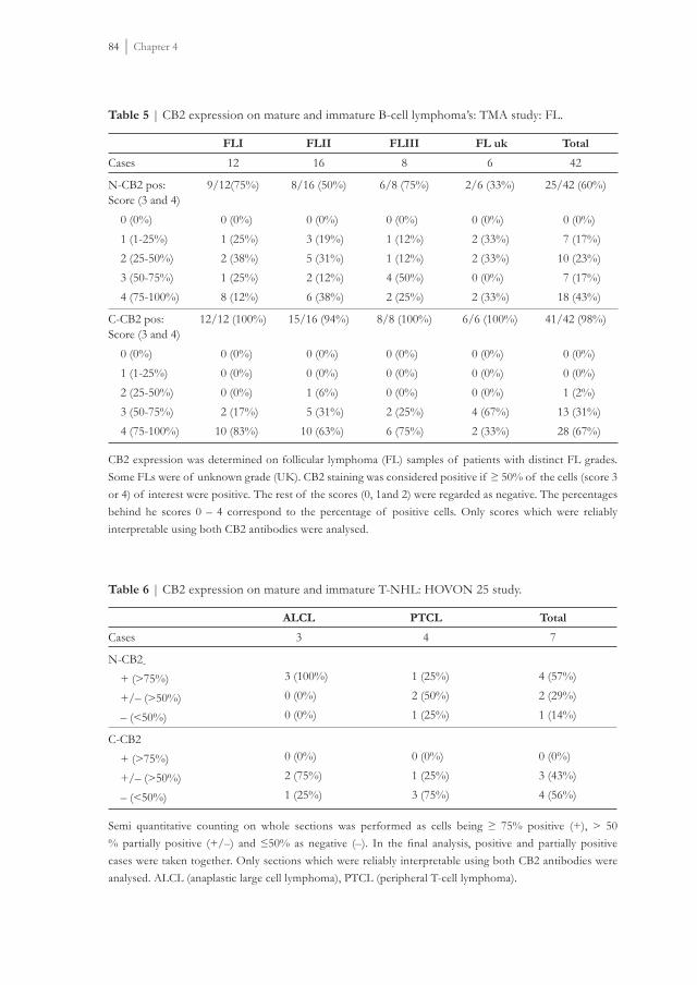
84 | Chapter 4
Table 5 | CB2 expression on mature and immature B-cell lymphoma’s: TMA study: FL.
FLI FLII FLIII FL uk Total
Cases 12 16 8 6 42
N-CB2 pos:Score (3 and 4)
9/12(75%) 8/16 (50%) 6/8 (75%) 2/6 (33%) 25/42 (60%)
0 (0%) 1 (1-25%) 2 (25-50%) 3 (50-75%) 4 (75-100%)
0 (0%) 1 (25%) 2 (38%) 1 (25%) 8 (12%)
0 (0%) 3 (19%) 5 (31%) 2 (12%) 6 (38%)
0 (0%)1 (12%)1 (12%)4 (50%)2 (25%)
0 (0%)2 (33%)2 (33%)0 (0%)2 (33%)
0 (0%) 7 (17%)10 (23%) 7 (17%)18 (43%)
C-CB2 pos:Score (3 and 4)
12/12 (100%) 15/16 (94%) 8/8 (100%) 6/6 (100%) 41/42 (98%)
0 (0%) 1 (1-25%) 2 (25-50%) 3 (50-75%) 4 (75-100%)
0 (0%) 0 (0%) 0 (0%) 2 (17%)10 (83%)
0 (0%) 0 (0%) 1 (6%) 5 (31%)10 (63%)
0 (0%)0 (0%)0 (0%)2 (25%)6 (75%)
0 (0%)0 (0%)0 (0%)4 (67%)2 (33%)
0 (0%) 0 (0%) 1 (2%)13 (31%)28 (67%)
CB2 expression was determined on follicular lymphoma (FL) samples of patients with distinct FL grades. Some FLs were of unknown grade (UK). CB2 staining was considered positive if ≥ 50% of the cells (score 3 or 4) of interest were positive. The rest of the scores (0, 1and 2) were regarded as negative. The percentages behind he scores 0 – 4 correspond to the percentage of positive cells. Only scores which were reliably interpretable using both CB2 antibodies were analysed.
Table 6 | CB2 expression on mature and immature T-NHL: HOVON 25 study.
ALCL PTCL Total
Cases 3 4 7
N-CB2 + (>75%) +/– (>50%) – (<50%)
3 (100%)0 (0%)0 (0%)
1 (25%)2 (50%)1 (25%)
4 (57%)2 (29%)1 (14%)
C-CB2 + (>75%) +/– (>50%) – (<50%)
0 (0%)2 (75%)1 (25%)
0 (0%)1 (25%)3 (75%)
0 (0%)3 (43%)4 (56%)
Semi quantitative counting on whole sections was performed as cells being ≥ 75% positive (+), > 50 % partially positive (+/–) and ≤50% as negative (–). In the fi nal analysis, positive and partially positive cases were taken together. Only sections which were reliably interpretable using both CB2 antibodies were analysed. ALCL (anaplastic large cell lymphoma), PTCL (peripheral T-cell lymphoma).

Peripheral cannabinoid receptors on immune cells and NHL | 85
Table 7 | CB2 expression on mature and immature T-NHL B: TMA study.
ALCL PTCL-nos T-LBL Total
Cases 10 8 7 25
N-CB2 pos:Score (3 and 4)
8/10 (80%) 7/8 (88%) 6/7 (86%) 21/25 (84%)
0 (0%) 1 (1-25%) 2 (25-50%) 3 (50-75%) 4 (75-100%)
0 (0%)0 (0%)2 (20%)1 (10%)7 (70%)
0 (0%)1 (12%)0 (0%)0 (0%) 7 (88%)
0 (0%)0 (0%)1 (14%)1 (14%)5 (72%)
0 (0%) 1 (4%) 3 (12%) 2 (8%)19 (76%)
C-CB2 pos:Score (3 and 4)
10/10 (100%) 8/8 (100%) 6/7 (86%) 24/25 (96%)
0 (0%) 1 (1-25%) 2 (25-50%) 3 (50-75%) 4 (75-100%)
0 (0%)0 (0%)0 (0%)2 (20%)8 (80%)
0 (0%)0 (0%)0 (0%)1 (12%)7 (88%)
0 (0%)0 (0%)1 (14%)1 (14%)5 (72%)
0 (0%) 0 (0%) 1 (4%) 4 (16%)20 (80%)
CB2 staining was considered positive if ≥ 50% of the cells (score 3 or 4) of interest were positive. The rest of the scores (0, 1and 2) were regarded as negative. The percentages behind he scores 0 – 4 correspond to the percentage of positive cells. . Only scores which were reliably interpretable using both CB2 antibodies were analysed. ALCL (anaplastic large cell lymphoma), PTCL-nos (peripheral T-cell lymphoma not otherwise specifi ed), T-LBL (T-cell lymphoblastic lymphoma).
DISCUSSION
CB2 receptor expression in normal lymphoid tissue
We investigated the expression of peripheral cannabinoid receptors in normal human lymphoid tissues and their malignant counterparts using CB2 antibodies. Positive staining with an antibody directed against the C-terminus is indicative for inactive receptors since it only recognizes non-phosphorylated Ser-352 and consequently inactive CB211. We demonstrated previously12 that B-cells in the MZ and MGZ of secondary follicles express inactive CB2, since they stain positively with this antibody. In contrast, these cells hardly stained for the N-terminal specifi c anti-CB2. Moreover, B-cells present in the GCs of secondary follicles, areas considered to contain a high number of active B-cells, showed strong staining with the N-terminal specifi c CB2 antibody. The staining pattern with C- and N- terminal specifi c antibodies seems mutually exclusive. Based on the combined

86 | Chapter 4
staining pattern we have hypothesized that the antibody recognizing the N-terminus of the receptor probably represents the recognition of the active state of the receptor. The non-staining of the N-terminal antibody in the B-cell areas which stain with the C-terminal specifi c antibody and therefore considered to be inactive as demonstrated by Bouaboula et al.11, may be caused by the occurrence of conformational or affi nity changes leading to epitope inaccessibility. An alternative explanation may be that indeed the N-CB2 does recognize the epitope, but the amount of receptors in the cells in these areas is too low, because the cells in these areas are assumed to be in a more inactive state. This is consistent with the positive staining with N-CB2 in the active germinal centre area of the lymph node. This latter staining pattern may correspond with activated/Ser-352 phosphorylated CB2 receptors on GC B-cells. However, additional functional studies investigating the phosphorylation status of the receptor in lymphocytes in relation to the antigen specifi c activation are needed to confi rm this hypothesis. These data may also be of importance, for the interpretation of CB2 expression on other haematopoietic cells. Using the N-terminal specifi c antibody, only a few CB2 positive cells in the paracortex could be found in normal reactive lymphoid tissues. Using the pan T-cell marker CD3, double staining was detected using only the N-terminal CB2. Moreover, double staining could not be found using anti CD1, CD2, CD5 and CD8 with both CB2 antibodies. Sub-typing with CD4 was unsuccessful, probably due to technical reasons. Based on these fi ndings one can state, that co-staining with the N-terminal antibody may suggest that the few CB2 positive T-cells in the paracortex of the lymph nodes may represent T-lymphocytes expressing activated peripheral cannabinoid receptors. On most T-cells, no CB2 was detectable using both antibodies, however, this does not necessarily imply that there are no receptors present at all. It has been reported that mRNA levels in normal human leukocyte subsets is very low in T-cells8-10. Consequently the levels of receptor expression may be below detection. Another study implies cannabinoid receptor involvement in T-lymphocytes using radio-ligand binding assays18. In this study, investigating the inhibition of T-cell-dependent immune responses by cannabinoids, only the mRNA expression of CB2 was investigated and found on T-lymphocytes using a T-cell line (HPB-ALL cells). However, no protein analysis was carried out and the mechanisms of the involvement of cannabinoid receptors have still not been fully elucidated. Moreover, recent studies suggest that effects of cannabinoid ligand on T-cells are independent of the presently known CB1 and CB2 cannabinoid receptors19. They and others19-22 have suggested a third receptor and / or non-receptor mediated mechanism which could explain these effects. We have studied CB2 expression

Peripheral cannabinoid receptors on immune cells and NHL | 87
on the NSB, HUT 102 and HPB-ALL T-cell lines and have found only minimal CB2 expression on HPB-ALL T-cell line (data not shown). To investigate protein expression on normal T-lymphocytes, we have analyzed two tonsils of healthy children and have carried out dual and triple fl owcytometry using T-cell specifi c antibodies and CB2. We were unable to detect CB2 protein using the N-terminal specifi c antibody (data not shown). This fi nding is confi rmed by dual staining immunohistochemistry experiments in which we could only detect sporadic double staining on T-cells. In the tumor cases the possibility remains that there is some unspecifi c staining. However, we have therefore only accepted positive staining when a clear distinctive cytoplasmatic or membrane staining was detected.
Expression of CB2 on follicular dendritic cells and a subset of macrophages
We observed a high number of cells which stain with the N-terminal specifi c CB2 antibody on several other smaller subsets of cells in lymph nodes, i.e. on follicular CD23+ dendritic cells and a sub population of CD68+ macrophages. It is possible that expression of activated CB2 on these cells also points to a particular activation status of the cells, corresponding with proliferation, differentiation or a role in immune modulation.
Expression of CB2 receptors in B-NHL and T-NHL
High levels of active CB2 on DLBCL was expected in a certain percentage of cases, since several studies showed that a large fraction of DLBCLs exhibit a GC B-cell phenotype. However, our previous fi ndings that normal B-cells in primary follicles or in the mantle zone and marginal zone of secondary follicles mainly stain with anti-C-terminal CB2, which would suggest that B-NHL of the FL-, MZ- or MGZ-type would stain positive with the C-terminal specifi c antibody and not with the N-terminal specifi c one. Although the C-terminal specifi c anti-CB2 identifi ed more CB2 positive B-cells in most tumours, the N-terminal specifi c antibody stained positive in most of the B-NHL cases as well. A similar discrepancy was also demonstrated in human acute myeloid leukaemia’s (AML). CD34 positive normal bone-marrow samples lack CB2 protein expression as determined with the N-terminal specifi c antibody, whereas AML blasts frequently show high expression of the CB2 receptor16. Moreover, a functional role of CB2 was demonstrated in AML. A block of neutrophilic differentiation was observed upon stimulation with synthetic cannabinoid ligands, using in vitro AML models16. In contrast to normal T-cells, we have found frequent CB2 expression on T-NHL, especially when using the N-terminal specifi c antibody. One explanation for this fi nding

88 | Chapter 4
could be, that T-NHL arises from the small, hard to detect fraction of normal CB2 expressing T-cells. However, since we have found CB2 expression on every T-NHL type that we studied, i.e. ALCL, PTCL as well as T-LBL, this explanation seems unlikely. Besides the fact that we found high CB2 levels on T-cell lymphoma’s, it was also striking that both CB2 antibodies frequently stained positive on these tissues. The fact that anti-CB2 directed to the non-phosphorylated C-terminal part of this receptor only stained positive in normal tissues when the N-terminal specifi c antibody does not, further underlines that T-cell lymphoma’s as well as B-cell lymphoma’s probably express an aberrantly functioning CB2 receptor. We conclude that peripheral cannabinoid receptor expression and activation status on B-NHL, T-NHL and AML differs from that observed on their normal counterparts. CB2 may be aberrantly expressed and may be involved in tumour development.

Peripheral cannabinoid receptors on immune cells and NHL | 89
REFERENCES
1. Matsuda LA, Lolait SJ, Brownstein MJ, Young AC, Bonner TI. Structure of a cannabinoid receptor and functional expression of the cloned cDNA. Nature 1990;346:561-564.
2. Matsuda LA, Bonner TI, and Lolait SJ. Localization of cannabinoid receptor mRNA in rat brain. J Comp Neurol 1993; 327:535-550.
3. Herkenham, M. Characterization and localization of cannabinoid receptors in brain: an in vitro technique using slide-mounted tissue sections. NIDA Res Monogr 1991;112:129145.
4. Kaminski NE, AboodME, KesslerFK, Martin BR and Schatz AR. Identifi cation of a functionally relevant cannabinoid receptor on mouse spleen cells that is involved in cannabinoid-mediated immune modulation. Mol Pharmacol 1992;42:736-742.
5. Twitchell W, Brown S and Mackie K. Cannabinoids inhibit N- and P/Q-type calcium channels in cultured rat hippocampal neurons. J Neurophysiol 1997;78:43-50.
6. Farquhar-Smith WP, Egertova M, Bradbury EJ, McMahon SB, Rice AS and Elphick MR. Cannabinoid CB(1) receptor expression in rat spinal cord. Mol Cell Neurosci 2000;15:510-521.
7. Munro S, Thomas KL and M. Abu-Shaar M. Molecular characterization of a peripheral receptor for cannabinoids. Nature 1993;365:61-65.
8. Bouaboula M, Rinaldi M, Carayon P, Carillon M, Delpech B and Shire D. Cannabinoid-receptor expression in human leukocytes. Eur J Biochem 1993 214:173-180.
9. Galiegue S, Mary S, Marchand J, Dussossoy D, Carriere D, Carayon P et al. Expression of central and peripheral cannabinoid receptors in human immune tissues and leukocyte subpopulations. Eur J Biochem 1995; 232:54-61.
10. Carayon P, Marchand J, Dussossoy D, Derocq JM, Jbilo O,. Bord A et al. Modulation and functional involvement of CB2 peripheral cannabinoid receptors during B-cell differentiation. Blood 1998;92:3605-3615.
11. Bouaboula M, Dussossoy D and Casellas. Regulation of peripheral cannabinoid receptor CB2 phosphorylation by the inverse agonist SR 144528. Implications for receptor biological responses. J Biol Chem 1997;274:20397-20405.
12. Rayman N, Lam KH, Laman JD, Simons PJ, Lowenberg B, Sonneveld P and Delwel R. Distinct expression profi les of the peripheral cannabinoid receptor in lymphoid tissues depending on receptor activation status. J Immunol 2004;172:2111-2117.
13. Buckley NE, McCoy EKL, Mezey M, Bonner T, Zimmer A, Felder CC et al. Immunomodulation by cannabinoids is absent in mice defi cient for the cannabinoid CB(2) receptor. Eur J Pharmacol 2000;396:141-149.
14. Flygare J, Gustafsson K, Kimby E, Christensson B and Sander B. Cannabinoid receptor ligands mediate growth inhibition and cell death in mantle cell lymphoma. FEBS Lett 2005;579:6885-6889.
15. McKallip RJ, Lombard C, Fisher M, Martin BR, Ryu S, Grant S et al. Targeting CB2 cannabinoid receptors as a novel therapy to treat malignant lymphoblastic disease. Blood 2002;100:627-634.
16. Jorda MA, Rayman N, Valk P, De Wee E and Delwel R. Identifi cation, characterization, and function of a novel oncogene: the peripheral cannabinoid receptor Cb2. Ann N Y Acad Sci 2003;996:10-16.
17. Doorduijn JK, van der Holt B, van Imhoff GW, van der Hem KG, Kramer MH, van Oers MH, et al. CHOP compared with CHOP plus granulocyte colony-stimulating factor in elderly patients with aggressive non-Hodgkin’s lymphoma. J Clin Oncol 2003;21:3041-3050.

90 | Chapter 4
18. Schatz AR, Lee M, Condie RB, Pulaski JT and Kaminski NE. Toxicology and applied Pharmacology 1997;142:278-287.
19. Rao GK, Kaminski N.E. Cannabinoid mediated elevation of intracellular calcium: a structure-activity relationship. J. Pharmacology and Exp.Ther 2006;317: 820-829.
20. Wiley JL, Martin BR. Cannabinoid pharmacology: implications for additional cannabinoid receptor subypes.Chemistry and Physics of Lipids 2002;121: 57-63.
21. Mo FM, Offertaler L, Kunos G. Atypical cannabinoid stimulates endothelial cell migration via a Gi/G-coupled receptor distinct from CB1, CB2 or EDG-1.Exp. J. of Pharmacology 2004; 489: 21-27.
22. Breivogel CS, Griffi n G, Di Marzo V, Martin B.R. Evidence for a new G protein-coupled cannabinoid receptor. Mol Pharmacol 2001;60:155-163.

Chapter 5
Prognostic relevance of immunohistochemical sub classifi cation of Diffuse Large B-cell Lymphoma in two prospective phase III clinical trials
Nazik Rayman1, King H. Lam2, Bronno van der Holt3, Clara Koss1, Dennis Veldhuizen4, Leo M. Budel6, Andries H. Mulder7, Leo F. Verdonck8, Ruud Delwel1, Daphne de Jong4, Gustaaf W. van Imhoff5 and Pieter Sonneveld1
1Department of Hematology, Erasmus Medical Center Rotterdam; 2Department of Pathology, Erasmus Medical Center Rotterdam; 3Department of Trials & Statistics, HOVON Data Center, Erasmus Medical Center, Rotterdam; 4Department of Pathology, Netherlands Cancer Institute Amsterdam; 5Department of Hematology, University Medical Center Groningen; 6Department of Pathology, Maasstad Hospital Rotterdam; 7Department of Pathology, Rijnstate Hospital Arnhem; 8Department of Internal Medicine, Isala Clinics Zwolle
Clinical Lymphoma, Myeloma & Leukemia 2011; 11: 23-32

92 | Chapter 5
ABSTRACT
Purpose: Until now molecular biological techniques are not easily used in daily clinical practice to stratify patients for therapeutic purposes. Therefore, we have investigated the prognostic relevance of the immunohistochemical (IHC) Germinal Center B-cell (GCB) versus (vs.) non-GCB diffuse large B-cell lymphoma (DLBCL) subtypes. Patients and methods: We have analyzed tumor samples from patients treated in two prospective, phase III multi-center trials, i.e. HOVON 25 (patients ≥ 65 years, n=153) and HOVON 26 (patients < 65 years, n=144) using whole sections (WS) or tissue micro array (TMA). CD10, BCL6 and MUM1 were applied in a specifi c IHC algorithm. The impact on clinical outcome using WS or TMA and variations in cut-off levels of these markers was also investigated.Results: The GCB subtype was not associated with a better OS in either trial. Small differences were observed in the HOVON 25 trial between techniques, with TMA showing a better outcome for GCB than did WS. Variation of cut off levels in the specifi c algorithm did not improve the prediction of clinical outcome. Conclusion: We did not observe a consistent predictive power of the GCB and non-GCB classifi cation by IHC in this large series of DLBCL patients treated with CHOP. This underscores the need to determine the biological variation and the standardization of the protein expression levels and to further study the relevance of prognostic IHC classifi cations, preferably in phase III clinical trials.
Keywords: DLBCL, phase III trial, germinal center B-cell, immunohistochemistry, prognosis

Clinical relevance of DLBCL classifi cation | 93
INTRODUCTION
Diffuse large B-cell lymphoma (DLBCL) as described by the World Health Organization (WHO) 2008 classifi cation is the most frequent lymphoma subtype of the aggressive Non-Hodgkin’s lymphomas (NHL)1. The addition of rituximab to CHOP (cyclophosphamide, doxorubicin, vincristine and prednisone) containing regimens (R-CHOP) highly improves outcome. However, survival of both elderly and young patients with NHL is still unsatisfactory, making improvement of current therapeutic strategies necessary2-12. One of the most promising strategies may be tailoring the therapy to the clinical and biological factors of patients and disease, which infl uence outcome. To this end, the International Prognostic Index (IPI) up till now has been the most powerful tool for predicting clinical outcome of patients with DLBCL. However, the IPI refl ects a mixture of underlying biologic differences and thus has its limits with respect to the aim of personalized therapy. Therefore, novel biological prognostic markers refl ecting the heterogeneous biology of DLBCL have been investigated with the ultimate goal to fi nd DLBCL subtypes with different prognosis13. Gene expression profi ling (GEP) using oligonucleotide arrays indeed confi rmed the existence of subgroups of patients, treated either with CHOP or CHOP-like regimens14-19. Most of these patients can be divided in having either a favorable germinal center B-cell (GCB) or an unfavorable activated B-cell (ABC) phenotype of DLBCL. This fi nding is supported by other investigators such as Tagawa et al. showing a correlation between cytogenetic alterations and GCB versus ABC expression subgroups, reiterating this biological difference20. However, up to now molecular techniques such as gene expression profi ling (GEP), cytogenetics, comparative genomic hybridization (CGH) and genome wide association studies (GWAS) are diffi cult to apply in clinical practice to stratify patients for therapeutic purposes. Because of its wide availability, histological sub classifi cation systems using immunohistochemistry (IHC) have been considered to be more feasible. Hans et al. published one approach to determine prognostic entities within the group of DLBCL patients treated with CHOP-like chemotherapy21. A panel of 3 antibodies against CD10, BCL6 and MUM1 were applied in a specifi c algorithm in a tissue micro array (TMA). Using this approach, these investigators could identify patients with a favorable GCB or a non-favorable non-GCB (ABC) phenotype. These IHC results correlated with the results obtained with GEP21. Using the same algorithm, other studies have reported comparable predictive power of IHC subgroups in DLBCL patients treated with anthracyclin-containing regimens. In these studies, patients with high IPI scores treated with high-dose chemotherapy and autologous stem cell transplantation

94 | Chapter 5
were analyzed as well22-24. However, other studies using similar algorithms have reported no predictive value, also in patients treated with R-CHOP25-27. In this paper, we analyzed the prognostic relevance of the IHC GCB versus non-GCB DLBCL subtypes in two prospective, randomized phase 3 multi-center studies. These studies were performed in 2 large cohorts of elderly and young patients, uniformly treated with CHOP. We also have made an attempt to optimize the algorithm as described by Hans et al., by making variations in the cut-off points of the algorithm21. The effect of the choice of IHC assays, i.e., WS or TMA on this algorithm was investigated as well. Also BCL2 expression has been analyzed since this marker has been described as being useful to sub classify the non-GCB group21,24,28.
PATIENTS, MATERIALS AND METHODS
Patients
We studied tissue samples of DLBCL patients who participated in 2 prospective randomized phase 3 clinical trials. All patients received an anthracyclin containing chemo-therapy regimen (CHOP). All diagnoses of DLBCL were confi rmed by central pathology review using the WHO 2001 criteria. In patient cohort 1, samples from patients who participated in the HOVON 25 (HO25) trial (n=389) were collected from the contributing centers using the HOVON (Dutch-Belgian Hemato-Oncology Cooperative Group) database (virtual tissue bank). Inclusion criteria were age ≥ 65 years, previously untreated intermediate or high-grade DLBCL according to the Working Formulation or the WHO 2001 classifi cation and Ann Arbor stage II, III or IV. Patients were included in this trial between August 1994 and September 20019. Suffi cient material was available for IHC analysis of 134 (34%) patients by TMA (HO25 TMA) and 153 (39%) patients using WS (HO25 WS). There were 73 patients (19%) of whom tissue was included in both HO25 WS and HO25 TMA. In cohort 2, we included patients who participated in the HO26 trial (n=513). Inclusion criteria were previously untreated aggressive NHL according to the intermediate-or high-grade Working Formulation, age < 65 years, and Ann Arbor stage II, III or IV. Patients were included between November 1994 and February 200411. Suffi cient material was available of 144 (28%) patients for IHC analysis on TMA. All studies were conducted according to regulations of the Dutch health authorities and in accordance with the

Clinical relevance of DLBCL classifi cation | 95
Declaration of Helsinki. The trials have been registered as ISRCTN26340837 (HO25) and ISRCTN11397785 (HO26), see e.g. www.controlled-trials.com.
Materials and methods
Patient samples (paraffi n-embedded tissue blocks) were collected from the participating centers. The TMA’s were constructed at the Department of Pathology of The Netherlands Cancer Institute (Amsterdam). H-E sections of paraffi n-embedded, formalin-fi xed tissue were used to defi ne the diagnostic areas. From each case 2 representative 0.6 mm cores were obtained and inserted adjacently into a recipient paraffi n block using a tissue arrayer (Beecher Instruments, Silver Spring, MD, USA). All IHC procedures and analyses were performed at the Department of Pathology at Erasmus Medical Center Rotterdam, according to standard IHC procedures. Antigen retrieval was performed by pre treating sections in a microwave oven (Salm and Kipp, Breukelen, The Netherlands). The primary antibodies CD10, BCL6, MUM1 and BCL2 were obtained from Novocastra and DAKO respectively. The secondary antibodies and conjugates (goat-anti-rabbit horse-radish peroxidase (HRP) and streptavidin biotin complex-HRP) were purchased from Labvision. Negative controls (omission of primary antibody) of all examined tissues were included. Stains were scored on a 5-point scale: 0=negative (all cells negative), 1= 1 – 25% cells positive, 2= 26 – 50% cells positive, 3= 51 – 75% cells positive and 4= 76 – 100% cells positive. Of the cores or whole sections with variable outcome, the mean expression was taken and reduced to the highest score. Cases were assigned to the GCB or non-GCB group, according the Hans algorithm21 in which positivity of the antibodies was defi ned as a score of > 2. Cases that were negative for all markers were assigned to the ABC group. Different cut-off points were taken for the variation in the assessment of positivity for each antibody resulting in different distributions of the GCB vs. non-GCB sub groups (GCB0, GCB1 and GCB2). This was performed in order to defi ne an optimal algorithm (i.e. ≥ 2 or 3 for CD10, ≥ 2 for BCL6 and ≥ 2 or ≥ 4 for MUM1). All samples were independently evaluated in a semi-quantitative way using the above-mentioned scoring system by two investigators (NR and KHL). Discordant scores were resolved by joint review on a multi-headed microscope.
Statistical analysis
All endpoints were defi ned according to the updated Cheson criteria 200729. Progression-free survival (PFS) was defi ned as the time from registration in the trial until lymphoma progression or death as a result of any cause. Event-free survival (EFS) was measured

96 | Chapter 5
from registration in the trial to any treatment failure including disease progression, or discontinuation of treatment for any reason. Overall survival (OS) was calculated from registration until death. Patients were censored at the date of last contact if they were still alive. The Kaplan-Meier method was used to estimate the survival endpoints PFS, EFS and OS30. Survival analysis was performed using univariate Cox regression to see whether there was a difference in survival between subgroups31. Hazard ratios (HRs) and 95% confi dence intervals (CIs) were determined for all three survival endpoints. Kaplan-Meier curves were generated to illustrate differences in PFS, EFS and OS between GCB and non-GCB patients, and were compared using the log-rank test (data not shown)32. Multivariate Cox regression analysis with GCB, BCL2 and a GCB х BCL2-interaction term was used to evaluate whether analyses of BCL2 within the subgroups of GCB and non-GCB patients were appropriate33,34. All reported P values were two-sided and, in view of the exploratory nature of these analyses, were calculated without adjustment for multiple testing. P-values ≤ 0.05 were considered statistically signifi cant.
RESULTS
Clinical characteristics and treatment outcome of patients
The treatment outcome for the two trials is summarized in Table 1. The 5-year PFS was 22% in trial 1 (HO25) using WS, 24% using TMA and 42% in trial 2. The 5-year EFS was 20%, 23% and 38% respectively. The 5-year-OS was 26%, 29% and 58% respectively. The main differences in the clinical characteristics of the DLBCL patients in the HO25 and HO26 trials were observed for median age, stage of the disease and the IPI (Supplemental Table 1). There were no major differences in patient characteristics between the IHC GCB and non-GCB sub types in both trials (Supplemental Table 2). We compared the initial patient characteristics of the DLBCL cases in the HO25 study with those included for IHC (HO25 WS and HO25 TMA) and found no signifi cant differences. The prognostic signifi cance in OS for the IPI in this initial study was high (P < 0.001)9. In the HO26 study, no major differences in patient characteristics were present either between the cases in our sample and the initial study population11. In this original HO26 study the predictive value of the IPI was weak (P=0.02) (data not shown).

Clinical relevance of DLBCL classifi cation | 97
Immunohistochemistry
CD10, BCL6 and MUM1 antibodies were used to assign patients into the GCB or the non-GCB DLBCL groups. Insuffi cient data or core loss resulted in the exclusion of 11 (WS) and 23 (TMA) patients in trial 1 and 2 patients (TMA) in trial 2. CD10, BCL6 and MUM1 positivity (score ≥ 2) varied signifi cantly (P ≤ 0.01) in the two trials. The distribution between the GCB and the non-GCB subtypes as determined by the Hans algorithm (cut-off point ≥ 2 for all markers) was not different between the patient groups from the 2 trials (P=0.10) (Supplemental Table 3). There were no discrepancies on the level of assigning each case to GCB or non-GCB in the TMA’s, because the discrepancies in scoring resulting in this assignment were resolved on the level of the individual markers. For HO25 WS, the assignment of each case was done by NR en KHL individually. This has resulted in the same overall percentage for both investigators as shown in Table 1. On case level, 52 out of 153 cases (34,6%) were assigned differently. All discrepancies were subsequently resolved at the individual marker level using a double-headed microscope and reassigned. This has not changed the overall percentage signifi cantly as shown in Table 1.
Prediction of the Hans algorithm for clinical outcome
The prognostic impact of GCB, the single markers CD10, BCL6, MUM1 and BCL2, as well as the IPI, on PFS, EFS and OS is shown in Table 1 and Figure 1-3. In both trials no statistically signifi cant differences were found between GCB versus non-GCB subtype. Only when using the TMA technique in the elderly trial (HO25), a small improvement of OS (P=0.03) was observed in the GCB subtype, which remained signifi cant in a multivariate analysis when adjusted for IPI (HR=0.52, 95% CI 0.30 – 0.90, P=0.01). Higher CD10 positivity was not associated with better or worse clinical outcome in either trial. A signifi cant positive impact was observed for BCL6 expression in the elderly trial (HO25 TMA; PFS, EFS and OS P < 0.01). BCL6 remained statistically signifi cant when adjusted for IPI in multivariate analyses: (for PFS, HR=0.66, 95% CI=0.50 – 0.88, P=0.001; for EFS, HR=0.66, 95% CI=0.50-0.87, P < 0.001); and for OS, HR=0.61, 95% CI=0.44-0.83, P < 0.001). No signifi cance was observed using WS, neither studying trial 2. MUM1 expression did not correlate with clinical outcome in any study. BCL2 expression was correlated with a worse PFS and EFS in trial 2 only (Table 1).

98 | Chapter 5
Table 1 | PFS, EFS and OS of the different patient groups.
PFS HR 95% CI EFS HR 95% CI OS HR 95% CI
Trial 1 HO25 WS
GCB 1.03 .71-1.49 1.11 .77-1.61 .98 .67-1.43CD10 1.05 .94-1.18 1.06 .95-1.18 1.02 .91-1.15BCL6 1.01 .90-1.14 1.02 .91-1.14 .99 .88-1.12MUM1 .96 .86-1.08 .96 .85-1.07 .95 .84-1.07BCL2 1.06 .94-1.19 1.04 .92-1.17 1.05 .93-1.19IPI 1.28 1.06-1.54a 1.27 1.06-1.52a 1.25 1.03-1.51a
Trial 1 HO25 TMA
GCB .64 .39-1.06 .63 .38-1.04 .56 .33-.96a
CD10 .94 .81-1.10 .95 .81-1.11 .93 .79-1.09BCL6 .70 .54-.91b .71 .55-.92b .65 .49-. 87b
MUM1 .90 .73-1.11 .89 .72-1.10 .87 .70-1.08BCL2 .91 .80-1.05 .92 .80-1.05 .91 .79-1.05IPI 1.19 .97-1.46 1.20 .98-1.47 1.14 .93-1.40
Trial 2 HO26 TMA
GCB 1.20 .77-1.85 1.02 .66-1.56 1.21 .73-2.01CD10 1.13 .97-1.30 1.06 .92-1.23 1.11 .94-1.31BCL6 1.07 .91-1.27 1.01 .86-1.19 1.06 .87-1.28MUM1 0.92 .78-1.09 .92 .78-1.09 .99 .82-1.20BCL2 1.19 1.04-1.36a 1.18 1.03-1.35a 1.13 .96-1.33IPI 1.32 .98-1.78 1.34 1.00-1.78 1.38 .98-1.94
HR indicates hazard ratio; and CI, confi dence interval; aP < 0.05 bP < 0.01
Clinical outcome using either WS or TMA
We next analyzed whether the use of either WS or TMA infl uenced the sub division in GCB vs. non-GCB groups in trial 1. Tissue of 73 patients of the HO25 trial was included in both TMA and WS analysis. In these patients, discordant scores were identifi ed for the markers CD10, BCL6, MUM1 and BCL2. Discordance was defi ned as > 25% difference in score system. We observed a large variation in scoring regarding BCL6, MUM1 and BCL2, resulting in a larger proportion of the GCB sub type in WS and non-GCB sub type in TMA setting. From the 73 patients, 58 could be assigned for sub classifi cation. Twenty discrepant cases were found using the Hans algorithm. Sixteen cases were classifi ed as GCB and 4 as non-GCB in the WS setting. The reverse holds true for TMA (Table 2A and B).

Clinical relevance of DLBCL classifi cation | 99
Figure 1 |
Figure 2 |

100 | Chapter 5
Figure 3 |
Table 2A | Difference in scores using WS compared to TMA in HO25 trial.
Marker Score% positive
00
11-25
226-50
351-75
476-100
Number discrepant (%)
CD10 (n=60)WS 2 0 1 2 6
11 (18)TMA 6 3 0 1 1
BCL6 (n=53)WS 2 0 7 12 10
31 (53)TMA 26 0 3 1 1
MUM1 (n=57)WS 1 0 5 11 17
34 (60)TMA 22 8 3 0 1
BCL2 (n=50)WS 3 1 4 5 12
25 (50)TMA 13 4 2 1 5

Clinical relevance of DLBCL classifi cation | 101
Table 2B | Distribution of discrepant GCB and ABC sub groups using WS and TMA (HO25 trial).
Discrepant case nr.
WS CD10 score
WSBCL6 score
WSMUM1 score
WSSub type
TMACD10 score
TMABCL6 score
TMAMUM1 score
TMASub type
1 4 0 1 GCB 1 0 0 ABC2 3 3 4 GCB 1 0 0 NGCB3 4 4 1 GCB 0 0 0 ABC4 4 1 2 GCB 0 0 0 ABC5 2 3 3 GCB 0 0 0 ABC6 0 4 1 GCB 0 0 0 ABC7 1 3 0 GCB 0 0 0 ABC8 0 2 0 GCB 0 0 4 ABC9 0 3 4 ABC 0 2 0 GCB10 4 3 4 GCB 1 0 0 ABC11 0 1 4 ABC 0 2 0 GCB12 0 3 0 GCB 0 0 0 ABC13 3 3 0 GCB 0 0 0 ABC14 0 3 0 GCB 0 0 0 ABC15 4 3 2 GCB 0 0 0 ABC16 0 0 3 ABC 4 2 1 GCB17 4 0 0 GCB 0 0 0 ABC18 0 0 1 ABC 3 0 0 GCB19 2 3 4 GCB 1 0 0 ABC20 0 2 1 GCB 1 1 0 ABC
GCB (germinal center B-cell); ABC (activated B-cell); WS (whole sections); TMA (tissue micro array).
Outcome prediction in the variations in cut-off points for the markers used in
the Hans algorithm
We next investigated whether variation in cut-off values of CD10, BCL6 and MUM1 expression resulted in better clinical outcome prediction. The defi nitions for the respective GCB sub types were as follows:
GCB0: CD10 ≥ 2, BCL6 ≥ 2 and MUM1 ≥ 2,GCB1: CD10 ≥ 3, BCL6 ≥ 2 and MUM1 ≥ 2,GCB2: CD10 ≥ 2, BCL6 ≥ 2 and MUM1 ≥ 4.
None of the variants resulted in a change of outcome prediction (Supplemental Table 4). We also applied more variations for BCL6 (scores ≥ 1, 3 and 4) since inter observer variations for this protein have been reported, again with no effect35 (data not

102 | Chapter 5
shown). A lower cut-off value for CD10 (≥ 1) had no impact either (data not shown). Given the above mentioned results, we did not perform statistical analysis for scores of individual markers other than 2.
The impact of BCL2 protein expression within GCB and non-GCB patients
As none of the Cox regression analyses showed a statistically signifi cant interaction between GCB and BCL2, subgroups analyses of BCL2 within GCB and non-GCB patients were not warranted. Therefore the best estimate of the hazard rate of BCL2 within these subgroups remains the overall estimate shown in Table 1, which implies an adverse effect of BCL2 positivity on PFS and EFS in the HO26 trial, but not in de HO25 trial.
Table 3 | Variation in algorithm to defi ne GCB and non-GCB DLBCL sub types.
PFS HR 95% CI EFS HR 95% CI OS HR 95% CI
Trial 1 HO25 WS GCB0 1.03 .71-1.49 1.11 .77-1.61 .98 .67-1.43GCB1 .94 .64-1.39 1.01 .69-1.48 .94 .64-1.40GCB2 1.08 .75-1.54 1.15 .81-1.64 1.01 .70-1.46
Trial 1 HO25 TMA
GCB0 .64 .39-1.06 .63 .38-1.04 .56 .33-.96a
GCB1 .69 .41-1.15 .68 .41-1.13 .66 .38-1.12GCB2 .64 .39-1.06 .63 .38-1.04 .56 .33-.96a
Trial 2 HO26 TMA
GCB0 1.20 .77-1.85 1.02 .66-1.56 1.21 .73-2.01GCB1 1.17 .75-1.83 1.01 .65-1.56 1.11 .66-1.88GCB2 1.14 .75-1.72 .92 .61-1.38 1.07 .66-1.74
GCB0=CD10, BCL6 and MUM1 ≥ 2; GCB1=CD10 ≥ 3, BCL6 and MUM1 ≥ 2; GCB2=CD10 and BCL6 ≥ 2, MUM1 ≥ 4, aP < 0.05.
DISCUSSION
We could not confi rm the prognostic relevance of the IHC GCB or non-GCB sub classifi cation in these 2 large cohorts of uniformly treated patients with DLBCL with a follow-up of 8 (HO25) or 7 years (HO26). Age difference is not a likely explanation for

Clinical relevance of DLBCL classifi cation | 103
this result, since the participants in the studies have different median ages. Moreover, the failure to demonstrate prognostic signifi cance of the GCB and non-GCB sub classifi cation did not result from the distribution of clinical characteristics, because the treatment (arm) was not signifi cantly different for the GCB or non-GCB subtype. Although we could not fi nd a predictive power of the Hans model in our study, others have corroborated this model as being predictive. Therefore we have used different cut-off levels in attributing classifi ers. More specifi cally, special attention was paid in varying the CD10 cut-off levels, because in most studies the positivity of CD10 is a prominent denominator of whether the case will be classifi ed as GCB or non-GCB. However, the variations did not result in improved or altered outcome prediction. This is in accordance with other investigators using a dichotomous system of scoring CD1022,35. Also, variation in the cut-off level of MUM1 or BCL6 (data not shown except for score 2) did not improve the predictive power22. The results of our study indicate that our fi ndings regarding the prognostic value of the Hans algorithm are most probably not caused by variations in the cut-off levels of the individual markers CD10, BCL6 and MUM1.Moreover, our cut-off value of 1 or partly 2 more or less corresponds with the cut-off value of 30% of the study of Hans and other studies reporting a predictive or a non-predictive effect22,25. The discrepancies in predictive value of the Hans model in the various studies are probably better explained by the variation of tissue processing procedures (paraffi n blocks originated from different pathology laboratories) prior to the construction of the TMAs and IHC procedures may have caused the above-mentioned variations. As reported earlier, IHC analysis in TMA is comparable with WS in NHL36,37. Indeed, we found no consistent differences in clinical outcome of the IHC GCB vs. non-GCB sub classifi cation between both techniques. However, for the individual markers CD10, BCL6 and MUM1, a trend towards lower positive scores was found in the HO25 TMA setting. We have therefore searched for discrepancies of more than 2 points in scoring between the 2 cores in the HO25 TMA. The number of discrepant cases (2 cores) for BCL2, MUM1 and CD10 were 3 out of 144 cases and for BCL6, 4 out of 144 cases (data not shown). The fact that the TMAs appear not to be representative of whole sections may be better explained by variance in the immunogenecity of the different areas of the tumour which were sampled. This may partly explain the relative high percentage of ABC sub type in the HO25 TMA as compared to the HO25 WS. In our clinical trials, the percentage of GCB classifi ed cases is slightly less than what has been reported in literature as lower

104 | Chapter 5
levels, ranging from 42 to 58% compared to 26 – 39% in our trials21-23,25. To analyze the difference in the distribution of GCB and non-GCB we have varied the cut-off levels in the scoring system. Indeed with the cut-off level 1 in the scoring of CD10 we have found a distribution that is comparable with other publications namely 50% (±10). However, even in these cases we could not detect a predictive effect (data not shown). Therefore, (semi-) quantitative expression analysis of each marker that contributes to the GCB and non-GCB DLBCL sub groups as well as BCL2 was investigated. Moreover, variable results have been reported concerning the relative value of these markers38-49. We also did not obtain consistent results with respect to the prognostic relevance of CD10, BCL6 and BCL2 in our data set. A possible explanation of the difference in outcome for BCL6 and BCL2 between the HO25 and HO26 studies may be that the patient characteristics of the individual studies are different, especially when considering age. The IPI (in which age is an important denominator) was only predictive for outcome in the HO25 WS study. This can be explained by the fact that no major differences were found in our sample and the initial study population of the HO25 study. Moreover, the IPI was weakly predictive in this initial study population. We do not have a plausible explanation for the discrepancy in predictive value of the IPI in the HO25 TMA sample. However, the GCB vs. non-GCB classifi cation has been described as a prognostic factor which is independent of the IPI score. Recently it was suggested that the huge variability in laboratory processing and staining techniques might ultimately explain the wide variation reported in the literature concerning the prognostic impact of these markers. These investigators reported highly variable results and very poor reproducibility in scoring for BCL6, MUM1, and BCL2, and to a lesser extent for CD1035. In this paper, IHC and scoring procedures of patient samples of the two clinical trials were performed in a central lab. However, sampling fi xation and paraffi n embedding of the specimens were performed in laboratories of local hospitals at which the patient presented at primary diagnosis. This may also have infl uenced the IHC results and is concordant with the reported concerns regarding the standardization of the IHC procedures and prognostic relevance of the investigated markers. It has recently been demonstrated that the addition of rituximab to the anthracyclin-based regimen does not eliminate the difference between clinical outcome between GCB and non-GCB sub types as determined by GEP. In keeping with the results based on CHOP regimens, controversial results are also reported in R-CHOP treated patients regarding the prognostic relevance of IHC based cell of origin algorithms. Possibly, with

Clinical relevance of DLBCL classifi cation | 105
ongoing research, techniques such as GEP, CGH and GWAS may become an alternative to determine the distinct biological DLBCL entities in clinical practice50. However, for the moment further development of immunohistochemical methods seems to be the most practical in terms of feasible usage in daily clinical practice when stratifying patients for therapeutic purposes. Ongoing immunohistochemical research using novel markers is warranted to dissect favorable and non-favorable subgroups of patients. However, when using these markers, changes to the Hans algorithm most probably have to be made because markers associated with a GCB profi le such as the HGAL protein did not fi nd a complete correlation with the GCB and non-GCB division as defi ned by Hans et al.51. In conclusion, we could not demonstrate a consistent predictive power of clinical outcome of the IHC GCB and non-GCB classifi cation in these large cohorts of prospectively treated and well documented DLBCL patients. This underscores the need to further determine the biological variation of the protein expression levels, to improve and standardize the detection of protein expression levels and to further study the relevance of prognostic classifi cations based on IHC algorithms using other markers in large cohorts of patients, preferably treated in prospective clinical trials.
ACKNOWLEDGEMENTS
We would like to thank the clinical investigators of the HOVON 25 and HOVON 26 trials for contributing patients and the patients for participating in the trials.This work was supported by a grant from the Dutch Cancer Society (Koningin Wilhelmina Fonds) and the Erasmus MC Revolving Fund.

106 | Chapter 5
Supplemental Table 1 | Patients characteristics in the trials.
Total Trial 1 H.25 WS 153 Trial 1 H.25 TMA 134 Trial 2 H.26 TMA 144
Age (P<0.001)
Median 73 72 50Range 65-85 65-90
Sex (%) (P=0.45)
Male 82 (54) 75 (56) 70 (49)Female 71 (46) 59 (44) 74 (51)
Stage (Ann Arbor) (%) (P<0.001)
II 45 (29) 44 (33) 30 (21)III 34 (22) 25 (19) 62 (43)IV 74 (48) 65 (49) 52 (36)
WHO performance status (%) (P=0.06)
0 64 (42) 61 (46) 74 (53)1 60 (39) 51 (39) 53 (38)2 22 (14) 15 (11) 9 (6)3 6 (4) 5 (4) 3 (2)Missing * 1 2 5
LDH (%) (P=0.06)
Normal 51 (34) 53 (40) 68 (47)Elevated 100 (66) 80 (60) 76 (53)Missing * 2 1 –
# Extra nodal sites involved (P=0.4)
0-1 130 (85) 115 (86) 129 (90)≥ 2 23 (15) 19 (14) 15 (10)
IPI (%) (P<0.001)
Low 19 (13) 21 (16) 62 (45)Low-intermediate 48 (32) 45 (34) 62 (45)High-intermediate 51 (34) 41 (31) 14 (10)High 32 (21) 24 (18) 1 (1)Missing * 3 3 5
Bone marrow involvement (%) (P=0.33)
No 121(79) 102(76) 115(83)Yes 32(21) 32(24) 23(17)Missing * – - 6
Bulky disease (%) (P=0.96)
No 121 (79) 104 (78) 112 (78)Yes 32 (21) 30 (22) 31 (22)
Treatment arm (%)
8cycles CHOP 74 (48) 68 (51) 65 (45)8cycles CHOP + G-CSF 79 (52) 66 (49) –6 cycles dose intensive CHOP – – 79 (55)
*Patients with missing data have been excluded when calculating percentages.

Clinical relevance of DLBCL classifi cation | 107
Supplemental Table 2 | Patient characteristics according to GCB and ABC sub classifi cation.
Total H.25 WS ABC 87
H.25 WS GCB 55
H.25 TMA ABC 82
H.25 TMA GCB 29
H. 26 TMA ABC 97
H.26 TMA GCB 45
Age
Median 72 73 73 69 51 50Range 65-83 65-85 65-90 65-83 16-65 17-65
Sex (%)
Male 48 (55) 30 (55) 44 (54) 17 (59) 44 (45) 25 (56)Female 39 (45) 25 (45) 38 (46) 12 (41) 53 (55) 20 (44)
Stage (Ann Arbor) (%)
II 28 (32) 15 (27) 27 (33) 27 (33) 21 (22) 9 (20)III 18 (21) 15 (27) 16 (20) 16 (20) 47 (48) 14 (31)IV 41 (47) 25 (45) 39 (48) 39 (48) 29 (30) 22 (49)
WHO performance status (%)
0 38 (44) 24 (44) 34 (41) 13 (45) 52 (54) 21 (47)1 32 (37) 23 (42) 32 (39) 12 (41) 35 (36) 17 (38)2 14 (16) 6 (11) 11 (13) 2 (7) 5 (5) 4 (9)3 3 (3) 1 (2) 3 (4) 2 (7) 1 (1) 2 (4)Missing * – 1 (2) 2 (2) – 4 (4) 1 (2)
LDH (%)
Normal 26 (30) 22 (40) 32 (39) 13 (45) 43 (44) 24 (53)Elevated 61 (79) 31 (56) 50 (61) 15 (52) 54 (56) 21 (47)Missing * – 2 (4) – 1 (3) – –
# Extra nodal sites involved
0-1 74 (85) 48 (87) 72 (88) 24 (83) 91 (94) 37 (82)≥ 2 13 (15) 7 (13) 10 (12) 5 (17) 6 (6) 8 (18)
International Prognostic Index (%)
Low 11 (13) 8 (15) 14 (17) 5 (17) 40 (41) 21 (47)Low-intermediate 28 (32) 18 (33) 26 (32) 8 (28) 43 (44) 19 (42)High-intermediate 27 (31) 17 (31) 23 (28) 12 (41) 10 (10) 3 (7)High 21 (24) 9 (16) 17 (21) 3 (10) – 1 (2)Missing * – 3 (5) 2 (2) 1 (3) 4 (4) 1 (2)
Bone marrow involvement (%)
No 70 (80) 46 (84) 64 (78) 20 (69) 78 (80) 35 (78)Yes 17 (20) 9 (16) 18 (22) 9 (31) 14 (14) 9 (20)Missing * – – – – 5 (5) 1 (2)
Bulky disease (%)
No 72 (83) 41 (75) 65 (79) 22 (76) 80 (82) 30 (67)Yes 15 (17) 14 (25) 17 (21) 7 (24) 17 (18) 14 (31)Missing * 1 (2)
Allocated treatment arm (%)
Control 42 (48) 27 (49) 40 (49) 17 (59) 44 (45) 19 (42)Experimental 45 (52) 28 (51) 42 (51) 12 (41) 53 (55) 26 (58)
*Patients with missing data have been excluded when calculating percentages.

108 | Chapter 5
Supplemental Table 3 | IHC data of the DLBCL sub classifi cation, IPI score and used markers.
Total HO25 WS 153 HO25 TMA 34 HO26 TMA 144
Sub type (%) P=0.10
GCB (0) ABC Missing*
55 (39)87 (61)11
29 (26)82 (74)23
45 (32)97 (68) 2
CD10 P=0.01
0% 1-25% 26-50% 51-75% 76-100% Missing*
97 (67) 7 (5) 7 (5) 6 (4)27 (19) 9
54 (48)37 (33) 3 (3) 6 (5)13 (12)21
57 (40)49 (35) 6 (4)12 (8)18 (13) 2
BCL6 P < 0.001
0% 1-25% B
26-50% 51-75% 76-100% Missing*
46 (33)18 (13)17 (12)31 (22)27 (19)14
79 (69)21 (18)11 (10) 1 (1) 3 (3)19
24 (17)48 (34)30 (21)20 (14)18 (13) 4
MUM1 P < 0.001
0% 1-25% 26-50% 51-75% 76-100% Missing*
33 (24)16 (12)18 (13)23 (17)46 (34)17
68 (58)36 (31) 6 (5) 2 (2) 6 (5)16
21 (15)45 (31)31 (22)24 (17)22 (15) 1
BCL2 P < 0.001
0% 1-25% 26-50% 51-75% 76-100% Missing*
22 (17)20 (15)18 (14)14 (11)56 (43)23
34 (31)31 (28)14 (13) 7 (6)24 (22)24
48 (34)34 (24)16 (11)21 (15)21 (15) 4
IPI P < 0.001
Low Low-interm High-interm High Missing*
19 (13)48 (32)51 (34)32 (21) 3
21 (16)45 (34)41 (31)24 (18) 3
62 (45)62 (45)14 (10)1 (1) 5
* Patients with missing data have been excluded when calculating percentages; GCB (0)=CD10, BCL6 and MUM1 ≥ 2.

Clinical relevance of DLBCL classifi cation | 109
REFERENCES
1. Swerdlow SH, Campo E, Harris NL, et al. WHO Classifi cation of Tumours of Haematopoietic and Lymphoid Tissues. 4th ed. Lyon: 2008.
2. Coiffi er B, Lepage E, Briere J, et al. CHOP chemotherapy plus rituximab compared with CHOP alone in elderly patients with diffuse large-B-cell lymphoma. N Engl J Med, 2002; 346:235-42.
3. Feugier P, Van Hoof A, Sebban C, et al. Long-Term results of the R-CHOP study in the treatment of elderly patients with Diffuse Large B-Cell Lymphoma: A Study by the Groupe d’Etude des Lymphomes de l’Adulte. J Clin Oncol, 2005; 23:4117-26.
4. Habermann TM, Weller EA, Morrison VA, et al. Rituximab-CHOP versus CHOP alone or with maintenance Rituximab inolder patients with Diffuse Large B-Cell Lymphoma. J Clin Oncol, 2006; 24:3121-27.
5. Pfreundschuh M, Trumper L, Osterborg A, et al. CHOP-like chemotherapy plus rituximab versus CHOP-like chemotherapy alone in young patients with good-prognosis diffuse large-B-cell lymphoma: a randomised controlled trial by the MabThera International Trial (MInT) Group. Lancet Oncol, 2006; 7:379-91.
6. Pfreundschuh M, Schubert J, Ziepert M, et al. Six versus eight cycles of bi-weekly CHOP-14 with or without rituximab in elderly patients with aggressive CD20+ B-cell lymphomas: a randomised controlled trial (RICOVER-60). Lancet Oncol, 2008; 9:105-16.
7. Sehn LH, Donaldson J, Chanabhai M, et al. Introduction of combined CHOP plus rituximab therapy dramatically improved outcome of diffuse large B-cell lymphoma in British Columbia. J Clin Oncol, 2005; 23:5027-33.
8. Sonneveld P, van Putten W, Holte H. Intensifi ed CHOP with Rituximab for Intermediate or High-Risk Non-Hodgkin’s Lymphoma: Interim Analysis of a Randomized Phase III Trial in Elderly Patients by the Dutch HOVON and Nordic Lymphoma Groups. ASH Annual Meeting Abstracts, 2005; 106:16.
9. Doorduijn JK, van der Holt B, van Imhoff GW, et al. CHOP compared with CHOP plus granulocyte colony-stimulating factor in elderly patients with aggressive non-Hodgkin’s lymphoma. J Clin Oncol, 2003; 21: 3041-3050.
10. A predictive model for aggressive non-Hodgkin’s lymphoma. The International Non-Hodgkin’s Lymphoma Prognostic Factors Project. N Engl J Med, 1993; 329: 987-94.
11. Verdonck LF, Notenboom A, de Jong D, et al. Intensifi ed 12-week CHOP (I-CHOP) plus G-CSF compared with standard 24-week CHOP (CHOP-21) for patients with intermediate-risk aggressive non-Hodkin Lymphoma: a phase 3 trial of the Dutch-Belgian Hemato-Oncology Cooperative Group (HOVON). Blood, 2007; 109:2759-66.
12. Fisher RI, Gaynor ER, Dahlberg S, et al . Comparison of a standard regimen (CHOP) with tree intensive chemotherapy regimens for advanced non-Hodgkin’s lymphoma. N Engl J Med, 1993; 328:1002-6.
13. Alizadeh AA, Eisen MB, Davis RE, et al. Distinct types of diffuse large B-cell lymphoma identifi ed by gene expression profi ling. Nature, 2000; 403: 503-11.
14. Wright G, Tan B, Rosenwald A, Hurt EH, et al. A gene expression-based method to diagnose clinically distinct subgroups of diffuse large B cell lymphoma. Proc Natl Acad Sci U S A, 2003; 100: 9991-6.
15. Rosenwald A, Wright G, Chan WC, et al. The use of molecular profi ling to predict survival after chemotherapy for diffuse large-B-cell lymphoma. N Engl J Med, 2002; 346: 1937-47.

110 | Chapter 5
16. Wilson WH, Dunleavy K, Pittaluga S, et al. Phase II study of dose-adjusted EPOCH and rituximab in untreated diffuse large B-cell lymphoma with analysis of germinal center and post-germinal center biomarkers. J Clin Oncol, 2008; 26:2717-24.
17. Fu K, Weisenburger DD, Choi WW, et al. Addition of rituximab to standard chemotherapy improves the survival of both the germinal center B-cell-like and non-germinal center B-cell-like subtypes of diffuse large B-cell lymphoma. J Clin Oncol, 2008; 26:4587-94.
18. Lenz G, Wright GW, Emre NC, et al. Molecular subtypes of diffuse large B-cell lymphoma arise by distinct genetic pathways. Proc Natl Acad Sci U S A, 2008; 105:13520-25.
19. Rimsza LM, LeBlanc ML, Joseph M, et al. Gene expression predicts overall survival in paraffi n-embedded tissues of diffuse large B-cell lymphoma treated with R-CHOP. Blood, 2008; 112: 3425 - 33.
20. Tagawa H, Suguro M, Tsuzuki S, et al. Comparison of genome profi les for identifi cation of distinct subgroups of diffuse large B-cell lymphoma. Blood, 2005; 1:1770-7.
21. Hans CP, Weisenburger DD, Greiner TC, et al. Confi rmation of the molecular classifi cation of diffuse large B-cell lymphoma by immunohistochemistry using a tissue microarray. Blood, 2004; 103: 275-82.
22. van Imhoff GW, Boerma E-JG, van der Holt B, et al. Prognostic impact of the germinal center-associated proteins and chromosomal breakpoints in poor-risk DLBCL. J Clin Oncol, 2006; 24: 4135-42.
23. Sjo LD, Poulsen CB, Hansen M, et al. Profi ling of diffuse large b-cell lymphoma by immunohistochemistry:identifi cation of prognostic subgroups. Eur J Haematol, 2007; 79:501-7.
24. Muris JJF, Meijer CJLM, Vos W, et al. Immunohistochemical profi ling based on Bcl-2, CD10 and MUM1 expression improves risk stratifi cation in patients with primary nodal diffuse large B cell lymphoma. J. Pathology, 2006; 208:714-23.
25. De Paepe P, Achten R, Verhoef G, et al. Large cleaved immunoblastic lymphoma may represent two distinct clinicopathologic entities within the group of diffuse large B-cell lymphomas. J Clin Oncol, 2005; 28: 7060-8.
26. Veeleken H, Dannheim S, Schulte Moenting J, et al. Immunophenotype as prognostic factor for diffuse large B-cell lymphoma in patients undergoing clinical risk-adapted therapy. Ann Oncol, 2007; 18:931-9.
27. Nyman H, Adde M, Karjalainen-Lindsberg ML, et al. Prognostic impact of immunohistochemically defi ned germinal center phenotype in diffuse large B-cell lymphoma patients treated with immunochemotherapy. Blood, 2007; 109:4930-5.
28. Barrans S, Carter I, Owen RG, et al. Germinal center phenotype and bcl-2 expression combined with the International Prognostic Index improves patient risk stratifi cation in diffuse large B-cell lymphoma. Blood, 2002; 15:1136-43.
29. Cheson BD, Pfi stner B, Malik E, et al. Revised Response Criteria for Malignant Lymphoma. J Clin Oncol, 2007; 25:1-8.
30. Kaplan EL, Meier P. Nonparametric estimation for incomplete observations. Journal Am Stat Assoc, 1958; 53: 457-81.
31. Cox, DR. Regression models and life tables. J R Stat Soc,1972; 34:187-220.
32. Mantel N. Evaluation of survival data and two new rank order statistics arising in its consideration. Cancer Chemother Rep, 1966; 50: 163-70.
33. Assmann SF, Pocock SJ, Enos LE, et al. Subgroup analysis and other (mis)uses of baseline data in clinical trials. Lancet, 2000; 355:1064-69.

Clinical relevance of DLBCL classifi cation | 111
34. Lagakos SW. The challenge of subgroup analyses-reporting without distorting. New Eng J. Med, 2006; 354:1667-9.
35. de Jong, D, Rosenwald A, Chanabhai M, et al. Immunohistochemical prognostic markers in diffuse large B-cell lymphoma: validation of tissue microarray as a prerequisite for broad clinical applications--a study from the Lunenburg Lymphoma Biomarker Consortium. J Clin Oncol, 2007; 25:805-12.
36. Hedvat CV, Hegde A, Chaganti RS, et al. Application of tissue microarray technology to the study of non-Hodgkin’s and Hodgkin’s lymphoma. Hum Pathol, 2002; 33: 968-74.
37. Seglison D. The tissue micro-array as a translational research tool for biomarker profi ling and validation. Biomarkers, 2005; 10 1: 77-82.
38. Dogan A, Bagdi E, Munson P, et al. CD10 and BCL-6 expression in paraffi n sections of normal lymphoid tissue and B-cell lymphomas. Am J Surg Pathol, 2000; 24: 846-52.
39. Falini B, Fizzotti M, Pileri S, et al. Bcl-6 protein expression in normal and neoplastic lymphoid tissues. Ann Oncol, 1997; 8 Suppl 2: 101-4.
40. Wang X, Li Z, Naganuma A, Ye BH. Negative autoregulation of BCL-6 is bypassed by genetic alterations in diffuse large B cell lymphomas. Proc Natl Acad Sci U S A, 2002; 99: 15018-23.
41. Xu Y, McKenna RW, Molberg KH, et al. Clinicopathologic analysis of CD10+ and CD10- diffuse large B-cell lymphoma. Identifi cation of a high-risk subset with coexpression of CD10 and bcl-2. Am J Clin Pathol, 2001; 116: 183-90.
42. Uherova P, Ross CW, Schnitzer B, et al. The clinical signifi cance of CD10 antigen expression in diffuse large B-cell lymphoma. Am J Clin Pathol, 2001; 115: 582-8.
43. Natkunam Y, Warnke RA, Montgomery K, et al. Analysis of MUM1/IRF4 protein expression using tissue microarrays and immunohistochemistry. Mod Pathol, 2001; 14: 686-94.
44. Chang CC, Cleveland RP, Perkins SL. CD10 expression and survival. Am J Clin Pathol, 2002; 117:660-1; author reply 661-2.
45. Chang CC, McClintock S, Cleveland RP, et al. Immunohistochemical expression patterns of germinal center and activation B-cell markers correlate with prognosis in diffuse large B-cell lymphoma. Am J Surg Pathol, 2004; 28:464-70.
46. Colomo L, Lopez-Guillermo A, Perales M, et al. Clinical impact of the differentiation profi le assessed by immunophenotyping in patients with diffuse large B-cell lymphoma. Blood, 2003; 101: 78-84 101-4.
47. Lossos IS, Jones CD, Warnke R, et al. Expression of a single gene, BCL-6, strongly predicts survival in patients with diffuse large B-cell lymphoma. Blood, 2001; 98: 945-51.
48. Fabiani B, Delmer A, Lepage E, et al. CD10 expression does not infl uence survival.Virchows Arch, 2004; 445:545-51.
49. Biasoli I, Morais JC, Scheliga A, et al. CD10 and BCL-2 expression combined with the IPI can identify subgroups of patients with diffuse large-cell lymphoma with very good or very poor prognoses. Histopathology, 2005; 46:328-33.
50. Lenz G, Wright GW, Dave SS, et al. Stromal Signatures in Large B-cell Lymphomas. N Engl J Med, 2008; 359:2313-23.
51. Natkunam Y, Lossos IS, Taidi B et al. Expression of the human germinal center–associated lymphoma (HGAL) protein, a new marker of germinal center B-cell derivation. Blood, 2005; 105:3979-86.


Chapter 6
The expression of the peripheral cannabinoid receptor CB2 has no effect on clinical outcome in Diffuse Large B-cell Lymphomas
Nazik Rayman1, King H. Lam2, Bronno van der Holt3, Clara Koss1, Joost van Leeuwen1,
Leo M. Budel4, Andries H. Mulder5, Pieter Sonneveld1 and Ruud Delwel 1.
1Department of Hematology, Erasmus Medical Center Rotterdam, the Netherlands; 2Department of Pathology, Erasmus Medical Center Rotterdam, the Netherlands; 3Department
of Trials & Statistics, HOVON Data Center, the Netherlands; Erasmus Medical Center,
Rotterdam, the Netherlands; 4Department of Pathology, Maasstad Hospital Rotterdam, the
Netherlands; 5Department of Pathology, Rijnstate Hospital Arnhem, the Netherlands
European Journal of Haematology 2011; 86:466-78

114 | Chapter 6
ABSTRACT
Background: The peripheral cannabinoid receptor (CB2) is mainly detected on B-cells in the Germinal centers (GCs) of the immune system, using an antibody directed against the extra cellular N-terminal domain of the receptor. We retrospectively investigated the CB2 receptor expression in Diffuse Large B-cell Lymphomas (DLBCL) and its clinical relevance for treatment outcome. Patients and Methods: We have constructed a tissue micro-array (TMA) using lymphoma tissue of a large cohort of DLBCL patients (N=104) who were treated with CHOP. Results: Forty-fi ve out of 79 evaluable cases (57%) were CB2 positive. The expression of CB2 receptors was variably present in both the Germinal Center B-cell (GCB) (n=31) and the non-GCB/activated B-cell (ABC) (n=43) DLBCL sub types. CB2 positivity was not associated with a different outcome in this patient cohort (CR; P=0.87, EFS; P=0.32, DFS; P=0.06 and OS; P=0.18). Implementation of CB2 expression in the Hans algorithm using the markers CD10, BCL6 and MUM1 did not result in added prognostic value (all P values > 0.1).Conclusions: We hypothesize that although CB2 is normally expressed in GCs, the expression in one of the malignant counterparts such as DLBCL is aberrant. This may be an explanation for the absence of prognostic relevance for the expression of this protein.

CB2 expression and prognosis in DLBCL | 115
INTRODUCTION
The peripheral cannabinoid receptor, encoded by the CB2 gene, is a seven transmembrane receptor, which shows high homology with the central cannabinoid receptor CB1. In healthy individuals CB2 is mainly expressed on a subset of B-lymphocytes1-6. Using the N-terminal specifi c CB2 antibody, it has been shown that B-lymphocytes in the germinal centers (GCs) highly express the protein, whereas weak or no staining was apparent on B-cells in the marginal- and mantle zones of secondary follicles. This suggests a germinal center like expression pattern6. The role of CB2 in the development or function of these B-cell subsets is at present unclear. Several pieces of evidence suggest that CB2 may act as an onco protein. Cb2 was fi rst identifi ed as a proto-oncogene in mouse myeloid leukemias by retroviral insertional mutagenesis7-9. Flow cytometric analysis using an N-terminal specifi c CB2 antibody revealed that in humans, the receptor was absent on normal bone marrow myeloblasts but was highly expressed on a signifi cant number of acute myeloid leukemia (AML) samples. Introduction of Cb2 into murine myeloid model 32D completely abolished neutrophil development, a hallmark of AML 0. Further evidence of altered expression in hematopoietic malignancies came from a recent study focusing on B- and T-cell lymphomas11. T-cell lymphomas frequently showed high CB2 expression, whereas no CB2 expression was evident on normal T-cells. The presence of CB2 on B-cell lymphomas was expected, but it is not clear yet whether distribution of CB2 protein on distinct B-cell lymphomas mirrors the expression pattern that we previously reported or whether CB2 expression may be altered in these malignancies as well. One study reports that signifi cant reduction of tumor size and mitotic index of human mantle cell lymphoma (MCL) occurred following in vivo treatment with cannabinoid receptor ligands in xenografted mice12. This suggests that the presence of CB2 receptors on those tumor cells is related to biological behavior which may have therapeutic implications in the human clinical setting. However MCL is a relative rare disease, which makes it diffi cult to use it as a model for studying the role of CB2 expression in B-cell non-Hodgkin’s lymphoma in a consistent manner. In contrast, diffuse large B-cell lymphomas (DLBCL) occur more commonly, making it a more suitable lymphoma subtype to be studied in this respect. DLBCL is a heterogeneous group of diseases responding variably to therapy. This possibly refl ects the differences in biology among patients13-23. Various attempts have been made to understand and unravel this heterogeneity by applying gene expression profi ling (GEP) or immunohistochemistry, also in search for novel biological prognostic markers. Two major forms of DLBCL

116 | Chapter 6
which appear to have a different physiological counterpart as distinct B-cell lymphoma types found in secondary follicles are currently distinguished using GEP24. The clinically favorable subtype is nowadays classifi ed as the GCB-type (germinal center) DLBCL and the clinically non-favorable sub type as non-GCB/ABC type DLBCL. The GEP technology is, however not or not readily available in clinical practice. Therefore, attempts have been made to replace GEP with immunohistochemistry on paraffi n embedded tissues, using combinations of immunological markers. Until now, these attempts have not been uniformly successful25-43. In this study, we retrospectively analyzed the expression pattern of the peripheral cannabinoid receptor expression in a cohort of 104 DLBCL samples using a tissue micro-array (TMA). We compared the results with those obtained using the immunohistochemical GCB- versus non-GCB DLBCL subtype classifi er as defi ned by Hans et al, applied on the same 104 cases33. We demonstrate that although CB2 expression is normally expressed on GCB-type B-cells in healthy individuals, DLBCL from both subtypes can be CB2 receptor positive or negative. Consequently, we could not demonstrate a predictive value of CB2 expression as determined with immunohistochemical staining on the survival of DLBCL. We hypothesize that the absence of clinical relevance of the CB2 protein is related to the aberrant expression on hematological malignancies including DLBCL.
PATIENTS AND METHODS
Patients
We retrieved all cases with a newly diagnosed, biopsy proven DLBCL from the archives of the Department of Pathology of the Erasmus Medical Center. In total, 104 cases were included for the retrospective analysis. The majority of the patients (78%) received an anthracycline-containing chemotherapy regimen (CHOP-like cyclophosphamide, doxorubicin, vincristine and prednisone). The remaining 34 patients were treated alternatively (17 radiotherapy, 3 rituximab, 2 leukeran, 1 methotrexate and cytarabine, 2 resection, 1 prednisone and 8 no treatment). The study was approved by the medical ethics committee of the Erasmus MC, and was conducted in accordance with the Declaration of Helsinki. Since this study is based on retrospective data it was not always possible to obtain informed consent. In these cases we proceeded according to the Dutch code of conduct for use of remaining tissues after diagnostic procedures (EUDRACT registration data).

CB2 expression and prognosis in DLBCL | 117
Construction of the TMA and scoring system
Hematoxylin-eosin (H-E) stained sections of paraffi n-embedded, formalin-fi xed tissue were used to defi ne the diagnostic areas. From each case, 3 representative 0.6 mm cores were obtained and inserted adjacently into a recipient paraffi n block using a tissue arrayer (Beecher Instruments, Silver Spring, MD). Only cases with suffi cient and representative material were included. Availability of at least one reliable core with a diameter of 0.6 mm was required for inclusion in this study. Cores were also stained with H-E and negative controls (omission of primary antibody) of all examined tissues were included. Stains were scored on a 5-point scale: 0=negative, 1=<26% cells positive, 2=26 – 50% cells positive, 3=51 – 75% cells positive, 4=>75% cells positive. Cases with scores 0, 1, 2 or 3 were designated as negative, whereas score 4 was designated as positive in case of CB2. Of the cores with variable outcome, the mean expression of the cores was taken and upgraded to the highest score. CD10, BCL6, MUM 1 antibodies (Abs) were used to assign patients into the GCB or the non-GCB DLBCL groups according to the Hans algorithm33. The GCB sub group was defi ned as CD10+ and/or BCL-6+ profi le, whereas CD10-/BCL-6-/+, MUM1+ cases were assigned to the non-GCB group. Cases that were negative for all markers were assigned into the non-GCB group. Positivity of the single markers including BCL2 was defi ned as a score 2 (>25% of the cells with positive staining (Table 1).
Table 1 | Defi nition of GCB and non-GCB DLBCL subtype.
Subtype DLBCL CD10 BCL-6 MUM1
GCB + - -
GCB – + –
GCB + + –
GCB + + +
GCB + – +
Non-GCB – – +
Non-GCB – + +
Non-GCB – – –
DLBCL, diffuse large B-cell lymphomas, GCB, germinal center B cell. – indicates a score of 0-1, + indicates a score of 2 – 4 . Scoring system (in % of positive cells):0=0, 1=1 – 25, 2=26 – 50, 3=51-75 and 4=76 – 100.

118 | Chapter 6
Immunohistochemistry
Table 2 summarizes the Abs and IHC procedures used in this study. The anti-CB2 receptor polyclonal antibody (Ab), raised against the fi rst 33 amino acid residues of the N terminus (N-CB2) of the receptor, was purchased from Affi nity BioReagents (ABR, Golden, CO). Five-micrometer paraffi n sections from formalin-fi xed material were dehydrated and deparaffi nized according to standard pathology laboratory procedures. Visualization was achieved using Diaminobenzidine (DAB; Fluka, Buchs, Switzerland) in dark for 7 minutes. Finally, sections were counterstained with hematoxylin, according to Harris (Klinipath, Duiven, Netherlands), dehydrated, and covered by pertex (Histolab, Göteborg, Sweden). Normal expression of the CB2 Ab was analyzed in sections of non-malignant human lymph nodes and spleens. All samples were independently evaluated in a semi-quantitative way by two medical doctors with suffi cient experience (NR and KHL). Discordant scores were resolved by joint review on a multi-headed microscope.
Table 2 | Antibodies and conjugates used for IHC analysis.
Primary antibodies Specifi city/incubation time Dilution/Epitope retrieval*
Source
CB2 Polyclonal 1:600/no ABRCD10 BCL6 (PG-B6P),MUM1 (MUM 1P)BCL2
Monoclonal / 30 minutesMonoclonal / overnightMonoclonal /40 minutesMonoclonal/ 30 minutes
1:10/yes1:10/yes1:200/yes1:200/yes
Novocastra DAKODAKODAKO
Secondary Abs and conjugates
GaM-HRP
S-ABC HRP-kit
Goat-anti-rabbit. horse radish peroxidase (HRP)/1hourStreptavidin biotin complex-HRP/15 minutes
1:200
1:100
DAKO
Labvision
Antigen retrieval was performed by pretreating sections in a microwave oven (Salm and Kipp, Breukelen, Netherlands). Antibodies were obtained from: Affi nity BioReagents (ABR, Golden, CO), Novocastra (Newcastle upon Tyne,UK), DAKO (Glostrup, Denmark) and Labvision (Fremont, California).
Statistical analysis
The clinical parameters of interest in this study were complete response (CR) rate, event-free survival (EFS), disease-free survival from CR (DFS) and overall survival (OS), CR was defi ned as disappearance of all symptoms and signs and all measurable lesions, normal lactate dehydrogenase (LDH) for at least 6 weeks, and no bone marrow infi ltration. EFS

CB2 expression and prognosis in DLBCL | 119
was measured from diagnosis until no CR, relapse, or death from any cause, whichever came fi rst. DFS was calculated from date of CR until relapse or death. OS was calculated from the date of diagnosis until death from any cause. Patients were censored if they were still alive at the date of last contact. Patient characteristics between subgroups of patients (CB2+ vs. CB2-, GCB vs. Non-GCB, and CHOP vs. no CHOP received) were compared using the Pearson χ2 test or the Fisher exact test in case of discrete variables, whichever appropriate, or the Wilcoxon rank sum test in case of continuous variables. The CR rate was compared between subgroups using univariate logistic regression analysis. EFS, DFS, and OS were estimated by the Kaplan-Meier method, and 95% confi dence intervals (CIs) were constructed. Survival analysis was performed using Cox regression to see whether there was a difference in survival between subgroups. Kaplan-Meier curves were generated to illustrate differences between the 2 treatment arms and compared using the log-rank test44-46. All reported P values are two-sided and in view of the exploratory nature of these analyses, were calculated without adjustment for multiple testing. P-values ≤ 0.05 were considered statistically signifi cant.
RESULTS
The expression of CB2 protein was initially studied in 104 DLBCL patients (66 males and 38 females). In total, 79 patients could be reliably evaluated using TMA. Of these 79 patients, 45 cases (57%) were CB2 positive (score 4). CB2 has been demonstrated to be expressed in GCB-cells of normal human lymph nodes, whereas weak or no staining with Abs directed to the N-terminus of CB2 has been reported in B cells outside the GCB areas of the secondary lymph nodes. Therefore, the next step was to evaluate whether CB2 positivity in DLBCLs correlated with a GCB-phenotype. To this end, we used the sub classifi cation as reported previously by Hans et al.33. Thirty-fi ve (42%) and 49 (58%) patients were classifi ed as GCB or non-GCB subtype respectively (Table 3 and 4). We demonstrated that of the 35 cases assigned to the GCB phenotype, only 19 cases showed CB2 positivity. Unexpectedly, 12 DLBCLs did not show CB2 expression. In the 49 cases assigned to the non-GCB phenotype, 19 cases were CB2 negative and 24 cases were CB2 positive. Of 20 cases, no correlation between the subtypes and CB2 could be made, due to unreliability of staining or core loss (Table 5). Thus, the absence of concordance in

120 | Chapter 6
Tab
le 3
| U
niva
riate
ana
lysis
of
the
imm
unoh
istoc
hem
ical
CB2
subg
roup
s and
the
IPI i
n th
e sin
gle-
cent
ered
stud
y.
Nu
mb
er
(%)
CR
%
(95%
CI)
P
EF
S %
At
5 ye
ars
(95%
CI)
PD
FS
% A
t 5
year
s (9
5% C
I)P
OS
% A
t 5
year
s (9
5% C
I)P
Tot
al10
4 (1
00)
44 (3
4-54
)25
(16-
34)
55
(37-
70)
41 (3
0-51
)C
B2
N=
791
.68
.13
.07
.05
1-25
%26
-50%
51-7
5%76
-100
%
11 (1
4)10
(13)
13 (1
6)45
(57)
45 (1
7-77
)30
(7-6
5)62
(32-
86)
49 (3
4-64
)
24
(4-5
3)~
0 2
5 (5
-52)
35
(21-
49)
53
(7-8
6) ~
0~
38 (6
-72)
71
(47-
86)
24 (4
-53)
30 (5
-62)
32 (6
-62)
52 (3
6-66
)C
B2
N=
791
.87
.32
.06
.18
Neg
ativ
ePo
sitiv
e34
(43)
45 (5
7)47
(30-
65)
49 (3
4-64
) 1
7 (5
-34)
35
(21-
49)
29
(6-5
9)
71 (4
7-86
)30
(14-
48)
52 (3
6-66
)C
D10
N=
841
.19
.009
.008
.01
Neg
ativ
ePo
sitiv
e57
(68)
27 (3
2)44
(31-
58)
59 (3
9-78
) 1
9 (9
-31)
47
(25-
66)
42
(21-
62)
76
(33-
94)
34 (2
1-47
)69
(44-
84)
BC
L6
N=
791
.48
.62
.59
.16
Neg
ativ
ePo
sitiv
e8
(10)
71 (9
0)38
(9-7
6)51
(39-
63)
13
(1-4
2) 2
9 (1
8-40
)
33 (1
-77)
56
(36-
73)
15 (1
-47)
48 (3
4-60
)M
UM
1N
=82
1.3
7.6
0.8
4.9
9N
egat
ive
Posit
ive
16 (2
0)66
(80)
38 (1
5-65
)50
(37-
63)
16
(1-4
5) 2
8 (1
7-39
)
42 (1
-84)
55
(35-
71)
38 (1
1-65
)43
(30-
56)
BC
L2
N=
831
.26
.01
.003
.13
Neg
ativ
ePo
sitiv
e18
(22)
65 (7
8)61
(36-
83)
46 (3
4-59
) 5
2 (2
6-73
) 2
2 (1
2-33
)
83 (2
7-97
)
46 (2
6-64
)57
(29-
78)
42 (2
8-54
)

CB2 expression and prognosis in DLBCL | 121
Nu
mb
er
(%)
CR
%
(95%
CI)
P
EF
S %
At
5 ye
ars
(95%
CI)
PD
FS
% A
t 5
year
s (9
5% C
I)P
OS
% A
t 5
year
s (9
5% C
I)P
DL
BC
L s
ub
gro
up
sN
=84
1.2
0.0
2.0
1.0
2G
CBN
on-G
CB35
(42)
49 (5
8)57
(39-
74)
43 (2
9-58
) 4
3 (2
4-61
) 1
7 (8
-29)
72
(34-
91)
41
(20-
61)
60 (3
7-77
)32
(19-
46)
IPI
N=
801
< .0
01<
.001
< .0
01<
.001
low
Low
-inte
rmH
igh-
inte
rmH
igh
44 (5
5)19
(24)
9 (1
1) 8
(10)
68 (5
2-81
)32
(13-
57)
33(7
-70)
0 (0
-37)
52
(35-
66)
7
(1-2
6)~
0~
0
76
(53-
89)
~0
~0
n.a.
67 (4
9-80
)31
(11-
55)
33 (8
-62)
~0
1) T
he n
umbe
rs o
f ca
ses
anal
yzed
for e
ach
imm
unoh
istoc
hem
ical
mar
ker a
re d
iffer
ent d
ue to
loss
of
patie
nt m
ater
ial o
r no
inte
rpre
tabl
e se
ctio
ns. ~
0 in
dica
tes
that
the
actu
aria
l est
imat
e w
as a
lread
y 0%
bef
ore
5 ye
ars.

122 | Chapter 6
Table 4 | Patient characteristics according to GCB and non-GCB subtypes.
Characteristics GCB (%)
nGCB (%)
Total classifi -able (%)
Missing (%)
Total (%)
Total 35 49 84 20 104Age at diagnosis Median Range
5928-90
6518-90
6318-90
5024-86
6018-90
Sex Male Female
23 (66)12 (34)
29 (59)20 (41)
52 (62)32 (38)
14 (70) 6 (30)
66 (63)38 (37)
Stage (Ann Arbor) I II III IV Unknown*
10 (30) 8 (24) 4 (12)11 (33) 2
14 (33) 5 (12) 8 (19)15 (36) 7
24 (32)13 (17)12 (16)26 (35) 9
3 (18) 5 (29) 2 (12) 7 (41) 3
27 (29)18 (20)14 (15)33 (36)12
WHO performance status 0 1 2 3 4 Unknown*
24 (73) 5 (15) 2 (6) 1 (3) 1 (3) 2
26 (59)11 (25) 2 (5) 3 (7) 2 (5) 5
50 (65)16 (21) 4 (5) 4 (5) 3 (4) 7
13 (68) 3 (16) 1 (5) 2 (11) – 1
63 (66)19 (20) 5 (5) 6 (6) 3 (3) 8
LDH Normal Elevated Unknown*
26 (76) 8 (24) 1
33 (70)14 (30) 2
59 (73)22 (27) 3
8 (42)11 (58) 1
67 (67)33 (33) 4
# Extra nodal sites involved 0-1 ≥ 2
26 (74) 9 (26)
43 (88) 6 (12)
69 (82)15 (18)
15 (75) 5 (25)
84 (81)20 (19)
International Prognostic Index (IPI) Low Low-intermediate High-intermediate High Unknown*
19 (63) 6 (20) 2 (7) 3 (10) 5
18 (51) 9 (26) 4 (11) 4 (11)14
37 (57)15 (23) 6 (9) 7 (11)19
7 (47) 4 (27) 3 (20) 1 (7) 5
44 (55)19 (24) 9 (11) 8 (10)24
Age-adjusted IPI Low Low-intermediate High-intermediate High Unknown*
16 (55) 6 (21) 5 (17) 2 (7) 6
14 (40)11 (31) 7 (20) 3 (9)14
30 (47)17 (27)12 (19) 5 (8)20
5 (38) 4 (31) 4 (31) – 7
35 (45)21 (27)16 (21) 5 (6)27
Bone marrow involvement 6 (18) 7 (16) 13 (16) 3 (17) 16 (16)Bulky disease 1 (3) 1 (2) 2 (3) 1 (6) 3 (3)
GCB, germinal center B cell.

CB2 expression and prognosis in DLBCL | 123
Table 5 | CB2 expression in GCB and non-GCB subtypes.
CB2 - CB2 + — Total
GCB
Non-GCB
—
1219 3
1924 2
4 615
35 49 20
Total 34 24 25 104
GCB, germinal center B cell; CB2 ± indicates the negative and positive cases respectively; — indicates missing or non-interpretable cases.
CB2 expression between the DLBCL subgroups and their physiological counterparts in the human normal B-cell population, may suggest sub lineage infi delity in these malignant B-lymphocytes. We next studied the prognostic value of CB2 detection in DLBCL. No major differences were observed in the clinical presentation between the CB2+ and CB2- cases (Table 6). Seventy patients received CHOP or CHOP-like therapy, whereas 34 patients received a non-CHOP based therapy. The median age of the patients was 60 years (range 18-90). The median follow-up of 41 surviving patients was 44 months (range 2 – 143). The fi ve-year EFS, DFS and OS of these 104 patients were 25%, 55% and 41% respectively. As expected, the IPI was a strong predictor of CR, EFS, DFS and OS (P < 0.001 for all four endpoints) (Table 3). The clinical outcome of the patients in the subgroups with a distinct CB2 expression is summarized in Table 4. CB2 positivity was not associated with a different clinical outcome in these 104 cases (CR; P=0.87, EFS; P=0.32, DFS; P=0.06 and OS; P=0.18). However, the amount of expression of this protein seems to be correlated with an improved survival (P=0.05) (Table 3, Figure 1). In contrast to the CB2 studies, we found a signifi cantly different clinical outcome between GCB and non-GCB sub types (Table 3; Figure 2). No difference in clinical parameters was observed between the two DLBCL sub groups (Table 4). No association with clinical outcome was found for the expression of the separate markers BCL6 and MUM1 (Table 3). We found no indication for a different effect of BCL2 within the subgroups of GCB and non-GCB patients. We next studied the additive value of CB2 expression in the distinct DLBCL subtypes. Nine-teen CB2 positive cases were present in the GCB group (n=31) and 19 in the non-GCB group (n=43) (Table 5). CB2 positivity was not associated with improved outcome in the GCB or the non-GCB sub types (Fi gures 3 and 4).

124 | Chapter 6
Fig
ure
1A
,B a
nd
C |
Eve
nt f
ree
surv
ival
(E
FS),
dise
ase
free
sur
viva
l (D
FS)
and
over
all
surv
ival
(O
S) o
f CB
2 po
sitiv
e an
d ne
gativ
e ca
ses.
Fig
ure
1D
|: C
B2 e
xpre
ssio
n ac
cord
ing
to d
iffer
ent c
ut-o
ff v
alue
s (sc
ore
1 –
4).

CB2 expression and prognosis in DLBCL | 125
Fig
ure
2 |
(A) E
vent
free
surv
ival
(EFS
), (B
) dise
ase
free
surv
ival
(DFS
) and
ove
rall
surv
ival
(C) (
OS)
of
patie
nts b
y D
LBCL
subg
roup
.
126 | Chapter 6
Table 6 | Baseline Patients Characteristics.
Characteristics CB2+(%)
CB2-(%)
Totalclassifi able (%)
Missing(%)
Total(%)
Total 45 34 79 25 104Age at diagnosis Median Range
6418-90
5928-89
6318-90
5628-84
6018-90
Sex Male Female
29 (64)16 (36)
21 (62)13 (38)
50 (63)29 (37)
16 (64) 9 (36)
66 (63)38 (37)
Stage (Ann Arbor) I II III IV Unknown*
15 (38) 8 (20) 5 (13)12 (30) 5
6 (21) 6 (21) 6 (21)11 (38) 5
21 (30)14 (20)11 (16)23 (33)10
6 (26) 4 (17) 3 (13)10 (43) 2
27 (29)18 (20)14 (15)33 (36)12
WHO performance status 0 1 2 3 4 Unknown*
25 (66) 6 (16) 3 (8) 2 (5) 2 (5) 7
22 (67) 8 (24) 0 3 (9) 0 1
47 (66)14 (20) 3 (4) 5 (7) 2 (3) 8
16 (64) 5 (20) 2 (8) 1 (4) 1 (4) 0
63 (66)19 (20) 5 (5) 6 (6) 3 (3) 8
LDH Normal Elevated Unknown*
33 (75)11 (25) 1
23 (72) 9 (28) 2
56 (74)20 (26) 3
11 (46)13 (54) 1
67 (67)33 (33) 4
# Extra nodal sites involved 0-1 ≥ 2
37 (82) 8 (18)
28 (82) 6 (18)
65 (82)14 (18)
19 (76) 6 (24)
84 (81)20 (19)
International Prognostic Index (IPI) Low Low-intermediate High-intermediate High Unknown*
21 (64) 6 (18) 3 (9) 3 (9)12
14 (54) 7 (27) 2 (8) 3 (12) 8
35 (59)13 (22) 5 (8) 6 (10)20
9 (43) 6 (29) 4 (19) 2 (10) 4
44 (55)19 (24) 9 (11) 8 (10)24
Age-adjusted IPI Low Low-intermediate High-intermediate High Unknown*
17 (50)10 (29) 4 (12) 3 (9)11
11 (48) 6 (26) 6 (26) 011
28 (49)16 (28)10 (18) 3 (5)22
7 (35) 5 (25) 6 (30) 2 (10) 5
35 (45)21 (27)16 (21) 5 (6)27
Bone marrow involvement 6 (14) 3 (10) 9 (12) 7 (29) 16 (16)Bulky disease 1 (2) 1 (3) 2 (3) 1 (5) 3 (3)
* Not included when calculating percentages; Because of rounding, percentages may not add up to 100%; Patient characteristics were not signifi cantly different between the CB2+ and CB2-subtypes.

CB2 expression and prognosis in DLBCL | 127
Fig
ure
3A
an
d B
| C
B2 e
xpre
ssio
n in
ger
min
al c
ente
r B c
ell (
GCB
) and
non
-GCB
subt
ypes
.

128 | Chapter 6
Figure 4 | CB2 expression in the germinal center B cell (GCB) subtype. A and B : CB2 negative case. C and D: CB2 positive case. Original object lens magnifi cations are 10× and 40× respectively. See page 175 for colour picture.
DISCUSSION
In this study, we found that using immunohistochemical methods the expression of the peripheral cannabinoid receptor CB2 did not defi ne cases with favorable or unfavorable clinical outcome. Although CB2 has a GCB-like expression pattern in normal lymph nodes, no association with clinical outcome was demonstrated in DLBCLs with either a GCB or non-GCB phenotype as defi ned by Hans et al.33. We suggest that these fi ndings are related with the aberrant CB2 protein expression in DLBCL. Other hematological malignancies including AML also demonstrate this phenomenon with differentiation block or confers abnormal migration in a ligand-dependent manner47. Likewise, almost no CB2 protein expression was observed on normal T-cells, whereas various T-cell lymphomas showed abundant CB2 protein expression suggesting an aberrant expression

CB2 expression and prognosis in DLBCL | 129
of this receptor11. The absence of correlation of the expression pattern of CB2 in B-cell lymphoma cells with their normal physiological counterparts, including the absence of correlation between the GCB and non-GCB DLBCL subtypes may represent yet another example of abnormal expression of the CB2 receptor. In contrast, in this study we were able to demonstrate two clinically discriminative DLBCL subtypes using the Hans algorithm whereas this is not always the case in other studies. This may be due to variation in the IHC techniques48,49. Careful interpretation is required regarding CB2 receptor expression. Lack of receptor expression using the N-terminal specifi c CB2 Ab, does not necessarily imply that the receptor is indeed absent on B-lymphocytes, since protein expression can be found using another C-terminal specifi c antibody which only recognizes a non-phosphorylated inactive receptor6. In fact, we observed that when cells were positive using this latter Ab, they nearly always stained negatively for the N-terminal specifi c anti-CB2 Ab. On the other hand, cells positive for the N-terminal specifi c CB2 Ab, stained negatively with the Ab directed against the C-terminus of the receptor. Thus, the distinct CB2 expression profi les on normal lymphoid tissues seem to be dependent on the activation status of the receptor6. This may also be the case in the malignant counterpart of these B-cells, meaning that the CB2 positive cells that we have identifi ed here most probably express an active CB2 receptor. We have found that migration is a major function of the CB2 receptor upon stimulation with its ligand 2-arachnoidoyl glycerol (2-AG)6. Thus, a higher expression of the active CB2 receptor may also play a role in enhancing the migratory capabilities of the malignant hematological cell. In fact, other investigators also found that on mRNA level, the majority of the lymphomas expressed higher CB2 mRNA levels as compared to reactive lymphoid tissue. Moreover, the levels of cannabinoid expression within lymphoma entities were highly variable12. Although we were not able to demonstrate a prognostic signifi cance of CB2 expression, there are indications that targeting this receptor may be useful as part of therapeutic strategies. In functional studies using lymphoma cell lines, a CB2 ligand anandamide analog R(+)-methanandamide (R(+)-MA) induced cell death in mantle cell lymphoma (MCL) cells, which over expressed CB2 receptors in vivo. Also, treatment with R(+)-MA caused a signifi cant reduction of tumor size and mitotic index in mice xenografted with human MCL12. These results suggest that therapies of malignant lymphomas (over)expressing CB2 using cannabinoid receptor ligands may favorably impact the clinical outcome.

130 | Chapter 6
In this study we could not demonstrate a clinical predictive signifi cance of CB2 expression in DLBCL. Furthermore, we found no correlation of CB2 expression with clinical outcome within the GCB or non-GCB DLBCL sub groups. We conclude that the expression of CB2 in DLBCL does not have a clinical signifi cance with respect to the survival parameters, but the therapeutic possibilities of targeting this receptor remains to be investigated.
AUTHORSHIP CONTRIBUTIONS
N. Rayman designed, performed the research and wrote the paper. K.H. Lam, R. Delwel and P. Sonneveld designed the research, co-written the paper and analyzed the data. B. van der Holt performed the statistical analysis of the data and co-written the paper. C. Koss performed the research and analyzed the data. J. van Leeuwen contributed to the design of the new analytical tools (TMA array) and performed the research. L.M. Budel and A.H. Mulder designed the research and co-written the paper.
ACKNOWLEDGEMENTS
The authors gratefully thank the technicians of the department of Pathology, Erasmus MC for their advice on immunohistochemical staining procedures. This research has been partly funded by the Dutch Cancer Society (Koningin Wilhelmina Fonds) and the Revolving Fund of the Erasmus MC (MRace).

CB2 expression and prognosis in DLBCL | 131
REFERENCES
1. Lynn AB, Herkenham M. Localization of cannabinoid receptors and nonsaturable high-density cannabinoid binding sites in peripheral tissues of the rat: implications for receptor-mediated immune modulation by cannabinoids. J Pharmacol Exp Ther. 1994; 268:1612-1623.
2. Bouaboula, M, Rinaldi M, Carayon P et al.. Cannabinoid-receptor expression in human leukocytes. Eur J Biochem. 1993; 214:173-180.
3. Galiegue, S., S. Mary, J. Marchand, D et al.. Expression of central and peripheral cannabinoid receptors in human immune tissues and leukocyte subpopulations. Eur J Biochem. 1995; 232:54-61.
4. Bouaboula, M., D. Dussossoy, and P. Casellas. Regulation of peripheral cannabinoid receptor CB2 phosphorylation by the inverse agonist SR 144528. Implications for receptor biological responses. J Biol Chem. 1999; 274:20397-20405.
5. Carayon, P., J. Marchand, D. Dussossoy, J et al. Modulation and functional involvement of CB2 peripheral cannabinoid receptors during B-cell differentiation. Blood 1998; 92:3605-3615.
6. Rayman N, Lam KH, Laman JD et al: Distinct expression profi les of the peripheral cannabinoid receptor in lymphoid tissues depending on receptor activation status. J Immunol. 2004; 172:2111-2117.
7. Matsuda LA,. Lolait SJ, Brownstein MJ et al. Structure of a cannabinoid receptor and functional expression of the cloned cDNA. Nature. 1990; 346:561-564.
8. Munro S, Thomas KL, and Abu-Shaar M. Molecular characterization of a peripheral receptor for cannabinoids. Nature. 1993; 365:61-65.
9. Valk PJ, Delwel R.The peripheral cannabinoid receptor, Cb2, in retrovirally-induced leukemic transformation and normal hematopoiesis. Leuk Lymphoma. 1998; 32:29-43.
10. Alberich Jordà M, Rayman N, Tas M et al. The peripheral cannabinoid receptor Cb2, frequently expressed on AML blasts, either induces a neutrophilic differentiation block or confers abnormal migration properties in a ligand-dependent manner.Blood. 2004 15;104:526-534.
11. Rayman N, Lam KH, van Leeuwen J et al: The expression of the Peripheral Cannabinoid Receptor on cells of the immune system and Non-Hodgkin’s lymphomas. Leuk Lymphoma. 2007 Jul;48(7):1389-1399.
12. Gustafsson K, Wang X, Severea D, Eriksson M et al. Exression of cannabinoid receptors type 1 and type 2 in non-Hodgkin lymphoma: growth inhibition by receptor activation. Int. J. Cancer. 2008; 123:1025-1033.
13. Swerdlow SH., Campo E., Harris, NL et al. WHO Classifi cation of Tumours of Haematopoietic and Lymphoid Tissues 4th ed, Lyon 2008.
14. Coiffi er B, Lepage E, Briere J et al. CHOP chemotherapy plus rituximab compared with CHOP alone in elderly patients with diffuse large-B-cell lymphoma. N Engl J Med. 2002; 346:235-242.
15. Feugier P, Van Hoof A, Sebban C et al. Long-Term Results of the R-CHOP Study in the Treatment of Elderly Patients With Diffuse Large B-Cell Lymphoma: A Study by the Groupe d’Etude des Lymphomes de l’Adulte. J Clin Oncol. 2005; 23:4117-4126.
16. Habermann TM, Weller EA, Morrison VA et al. Rituximab-CHOP versus CHOP Alone or With Maintenance Rituximab in Older Patients With Diffuse Large B-Cell Lymphoma. J Clin Oncol. 2006; 24:3121-3127.

132 | Chapter 6
17. Pfreundschuh M, Trumper L, Osterborg A et al. CHOP-like chemotherapy plus rituximab versus CHOP-like chemotherapy alone in young patients with good-prognosis diffuse large-B-cell lymphoma: a randomised controlled trial by the MabThera International Trial (MInT) Group. The Lancet Oncology 2006; 7:379-391.
18. Pfreundschuh M, Schubert J, Ziepert M et al. Six versus eight cycles of bi-weekly CHOP-14 with or without rituximab in elderly patients with aggressive CD20+ B-cell lymphomas: a randomised controlled trial (RICOVER-60). Lancet Oncol. 2008; 9:105-116.
19. Sehn LH, Donaldson J, Chhanabhai M et al. Introduction of combined CHOP plus rituximab therapy dramatically improved outcome of diffuse large B-cell lymphoma in British Columbia. J.Clin Oncol. 2005; 23:5027-5033.
20. Sonneveld P, van Putten W, Holte H et al. Intensifi ed CHOP with Rituximab for Intermediate or High-Risk Non-Hodgkin‘s Lymphoma: Interim Analysis of a Randomized Phase III Trial in Elderly Patients by the Dutch HOVON and Nordic Lymphoma Groups. ASH Annual Meeting Abstracts 2005; 106:116.
21. Doorduijn JK., van der Holt B, van Imhoff GW, et al. CHOP compared with CHOP plus granulocyte colony-stimulating factor in elderly patients with aggressive non-Hodgkin’s lymphoma. J Clin Oncol. 2003; 21: 3041-3050.
22. A predictive model for aggressive non-Hodgkin’s lymphoma. The International Non-Hodgkin’s Lymphoma Prognostic Factors Project. N Engl J Med. 1993; 329: 987-994.
23. Verdonck LF, Notenboom A, de Jong et al. Intensifi ed 12-week CHOP (I-CHOP) plus G-CSF compared with standard 24-week CHOP (CHOP-21) for patients with intermediate-risk aggressive non-Hodkin Lymphoma: a phase 3 trial of the Dutch-Belgian Hemato-Oncology Cooperative Group (HOVON). Blood. 2007; 109:2759-2766.
24. Alizadeh AA, Eisen MB, Davis RE et al. Distinct types of diffuse large B-cell lymphoma identifi ed by gene expression profi ling. Nature. 2000; 403: 503-511.
25. Fu K, Weisenburger DD, Choi WW et al. Addition of rituximab to standard chemotherapy improves the survival of both the germinal center B-cell-like and non-germinal center B-cell-like subtypes of diffuse large B-cell lymphoma. J Clin Oncol. 2008 Oct 1;26(28):4587-4594.
26. van Imhoff GW, Boerma E-JG, van der Holt B et al. Prognostic impact of the germinal center-associated proteins and chromosomal breakpoints in poor-risk DLBCL J Clin Oncol. 2006; 24: 4135-4142.
27. Sjo LD, Poulsen CB, Hansen M, Moller MB et al. Profi ling of diffuse large b-cell lymphoma by immunohistochemistry:identifi cation of prognostic subgroups. Eur J Haematol. 2007; 79:501-507.
28. De Paepe P, Achten R, Verhoef G et al. Large cleaved immunoblastic lymphoma may represent two distinct clinicopathologic entities within the group of diffuse large B-cell lymphomas. J Clin Oncol. 2005; 28:7060-7068.
29. Muris JJF, Meijer CJLM, Vos W et al. Immunohistochemical profi ling based on Bcl-2, CD10 and MUM1 expression improves risk stratifi cation in patients with primary nodal diffuse large B cell lymphoma. J. Pathology. 2006; 208:714-723.
30. Veeleken H, Dannheim S, Schulte Moenting J et al. Immunophenotype as prognostic factor for diffuse large B-cell lymphoma in patients undergoing clinical risk-adapted therapy. Ann Oncol. 2007; 18:931-939.
31. Nyman H, Adde M, Karjalainen-Lindsberg ML et al. Prognostic impact of immunohistochemically defi ned germinal center phenotype in diffuse large B-cell lymphoma patients treated with immunochemotherapy. Blood. 2007; 109:4930-4935.

CB2 expression and prognosis in DLBCL | 133
32. Barrans S,Carter I, Owen RG et al. Germinal center phenotype and bcl-2 expression combined with the International Prognostic Index improves patient risk stratifi cation in diffuse large B-cell lymphoma. Blood. 2002; 15:1136-1143.
33. Hans CP, Weisenburger DD, Greiner TC et al. Confi rmation of the molecular classifi cation of diffuse large B-cell lymphoma by immunohistochemistry using a tissue microarray. Blood. 2004; 103: 275-282.
34. Wilson WH, Dunleavy K, Pittaluga S et al. Phase II study of dose-adjusted EPOCH and rituximab in untreated diffuse large B-cell lymphoma with analysis of germinal center and post-germinal center biomarkers. J Clin Oncol. 2008; 26:2717-2724.
35. Lenz G, Wright GW, Emre NC et al. Molecular subtypes of diffuse large B-cell lymphoma arise by distinct genetic pathways.. Proc Natl Acad Sci U S A. 2008; 105(36):13520-13525.
36. Rimsza LM, LeBlanc ML, Joseph M et al. Gene expression predicts overall survival in paraffi n-embedded tissues of diffuse large B-cell lymphoma treated with R-CHOP. Blood. 2008; 112: 3425-3433.
37. Tagawa H, Suguro M, Tsuzuki S et al. Comparison of genome profi les for identifi cation of distinct subgroups of diffuse large B-cell lymphoma. Blood. 2005; 1:1770-1777.
38. Hedvat CV, Hegde A, Chaganti RS et al. Application of tissue microarray technology to the study of non-Hodgkin’s and Hodgkin’s lymphoma. Hum Pathol. 2002; 33:968-974.
39. Seglison D. The tissue micro-array as a translational research tool for biomarker profi ling and validation. Biomarkers. 2005; 10:77-82.
40. Natkunam Y, Warnke RA, Montgomery K et al. Analysis of MUM1/IRF4 protein expression using tissue microarrays and immunohistochemistry. Mod Pathol. 2001; 14:686-694.
41. Chang CC, McClintock S, Cleveland RP et al. Immunohistochemical expression patterns of germinal center and activation B-cell markers correlate with prognosis in diffuse large B-cell lymphoma. Am J Surg Pathol. 2004; 28:464-470
42. Colomo L, Lopez-Guillermo A, Perales M et al. Clinical impact of the differentiation profi le assessed by immunophenotyping in patients with diffuse large B-cell lymphoma. Blood. 2003; 101:78-84 101-104.
43. Biasoli I, Morais JC, Scheliga A et al. CD10 and BCL-2 expression combined with the IPI can identify subgroups of patients with diffuse large-cell lymphoma with very good or very poor prognoses. Histopathology 2005; 46:328-333.
44. Kaplan EL, Meier P. Nonparametric estimation for incomplete observations. Journal Am Stat Assoc. 1958; 53:457-481.
45. Cox, DR. Regression models and life tables. J R Stat Soc. 1972; 34:187-220.
46. Mantel N. Evaluation of survival data and two new rank order statistics arising in its consideration. Cancer Chemother Rep. 1966; 50:163-170.
47. Jordà MA, Lowenberg B, Delwel R. The peripheral cannabinoid receptor Cb2, a novel oncoprotein, induces a reversible block in neutrophilic differentiation. Blood. 2003; 101:1336-1343.
48. de Jong, D, Rosenwald A, Chanabhai M et al. Immunohistochemical prognostic markers in diffuse large B-cell lymphoma: validation of tissue microarray as a prerequisite for broad clinical applications--a study from the Lunenburg Lymphoma Biomarker Consortium. J Clin Oncol. 2007; 25:805-812.
49. Rayman N, Lam KH, Koss C et al. Prognostic assessment of immunohistochemical GCB and ABC type Diffuse Large B-cell Lymphoma’s treated with CHOP in two prospective clinical trials. Submitted 2010.


Chapter 7
DiscussionDiscussion

136 | Chapter 7
In this thesis we have investigated the function and expression of the CB2 receptor in normal and malignant hematological cells and analyzed its potential as a novel prognostic factor in especially DLBCLs. We have used clinical data and histological material from well-defi ned patient groups and correlated the immunohistochemical data with existing immunohistochemical classifi cation systems.
Expression and function of CB2 receptors in AML cells
In chapter 2 we have shown that the CB2 expression pattern on AML blasts is different from normal immature myeloid cells in the bone marrow. Very low to absent protein expression was detected on normal immature myeloid cells, whereas approximately 50% of the primary AML cells showed high CB2 receptor expression. Moreover, using a 32-D cell line model, in which Cb2 was introduced, we have shown that the function of the receptor was dependent on the type of agonist. A block of neutrophilic differentiation was induced using a synthetic cannabinoid agonist CP 55940 and migration of these cells occurred upon stimulation with the endogenous cannabinoid 2-AG. This effect is Cb2 specifi c since a specifi c antagonist SR 144528 abolished both effects. It would be interesting to further investigate whether these effects also occur in primary AML cells upon stimulation with cannabinoid ligands. Targeting the Cb2 receptor by their ligands may become part of novel treatment modalities. It would be therefore very interesting to investigate whether these or other (endo) cannabinoid ligands could induce differentiation in primary AMLs. Also, hampering the migratory or homing capacity of malignant cells in vivo may lead to less spreading of the initial tumor to other organs or lymph nodes. However, little is known about this function of this G-protein coupled receptor. The functional mechanisms of other GPCRs such as CXCR, which has been shown to be very important in the migration and homing of malignant hematopoietic cells, could give clues how to target CB2 receptors in future research1.
Expression and function of CB2 receptors in normal immune tissues
In chapter 3, we have investigated the distribution of CB2 receptors as well as their activation status in human immune tissues using two distinct anti-CB2 antibodies. We found that the N-terminal specifi c antibody was mainly present in areas of active immunomodulation, e.g. on B-cells in the GCCs of secondary follicles. The C-terminal specifi c anti-CB2 antibody, which only recognized the inactive non-phosphorylated receptors, was mainly present in the mantle (MZ) and marginal zones (MGZ) of the

Discussion | 137
secondary follicles areas where mainly resting B-cells reside2. The results in Chapter 3 suggests that activated phosphorylated CB2 receptors were present on immunologically active areas such as the GCs. Investigation of the CB2 phosphorylation in B-lymphocytes in relation to the antigen specifi c activation by cannabinoid ligands is needed to validate this observation. Also, careful comparison of the differences in distribution patterns of the CB2 receptor in lymphoid tissues under various physiological activation states is required, since these differences may be explained by affi nity or epitope accessibility of the receptors. It would be interesting to further investigate whether the activation status of the receptor is indeed related to an immunomodulatory function of the receptor in primary lymphoma cells. An immunomodulatory function of these ligands via the CB2 receptor have been shown in mice defi cient for the receptor in which formation of B-and T-cell subsets were absent3. Furthermore, functional cross-talk between CB2 receptors and other receptors such as CD40, which has been involved in GPCRs, is demonstrated in virgin tonsillar B-cells, which showed augmented proliferation upon stimulation with CD40 ligands4. We also showed that signifi cant migration occurred in the Raji-B-cell line upon stimulation with the endocannabinoid 2-AG. This migration was signifi cantly augmented by CD40 stimulation. This observation was made in a malignant B-cell line and therefore raised the question whether this receptor was also involved in the malignant transformation of B-cells resulting in lymphomas or whether this receptor contributed to a specifi c function of malignant B-cells, such as homing of malignant B-cells in vivo or that CB2 receptors may be involved in B-cell activation. Analysis of the CB2 receptor and its downstream effects upon stimulation on molecular level is therefore necessary to assess its possible role in tumorigenesis and spreading of tumor cells from its initial localization. Data of these investigations may be useful as part of a therapeutic strategy.
Expression of CB2 receptors in B-NHL and T-NHL
High levels of active CB2 on DLBCL was expected in a certain percentage of cases, since several studies showed that a large fraction of DLBCLs exhibit a GCB-cell phenotype and the N-terminal specifi c antibody staining was mainly present in the GCs. Also, a high CB2 expression using the N-terminal antibody was expected in follicular lymphoma (FL). In contrast, high C-terminal CB2 expression was expected in the lymphoma subtypes such as mantel cell lymphoma (MCL) or marginal zone lymphoma (MGZL), which have their normal counterparts in the primary follicles (mantle zone) or marginal zone of secondary follicles with low levels of active CB receptors. However, we have found the N-terminal

138 | Chapter 7
specifi c antibody staining positive in most of the B-NHL, including MCL and MZL, although the C-terminal specifi c anti-CB2 antibody also stained more B-cells in most tumors. These data indicate that the presence or absence of active CB2 receptors as detected by the N-terminal specifi c CB2 antibody, on normal B-cells does not (always) correspond with the expression in their malignant counterparts. This suggests that CB2 receptor expression may be aberrant in B-cell malignancies. This observation is further supported by the fact that in contrast to normal T-cells, frequent CB2 expression was present on T-NHL. This fi nding is probably not explained by the fact that certain T-NHL arise from a small, hard to detect fraction of normal CB2 expressing T-cells since CB2 expression was detected using both CB2 antibodies on almost all T-NHL subtypes. The fact that, in contrast to the many malignant B-cell lymphoma subtypes, anti-CB2 directed to the non-phosphorylated C-terminal part of this receptor only stained positive in normal tissues when the N-terminal specifi c antibody did not, further supports the conclusion that T-cell lymphomas as well as B-cell lymphomas probably express an aberrantly functioning CB2 receptor. The question thus arises whether this difference may indicate a possible use of CB2 expression as a prognostic factor. Therefore, we proceeded to immunohistochemical analysis of the CB2 receptor expression using lymphoma tissue of large groups of patients treated in randomized clinical trials.
The GCB vs. non-GCB classifi cation in DLBCL according to the Hans model
In order to investigate the possible effect of the presence or absence of the CB2 receptor on the prognosis in DLBCLs, we fi rst have investigated the clinical signifi cance of the classifi cation system as described by Hans et al5. This was the fi rst system based on immunohistochemistry and the results correlated quite well with those of the gene expression array. Likewise, we have used a panel consisting of CD10, BCL-6 and MUM1 antibodies in a specifi c algorithm to assign cases in the GCB -or non-GCB DLBCL sub groups. The clinical data and histological material were from patients who were enrolled in 2 large prospective randomized clinical trials with a uniform treatment protocol. We could not confi rm the prognostic relevance of the immunohistochemical GCB or non-GCB sub classifi cation in these large cohorts of patients with a long follow-up of 7 to 8 years. Differences in the clinical characteristics, including age or being included in a specifi c treatment arm were not a likely explanation for this result, since for example, the participants in the studies had different median ages in the respective studies.

Discussion | 139
Although we could not fi nd a predictive power of the Hans model in our study, others have corroborated this model as being predictive6,7,8,9. Therefore, we have investigated different cut-off levels in attributing classifi ers. We have looked more closely at CD10 cut-off levels because it is the principal denominator in this classifi cation system. In accordance with other investigators who have used a dichotomous scoring system for CD10, variation in cut-off levels resulting in shifts towards GCB or non-GCB sub types did not result in improved or altered outcome prediction. Also, variation in the cut-off level of MUM1 or BCL6 did not infl uence the predictive power. These results indicate that the prognostic value of the Hans algorithm is probably not caused by variations in the cut-off levels of the individual markers CD10, BCL6 and MUM1. In our study, we have used TMA as a novel approach to standardize staining for each antibody and compared this with whole section staining. We found lower scores for the antibodies used in the TMA and this has probably resulted in more non-GCB cases in our TMA series compared to the whole section series in one of the studies (the HOVON 25 study). Discrepancies in scoring as such were investigated and were found not responsible for this effect. A more plausible explanation for our fi ndings using TMA as well as whole tissue sections was that variations in tissue processing procedures in the various laboratories contributing tissue for these studies have caused the differences in outcome prediction. A recent report suggests that the huge variability in laboratory processing and staining techniques might ultimately explain the wide variation reported in the literature concerning the prognostic impact of these markers. Variable results and very poor reproducibility in scoring for BCL6, MUM1, and BCL2, and to a lesser extent for CD10 have been reported10. Concerns regarding the standardization of the IHC procedures and prognostic relevance of the investigated markers are thus very important and need careful attention. One might argue that these data are from trials conducted in the pre-rituximab era. However, the addition of rituximab to the anthracyclin-based regimens did not eliminate the difference between clinical outcome between GCB and non-GCB sub types as determined by GEP11. Moreover, controversial results have also been reported in R-CHOP treated patients regarding the prognostic relevance of IHC based cell of origin algorithms12,13,14. Other novel markers associated with a GCB profi le such as the HGAL protein also did not provide a complete correlation with the GCB and non-GCB division as defi ned by Hans et al15. Altogether, the need to further determine the biological variation of the protein expression levels and to further study the relevance of

140 | Chapter 7
prognostic classifi cations based on immunohistochemical algorithms preferably treated in prospective clinical trials, is obvious.
Prognostic value of CB2 in the context of the GCB vs. non-GCB classifi cation
system
In an attempt to avoid a possible effect of technical variations of tissue processing in the various laboratories, we have employed the TMA technique in a retrospective cohort of DLBCL patients of which tissues were processed in one laboratory using standard procedures. Using immunohistochemistry, we have found that the expression of the peripheral cannabinoid receptor CB2 did not defi ne cases with favorable or unfavorable clinical outcome. Although CB2 has a GCB-like expression pattern in normal lymph nodes, no association with clinical outcome was demonstrated in DLBCLs as a group and in the respective subgroups with either a GCB or non-GCB phenotype as defi ned by Hans et al. This is a remarkable fi nding, because in this study we could indeed confi rm the results of Hans et al. using CD10, BCL6 and MUM1 in their specifi c algorithm. We could also confi rm the improved outcome for CD10 positive cases and a worse outcome for BCL-2 positive cases whereas no clinical signifi cance was found for the expression of BCL-6 and MUM1. However, the latter results have to be interpreted with caution, because contradicting results have also been reported using the Hans algorithm for which the possible causes have already been mentioned. These results may yet represent another example of abnormal CB2 expression in B-cell lymphoma cells.
Targeting the CB2 receptor as a novel treatment modality in NHL en AML
We have not been able to demonstrate a correlation between prognosis and CB2 expression. However, there are indications that targeting this receptor might be useful as part of therapeutic strategies. Expression of CB2 is absent on normal human myeloid bone marrow cells, whereas certain types of AML highly express this protein. Functional assays on myeloid cells showed that stimulation of Cb2 either induces a neutrophilic differentiation block or abnormal migration in a ligand-dependent manner20. Within the cohort of AML samples, the largest group of patients is classifi ed as intermediate risk AML. In this group, an improved OS was detected for the active CB2 receptor using the N-terminal CB2 antibody (unpublished observation). It is however obvious that the exact role, including the prognostic value, of CB2 expression on AML remains to be investigated. Future research may include analyzing a large cohort of primary AMLs to

Discussion | 141
detect a potentially clinical impact on outcome and studying the impact on the survival of AML cells in vitro and vivo using ligands to either stimulate of block the CB2 receptor. Cannabinoid ligands have been described to induce apoptosis in malignant cells of immune origin including lymphomas and leukemia via the caspase and BID pathways16, 17,18 . It was demonstrated that human leukemia and lymphoma cell lines such as Jurkat cells and Molt-4 expressed CB2 receptors and were susceptible to apoptosis induced by cannabinoid ligands18. Further evidence of CB2 mediated apoptosis was shown in murine tumors in which exposure to endocannabinoids such as THC (delta-9-tetrahydrocannabinol) in vitro and in vivo led to a signifi cant reduction in cell viability and an increase in apoptosis19. In human leukemia and lymphoma cell lines (Jurkat, Molt-4, and Sup-T1) expressing CB2, apoptosis was induced, by THC, HU-210, anandamide, and the CB2-selective agonist JWH-015. This effect was mediated at least in part through the CB2 receptors because pretreatment with the CB2 antagonist SR144528 partially reversed the THC-induced apoptosis20. Culture of primary acute lymphoblastic leukemia cells with THC in vitro reduced cell viability and induced apoptosis. In functional studies using lymphoma cell lines, a CB2 ligand anandamide analog R(+)-methanandamide (R(+)-MA) induced cell death in mantle cell lymphoma (MCL) cells, which over expressed CB2 receptors in vivo. Also, treatment with R(+)-MA caused a signifi cant reduction of tumor size and mitotic index in mice xenografted with a human MCL21. These results suggest that therapies of malignant lymphomas (over)expressing CB2 using cannabinoid receptor ligands may favorably impact the clinical outcome.

142 | Chapter 7
REFERENCES
1. Liekens S, Schols D, Hatse S. CXCL12-CXCR4 axis in angiogenesis, metastasis and stemcell mobilization. Curr Pharm Des. 2010; 16(35):3903-20.
2. Bouaboula, M, D. Dussossoy, and P. Casellas. 1999. Regulation of peripheral cannabinoid receptor CB2 phosphorylation by the inverse agonist SR 144528. Implications for receptor biological responses. J Biol Chem. 274:20397.
3. Buckley NE, McCoy KL, Mezey E, Bonner T, Zimmer A, Felder CC, Glass M, Zimmer A.A.Immunomodulation by cannabinoids is absent in mice defi cient for the cannabinoid CB(2) receptor. Eur J Pharmacol. 2000 May 19;396(2-3):141-9.
4. Carayon P, Marchand J, Dussossoy D, Derocq JM, Jbilo O, Bord A, Bouaboula M, Galiègue S, Mondière P, Pénarier G, Fur GL, Defrance T, Casellas P. Modulation and functional involvement of CB2 peripheral cannabinoid receptors during B-cell differentiation. Blood. 1998 Nov 15;92(10):3605-15.
5. Hans CP, Weisenburger DD, Greiner TC, Gascoyne RD, Delabie J, Ott G, Müller-Hermelink HK, Campo E, Braziel RM, Jaffe ES, Pan Z, Farinha P, Smith LM, Falini B, Banham AH, Rosenwald A, Staudt LM, Connors JM, Armitage JO, Chan WC. Confi rmation of the molecular classifi cation of diffuse large B-cell lymphoma by immunohistochemistry using a tissue microarray. Blood. 2004 Jan 1;103(1):275-82.
6. van Imhoff GW, Boerma E-JG, van der Holt B, et al. Prognostic impact of the germinal center-associated proteins and chromosomal breakpoints in poor-risk DLBCL. J Clin Oncol, 2006; 24:4135-42.
7. Sjo LD, Poulsen CB, Hansen M, et al. Profi ling of diffuse large b-cell lymphoma by immunohistochemistry:identifi cation of prognostic subgroups. Eur J Haematol, 2007; 79:501-7.
8. Muris JJF, Meijer CJLM, Vos W, et al. Immunohistochemical profi ling based on Bcl-2, CD10 and MUM1 expression improves risk stratifi cation in patients with primary nodal diffuse large B cell lymphoma. J. Pathology, 2006; 208:714-23.
9. Barrans S, Carter I, Owen RG, et al. Germinal center phenotype and bcl-2 expression combined with the International Prognostic Index improves patient risk stratifi cation in diffuse large B-cell lymphoma. Blood, 2002; 15:1136-43.
10. de Jong, D, Rosenwald A, Chanabhai M, et al. Immunohistochemical prognostic markers in diffuse large B-cell lymphoma: validation of tissue microarray as a prerequisite for broad clinical applications--a study from the Lunenburg Lymphoma Biomarker Consortium. J Clin Oncol, 2007; 25:805-12.
11. Fu K, Weisenburger DD, Choi WW, et al. Addition of rituximab to standard chemotherapy improves the survival of both the germinal center B-cell-like and non-germinal center B-cell-like subtypes of diffuse large B-cell lymphoma. J Clin Oncol, 2008; 26:4587-94.
12. Wilson WH, Dunleavy K, Pittaluga S, et al. Phase II study of dose-adjusted EPOCH and rituximab in untreated diffuse large B-cell lymphoma with analysis of germinal center and post-germinal center biomarkers. J Clin Oncol, 2008; 26:2717-24.
13. De Paepe P, Achten R, Verhoef G, et al. Large cleaved immunoblastic lymphoma may represent two distinct clinicopathologic entities within the group of diffuse large B-cell lymphomas. J Clin Oncol, 2005; 28:7060-8.

Discussion | 143
14. Nyman H, Adde M, Karjalainen-Lindsberg ML, et al. Prognostic impact of immunohistochemically defi ned germinal center phenotype in diffuse large B-cell lymphoma patients treated with immunochemotherapy. Blood, 2007; 109:4930-5.
15. Natkunam Y, Lossos IS, Taidi B et al. Expression of the human germinal center- associated lymphoma (HGAL) protein, a new marker of germinal center B-cell derivation. Blood, 2005; 105:3979-86.
16. Lombard C, Nagarkatti M, Nagarkatti PS. Targeting cannabinoid receptors to treat leukemia: role of cross-talk between extrinsic and intrinsic pathways in Delta9-tetrahydrocannabinol (THC)-induced apoptosis of Jurkat cells. Leuk Res. 2005 Aug;29(8):915-22. Epub 2005 Mar 2.
17. McKallip RJ, Jia W, Schlomer J, Warren JW, Nagarkatti PS, Nagarkatti M Cannabidiol-induced apoptosis in human leukemia cells: A novel role of cannabidiol in the regulation of p22phox and Nox4 expression. Mol Pharmacol. 2006 Sep;70(3):897-908. Epub 2006 Jun 5.
18. McKallip RJ, Lombard C, Fisher M, Martin BR, Ryu S, Grant S, Nagarkatti PS, Nagarkatti M. Targeting CB2 cannabinoid receptors as a novel therapy to treat malignant lymphoblastic disease. Blood. 2002 Jul 15;100(2):627-34.
19. Zhu W, Friedman H, Klein TW. Delta9-tetrahydrocannabinol induces apoptosis in macrophages and lymphocytes: involvement of Bcl-2 and caspase-1. J. Pharmacol Exp Ther. 1998 Aug;286(2):1103-9.
20. Jia W, Hegde VL, Singh NP, Sisco D, Grant S, Nagarkatti M, Nagarkatti PS. Delta9- tetrahydrocannabinol-induced apoptosis in Jurkat leukemia T cells is regulated by translocation of Bad to mitochondria. Mol Cancer Res. 2006 Aug;4(8):549-62.
21. Gustafsson K, Wang X, Severa D, Eriksson M, Kimby E, Merup M, Christensson B, Flygare J, Sander B. Expression of cannabinoid receptors type 1 and type 2 in non-Hodgkin lymphoma: growth inhibition by receptor activation. Int J Cancer. 2008 Sep 1;123(5):1025-33.


Summary | 145
Summary
Non-Hodgkin lymphomas (NHL) and acute myeloid leukemia’s (AML) are heterogeneous groups of haematological malignancies with a large difference in clinical behavior and prognosis. NHLs are malignancies which arise from cells of the immune system as a result of accumulating genetic aberrations and usually resulting in either B-cell or T-cell lymphomas. NHLs usually demonstrate characteristics of their normal counterpart representing the various differentiation stages in the lymph nodes. The B-cell lymphomas are the largest group within the group of NHLs. B-cell NHLs make up for approximately 85% of all lymphomas, with diffuse large B-cell lymphomas (DLBCLs) as the largest subtype. The most recent WHO classifi cation (2008) distinguishes about 50 different clinico-pathological entities. The IPI is one of the most powerful tools to separate NHL patients with a good from those with a poor prognosis. However, it is a clinical classifi cation system which in itself is cannot fully encompass the heterogenic biological behavior of this malignancy. Therefore, biological classifi cation systems have been developed in order to improve outcome prediction. For this purpose, DNA micro-array techniques are nowadays widely investigated. Using gene expression profi ling (GEP), two sub types of DLBCL were identifi ed i.e. a germinal center B-cell (GCB) type with a relatively favorable prognosis and an ABC (activated B-cell) type with a relatively unfavorable prognosis. Since GEP is still diffi cult to implement in clinical practice, histological sub classifi cation systems using immunohistochemistry were designed, which could identify patients with a favorable GCB or a non-favorable ABC phenotype. The fi rst of these systems used 3 antibodies (CD10, BCL6 and MUM1). The results regarding clinical outcome correlated surprisingly well with those using GEP of the same tissue samples. However, these fi ndings could not always be confi rmed by other investigators, indicating the need for novel prognostic markers or improvement of the technique. This is also the case for AML which is also a heterogeneous disease, characterized by a large diversity of morphologic, cytogenetic, immunophenotypic and molecular features. AML is the most common type of acute leukemia in adults. Analogous to NHL, accumulating genetic aberrations lead to maturation and differentiation arrest at the various stages of development resulting in accumulation of immature cell types in the

146 |
bone marrow. The underlying correlation with developmental state is partly refl ected by the distinct AML subtypes with a different biological and clinical behavior in the latest WHO 2008 classifi cation. Methods used to classify myeloid leukemias include morphology, cytochemistry, immunophenotyping, cytogenetic and molecular genetic studies. Immuno-phenotyping separates the various AML entities having widely variable aberrant or unusual immunophenotypical characteristics. Additional genetic studies such as gene-rearrangement analysis by RT-PCR or FISH and mutation detection by gene sequencing or allelic PCR may be performed in order to refi ne the classifi cation. This is especially important in AML, in which mutation analysis have revealed novel important diagnostic and prognostic subtypes. Examples of mutations in AML which have diagnostic and prognostic implication are RAS, JAK2, PTPN11, NMP1, CEBPA, RUNX1, KIT and FLT3 and EVI. However, ongoing research is necessary to defi ne novel genes or their products to achieve more patient-tailored treatment modalities. Cannabinoid receptors are members of the family of seven trans membrane G-protein-coupled receptors (GPCR). In mice, using retroviral insertional mutagenesis, Cb2 was identifi ed as the target gene in the common virus integration site Evi11, indicating that Cb2 may act as a proto-oncogene in leukemogenesis. Up till now, two cannabinoid receptors have been identifi ed. The human central cannabinoid receptor (CB1) is mainly located in the central and peripheral nervous system, whereas the human peripheral cannabinoid receptor (CB2) is mainly found on cells of the immune system. Many aspects of the mechanisms of CB2 activation and its downstream effects are still elusive. It has been reported that cannabinoid receptor ligands may interfere with the immune response having an anti-infl ammatory effect by inducing apoptosis, inhibition of proliferation, suppression of the production of chemokines and cytokines and induction of regulatory T-cells. They also enhance human B-cell growth at nanomolar concentrations and migration of these cells. In myeloid cells, CB2 enhances migration and block of neutrophilic differentiation. The aim of this thesis is to investigate the expression of the Cb2 receptor and its potential as a novel diagnostic and/or prognostic marker in hematological malignancies. More specifi cally, we have investigated the expression of the CB2 receptor in normal and malignant hematopoietic and immune tissues and its relation with clinical outcome with a focus on DLBCL. In chapter 2 we have studied the CB2 expression in normal human bone marrow and primary AMLs. We have found that CB2 protein was not detectable on normal

Summary | 147
myeloid precursors whereas CB2 protein was frequently expressed on AML blasts. In a ligand-dependent manner, the receptor either induced a neutrophilic differentiation block or conferred abnormal migration properties. In chapter 3 we have found that distinct expression profi les are present on normal human B-cells in secondary follicles of lymph nodes depending on the receptor activation status. Using the N-terminal specifi c anti CB2 antibody, we have found that GC-B-cells highly express the activated CB2 receptor, whereas using the inactive non-phosphorylated anti C-terminal CB2 antibody, the inactive CB2 receptors were mainly detected in areas outside the GCs e.g. in the mantle zones and marginal zones of the secondary follicles. The major function of the CB2 receptor as tested in a human malignant B-cell line was migration upon stimulation with the endocannabinoid 2-AG. This migration was signifi cantly augmented with co-stimulation using CD40 ligands. In chapter 4, we have described the CB2 protein expression in various NHL sub types. We have found that various B- and T-cell lymphoma express the active or inactive CB2 receptor. Remarkably, the presence or absence of active CB2 receptors as detected by the N-terminal specifi c CB2 antibody, on normal B-cells does not (always) correspond with the expression in their malignant counterparts. This suggests that CB2 receptor expression may be aberrant in B-cell malignancies. This observation is further supported by the fact that in contrast to normal T-cells, frequent CB2 expression was present on T-NHL. In chapter 5, we have investigated the clinical impact of known immuno-histochemical classifi cation systems of DLBCL in the GCB (germinal center B-cell) and non-GCB-cell (ABC-activated B-cell) subtype. We could not demonstrate an impact of this classifi cation system on outcome using 2 well defi ned randomized clinical trials in which patients received uniform (CHOP) treatment. Also variations in the algorithm used for this classifi cation did not infl uence clinical outcome. We hypothesize that this outcome is probably caused by non-uniform fi xation techniques in different laboratories. In chapter 6, we have investigated the potential use of CB2 expression as a prognostic factor using TMA and immunohistochemistry in a retrospective study, in which patients received an anthracyclin containing regimen. We have not found a correlation between the expression of CB2 and the clinical outcome of this receptor. However, in this study and in contrast to the results described in chapter 4, we have been able to demonstrate an improved clinical outcome in patients with the GCB phenotype according to the algorithm of Hans et al.

148 |
Although we could not demonstrate a correlation between CB2 expression and prognosis in DLBCL, our results indicate that CB2 is probably aberrantly expressed on human B-and T-cell NHLs and AMLs. This aberrant expression is already being tested in various preclinical studies as a potential target in the quest for more patient-tailored therapies.

Samenvatting | 149
Samenvatting
Non-Hodgkin lymfomen (NHL) en acute myeloide leukemieën (AML) kunnen worden beschouwd als groepen hematologische maligniteiten waarvan de afzonderlijke entiteiten onderling een grote klinische en prognostische verscheidenheid tonen. Non-Hodgkin lymfomen hebben hun oorsprong in het immuun systeem. Hierbij leiden genetische aberraties tot het ontstaan van een B-cel of T-cel lymfoom. Deze lymfomen kunnen als een maligne tegenhanger beschouwd worden van de verschillende stadia van de ontwikkeling van een naïeve lymfocyt tot een immuun competente cel in het lymfoide weefsel. De meest recente WHO classifi catie uit 2008 kent ongeveer 50 verschillende lymfomen als een klinisch-pathologische entiteit. B-cel lymfomen vormen de grootste groep (85%) NHL. Op zijn beurt is het diffuus grootcellig B-cel lymfoom (DLBCL) de grootste entiteit binnen de groep van B-cel NHL. De verschillende vormen van het NHL hebben een uiteenlopend klinisch beloop variërend van relatief indolent tot zeer agressief. Grotendeels hiermee samenhangend varieert de prognose van relatief goed tot zeer slecht. De IPI (International Prognostic Index) wordt beschouwd als de beste indicator bestaande uit klinische parameters die het onderscheid maakt tussen NHL met een relatief goede dan wel een relatief slechte prognose. De IPI is echter een klinisch classifi catie systeem dat onvoldoende rekening houdt met de onderliggende biologische diversiteit, terwijl deze zeker ook van invloed is op de prognose. Om deze reden worden ook classifi catie systemen ontwikkeld waarin biologische parameters zijn geïncorporeerd om het klinisch beloop beter te kunnen voorspellen en vervolgens het therapeutisch beleid hier beter op aan te passen. Een van de recent ontwikkelde technieken die hiervoor gebruikt kan worden, is het maken van genexpressie profi elen. Door gebruik te maken van deze genexpressie profi elen kunnen we twee DLBCL subtypen onderscheiden; de germinal-center B-cel (GCB) subtype met een relatief goede prognose en de non-germinal-center B-cel (non-GCB), ook wel geduid als activated-B-cel (ABC) subtype met een relatief slechte prognose. Het maken van genexpressie profi elen is echter nog steeds niet goed in te passen in de klinische praktijk. Daarom worden ook praktisch beter toepasbare histologische/immunohistochemische sub classifi catie systemen ontwikkeld. In een van de eerste immunohistochemische classifi catiesystemen werd gebruik gemaakt van slechts 3 antilichamen (CD10, BCL6 en MUM1). De resultaten van dit classifi catie systeem blijkt voor wat betreft de klinische uitkomsten goed overeen te komen met de resultaten van de

150 |
genexpressie profi leringstechnieken. Deze bevindingen konden echter niet altijd worden bevestigd door andere onderzoekers zodat nieuwe prognostische merkers of verbetering van deze immunohistochemische methoden noodzakelijk zijn. AML is de meest voorkomende acute leukemie bij ouderen. Min of meer analoog aan NHL is hierbij door genetische aberraties sprake van een maturatie en differentiatie stop tijdens de verscheidene rijpingsstadia van de myeloide cel. Hierdoor treedt accumulatie op van de afwijkende onrijpe (leukemische) cellen in het beenmerg. De onderliggende samenhang met het ontwikkelingsstadium komt deels ook tot uiting in de verschillende AML subtypen in de WHO 2008 classifi catie. Naar analogie met NHL blijkt ook AML een zeer heterogene ziekte, waarbij de verschillende subtypen worden gekarakteriseerd door sterk uiteenlopende morfologische, cytogenetische, immunologische en moleculaire kenmerken. De heterogeniteit van het ziektebeeld blijkt ook uit het vaak voorkomen van aberrante of ongewone immunofenotypische kenmerken binnen de AML subtypen. Om AML te classifi ceren worden verschillende technieken gebruikt. Deze zijn cytomorfologische beoordeling, immunofenotypering en karyotypering. Om de initiële classifi catie te verfi jnen worden additionele moleculair biologische technieken gebruikt. Voorbeelden zijn RT-PCR, FISH, gene sequencing en allelische PCR analyse. Bij AML heeft het toepassen van deze technieken geleid tot nieuwe entiteiten op grond van genetische afwijkingen, Voorbeelden zijn: RAS, JAK2, PTPN11, NMP1, CEBPA, RUNX1, KIT, FLT3 en EVI. Deze genetische afwijkingen leiden tot verschillen in prognose. Cannabinoid receptoren behoren tot de familie van zeven transmembraan G-eiwit-gekoppelde receptoren (GPCR). Cb2 werd in de muis geïdentifi ceerd als het doelwit in de common virus integration site Evi11 met de retrovirale insertie mutagenese techniek. Dit wijst op een mogelijke rol als een proto-oncogen in de leukemogenese. Er werden tot nu toe twee cannabinoid receptoren gevonden. De humane centrale cannabinoid receptor (CB1) is voornamelijk gelokaliseerd op cellen in het centrale en perifere zenuwstelsel, terwijl de humane perifere cannabinoide receptor (CB2) voornamelijk gevonden wordt op de cellen van het immuunsysteem. Veel van het mechanisme van CB2 activatie en de stroomafwaarts effecten hiervan in de cel is nog onbekend. Wel zijn effecten van activatie van de CB2 receptor op het immuunsysteem beschreven. Deze zijn een ontstekingsremmend effect door het stimuleren van apoptose, remming van proliferatie, onderdrukking van de productie van chemokines en cytokines en stimuleren van de regulatoire T-cellen. Ook bevordert de stimulatie van CB2 receptor de groei en migratie van humane B-cellen. Bij myeloide cellen bevordert CB2 migratie van de cellen en wordt de neutrofi ele differentiatie geblokkeerd.

Samenvatting | 151
In dit proefschrift worden de resultaten beschreven van het onderzoek naar de mogelijkheid om de expressie van het cannabinoid receptor type 2 (CB2) te gebruiken als nieuwe diagnostische en/of prognostische merker bij NHL en AML. In hoofdstuk 2 hebben we de CB2 expressie in myeloide cellen in normaal humaan beenmerg en in primaire AML onderzocht. Het CB2 eiwit was niet aantoonbaar op normale myeloïde voorloper cellen maar wel zeer frequent aantoonbaar op de AML blasten. Op een ligand afhankelijke manier induceert de receptor ofwel een blok in neutrofi ele differentiatie of abnormale migratie eigenschappen. In hoofdstuk 3 hebben we aangetoond dat expressie van de CB2 receptor op normale humane B-cellen in de lymfklier verschillend is. De mate van expressie lijkt te correleren met de activatie status van de receptor. Door gebruik te maken van een antilichaam gericht tegen het N-terminus van de CB2 receptor, hebben we aannemelijk gemaakt dat de B-cellen in het kiemcentrum de geactiveerde CB2 receptor tot expressie brengen. Door gebruik te maken van een antilichaam dat gericht was tegen de C-terminus van de CB2 receptor kon ook aannemelijk worden gemaakt dat de inactieve, niet-gefosforyleerde receptor voornamelijk op B-cellen voor komt die liggen in de gebieden buiten het kiemcentrum, namelijk in de mantel- en marginale zone. In een maligne humane B (Raji)-cellijn bleek stimulatie van de CB2 receptor met de endocannabinoid 2-AG migratie te induceren. Deze migratie werd bovendien versterkt door co-stimulatie met CD40 ligand. In hoofdstuk 4 werd de CB2 expressie bij de verschillende NHL subtypen onderzocht. We vonden dat verschillende B- en T-cel lymfomen de actieve CB2 receptor tot expressie brachten terwijl hun normale fysiologische tegenhangers geen actieve CB2 receptor tot expressie brachten. Deze bevindingen wijzen er op dat de CB2 receptor mogelijkerwijs aberrant tot expressie wordt gebracht in humane B-cel en T-cel lymfomen. In hoofdstuk 5 werd de betekenis van de immunohistologische classifi catie systeem volgens Hans et al. in de klinische praktijk onderzocht. Bij patiënten die in 2 gerandomiseerde prospectieve klinische trials weren behandeld met CHOP konden wij met dit classifi catie systeem geen verschil in prognose aantonen tussen GCB en non-GCB subtype DLBCL. Evenzo heeft variëren van de afkappunten in het algoritme die werd gebruikt voor dit classifi catie systeem geen invloed op voorspelbaarheid van de prognose gehad. Een waarschijnlijke verklaring voor deze bevindingen is dat er niet-uniforme fi xatie technieken zijn gebruikt in verschillende laboratoria voor de vervaardiging van de paraffi ne blokjes waarvan de coupes op de glaasjes zijn gemaakt of van waaruit de puncties voor TMA zijn genomen. Dit pleit voor verdere standaardisatie van deze technieken.

152 |
In hoofdstuk 6 werd de mogelijk klinisch voorspellende waarde van CB2 expressie d.m.v. immunohistochemische technieken onderzocht. Om de effecten van de verschillende fi xatie en weefsel bewerkingstechnieken te minimaliseren hebben we gebruik gemaakt van weefselmateriaal dat in één laboratorium was bewerkt. De patiënten van deze retrospectieve studie hebben een anthracycline bevattende chemotherapie ontvangen. Het al dan niet tot expressie komen van de CB2 receptor bleek niet voorspellend te zijn voor de prognose. Overigens bleek in deze studie de sub classifi catie in GCB-en non-GCB wel prognostisch van belang te zijn. Wij konden geen correlatie aantonen tussen expressie van de CB2 receptor en de prognose bij NHL. We hebben wel aannemelijk gemaakt dat NHL en AML deze receptor aberrant tot expressie te brengen. Dit gegeven zou een aangrijpingspunt kunnen vormen voor de verdere ontwikkeling van op maat gemaakte behandelingen van deze maligniteiten en wordt momenteel in preklinische studies getest.

List of abbreviations | 153
List of abbreviations
Ab AntibodyABC Activated B-cellALCL Anaplastic large cell lymphomaAML Acute myeloid leukemiaCB1 Cannabinoid receptor type / central cannabinoid receptorCB2 Cannabinoid receptor type 2 / peripheral cannabinoid receptorCCR7 chemokine-receptor-7CD Cluster of differentiationCHO Chinese hamster ovaryCR Complete response CRE Cyclic AMP responsive elementcVIS Common virus integration siteDFS Disease free survivalDLBCL Diffuse large B-cell lymphomaEFS Event free survivalEVI Ecotropic virus integration siteFL Follicular lymphomaGC Germinal centerGCB Germinal center B-cellGEP Gene expression profi lingGPCR G-protein coupled receptorG-CSF Granulocyte colony stimulating factorGM-CSF Granulocyte macrophage colony stimulating factorGWAS Genome wide association studyHOVON Hemato-oncologie voor volwassenen NederlandIHC ImmunohistochemistryIl InterleukinIPI International prognostic indexLBL Lymphoblastic B- cell lymphomaMCL Mantel cell lymphomaMGZL Marginal zone lymphoma

154 |
NHL Non-Hodgkin’s lymphomaOS Overall survivalPFS Progression free survivalPTCL Peripheral T-cell lymphomaPTX Pertussis toxinR RituximabRIM Retroviral insertional mutagenesisTHC Delta-9- tetrahydrocannabinolTMA Tissue micro arrayTNF Tumor necrosis factorT-NHL T-cell non-Hodgkin’s lymphomaWHO World health organizationWS Whole section2-AG 2-arachidonoyl glycerol7-TM 7-trans membrane

Curriculum Vitae | 155
Curriculum Vitae
Nazik Rayman werd geboren op 27 augustus 1975 te Sarıkaya, Turkije. In het kader van gezinshereniging kwam zij in 1983 naar Nederland. Haar eerste woorden Nederlands heeft zij geleerd op de Hietweide school in Twello. In 1994 behaalde zij haar Gymnasium diploma aan het Alexander Hegius Scholengemeenschap te Deventer. In hetzelfde jaar begon zij met haar studie Geneeskunde aan de Erasmus Universiteit Rotterdam. In 2000 behaalde zij haar artsexamen en ving haar promotie onderzoek aan op de afdeling Hematologie (toenmalig hoofd: Prof.dr. B. Löwenberg, huidig hoofd: Prof.dr. P. Sonneveld) onder supervisie van Prof.dr. H.R. Delwel. Haar opleiding tot internist volgde zij in het Erasmus MC (opleiders Prof. dr. H.A.P Pols en Prof.dr. J.C.L.M van Saase) en het Albert Schweitzer Ziekenhuis (opleiders dr. A. van Vliet en dr. E.F.H. van Bommel). Op 1 september 2009 begon zij met de opleiding in haar aandachtsgebied Hematologie in het Erasmus MC (opleiders Prof.dr. B. Löwenberg en Prof.dr. P. Sonneveld). Op 1 september 2011 heeft zij zich geregistreerd als internist-hematoloog. Nazik is getrouwd met Hayrettin Durdu. Samen hebben zij een zoon Kerim en een dochter Irem.


Dankwoord | 157
Dankwoord
Eindelijk is het “boekje” dan af. Het heeft lang geduurd, maar de aanhouder wint. Zonder de hulp en vooral de steun van velen was het er niet van gekomen. Ik zal vast vergeten enkele mensen te noemen, waarvoor bij voorbaat mijn excuses. Ik heb zelfs het voorrecht om 2 promotoren te mogen noemen die mij hebben begeleid tijdens mijn promotietraject, een klinische wetenschapper en een hardcore basic scientist. Prof.dr.P. Sonneveld, beste Pieter. Hier is het boekje dan. Het was niet gelukt zonder jouw onvoorwaardelijke steun en vertrouwen. Het heeft lang geduurd, maar je hebt mij nooit het gevoel gegeven dat je niet (meer) in mij geloofde. Op de momenten dat het er echt toe deed was je er. Ik denk dat het Erasmus MC trots mag zijn op zijn nieuwe afdelingshoofd Hematologie. Dank ook voor de mogelijkheid die je mij hebt geboden om opgeleid te worden tot internist-hematoloog. Prof.dr. R. Delwel, beste Ruud, je hebt me vanaf het begin geboeid met je enthousiasme voor de wetenschap. Terecht ben jij een van de wetenschappelijke steunpilaren van de afdeling, maar wat ik bijzonder vind aan jou, is dat je vooral ook een sociaal mens bent. Je hebt altijd een luisterend oor gehad, en ik kon heel veel bespreken met jou ook zaken die niets met de wetenschap te maken hadden. Toen ik zware en moeilijke tijden doormaakte met mijn zieke vader heb je me volop gesteund. Ik zal dit nooit vergeten. Ik bedank je uit het diepst van mijn hart voor alle steun en input tijdens mijn promotie periode en tijdens mijn opleiding tot internist. Prof.dr. B. Lowenberg, beste Bob, ik wil je bedanken voor de belangrijke input die je hebt geleverd en natuurlijk ook voor de mogelijkheid die je me hebt geboden om op jouw afdeling onderzoek te doen en opgeleid te worden tot internist-hematoloog. Je oprechte interesse voor de thuissituatie gaf me telkens een boost om verder te gaan. Ik wens je al het goede in je nieuwe carrière na jouw emeritaat. Prof.dr. A. Hagenbeek, Prof.dr. H. Hooijkaas en Dr. D. de Jong wil ik danken voor het plaatsnemen in de kleine commissie en voor het kritisch beoordelen van het proefschrift in zeer korte tijd. Daphne, dit proefschrift was niet in deze vorm tot stand gekomen zonder jouw belangrijke bijdrage, zowel voor het ter beschikking stellen van de tissue micro arrays als jouw kritische commentaar op de onderzoeksvoorstellen. Dr. K.H. Lam, beste King. En opeens was je gebombardeerd tot co-promotor! Je kwam pas echt in beeld tijdens het tweede jaar van mijn onderzoek, maar algauw bleek je onmisbaar. Je hebt mij altijd op een positieve manier begeleid en gestimuleerd. Je stuurde

158 |
me zelfs sms’jes en e-mails (waar blijft de laatste versie?) zelfs midden in de nacht wat resulteerde in klusjes klaren midden in de nacht. Je hebt altijd rekening gehouden met mijn gezinsleven voor het maken van afspraken. Deze vonden dan ook plaats in de late uurtjes, maar het was (bijna!) altijd erg gezellig. Ik moet bekennen dat je een hele geduldige man bent, en dat voor een Antilliaan! Je bent een enthousiaste, stimulerende en leergierige man. Wellicht kan je later nog andere promovendi begeleiden, daar je mij in ieder geval overleefd hebt! Ik bedank jou heel hartelijk voor alle steun, begeleiding en de gezellige momenten. Het zal je misschien verbazen, maar ik ga dit alles zeker missen. Dr. L.M. Budel wil ik bedanken voor het meeschrijven van de onderzoeksvoorstellen die de basis hebben gevormd voor dit onderzoek. Dr. A. H. Mulder, beste Andries, dank voor het bewaren en ter beschikking stellen van de blanco coupes en je gastvrijheid op de afdeling Pathologie in het Rijnstate Ziekenhuis. Prof.dr. J.L.C.M. van Saase, beste Jan, ik vind het een eer dat je zitting wilt nemen in de grote commissie. Ik weet niet of je erin geloofde dat ook ik uiteindelijk zou promoveren (de voorlaatste van de 3 van de afdeling Hematologie die bij de algemene interne in opleiding waren), maar hier is dan het bewijs. Ik wil je hartelijk danken voor je steun en begeleiding tijdens mijn opleiding tot internist. De overige leden van de grote commissie, dr. P. Valk en dr. E. Lugtenburg wil ik ook hartelijk bedanken voor hun deelname. Peter, jij hebt met basaal wetenschappelijk onderzoek de perifere cannabinoid receptor in beeld gebracht. Ik als clinicus heb dit verder met veel plezier mogen uitspitten. Elly, bedankt voor de steun en feedback die je mij de afgelopen maanden hebt gegeven tijdens mijn opleiding tot hematoloog. Dr. B. van der Holt, beste Ronnie, dank voor al je statistische inspanningen als ik weer eens de zoveelste report nodig had van de HOVON studies. Het mag duidelijk zijn dat het zonder jou mij niet gelukt was te promoveren. De groep van Ruud Delwel (het is zo lang geleden dat het mij zelfs niet lukt om iedereen apart op te noemen) wil ik bedanken voor hun collegialiteit en de gezellige momenten, ondanks het feit dat ik vaker op de afdeling Pathologie te vinden was. Meri, je was een zeer enthousiaste collega die mede onderzoek deed naar de cannabis receptor en je hebt mij menigmaal geholpen met de functionele studies. Muchas gracias. Ik hoop dat je in alle opzichten succesvol bent in het buitenland. Sahar, mijn lieve en gepassioneerde collega, dank voor jouw collegialiteit en vriendschap tijdens het onderzoek. Ik hoop dat alles goed gaat met jou en je familie. Buiten de Delwel groep wil ik van de afdeling Hematologie prof.dr. Ivo Touw, dr. Marieke von Lindern, Ans Mannens, Stefan en Karola van Rooyen bedanken voor

Dankwoord | 159
hun hulp en bijdrage. Het transplantatie laboratorium (Peter, Hans, Pia, Hannie, Arie en Lianne) wil ik vooral bedanken voor hun hulp bij het opzoeken van de AML monsters en het leveren van de humane antilichamen voor de FACS analyses. Ik vond het erg leuk om jullie af en toe op de afdeling te zien bij de autologe stamceltransplantaties. Ineke en de andere medewerkers van de 13e verdieping die ik niet genoemd, wil ik verder bedanken voor de prettige samenwerking. Ook wil ik alle collega’s van de klinische afdeling Hematologie hartelijk danken voor hun collegialiteit en gezelligheid in het afgelopen 2 jaar. Ik wil niet vergeten alle analisten van het diagnostisch laboratorium van de afdeling Pathologie te noemen. Frieda, Monique, Jeanine, Pieter en Marcel, bedankt voor jullie hulp en adviezen bij de immunohistochemische kleuringen. Jullie hebben mij nooit het gevoel gegeven dat ik een passagiere gast was op het laboratorium. Ik was daar meer een vaste stamgast die altijd welkom was. Nel en Ronald, bedankt voor jullie hulp bij het maken van de histologische coupes. Johan, eindelijk heb je je microscoop terug. Bedankt voor het geduld. Prof. dr. J.W. Oosterhuis wil ik bedanken voor de mogelijkheid om op de afdeling Pathologie onderzoek te mogen doen. Alle ex-collega’s van het Albert Schweitzer ziekenhuis en de opleiders (dr. Adrie van Vliet, dr. Eric van Bommel, assistenten interne geneeskunde en cardiologie) wil ik bij deze ook bedanken voor de leerzame en gezellige periode tijdens mijn opleiding tot internist. Ook wil ik van de afdeling Interne Geneeskunde van het Erasmus MC, Paula van Biezen, dr. Paul van Daele, dr. Jan van Laar en natuurlijk prof.dr. Martin van Hagen bedanken voor hun interesse en toewijding om mij de beginselen van het vak Interne Geneeskunde bij te brengen. Ik denk nog met veel plezier en weemoed terug aan jullie allen. En dan nu mijn beide paranimfen: Emine, lang geleden zijn wij samen begonnen met het promotie onderzoek, ik heb jou toen beloofd dat jij mijn paranimf zou worden. Ik bedank jou voor je waardevolle vriendschap tijdens de studie geneeskunde en ook in de periode erna. Ik hoop dat jij je ambities als stafl id van de afdeling Oogheelkunde verder kunt vervullen. Behiye, mijn lieve vriendin, niet alleen zijn wij al heel lang vriendinnen maar ook onze Kayseriaanse mannen zijn vrienden geworden. Ik verheug me op vele leuke en gezellige momenten samen met onze gezinnen. Emine en Behiye, dank voor jullie tomeloze inzet om mijn promotie tot een gedenkvolle dag te maken.
160 |
Mijn andere vrienden wil ik natuurlijk niet vergeten: Caner en Shakib, jullie hebben een bijzondere plek in mijn hart. Caner, jij wordt vast een hele goede psychiater, het is jou op het lijf geschreven. Shakib, zorg goed voor Sevilay, zij is denk ik het beste wat jou is overkomen. Aysel, jij bent een bijzonder persoon, ik hoop dat je je oprechtheid nooit zult verliezen. Dit geldt eveneens voor Yavuz, het was erg prettig om met je samen gewerkt te hebben. Nu jij nog over een aantal dagen!! Nuray en Bilge, hoewel we elkaar niet vaak meer zien, blijven jullie in mijn vaste vriendenkring. Dank voor de gezellige etentjes en de humor. En dan natuurlijk niet te vergeten, mijn lieve vriendin Serpil. Jij hebt altijd voor mij klaargestaan in voor-en tegenspoed. Ik zal nooit jouw steun nooit vergeten. Jij hebt een gouden hart dat je graag deelt met anderen. Ik dank je dat jij mij waardig vindt om te delen. Hatice en Suleyman, jullie zijn onmisbare vrienden voor ons. Wanneer gaan we weer naar Zeeland? Matieu, Consuelo, Menno, Andrea en Vernon, jullie zijn niet alleen vrienden van mijn man, maar ook mijn onmisbare vrienden. Ik ben blij dat ik jullie heb leren kennen. Mijn basis school vriendin Vera, ook jou zie ik niet vaak, maar ik denk wel vaak aan jou. Mede dankzij jou ben ik zover gekomen. Juffrouw Irma Koers, mijn eerste woorden Nederlands heb ik van jou geleerd. Je bent altijd een groot voorbeeld geweest voor mij. Natuurlijk heb ik dit alles niet kunnen bereiken zonder de hulp van mijn familie. Annecigim, seni cok seviyorum. Babamin yoklugunu hafi fl ettin hayatimizda. Rabbim sana saglikli ve uzun omur versin. Babam belki hissedip benimle gurur duyuyordur. Abi ve yengecigim, bana her zaman destek verdiniz, hakkinizi zor odeyecegim. Fadime ve Gamze ablam, ve eniştelerim sizinde yardimlariniz ve desteginiz icin cok tesekkur ederim. Sevgili Nusret kardesim ve esi Ozlem, bana en yakin sizlersiniz. Kardesim, senin o temiz yuregin hic bir zaman kararmasin, Ozlemle ve gelecek ufaklikla aydin bir gelecege yuruyeceksiniz. Belki ufaklik bana benzer! Sevgili kardesim Guli ve esin Asgar, hayatinizdaki attiginiz her adimda basari, huzur ve saglik dilerim. Sizde yakinda evlenecek ve yuva kuracaksiniz. Unutmayin, yaninizda ben varim! Omer, Serif, Semih, Mustafa, Ferzi, Yasin, Semra ve Alperen, halaniz/teyzeniz sizleri cok seviyor. Hayatinizda basarili ve azimli olmanizi dilerim. Van mijn schoonfamilie wil ik allereerst mijn schoonzus Marit en mijn zwager Keramettin bedanken. Marit, jij bent niet alleen een schoonzus maar ook een vriendin voor mij. Wat er ook gebeurt, ik kan altijd bij jou terecht. Dit geldt ook voor mijn lieve zwager Keramettin, abi desteginiz ve yardimlariniz icin cok tesekkur ederim. Ik kan mij geen betere schoonfamilie wensen. Ik hoop dat we samen nog mooie dagen gaan beleven in onze huizen in Kayseri. Ayse annem, Hasan babam, hersey icin cok tesekkur. Rabbim

Dankwoord | 161
sizlere agiz tadi ve uzun omur versin. Fatma, Funda ve Sadullah, bana ve aileme sonsuz desteginiz icin cok tesekkur ederim. Funda, anne olacaksin, welcome to the club!! En dan mijn andere neefjes en nichtjes, Emre, Maside, Sueda, Zahra, Leyli, tante houdt heel veel van jullie! En dan mijn woorden voor mijn lieve echtgenoot, Hayrettin. Het valt niet mee om een dankwoord voor jou te schrijven, daar mijn gevoelens voor jou zo diep zijn dat het voor mij te emotioneel is om alles op papier te zetten. Jij hebt mij altijd gestimuleerd en gesteund om het maximale uit mijzelf te halen. Zonder jouw steun zou alles te zwaar voor mij zijn geweest. Dank liefste. Ik hoop dat wij samen onze kinderen een mooie toekomst kunnen bieden. Mijn lieve kinderen Kerim en Irem, later als jullie kunnen lezen en Turks hebben geleerd, zullen jullie lezen dat mama heel veel van jullie houdt. Benim dunyadaki meleklerimsiniz.

162 |
Color fi gures
CHAPTER 3
Figure 2 | CB2 expression in primary follicles of the human spleen. CB2 expression is demonstrated using the C-terminal (A,*5; B *20) and the N-terminal (C, *5; D, *20) anti-CB2 antibodies respectively. Specifi city of the C-terminal anti-CB2 antibody was confi rmed by pre-incubation with a specifi c C-terminal peptide (E; 5*). Staining with only the secondary antibody (F; 5*) served as a negative control. P=primary follicle; red staining=C-or N-terminal CB2 antibody staining; blue staining=counterstaining with haematoxylin. See page 59
Figure 6 | CD40 and CB2 expression in Raji cell line. Intra cellular CB2 protein in permeabilised
cells (C) was analysed using the N-terminal specifi c anti-CB2 receptor antibody. The green dots
represent CB2 protein in the cytoplasm; blue counter stain with diamino-2-phenylindole refl ects the
nuclei of Raji cells. See page 64..

Color fi gures | 163
Figure 3 | Single immunohistochemical analysis in primary and secondary follicles of the human lymph node. C-terminal anti-CB2 antibody staining pattern is depicted in fi gures (A; 1.25*), (B; 10*) and (C; 20*). N-terminal anti-CB2 antibody staining pattern is shown in fi gures (D; 1.25*), (E; 10*) and ((F;20*, insert *40). P= primary follicle; s= secondary follicle; MZ=mantle zone; MGZ=marginal zone; GC=germinal centre. Black rectangles represent magnifi cations of areas of interest. Red staining=C-or N-terminal CB2 staining; blue staining=counterstaining with haematoxylin. See page 60.

164 |

Color fi gures | 165

166 |
Figure 4 |CB2 coexpression analysis in normal human lymph nodes. Co expression was analysed in primary and secondary follicles using the C-terminal anti-CB2 antibody with the pan B-cell marker CD79a (A; 10*) or with IgD (B; 20*). Using the N-terminal anti-CB2 antibody, co expression of CD79a expressing cells (C; 10* and 40*), or IgD (D; 10*) expressing cells was also analysed. Detailed co expression analysis of CD79a positive GC cells using the C-terminal anti-CB2 antibody is shown in fi gure (E; 10*). Open and fi lled arrows indicate coexpression; red arrows inidcate lack of coexpression. Red staining= C-or N-terminal CB2; blue staining= CD79a (A,C, and E) or IgD (B and D); purple staining=coexpression. The fi lled rectangle represents the magnifi cation of the area of interest. P=primary follicle. See page 62.

Color fi gures | 167
Figure 5 | Coexpression of CD40 and Ki67 positive GC cells. Coexpression of CD40 and Ki67 positive cells in the GCs of secondary follicles of lymph nodes using the N-terminal anti-CB2 antibody (A; 10 and 20* respectively; and B; 20 and 40* respectively). The C-terminal anti-CB2 antibody was used to determine co expression of Ki-67 positive GC cells (C; 20 and 40*respectively). Open rectangles represent magnifi cations of areas of interest. Costaining is indicated by purple membrane staining of CB2 and CD40 (A) or red membrane staining (N-terminal CB2) with blue nuclear staining (Ki-67; B). Cells are either highly red (membrane/cytoplasmatic) or blue (nuclear) stained when costaining is absent (C). See page 63.

168 |

Color fi gures | 169

170 |
CHAPTER 4
Figure 1 | CB2/CD3 dual staining of normal reactive lymph nodes. Dual staining procedures using the C-terminal (panel A, C and E) or N-terminal (panel B, D and F) anti-CB2 antibodies (red staining) combined with the pan-T-cell marker CD3 (all panels) (blue staining) on normal lymph node sections. Note the almost complete separation of red and blue staining areas. See text for further discussion. The following areas in the lymph node are indicated: GC; germinal centre, MZ: mantle zone. MGZ: marginal zone, P: para-cortical (T-cell) area. Original object lens magnifi cations: A: 1,25x, B: 2.5x, C and D: 10x, E and F: 40x. See page 78.

Color fi gures | 171
Figure 2 | CB2/CD23dual staining of normal reactive lymph nodes. Dual staining of secondary follicles of human normal reactive lymph nodes using the C-terminal (A and B) or N-terminal (C and D) specifi c anti-CB2 antibodies (red staining) with CD23 (blue staining). Note the reticular blue-purple staining pattern in the germinal centre (C and D). Co-staining was absent in the GC B-cells (A and B). The following areas in the lymph node are indicated: GC; germinal centre, MZ: mantle zone. MGZ: marginal zone, P: para-cortical (T-cell) area. Original object lens magnifi cations: A and C: 10x, B and D: 20x. Panels A and B were taken form different germinal centres for technical reasons. See page 80.

172 |

Color fi gures | 173

174 |
Figure 3 | CB2 expression in malignant lymphoma’s. Whole sections of malignant lymphomas were stained with C- and N-terminal CB2 antibodies respectively (brown staining). Representative cases showing positive staining with both antibodies including negative controls are shown: A-C: T-cell NHL; D-F: FL; G-I: DLBCL; J-L: MCL. Original object lens magnifi cation: 40x (inserts,20x). See page 82.

Color fi gures | 175
CHAPTER 6
Figure 4 | CB2 expression in the germinal center B cell (GCB) subtype. A and B : CB2 negative case. C and D: CB2 positive case. Original object lens magnifi cations are 10× and 40× respectively. See page 128.


The expression and prognostic value of the peripheral cannabinoid receptor
in hematological malignancies
Nazik Durdu-Rayman
UITNODIGING
Voor het bijwonen van de openbare verdediging van het
proefschrift:
The expression and prognostic value of the peripheral
cannabinoid receptor in hematological malignancies
De openbare verdediging zal plaatsvinden op
23 september 2011 om 9:30 uurErasmus Universiteit,
Senaatszaal A locatie Woudestein.
Burgemeester Oudlaan 503062 PA Rotterdam
U bent van harte uitgenodigd voor de receptie na a oop van de
promotie
Paranimfen:Emine Kiliç
Behiye Salkml-Özcan
Nazik Durdu-RaymanBinnengracht 47 3162 WD RhoonTel: 0641518756
The expression and prognostic value of the peripheral cannabinoid receptor in hem
atological malignancies N
azik Durdu-R
ayman