Embed Size (px)
Citation preview

General Papers ARKIVOC 2013 (iv) 107-125
Page 107 ©ARKAT-USA, Inc
Unambiguous assignment of 13C NMR signals in epimeric 4,5-epoxy-
3-oxo-steroids assisted by X-ray diffraction and gauge invariant
atomic orbitals calculation of absolute isotropic shieldings
Pablo Labra-Vázquez,a Annia Galano, b Margarita Romero-Ávila,a
Marcos Flores-Álamo a and Martín A. Iglesias-Arteagaa *
a Facultad de Química, Universidad Nacional Autónoma de México,
Ciudad Universitaria, 04510 México, D.F., México b Departamento de Química, División de Ciencias Básicas e Ingeniería, Universidad Autónoma
Metropolitana Iztapalapa, 09340 México D.F., México
E-mail: [email protected]
DOI: http://dx.doi.org/10.3998/ark.5550190.p008.237
Abstract
Complete assignments of the 13C signals of diastereomeric 4,5-epoxy-3-oxo steroids based on a
combination of 1D and 2D NMR techniques are described The assignments were corroborated or
corrected by calculation of the absolute isotropic 13C NMR shieldings using the Gauge Invariant
Atomic Orbitals (GIAO) method at B3LYP/6-31+G(d,p) level.
Keywords: 4,5-Epoxy-3-oxo-steroids, NMR, Absolute isotropic 13C NMR shielding
calculations, X-ray structure
Introduction
Steroids play different roles in living organisms from both animal and vegetal kingdoms. In a
wide variety of steroids, the coexistence of different functionality in the steroidal nucleus confers
several properties that are interesting from both the biological and the synthetic points of view.
In particular, steroids containing the -epoxy-3-oxo moiety are of special utility as synthetic
precursors of a wide variety of polyfunctional derivatives or rearranged compounds.1-5
4,5-Epoxy-3-oxo-steroids can be prepared in moderate to good yield and varying
diastereoselectivity by treatment with different reagents that include peracids,4 H2O2 in alkaline
media,5 dioxiranes6 and more recently magnesium bis(monoperoxyphthalate) hexahydrate.7 In
spite that such compounds have been extensively employed as synthetic precursors for different
polyfunctional or rearranged steroids, no complete and unambiguous assignment of the 13C NMR
signals of such compounds is available. This may obey to the fact that in most of the synthetic

General Papers ARKIVOC 2013 (iv) 107-125
Page 108 ©ARKAT-USA, Inc
applications, 4,5-epoxy-3-oxo-steroids have been employed as diastereomeric mixtures,
regardless that the different reactivity profile of each diastereomer may affect (or not) the yield.
As a part of our ongoing program on the synthesis of potentially bioactive steroids,8-11 we
require the unambiguous NMR characterization several pairs of epimeric epoxides derived from
different 4-en-3-oxo-steroids that are being employed as synthetic precursors. Herein we report
on the unambiguous 13C NMR assignments of series of epimeric 4,5-epoxy-3-oxo-steroids,
assisted by X-ray diffraction and Gauge Invariant Atomic Orbitals Calculation of Absolute
Isotropic Shieldings.
Results and Discussion
Treatment of methanol solutions of the 4-en-3-oxo-steroids 1-3 with H2O2 and NaOH produced
mixtures of the corresponding and -epoxides. In the case of testosterone acetate (1) the
epoxidation was accompanied with the partial saponification of the acetate at C-17 that was re-
acetylated by treatment with acetic anhydride in pyridine (see Scheme 1). The diastereomeric
relation in each epoxide pair was calculated by relative integration of the 1H NMR signals
corresponding to H-4 in the crude mixtures.
Scheme 1. Epoxidation of 4-en-3-oxo-steroids.
General Papers ARKIVOC 2013 (iv) 107-125
Page 109 ©ARKAT-USA, Inc
1H NMR Analysis and structure determination
At first glance the -orientation of H-4 in a 4,5-epoxy-steroid suggests that observation of a H-
19↔H-4 NOE effect should provide evidence enough to differentiate the -epoxide from its-
epimer in which such effect should be not observed due to the -orientation of H-4.
The NOESY experiment ran with the epoxide 1a showed no conclusive NOE correlation
between the H-4 signal at 3.02 ppm and the signal corresponding to the 19-methyl group at 1.05
ppm. This experiment showed a cross peak between the signal of H-4 ppm with a proton that
resonates at 1.11 ppm that was identified as the equatorial H-6. Unfortunately this NOE
correlation provides no evidence on the orientation of the oxirane ring because it is possible in
both the and -epoxides.
Determination of the orientation of the oxirane ring in epoxide 1b by observation of H-19↔
H-4 NOE effect is also hindered by overlapping of the appropriate 1H signals. The NOESY
experiment ran with the epoxide 1b showed a weak cross peak between the signals at 2.96 ppm
(H-4) and 1.14 ppm (circa resonance of H-19) that may lead to assume the -orientation of H-4,
and consequently to the wrong (vide infra) assignation of the -orientation to the oxirane ring in
epoxide 1b. Carefully examination of HSQC correlations evidenced that the signal
corresponding to at least one of the protons attached to C-6 appears between 1.14 and 1.13 ppm
overlapping the signal of H-19. This results in the unsolvable disjunctive to assume that the
mentioned cross peak is due to a H-4↔H-19 NOE effect that would lead to the mistaken
assignment of the -orientation to epoxide 1b (vide infra) or attribute the mentioned cross peak
to a H-4↔H-6 NOE effect, that is possible in both, the - and -epoxides.
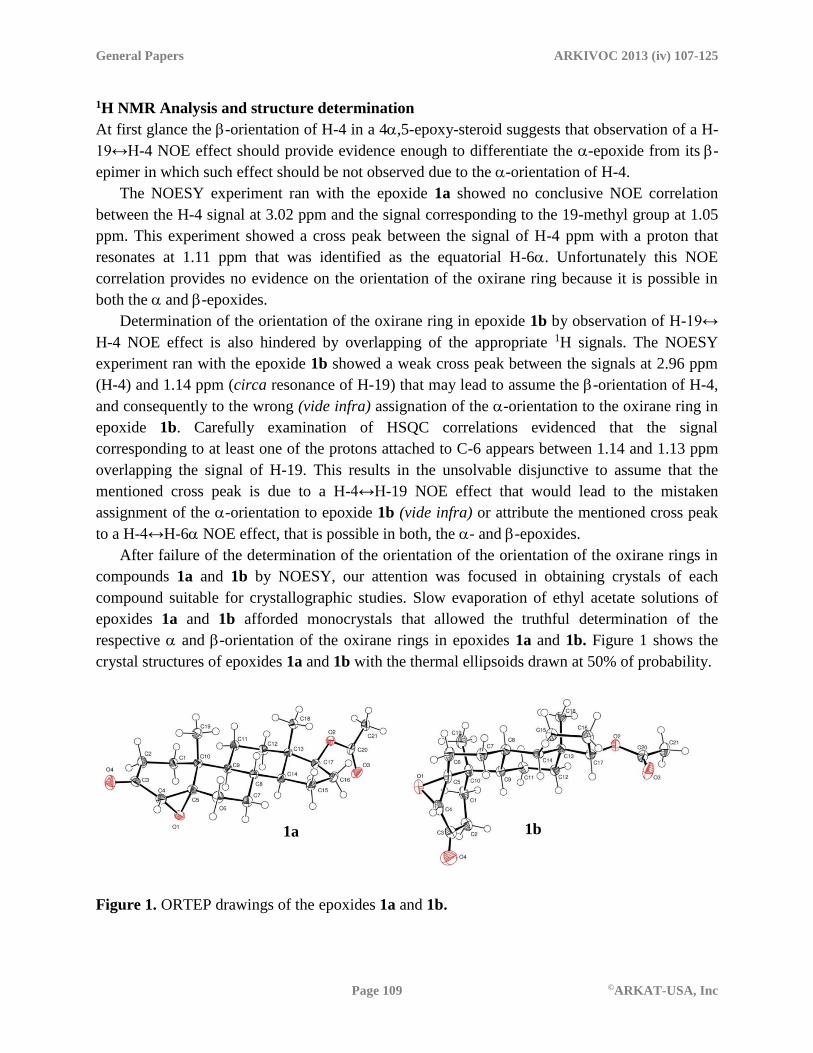
After failure of the determination of the orientation of the orientation of the oxirane rings in
compounds 1a and 1b by NOESY, our attention was focused in obtaining crystals of each
compound suitable for crystallographic studies. Slow evaporation of ethyl acetate solutions of
epoxides 1a and 1b afforded monocrystals that allowed the truthful determination of the
respective and -orientation of the oxirane rings in epoxides 1a and 1b. Figure 1 shows the
crystal structures of epoxides 1a and 1b with the thermal ellipsoids drawn at 50% of probability.
Figure 1. ORTEP drawings of the epoxides 1a and 1b.
1a 1b
General Papers ARKIVOC 2013 (iv) 107-125
Page 110 ©ARKAT-USA, Inc
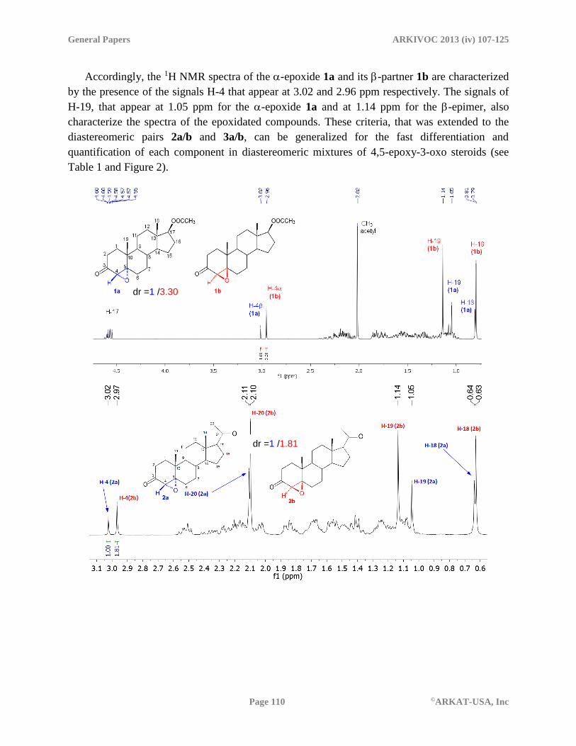
Accordingly, the 1H NMR spectra of the -epoxide 1a and its -partner 1b are characterized
by the presence of the signals H-4 that appear at 3.02 and 2.96 ppm respectively. The signals of
H-19, that appear at 1.05 ppm for the -epoxide 1a and at 1.14 ppm for the -epimer, also
characterize the spectra of the epoxidated compounds. These criteria, that was extended to the
diastereomeric pairs 2a/b and 3a/b, can be generalized for the fast differentiation and
quantification of each component in diastereomeric mixtures of 4,5-epoxy-3-oxo steroids (see
Table 1 and Figure 2).
dr =1 /1.81
dr =1 /3.30

General Papers ARKIVOC 2013 (iv) 107-125
Page 111 ©ARKAT-USA, Inc
Figure 2. 1H NMR spectra of the mixtures of the diastereomeric pairs 1a/b, 2a/b and 3a/b.
Table 1. NMR signals of H-4 and H-19 in each pair of epoxides 1a/b, 2a/b and 3a/b
-epoxides -epoxides
1a 2a 3a 1b 2b 3b
H-4 3.02 3.02 3.03 2.96 2.97 2.96
H-19 1.05 1.05 1.05 1.14 1.14 1.14
13C NMR Analysis
Computational studies
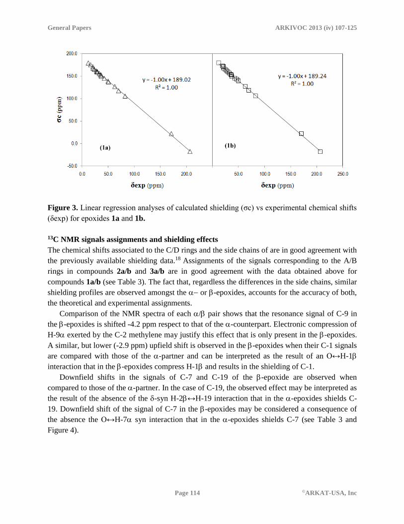
With the crystalline structures of epoxide 1a and 1b in hand, the 13C NMR spectra such
compounds were simulated in order to assist, confirm or correct the assignments of the NMR
signals. Even though the employed computational code allows obtaining absolute chemical
shifts, the values are reported as relative to TMS to facilitate comparisons with the experimental
data.
Relative chemical shifts (δc) were estimated by using the corresponding tetramethylsilane
(TMS) shielding calculated as:
δc = σTMS-σ (1)
In addition, the δc values were improved by using the procedure suggested by Forsyth and
Seabag.12 It consists on scaling the theoretical shielding values using the slope (a) and intercept
(b) obtained from a linear regression analysis of experimental chemical shifts and the calculated
dr =1 /3.92

General Papers ARKIVOC 2013 (iv) 107-125
Page 112 ©ARKAT-USA, Inc
shieldings. It should be noted that the values of parameters a and b are generally method- and
basis-set dependent, but the a value is expected to be close to 1. This procedure has been
successfully employed,13-16 and is currently accepted as a reliable way of improving NMR data
obtained from calculations. In the present study the correlation σc vs. δexp has been used for that
purpose, and thus the scaled values correspond to:
δcscal = (σc –b)/a (2)
The calculated 13C isotropic chemical shielding of TMS was found to be 193.6 ppm, i.e. 5.5
ppm larger than the experimental value is (188.1 ppm).17 Since it is a significant deviation, to
calculate the relative 13C chemical shifts of epoxides 1a and 1b, two different approaches were
employed. The first one is to use the computed absolute shift of TMS as reference in the
calculation of the relative values to obtain δc (Equation 1), and the other one to use the Forsyth
and Seabag procedure12 to obtain scaled values of the chemical shifts (δcscal, Equation 2)
The linear correlations for the latter case are shown in Figure 3. The slope in both cases is ≈ -
1 and the correlation coefficients (R2) values are ≈ 1, which supports the reliability of the
calculated 13C NMR data. In addition, this value simplifies the form of Equation 2, leading to a
scaling procedure consisting only on adjustments based on subtraction from a fixed reference.
Accordingly, the expressions used to calculate the δcscal values for epoxides 1a and 1b are:
δcscal (1a) = 189.02 –σc(1a) (3)
δcscal (1b) = 189.24 –σc(1b) (4)
It is interesting to notice that the b values obtained from the correlations are very similar for
epoxides 1a and 1b (189.02 and 189.24 ppm, respectively), supporting the consistency of the
calculations at the given level of theory.
The calculated values are listed in Table 2, together with the deviations from the
experimental values and the corresponding mean unsigned errors (MUE). It was found that the
non-scaled chemical shifts (δc, Equation 1) lead to Mean Unsigned Errors (MUE) values of 4.3
and 4.2 ppm, for epoxides 1a and 1b, respectively. The agreement between the calculated and
the experimental data is significantly improved when the scaling procedure is used (δcscal,
Equations 3 and 4), leading to MUE values equal to 1.1 ppm for both epoxides 1a and 1b. It
should be noted, however, that the trend of the calculated signals is the same regardless the
procedure used to calculate the chemical shifts.

General Papers ARKIVOC 2013 (iv) 107-125
Page 113 ©ARKAT-USA, Inc
Table 2. Experimental and calculated 13C NMR signals of epoxides 1a and 1b (δc, ppm),
deviations with respect to the experimental values (Dv, ppm),(a,b) and Mean Unsigned Errors
(MUE, ppm)(c)
C# 1a 1b
δexp δc Dv(a) δcscal Dvscal(b) δexp δc Dv(a) δcscal Dvscal
1 29.0 33.4 4.4 28.8 -0.2 26.1 29.9 3.8 25.6 -0.5
2 33.0 37.3 4.3 32.7 -0.3 32.4 36.8 4.4 32.4 0.0
3 206.6 211.3 4.7 206.7 0.1 206.4 211.8 5.4 207.4 1.0
4 62.7 66.5 3.8 61.9 -0.8 62.5 66.3 3.8 61.9 -0.6
5 69.9 76.0 6.1 71.4 1.5 70.0 75.1 5.1 70.8 0.8
6 29.5 34.0 4.5 29.4 -0.1 29.7 34.1 4.4 29.7 0.0
7 28.4 33.4 5.0 28.9 0.5 29.8 34.3 4.5 29.9 0.1
8 35.2 40.5 5.3 36.0 0.8 34.8 40.1 5.3 35.8 1.0
9 50.6 56.4 5.8 51.8 1.2 46.4 50.9 4.5 46.5 0.1
10 36.7 44.0 7.3 39.4 2.7 37.2 44.3 7.1 40.0 2.8
11 21.1 25.9 4.8 21.4 0.3 21.1 26.0 4.9 21.7 0.6
12 36.6 40.8 4.2 36.3 -0.3 36.4 40.7 4.3 36.4 0.0
13 42.5 48.3 5.8 43.7 1.2 42.6 48.4 5.8 44.1 1.5
14 49.9 54.7 4.8 50.1 0.2 50.2 54.7 4.5 50.3 0.1
15 23.4 28.0 4.6 23.4 0.0 23.4 28.1 4.7 23.8 0.4
16 27.4 31.6 4.2 27.0 -0.4 27.4 31.9 4.5 27.5 0.1
17 82.4 87.8 5.4 83.2 0.8 82.4 88.1 5.7 83.8 1.4
18 11.9 14.1 2.2 9.6 -2.3 12.0 14.3 2.3 10.0 -2.0
19 16.5 18.3 1.8 13.8 -2.7 18.9 21.0 2.1 16.7 -2.2
CH3 acetyl 20.8 22.5 1.7 18.0 -2.8 21.0 22.4 1.4 18.0 -3.0
C=O acetyl 170.9 171.4 0.5 166.9 -4.0 171.0 171.4 0.4 167.1 -3.9
MUE(c) 4.3 1.1 4.2 1.1
Legend: (a) Dv = δc-δexp (b) Dvscal = δcscal-δexp (c)
General Papers ARKIVOC 2013 (iv) 107-125
Page 114 ©ARKAT-USA, Inc
Figure 3. Linear regression analyses of calculated shielding (σc) vs experimental chemical shifts
(δexp) for epoxides 1a and 1b.
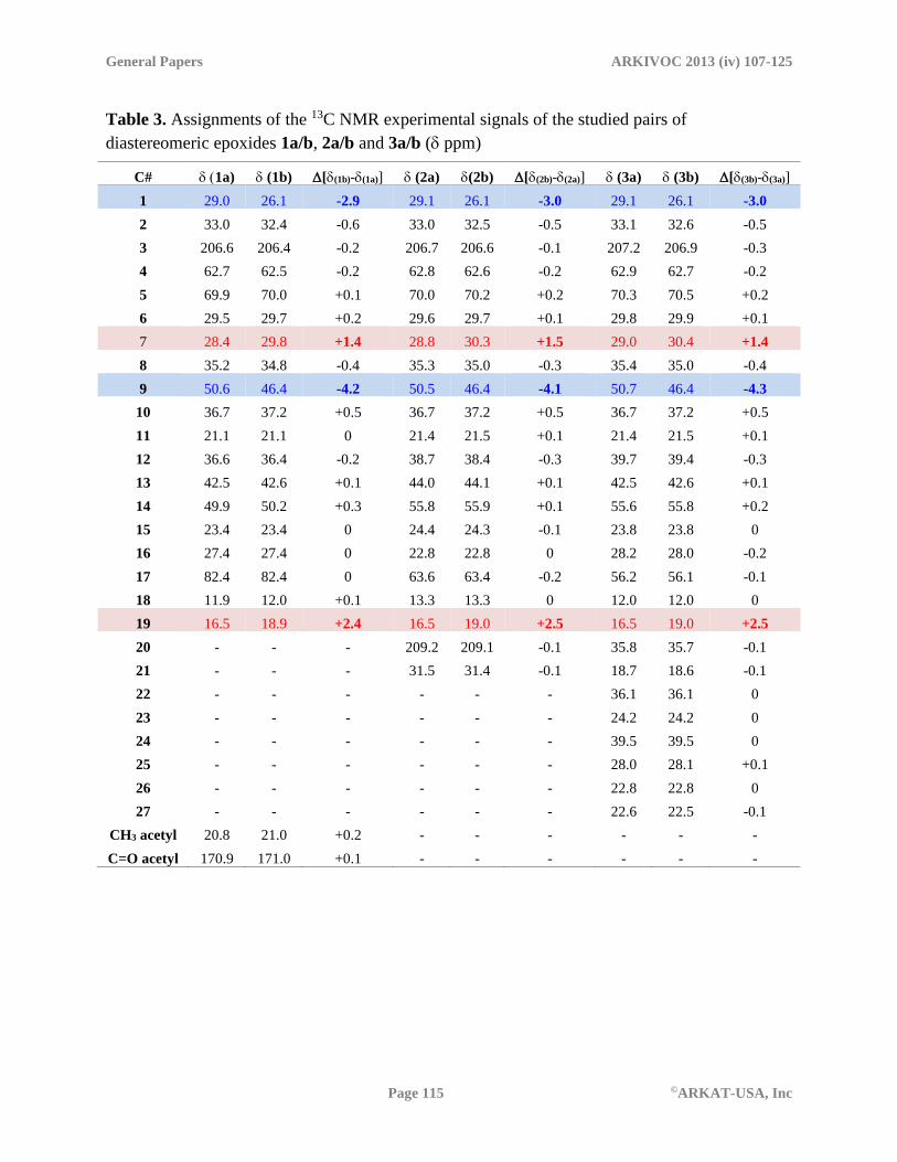
13C NMR signals assignments and shielding effects
The chemical shifts associated to the C/D rings and the side chains of are in good agreement with
the previously available shielding data.18 Assignments of the signals corresponding to the A/B
rings in compounds 2a/b and 3a/b are in good agreement with the data obtained above for
compounds 1a/b (see Table 3). The fact that, regardless the differences in the side chains, similar
shielding profiles are observed amongst the or -epoxides, accounts for the accuracy of both,
the theoretical and experimental assignments.
Comparison of the NMR spectra of each / pair shows that the resonance signal of C-9 in
the -epoxides is shifted -4.2 ppm respect to that of the -counterpart. Electronic compression of
H-9 exerted by the C-2 methylene may justify this effect that is only present in the -epoxides.
A similar, but lower (-2.9 ppm) upfield shift is observed in the -epoxides when their C-1 signals
are compared with those of the -partner and can be interpreted as the result of an O↔H-1
interaction that in the -epoxides compress H-1and results in the shielding of C-1.
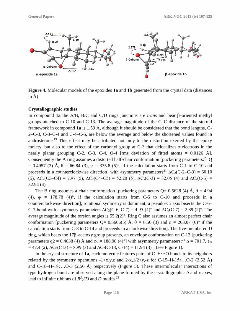
Downfield shifts in the signals of C-7 and C-19 of the -epoxide are observed when
compared to those of the -partner. In the case of C-19, the observed effect may be interpreted as
the result of the absence of the -syn H-2↔H-19 interaction that in the -epoxides shields C-
19. Downfield shift of the signal of C-7 in the -epoxides may be considered a consequence of
the absence the O↔H-7 syn interaction that in the -epoxides shields C-7 (see Table 3 and
Figure 4).
General Papers ARKIVOC 2013 (iv) 107-125
Page 115 ©ARKAT-USA, Inc
Table 3. Assignments of the 13C NMR experimental signals of the studied pairs of
diastereomeric epoxides 1a/b, 2a/b and 3a/b ( ppm)
C# 1a) (1b) [(1b)-(1a)] (2a) (2b) [(2b)-(2a)] (3a) (3b) [(3b)-(3a)]
1 29.0 26.1 -2.9 29.1 26.1 -3.0 29.1 26.1 -3.0
2 33.0 32.4 -0.6 33.0 32.5 -0.5 33.1 32.6 -0.5
3 206.6 206.4 -0.2 206.7 206.6 -0.1 207.2 206.9 -0.3
4 62.7 62.5 -0.2 62.8 62.6 -0.2 62.9 62.7 -0.2
5 69.9 70.0 +0.1 70.0 70.2 +0.2 70.3 70.5 +0.2
6 29.5 29.7 +0.2 29.6 29.7 +0.1 29.8 29.9 +0.1
7 28.4 29.8 +1.4 28.8 30.3 +1.5 29.0 30.4 +1.4
8 35.2 34.8 -0.4 35.3 35.0 -0.3 35.4 35.0 -0.4
9 50.6 46.4 -4.2 50.5 46.4 -4.1 50.7 46.4 -4.3
10 36.7 37.2 +0.5 36.7 37.2 +0.5 36.7 37.2 +0.5
11 21.1 21.1 0 21.4 21.5 +0.1 21.4 21.5 +0.1
12 36.6 36.4 -0.2 38.7 38.4 -0.3 39.7 39.4 -0.3
13 42.5 42.6 +0.1 44.0 44.1 +0.1 42.5 42.6 +0.1
14 49.9 50.2 +0.3 55.8 55.9 +0.1 55.6 55.8 +0.2
15 23.4 23.4 0 24.4 24.3 -0.1 23.8 23.8 0
16 27.4 27.4 0 22.8 22.8 0 28.2 28.0 -0.2
17 82.4 82.4 0 63.6 63.4 -0.2 56.2 56.1 -0.1
18 11.9 12.0 +0.1 13.3 13.3 0 12.0 12.0 0
19 16.5 18.9 +2.4 16.5 19.0 +2.5 16.5 19.0 +2.5
20 - - - 209.2 209.1 -0.1 35.8 35.7 -0.1
21 - - - 31.5 31.4 -0.1 18.7 18.6 -0.1
22 - - - - - - 36.1 36.1 0
23 - - - - - - 24.2 24.2 0
24 - - - - - - 39.5 39.5 0
25 - - - - - - 28.0 28.1 +0.1
26 - - - - - - 22.8 22.8 0
27 - - - - - - 22.6 22.5 -0.1
CH3 acetyl 20.8 21.0 +0.2 - - - - - -
C=O acetyl 170.9 171.0 +0.1 - - - - - -
General Papers ARKIVOC 2013 (iv) 107-125
Page 116 ©ARKAT-USA, Inc
Figure 4. Molecular models of the epoxides 1a and 1b generated from the crystal data (distances
in Å)
Crystallographic studies
In compound 1a the A/B, B/C and C/D rings junctions are trans and bear -oriented methyl
groups attached to C-10 and C-13. The average magnitude of the C–C distance of the steroid
framework in compound 1a is 1.53 Å, although it should be considered that the bond lengths, C-
2–C-3, C-3–C-4 and C-4–C-5, are below the average and below the shortened values found in
androsterone.19 This effect may be attributed not only to the distortion exerted by the epoxy
moiety, but also to the effect of the carbonyl group at C-3 that delocalizes electrons in the
nearly planar grouping C-2, C-3, C-4, O-4 [rms deviation of fitted atoms = 0.0126 Å].
Consequently the A ring assumes a distorted half-chair conformation [puckering parameters:20 Q
= 0.4957 (2) Å, = 66.84 (3), = 335.8 (5)°, if the calculation starts from C-1 to C-10 and
proceeds in a counterclockwise direction] with asymmetry parameters21 ∆C2(C-2–C-3) = 60.10
(5), ∆C2(C3–C4) = 7.97 (5), ∆C2(C4–C5) = 52.20 (5), ∆Cs(C-3) = 32.05 (4) and ∆Cs(C-5) =
52.94 (4)°.
The B ring assumes a chair conformation [puckering parameters Q= 0.5628 (4) Å, = 4.94
(4), = 178.78 (4)°, if the calculation starts from C-5 to C-10 and proceeds in a
counterclockwise direction]; rotational symmetry is dominant; a pseudo-C2 axis bisects the C-6–
C-7 bond with asymmetry parameters ∆C2(C-6–C-7) = 4.95 (4)° and ∆Cs(C-7) = 2.89 (2)°. The
average magnitude of the torsion angles is 55.2(2)°. Ring C also assumes an almost perfect chair
conformation [puckering parameters Q= 0.5666(5) Å, = 8.50 (3) and = 263.07 (6)° if the
calculation starts from C-8 to C-14 and proceeds in a clockwise direction]. The five-membered D
ring, which bears the 17-acetoxy group presents, an envelope conformation on C-13 [puckering
parameters q2 = 0.4638 (4) Å and 2 = 188.90 (4)°] with asymmetry parameters:22 ∆ = 701.7, m
= 47.4 (2), ∆Cs(C13) = 8.99 (3) and ∆C2(C-13, C-14) = 11.94 (3)°; (see Figure 1).
In the crystal structure of 1a, each molecule features pairs of C–H···O bonds to its neighbors
related by the symmetry operations -1+x,y,z and 2-x,1/2+y,-z for C-15–H-15a…O-2 (2.52 Å)
and C-18–H-18c…O-3 (2.56 Å) respectively (Figure 5). These intermolecular interactions of
type hydrogen bond are observed along the plane formed by the crystallographic b and c axes,
lead to infinite ribbons of R22(7) and D motifs.23
H-2 C-19
-Oxirane H-7
-epoxide 1a
-Oxirane
-epoxide 1b
H-1
H-9
C-2
C-19

General Papers ARKIVOC 2013 (iv) 107-125
Page 117 ©ARKAT-USA, Inc
Figure 5. Crystal structure of compound 1a view along the axis a; with perspective to plane
formed by b and c axes emphasizing the R22(7) and D motifs.
The asymmetric unit of the -epoxide 1b consist of two independent units of 17β-acetoxy-
4,5-epoxy-5β-androstan-3-one, molecule A and molecule B with cis A/B. Compound 1b has
trans B/C and C/D rings junctions and bears methyl groups attached to C-10 and C-13 in the -
side (see Figure 6). As described for compound 1a, in both molecules A and B of the epoxide 1b
the average magnitude of the C-2–C-3, C-3–C-4 and C-4–C-5 distances are rather below the 1.53
Å average and below the shortened values found in androsterone.19
Table 4. Least-square overlay analysis for each pair of molecules A and B of compound 1b
Rings Molecule-A vs Molecule-B
A 0.0672
B 0.0194
C 0.0163
D 0.0101
In molecule B the O-3 of the 17β-acetoxy shows disorder in two positions (O-3b and O-3p)
with 70:30 of O-3b:O-3p occupancy respectively. In order to establish differences among the
studied molecules A and B of compound 1b, a least-squares overlay analysis of the structures by
pairs was performed. Table 4 shows the results obtained with the Mercury program.24 Small
differences are observed in the A ring (rms 0.0672) and in the torsion angles of 170.64 and
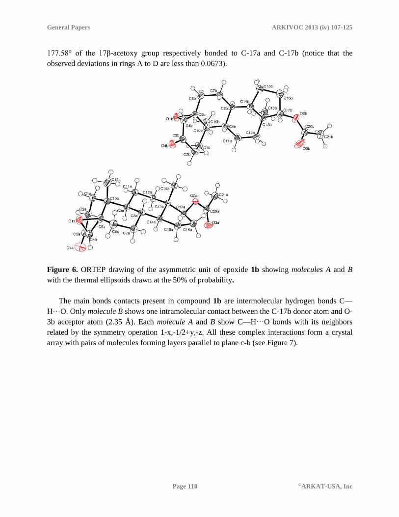
General Papers ARKIVOC 2013 (iv) 107-125
Page 118 ©ARKAT-USA, Inc
177.58° of the 17β-acetoxy group respectively bonded to C-17a and C-17b (notice that the
observed deviations in rings A to D are less than 0.0673).
Figure 6. ORTEP drawing of the asymmetric unit of epoxide 1b showing molecules A and B
with the thermal ellipsoids drawn at the 50% of probability.
The main bonds contacts present in compound 1b are intermolecular hydrogen bonds C—
H···O. Only molecule B shows one intramolecular contact between the C-17b donor atom and O-
3b acceptor atom (2.35 Å). Each molecule A and B show C—H···O bonds with its neighbors
related by the symmetry operation 1-x,-1/2+y,-z. All these complex interactions form a crystal
array with pairs of molecules forming layers parallel to plane c-b (see Figure 7).

General Papers ARKIVOC 2013 (iv) 107-125
Page 119 ©ARKAT-USA, Inc
Figure 7. Crystal structure of epoxide 1b viewed along the b axis, showing the short contacts
between the symmetry equivalent for molecule A (green) and molecule B (blue) extending along
the c-b plane.
Conclusion
The generalized practice for the determination of orientation of substituents in the steroid
framework by observation of NOE effects, fails in the case of 4,5-epoxy-3-oxo steroids. After
the unambiguous identification of the and -diastereomers by X-ray studies, a fast and
reliable criteria, based on the chemical shifts of H-4 and H-19 of each diastereomer, allows both
the identification and quantification of each component in the crude reaction mixtures.
Complete and unambiguous assignments of the 13C signals of the studied compounds based
on a combination of 1D and 2D NMR techniques was assisted by calculation of absolute
isotropic 13C NMR shieldings using the Gauge. The agreement between the experimental
assignment and theoretical results accounts for the accuracy of both, the experimental and
theoretical data.
Experimental Section
Synthesis and NMR
Reactions were monitored by TLC on ALUGRAM SIL G/UV254 plates from MACHEREY-
NAGEL. Chromatographic plates were sprayed with a 1% solution of vanillin in 50% HClO4 and
heated until color developed. Melting points were measured on a Melt-Temp II equipment and
are uncorrected. Mass spectra were registered in a Thermo-Electron spectrometer model DFS

General Papers ARKIVOC 2013 (iv) 107-125
Page 120 ©ARKAT-USA, Inc
(Double Focus Sector). NMR spectra were recorded in CDCl3 solution in a Varian INOVA 400
spectrometer using the solvent signal 7.26 ppm for 1H and 77.00 ppm for 13C as references. 13C
NMR signals assignments in each epoxide were made with the aid of a combination of 2D
homonuclear (1H–1H) and heteronuclear (1H–13C) correlation techniques, which included
Correlation Spectroscopy (COSY), Nuclear Overhauser Effect Spectroscopy (NOESY),
Heteronuclear Single Quantum Correlation (HSQC) and Heteronuclear Multiple Bond
Correlation (HMBC). All 2D NMR spectra were recorded using the standard pulse sequences
and parameters recommended by the manufacturer and processed employing the NMR
processing program MestreNova [See http://mestrelab.com/].
General epoxidation procedure. NaOH 6N (2 mL) and H2O2 30% (3.1 ml) were added to a
solution of the 4-en-3-oxo-steroid (2 mmol) and the mixture was stirred for 90 min before
pouring into ice-water (300 mL) and extraction with ethyl acetate (2x125 mL). The organic layer
was washed with water to achieve neutrality (12x50 mL), dried (anh. Na2SO4) and evaporated to
afford a mixture of the corresponding epimeric 4,5-epoxy-3-oxo-steroids.
Reacetylation procedure for the mixture of 1a/1b. The crude mixture resulting from the
epoxidation of testosterone acetate (1) was dissolved in pyridine (5 ml). Acetic anhydride (1 mL)
and a few crystals of DMAP were added and the mixture was stirred for 24 h, poured into ice-
H2O (200 mL) and extracted with ethyl acetate (4 x 50 mL). The organic layer was washed with
H2O (8x40 mL), 10% aqueous CuSO4 solution, water (8x40 mL), dried (anh. Na2SO4) and
evaporated to afford 487.3 mg (70.3 % overall yield for the epoxidation-reacetylation sequence)
of the 1/3.30 mixture of the epimeric epoxides 1a and 1b. Exhaustive chromatographic
separation afforded analytical samples of each epoxide.
17-Acetoxy-4,5-epoxy-5-androstan-3-one (1a). Mp 160-162 °C (from ethyl acetate) 1H
NMR (CDCl3, 400 MHz) δ 4.60 (dd, J 9.2, 7.8 Hz, 1H, H-17), 3.02 (s, 1H, H-4), 2.38 (1H,
ddd, J 19.9, 7.4, 1.7 Hz H-2), 2.24 (1H, dd, J 19.6, 7.7 Hz, H-2), 2.01 (s, 3H, CH3 acetyl),
1.05 (d, J 0.8 Hz, 3H, H-19), 0.81 (s, 3H, H-18). For 13C NMR (CDCl3, 100 MHz) see Table 3.
MS (EI, 70 eV) 347 (MH+, 4%), 346 (M+, 13%), 328 (M+- H2O, 12), 319 (10), 318 (M+- C=O,
50), 304 (25), 303 (36), 287 (11), 286 (M+- CH3COOH, 42), 274 (74), 273 (13), 272 (15), 271
(28), 268 (16), 258 (15), 257 (28), 240 (16), 239 (25), 231 (24), 229 (10), 225 (11), 215 (36), 213
(77), 212 (18), 211 (11), 202 (11), 201 (49), 200 (18), 199 (60), 197 (19), 189 (12), 187 (25), 186
(16), 185 (26), 183 (12), 175 (18), 174 (19), 173 (62), 172 (17), 171 (33), 165 (11), 163 (14), 161
(28), 160 (21), 159 (49), 158 (14), 157 (26), 151 (11), 149 (29), 148 (33), 147 (100), 146 (41), 45
(70), 144 (21), 143 (24), 138 (10), 137 (14), 136 (12), 135 (36), 134 (26), 133 (63), 132 (18), 131
(50), 129 (13), 125 (17), 124 (10), 123 (38), 122 (14), 121 (50), 120 (26), 119 (61), 118 (12), 117
(20), 113 (11), 111 (16), 110 (21), 109 (38), 108 (17), 107 (66), 106 (19), 105 (80), 97 (27), 96
(13), 95 (57), 94 (25), 93 (89), 92 (13), 91 (70), 83 (14), 81 (78), 80 (13), 79 (72), 77 (37), 68
(10), 67 (57), 65 (12), 57 (14), 55 (68), 53 (25). HRMS (EI, 70 eV) observed 346.2132 required
for C21H30O4 346.2139.

General Papers ARKIVOC 2013 (iv) 107-125
Page 121 ©ARKAT-USA, Inc
17-Acetoxy-4,5-epoxy-5-androstan-3-one (1b). Mp 140-142 °C (from ethyl acetate) 1H
NMR (CDCl3, 400 MHz) δ 4.57 (ddd, J 9.2, 7.7, 1.2 Hz, 1H, H-17), 2.96 (d, J 1.4 Hz, 1H, H-
4), 2.28 (H-2), 2.12 (H-2), 2.02 (s, 3H, CH3 acetyl), 1.14 (s, 3H, H-19), 0.80 (s, 3H, H-18).
For 13C NMR (CDCl3, 100 MHz) see Table 3. MS (EI, 70 eV) 347 (MH+, 3%), 346 (M+, 10%),
318 (M+- C=O, 37) 304 (25), 303 (55), 286 (M+- CH3COOH, 35), 275 (20), 274 (56), 273 (11),
272 (13), 271 (22), 268 (15), 258 (18), 257 (25), 243 (11), 240 (22), 239 (27), 232 (11), 231 (34),
230 (15), 229 (11), 225 (14), 215 (38), 214 (100), 213 (88), 212 (21), 211 (12), 203 (13), 202
(10), 201 (44), 200 (18), 199 (58), 197 (19), 191 (12), 189 (12), 187 (28), 186 (15), 185 (27), 183
(14), 175 (19), 174 (19), 173 (69), 172 (16), 171 (35), 163 (13), 161 (27), 159 (50), 158 (14), 157
(31), 148 (35), 147 (93), 146 (33), 145 (67), 144 (19), 143 (26), 137 (14), 136 (11), 135 (33), 134
(23), 133 (60), 132 (16), 131 (50), 129 (14), 125 (19), 124 (11), 123 (39), 122 (16), 121 (46), 120
(23), 119 (62), 118 (11), 117 (22), 113 (10), 111 (17), 110 (25), 109 (40), 108 (17), 107 (63), 106
(19), 105 (81), 97 (26), 95 (56), 94 (25), 93 (91), 92 (13), 91 (77), 83 (15), 82 (11), 81 (76), 80
(13), 79 (75), 77 (35), 69 (20), 68 (10), 67 (57), 65 (10), 55 (66), 53 (20). HRMS (EI, 70 eV)
observed 346.2127 required for C21H30O4 346.2139.
The general epoxidation procedure when applied to 2 mmol of progesterone (2) afforded 405 mg
(61.3 %) of a 1/1.81 mixture of the epoxides 2a/2b.
4,5-Epoxy-5-pregnan-3,20-dione (2a). Colorless oil as a mixture with 2b. 1H NMR (400
MHz, CDCl3) δ 3.02 (s, 1H, H-4), 2.39 (H-2), 2.20 (H-2), 2.11 (s, 3H, H-21), 1.05 (s, 3H, H-
19), 0.64 (s, 3H, H-18). For 13C NMR (CDCl3, 100 MHz) see Table 3.
4,5-Epoxy-5- pregnan-3,20-dione (2b). Colorless oil as a mixture with 2a. 1H NMR (400
MHz, CDCl3) δ 2.97 (s, 1H, H-4), 2.27 (H-2), 2.15 (H-2), 2.10 (s, 3H, H-21), 1.14 (s, 3H, H-
19), 0.63 (s, 3H, H-18). For 13C NMR (CDCl3, 100 MHz) see Table 3.
The general epoxidation procedure when applied to 2 mmol of cholest-4-en-3-one (3) afforded
733 mg (86.7 %) of a 1/3.92 mixture of the epoxides 3a/3b. Exhaustive chromatographic
separation afforded analytical samples of each epoxide.
4,5-Epoxy-5-cholestan-3-one (3a). Mp 118°C (from hexane-acetone) Lit.4 123-124 °C. 1H
NMR (400 MHz, CDCl3) δ 3.03 (s, 1H, H-4), 2.39 (ddd, J 19.7, 7.2, 1.2 Hz, 1H, H-2), 2.24
(dd, J 19.8, 7.2 Hz, 1H, H-2), 1.05 (s, 3H H-19), 0.91 (d, J = 6.5 Hz, 3H, H-21), 0.87 (d, J = 6.6
Hz, 3H, H-26), 0.86 (d, J = 6.6 Hz, 3H, H-27), 0.69 (s, 3H, H-18). For 13C NMR (CDCl3, 100
MHz) see Table 3.
4,5-Epoxy-5-cholestan-3-one (3b). Mp 118-119 °C (from hexane-acetone) Lit.4 118-119. 1H
NMR (400 MHz, CDCl3) δ 2.96 (s, 1H, H-4), 2.28 (ddd, J 19.4, 5.9, 2.2 Hz, 1H, H-2), 2.11
(dd, J 19.4, 6.6 Hz, 1H, H-2), 1.14 (s, 3H, H-19), 0.89 (d, J = 6.5 Hz, 3H, H-21), 0.86 (d, J =
6.6 Hz, 3H, H-26), 0.85 (d, J = 6.6 Hz, 3H, H-27), 0.68 (s, 3H, H-18). For 13C NMR (CDCl3, 100
MHz) see Table 3.
X-ray crystal structure determination
Suitable crystals for X-Ray diffraction studies were obtained by slow evaporation of the ethyl
acetate solutions of epoxides 1a and 1b at room temperature. Crystals of compounds 1a and 1b

General Papers ARKIVOC 2013 (iv) 107-125
Page 122 ©ARKAT-USA, Inc
mounted on glass fiber were studied with Oxford Diffraction Gemini "A" diffractometer with a
CCD area detector (MoK = 0.71073 Å, monochromator: graphite) source equipped with a sealed
tube X-ray source at 130 K. Unit cell constants were determined with a set of 15/3 narrow
frame/runs (1° in ) scans. A data sets consisted of 133 and 183 frames of intensity data
collected for the epoxides 1a and 1b respectively with a frame width of 1° in , a counting time
of 10 s/frame, and a crystal-to-detector distance of 55.00 mm. The double pass method of
scanning was employed to exclude any noise. The collected frames were integrated by using an
orientation matrix determined from the narrow frame scans.
Crystallographic data have been deposited with the Cambridge Crystallographic Data Center as
supplementary material numbers CCDC 899268 (compound 1a) and CCDC 899269 (compound
1b). Copies of the data can be obtained free of charge on application to CCDC, 12 Union Road,
Cambridge CB2 1EZ, UK. fax: +44(0)1223-336033; email: [email protected], or from
www.ccdc.cam.ac.uk/conts/retrieving.html.
Table 5. Crystal data and structure refinement parameters for epoxides 1a and 1b
Parameter 1a 1b
Empirical formula C21 H30O4 C21H30O4
Formula weight 346.45 346.45
Temperature 130(2) K 130(2) K
Wavelength 0.71073 Å 0.71073 Å
Crystal system Othorhombic Monoclinic
Space group P 21 21 21 P 21
Unit cell dimensions a = 11.0027(5) Å a = 8.0076(4) Å
b = 12.4699(6) Å b = 11.0905(5) Å
c = 13.3482(7) Å c = 20.8099(10) Å
β = 93.367(5)°
Volume 1831.41(15) Å3 1844.90(15) Å3
Z 4 4
Density (calculated) 1.257 Mg/m3 1.247 Mg/m3
Absorption coefficient 0.085 mm-1 0.085 mm-1
F(000) 752 752
Crystal size 0.5838 x0.5547 x 0.5178 mm3 0.5078 0.4554 0.1292mm3
Theta range for data collection 3.61 to 26.05°. 3.47 to 26.04°.
Index ranges -13≤ h≤ 9, -15≤ k≤1 1, -15≤
l≤16
-7≤ h≤ 9, -10≤ k≤ 13, -25≤
l≤ 19
Reflections collected 5700 8127
Independent reflections 3183 [R(int) = 0.0185] 5532 [R(int) = 0.0331]
Completeness to theta = 26.3° 99.6 % 99.7 %

General Papers ARKIVOC 2013 (iv) 107-125
Page 123 ©ARKAT-USA, Inc
Table 5. Continued
Parameter 1a 1b
Refinement method Full-matrix least-squares on F2 Full-matrix least-squares on
F2
Data / restraints / parameters 3183 / 0 / 229 5532 / 7 / 461
Goodness-of-fit on F2 1.037 1.056
Final R indices [I>2sigma(I)] R1 = 0.0342, wR2 = 0.0781 R1 = 0.0440, wR2 = 0.1132
R indices (all data) R1 = 0.0402, wR2 = 0.0819 R1 = 0.0514, wR2 = 0.1215
Absolute structure parameter -0.1(10) -0.2(10)
Largest diff. peak and hole 0.151 and -0.196 e.Å-3 0.173 and -0.208 e.Å-3
CrysAlisPro and CrysAlis RED software packages25 were employed for collection and
integration of data. Analysis of the integrated data did not reveal any decay. Final cell constants
were determined by a global refinement of 5818 and 8298 reflections ( < 26.3 °) for 1a and 1b
respectively. Collected data were corrected for absorbance by using analytical numeric
absorption correction26 using a multifaceted crystal model based on expressions upon the Laue
symmetry employing equivalent reflections. Structure solution and refinement were carried out
with the programs SHELXS97 and SHELXL97.27 Molecular graphics were generated with
ORTEP-3 for Windows and software employed for preparation of the material for publication
was WinGX.28
Full-matrix least-squares refinement was carried out by minimizing (Fo2 - Fc2)2. All non-
hydrogen atoms were refined anisotropically. Hydrogens attached to carbon atoms were placed
in geometrically idealized positions and refined employing the riding model, with C–H distances
= 0.98 – 1.00 Å with Uiso (H) = 1.2Ueq(C) for methylene and methyne groups, and Uiso (H) = 1.5
Ueq(C) for methyl group. Crystal data and experimental details of the structure refinement are
summarized in Table 7.
Computational details. Geometry optimizations and frequency calculations were performed
using the B3LYP functional and the 6-31+G(d,p) basis set. They were carried out in solution,
using the SMD continuum model29 and chloroform as solvent. Geometries were fully optimized
without imposing any restriction, using the X-ray structures as starting points. Local minima
were confirmed by the absence of imaginary frequencies. All the electronic calculations were
performed with Gaussian 09 package of programs.30 After optimization, the absolute isotropic 13C NMR shieldings (σc) were calculated using the GIAO (Gauge Invariant Atomic Orbitals)
method31,32 in chloroform also at B3LYP/6-31+G(d,p) level.
Acknowledgments
The authors thank to Dirección General de Asuntos del Personal Académico (DGAPA-UNAM)
for financial support via Project IN221911-3 and Laboratorio de Visualización y Cómputo

General Papers ARKIVOC 2013 (iv) 107-125
Page 124 ©ARKAT-USA, Inc
Paralelo at UAM-Iztapalapa for the access to its computer facilities. Thanks are due to Dr. Nuria
Esturau Escofet and M.Sc.Georgina Duarte Lisci (USAI-UNAM) for registering NMR and MS
spectra. We want to express our gratitude to Dr. Carlos Cobas from Mestrelab® for assistance
with the MestreNova NMR processing program.
References
1. Uyanic, C.; Malay, A.; Ayna, A. S.; Hanson, J. R.; Hitchcock, P. B. Steroids 2005, 70, 71.
http://dx.doi.org/10.1016/j.steroids.2004.09.002
PMid:15631862
2. Back, T. G.; Chau, J. H-L.; Codding, O. W.; Gladstone, P. L.; Jones, D. H.; Morzycki, J. W.;
Roszak, A. W. J. Org. Chem. 1992, 57, 4110.
http://dx.doi.org/10.1021/jo00041a013
3. Neeman, M.; O’Grodnick, J.S. Tetrahedron Lett. 1972, 13, 783.
http://dx.doi.org/10.1016/S0040-4039(01)84438-5
4. Nickon, A.; Mendelson, W. L. J. Am. Chem. Soc. 1965, 87, 3921–3928.
http://dx.doi.org/10.1021/ja01095a023
5. Jennings, B. R.; Bengtson, J. M. Steroids 1978, 31, 49–67.
http://dx.doi.org/10.1016/0039-128X(78)90019-3
6. Bovicelli, P.; Lupattelli, P.; Mincione, E. J. Org. Chem. 1992, 57, 2182–2184.
http://dx.doi.org/10.1021/jo00033a053
7. Carvalho, J. F. S.; Cruz Silva, M. M.; Sá e Melo, M. L. Tetrahedron 2009, 65, 2773.
http://dx.doi.org/10.1016/j.tet.2009.01.100
8. Romero-Ávila, M.; de Dios-Bravo, G.; Méndez-Stivalet, J. M.; Rodríguez-Sotres, R.;
Iglesias-Arteaga, M. A. Steroids 2007, 72, 955.
http://dx.doi.org/10.1016/j.steroids.2007.08.007
PMid:17905389
9. Rosado-Abón, A.; Romero-Avila, M.; Iglesias-Arteaga, M. A. Arkivoc 2010, (x), 110.
http://dx.doi.org/10.3998/ark.5550190.0011.a10
10. Rosado-Abón, A.; de Dios-Bravo, G.; Rodríguez-Sotres, R.; Iglesias-Arteaga, M. A. Steroids
2012, 77, 461.
http://dx.doi.org/10.1016/j.steroids.2012.01.004
PMid:22273808
11. Rosado-Abón, A.; de Dios-Bravo, G.; Rodríguez-Sotres, R.; Iglesias-Arteaga, M. A. J.
Steroid. Biochem. & Mol. Biol. 2013, 134, 45.
12. Forsyth, D. A.; Seabag, A. B. J. Am. Chem. Soc. 1997, 119, 9483.
http://dx.doi.org/10.1021/ja970112z
13. Barone, G.; Gomez-Paloma, L.; Duca, D.; Silvestri, A.; Riccio, R.; Bifulco, G. Chem. Eur. J.
2002, 8, 3233.
http://dx.doi.org/10.1002/1521-3765(20020715)8:14<3233::AID-CHEM3233>3.0.CO;2-0
General Papers ARKIVOC 2013 (iv) 107-125
Page 125 ©ARKAT-USA, Inc
14. Barone, G.; Duca, D.; Silvestri, A.; Gomez-Paloma, L.; Riccio, R.; Bifulco, G. Chem. Eur. J.
2002, 8, 3240.
http://dx.doi.org/10.1002/1521-3765(20020715)8:14<3240::AID-CHEM3240>3.0.CO;2-G
15. Cheng F, Sun H, Zhang Y, Mukkamala D, Oldfield E J. Am. Chem. Soc. 2005, 127, 12544.
http://dx.doi.org/10.1021/ja051528c
PMid:16144402
16. Mukkamala D.; Zhang Y.; Oldfield E. J. Am. Chem. Soc. 2007, 129, 7385.
http://dx.doi.org/10.1021/ja071227y
PMid:17506558
17. Cheeseman, J. R.; Trucks, G. W.; Keith, T. A.; Frisch, M. J. J. Chem. Phys. 1996, 104, 5497.
18. Blunt, J. W.; Stothers, J. B. Org. Magn. Resonance 1977, 9, 439.
http://dx.doi.org/10.1002/mrc.1270090802
19. High, D.F.; Kraut, J. Acta. Crystallogr. 1966, 21, 88-96.
http://dx.doi.org/10.1107/S0365110X66002378
PMid:5952605
20. Cremer, D.; Pople, J. A. J. Am. Chem. Soc. 1975, 97, 1354.
http://dx.doi.org/10.1021/ja00839a011
21. Duax, W. L.; Weeks, C. M.; Rohrer, D. C. Topics in Stereochemistry Eliel, E. L.; Allinger,
N. New York: John Wiley, 1976; Vol. 2, p 271.
http://dx.doi.org/10.1002/9780470147184.ch5
22. Altona, C.; Geise, H. J.; Romers, C. Tetrahedron 1968, 24, 132.
23. Etter, M. C. Acc. Chem. Res. 1990, 23, 120.
http://dx.doi.org/10.1021/ar00172a005
24. Macrae, C. F.; Edgington, P. R.; McCabe, P.; Pidcock, E.; Shields, G. P.; Taylor, R.; Towler,
M.; van De Streek, J. J. Appl. Cryst. 2006, 39, 453.
http://dx.doi.org/10.1107/S002188980600731X
25. CrysAlis CCD and CrysAlis R. 2009 Oxford Diffraction, Abingdon.
26. Clark, R. C.; Reid, J. S. Acta Crystallogr. 1995, A51, 887.
27. Sheldrick, G. M. Acta Crystallog. 2008, A64, 112.
28. Farrugia, L. J. J. Appl. Crystallogr. 2012, 45, 849.
http://dx.doi.org/10.1107/S0021889812029111
29. Marenich, A. V.; Cramer, C. J.; Truhlar, D. G. J. Phys. Chem. B. 2009, 113, 6378.
http://dx.doi.org/10.1021/jp810292n
PMid:19366259
30. Gaussian 09, Revision A.08, Frisch, M. J.; Trucks, G. W.; Schlegel, H. B.; Scuseria, G. E.;
Robb, M. A.; Cheeseman, J. R.; Scalmani, G.; Barone, V.; Mennucci, B.; Petersson, G. A.; et
al. Gaussian, Inc., Wallingford CT, 2009.
31. Wolinski, K.; Hilton, J. F.; Pulay, P. J. Am. Chem. Soc. 1990, 112, 8251.
http://dx.doi.org/10.1021/ja00179a005
32. Cheeseman, J. R.; Trucks, G. W.; Keith, T. A.; Frisch, M. J. J. Chem. Phys. 1996, 104, 5497.